The Role of the Inflammasome in Neurodegenerative Diseases
12
Molecules 2021, 26, 953. https://doi.org/10.3390/molecules26040953 www.mdpi.com/journal/molecules Review The Role of the Inflammasome in Neurodegenerative Diseases Federica Piancone 1, *, Francesca La Rosa 1 , Ivana Marventano 1 , Marina Saresella 1 and Mario Clerici 1,2 1 IRCCS Fondazione Don Carlo Gnocchi, 20148 Milano, Italy; [email protected] (F.L.R.); [email protected] (I.M.); [email protected] (M.S.); [email protected] (M.C.) 2 Department of Pathophysiology and Transplantation, University of Milano, 20122 Milano, Italy * Correspondence: [email protected] Abstract: Neurodegenerative diseases are chronic, progressive disorders that occur in the central nervous system (CNS). They are characterized by the loss of neuronal structure and function and are associated with inflammation. Inflammation of the CNS is called neuroinflammation, which has been implicated in most neurodegenerative diseases, including Alzheimer’s disease (AD), Parkinson’s disease (PD), amyotrophic lateral sclerosis (ALS) and multiple sclerosis (MS). Much evidence indicates that these different conditions share a common inflammatory mechanism: the activation of the inflammasome complex in peripheral monocytes and in microglia, with the con‐ sequent production of high quantities of the pro‐inflammatory cytokines IL‐1β and IL‐18. Inflam‐ masomes are a group of multimeric signaling complexes that include a sensor Nod‐like receptor (NLR) molecule, the adaptor protein ASC, and caspase‐1. The NLRP3 inflammasome is currently the best‐characterized inflammasome. Multiple signals, which are potentially provided in combi‐ nation and include endogenous danger signals and pathogens, trigger the formation of an active inflammasome, which, in turn, will stimulate the cleavage and the release of bioactive cytokines including IL‐1β and IL‐18. In this review, we will summarize results implicating the inflammasome as a pivotal player in the pathogenesis of neurodegenerative diseases and discuss how compounds that hamper the activation of the NLRP3 inflammasome could offer novel therapeutic avenues for these diseases. Keywords: inflammasome; NLRP3 inflammasome; neuroinflammation; neurodegenerative dis‐ eases; Multiple Sclerosis; Alzheimer’s disease; Parkinson’s diseases; Amyotrophic Lateral Sclerosis 1. Introduction Neuroinflammation plays a key role in the onset and the progression of several neurodegenerative diseases such as Alzheimer’s disease (AD), Parkinson’s disease (PD), Multiple Sclerosis (MS), and Amyotrophic Lateral Sclerosis (ALS) [1–3]. Nevertheless, it has to be considered that, in primary neurodegenerative diseases characterized by the accumulation of misfolded proteins like AD and PD, it is not clear if inflammation might be the primary cause of disease or a reaction to pathology. Indeed, the pathophysiological hypothesis of neurodegenerative diseases relies on the fact that some proteins, changing their conformations, aggregate into fibrils or oligomers, result‐ ing in neurotoxicity and leading to neurodegeneration and inflammation [4–7]. Neu‐ roinflammation is a physiological response to exogenous and endogenous insults that target the central nervous system (CNS) and represents a protective response in the brain, but excessive inflammatory responses are detrimental to the CNS. Several in‐ flammation‐inducing stimuli, such as damage‐associated molecular patterns (DAMPs) or pathogen‐associated molecular patterns (PAMPs), are recognized by multiprotein com‐ plexes, called inflammasomes. This elicits a pro‐inflammatory response mediated by the release of the inflammatory cytokines IL‐1β and IL‐18 [8]. The nucleotide‐binding oli‐ gomerization domain leucine‐rich repeat and pyrin domain‐containing protein 3 Citation: Piancone, F.; La Rosa, F.; Marventano, I.; Saresella, M.; Clerici, M. The Role of the Inflammasome in Neurodegenerative Diseases. Molecules 2021, 26, 953. https:// doi.org/10.3390/molecules26040953 Academic Editor: Massimo Bertinaria Received: 28 December 2020 Accepted: 6 February 2021 Published: 11 February 2021 Publisher’s Note: MDPI stays neu‐ tral with regard to jurisdictional claims in published maps and insti‐ tutional affiliations. Copyright: © 2021 by the authors. Licensee MDPI, Basel, Switzerland. This article is an open access article distributed under the terms and conditions of the Creative Commons Attribution (CC BY) license (http://creativecommons.org/licenses /by/4.0/).
Transcript of The Role of the Inflammasome in Neurodegenerative Diseases
Molecules 2021, 26, 953. https://doi.org/10.3390/molecules26040953 www.mdpi.com/journal/molecules
Review
The Role of the Inflammasome in Neurodegenerative Diseases
Federica Piancone 1,*, Francesca La Rosa 1, Ivana Marventano 1, Marina Saresella 1 and Mario Clerici 1,2
1 IRCCS Fondazione Don Carlo Gnocchi, 20148 Milano, Italy; [email protected] (F.L.R.);
[email protected] (I.M.); [email protected] (M.S.); [email protected] (M.C.) 2 Department of Pathophysiology and Transplantation, University of Milano, 20122 Milano, Italy
* Correspondence: [email protected]
Abstract: Neurodegenerative diseases are chronic, progressive disorders that occur in the central
nervous system (CNS). They are characterized by the loss of neuronal structure and function and
are associated with inflammation. Inflammation of the CNS is called neuroinflammation, which
has been implicated in most neurodegenerative diseases, including Alzheimer’s disease (AD),
Parkinson’s disease (PD), amyotrophic lateral sclerosis (ALS) and multiple sclerosis (MS). Much
evidence indicates that these different conditions share a common inflammatory mechanism: the
activation of the inflammasome complex in peripheral monocytes and in microglia, with the con‐
sequent production of high quantities of the pro‐inflammatory cytokines IL‐1β and IL‐18. Inflam‐
masomes are a group of multimeric signaling complexes that include a sensor Nod‐like receptor
(NLR) molecule, the adaptor protein ASC, and caspase‐1. The NLRP3 inflammasome is currently
the best‐characterized inflammasome. Multiple signals, which are potentially provided in combi‐
nation and include endogenous danger signals and pathogens, trigger the formation of an active
inflammasome, which, in turn, will stimulate the cleavage and the release of bioactive cytokines
including IL‐1β and IL‐18. In this review, we will summarize results implicating the inflammasome
as a pivotal player in the pathogenesis of neurodegenerative diseases and discuss how compounds
that hamper the activation of the NLRP3 inflammasome could offer novel therapeutic avenues for
these diseases.
Keywords: inflammasome; NLRP3 inflammasome; neuroinflammation; neurodegenerative dis‐
eases; Multiple Sclerosis; Alzheimer’s disease; Parkinson’s diseases; Amyotrophic Lateral Sclerosis
1. Introduction
Neuroinflammation plays a key role in the onset and the progression of several
neurodegenerative diseases such as Alzheimer’s disease (AD), Parkinson’s disease (PD),
Multiple Sclerosis (MS), and Amyotrophic Lateral Sclerosis (ALS) [1–3].
Nevertheless, it has to be considered that, in primary neurodegenerative diseases
characterized by the accumulation of misfolded proteins like AD and PD, it is not clear if
inflammation might be the primary cause of disease or a reaction to pathology. Indeed,
the pathophysiological hypothesis of neurodegenerative diseases relies on the fact that
some proteins, changing their conformations, aggregate into fibrils or oligomers, result‐
ing in neurotoxicity and leading to neurodegeneration and inflammation [4–7]. Neu‐
roinflammation is a physiological response to exogenous and endogenous insults that
target the central nervous system (CNS) and represents a protective response in the
brain, but excessive inflammatory responses are detrimental to the CNS. Several in‐
flammation‐inducing stimuli, such as damage‐associated molecular patterns (DAMPs) or
pathogen‐associated molecular patterns (PAMPs), are recognized by multiprotein com‐
plexes, called inflammasomes. This elicits a pro‐inflammatory response mediated by the
release of the inflammatory cytokines IL‐1β and IL‐18 [8]. The nucleotide‐binding oli‐
gomerization domain leucine‐rich repeat and pyrin domain‐containing protein 3
Citation: Piancone, F.; La Rosa, F.;
Marventano, I.; Saresella, M.; Clerici,
M. The Role of the Inflammasome in
Neurodegenerative Diseases.
Molecules 2021, 26, 953. https://
doi.org/10.3390/molecules26040953
Academic Editor: Massimo
Bertinaria
Received: 28 December 2020
Accepted: 6 February 2021
Published: 11 February 2021
Publisher’s Note: MDPI stays neu‐
tral with regard to jurisdictional
claims in published maps and insti‐
tutional affiliations.
Copyright: © 2021 by the authors.
Licensee MDPI, Basel, Switzerland.
This article is an open access article
distributed under the terms and
conditions of the Creative Commons
Attribution (CC BY) license
(http://creativecommons.org/licenses
/by/4.0/).
Molecules 2021, 26, 953 2 of 12
(NLRP3) inflammasome, one of the most intensively investigated inflammasomes, has
been reported to play a key role in neurodegenerative diseases.
Inflammasomes are a group of cytosolic multiprotein complexes that consist of a
sensor molecule (NLR, AIM2‐like receptors, ALR, and pyrin receptors), the adaptor
apoptosis‐associated speck‐like protein, which contains a caspase recruitment domain
(ASC), and pro‐caspase‐1 [9,10]. Inflammasome‐inducing stimuli trigger the oligomeri‐
zation of pattern recognition receptors (PRR) and the recruitment of pro‐caspase‐1 into
the complex, leading to the generation of active caspase‐1 that, consequently, will cleave
inactive pro‐peptides pro‐IL‐1β and pro‐IL‐18 into mature cytokines. Importantly,
caspase‐1 can also induce a pro‐inflammatory form of cell death, pyroptosis, that fea‐
tures early plasma membrane rupture, thereby releasing the soluble intracellular frac‐
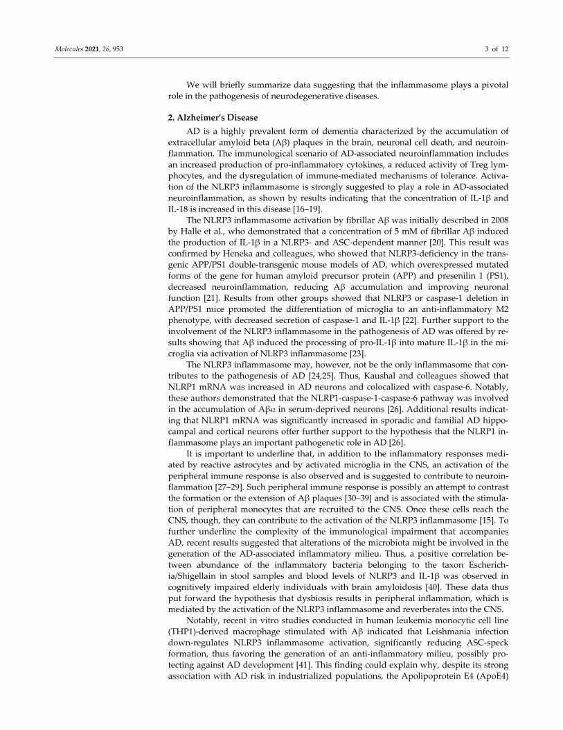
tion that fuels the inflammatory response [9,11]. NLRP3, as shown in Figure 1, is the
best‐characterized inflammasome; its activation involves a two‐step process. A first sig‐
nal, or “priming” signal, results in the NF‐kB‐dependent transcriptional upregulation of
NLRP3 and pro‐IL‐1β, but also controls post‐translational modifications of NLRP3 [12].
This initial trigger is followed by a second “activation” signal that induces the oligomer‐
ization and activation of the NLRP3 inflammasome. Besides this “canonical” NLRP3 in‐
flammasome activation pathway, a “noncanonical” NLRP3 activatory pathway has been
described. This pathway involves the activation of caspase‐11 in mice (or its human
orthologs caspase‐4 and caspase‐5) by cytosolic LPS, the induction of pyroptosis through
the cleavage of gasdermin D (GSDMD), and the release of high mobility group box 1
protein (HMGB1), resulting in the production of IL‐1β [9,13]. In both cases, the activa‐
tion of NLRP3 inflammasome results in the cleavage of the pro‐inflammatory IL‐1β and
IL‐18; this leads to the generation of the biological active form of these proteins that ini‐
tiates inflammatory signaling cascades, contributing to neuronal injury, cell death, and
neuroinflammation [14,15].
Figure 1. Inflammasome activation process and signaling mechanism: A Two‐Signal Model for
NLRP3 Inflammasome Activation. The priming signal (signal 1, left) is provided by dam‐
age‐associated molecular patterns (DAMP) or by pathogen‐associated molecular patterns (PAMP),
leading to the activation of the transcription factor NF‐κB and subsequent upregulation of NLRP3,
pro‐interleukin‐1β (pro‐IL‐1β), and pro‐interleukin‐IL‐18 (pro‐IL‐18). The activation signal (signal
2, right) is provided by a variety of stimuli including β‐amyloid, α‐synuclein, and Monosodium
Urate Crystals (MSU), leading to the assembly and formation of the NLRP3 inflammasome through
the combination of NLRP3, ASC, and procaspase‐1, and leading to the production of caspase‐1,
which catalyzes the transformation from pro‐IL‐1β and pro‐IL‐18 into IL‐1β and IL‐18.
Molecules 2021, 26, 953 3 of 12
We will briefly summarize data suggesting that the inflammasome plays a pivotal
role in the pathogenesis of neurodegenerative diseases.
2. Alzheimer’s Disease
AD is a highly prevalent form of dementia characterized by the accumulation of
extracellular amyloid beta (Aβ) plaques in the brain, neuronal cell death, and neuroin‐
flammation. The immunological scenario of AD‐associated neuroinflammation includes
an increased production of pro‐inflammatory cytokines, a reduced activity of Treg lym‐
phocytes, and the dysregulation of immune‐mediated mechanisms of tolerance. Activa‐
tion of the NLRP3 inflammasome is strongly suggested to play a role in AD‐associated
neuroinflammation, as shown by results indicating that the concentration of IL‐1β and
IL‐18 is increased in this disease [16–19].
The NLRP3 inflammasome activation by fibrillar Aβ was initially described in 2008
by Halle et al., who demonstrated that a concentration of 5 mM of fibrillar Aβ induced
the production of IL‐1β in a NLRP3‐ and ASC‐dependent manner [20]. This result was
confirmed by Heneka and colleagues, who showed that NLRP3‐deficiency in the trans‐
genic APP/PS1 double‐transgenic mouse models of AD, which overexpressed mutated
forms of the gene for human amyloid precursor protein (APP) and presenilin 1 (PS1),
decreased neuroinflammation, reducing Aβ accumulation and improving neuronal
function [21]. Results from other groups showed that NLRP3 or caspase‐1 deletion in
APP/PS1 mice promoted the differentiation of microglia to an anti‐inflammatory M2
phenotype, with decreased secretion of caspase‐1 and IL‐1β [22]. Further support to the
involvement of the NLRP3 inflammasome in the pathogenesis of AD was offered by re‐
sults showing that Aβ induced the processing of pro‐IL‐1β into mature IL‐1β in the mi‐
croglia via activation of NLRP3 inflammasome [23].
The NLRP3 inflammasome may, however, not be the only inflammasome that con‐
tributes to the pathogenesis of AD [24,25]. Thus, Kaushal and colleagues showed that
NLRP1 mRNA was increased in AD neurons and colocalized with caspase‐6. Notably,
these authors demonstrated that the NLRP1‐caspase‐1‐caspase‐6 pathway was involved
in the accumulation of Aβ42 in serum‐deprived neurons [26]. Additional results indicat‐
ing that NLRP1 mRNA was significantly increased in sporadic and familial AD hippo‐
campal and cortical neurons offer further support to the hypothesis that the NLRP1 in‐
flammasome plays an important pathogenetic role in AD [26].
It is important to underline that, in addition to the inflammatory responses medi‐
ated by reactive astrocytes and by activated microglia in the CNS, an activation of the
peripheral immune response is also observed and is suggested to contribute to neuroin‐
flammation [27–29]. Such peripheral immune response is possibly an attempt to contrast
the formation or the extension of Aβ plaques [30–39] and is associated with the stimula‐
tion of peripheral monocytes that are recruited to the CNS. Once these cells reach the
CNS, though, they can contribute to the activation of the NLRP3 inflammasome [15]. To
further underline the complexity of the immunological impairment that accompanies
AD, recent results suggested that alterations of the microbiota might be involved in the
generation of the AD‐associated inflammatory milieu. Thus, a positive correlation be‐
tween abundance of the inflammatory bacteria belonging to the taxon Escherich‐
ia/Shigellain in stool samples and blood levels of NLRP3 and IL‐1β was observed in
cognitively impaired elderly individuals with brain amyloidosis [40]. These data thus
put forward the hypothesis that dysbiosis results in peripheral inflammation, which is
mediated by the activation of the NLRP3 inflammasome and reverberates into the CNS.
Notably, recent in vitro studies conducted in human leukemia monocytic cell line
(THP1)‐derived macrophage stimulated with Aβ indicated that Leishmania infection
down‐regulates NLRP3 inflammasome activation, significantly reducing ASC‐speck
formation, thus favoring the generation of an anti‐inflammatory milieu, possibly pro‐
tecting against AD development [41]. This finding could explain why, despite its strong
association with AD risk in industrialized populations, the Apolipoprotein E4 (ApoE4)

Molecules 2021, 26, 953 4 of 12
allele was shown to be associated with improved cognitive functions in members of re‐
mote Amazonian tribes. In these populations, the E4 allele has been demonstrated to
confer survival in response to infection by parasites that, in turn, could reduce inflam‐
mation by reducing the activation of NLRP3 [42].
3. Multiple Sclerosis
MS is an autoimmune demyelinating disease of the CNS characterized by immune
cell infiltration from the periphery into the CNS as well as by the activation of the mi‐
croglia and astrocytes, which together promote neuroinflammation and neurodegenera‐
tion [43]. A number of studies have suggested the involvement of the NLRP3 inflam‐
masome in the pathogenesis of MS. Gris and colleagues, in 2010 [44], were the first to
suggest the critical role of Nlrp3 gene in the development of experimental autoimmune
encephalomyelitis (EAE), the most commonly used experimental model for human MS
[45]. Results from their study showed that the absence of Nlrp3 gene resulted in dimin‐
ished Th1 and Th17 encephalitogenic responses [44]. In line with this evidence, Peelen et
al. reported that the expression level of the inflammasome‐related genes NLRP3, IL‐1β,
and caspase‐1, was increased in peripheral blood mononuclear cell (PBMC) from re‐
lapsing‐remitting (RR) MS patients compared to healthy controls [46].
Results from other groups showed the up‐regulation of caspase‐1 and IL‐1β pro‐
teins in PBMCs and cerebrospinal fluid (CSF) of MS patients [47,48]. Moreover, caspa‐
se‐1 expression was shown to be elevated in MS plaques and PBMC of MS patients
[49,50]; taken together these observations lead to the proposal of using serum caspase‐1
and ASC protein concentrations as candidate biomarkers for MS onset [51]. IL‐18 con‐
centration was observed to be augmented, as well, in serum, CSF, and PBMCs of MS pa‐
tients [44,52–54]. Furthermore, a study by de Jong et al. showed that the increase of IL‐1β
in CSF was concomitant with a depletion of the IL‐1 receptor antagonist (IL‐1Ra), an an‐
ti‐inflammatory protein that antagonizes the binding of IL‐1β to its receptor [55]. An in‐
direct support to the role played by IL‐1β—a prototypical NLRP3 inflammasome activa‐
tion‐derived cytokine—in the pathogenesis of MS stems from the observation that suc‐
cessful treatment of disease relapses in MS patients with glatiramer acetate or IFNβ re‐
sults in the increase of endogenous IL‐1Ra concentration [56,57]. Notably, IL‐18 and
IL‐1β promote, respectively, IFNγ and IL‐17 production by Th1 cells and Th17 cells, two
functional T helper lymphocyte subsets that we repeatedly described to play a pivotal
role in MS pathogenesis.
The canonical NLRP3 inflammasome requires caspase‐1 activation for IL‐1β and
IL‐18 processing. Recent results nevertheless indicated that T cell intrinsic inflam‐
masome activity could drive IL‐1β and IL‐18 production via caspase‐8 activation inde‐
pendently from caspase‐1 activation [58,59]. Recent results reinforced a central role for
the NLRP3/caspase‐8 inflammasome pathway in MS by showing that stimulation of
PBMCs from primary progressive MS (PPMS) patients with Monosodium Urate Crystals
(MSU) resulted in a significant increase in the expression of NLRP3 and ASC‐speck pro‐
tein and in IL‐18 and caspase‐8 production. The NLRP3/caspase‐8 inflammasome path‐
way is activated in PPMS, possibly as a consequence of hyperuricemia. Thus, levels of
uric acid are upregulated in the CSF of MS patients [60], and the serum uric acid level in
patients is potentially associated with susceptibility of MS [61]. Taken together, these
results support the hypothesis of hyperuricemia as a common detrimental condition that
characterizes MS via the activation of the NLRP3/caspase‐8 inflammasome pathway [62].
Finally, the expression of P2 × 7R, a purinergic receptor that detects and amplifies
the release of ATP and, as a consequence, the activation of NLRP3 inflammasome, was
shown to be elevated in spinal cords of MS patients [63,64]. In line with this evidence,
other studies have shown an association between gain‐of‐function single nucleotide
polymorphisms in the P2X7 receptor gene and MS [65]. On the other hand, glatiramer
acetate, one the immunomodulator drugs used for MS, was shown to reduce P2X7R ex‐
pression [66], suggesting the contribution of extracellular ATP to the pathogenesis of

Molecules 2021, 26, 953 5 of 12
MS. Taken together, these results seem to suggest that endogenous metabolic danger
signals, ATP, and uric acid are likely to all be involved in the activation of the NLRP3 in‐
flammasome pathway observed in MS.
4. Parkinson’s Disease
PD is a progressive neurodegenerative disorder characterized by the depletion of
dopaminergic (DA) neurons in the substantia nigra (SN) and by the accumulation of cy‐
toplasmatic inclusions of fibrillar α‐synuclein (α‐syn), also called Lewy bodies [67]. Dif‐
ferent intracellular mechanisms allow the release of α‐syn outside of the cell [68], but the
common endpoint of α‐syn accumulation is the activation of astrocytes and microglia to
produce IL‐1β [68,69]. Notably, this phenomenon also facilitates the recruitment of im‐
mune cells from the periphery into the CNS [70].
A possible involvement of the NLRP3 inflammasome in the pathogenesis of PD was
initially suggested in 2013 by Codolo et al., who demonstrated in vitro that, while both
monomeric and fibrillary α‐syn increased pro‐IL‐1β levels via toll‐like receptor (TLR)‐2
signaling, the fibrillary form of α‐syn, alone, stimulated the inflammasome by activating
caspase‐1, resulting in IL‐1β production [71]. The result of this study was further con‐
firmed in vivo using an animal model of PD. Thus, injections of neurotoxin
1‐methyl‐4‐phenyl‐1,2,3,6‐tetrahydropyridine (MPTP) caused the loss of dopaminergic
neurons in the substantia nigra and a PD‐like pathology. Nlrp3 deficient mice were
shown to be resistant to PD, strongly suggesting an important role of the NLRP3 in‐
flammasome in the pathogenesis of PD [72]. Further results showed that miR‐7, a mi‐
croRNA known to regulate α‐syn gene expression [73], was present in the midbrain of
the MPTP‐induced PD mice model. Notably, as Nlrp3 is a target gene of miR‐7, the ste‐
reotactic injection of miR‐7 mimics in the mouse brain was demonstrated to inhibit the
NLRP3 inflammasome activation, reducing neuroinflammation [74]. Additional results
indicated that the exogenous administration of IL‐1Ra attenuated the MPTP‐induced PD
phenotypes in mice [75]. Further support to the possible role of the NLRP3 inflam‐
masome in PD is based on the knowledge that dopamine neurons negatively regulate
NLRP3 by the dopamine D1 receptor (DRD1)‐cyclic adenosine monophosphate (cAMP)
signaling pathway. Based on this information, it was observed that DRD1−/− mice were
less resistant to MPTP‐induced neuroinflammation, as shown by the increased IL‐18 and
IL‐1β production and the more extensive damages inflicted on dopaminergic neurons
[76].
Whereas the overall consensus of results obtained in the animal model strongly
supports an association between the NLRP3 inflammasome and PD, data stemming
from analyses performed in patients with a diagnosis of PD are much less convincing.
To summarize, in PD patients compared to healthy controls, (1) CSF concentration of
IL‐1β and IL‐18 was found to be higher [77]; (2) serum concentration of IL‐1β as well as
caspase‐1 activity were shown to be increased [78]; and (3) protein levels of NLRP3,
caspase‐1, and IL‐1β were seen to be augmented in PBMCs [78]. Nevertheless, results
from other groups showed that, whereas NLRP3 serum levels were increased in PD pa‐
tients compared to healthy controls (HC), no differences in IL‐1β and IL‐18 serum levels
could be detected [77]. Even more recently, we observed that stimulation of PBMC with
monomeric or aggregated α‐syn induced a comparable NLRP3 and ASC‐speck expres‐
sion, as well as IL‐18 and caspase‐1 production in cells of PD patients and healthy con‐
trols, indicating that α‐syn does not stimulate the NLRP3 inflammasome activity. Inter‐
estingly, IL1β and IL‐6 production was increased, whereas that of IL‐10 was reduced in
α‐syn‐stimulated cells of PD patients, suggesting that PD‐associated neuroinflammation
is not the consequence of the activation of the NLRP3 inflammasome but rather of an
imbalance between pro‐ and anti‐inflammatory cytokines.
In conclusion, although several studies have shown that α‐synuclein can elicit acti‐
vation of inflammasome in monocyte and microglial cell lines and in PD animal models,
the possible role of NLRP3 in patients with a diagnosis of PD still needs to be clarified.
Molecules 2021, 26, 953 6 of 12
5. Amyotrophic Lateral Sclerosis
ALS is a neurodegenerative disease characterized by the selective loss of motor
neurons in the motor cortex, the brainstem, and the spinal cord. The vast majority of
ALS cases are sporadic, (sALS), but a small fraction (about 5–10%) of cases are familiar
(fALS). In this situation, mutations in a number of genes, the most frequent of which is
the mutation in Cu2+/Zn2+ superoxide dismutase (SOD1), are known to associate with the
disease.
Increasing evidence has proposed an important role for neuroinflammation in the
pathogenesis of ALS, as demonstrated by the infiltration of lymphocytes and macro‐
phages in the CNS, the activation of the microglia, and the presence of reactive astro‐
cytes in the same anatomical sites where motor neuron injuries are observed. Recent
studies have suggested that a dysregulated and excessive inflammasome activation con‐
tributes to the neuroinflammation observed in ALS [79,80]. Thus, data obtained in the
G93A‐SOD1 transgenic mice, the most common animal model for ALS, showed the ac‐
tivation of caspase‐1 and IL‐1β in the microglia by ALS‐linked mutant SOD1 and
demonstrated that caspase‐1 or IL‐1β genes knockout or the use of recombinant IL‐1Ra
resulted in a reduction of inflammation. Notably, augmented caspase‐1 and IL‐1β pro‐
duction appeared to be NLRP3‐independent in this model, suggesting the possible in‐
volvement of other inflammasome complexes [81]. In partial contrast with these results,
other analyses performed in the SOD1 transgenic mice showed an upregulation of
NLRP3 and ASC in the anterior dorsal thalamic nucleus (AD) of G93A‐ [82] and of the
transactive response DNA‐binding protein‐43 (TDP‐43) in the microglia [83]. Other re‐
sults showed that in G93A‐SOD1 transgenic mice and in human tissues, spinal cord as‐
trocytes were activated, expressed NLRP3‐inflammasome proteins, and contributed to
inflammation in ALS by releasing proinflammatory cytokines [84]. In the same work, the
authors noticed the microglial expression of ASC but not that of NLRP3, suggesting that
other inflammasome sensor molecules may play a role in microglia‐driven neuroin‐
flammation in ALS.
Fewer results are available in humans; in ALS patients, serum concentration of
IL‐18, but not of IL‐1β, was observed to be increased [85], and NLRP3 and caspase‐1 ex‐
pression was shown to be augmented in brain tissues [86]. It is also important to under‐
line that clinical studies using the Interleukin‐1 receptor antagonist Anakinra have not
demonstrated a reduction in the neuroinflammation in ALS, suggesting that NLRP3 in‐
flammasome might not play a major role in ALS or that this disease is mainly driven by
IL‐18 and not IL‐1β [87]. To further augment the uncertainty of the possible role of the
inflammasome in ALS, data show that 17β‐estradiol, a steroid hormone that
down‐regulates inflammasome activation, improves motor neuron survival in a hu‐
manized animal model of ALS that carries the human SOD1 (G93A) mutation [88].
However, a conclusion has still not been reached; extensive analyses will be needed to
dissect the possibility that the inflammasome is involved in the pathogenesis of ASL.
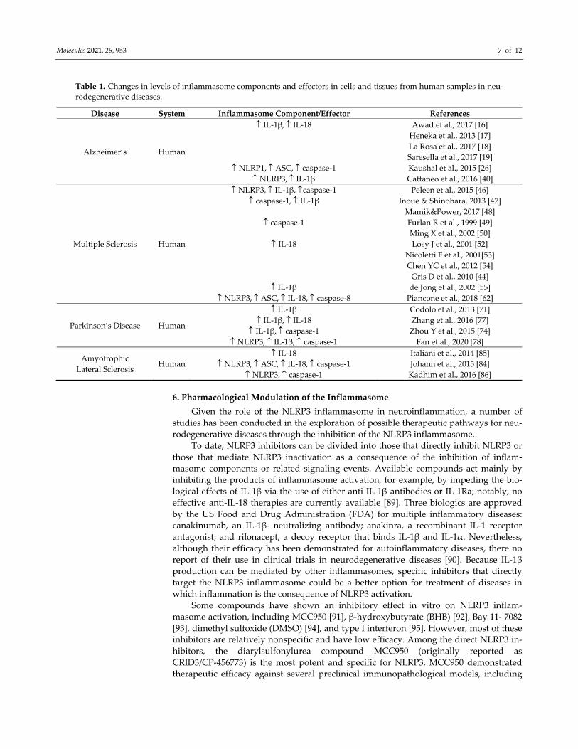
Literature data concerning expression of inflammasome proteins and effector cells
in neurodegenerative disease are summarized in Table 1.
Molecules 2021, 26, 953 7 of 12
Table 1. Changes in levels of inflammasome components and effectors in cells and tissues from human samples in neu‐
rodegenerative diseases.
Disease System Inflammasome Component/Effector References
Alzheimer’s Human
IL‐1β, IL‐18
NLRP1, ASC, caspase‐1 NLRP3, IL‐1β
Awad et al., 2017 [16]
Heneka et al., 2013 [17]
La Rosa et al., 2017 [18]
Saresella et al., 2017 [19]
Kaushal et al., 2015 [26]
Cattaneo et al., 2016 [40]
Multiple Sclerosis Human
NLRP3, IL‐1β, caspase‐1 caspase‐1, IL‐1β
caspase‐1
IL‐18
IL‐1β NLRP3, ASC, IL‐18, caspase‐8
Peleen et al., 2015 [46]
Inoue & Shinohara, 2013 [47]
Mamik&Power, 2017 [48]
Furlan R et al., 1999 [49]
Ming X et al., 2002 [50]
Losy J et al., 2001 [52]
Nicoletti F et al., 2001[53]
Chen YC et al., 2012 [54]
Gris D et al., 2010 [44]
de Jong et al., 2002 [55]
Piancone et al., 2018 [62]
Parkinson’s Disease Human
IL‐1β IL‐1β, IL‐18
IL‐1β, caspase‐1 NLRP3, IL‐1β, caspase‐1
Codolo et al., 2013 [71]
Zhang et al., 2016 [77]
Zhou Y et al., 2015 [74]
Fan et al., 2020 [78]
Amyotrophic
Lateral Sclerosis Human
IL‐18 NLRP3, ASC, IL‐18, caspase‐1
NLRP3, caspase‐1
Italiani et al., 2014 [85]
Johann et al., 2015 [84]
Kadhim et al., 2016 [86]
6. Pharmacological Modulation of the Inflammasome
Given the role of the NLRP3 inflammasome in neuroinflammation, a number of
studies has been conducted in the exploration of possible therapeutic pathways for neu‐
rodegenerative diseases through the inhibition of the NLRP3 inflammasome.
To date, NLRP3 inhibitors can be divided into those that directly inhibit NLRP3 or
those that mediate NLRP3 inactivation as a consequence of the inhibition of inflam‐
masome components or related signaling events. Available compounds act mainly by
inhibiting the products of inflammasome activation, for example, by impeding the bio‐
logical effects of IL‐1β via the use of either anti‐IL‐1β antibodies or IL‐1Ra; notably, no
effective anti‐IL‐18 therapies are currently available [89]. Three biologics are approved
by the US Food and Drug Administration (FDA) for multiple inflammatory diseases:
canakinumab, an IL‐1β‐ neutralizing antibody; anakinra, a recombinant IL‐1 receptor
antagonist; and rilonacept, a decoy receptor that binds IL‐1β and IL‐1α. Nevertheless,
although their efficacy has been demonstrated for autoinflammatory diseases, there no
report of their use in clinical trials in neurodegenerative diseases [90]. Because IL‐1β
production can be mediated by other inflammasomes, specific inhibitors that directly
target the NLRP3 inflammasome could be a better option for treatment of diseases in
which inflammation is the consequence of NLRP3 activation.
Some compounds have shown an inhibitory effect in vitro on NLRP3 inflam‐
masome activation, including MCC950 [91], β‐hydroxybutyrate (BHB) [92], Bay 11‐ 7082
[93], dimethyl sulfoxide (DMSO) [94], and type I interferon [95]. However, most of these
inhibitors are relatively nonspecific and have low efficacy. Among the direct NLRP3 in‐
hibitors, the diarylsulfonylurea compound MCC950 (originally reported as
CRID3/CP‐456773) is the most potent and specific for NLRP3. MCC950 demonstrated
therapeutic efficacy against several preclinical immunopathological models, including
Molecules 2021, 26, 953 8 of 12
EAE [91], AD [22], and PD [96]. However, this compound is currently not approved by
the FDA for the therapy of neurodegenerative disease.
Using a different approach, Stavudine (d4T), an antiviral nucleoside reverse tran‐
scriptase inhibitor (NRTIs) designed to target HIV, was recently shown to
down‐modulate NLRP3 inflammasome activation in mice [97]. Additional data con‐
firmed the ability of this compound to hamper NLRP3 inflammasome activation in an in
vitro model of AD by reducing NLRP3 assembly as well as IL‐18 and caspase‐1 produc‐
tion and stimulating amyloid‐beta autophagy by macrophages [18].
Given the lack of effective drugs in the therapy of chronic neurodegenerative con‐
ditions and the role of NLRP3 inflammasome in the pathogenesis and progression of
these diseases, efforts should be made to develop effective therapeutic strategies, possi‐
bly including those targeting the NLRP3 inflammasome.
Author Contributions: F.P. wrote the manuscript with guidance from M.C., M.S., F.L.R. and I.M.
provided intellectual contributions in this study. All authors have read and agreed to the pub‐
lished version of the manuscript.
Funding: This research received no external funding.
Acknowledgments: This work was supported by grants from the Italian Ministry of Health (Ri‐
cerca Corrente 2019–2020).
Conflicts of Interest: The authors declare no conflict of interest.
References
1. Schain, M.; Kreisl, W.C. Neuroinflammation in neurodegenerative disorders‐a review. Curr. Neurol. Neurosci. Rep. 2017, 17, 25,
doi:10.1007/s11910‐017‐0733‐2.
2. Chen, W.W.; Zhang, X.; Huang, W.J. Role of neuroinflammation in neurodegenerative diseases. Mol. Med. Rep. 2016, 13,
3391–3396, doi:10.3892/mmr.2016.4948.
3. Liu, Z.; Cheng, X.; Zhong, S.; Liu, C.; Liu, F.; Zhao, C. Peripheral and Central Nervous System Immune Response Crosstalk in
Amyotrophic Lateral Sclerosis. Front. Neurosci. 2020, 14, 575, doi:10.3389/fnins.2020.00575.
4. Ciccocioppo, F.; Bologna, G.; Ercolino, E.; Pierdomenico, L.; Simeone, P.; Lanuti, P.; Pieragostino, D.; Del Boccio, P.; Marchisio,
M.; Miscia, S. Neurodegenerative diseases as proteinopathies‐driven immune disorders. Neural. Regen. Res. 2020, 15, 850–856,
doi:10.4103/1673‐5374.268971.
5. Bayer, T.A. Proteinopathies, a core concept for understanding and ultimately treating degenerative disorders? Eur. Neuropsy‐
chopharmacol. 2015, 25, 713–24, doi:10.1016/j.euroneuro.2013.03.007.
6. Sami, N.; Rahman, S.; Kumar, V.; Zaidi, S.; Islam, A.; Ali, S.; Ahmad, F.; Hassan, M.I. Protein aggregation, misfolding and
consequential human neurodegenerative diseases. Int. J. Neurosci. 2017, 127, 1047–1057, doi:10.1080/00207454.2017.1286339.
7. Soto, C.; Pritzkow, S. Protein misfolding, aggregation, and conformational strains in neurodegenerative diseases. Nat. Neurosci
2018; 21, 1332–1340, doi:10.1038/s41593‐018‐0235‐9.
8. Strowig, T.; Henao‐Mejia, J.; Elinav, E.; Flavell, R. Inflammasomes in health and disease. Nature 2012, 481, 278–286,
doi:10.1038/nature10759.
9. Lamkanfi, M.; Dixit, V.M. Mechanisms and functions of inflammasomes. Cell 2014, 157, 1013–1022,
doi:10.1016/j.cell.2014.04.007.
10. Broz, P.; Dixit, V.M. Inflammasomes: Mechanism of assembly, regulation and signalling. Nat. Rev. Immunol. 2016, 16, 407–420,
doi:10.1038/nri.2016.58.
11. Lamkanfi, M.; Dixit, V.M. Inflammasomes and their roles in health and disease. Ann. Rev. Cell. Dev. Biol. 2012, 28,137‐161,
doi:10.1146/annurev‐cellbio‐101011‐155745.
12. Yang, J.; Liu, Z.; Xiao, TS. Post‐translational regulation of inflammasomes. Cell. Mol. Immunol. 2017, 14, 65–79,
doi:10.1038/cmi.2016.29.
13. Shi, J.Q.; Zhang, C.C.; Sun, X.L.; Cheng, X.X.; Wang, J.B.; Zhang, Y.D.; Xu, J.; Zou, H.Q. Antimalarial drug artemisinin extenu‐
ates amyloidogenesis and neuroinflammation in APPswe/PS1dE9 transgenic mice via inhibition of nuclear factor‐kappaB and
NLRP3 inflammasome activation. CNS. Neurosci. Ther. 2013, 19, 262–268, doi:10.1111/cns.12066.
14. Allan, S.M. Pragmatic target discovery from novel gene to functionally defined drug target: The interleukin‐1 story. Methods
Mol. Med. 2005, 104, 333–46, doi:10.1385/1‐59259‐836‐6:333.
15. Alboni, S.; Cervia, D.; Sugama, S.; Cont,i B. Interleukin 18 in the CNS. J. Neuroinflammation 2010, 7, 9, doi:10.1186/1742‐2094‐7‐9.
16. Awad, F.; Assrawi, E.; Jumeau, C.; Georgin‐Lavialle, S.; Cobret, L.; Duquesnoy, P.; Piterboth, W.; Thomas, L.;
Stankovic‐Stojanovic, K.; Louvrier, C.; et al. Impact of human monocyte and macrophage polarization on NLR expression and
NLRP3 inflammasome activation. PLoS ONE 2017, 12, e0175336, doi:10.1371/journal.pone.0175336.
Molecules 2021, 26, 953 9 of 12
17. Heneka, M.T.; Kummer, M.P.; Stutz, A.; Delekate, A.; Schwartz, S.; Vieira‐Saecker, A.; Griep, A.; Axt, D.; Remus, A.; Tzeng,
T.C.; et al. NLRP3 is activated in Alzheimer’s disease and contributes to pathology in APP/PS1 mice. Nature 2013, 493,
674–678.
18. La Rosa, F.; Saresella, M.; Marventano, I.; Piancone, F.; Ripamonti, E.; Al‐Daghri, N.; Bazzini, C.; Zoia, C.P.; Conti, E.;
Ferrarese, C.; Clerici, M. Stavudine Reduces NLRP3 Inflammasome Activation and Modulates Amyloid‐β Autophagy. J.
Alzheimers Dis. 2019, 72, 401–412, doi:10.3233/JAD‐181259.
19. Saresella, M.; La Rosa, F.; Piancone, F.; Zoppis, M.; Marventano, I.; Calabrese, E.; Rainone, V.; Nemni, R.; Mancuso, R.; Clerici,
M.; The NLRP3 and NLRP1 inflammasomes are activated in Alzheimer’s disease. Mol. Neurodegener. 2016, 3,11, 23,
doi:10.1186/s13024‐016‐0088‐1.
20. Halle, A.; Hornung, V.; Petzold, G.C.; Stewart, C.R.; Monks, B.G.; Reinheckel, T.; Fitzgerald, K.A.; Latz, E.; Moore, K.J.; Go‐
lenbock, D.T. The NALP3 inflammasome is involved in the innate immune response to amyloid‐β. Nat. Immunol. 2008, 9,
857–865, doi:10.1038/ni.1636.
21. Heneka, M.T. Inflammasome activation and innate immunity in Alzheimer’s disease. Brain Pathol. 2017, 27, 220–222,
doi:10.1111/bpa.12483.
22. Dempsey, C.; Rubio Araiz, A.; Bryson, K.J.; Finucane, O.; Larkin, C.; Mills, E.L.; Robertson, A.A.B.; Cooper, M.A.; O’Neill,
L.A.J.; Lynch, M.A. Inhibiting the NLRP3 inflammasome with MCC950 promotes non‐phlogistic clearance of amyloid‐β and
cognitive function in APP/PS1 mice. Brain Behav. Immun. 2017, 61, 306–316, doi:10.1016/j.bbi.2016.12.014.
23. Parajuli, B.; Sonobe, Y.; Horiuchi, H.; Takeuchi, H.; Mizuno, T.; Suzumura, A. Oligomeric amyloid β induces IL‐1β processing
via production of ROS: Implication in Alzheimer’s disease. Cell Death Dis. 2013, 4, e975, doi:10.1038/cddis.2013.503.
24. Pontillo, A.; Catamo, E.; Arosio, B.; Mari, D.; Crovella, S. NALP1/NLRP1 genetic variants are associated with Alzheimer
disease. Alzheimer Dis. Assoc. Disord. 2012, 26, 277–281, doi:10.1097/WAD.0b013e318231a8ac.
25. Tan, M.S.; Tan, L.; Jiang, T.; Zhu, X.C.; Wang, H.F.; Jia, C.D.; Yu, J.T. Amyloid‐beta induces NLRP1‐dependent neuronal py‐
roptosis in models of Alzheimer’s disease. Cell Death Dis. 2014, 5, e1382, doi:10.1038/cddis.2014.348.
26. Kaushal, V.; Dye, R.; Pakavathkumar, P.; Foveau, B.; Flores, J.; Hyman, B.; Ghetti, B.; Koller, B.H.; LeBlanc, A.C. Neuronal
NLRP1 inflammasome activation of Caspase‐1 coordinately regulates inflammatory interleukin‐1‐beta production and axonal
degeneration‐associated Caspase‐6 activation. Cell Death Differ. 2015, 22, 1676–1686, doi:10.1038/cdd.2015.16.
27. Rezai‐Zadeh, K.; Gate, D.; Gowing, G.; Town, T. How to get from here to there: Macrophage recruitment in Alzheimer’s dis‐
ease. Curr. Alzheimer Res. 2011, 8, 156–163, doi:10.2174/156720511795256017.
28. Nascimento, C.M.; Pereira, J.R.; de Andrade, L.P.; Garuffi, M.; Talib, L.L.; Forlenza, O.V.; Cancela, J.M.; Cominetti, M.R.; Stella,
F. Physical exercise in MCI elderly promotes reduction of pro‐inflammatory cytokines and improvements on cognition and
BDNF peripheral levels. Curr. Alzheimer Res. 2014, 11, 799–805, doi:10.2174/156720501108140910122849.
29. Dionisio‐Santos, D.A.; Olschowka, J.A.; O’Banion, M.K. Exploiting microglial and peripheral immune cell crosstalk to treat
Alzheimer’s disease. J. Neuroinflammation 2019, 16, 74, doi:10.1186/s12974‐019‐1453‐0.
30. Town, T.; Tan, J.; Mullan, M. CD40 signaling and Alzheimer’s disease pathogenesis. Neurochem. Int. 2001, 39, 371–380,
doi:10.1016/s0197‐0186(01)00044‐4.
31. Town, T.; Nikolic, V.; Tan, J. The microglial “activation” continuum: From innate to adaptive responses. J. Neuroinflammation
2005, 2, 24, doi:10.1186/1742‐2094‐2‐24.
32. Town, T.; Laouar, Y.; Pittenger, C.; Mori, T.; Szekely, C.A.; Tan, J.; Duman, R.S.; Flavell, R.A. Blocking TGF‐beta‐Smad2/3 in‐
nate immune signaling mitigates Alzheimer‐like pathology. Nat. Med. 2008, 14, 681–687, doi:10.1038/nm1781.
33. Hawkes, C.A.; McLaurin, J. Selective targeting of perivascular macrophages for clearance of beta‐amyloid in cerebral amyloid
angiopathy. Proc. Natl. Acad. Sci. USA 2009, 106, 1261–1266, doi:10.1073/pnas.0805453106.
34. Tan, M.S.; Yu, J.T.; Jiang, T.; Zhu, X.C.; Tan, L. The NLRP3 inflammasome in Alzheimer’s disease. Mol. Neurobiol. 2013, 48,
875–82, doi:10.1007/s12035‐013‐8475‐x.
35. Townsend, K.P.; Town, T.; Mori, T.; Lue, L.F.; Shytle, D.; Sanberg, P.R.; Morgan, D.; Fernandez, F.; Flavell, R.A.; Tan, J. CD40
signaling regulates innate and adaptive activation of microglia in response to amyloid beta‐peptide. Eur. J. Immunol. 2005, 35,
901–910, doi:10.1002/eji.200425585.
36. Feng, Y.; Li, L.; Sun, X.H. Monocytes and Alzheimer’s disease. Neurosci. Bull. 2011, 2, 115–122, doi:10.1007/s12264‐011‐1205‐3.
37. Fiala, M.; Lin, J.; Ringman, J.; Kermani‐Arab, V.; Tsao, G.; Patel, A.; Lossinsky, A.S.; Graves, M.C.; Gustavson, A.; Sayre, J.; et al.
Ineffective phagocytosis of amyloid‐beta by macrophages of Alzheimer’s disease patients. J. Alzheimers Dis. 2005, 3, 221–232,
doi:10.3233/jad‐2005‐7304.
38. Simard, A.R.; Rivest, S. Neuroprotective properties of the innate immune system and bone marrow stem cells in Alzheimer’s
disease. Mol. Psychiatry 2006, 11, 327–335, doi:10.1038/sj.mp.4001809.
39. Saresella, M.; Marventano, I.; Calabrese, E.; Piancone, F.;Rainone, V.; Gatti, A.; Alberoni, M.; Nemni, R.; Clerici, M. A complex
proinflammatory role for peripheral monocytes in Alzheimer’s disease. J. Alzheimers Dis. 2014, 38, 403–413,
doi:10.3233/JAD‐131160. PMID: 23979026.
40. Cattaneo, A.; Cattane, N.; Galluzzi, S.; Provasi, S.; Lopizzo, N.; Festari, C.; Ferrari, C.; Guerra, U.P.; Paghera, B.; Muscio, C.; et
al. Association of brain amyloidosis with pro‐inflammatory gut bacterial taxa and peripheral inflammation markers in
cognitively impaired elderly. Neurobiol. Aging. 2017, 49, 60–68, doi:10.1016/j.neurobiolaging.2016.08.019.
Molecules 2021, 26, 953 10 of 12
41. Saresella, M.; Basilico, N.; Marventano, I.; Perego, F.; La Rosa, F.; Piancone, F.; Taramelli, D.; Banks, H.; Clerici, M. Leishmania
infantum infection reduces the amyloid β42‐stimulated NLRP3 inflammasome activation. Brain Behav. Immun. 2020, 88,
597–605, doi:10.1016/j.bbi.2020.04.058.
42. Trumble, B.C.; Stieglitz, J.; Blackwell, A.D.; Allayee, H.; Beheim, B.; Finch, C.E.; Gurven, M.; Kaplan, H. Apolipoprotein E4 is
associated with improved cognitive function in Amazonian forager‐horticulturalists with a high parasite burden. FASEB J.
2017, 31, 1508–1515, doi:10.1096/fj.201601084R.
43. Baecher‐Allan, C.; Kaskow, B.J.; Weiner, H.W. Multiple Sclerosis: Mechanisms and Immunotherapy. Neuron 2018, 97, 742–768,
doi:10.1016/j.neuron.2018.01.021.
44. Gris, D.; Ye, Z.; Iocca, H.A.; Wen, H.; Craven, R.R.; Gris, P.; Huang, M.; Schneider, M.; Miller, S.D.; Ting, J.P. NLRP3 plays a
critical role in the development of experimental autoimmune encephalomyelitis by mediating Th1 and Th17 responses. J. Im‐
munol. 2010, 185, 974–981, doi:10.4049/jimmunol.0904145.
45. Ransohoff, R.M. Animal models of multiple sclerosis: The good, the bad and the bottom line. Nat. Neurosci. 2012, 15, 1074–7,
doi:10.1038/nn.3168.
46. Peelen, E.; Damoiseaux, J.; Muris, A.H.; Knippenberg, S.; Smolders, J.; Hupperts, R.; Thewissen, M. Increased inflammasome
related gene expression profile in PBMC may facilitate T helper 17 cell induction in multiple sclerosis. Mol. Immunol. 2015, 63,
521–529, doi:10.1016/j.molimm.2014.10.008.
47. Inoue, M.; Shinohara, M.L. NLRP3 Inflammasome and MS/EAE. Autoimmune Dis. 2013, 2013, 859145, doi:10.1155/2013/859145.
48. Mamik, M.K.; Power, C. Inflammasomes in neurological diseases: Emerging pathogenic and therapeutic concepts. Brain 2017,
140, 2273–2285, doi:10.1093/brain/awx133.
49. Furlan, R.; Filippi, M.; Bergami, A.; Rocca, M.A.; Martinelli, V.; Poliani, P.L.; Grimaldi, L.M.; Desina, G.; Comi, G.; Martino, G.
Peripheral levels of caspase‐1 mRNA correlate with disease activity in patients with multiple sclerosis; a preliminary study. J.
Neurol. Neurosurg. Psychiatry 1999, 67, 785–788, doi:10.1136/jnnp.67.6.785.
50. Ming, X.; Li, W.; Maeda, Y.; Blumberg, B.; Raval, S.; Cook, S.D.; Dowling, P.C. Caspase‐1 expression in multiple sclerosis
plaques and cultured glial cells. J. Neurol. Sci. 2002, 197, 9–18, doi:10.1016/s0022‐510x(02)00030‐8.
51. Keane, R.W.; Dietrich, W.D.; de Rivero Vaccari, J.P. Inflammasome Proteins As Biomarkers of Multiple Sclerosis. Front. Neurol.
2018, 9, 135, doi:10.3389/fneur.2018.00135.
52. Losy, J.; Niezgoda, A. IL‐18 in patients with multiple sclerosis. Acta Neurol. Scand. 2001, 104, 171–173,
doi:10.1034/j.1600‐0404.2001.00356.x.
53. Nicoletti, F.; Di Marco, R.; Mangano, K.; Patti, F.; Reggio, E.; Nicoletti, A.; Bendtzen, K.; Reggio, A. Increased serum levels of
interleukin‐18 in patients with multiple sclerosis. Neurology 2001, 57, 342–344, doi:10.1212/wnl.57.2.342.
54. Chen, Y.C.; Chen, S.D.; Miao, L.; Liu, Z.G.; Li, W.; Zhao, Z.X.; Sun, X.J.; Jiang, G.X.; Cheng, Q. Serum levels of interleukin
(IL)‐18, IL‐23 and IL‐17 in Chinese patients with multiple sclerosis. J. Neuroimmunol. 2012, 243, 56‐60,
doi:10.1016/j.jneuroim.2011.12.008.
55. de Jong, B.A.; Huizinga, T.W.; Bollen, E.L.; Uitdehaag, B.M.; Bosma, G.P.; van Buchem, M.A.; Remarque, E.J.; Burgmans, A.C.;
Kalkers, N.F.; Polman, C.H.; et al. Production of IL‐1beta and IL‐1Ra as risk factors for susceptibility and progression of
relapse‐onset multiple sclerosis. J. Neuroimmunol. 2002, 126, 172–179, doi:10.1016/s0165‐5728(02)00056‐5.
56. Burger, D.; Molnarfi, N.; Weber, M.S.; Brandt, K.J.; Benkhoucha, M.; Gruaz, L.; Chofflon, M.; Zamvil, S.S.; Lalive, P.H. Glati‐
ramer acetate increases IL‐1 receptor antagonist but decreases T cell‐induced IL‐1β in human monocytes and multiple sclerosis.
Proc. Natl. Acad. Sci. USA 2009, 106, 4355–4359, doi:10.1073/pnas.0812183106.
57. Nicoletti, F.; Patti, F.; DiMarco, R.; Zaccone, P.; Nicoletti, A.; Meroni, P.; Reggio, A. Circulating serum levels of IL‐1ra in pa‐
tients with relapsing remitting multiple sclerosis are normal during remission phases but significantly increased either during
exacerbations or in response to IFN‐β treatment. Cytokine 1996, 8, 395–400, doi:10.1006/cyto.1996.0054.
58. Martin, B.N.; Wang, C.; Zhang, C.J.; Kang, Z.; Gulen, M.F.; Zepp, J.A.; Zhao, J.; Bian, G.; Do, J.S.; Min, B.; et al. T cell‐intrinsic
ASC critically promotes T(H)17‐mediated experimental autoimmune encephalomyelitis. Nat. Immunol. 2016, 17, 583–592,
doi:10.1038/ni.3389.
59. Pierini, R.; Perret, M.; Djebali, S.; Juruj, C.; Michallet, M.C.; Förster, I.; Marvel, J.; Walzer, T.; Henry, T. ASC controls IFN‐γ
levels in an IL‐18‐dependent manner in caspase‐1‐deficient mice infected with Francisella novicida. J. Immunol. 2013, 191,
3847–3857, doi:10.4049/jimmunol.1203326.
60. Amorini, A.M.; Petzold, A.; Tavazzi, B.; Eikelenboom, J.; Keir, G.; Belli, A.; Giovannoni, G.; Di Pietro, V.; Polman, C.; D’Urso,
S.; et al. Increase of uric acid and purine compounds in biological fluids of multiple sclerosis patients. Clin. Biochem. 2009, 42,
1001–1006, doi:10.1016/j.clinbiochem.2009.03.020.
61. Liu, B.; Shen, Y.; Xiao, K.; Tang, Y.; Cen, L.; Wei, J. Serum uric acid levels in patients with multiple sclerosis: A meta‐analysis.
Neurol Res, 2012, 34, 163–171, doi:10.1179/1743132811Y.0000000074.
62. Piancone, F.; Saresella, M.; Marventano, I.; La Rosa, F.; Santangelo, M.A.; Caputo, D.; Mendozzi, L.; Rovaris, M.; Clerici, M.
Monosodium Urate Crystals Activate the Inflammasome in Primary Progressive Multiple Sclerosis. Front. Immunol. 2018, 4, 9,
983, doi:10.3389/fimmu.2018.00983.
63. Yiangou, Y.; Facer, P.; Durrenberger, P.; Chessell, I.P.; Naylor, A.; Bountra, C.; Banati, R.R.; Anand, P. COX‐2, CB2 and
P2X7‐immunoreactivities are increased in activated microglial cells/macrophages of multiple sclerosis and amyotrophic lateral
sclerosis spinal cord. BMC Neurol. 2006, 6, 12, doi:10.1186/1471‐2377‐6‐12.
Molecules 2021, 26, 953 11 of 12
64. Matute, C. Interaction between glutamate signalling and immune attack in damaging oligodendrocytes. Neuron. Glia. Biol.
2007, 3, 281–285, doi:10.1017/S1740925X08000033.
65. Oyanguren‐Desez, O.; Rodriguez‐Antiguedad, A.; Villoslada, P.; Domercq, M.; Alberdi, E.; Matute, C. Gain‐of‐function of
P2X7 receptor gene variants in multiple sclerosis. Cell Calcium. 2011, 50, 468–472, doi:10.1016/j.ceca.2011.08.002.
66. Caragnano, M.; Tortorella, P.; Bergami, A.; Ruggieri, M.; Livrea, P.; Specchio, L.M.; Martino, G.; Trojano, M.; Furlan, R.;
Avolio, C. Monocytes P2X7 purinergic receptor is modulated byglatiramer acetate in multiple sclerosis. J. Neuroimmunol. 2012,
245, 93–97, doi:10.1016/j.jneuroim.2012.02.002.
67. Petrucci, S.; Consoli, F.; Valente, E.M. Parkinson Disease Genetics: A “Continuum” from Mendelian to Multifactorial Inher‐
itance. Curr. Mol. Med. 2014, 14, 1079–1088, doi:10.2174/1566524014666141010155509.
68. Lee, S.J. Origins and effects of extracellular alpha‐synuclein: Implications in Parkinson’s disease. J. Mol. Neurosci. 2008, 34,
17–22, doi:10.1007/s12031‐007‐0012‐9.
69. Beraud, D.; Maguire‐Zeiss, K.A. Misfolded alpha‐synuclein and Toll‐like receptors: Therapeutic targets for Parkinson’s dis‐
ease. Parkinsonism Relat. Disord. 2012, 18 (Suppl. 1), S17–S20, doi:10.1016/S1353‐8020(11)70008‐6.
70. Harms, A.S.; Delic, V.; Thome, A.D.; Bryant, N.; Liu, Z.; Chandra, S.; Jurkuvenaite, A.; West, A.B. α‐Synuclein fibrils recruit
peripheral immune cells in the rat brain prior to neurodegeneration. Acta Neuropathol. Commun. 2017, 5, 85,
doi:10.1186/s40478‐017‐0494‐9.
71. Codolo, G.; Plotegher, N.; Pozzobon, T.; Brucale, M.; Tessari, I.; Bubacco, L.; de Bernard, M. Triggering of inflammasome by
aggregated alpha‐synuclein, an inflammatory response in synucleinopathies. PLoS ONE 2013, 8, e55375,
doi:10.1371/journal.pone.0055375.
72. Yan, Y.; Jiang, W.; Liu, L.; Wang, X.; Ding, C.; Tian, Z.; Zhou, R. Dopamine controls systemic inflammation through inhibition
of NLRP3 inflammasome. Cell 2015, 160, 62–73, doi:10.1016/j.cell.2014.11.047.
73. Junn, E.; Lee, K.W.; Jeong, B.S.; Chan, TW.; Im, J.Y.; Mouradian, M.M. Repression of alpha‐synuclein expression and toxicity
by microRNA‐7. Proc. Natl. Acad. Sci. USA 2009, 106, 13052–13057, doi:10.1073/pnas.0906277106.
74. Zhou, Y.; Lu, M.; Du, R.H.; Qiao, C.; Jiang, C.Y.; Zhang, K.Z.; Ding, J.H.; Hu, G. MicroRNA‐7 targets Nod‐like receptor protein
3 inflammasome to modulate neuroinflammation in the pathogenesis of Parkinson’s disease. Mol. Neurodegener. 2016, 11, 28,
doi:10.1186/s13024‐016‐0094‐3.
75. Lee, E.; Hwang, I.; Park, S.; Hong, S.; Hwang, B.; Cho, Y.; Son, J.; Yu, J.W. MPTP‐driven NLRP3 inflammasome activation in
microglia plays a central role in dopaminergic neurodegeneration. Cell Death Differ. 2019, 26, 213–228,
doi:10.1038/s41418‐018‐0124‐5.
76. Daniels, M.J.D.; Rivers‐Auty, J.; Schilling, T.; Spencer, N.G.; Watremez, W.; Fasolino, V.; Booth, S.J.; White, C.S.; Baldwin,
A.G.; Freeman, S.; et al. Fenamate NSAIDs inhibit the NLRP3 inflammasome and protect against Alzheimer’s disease in ro‐
dent models. Nat. Commun. 2016, 7, 12504, doi:10.1038/ncomms12504.
77. Zhang, P.; Shao, X.Y.; Qi, G.J.; Chen, Q.; Bu, L.L.; Chen, L.J.; Shi, J.; Ming, J.; Tian, B. Cdk5‐Dependent Activation of Neuronal
Inflammasomes in Parkinson’s Disease. Mov. Disord. 2016, 31, 366–376.
78. Fan, Z.; Pan, Y.T.; Zhang, Z.Y.; Yang, H.; Yu, S.Y.; Zheng, Y.; Ma, J.H.; Wang, X.M. Systemic activation of NLRP3 inflam‐
masome and plasma α‐synuclein levels are correlated with motor severity and progression in Parkinson’s disease. J. Neuroin‐
flamm. 2020, 17, 11, doi:10.1186/s12974‐019‐1670‐6.
79. McCombe, P.A.; Henderson, R.D. The Role of immune and inflammatory mechanisms in ALS. Curr. Mol. Med. 2011, 11,
246–254, doi:10.2174/156652411795243450.
80. Lall, D.; Baloh, R.H. Microglia and C9orf72 in neuroinflammation and ALS and frontotemporal dementia. J. Clin. Invest. 2017,
127, 3250–3258, doi:10.1172/JCI90607.
81. Meissner, F.; Molawi, K.; Zychlinsky, A. Mutant superoxide dismutase 1‐induced IL‐1β accelerates ALS pathogenesis. Proc.
Natl. Acad. Sci. USA 2010, 107, 13046–13050, doi:10.1073/pnas.1002396107.
82. Debye, B.; Schmülling, L.; Zhou, L.; Rune, G.; Beyer, C.; Johann, S. Neurodegeneration and NLRP3 inflammasome expression
in the anterior thalamus of SOD1(G93A) ALS mice. Brain Pathol. 2018, 28, 14–27, doi:10.1111/bpa.12467.
83. Zhao, W.; Beers, D.R.; Bell, S.; Wang, J.; Wen, S.; Baloh, R.H.; Appel, S.H. TDP‐43 activates microglia through NF‐κB and
NLRP3 inflammasome. Exp Neurol. 2015, 273, 24–35, doi:10.1016/j.expneurol.2015.07.019.
84. Johann, S.; Heitzer, M.; Kanagaratnam, M.; Goswami, A.; Rizo, T.; Weis, J.; Troost, D.; Beyer, C. NLRP3 inflammasome is ex‐
pressed by astrocytes in the SOD1 mouse model of ALS and in human sporadic ALS patients. Glia 2015, 63, 2260–2273,
doi:10.1002/glia.22891.
85. Italiani, P.; Carlesi, C.; Giungato, P.; Puxeddu, I.; Borroni, B.; Bossu, P.; Migliorini, P.; Siciliano, G.; Boraschi, D. Evaluating the
levels of interleukin‐1 family cytokines in sporadic amyotrophic lateral sclerosis. J. Neuroinflamm. 2014, 11, 94,
doi:10.1186/1742‐2094‐11‐94.
86. Kadhim, H.; Deltenre, P.; Martin, J.J.; Sebire, G. In‐situ expression of Interleukin‐18 and associated mediators in the human
brain of sALS patients: Hypothesis for a role for immune‐inflammatory mechanisms. Med. Hypotheses 2016, 86, 14–17,
doi:10.1016/j.mehy.2015.11.022.
87. Maier, N.K.; Leppla, S.H.; Moayeri, M. The cyclopentenone prostaglandin 15d‐PGJ2 inhibits the NLRP1 and NLRP3 inflam‐
masomes. J. Immunol. 2015, 194, 2776–2785, doi:10.4049/jimmunol.1401611.
Molecules 2021, 26, 953 12 of 12
88. Heitzer, M.; Kaiser, S.; Kanagaratnam, M.; Zendedel, A.; Hartmann, P.; Beyer, C.; Johann, S. Administration of 17beta‐Estradiol
Improves Motoneuron Survival and Down‐regulates Inflammasome Activation in Male SOD1 (G93A) ALS Mice. Mol. Neuro‐
biol. 2017, 54, 8429–8443, doi:10.1007/s12035‐016‐0322‐4.
89. Netea, M.G.; van de Veerdonk, F.L.; van der Meer, J.W.; Dinarello, C.A.; Joosten, L.A. Inflammasome‐independent regulation
of IL‐1‐family cytokines. Annu. Rev. Immunol. 2015, 33, 49–77, doi:10.1146/annurev‐immunol‐032414‐112306.
90. Duan, Y.; Kelley, N.; He, Y. Role of the NLRP3 inflammasome in neurodegenerative diseases and therapeutic implications.
Neural Regen Res. 2020, 15, 1249–1250, doi:10.4103/1673‐5374.272576.
91. Coll, R.C.; Robertson, A.A.; Chae, J.J.; Higgins, S.C.; Muñoz‐Planillo, R.; Inserra, M.C.; Vetter, I.; Dungan, L.S.; Monks, B.G.;
Stutz, A.; et al. A small‐molecule inhibitor of the NLRP3 inflammasome for the treatment of inflammatory diseases. Nat. Med.
2015, 21, 248–255, doi:10.1038/nm.3806.
92. Youm, Y.H.; Nguyen, K.Y.; Grant, R.W.; Goldberg, E.L.; Bodogai, M.; Kim, D.; D’Agostino, D.; Planavsky, N.; Lupfer, C.;
Kanneganti, T.D.; et al. The ketone metabolite β‐hydroxybutyrate blocks NLRP3 inflammasome‐mediated inflammatory dis‐
ease. Nat. Med. 2015, 21, 263–269, doi:10.1038/nm.3804.
93. Juliana, C.; Fernandes‐Alnemri, T.; Wu, J.; Datta, P.; Solorzano, L.; Yu, J.W.; Meng, R.; Quong, A.A.; Latz, E.; Scott, C.P.; et al.
Anti‐inflammatory compounds parthenolide and Bay 11‐7082 are direct inhibitors of the inflammasome. J. Biol. Chem. 2010,
285, 9792–9802, doi:10.1074/jbc.M109.082305.
94. Ahn, H.; Kim, J.; Jeung, E.B.; Lee, G.S. Dimethyl sulfoxide inhibits NLRP3 inflammasome activation. Immunobiology 2014, 219,
315–322, doi:10.1016/j.imbio.2013.11.003.
95. Inoue, M.; Williams, K.L.; Oliver, T.; Vandenabeele, P.; Rajan, J.V.; Miao, E.A.; Shinohara, M.L. Interferon‐β therapy against
EAE is effective only when development of the disease depends on the NLRP3 inflammasome. Sci. Signal. 2012, 5, ra38,
doi:10.1126/scisignal.2002767.
96. Gordon, R.; Albornoz, E.A.; Christie, D.C.; Langley, M.R.; Kumar, V.; Mantovani, S.; Robertson, A.A.B.; Butler, M.S.; Rowe,
D.B.; O’Neill, L.A.; et al. Inflammasome inhibition prevents α‐synuclein pathology and dopaminergic neurodegeneration in
mice. Sci. Transl. Med. 2018, 10, eaah4066, doi:10.1126/scitranslmed.aah4066.
97. Fowler, B.J.; Gelfand, B.D.; Kim, Y.; Kerur, N.; Tarallo, V.; Hiran, Y.; Amarnath, S.; Fowler, D.H.; Radwan, M.; Young, M.T.
Nucleoside reverse transcriptase inhibitors possess intrinsic anti‐inflammatory activity. Science 2014, 346, 1000–1003.