The Role of Sleep in Emotional Brain Function
35
The Role of Sleep in Emotional Brain Function Andrea N. Goldstein 1, 2 and Matthew P. Walker 1, 2 1 Helen Wills Neuroscience Institute, University of California, Berkeley, California 94720-1650; email: [email protected] 2 Department of Psychology, University of California, Berkeley, California 94720-1650 Annu. Rev. Clin. Psychol. 2014. 10:679–708 First published online as a Review in Advance on January 31, 2014 The Annual Review of Clinical Psychology is online at clinpsy.annualreviews.org This article’s doi: 10.1146/annurev-clinpsy-032813-153716 Copyright c 2014 by Annual Reviews. All rights reserved Keywords sleep, rapid-eye movement (REM), emotion, memory, major depression, posttraumatic stress disorder (PTSD) Abstract Rapidly emerging evidence continues to describe an intimate and causal relationship between sleep and emotional brain function. These findings are mirrored by long-standing clinical observations demonstrating that nearly all mood and anxiety disorders co-occur with one or more sleep abnormalities. This review aims to (a) provide a synthesis of recent findings describing the emotional brain and behavioral benefits triggered by sleep, and conversely, the detrimental impairments following a lack of sleep; (b) outline a proposed framework in which sleep, and specifically rapid-eye movement (REM) sleep, supports a process of affective brain homeostasis, optimally preparing the organism for next-day social and emotional functioning; and (c) describe how this hypothesized framework can explain the prevalent relationships between sleep and psychiatric disorders, with a particular focus on posttraumatic stress disorder and major depression. 679 Annu. Rev. Clin. Psychol. 2014.10:679-708. Downloaded from www.annualreviews.org by ${individualUser.displayName} on 04/02/14. For personal use only.
Transcript of The Role of Sleep in Emotional Brain Function
CP10CH25-Walker ARI 11 February 2014 12:9
The Role of Sleep in EmotionalBrain FunctionAndrea N. Goldstein1,2 and Matthew P. Walker1,2
1Helen Wills Neuroscience Institute, University of California, Berkeley,California 94720-1650; email: [email protected] of Psychology, University of California, Berkeley, California 94720-1650
Annu. Rev. Clin. Psychol. 2014. 10:679–708
First published online as a Review in Advance onJanuary 31, 2014
The Annual Review of Clinical Psychology is online atclinpsy.annualreviews.org
This article’s doi:10.1146/annurev-clinpsy-032813-153716
Copyright c© 2014 by Annual Reviews.All rights reserved
Keywords
sleep, rapid-eye movement (REM), emotion, memory, major depression,posttraumatic stress disorder (PTSD)
Abstract
Rapidly emerging evidence continues to describe an intimate and causalrelationship between sleep and emotional brain function. These findings aremirrored by long-standing clinical observations demonstrating that nearly allmood and anxiety disorders co-occur with one or more sleep abnormalities.This review aims to (a) provide a synthesis of recent findings describing theemotional brain and behavioral benefits triggered by sleep, and conversely,the detrimental impairments following a lack of sleep; (b) outline a proposedframework in which sleep, and specifically rapid-eye movement (REM) sleep,supports a process of affective brain homeostasis, optimally preparing theorganism for next-day social and emotional functioning; and (c) describe howthis hypothesized framework can explain the prevalent relationships betweensleep and psychiatric disorders, with a particular focus on posttraumatic stressdisorder and major depression.
679
Ann
u. R
ev. C
lin. P
sych
ol. 2
014.
10:6
79-7
08. D
ownl
oade
d fr
om w
ww
.ann
ualr
evie
ws.
org
by $
{ind
ivid
ualU
ser.
disp
layN
ame}
on
04/0
2/14
. For
per
sona
l use
onl
y.
CP10CH25-Walker ARI 11 February 2014 12:9
Contents
INTRODUCTION . . . . . . . . . . . . . . . . . . . . . . . . . . . . . . . . . . . . . . . . . . . . . . . . . . . . . . . . . . . . . . . 680NEUROBIOLOGY OF THE SLEEPING BRAIN . . . . . . . . . . . . . . . . . . . . . . . . . . . . . . . . 681IMPACT OF SLEEP LOSS ON EMOTIONAL BRAIN FUNCTION
REACTIVITY AND RECOGNITION . . . . . . . . . . . . . . . . . . . . . . . . . . . . . . . . . . . . . . . . 681Emotional Reactivity . . . . . . . . . . . . . . . . . . . . . . . . . . . . . . . . . . . . . . . . . . . . . . . . . . . . . . . . . . . 681Emotion Recognition and Expression . . . . . . . . . . . . . . . . . . . . . . . . . . . . . . . . . . . . . . . . . . . . 684
BENEFITS OF SLEEP ON EMOTIONAL BRAIN FUNCTION . . . . . . . . . . . . . . . . . 685Fear Conditioning: Acquisition, Generalization, and Extinction . . . . . . . . . . . . . . . . . . . 685Emotional Memory . . . . . . . . . . . . . . . . . . . . . . . . . . . . . . . . . . . . . . . . . . . . . . . . . . . . . . . . . . . . . 686
RAPID-EYE MOVEMENT SLEEP HOMEOSTASIS OFAFFECTIVE BRAIN FUNCTION . . . . . . . . . . . . . . . . . . . . . . . . . . . . . . . . . . . . . . . . . . . . 687Emotional Memory Resolution: Sleeping to Forget and Sleeping to Remember . . . . 689Rapid-Eye Movement Sleep Emotion Recalibration . . . . . . . . . . . . . . . . . . . . . . . . . . . . . . 691
IMPLICATIONS FOR PSYCHIATRIC CONDITIONS . . . . . . . . . . . . . . . . . . . . . . . . . . 697Posttraumatic Stress Disorder . . . . . . . . . . . . . . . . . . . . . . . . . . . . . . . . . . . . . . . . . . . . . . . . . . . 697Major Depression . . . . . . . . . . . . . . . . . . . . . . . . . . . . . . . . . . . . . . . . . . . . . . . . . . . . . . . . . . . . . . 699
CONCLUSION . . . . . . . . . . . . . . . . . . . . . . . . . . . . . . . . . . . . . . . . . . . . . . . . . . . . . . . . . . . . . . . . . . 701
INTRODUCTION
The ability of the human brain to generate, regulate, and be guided by emotions represents afundamental process governing our personal lives, our mental health, and our societal structure.Advances in cognitive neuroscience over the past two decades have helped characterize the mecha-nisms underlying affective brain processes (Critchley 2005, Delgado et al. 2006, Hartley & Phelps2010, Ochsner et al. 2009), translationally bridging animal models of emotion regulation andrelevant clinical disorders (Davidson 2002, Delgado et al. 2006, Drevets et al. 2008, Etkin 2010).In parallel, an exciting collection of recent human neuroscience findings has established a causalrole for sleep in the optimal regulation of affective brain function. Moreover, these reports offertentative neural explanations for the pervasive co-occurrence of sleep abnormalities in psychiatricdisorders (Armitage 2007, Buysse 2004, Franzen & Buysse 2008, Gottesmann & Gottesman 2007,Harvey et al. 2003, Tsuno et al. 2005).
Here, we first review basic experimental data in humans that establish an obligate symbiosisbetween sleep and affect, both the maladaptive consequences caused by the absence of sleep andadaptive benefits following the presence of sleep, with an emphasis on rapid-eye movement (REM)sleep. Building on these findings, we next propose a REM sleep neurobiological framework thatmay account for the observed interactions between sleep and affective brain function. We concludeby discussing how basic experimental evidence, and our hypothesized REM sleep framework, mayprovide a mechanistic and therapeutic understanding of the prominent co-occurrence of sleepdisruption and affective disorders, with specific emphases on posttraumatic stress disorder (PTSD)and major depression. It should be noted that this review targets the relationship between sleepand emotional processing. In this necessarily focused capacity, it does not consider the nonethelessfascinating, but currently less well-characterized, interaction between sleep and mood states; thelatter is considered distinct from emotions by us and others. Emotions are short-lived events, often
680 Goldstein ·Walker
Ann
u. R
ev. C
lin. P
sych
ol. 2
014.
10:6
79-7
08. D
ownl
oade
d fr
om w
ww
.ann
ualr
evie
ws.
org
by $
{ind
ivid
ualU
ser.
disp
layN
ame}
on
04/0
2/14
. For
per
sona
l use
onl
y.
CP10CH25-Walker ARI 11 February 2014 12:9
in response to external stimuli, whereas mood states are more sustained events, often internallygenerated (Mendl et al. 2010).
NEUROBIOLOGY OF THE SLEEPING BRAIN
Before considering the impact of sleep on emotional brain function, we first outline neurobiologicalfeatures that are important links between these two processes in subsequent sections. In humans(and other mammals), sleep is separated into two main types: REM sleep and non-rapid-eyemovement (NREM) sleep, the latter is further subdivided into four stages (corresponding toincreasing depth of sleep). These sleep stages are associated with dramatic alterations in functionalbrain activity and brain neurochemistry, with changes in REM sleep the most relevant to thisreview. Neuroimaging studies reveal significant activity increases during REM sleep in emotion-related regions both subcortically, in the amygdala, striatum, and hippocampus, and cortically,in the insula and medial prefrontal cortex (mPFC) (Dang-Vu et al. 2010, Miyauchi et al. 2009,Nofzinger 2005). These changes in functional brain activity are paralleled (and likely governed) bystriking alterations in neurochemistry (Kametani & Kawamura 1990, Marrosu et al. 1995, McGinty& Harper 1976). Perhaps most remarkable is a substantial reduction in levels of noradrenaline(norepinephrine) during REM sleep, falling to concentrations below that of either NREM sleepor wake (Kametani & Kawamura 1990, Marrosu et al. 1995, Ouyang et al. 2004, Park 2002,Shouse et al. 2000), the lowest of any time during the 24-h period. This REM sleep reductionis pertinent to emotion processing because noradrenaline is associated with numerous arousal-related emotional processes within the brain (and body) and, in dysfunctional ranges, is associatedwith specific psychopathologies, including PTSD and major depression.
Therefore, the neuroanatomical and neurochemical changes that dominate REM sleep show astrong convergence with waking brain mechanisms of emotion reactivity, regulation, and conse-quential action (Dolcos et al. 2005); themes that we return to throughout the remaining sections.
IMPACT OF SLEEP LOSS ON EMOTIONAL BRAIN FUNCTIONREACTIVITY AND RECOGNITION
Emotional Reactivity
Together with impairments of attention, alertness, and memory, sleep loss has consistently beenassociated with subjective reports of irritability and emotional volatility (Horne 1985). Restrict-ing sleep to only five hours a night across a one-week period leads to a progressive increase inemotional disturbance in participants on the basis of questionnaire mood scales, together with di-ary documentation of increasing subjective emotional difficulties (Dinges et al. 1997). Moreover,accumulated sleep loss leads to an amplification of negative emotions in response to disruptivedaytime experiences while blunting the affective benefit associated with goal-enhancing activities(Zohar et al. 2005). Congruently, one night of experimentally controlled sleep loss increases sub-jective reports of stress, anxiety, and anger in response to low-stress situations (Minkel et al. 2012)and increases impulsivity towards negative stimuli (Anderson & Platten 2011). This is of particularclinical interest considering that impulsivity is significantly correlated with aggressive behaviorand suicidality (Plutchik 1995), both of which are associated with sleep disruption (Bernert &Joiner 2007, Kamphuis et al. 2012).
Studies assessing objective physiological and neural measures of affect have provided addi-tional verification of, and explanatory mechanisms for, emotional dysregulation following sleepdeprivation. Assessed using functional magnetic resonance imaging, one night of sleep deprivation
www.annualreviews.org • Sleep and Emotion 681
Ann
u. R
ev. C
lin. P
sych
ol. 2
014.
10:6
79-7
08. D
ownl
oade
d fr
om w
ww
.ann
ualr
evie
ws.
org
by $
{ind
ivid
ualU
ser.
disp
layN
ame}
on
04/0
2/14
. For
per
sona
l use
onl
y.
CP10CH25-Walker ARI 11 February 2014 12:9
* *
x
Sleep
No sleep
0
25
50
75
100
Pe
ak
am
yg
da
la s
ign
al
(pa
ram
ete
r e
stim
ate
s)
Ex
ten
t of a
my
gd
ala
activ
ate
d(n
um
be
r of v
ox
els)
a
L t-score
Sleep No sleep b
Sleep
Amygdala
c d
Amygdala
No sleep
0
3
6
9
12
–19 –19
mPFC mPFC
0 6
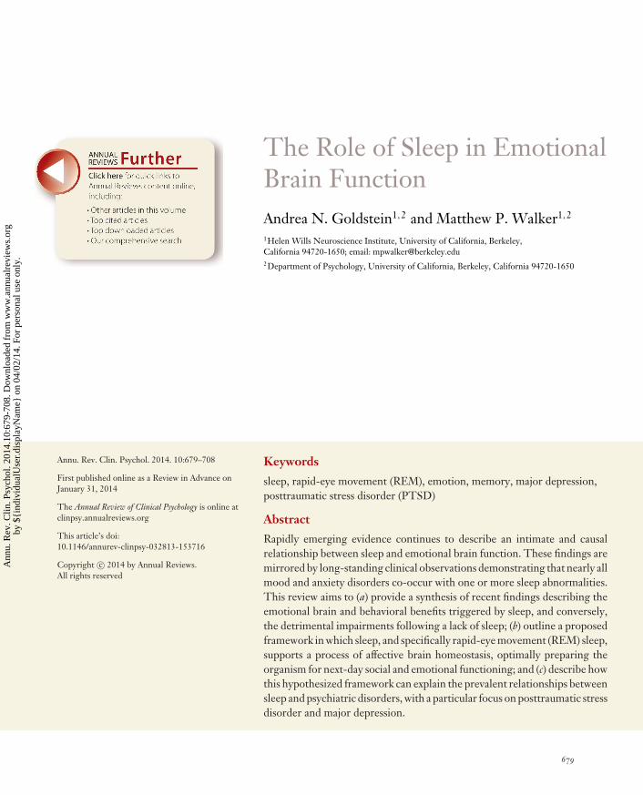
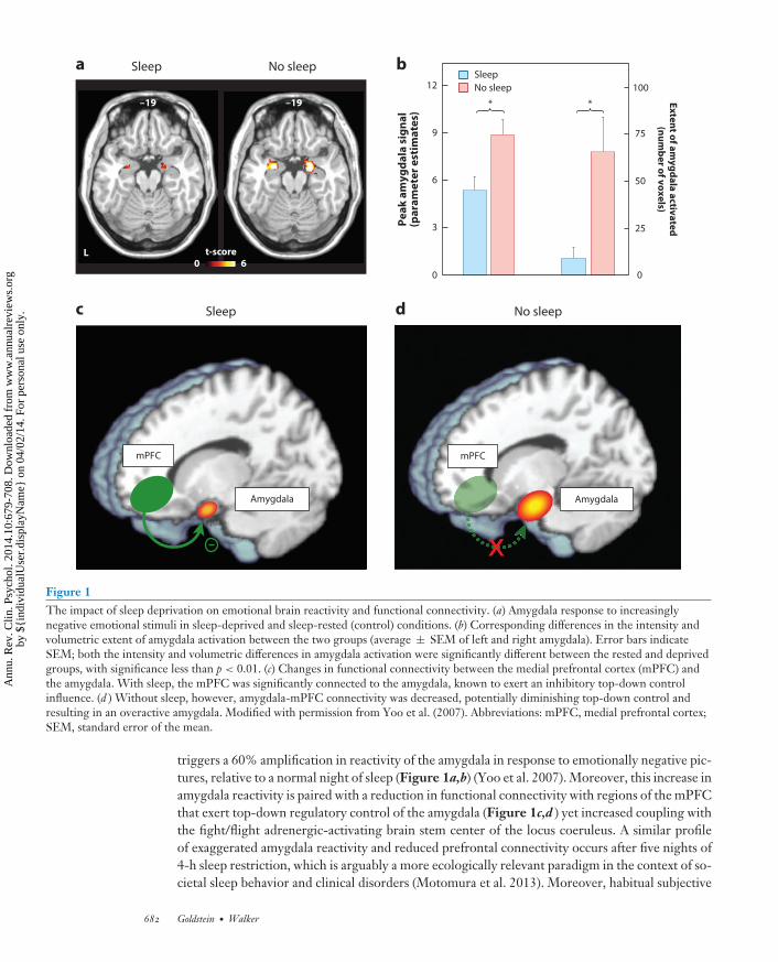
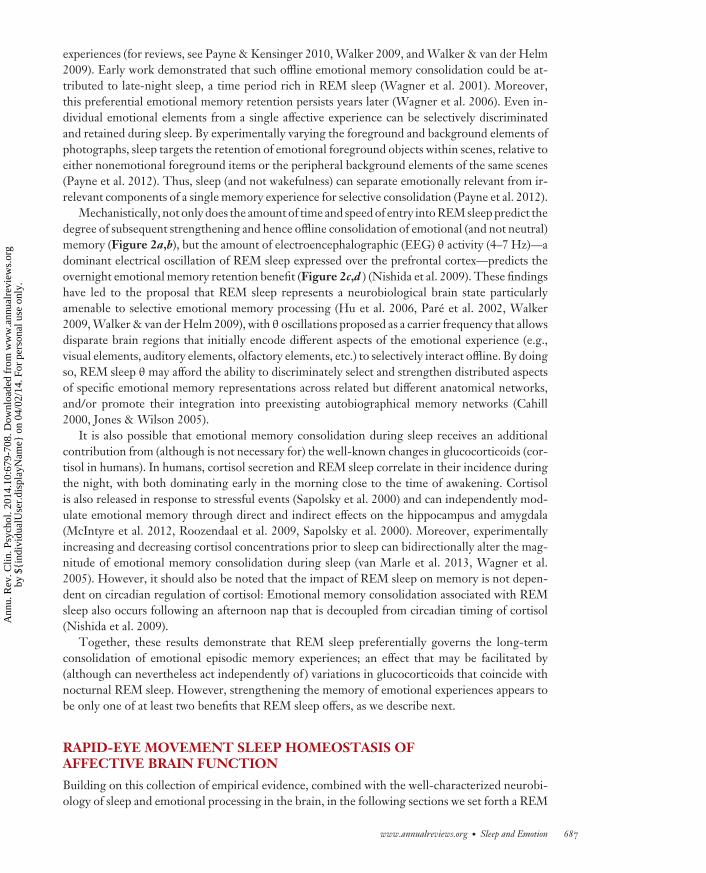
Figure 1The impact of sleep deprivation on emotional brain reactivity and functional connectivity. (a) Amygdala response to increasinglynegative emotional stimuli in sleep-deprived and sleep-rested (control) conditions. (b) Corresponding differences in the intensity andvolumetric extent of amygdala activation between the two groups (average ± SEM of left and right amygdala). Error bars indicateSEM; both the intensity and volumetric differences in amygdala activation were significantly different between the rested and deprivedgroups, with significance less than p < 0.01. (c) Changes in functional connectivity between the medial prefrontal cortex (mPFC) andthe amygdala. With sleep, the mPFC was significantly connected to the amygdala, known to exert an inhibitory top-down controlinfluence. (d ) Without sleep, however, amygdala-mPFC connectivity was decreased, potentially diminishing top-down control andresulting in an overactive amygdala. Modified with permission from Yoo et al. (2007). Abbreviations: mPFC, medial prefrontal cortex;SEM, standard error of the mean.
triggers a 60% amplification in reactivity of the amygdala in response to emotionally negative pic-tures, relative to a normal night of sleep (Figure 1a,b) (Yoo et al. 2007). Moreover, this increase inamygdala reactivity is paired with a reduction in functional connectivity with regions of the mPFCthat exert top-down regulatory control of the amygdala (Figure 1c,d ) yet increased coupling withthe fight/flight adrenergic-activating brain stem center of the locus coeruleus. A similar profileof exaggerated amygdala reactivity and reduced prefrontal connectivity occurs after five nights of4-h sleep restriction, which is arguably a more ecologically relevant paradigm in the context of so-cietal sleep behavior and clinical disorders (Motomura et al. 2013). Moreover, habitual subjective
682 Goldstein ·Walker
Ann
u. R
ev. C
lin. P
sych
ol. 2
014.
10:6
79-7
08. D
ownl
oade
d fr
om w
ww
.ann
ualr
evie
ws.
org
by $
{ind
ivid
ualU
ser.
disp
layN
ame}
on
04/0
2/14
. For
per
sona
l use
onl
y.
CP10CH25-Walker ARI 11 February 2014 12:9
sleep quality outside of the laboratory is also significantly related to amygdala reactivity, as wellas features of negative affect and stress (Prather et al. 2013). Additionally, interindividual differ-ences in the change in connectivity between the amygdala and mPFC caused by sleep deprivationaccurately predict the concurrent increase in subjective anxiety (Motomura et al. 2013).
Although the majority of studies to date have focused on changes within the central nervoussystem (and specifically brain), it is of note that such central alterations are paralleled by changesin peripheral nervous system physiology. One night of sleep deprivation amplifies pupil diame-ter responses—an index of peripheral autonomic nervous system reactivity—during the passiveviewing of negative emotional picture stimuli (Franzen & Buysse 2008). Congruently, sleep depri-vation also increases sympathetic dominance of the autonomic nervous system, indexed by changesin heart rate variability (Sauvet et al. 2010, Zhong et al. 2005). The latter is important becausethis sympathetic bias is associated with a lack of flexibility and capacity to respond to emotionalchallenges, and has been positively associated with psychopathology (Appelhans & Luecken 2006).
Growing evidence suggests that sleep loss imposes a bidirectional nature of affective imbalance,additionally triggering excessive reactivity to positive, reward-relevant stimuli. One night of sleeploss enhances reactivity throughout regions of the dopaminergic mesolimbic systems in responseto pleasure-evoking emotional picture stimuli (Gujar et al. 2011). As with aversive reactivity, thisenhanced mesolimbic reward sensitivity is further associated with decreased functional connectiv-ity in regions of the medial and orbital prefrontal cortex. Similar enhanced mesolimbic reactivityfollowing sleep deprivation can occur using monetary reward incentive paradigms (Libedinskyet al. 2011, McKenna et al. 2007, Venkatraman et al. 2007). Beyond more abstract reward stimuli,such as money, this impact of sleep loss extends to more primary reward-motivated behaviors,including that of appetitive food desire. Both acute and chronic sleep loss are associated withelevated reactivity to food stimuli in salience and hedonic regions of the striatum and amygdala,together with blunted activity in appetitive choice and decision-making regions of the frontal cor-tex (Benedict et al. 2012, Greer et al. 2013, Killgore et al. 2013, St-Onge et al. 2012). Moreover,these neural changes are accompanied by an increased preference for higher-calorie foods (Greeret al. 2013) and greater tendencies to overeat (Killgore et al. 2013).
In addition to changes in neural reactivity at the time of experiencing an emotional event, sleeploss further alters the preemptive neural anticipation of impending emotional experiences in boththe amygdala and anterior insula cortex (Goldstein et al. 2013). Of special clinical relevance, traitanxiety levels predict interindividual differences in the extent of this exaggerated sleep-deprivedanticipation response (Goldstein et al. 2013). Specifically, high trait-anxious participants expressthe most severe increase in anticipatory reactivity under conditions of sleep loss; this is a concernbecause these are already the individuals at greatest risk for developing an anxiety disorder.
Amplified anticipatory responses following sleep deprivation are also observed for reward-relevant cues. One night of sleep deprivation elevates anticipatory activity within the reward-sensitive region of the striatum to monetary decisions that can lead to either future reward payoffsor losses—especially when those gambles are risky (Venkatraman et al. 2007). Consistent withstudies discussed above regarding appetitive food stimuli, the increases in subcortical striatal re-activity following sleep deprivation co-occurs with blunted activity in anterior insula cortex andorbitofrontal cortex, specifically to monetary losses. Thus, sleep deprivation appears to trigger astate where rewards are overvalued (increased striatal sensitivity), yet losses (through punishment)are undervalued.
Taken as a whole, these data establish that insufficient sleep exaggerates subcortical limbic andstriatal responses to both negative and positive affective stimuli, commonly associated with impov-erished prefrontal cortex activity and/or connectivity. The consequence appears to be a pendulumlike, bidirectional state of emotion imbalance at both ends of the valence spectrum, fitting early
www.annualreviews.org • Sleep and Emotion 683
Ann
u. R
ev. C
lin. P
sych
ol. 2
014.
10:6
79-7
08. D
ownl
oade
d fr
om w
ww
.ann
ualr
evie
ws.
org
by $
{ind
ivid
ualU
ser.
disp
layN
ame}
on
04/0
2/14
. For
per
sona
l use
onl
y.
CP10CH25-Walker ARI 11 February 2014 12:9
anecdotal reports of affective liability following a lack of sleep (Dahl 1996). Furthermore, thisexaggerated reactivity can, if cued, be observed preemptively before the emotional stimulus itself.
Such a model of altered emotion reactivity following sleep loss is of translational relevance for atleast three clinical areas. First, remarkably similar patterns of altered mesolimbic system emotionreactivity, as well as limbic-prefrontal cortex connectivity, have been reported in several affectivepsychopathologies, including major depression, bipolar disorder, generalized anxiety disorder,and PTSD (Davidson 2002, Drevets et al. 2008, Etkin 2010, Etkin & Wager 2007, Nitschke et al.2009, Paulus & Stein 2006, Rauch et al. 2000, Shin et al. 2006, Siegle et al. 2007). Crucially,every one of these clinical conditions expresses highly comorbid sleep disruption (Harvey 2011,Peterson & Benca 2006), and in some of these disorders, sleep abnormalities form part of theirdiagnostic criteria. Considering the overlap between the neural correlates of such conditions thatdemonstrate co-occurring sleep disruption and the patterns of neural dysfunction that can beexperimentally induced by sleep deprivation, these findings raise the important issue of whethersleep loss plays a causal role in the etiology of these conditions. Moreover, should sleep be acontributing factor, it would represent a novel treatment intervention target (Harvey et al. 2011).
Second, in the context of reward sensitivity, sleep disturbance is a recognized hallmark of ad-diction (Arnedt et al. 2007, Brower & Perron 2010, Dimsdale et al. 2007, Pace-Schott et al. 2005),leading to the recent proposal that sleep loss represents a common and reliable predictor of relapsein numerous addiction disorders (Brower & Perron 2010). Additionally, a prospective study hasdemonstrated that sleep problems assessed during childhood significantly predict early onset ofdrug and alcohol use years later in adolescence, even when controlling for effects of anxiety andattention deficits (Volkow et al. 2009). As such, the mesolimbic dopaminergic system appears torepresent one common pathway through which the effects of sleep loss and heightened addictionsensitivity can be understood. The experimental evidence discussed above, demonstrating an inter-action between a lack of sleep and enhanced mesolimbic reward reactivity, implicates sleep loss asa predisposing and causal (rather than co-occurring) risk factor in heightened responsiveness andhence acquired addiction potential for use of reward-stimulating drugs. Moreover, beyondacquisition, these findings also indicate a possible role for sleep disruption in the maintenance ofaddiction habits, especially during attempted withdrawal, leading to higher relapse rates.
Third, although anticipation is generally an adaptive process, aiding preparatory responses topotentially threatening or rewarding events, exaggerated expectancy activity, such as that observedfollowing sleep deprivation, can be maladaptive. In the context of aversive events, increased antic-ipatory limbic activity positively predicts clinical features of anxiety disorders, such as worry andrumination (Etkin & Wager 2007, Nitschke et al. 2009, Paulus & Stein 2006), many of which ex-press co-occurring impairments in the quantity and quality of sleep (Papadimitriou & Linkowski2005). Perhaps more concerning is that individuals with higher levels of trait anxiety, who arealready at higher risk for developing an anxiety disorder, appear to be the most vulnerable to theseanxiogenic effects of insufficient sleep (Goldstein et al. 2013).
Emotion Recognition and Expression
Intriguingly, a number of studies have reported what may at first be considered a paradoxicalblunting, rather than overestimation, in the subjective rating of emotions in others by sleep-deprived participants. For example, sleep deprivation decreases the subjective intensity ratings ofthreat-relevant (angry) and reward-relevant (happy) static facial expressions (van der Helm et al.2010). Sleep loss also decreases the outward expression of emotion by sleep-deprived individuals, asjudged by expert raters (Minkel et al. 2011). Similarly, decreases in the vocal expression of positiveemotion by deprived participants are observed after a single night of sleep loss, suggesting that
684 Goldstein ·Walker
Ann
u. R
ev. C
lin. P
sych
ol. 2
014.
10:6
79-7
08. D
ownl
oade
d fr
om w
ww
.ann
ualr
evie
ws.
org
by $
{ind
ivid
ualU
ser.
disp
layN
ame}
on
04/0
2/14
. For
per
sona
l use
onl
y.
CP10CH25-Walker ARI 11 February 2014 12:9
multiple routes of emotional expression (e.g., facial muscles, vocalizations) are compromised byinsufficient sleep (McGlinchey et al. 2011). In addition to diminishing outward emotive expression,sleep deprivation also slows the generation of facial reactions in response to a visual presentationof faces (Schwarz et al. 2013). Of concern, insufficient sleep appears to trigger as much, if notmore, of an impact on emotional expression in young children. Recent evidence demonstratesthat three-year-olds who do not obtain an afternoon nap show dysregulation of both positive andnegative emotional expression in response to emotional stimuli, relative to those who have takena nap (Berger et al. 2012).
The potential disparity between these impairments and the objective neural data describedabove, which have reported amplifications (rather than impairments) in limbic brain reactivityfollowing sleep deprivation, can be reconciled when considering the concomitant neural impair-ments in the prefrontal cortex. Not only are prefrontal regions implicated in top-down regulatorycontrol of subcortical limbic networks, but they also critically integrate primary affective signalsarising from these subcortical systems (such as the brain stem, limbic system, and basal ganglia) intosecond-order maps of the internal state of the organism (Craig 2010, 2011; Critchley 2005, 2009;Harrison et al. 2010). It has been argued that only through such mapping, and hence appreciationof the current state of the body in the frontal lobe, can the brain select appropriate behavioral ac-tions for the organism (actions that include emotion expression) (Craig 2010, 2011; Critchley 2005,2009; Harrison et al. 2010). Set against this evidence, the above disparate findings may be resolved.Specifically, the sleep-deprived brain may suffer a mismatch between excessive subcortical reac-tivity and impaired higher-order prefrontal functioning, the latter preventing optimal integrationand hence use of the former, as well as control over the former. As a consequence, there can be afailure of affectively guided judgments, decisions, and downstream behavioral emotive (re)actions.
BENEFITS OF SLEEP ON EMOTIONAL BRAIN FUNCTION
Fear Conditioning: Acquisition, Generalization, and Extinction
Beyond emotion reactivity, recognition, and expression, sleep has been demonstrated to alsoplay an influential role in modulating conditioned fear. In classical fear conditioning paradigms,neutral items (e.g., a tone) are repeatedly paired with a coinciding noxious event (unconditionedstimulus, e.g., electric shock), triggering fear reactions. After an association is formed betweenthese two elements, the presentation of the previously neutral item alone (now referred to as theconditioned stimulus, the tone, in this example) is sufficient to elicit a conditioned fear response.However, if this conditioned stimulus is subsequently re-presented (the tone), but now in theabsence of the coinciding unconditioned stimulus (the shock), the fear response to the conditionedstimulus (the tone) gradually dissipates—a process known as extinction. Importantly, extinctionis not accomplished by unlearning the old fear association but instead by new learning of fearinhibition (Phelps et al. 2004). Beyond learning such direct associations, contextual cues canalso modify fear responses. After fear conditioning, even the testing environment itself—wherethe conditioned stimulus (the tone) is associated with the unconditioned stimulus (the shock)—can signal an unsafe context and trigger a fear reaction. Conversely, a safe context that was notpreviously associated with the conditioned-unconditioned stimulus pairing, such as a novel orextinction environment, can promote inhibition of the fear response, even when the conditionedstimulus (tone) is presented. In the following sections, we describe the impact of sleep and sleeploss on (a) the consolidation of fear-conditioned learning, (b) the extinction of conditioned fear,and (c) the appropriate maintenance or inhibition of a fear response depending on unsafe or safecontexts.
www.annualreviews.org • Sleep and Emotion 685
Ann
u. R
ev. C
lin. P
sych
ol. 2
014.
10:6
79-7
08. D
ownl
oade
d fr
om w
ww
.ann
ualr
evie
ws.
org
by $
{ind
ivid
ualU
ser.
disp
layN
ame}
on
04/0
2/14
. For
per
sona
l use
onl
y.

CP10CH25-Walker ARI 11 February 2014 12:9
Focusing first on consolidation, a night of sleep strengthens conditioned fear responses(neural, physiological, and behavioral) compared to the absence of intervening sleep, and as aconsequence, results in superior discrimination of fear-related from non-fear-related cues (Menzet al. 2013). Furthermore, the magnitude of this sleep-dependent beneficial fear discriminationwas positively predicted by the amount of intervening REM sleep (Menz et al. 2013). Together,these findings suggest that sleep, and specifically REM sleep, consolidates conditioned fearmemories, allowing for an improved next-day sensitivity to discriminate between threatening andnonthreatening stimuli.
In addition to strengthening fear memories, sleep also adaptively facilitates the subsequentextinction of conditioned fear, a process that is known to be accomplished by way of top-downPFC inhibition of the amygdala. An intervening period of sleep, and specifically one containingREM sleep, subsequently promotes more rapid diminution of fear responses (here, skin conduc-tance) and with it the beneficial reengagement of ventromedial PFC involvement during postsleepfear extinction recall (Spoormaker et al. 2012). The latter finding is relevant considering that theventromedial PFC is a region known to be necessary for successful development of lasting fear ex-tinction (Phelps et al. 2004). Combined with the findings described above, these data indicate thatintervening sleep not only strengthens conditioned fear but also primes the neural mechanisms toextinguish fear the next day, should experience dictate it. Moreover, that the extent of these bene-fits appears to correlate with the amount of REM sleep is perhaps no coincidence: The brain areasrecognized to support fear acquisition and extinction (mPFC, amygdala, hippocampus) are all reac-tivated during REM sleep (Dang-Vu et al. 2010, Miyauchi et al. 2009, Nofzinger 2005, Phelps et al.2004). And through such REM sleep, brain network restoration may aid the appropriate returnof top-down PFC action on the amygdala, governing these processes (van der Helm et al. 2011).
Beyond simply strengthening or extinguishing fear responses, recent evidence demonstratesthat sleep, relative to time spent awake, preferentially modifies the appropriate expression offear, depending on whether the conditioned stimulus (the tone) is presented in an unsafe or a safesurrounding context (Menz et al. 2013, Pace-Schott et al. 2009). As a consequence, individuals whosleep demonstrate significantly more adaptive expressions of fear, either maintaining or inhibitingfear responses, depending on the presence of unsafe or safe contextual cues, respectively (Menzet al. 2013, Pace-Schott et al. 2009). Sleep therefore facilitates the most appropriate or “intelligent”expression of conditioned fear, based on environmental information signaling threat or safety.
As a whole, these data from fear conditioning experiments indicate that sleep, including REMsleep, supports adaptive fear responses across numerous levels. The benefits of such processesencourage proper generation and maintenance of fear responses in dangerous situations, whileinhibiting fear responses to nondangerous situations. In contrast, sleep deprivation impairs thesesame processes due, in part, to a loss of top-down prefrontal cortex control of subcortical limbicregions. This is particularly relevant in a clinical context because deficits in the extinction and abilityto appropriately utilize surrounding contextual information underlie fear-related disorders, suchas specific phobia and PTSD (Phelps et al. 2004), the latter associated with marked sleep disruption(Germain 2013). Early evidence already suggests a treatment intervention promise: Patients whoslept immediately after exposure treatment—a form of fear extinction learning—showed greatersubjective reductions in anxiety and negative cognition one week after treatment relative to thosewho remain awake for some time after treatment (Kleim et al. 2013).
Emotional Memory
REM sleep also plays an influential role in the processing of affective information beyond basic fearconditioning, particularly in the “offline” consolidation of emotional episodic (autobiographical)
686 Goldstein ·Walker
Ann
u. R
ev. C
lin. P
sych
ol. 2
014.
10:6
79-7
08. D
ownl
oade
d fr
om w
ww
.ann
ualr
evie
ws.
org
by $
{ind
ivid
ualU
ser.
disp
layN
ame}
on
04/0
2/14
. For
per
sona
l use
onl
y.
CP10CH25-Walker ARI 11 February 2014 12:9
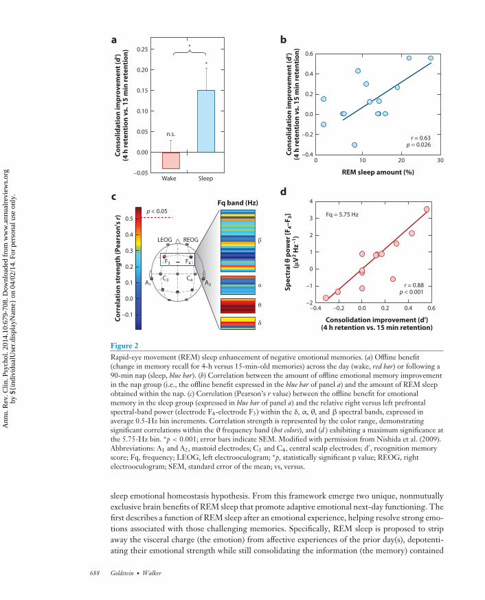
experiences (for reviews, see Payne & Kensinger 2010, Walker 2009, and Walker & van der Helm2009). Early work demonstrated that such offline emotional memory consolidation could be at-tributed to late-night sleep, a time period rich in REM sleep (Wagner et al. 2001). Moreover,this preferential emotional memory retention persists years later (Wagner et al. 2006). Even in-dividual emotional elements from a single affective experience can be selectively discriminatedand retained during sleep. By experimentally varying the foreground and background elements ofphotographs, sleep targets the retention of emotional foreground objects within scenes, relative toeither nonemotional foreground items or the peripheral background elements of the same scenes(Payne et al. 2012). Thus, sleep (and not wakefulness) can separate emotionally relevant from ir-relevant components of a single memory experience for selective consolidation (Payne et al. 2012).
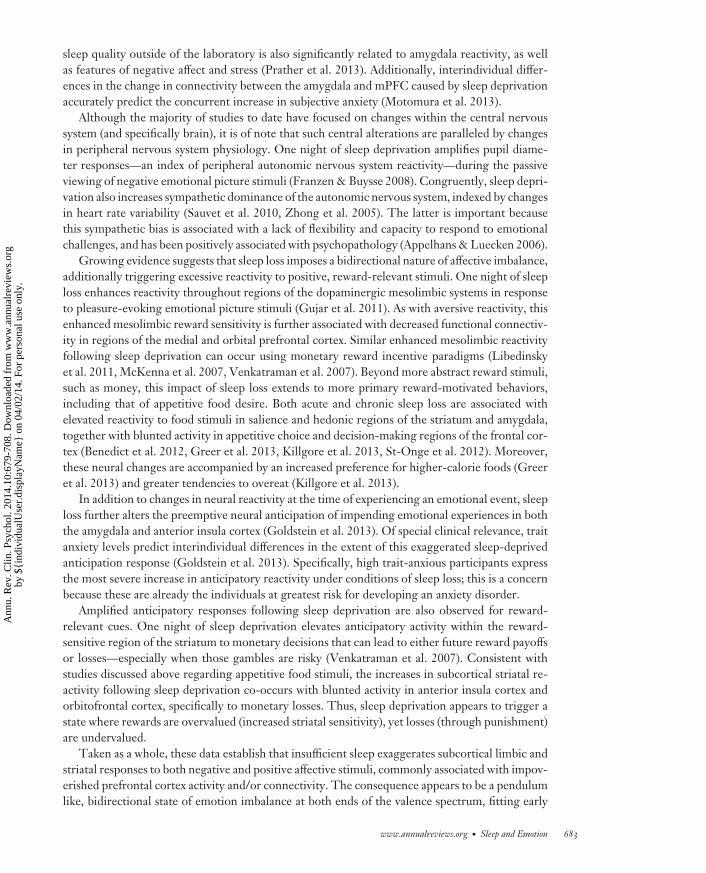
Mechanistically, not only does the amount of time and speed of entry into REM sleep predict thedegree of subsequent strengthening and hence offline consolidation of emotional (and not neutral)memory (Figure 2a,b), but the amount of electroencephalographic (EEG) θ activity (4–7 Hz)—adominant electrical oscillation of REM sleep expressed over the prefrontal cortex—predicts theovernight emotional memory retention benefit (Figure 2c,d ) (Nishida et al. 2009). These findingshave led to the proposal that REM sleep represents a neurobiological brain state particularlyamenable to selective emotional memory processing (Hu et al. 2006, Pare et al. 2002, Walker2009, Walker & van der Helm 2009), with θ oscillations proposed as a carrier frequency that allowsdisparate brain regions that initially encode different aspects of the emotional experience (e.g.,visual elements, auditory elements, olfactory elements, etc.) to selectively interact offline. By doingso, REM sleep θ may afford the ability to discriminately select and strengthen distributed aspectsof specific emotional memory representations across related but different anatomical networks,and/or promote their integration into preexisting autobiographical memory networks (Cahill2000, Jones & Wilson 2005).
It is also possible that emotional memory consolidation during sleep receives an additionalcontribution from (although is not necessary for) the well-known changes in glucocorticoids (cor-tisol in humans). In humans, cortisol secretion and REM sleep correlate in their incidence duringthe night, with both dominating early in the morning close to the time of awakening. Cortisolis also released in response to stressful events (Sapolsky et al. 2000) and can independently mod-ulate emotional memory through direct and indirect effects on the hippocampus and amygdala(McIntyre et al. 2012, Roozendaal et al. 2009, Sapolsky et al. 2000). Moreover, experimentallyincreasing and decreasing cortisol concentrations prior to sleep can bidirectionally alter the mag-nitude of emotional memory consolidation during sleep (van Marle et al. 2013, Wagner et al.2005). However, it should also be noted that the impact of REM sleep on memory is not depen-dent on circadian regulation of cortisol: Emotional memory consolidation associated with REMsleep also occurs following an afternoon nap that is decoupled from circadian timing of cortisol(Nishida et al. 2009).
Together, these results demonstrate that REM sleep preferentially governs the long-termconsolidation of emotional episodic memory experiences; an effect that may be facilitated by(although can nevertheless act independently of ) variations in glucocorticoids that coincide withnocturnal REM sleep. However, strengthening the memory of emotional experiences appears tobe only one of at least two benefits that REM sleep offers, as we describe next.
RAPID-EYE MOVEMENT SLEEP HOMEOSTASIS OFAFFECTIVE BRAIN FUNCTION
Building on this collection of empirical evidence, combined with the well-characterized neurobi-ology of sleep and emotional processing in the brain, in the following sections we set forth a REM
www.annualreviews.org • Sleep and Emotion 687
Ann
u. R
ev. C
lin. P
sych
ol. 2
014.
10:6
79-7
08. D
ownl
oade
d fr
om w
ww
.ann
ualr
evie
ws.
org
by $
{ind
ivid
ualU
ser.
disp
layN
ame}
on
04/0
2/14
. For
per
sona
l use
onl
y.
CP10CH25-Walker ARI 11 February 2014 12:9
*
*a
0.25
0.20
0.15
0.10
0.05
0.00
–0.05Wake Sleep
n.s.
Co
nso
lid
ati
on
im
pro
ve
me
nt
(d')
(4 h
re
ten
tio
n v
s. 1
5 m
in r
ete
nti
on
)
b
Co
nso
lid
ati
on
im
pro
ve
me
nt
(d')
(4 h
re
ten
tio
n v
s. 1
5 m
in r
ete
nti
on
)
REM sleep amount (%)
0.6
0.4
0.2
0.0
–0.2
–0.410 20 30
r = 0.63p = 0.026
0
d
–0.4
r = 0.88p < 0.001
–0.2 0.0 0.2 0.4 0.6
–1
0
1
2
3
4
Consolidation improvement (d')(4 h retention vs. 15 min retention)
Sp
ect
ral
θ p
ow
er
[F4–
F3]
(μV
2 H
z–
1)
–2
c
Co
rre
lati
on
str
en
gth
(P
ea
rso
n's
r)
0.0
–0.1
0.1
0.2
0.3
0.4
0.5p < 0.05
LEOG REOG
A1
C3 C4A2
F3 F4
Fq band (Hz)
β
α
θ
δ
Fq = 5.75 Hz
Figure 2Rapid-eye movement (REM) sleep enhancement of negative emotional memories. (a) Offline benefit(change in memory recall for 4-h versus 15-min-old memories) across the day (wake, red bar) or following a90-min nap (sleep, blue bar). (b) Correlation between the amount of offline emotional memory improvementin the nap group (i.e., the offline benefit expressed in the blue bar of panel a) and the amount of REM sleepobtained within the nap. (c) Correlation (Pearson’s r value) between the offline benefit for emotionalmemory in the sleep group (expressed in blue bar of panel a) and the relative right versus left prefrontalspectral-band power (electrode F4-electrode F3) within the δ, α, θ, and β spectral bands, expressed inaverage 0.5-Hz bin increments. Correlation strength is represented by the color range, demonstratingsignificant correlations within the θ frequency band (hot colors), and (d ) exhibiting a maximum significance atthe 5.75-Hz bin. ∗p < 0.001; error bars indicate SEM. Modified with permission from Nishida et al. (2009).Abbreviations: A1 and A2, mastoid electrodes; C3 and C4, central scalp electrodes; d′, recognition memoryscore; Fq, frequency; LEOG, left electrooculogram; ∗p, statistically significant p value; REOG, rightelectrooculogram; SEM, standard error of the mean; vs, versus.
sleep emotional homeostasis hypothesis. From this framework emerge two unique, nonmutuallyexclusive brain benefits of REM sleep that promote adaptive emotional next-day functioning. Thefirst describes a function of REM sleep after an emotional experience, helping resolve strong emo-tions associated with those challenging memories. Specifically, REM sleep is proposed to stripaway the visceral charge (the emotion) from affective experiences of the prior day(s), depotenti-ating their emotional strength while still consolidating the information (the memory) contained
688 Goldstein ·Walker
Ann
u. R
ev. C
lin. P
sych
ol. 2
014.
10:6
79-7
08. D
ownl
oade
d fr
om w
ww
.ann
ualr
evie
ws.
org
by $
{ind
ivid
ualU
ser.
disp
layN
ame}
on
04/0
2/14
. For
per
sona
l use
onl
y.
CP10CH25-Walker ARI 11 February 2014 12:9
within that experience—a form of “overnight therapy.” The second outlines a role for REM sleepbefore an emotional experience, recalibrating the sensitivity and specificity of the brain’s responseto initial emotional events. As a consequence REM sleep primes key brain regions to appropriatelyreact to affective experiences, allowing accurate and adaptive next-day discrimination of one emo-tional experience from another by faithfully registering their respective salient values.
Emotional Memory Resolution: Sleeping to Forget and Sleeping to Remember
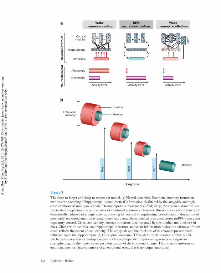
Our first proposed function of REM sleep takes place after the occurrence of emotional eventsand involves aiding the reprocessing of prior affective experiences, including those most traumatic.Although there is abundant evidence to suggest that emotional experiences persist in our autobi-ographies over time (strengthening the memory) (Dolcos et al. 2005), an equally remarkable butless noted change is a reduction in the affective tone associated with their recall (depotentiationof emotion). Affective experiences appear to be remembered more robustly than neutral memo-ries due to a well-characterized set of adrenergic and peripheral autonomic reactions elicited atthe time of the experience, orchestrated, in part, by the locus coeruleus (McGaugh 2004). Theseadrenergic bursts at the time of affectively charged experiences are believed to adaptively priori-tize the formation (and hence long-term retention) of such salient information within the brain,creating what is commonly termed an emotional memory (Figure 3a). However, the later recallof these memories tends not to be associated with the same magnitude of autonomic (re)activationas that elicited at the moment of experience—suggesting that, over time, the affective “blanket”(the emotion) that originally tagged the memory at the time of learning has been removed, whereasthe information of the experience (the memory) remains (Figure 3b). We propose here [and else-where (van der Helm & Walker 2012, Walker 2009, Walker & van der Helm 2009)] that the uniqueneurobiological state of REM sleep supports decoupling of emotion from memory such that wesleep to forget the emotional tone, yet sleep to remember the memory of that experience. Thismodel further posits that, if this process is not achieved, the magnitude of affective “charge” wouldpersist, resulting in a condition of chronic anxiety within autobiographical memory networks.
Three specific biological features of REM sleep are proposed to provide an optimal biologicalmilieu within which this form of overnight therapy can be achieved: neuroanatomical, neurophys-iological, and neurochemical (Figure 3a). First, the prominent increase in activity within limbicand paralimbic structures during REM sleep (Nofzinger 2005) supports the ability for reactivationand hence (re)processing of previously acquired affective memories. Second, the neurophysiolog-ical signature of REM sleep involving dominant θ oscillations within subcortical as well as corticalnodes offers large-scale network cooperation during REM sleep for the strengthening of dis-tributed aspects of the emotional memory representation (e.g., perceptual, contextual), acrosssuch related but different anatomical networks. This then results in enhanced consolidation andintegration of that memory. Third, these interactions during REM sleep (and perhaps through theconscious process of dreaming) critically and perhaps most importantly take place within a brainthat is low in aminergic neurochemical concentration (Pace-Schott & Hobson 2002), particularlythe suppressed noradrenergic input from the locus coeruleus (associated with stress and anxietyresponses when highly active) (Itoi & Sugimoto 2010, Ramos & Arnsten 2007, Sullivan et al.1999, Valentino & Van Bockstaele 2008). Therefore, REM sleep is proposed to offer a uniquebiological condition to achieve the strengthening and consolidation of the informational core ofemotional experiences (the memory) yet additionally depotentiates and ultimately ameliorates theautonomic arousing charge originally acquired at the time of learning (the emotion). Throughthe process of developing stronger cortico-cortical connections, integration and assimilation of theaffective event(s) in the context of past knowledge are supported. As a result, emotional experiences
www.annualreviews.org • Sleep and Emotion 689
Ann
u. R
ev. C
lin. P
sych
ol. 2
014.
10:6
79-7
08. D
ownl
oade
d fr
om w
ww
.ann
ualr
evie
ws.
org
by $
{ind
ivid
ualU
ser.
disp
layN
ame}
on
04/0
2/14
. For
per
sona
l use
onl
y.
CP10CH25-Walker ARI 11 February 2014 12:9
Amygdala
Wakememory encoding
REMneural reactivation
Wakememory recollection
Corticalmodules
Hippocampus
Adrenergic
Cholinergic
Activity level Activity level Activity level
Ne
uro
che
mic
al
Ne
uro
an
ato
mic
al
Memory
Emotion
Memory Emotional
memory
a
b
mP
FC
Log time
Sle
ep
Sle
ep
Sle
ep
Figure 3The sleep to forget and sleep to remember model. (a) Neural dynamics. Emotional memory formationinvolves the encoding of hippocampal-bound cortical information, facilitated by the amygdala and highconcentrations of aminergic activity. During rapid-eye movement (REM) sleep, these neural structures arereactivated, supporting the reprocessing of emotional memories. However, this occurs in a brain state withdramatically reduced adrenergic activity, allowing for cortical strengthening (consolidation), dissipation ofpreviously associated emotion (visceral tone), and reestablished medial prefrontal cortex (mPFC)-amygdalaregulatory control. Cross connectivity between structures is represented by the number and thickness oflines. Circles within cortical and hippocampal structures represent information nodes; the darkness of theirshade reflects the extent of connectivity. The amygdala and the thickness of its arrows represent theirinfluence upon the hippocampus. (b) Conceptual outcome. Through multiple iterations of this REMmechanism across one or multiple nights, such sleep-dependent reprocessing results in long-termstrengthening of salient memories, yet a dissipation of the emotional charge. Thus, sleep transforms anemotional memory into a memory of an emotional event that is no longer emotional.
690 Goldstein ·Walker
Ann
u. R
ev. C
lin. P
sych
ol. 2
014.
10:6
79-7
08. D
ownl
oade
d fr
om w
ww
.ann
ualr
evie
ws.
org
by $
{ind
ivid
ualU
ser.
disp
layN
ame}
on
04/0
2/14
. For
per
sona
l use
onl
y.
CP10CH25-Walker ARI 11 February 2014 12:9
are preferentially retained long-term, but importantly, the emotion, which was initially criticalto signify salience and priority at the time of learning, has been dissipated. The brain thereforepreserves a memory of an emotional event, but the event no longer arouses strong emotions.
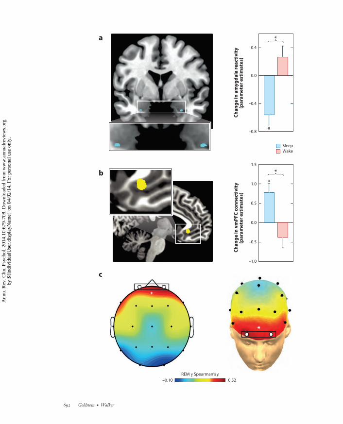
Specific predictions emerge from this model. As was partially demonstrated (see the Emo-tional Memory section, above), the first prediction is that the degree to which the informationof those emotional experiences is retained for a long term would be proportional to the amountof post-encoding REM sleep obtained, how quickly it is achieved (REM latency), and the powerof θ oscillations during REM sleep. Evidence for all three of these predictions exists (Nishida et al.2009, Pare et al. 2002). Second, the inverse REM sleep relationship would hold for the magni-tude of emotional depotentiation after sleep. This too appears to be the case; compared to thosethat remained awake, individuals who slept prior to reexposure displayed a significant overnightdecrease in amygdala response to previously seen emotional images (Figure 4a), together witha concomitant increase in amygdala-mPFC connectivity (Figure 4b). Furthermore, sleep alsoresults in a significant dissipation of subjective emotional intensity ratings relative to the equiva-lent period of wakefulness. Additionally, the success of overnight emotion depotentiation at boththe brain (amygdala) and behavioral (intensity ratings) levels is predicted by γ EEG activity—avalidated but indirect measure of central adrenergic activity (Berridge & Foote 1991, Cape &Jones 1998, Keane et al. 1976). Specifically, those participants who express the lowest REM γ
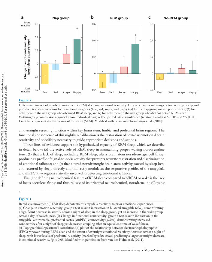
measurements show the greatest beneficial overnight reduction in emotion intensity (Figure 4c).Similarly, a daytime nap dissipates the intensity ratings of previously exposed negative emotionalface expressions (Gujar et al. 2010). However, not all participants who slept demonstrated thisresetting of emotional reactivity. Instead, only those who obtained REM sleep during the nap dis-played such a change in emotional sensitivity to the reexperience of the same previous face stimuli(Figure 5). In addition to fitting with a role for sleep in emotional depotentiation, if participantsare not allowed to sleep the first night after being exposed to aversive emotional stimuli, there is afailure of subsequent amygdala depotentiation to the same emotional stimulus during a memorytest, even following two nights of recovery sleep (Sterpenich et al. 2007). The latter finding isrelevant clinically and argues against some proposals for the use of sleep deprivation after traumaexperience to facilitate forgetting. Our model would argue the opposite: Sleep deprivation wouldprevent the beneficial removing of emotion from the memory (van der Helm & Walker 2012,Walker 2009, Walker & van der Helm 2009). Furthermore, although the emotion of the traumacan be clinically problematic, the information from the experience (the memory) is neverthelessuseful, helping guide future actions based on past experience, which may prevent repetition of thesame negative experience. Thus, it is the emotion that needs to be targeted, not the memory.
In summary, these studies provide support to the proposed neurobiological model of sleep-dependent emotional memory depotentiation, one in which REM sleep is capable of divorcingthe visceral affective charge from prior emotional experiences while preserving the informationof those episodes.
Rapid-Eye Movement Sleep Emotion Recalibration
Our second proposed function of REM sleep takes place before the occurrence of next-day emo-tional events, restoring optimal emotional reactivity. Adaptive emotional responses require ac-curate discrimination between salient stimuli (a potential threat to be avoided, or reward to beapproached) and nonsalient stimuli within the environment. Failures in the ability to differen-tiate salient from nonsalient information can lead to maladaptive behaviors, such as generatingexcessive fear responses to nondangerous situations (e.g., a car backfiring). On the basis of themarked changes in adrenergic neurochemistry described above, we propose that REM sleep offers
www.annualreviews.org • Sleep and Emotion 691
Ann
u. R
ev. C
lin. P
sych
ol. 2
014.
10:6
79-7
08. D
ownl
oade
d fr
om w
ww
.ann
ualr
evie
ws.
org
by $
{ind
ivid
ualU
ser.
disp
layN
ame}
on
04/0
2/14
. For
per
sona
l use
onl
y.
CP10CH25-Walker ARI 11 February 2014 12:9
a
b
REM γ Spearman’s ρ
c
0.52 –0.10
*
*
Sleep
Wake
Ch
an
ge
in
am
yg
da
la r
ea
ctiv
ity
(pa
ram
ete
r e
stim
ate
s)
–0.8
–0.4
0.0
0.4
*
*
Ch
an
ge
in
vm
PF
C c
on
ne
ctiv
ity
(pa
ram
ete
r e
stim
ate
s)
–1.0
–0.5
0.0
0.5
1.0
1.5
*
*
692 Goldstein ·Walker
Ann
u. R
ev. C
lin. P
sych
ol. 2
014.
10:6
79-7
08. D
ownl
oade
d fr
om w
ww
.ann
ualr
evie
ws.
org
by $
{ind
ivid
ualU
ser.
disp
layN
ame}
on
04/0
2/14
. For
per
sona
l use
onl
y.
CP10CH25-Walker ARI 11 February 2014 12:9
a b c
*
*
–0.2
–0.1
0
0.1
0.2
0.3C
ha
ng
e i
n e
mo
tio
na
l re
act
ivit
y(p
ost
-sle
ep
vs.
pre
-sle
ep
)
Morereactive
Lessreactive Fear Sad Anger Happy
Nap group
*
*
0.6
0.4
0.2
0
–0.2Fear Sad Anger Happy
REM group
0.6
0.4
0.2
0
–0.2
No-REM group
Fear Sad Anger Happy
Figure 5Differential impact of rapid-eye movement (REM) sleep on emotional reactivity. Difference in mean ratings between the presleep andpostsleep test sessions across four emotion categories (fear, sad, anger, and happy) (a) for the nap group overall performance, (b) foronly those in the nap group who obtained REM sleep, and (c) for only those in the nap group who did not obtain REM sleep.Within-group comparisons (symbol above individual bars) reflect paired t-test significance (relative to null) at ∗<0.05 and ∗∗<0.01.Error bars represent standard error of the mean (SEM). Modified with permission from Gujar et al. (2010).
an overnight resetting function within key brain stem, limbic, and prefrontal brain regions. Thefunctional consequence of this nightly recalibration is the restoration of next-day emotional brainsensitivity and specificity necessary to guide appropriate decisions and actions.
Three lines of evidence support the hypothesized capacity of REM sleep, which we describein detail below: (a) the active role of REM sleep in maintaining proper waking noradrenalinetone; (b) that a lack of sleep, including REM sleep, alters brain stem noradrenergic cell firing,producing a profile of signal-to-noise activity that prevents accurate registration and discriminationof emotional salience; and (c) that altered noradrenergic brain stem activity caused by sleep loss,and restored by sleep, directly and indirectly modulates the responsive profiles of the amygdalaand mPFC, two regions critically involved in detecting emotional salience.
First, the defining neurochemical feature of REM sleep compared to NREM or wake is the lackof locus coeruleus firing and thus release of its principal neurochemical, noradrenaline (Ouyang
←−−−−−−−−−−−−−−−−−−−−−−−−−−−−−−−−−−−−−−−−−−−−−−−−−−−−−−−−−−−−−−−−−−−−−−−−Figure 4Rapid-eye movement (REM) sleep depotentiates amygdala reactivity to prior emotional experiences.(a) Change in emotion reactivity: group x test session interaction in bilateral amygdala (blue), demonstratinga significant decrease in activity across a night of sleep in the sleep group, yet an increase in the wake groupacross a day of wakefulness. (b) Change in functional connectivity: group x test session interaction in theamygdala-ventromedial prefrontal cortex (vmPFC) connectivity ( yellow), demonstrating increasedconnectivity after a night of sleep yet decreased coupling after an equivalent time of wakefulness.(c) Topographical Spearman’s correlation (ρ) plot of the relationship between electroencephalographic(EEG) γ power during REM sleep and the extent of overnight emotional reactivity decrease across a night ofsleep, with lower levels of prefrontal γ activity (marked by white circles) predicting a larger overnight decreasein emotional reactivity. ∗p < 0.05. Modified with permission from van der Helm et al. (2011).
www.annualreviews.org • Sleep and Emotion 693
Ann
u. R
ev. C
lin. P
sych
ol. 2
014.
10:6
79-7
08. D
ownl
oade
d fr
om w
ww
.ann
ualr
evie
ws.
org
by $
{ind
ivid
ualU
ser.
disp
layN
ame}
on
04/0
2/14
. For
per
sona
l use
onl
y.
CP10CH25-Walker ARI 11 February 2014 12:9
et al. 2004, Park 2002, Shouse et al. 2000). As a result, the near absence of noradrenergic con-centration permeating the brain during REM sleep is potentially capable of restoring postsleepwaking levels of noradrenergic tone within the brain (Kametani & Kawamura 1990, Mallick &Singh 2011, Marrosu et al. 1995, Siegel & Rogawski 1988). Conversely, a lack of REM sleep, byway of selective deprivation, increases central noradrenaline concentrations to levels that exceedthose of rested waking brain function (Mallick & Singh 2011, Siegel & Rogawski 1988). Thus,a dose-dependent relationship may exist between the quantity/quality of REM sleep and the de-crease of noradrenaline through the night. Moreover, an increase in REM sleep quantity/quality atnight, either through pharmacological intervention (Stern & Morgane 1974, Taylor et al. 2008)or through psychopathology states such as major depression (Blier & Briley 2011, El Mansariet al. 2010, Hamon & Blier 2013, Ordway et al. 2003), would predict a proportional decrease ofnoradrenergic tone the next day and, with it, alterations in emotional brain function. Therefore,REM sleep may serve a noradrenergic “housekeeping” function, one that reduces and thus restoresconcentrations of noradrenaline to baseline each day (in humans), allowing for optimal wakefulfunctioning (Mallick & Singh 2011, Siegel & Rogawski 1988).
Second, experimental manipulations that evoke elevated noradrenaline levels, similar to thoseinduced by sleep loss, impair sensitivity to salience and specificity of locus coeruleus responses.During wake, noradrenaline neurons within the locus coeruleus display two distinct modes of over-all activity. In one of these modes, the locus coeruleus responds in a predominantly phasic manner,i.e., selectively to salient stimuli within the environment (i.e., salient signals) while maintaininga low level of baseline (tonic) ongoing activity (i.e., background “noise”) (Aston-Jones & Cohen2005, Mallick & Singh 2011, Valentino & Van Bockstaele 2008). Thus, the overall threshold of re-activity to external events is optimal along a gradient of potential emotional strengths (Figure 6a).However, the second mode of locus coeruleus activity, one that can occur under specific condi-tions (e.g., stress), is characterized by a shift to high levels of persistent baseline tonic firing andelevated noradrenaline levels. As a consequence of this high tonic background activity, the phasicsignals in response to external emotional stimuli result in poor signal to noise within the system
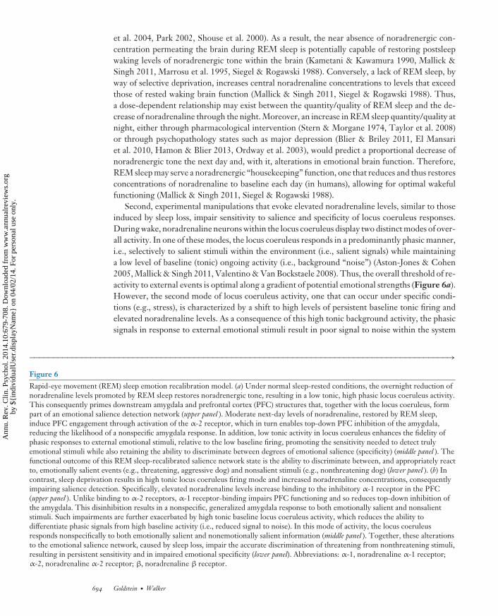
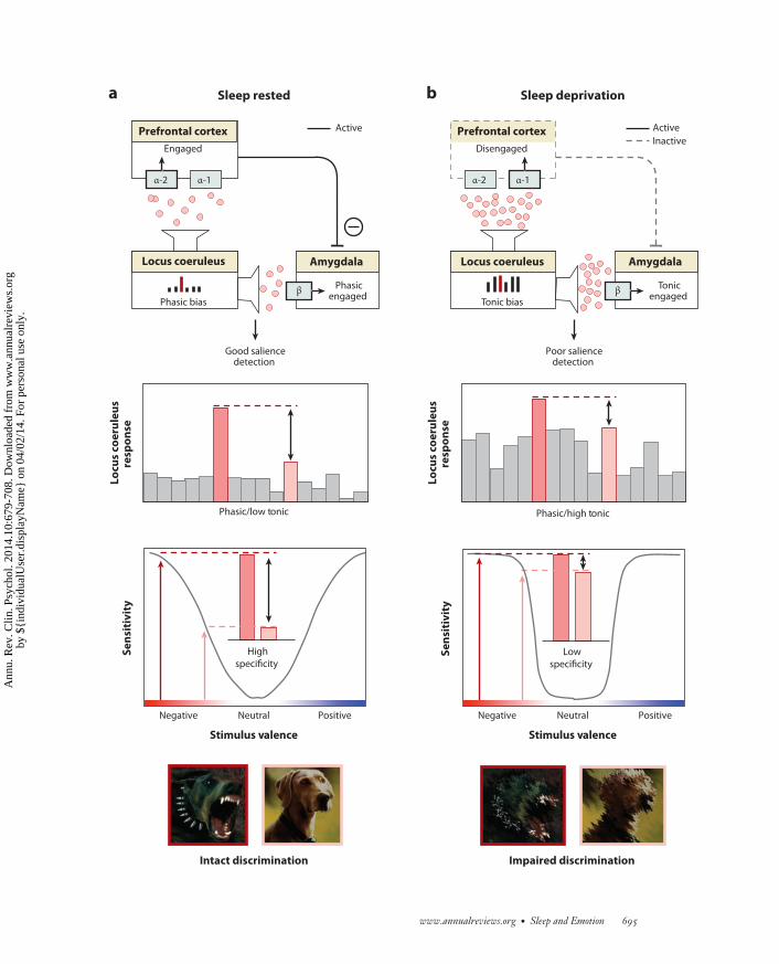
−−−−−−−−−−−−−−−−−−−−−−−−−−−−−−−−−−−−−−−−−−−−−−−−−−−−−−−−−−−−−−−−−−−−−−−−−−−−−−−−−−−−−−−−−→Figure 6Rapid-eye movement (REM) sleep emotion recalibration model. (a) Under normal sleep-rested conditions, the overnight reduction ofnoradrenaline levels promoted by REM sleep restores noradrenergic tone, resulting in a low tonic, high phasic locus coeruleus activity.This consequently primes downstream amygdala and prefrontal cortex (PFC) structures that, together with the locus coeruleus, formpart of an emotional salience detection network (upper panel ). Moderate next-day levels of noradrenaline, restored by REM sleep,induce PFC engagement through activation of the α-2 receptor, which in turn enables top-down PFC inhibition of the amygdala,reducing the likelihood of a nonspecific amygdala response. In addition, low tonic activity in locus coeruleus enhances the fidelity ofphasic responses to external emotional stimuli, relative to the low baseline firing, promoting the sensitivity needed to detect trulyemotional stimuli while also retaining the ability to discriminate between degrees of emotional salience (specificity) (middle panel ). Thefunctional outcome of this REM sleep-recalibrated salience network state is the ability to discriminate between, and appropriately reactto, emotionally salient events (e.g., threatening, aggressive dog) and nonsalient stimuli (e.g., nonthreatening dog) (lower panel ). (b) Incontrast, sleep deprivation results in high tonic locus coeruleus firing mode and increased noradrenaline concentrations, consequentlyimpairing salience detection. Specifically, elevated noradrenaline levels increase binding to the inhibitory α-1 receptor in the PFC(upper panel ). Unlike binding to α-2 receptors, α-1 receptor-binding impairs PFC functioning and so reduces top-down inhibition ofthe amygdala. This disinhibition results in a nonspecific, generalized amygdala response to both emotionally salient and nonsalientstimuli. Such impairments are further exacerbated by high tonic baseline locus coeruleus activity, which reduces the ability todifferentiate phasic signals from high baseline activity (i.e., reduced signal to noise). In this mode of activity, the locus coeruleusresponds nonspecifically to both emotionally salient and nonemotionally salient information (middle panel ). Together, these alterationsto the emotional salience network, caused by sleep loss, impair the accurate discrimination of threatening from nonthreatening stimuli,resulting in persistent sensitivity and in impaired emotional specificity (lower panel). Abbreviations: α-1, noradrenaline α-1 receptor;α-2, noradrenaline α-2 receptor; β, noradrenaline β receptor.
694 Goldstein ·Walker
Ann
u. R
ev. C
lin. P
sych
ol. 2
014.
10:6
79-7
08. D
ownl
oade
d fr
om w
ww
.ann
ualr
evie
ws.
org
by $
{ind
ivid
ualU
ser.
disp
layN
ame}
on
04/0
2/14
. For
per
sona
l use
onl
y.
CP10CH25-Walker ARI 11 February 2014 12:9
a bL
ocu
s co
eru
leu
sre
spo
nse
Lo
cus
coe
rule
us
resp
on
se
Se
nsi
tiv
ity
Stimulus valence
Positive Neutral Negative Neutral Negative
Stimulus valence
Impaired discrimination Intact discrimination
Sleep deprivation Sleep rested
Phasic/low tonic Phasic/high tonic
High
specificity Low
specificity
Positive
α-2
Disengaged
Prefrontal cortex
α-1
Amygdala
Tonicengaged
Tonic bias
Locus coeruleus
Active
Inactive
Poor saliencedetection
β
α-2
Engaged
Prefrontal cortex
α-1
Amygdala
Phasicengaged
Phasic bias
Locus coeruleus
Good saliencedetection
Active
β
Se
nsi
tiv
ity
www.annualreviews.org • Sleep and Emotion 695
Ann
u. R
ev. C
lin. P
sych
ol. 2
014.
10:6
79-7
08. D
ownl
oade
d fr
om w
ww
.ann
ualr
evie
ws.
org
by $
{ind
ivid
ualU
ser.
disp
layN
ame}
on
04/0
2/14
. For
per
sona
l use
onl
y.
CP10CH25-Walker ARI 11 February 2014 12:9
and thus reduced specificity (Figure 6b) (Aston-Jones & Cohen 2005, Mallick & Singh 2011,Valentino & Van Bockstaele 2008). It is precisely this pattern of locus coeruleus activity and theassociated increased concentrations of noradrenaline that develop under conditions of sleep de-privation (Mallick & Singh 2011). It is therefore possible that the changes in the noradrenergicsystem caused by sleep deprivation produce a hypervigilant brain state far less able to discriminatesalient from nonsalient stimuli (Figure 6b). Conversely, the reduced next-day noradrenergic con-centrations resulting from a recalibration during prior REM sleep (Mallick & Singh 2011) mayadvantageously restore this system, promoting the return of low tonic and strong phasic activitynecessary for accurate emotional sensitivity and specificity.
This model complements a hypothesis posed by Siegel & Rogawski (1988), which proposed abeneficial role for REM sleep in promoting optimal next-day noradrenergic signaling. However,the mechanism underlying this prior proposal of noradrenergic modulation by REM sleep isdifferent from the current model. The former proposes that REM sleep alters noradrenalinereceptor density and, by way of this structural change, adjusts noradrenergic action. In contrast,the current theory proposes a role for REM sleep in directly regulating the concentrations ofnoradrenaline itself, and through this functional (rather than structural) alteration, REM sleepgoverns optimal next-day performance of the system.
The present model can provide mechanistic insight into how REM sleep, and converselysleep deprivation, may influence salience detection in emotion by brain regions beyond the locuscoeruleus, specifically the amygdala and PFC. The locus coeruleus modulates amygdala primarilythrough the “β” adrenergic receptor, while influencing the PFC through two types of α recep-tors (α-1/α 2) (Ramos & Arnsten 2007). Under conditions of low noradrenergic activity and lowtonic/high phasic signaling promoted by REM sleep, the locus coeruleus facilitates selective amyg-dala responsiveness to salient stimuli in a phasic manner (through β receptor control) (Figure 6a)(Hermans et al. 2011, van Marle et al. 2009). This profile of amygdala responsiveness is furtherenhanced by the effects of the locus coeruleus on mPFC functioning by acting on the α-2 PFCreceptor (Figure 6a). In this mode of activity, the locus coeruleus increases mPFC engagementthat enables top-down control of the amygdala, preventing the amygdala from responding non-specifically, i.e., to nonsalient stimuli (Aston-Jones & Cohen 2005, Ramos & Arnsten 2007). Thus,REM sleep optimally restores the emotional salience sensitivity and specificity of this adrener-gic locus coeruleus-mPFC-amygdala functional network. In contrast, a state of sleep deprivationpromotes a high background level of tonic locus coeruleus firing and increased noradrenaline con-centration. This results in a similar tonic firing profile in the amygdala (Figure 6b) (again throughthe β receptor) (Hermans 2011, Mallick & Singh 2011, van Marle et al. 2009), resulting in lowemotional signal-to-noise reactivity and decreasing specificity (the ability to discriminate stimulialong a gradient of emotional strength) while maintaining or even elevating sensitivity (magni-tude of reactivity) to salient events. Moreover, this situation is further exacerbated by high nora-drenaline concentrations that now impair PFC function due to a shift in binding to the (inhibitory)α-1 receptor. As a result, there is diminished top-down control from the mPFC to the amygdala,releasing amygdala inhibition (Figure 6a) and resulting in additional generalized amygdala re-sponses. In summary, the quiescence of locus coeruleus activity during REM sleep throughoutthe night restores the appropriate next-day tonic/phasic response specificity within this emotionalsalience network (locus coeruleus, amygdala, PFC). The functional outcome of this recalibrationis the precise capacity for responding to, and discriminating between, signals of varying affectiveimportance, resulting in a balanced degree of emotion sensitivity as well as specificity (Figure 6a).
Several specific predictions arise from this framework. First, REM sleep should promoteincreased top-down PFC control of the amygdala, whereas sleep deprivation should be associatedwith decreased connectivity between the PFC and amygdala, and exaggerated amygdala reactivity.
696 Goldstein ·Walker
Ann
u. R
ev. C
lin. P
sych
ol. 2
014.
10:6
79-7
08. D
ownl
oade
d fr
om w
ww
.ann
ualr
evie
ws.
org
by $
{ind
ivid
ualU
ser.
disp
layN
ame}
on
04/0
2/14
. For
per
sona
l use
onl
y.
CP10CH25-Walker ARI 11 February 2014 12:9
Early evidence supports these predictions. Sleep deprivation has been demonstrated to decreaseamygdala-PFC connectivity, relative to a full night of sleep (Gujar et al. 2011, Yoo et al. 2007).Moreover, a night of sleep restores amygdala-mPFC connectivity; the extent of this is selectivelyand specifically predicted by the physiological quality of prior REM sleep (van der Helm et al.2011).
Second, sleep loss, including that of REM sleep, should impair the ability for emotional dis-crimination while still resulting in as much (or more) outright sensitivity. It is just such a profileof heightened sensitivity to strongly emotional stimuli that has been observed at a neural level(Gujar et al. 2011, Yoo et al. 2007), together with a decrease in discrimination specificity at abehavioral level (van der Helm et al. 2010, Yoo et al. 2007). It should be noted, however, thatthese data employ total sleep deprivation, and confirmation of the involvement of REM sleep(either through selective deprivation or by demonstrating that qualitative features of REM sleepreflect reduced adrenergic tone, such as EEG activity) has yet to be published.
Third, achieving optimal noradrenergic recalibration by way of REM sleep should facilitate theappropriate downstream physiological and behavioral responses to salient relative to nonsalientstimuli. Indeed, in fear conditioning and extinction paradigms, REM sleep has been associatedwith more nuanced fear responses (measured by subjective shock expectancy as well as skin con-ductance) and is capable of appropriately generating or inhibiting fear responses, depending onenvironmental cues signaling threatening or nonthreatening circumstances. In contrast, sleep de-privation is associated with increased fear responses to nonthreatening cues (Menz et al. 2013;Pace-Schott et al. 2009; Spoormaker et al. 2010, 2012), indicative of a failure to appropriatelydiscriminate between threatening and nonthreatening information.
IMPLICATIONS FOR PSYCHIATRIC CONDITIONS
The empirical evidence reviewed in this review, combined with these two proposed REM sleepfunctional models, suggests that pathological disruption to sleep, including REM sleep, shouldlead to maladaptive neural and behavioral emotional responses. From a clinical standpoint, this isof particular relevance considering that numerous psychiatric disorders demonstrate (a) comorbidabnormalities in REM sleep, (b) alterations in noradrenaline activity, and (c) corresponding disrup-tions in limbic-prefrontal functioning. Below, we focus the discussion on two disorders whereinboth of these models may be of causal importance: (a) PTSD in which there is diminished anddisrupted REM sleep, leading to excessive noradrenergic tone; and (b) major depression in whichthere may be excess REM sleep and consequently diminutive noradrenergic tone.
Posttraumatic Stress Disorder
Building on emerging empirical evidence (Germain et al. 2008, Spoormaker & Montgomery 2008),both of the above-proposed REM sleep models have relevance to the etiology and pathophysiologyof PTSD, as well as for understanding recent pharmacological treatment interventions.
PTSD has been associated not only with decreases in the total time spent in REM sleep(Germain 2013, Lavie et al. 1979, Mellman et al. 1997) but also with qualitative REM sleepabnormalities. These included marked fragmentation of REM sleep (Breslau et al. 2004, Habukawaet al. 2007, Mellman et al. 2002) (indicative of arousal-related awakenings from REM sleep linkedto adrenergic surges) and increases in the sympathetic tone of the autonomic nervous system(Harvey et al. 2003, Mellman & Hipolito 2006) (reflective of heightening adrenergic activity). Ofadditional relevance, both objective sleep disturbances occurring in the immediate aftermath oftrauma exposure, as well as heightened sympathetic vagal tone during REM sleep, are associated
www.annualreviews.org • Sleep and Emotion 697
Ann
u. R
ev. C
lin. P
sych
ol. 2
014.
10:6
79-7
08. D
ownl
oade
d fr
om w
ww
.ann
ualr
evie
ws.
org
by $
{ind
ivid
ualU
ser.
disp
layN
ame}
on
04/0
2/14
. For
per
sona
l use
onl
y.
CP10CH25-Walker ARI 11 February 2014 12:9
with an increased risk of PTSD (Koren et al. 2002, Mellman et al. 2002). Congruently, the presenceof insomnia in war veterans during the four months after deployment is a significant predictor ofPTSD symptom development (Wright et al. 2011).
These sleep abnormalities are also paralleled by significant alterations in the noradrenergicsystem in PTSD. On the basis of recent neuroimaging data, patients with PTSD express exag-gerated locus coeruleus activity during REM sleep compared to controls, suggesting a persistence(rather than normal quiescence) of the noradrenergic system in REM sleep (Germain et al. 2013).Consistent with this finding, PTSD patients do not show the normal overnight reduction in nora-drenaline concentrations (as indexed by noradrenaline metabolite concentrations in urine) relativeto daytime concentrations (Mellman et al. 1995). Furthermore, the degree of the nocturnal no-radrenaline alterations are negatively correlated with measures of sleep quality in both controland PTSD groups, such that greater noradrenaline concentrations across the night are associatedwith worse sleep quality (Mellman et al. 1995). PTSD patients also experience elevated postsleepnoradrenaline concentrations in cerebrospinal fluid, and the amount of this positively predictsthe severity of PTSD symptoms (Mellman et al. 1995). Given recent findings suggesting a similarhypernoradrenaline profile following REM sleep deprivation (Mallick & Singh 2011), it is possiblethat the elevated noradrenergic activity in PTSD is a direct consequence of REM sleep disruptions.
These reported PTSD deficits in REM sleep amount, together with adrenergic activity duringREM, have significant implications regarding the two above proposed functional models of REMsleep. Regarding the first emotional memory depotentiation hypothesis, if the decoupling emotionfrom memory is not achieved across the first night following a trauma experience, as may occurin PTSD, the model predicts a repeat attempt of the affective demodulation on subsequent nightsas the strength of the emotional tag associated with the memory would remain high. If thisprocess fails a second time, the same events will continue to repeat across ensuing nights. It isjust such a cycle of REM sleep dreaming (nightmares) that represents a key diagnostic featureof PTSD (Lavie 2001). It may not be coincidental that these patients also continue to displayhyperarousal reactions to associated trauma cues (Harvey et al. 2003, Pole 2007). In addition,this suggests that the process of separating the affective tone from the emotional experience hasnot been accomplished and is hence consistently relived during subsequent cued or deliberatewaking recollection (Figure 7a). We offer the thesis that the PTSD pathological persistence ofcentral brain adrenergic activity during REM sleep in particular, reflected in hyperarousal signals(Harvey et al. 2003, Pole 2007, Strawn & Geracioti 2008), prevents the capacity of REM sleepto palliatively decrease the emotion from the traumatic memory, leaving some patients unableto depotentiate the emotion of this stored experience. Moreover, the consequential next-daypersistent amygdala hyperreactivity may further prevent the capacity for a return of adaptiveamygdala-PFC connectivity and hence regulation.
The second model of REM sleep emotion recalibration would separately predict that thesleep disruption and related hypernoradrenergic tone observed in PTSD should shift the locuscoeruleus-amygdala-PFC network functioning into a mode of poor emotion stimulus specificityyet with persistent (or excessive) sensitivity (described in Figure 8). Consistent with this pre-diction, common symptoms of PTSD not only include hyperarousal and an increased startleresponse but also a generalization of the fear response to commonly nonthreatening stimuli. Theformer two are indicative of heightened sensitivity, the latter of decreased emotional specificity( Jovanovic et al. 2009, 2012). Similarly, PTSD patients produce a fear response of equal magni-tude to both threat- and nonthreat-related stimuli, preventing emotion discrimination, whereashealthy controls show specificity only to threat cues (Grillon & Morgan 1999). Further supportingthis theory, and potentially underlying the alterations in salience discrimination, PTSD patientsexpress hyperactivity of the amygdala yet hypoactivity of the PFC, which is a profile similar to that
698 Goldstein ·Walker
Ann
u. R
ev. C
lin. P
sych
ol. 2
014.
10:6
79-7
08. D
ownl
oade
d fr
om w
ww
.ann
ualr
evie
ws.
org
by $
{ind
ivid
ualU
ser.
disp
layN
ame}
on
04/0
2/14
. For
per
sona
l use
onl
y.
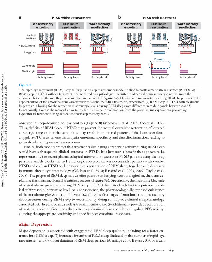
CP10CH25-Walker ARI 11 February 2014 12:9
Amygdala
Wake memoryencoding
REM neuralreactivation
Wake memoryrecollection
Corticalmodules
Hippocampus
Adrenergic
Cholinergic
Activity level Activity level Activity level
a
mP
FC
PTSD without treatment
Wake memoryencoding
REM neuralreactivation
Wake memoryrecollection
Activity level Activity level Activity level
bm
PF
C
PTSD with treatment
Prazosin
Figure 7The rapid-eye movement (REM) sleep to forget and sleep to remember model applied to posttraumatic stress disorder (PTSD). (a)REM sleep in PTSD without treatment, characterized by a pathological persistence of central brain adrenergic activity (note thedifference between the middle panel a and the middle panel of Figure 3a). Elevated adrenergic activity during REM sleep prevents thedepotentiation of the emotional tone associated with salient, including traumatic, experiences. (b) REM sleep in PTSD with treatmentby prazosin, allowing for the reduction in adrenergic levels during REM sleep (note difference in middle panels between a and b).Consequently, there is the restored opportunity for the dissipation of emotion from the prior trauma experiences, preventinghyperarousal reactions during subsequent postsleep memory recall.
observed in sleep-deprived healthy controls (Figure 8) (Motomura et al. 2013, Yoo et al. 2007).Thus, deficits of REM sleep in PTSD may prevent the normal overnight restoration of loweredadrenergic tone and, at the same time, may result in an altered pattern of the locus coeruleus-amygdala-PFC activity, one that impairs emotional specificity and thus discrimination, leading togeneralized and hypersensitive responses.
Finally, both models predict that treatments dissipating adrenergic activity during REM sleepwould have a therapeutic clinical outcome in PTSD. It is just such a benefit that appears to berepresented by the recent pharmacological intervention success in PTSD patients using the drugprazosin, which blocks the α-1 adrenergic receptor. Given nocturnally, patients with combatPTSD and civilian PTSD both demonstrate a restoration of REM sleep, together with decreasesin trauma-dream symptomatology (Calohan et al. 2010; Raskind et al. 2003, 2007; Taylor et al.2008). The proposed REM sleep models offer putative underlying neurobiological mechanisms ex-plaining this pharmacological treatment success (Figure 7b). Specifically, the nighttime blockadeof central adrenergic activity during REM sleep in PTSD dissipates levels back to a potentially crit-ical subthreshold, normative level. As a consequence, the pharmacologically imposed quiescenceof the noradrenergic system at night would (a) allow the first stages of emotional (trauma) memorydepotentiation during REM sleep to occur and, by doing so, improve clinical symptomatologyassociated with hyperarousal as well as trauma memory; and (b) additionally provide a recalibrationof next-day noradrenaline levels that restore appropriate locus coeruleus-amygdala-PFC activity,allowing the appropriate sensitivity and specificity of emotional responses.
Major Depression
Major depression is associated with exaggerated REM sleep qualities, including (a) a faster en-trance into REM sleep, (b) increased intensity of REM sleep (indexed by the number of rapid-eyemovements), and (c) longer duration of REM sleep periods (Armitage 2007, Buysse 2004, Franzen
www.annualreviews.org • Sleep and Emotion 699
Ann
u. R
ev. C
lin. P
sych
ol. 2
014.
10:6
79-7
08. D
ownl
oade
d fr
om w
ww
.ann
ualr
evie
ws.
org
by $
{ind
ivid
ualU
ser.
disp
layN
ame}
on
04/0
2/14
. For
per
sona
l use
onl
y.
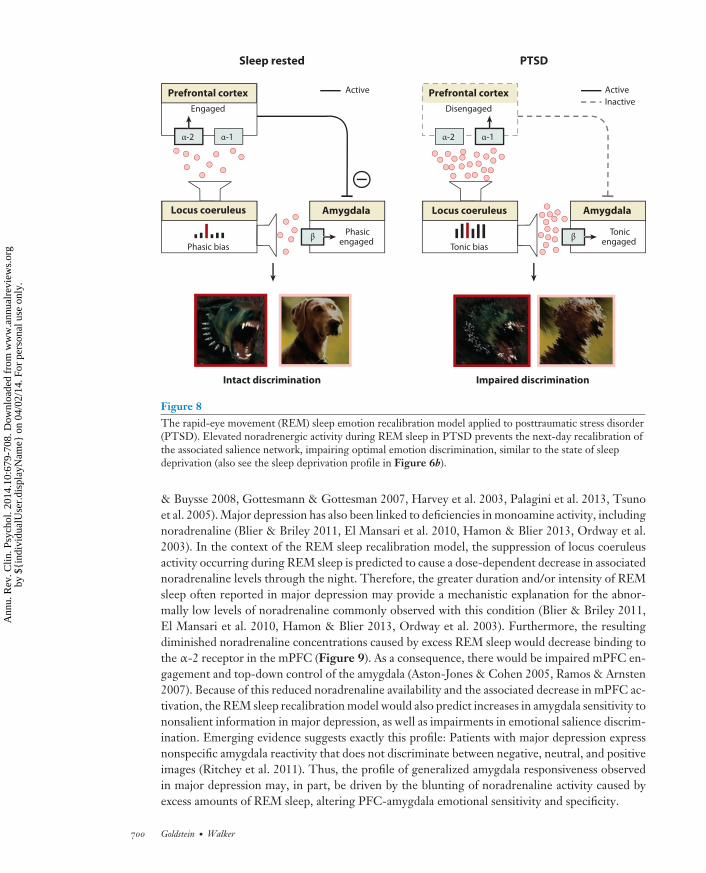
CP10CH25-Walker ARI 11 February 2014 12:9
PTSDSleep rested
α-2
Disengaged
Prefrontal cortex
α-1
Amygdala
Tonicengaged
Tonic bias
Locus coeruleus
Active
Inactive
β
α-2
Engaged
Prefrontal cortex
α-1
Amygdala
Phasicengaged
Phasic bias
Locus coeruleus
Active
β
Impaired discrimination Intact discrimination
Figure 8The rapid-eye movement (REM) sleep emotion recalibration model applied to posttraumatic stress disorder(PTSD). Elevated noradrenergic activity during REM sleep in PTSD prevents the next-day recalibration ofthe associated salience network, impairing optimal emotion discrimination, similar to the state of sleepdeprivation (also see the sleep deprivation profile in Figure 6b).
& Buysse 2008, Gottesmann & Gottesman 2007, Harvey et al. 2003, Palagini et al. 2013, Tsunoet al. 2005). Major depression has also been linked to deficiencies in monoamine activity, includingnoradrenaline (Blier & Briley 2011, El Mansari et al. 2010, Hamon & Blier 2013, Ordway et al.2003). In the context of the REM sleep recalibration model, the suppression of locus coeruleusactivity occurring during REM sleep is predicted to cause a dose-dependent decrease in associatednoradrenaline levels through the night. Therefore, the greater duration and/or intensity of REMsleep often reported in major depression may provide a mechanistic explanation for the abnor-mally low levels of noradrenaline commonly observed with this condition (Blier & Briley 2011,El Mansari et al. 2010, Hamon & Blier 2013, Ordway et al. 2003). Furthermore, the resultingdiminished noradrenaline concentrations caused by excess REM sleep would decrease binding tothe α-2 receptor in the mPFC (Figure 9). As a consequence, there would be impaired mPFC en-gagement and top-down control of the amygdala (Aston-Jones & Cohen 2005, Ramos & Arnsten2007). Because of this reduced noradrenaline availability and the associated decrease in mPFC ac-tivation, the REM sleep recalibration model would also predict increases in amygdala sensitivity tononsalient information in major depression, as well as impairments in emotional salience discrim-ination. Emerging evidence suggests exactly this profile: Patients with major depression expressnonspecific amygdala reactivity that does not discriminate between negative, neutral, and positiveimages (Ritchey et al. 2011). Thus, the profile of generalized amygdala responsiveness observedin major depression may, in part, be driven by the blunting of noradrenaline activity caused byexcess amounts of REM sleep, altering PFC-amygdala emotional sensitivity and specificity.
700 Goldstein ·Walker
Ann
u. R
ev. C
lin. P
sych
ol. 2
014.
10:6
79-7
08. D
ownl
oade
d fr
om w
ww
.ann
ualr
evie
ws.
org
by $
{ind
ivid
ualU
ser.
disp
layN
ame}
on
04/0
2/14
. For
per
sona
l use
onl
y.
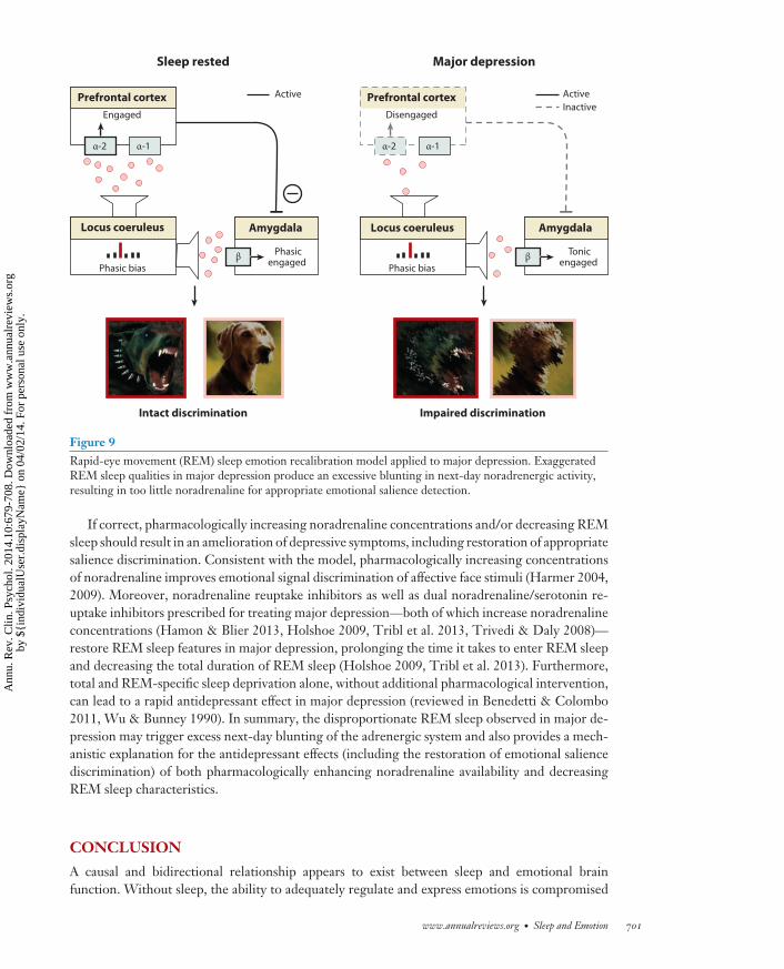
CP10CH25-Walker ARI 11 February 2014 12:9
Major depressionSleep rested
α-2
Disengaged
Prefrontal cortex
α-1
Amygdala
Tonicengaged
Locus coeruleus
Active
Inactive
β
α-2
Engaged
Prefrontal cortex
α-1
Amygdala
Phasicengaged
Phasic bias
Locus coeruleus
Active
β
Impaired discrimination Intact discrimination
Phasic bias
Figure 9Rapid-eye movement (REM) sleep emotion recalibration model applied to major depression. ExaggeratedREM sleep qualities in major depression produce an excessive blunting in next-day noradrenergic activity,resulting in too little noradrenaline for appropriate emotional salience detection.
If correct, pharmacologically increasing noradrenaline concentrations and/or decreasing REMsleep should result in an amelioration of depressive symptoms, including restoration of appropriatesalience discrimination. Consistent with the model, pharmacologically increasing concentrationsof noradrenaline improves emotional signal discrimination of affective face stimuli (Harmer 2004,2009). Moreover, noradrenaline reuptake inhibitors as well as dual noradrenaline/serotonin re-uptake inhibitors prescribed for treating major depression—both of which increase noradrenalineconcentrations (Hamon & Blier 2013, Holshoe 2009, Tribl et al. 2013, Trivedi & Daly 2008)—restore REM sleep features in major depression, prolonging the time it takes to enter REM sleepand decreasing the total duration of REM sleep (Holshoe 2009, Tribl et al. 2013). Furthermore,total and REM-specific sleep deprivation alone, without additional pharmacological intervention,can lead to a rapid antidepressant effect in major depression (reviewed in Benedetti & Colombo2011, Wu & Bunney 1990). In summary, the disproportionate REM sleep observed in major de-pression may trigger excess next-day blunting of the adrenergic system and also provides a mech-anistic explanation for the antidepressant effects (including the restoration of emotional saliencediscrimination) of both pharmacologically enhancing noradrenaline availability and decreasingREM sleep characteristics.
CONCLUSION
A causal and bidirectional relationship appears to exist between sleep and emotional brainfunction. Without sleep, the ability to adequately regulate and express emotions is compromised
www.annualreviews.org • Sleep and Emotion 701
Ann
u. R
ev. C
lin. P
sych
ol. 2
014.
10:6
79-7
08. D
ownl
oade
d fr
om w
ww
.ann
ualr
evie
ws.
org
by $
{ind
ivid
ualU
ser.
disp
layN
ame}
on
04/0
2/14
. For
per
sona
l use
onl
y.
CP10CH25-Walker ARI 11 February 2014 12:9
at both a brain and behavioral level, common to both the positive and negative domains of theemotional spectrum. In contrast, sleep, and specifically REM sleep, provides a restoration ofappropriate next-day emotional reactivity and salience discrimination. Basic emotional stimulusresponsiveness, sleep, and consistent REM sleep promote the offline processing and consolidationof emotional memories.
Uniting these empirical findings with the basic neurobiological features of REM sleep, anemotional homeostasis framework emerges in which REM sleep provides at least two discretefunctional benefits (a) the therapeutic depotentiation of emotion from prior affective experiencesand (b) a next-day recalibration of noradrenergic salience signaling by the brain, restoring opti-mal emotional sensitivity and specificity. Moreover, these models offer clarifying neurobiologicalinsights underlying the neural etiology and expressed symptomatology of PTSD and major depres-sion, as well as novel mechanistic routes through which pharmacological interventions currentlyused in PTSD and major depression may operate.
Although considerable progress has been made in understanding the relationship between sleepand affective brain function, the field is still only in its infancy, with much yet to understand. Wecurrently know little about the interplay between the peripheral and central nervous system mech-anisms that lead to abnormalities of emotion processing caused by sleep deprivation. Similarly, theprecise combination of sleep factors that reset the balance for optimal next-day emotional brainfunction is unclear. Is it the stages of sleep, their quantity or quality, their brain oscillations, ortheir neurochemistry? Or is it the cycling nature of sleep and/or the timing of sleep? Conversely,why do some emotional events we experience while awake impact our sleep at night, and others donot? Is it their intensity, novelty, salience, valence, temporal proximity to sleep, relationship withpast experiences, or the degree of unresolved understanding? We need to look no further thanmood disorders to appreciate this wake-sleep reciprocity that modulates affective states. Whatdoes seems clear, however, is that the clinical, professional, and public health ramifications of thisemerging association between sleep and affective brain function are substantive.
DISCLOSURE STATEMENT
The authors are not aware of any affiliations, memberships, funding, or financial holdings thatmight be perceived as affecting the objectivity of this review.
ACKNOWLEDGMENTS
The authors thank Allison Harvey, Stephanie Greer, Jared Saletin, and Els van der Helm forhelping insights and discussion regarding the content of this review. This work was supported bygrants R01AG031164, R01MH093537, and R21DA031939 (to M.P.W.) and F31MH094075 (toA.N.G.) from the National Institutes of Health.
LITERATURE CITED
Anderson C, Platten CR. 2011. Sleep deprivation lowers inhibition and enhances impulsivity to negativestimuli. Behav. Brain Res. 217:463–66
Appelhans B, Luecken L. 2006. Heart rate variability as an index of regulated emotional responding. Rev. Gen.Psychol. 10:229–40
Armitage R. 2007. Sleep and circadian rhythms in mood disorders. Acta Psychiatr. Scand. 115(Suppl. 433):104–15
Arnedt JT, Conroy DA, Brower KJ. 2007. Treatment options for sleep disturbances during alcohol recovery.J. Addict. Dis. 26:41–54
702 Goldstein ·Walker
Ann
u. R
ev. C
lin. P
sych
ol. 2
014.
10:6
79-7
08. D
ownl
oade
d fr
om w
ww
.ann
ualr
evie
ws.
org
by $
{ind
ivid
ualU
ser.
disp
layN
ame}
on
04/0
2/14
. For
per
sona
l use
onl
y.
CP10CH25-Walker ARI 11 February 2014 12:9
Aston-Jones G, Cohen JD. 2005. An integrative theory of locus coeruleus-norepinephrine function: adaptivegain and optimal performance. Annu. Rev. Neurosci. 28:403–50
Benedetti F, Colombo C. 2011. Sleep deprivation in mood disorders. Neuropsychobiology 64:141–51Benedict C, Brooks SJ, O’Daly OG, Almen MS, Morell A, et al. 2012. Acute sleep deprivation enhances the
brain’s response to hedonic food stimuli: an fMRI study. J. Clin. Endocrinol. Metab. 97:E443–47Berger RH, Miller AL, Seifer R, Cares SR, LeBourgeois MK. 2012. Acute sleep restriction effects on emotion
responses in 30- to 36-month-old children. J. Sleep Res. 21:235–46Bernert RA, Joiner TE. 2007. Sleep disturbances and suicide risk: a review of the literature. Neuropsychiatr.
Dis. Treat. 3:735–43Berridge CW, Foote SL. 1991. Effects of locus coeruleus activation on electroencephalographic activity in
neocortex and hippocampus. J. Neurosci. 11:3135–45Blier P, Briley M. 2011. The noradrenergic symptom cluster: clinical expression and neuropharmacology.
Neuropsychiatr. Dis. Treat. 7:15–20Breslau N, Roth T, Burduvali E, Kapke A, Schultz L, Roehrs T. 2004. Sleep in lifetime posttraumatic stress
disorder: a community-based polysomnographic study. Arch. Gen. Psychiatry 61:508–16Brower KJ, Perron BE. 2010. Sleep disturbance as a universal risk factor for relapse in addictions to psychoactive
substances. Med. Hypotheses 74:928–33Buysse DJ. 2004. Insomnia, depression and aging. Assessing sleep and mood interactions in older adults.
Geriatrics 59:47–51, quiz 52Cahill L. 2000. Neurobiological mechanisms of emotionally influenced, long-term memory. Prog. Brain Res.
126:29–37Calohan J, Peterson K, Peskind ER, Raskind MA. 2010. Prazosin treatment of trauma nightmares and sleep
disturbance in soldiers deployed in Iraq. J. Trauma. Stress 23:645–48Cape EG, Jones BE. 1998. Differential modulation of high-frequency γ-electroencephalogram activity and
sleep-wake state by noradrenaline and serotonin microinjections into the region of cholinergic basalisneurons. J. Neurosci. 18:2653–66
Craig AD. 2010. The sentient self. Brain Struct. Funct. 214:563–77Craig AD. 2011. Significance of the insula for the evolution of human awareness of feelings from the body.
Ann. NY Acad. Sci. 1225:72–82Critchley HD. 2005. Neural mechanisms of autonomic, affective, and cognitive integration. J. Comp. Neurol.
493:154–66Critchley HD. 2009. Psychophysiology of neural, cognitive and affective integration: fMRI and autonomic
indicants. Int. J. Psychophysiol. 73:88–94Dahl RE. 1996. The impact of inadequate sleep on children’s daytime cognitive function. Semin. Pediatr.
Neurol. 3:44–50Dang-Vu TT, Schabus M, Desseilles M, Sterpenich V, Bonjean M, Maquet P. 2010. Functional neuroimaging
insights into the physiology of human sleep. Sleep 33:1589–603Davidson RJ. 2002. Anxiety and affective style: role of prefrontal cortex and amygdala. Biol. Psychiatry 51:68–80Delgado MR, Olsson A, Phelps EA. 2006. Extending animal models of fear conditioning to humans. Biol.
Psychol. 73:39–48Dimsdale JE, Norman D, DeJardin D, Wallace MS. 2007. The effect of opioids on sleep architecture. J. Clin.
Sleep Med. 3:33–36Dinges DF, Pack F, Williams K, Gillen KA, Powell JW, et al. 1997. Cumulative sleepiness, mood disturbance,
and psychomotor vigilance performance decrements during a week of sleep restricted to 4–5 hours pernight. Sleep 20:267–77
Dolcos F, LaBar KS, Cabeza R. 2005. Remembering one year later: role of the amygdala and the medialtemporal lobe memory system in retrieving emotional memories. Proc. Natl. Acad. Sci. USA 102:2626–31
Drevets WC, Savitz J, Trimble M. 2008. The subgenual anterior cingulate cortex in mood disorders. CNSSpectr. 13:663–81
El Mansari M, Guiard BP, Chernoloz O, Ghanbari R, Katz N, Blier P. 2010. Relevance of norepinephrine-dopamine interactions in the treatment of major depressive disorder. CNS Neurosci. Ther. 16:e1–17
Etkin A. 2010. Functional neuroanatomy of anxiety: a neural circuit perspective. Curr. Top. Behav. Neurosci.2:251–77
www.annualreviews.org • Sleep and Emotion 703
Ann
u. R
ev. C
lin. P
sych
ol. 2
014.
10:6
79-7
08. D
ownl
oade
d fr
om w
ww
.ann
ualr
evie
ws.
org
by $
{ind
ivid
ualU
ser.
disp
layN
ame}
on
04/0
2/14
. For
per
sona
l use
onl
y.
CP10CH25-Walker ARI 11 February 2014 12:9
Etkin A, Wager TD. 2007. Functional neuroimaging of anxiety: a meta-analysis of emotional processing inPTSD, social anxiety disorder, and specific phobia. Am. J. Psychiatry 164:1476–88
Franzen PL, Buysse DJ. 2008. Sleep disturbances and depression: risk relationships for subsequent depressionand therapeutic implications. Dialogues Clin. Neurosci. 10:473–81
Germain A. 2013. Sleep disturbances as the hallmark of PTSD: Where are we now? Am. J. Psychiatry 170:372–82
Germain A, Buysse DJ, Nofzinger E. 2008. Sleep-specific mechanisms underlying posttraumatic stress disor-der: integrative review and neurobiological hypotheses. Sleep Med. Rev. 12:185–95
Germain A, James J, Insana S, Herringa RJ, Mammen O, et al. 2013. A window into the invisible wound ofwar: functional neuroimaging of REM sleep in returning combat veterans with PTSD. Psychiatry Res.211:176–79
Goldstein AN, Greer SM, Saletin JM, Harvey AG, Nitschke JB, Walker MP. 2013. Tired and apprehensive:anxiety amplifies the impact of sleep loss on aversive brain anticipation. J. Neurosci. 33:10607–15
Gottesmann C, Gottesman I. 2007. The neurobiological characteristics of rapid eye movement (REM) sleep arecandidate endophenotypes of depression, schizophrenia, mental retardation and dementia. Prog. Neurobiol.81:237–50
Greer SM, Goldstein AN, Walker MP. 2013. The impact of sleep deprivation on food desire in the humanbrain. Nat. Commun. 4:2259
Grillon C, Morgan CA 3rd. 1999. Fear-potentiated startle conditioning to explicit and contextual cues in GulfWar veterans with posttraumatic stress disorder. J. Abnorm. Psychol. 108:134–42
Gujar N, McDonald SA, Nishida M, Walker MP. 2010. A role for REM sleep in recalibrating the sensitivityof the human brain to specific emotions. Cereb. Cortex 21:115–23
Gujar N, Yoo SS, Hu P, Walker MP. 2011. Sleep deprivation amplifies reactivity of brain reward networks,biasing the appraisal of positive emotional experiences. J. Neurosci. 31:4466–74
Habukawa M, Uchimura N, Maeda M, Kotorii N, Maeda H. 2007. Sleep findings in young adult patients withposttraumatic stress disorder. Biol. Psychiatry 62:1179–82
Hamon M, Blier P. 2013. Monoamine neurocircuitry in depression and strategies for new treatments. Prog.Neuropsychopharmacol. Biol. Psychiatry 45:54–63
Harmer CJ, O’Sullivan U, Favaron E, Massey-Chase R, Ayres R, et al. 2009. Effect of acute antidepressantadministration on negative affective bias in depressed patients. Am. J. Psychiatry 166:1178–84
Harmer CJ, Shelley NC, Cowen PJ, Goodwin GM. 2004. Increased positive versus negative affective per-ception and memory in healthy volunteers following selective serotonin and norepinephrine reuptakeinhibition. Am. J. Psychiatry 161:1256–63
Harrison NA, Gray MA, Gianaros PJ, Critchley HD. 2010. The embodiment of emotional feelings in thebrain. J. Neurosci. 30:12878–84
Hartley CA, Phelps EA. 2010. Changing fear: the neurocircuitry of emotion regulation. Neuropsychopharma-cology 35:136–46
Harvey AG. 2011. Sleep and circadian functioning: critical mechanisms in the mood disorders? Annu. Rev.Clin. Psychol. 7:297–319
Harvey AG, Jones C, Schmidt DA. 2003. Sleep and posttraumatic stress disorder: a review. Clin. Psychol. Rev.23:377–407
Harvey AG, Murray G, Chandler RA, Soehner A. 2011. Sleep disturbance as transdiagnostic: considerationof neurobiological mechanisms. Clin. Psychol. Rev. 31:225–35
Hermans EJ, van Marle HJ, Ossewaarde L, Henckens MJ, Qin S, et al. 2011. Stress-related noradrenergicactivity prompts large-scale neural network reconfiguration. Science 334:1151–53
Holshoe JM. 2009. Antidepressants and sleep: a review. Perspect. Psychiatr. Care 45:191–97Horne JA. 1985. Sleep function, with particular reference to sleep deprivation. Ann. Clin. Res. 17:199–208Hu P, Stylos-Allen M, Walker MP. 2006. Sleep facilitates consolidation of emotionally arousing declarative
memory. Psychol. Sci. 17:891–98Itoi K, Sugimoto N. 2010. The brainstem noradrenergic systems in stress, anxiety and depression. J. Neuroen-
docrinol. 22:355–61Jones MW, Wilson MA. 2005. Theta rhythms coordinate hippocampal-prefrontal interactions in a spatial
memory task. PLoS Biol. 3:e402
704 Goldstein ·Walker
Ann
u. R
ev. C
lin. P
sych
ol. 2
014.
10:6
79-7
08. D
ownl
oade
d fr
om w
ww
.ann
ualr
evie
ws.
org
by $
{ind
ivid
ualU
ser.
disp
layN
ame}
on
04/0
2/14
. For
per
sona
l use
onl
y.
CP10CH25-Walker ARI 11 February 2014 12:9
Jovanovic T, Kazama A, Bachevalier J, Davis M. 2012. Impaired safety signal learning may be a biomarker ofPTSD. Neuropharmacology 62:695–704
Jovanovic T, Norrholm SD, Fennell JE, Keyes M, Fiallos AM, et al. 2009. Posttraumatic stress disorder maybe associated with impaired fear inhibition: relation to symptom severity. Psychiatry Res. 167:151–60
Kametani H, Kawamura H. 1990. Alterations in acetylcholine release in the rat hippocampus during sleep-wakefulness detected by intracerebral dialysis. Life Sci. 47:421–26
Kamphuis J, Meerlo P, Koolhaas JM, Lancel M. 2012. Poor sleep as a potential causal factor in aggressionand violence. Sleep Med. 13:327–34
Keane PE, Candy JM, Bradley PB. 1976. The role of endogenous catecholamines in the regulation of elec-trocortical activity in the encephale isole cat. Electroencephalogr. Clin. Neurophysiol. 41:561–70
Killgore WD, Schwab ZJ, Weber M, Kipman M, Deldonno SR, et al. 2013. Daytime sleepiness affectsprefrontal regulation of food intake. Neuroimage 71:216–23
Kleim B, Wilhelm FH, Temp L, Margraf J, Wiederhold BK, Rasch B. 2013. Sleep enhances exposure therapy.Psychol. Med. In press
Koren D, Arnon I, Lavie P, Klein E. 2002. Sleep complaints as early predictors of posttraumatic stress disorder:a 1-year prospective study of injured survivors of motor vehicle accidents. Am. J. Psychiatry 159:855–57
Lavie P. 2001. Sleep disturbances in the wake of traumatic events. N. Engl. J. Med. 345:1825–32Lavie P, Hefez A, Halperin G, Enoch D. 1979. Long-term effects of traumatic war-related events on sleep.
Am. J. Psychiatry 136:175–78Libedinsky C, Smith DV, Teng CS, Namburi P, Chen VW, et al. 2011. Sleep deprivation alters valuation
signals in the ventromedial prefrontal cortex. Front. Behav. Neurosci. 5:70Mallick BN, Singh A. 2011. REM sleep loss increases brain excitability: role of noradrenaline and its mechanism
of action. Sleep Med. Rev. 15:165–78Marrosu F, Portas C, Mascia MS, Casu MA, Fa M, et al. 1995. Microdialysis measurement of cortical and
hippocampal acetylcholine release during sleep-wake cycle in freely moving cats. Brain Res. 671:329–32McGaugh JL. 2004. The amygdala modulates the consolidation of memories of emotionally arousing experi-
ences. Annu. Rev. Neurosci. 27:1–28McGinty DJ, Harper RM. 1976. Dorsal raphe neurons: depression of firing during sleep in cats. Brain Res.
101:569–75McGlinchey EL, Talbot LS, Chang KH, Kaplan KA, Dahl RE, Harvey AG. 2011. The effect of sleep depri-
vation on vocal expression of emotion in adolescents and adults. Sleep 34:1233–41McIntyre CK, McGaugh JL, Williams CL. 2012. Interacting brain systems modulate memory consolidation.
Neurosci. Biobehav. Rev. 36:1750–62McKenna BS, Dickinson DL, Orff HJ, Drummond SP. 2007. The effects of one night of sleep deprivation on
known-risk and ambiguous-risk decisions. J. Sleep Res. 16:245–52Mellman TA, Bustamante V, Fins AI, Pigeon WR, Nolan B. 2002. REM sleep and the early development of
posttraumatic stress disorder. Am. J. Psychiatry 159:1696–701Mellman TA, Hipolito MM. 2006. Sleep disturbances in the aftermath of trauma and posttraumatic stress
disorder. CNS Spectr. 11:611–15Mellman TA, Kumar A, Kulick-Bell R, Kumar M, Nolan B. 1995. Nocturnal/daytime urine noradrenergic
measures and sleep in combat-related PTSD. Biol. Psychiatry 38:174–79Mellman TA, Nolan B, Hebding J, Kulick-Bell R, Dominguez R. 1997. A polysomnographic comparison of
veterans with combat-related PTSD, depressed men, and non-ill controls. Sleep 20:46–51Mendl M, Burman OH, Paul ES. 2010. An integrative and functional framework for the study of animal
emotion and mood. Proc. Biol. Sci. 277:2895–904Menz MM, Rihm JS, Salari N, Born J, Kalisch R, et al. 2013. The role of sleep and sleep deprivation in
consolidating fear memories. Neuroimage 75:87–96Minkel J, Htaik O, Banks S, Dinges D. 2011. Emotional expressiveness in sleep-deprived healthy adults. Behav.
Sleep Med. 9:5–14Minkel JD, Banks S, Htaik O, Moreta MC, Jones CW, et al. 2012. Sleep deprivation and stressors: evidence
for elevated negative affect in response to mild stressors when sleep deprived. Emotion 12:1015–20Miyauchi S, Misaki M, Kan S, Fukunaga T, Koike T. 2009. Human brain activity time-locked to rapid eye
movements during REM sleep. Exp. Brain Res. 192:657–67
www.annualreviews.org • Sleep and Emotion 705
Ann
u. R
ev. C
lin. P
sych
ol. 2
014.
10:6
79-7
08. D
ownl
oade
d fr
om w
ww
.ann
ualr
evie
ws.
org
by $
{ind
ivid
ualU
ser.
disp
layN
ame}
on
04/0
2/14
. For
per
sona
l use
onl
y.
CP10CH25-Walker ARI 11 February 2014 12:9
Motomura Y, Kitamura S, Oba K, Terasawa Y, Enomoto M, et al. 2013. Sleep debt elicits negative emotionalreaction through diminished amygdala-anterior cingulate functional connectivity. PLoS ONE 8:e56578
Nishida M, Pearsall J, Buckner RL, Walker MP. 2009. REM sleep, prefrontal theta, and the consolidation ofhuman emotional memory. Cereb. Cortex 19:1158–66
Nitschke JB, Sarinopoulos I, Oathes DJ, Johnstone T, Whalen PJ, et al. 2009. Anticipatory activation in theamygdala and anterior cingulate in generalized anxiety disorder and prediction of treatment response.Am. J. Psychiatry 166:302–10
Nofzinger EA. 2005. Functional neuroimaging of sleep. Semin. Neurol. 25:9–18Ochsner KN, Ray RR, Hughes B, McRae K, Cooper JC, et al. 2009. Bottom-up and top-down processes in
emotion generation: common and distinct neural mechanisms. Psychol. Sci. 20:1322–31Ordway GA, Schenk J, Stockmeier CA, May W, Klimek V. 2003. Elevated agonist binding to α2-adrenoceptors
in the locus coeruleus in major depression. Biol. Psychiatry 53:315–23Ouyang M, Hellman K, Abel T, Thomas SA. 2004. Adrenergic signaling plays a critical role in the maintenance
of waking and in the regulation of REM sleep. J. Neurophysiol. 92:2071–82Pace-Schott EF, Hobson JA. 2002. The neurobiology of sleep: genetics, cellular physiology and subcortical
networks. Nat. Rev. Neurosci. 3:591–605Pace-Schott EF, Milad MR, Orr SP, Rauch SL, Stickgold R, Pitman RK. 2009. Sleep promotes generalization
of extinction of conditioned fear. Sleep 32:19–26Pace-Schott EF, Stickgold R, Muzur A, Wigren PE, Ward AS, et al. 2005. Sleep quality deteriorates over a
binge-abstinence cycle in chronic smoked cocaine users. Psychopharmacology 179:873–83Palagini L, Baglioni C, Ciapparelli A, Gemignani A, Riemann D. 2013. REM sleep dysregulation in depression:
state of the art. Sleep Med. Rev. 17:377–90Papadimitriou GN, Linkowski P. 2005. Sleep disturbance in anxiety disorders. Int. Rev. Psychiatry 17:229–36Pare D, Collins DR, Pelletier JG. 2002. Amygdala oscillations and the consolidation of emotional memories.
Trends Cogn. Sci. 6:306–14Park SP. 2002. In vivo microdialysis measures of extracellular norepinephrine in the rat amygdala during
sleep-wakefulness. J. Korean Med. Sci. 17:395–99Paulus MP, Stein MB. 2006. An insular view of anxiety. Biol. Psychiatry 60:383–87Payne JD, Chambers AM, Kensinger EA. 2012. Sleep promotes lasting changes in selective memory for
emotional scenes. Front. Integr. Neurosci. 6:108Payne JD, Kensinger EA. 2010. Sleep’s role in the consolidation of emotional episodic memories. Curr. Dir.
Psychol. Sci. 19:290–95Peterson MJ, Benca RM. 2006. Sleep in mood disorders. Psychiatr. Clin. North Am. 29:1009–32, abstract ixPhelps EA, Delgado MR, Nearing KI, LeDoux JE. 2004. Extinction learning in humans: role of the amygdala
and vmPFC. Neuron 43:897–905Plutchik R. 1995. Outward and inward directed aggressiveness: the interaction between violence and suicidal-
ity. Pharmacopsychiatry 28(Suppl. 2):47–57Pole N. 2007. The psychophysiology of posttraumatic stress disorder: a meta-analysis. Psychol. Bull. 133:725–46Prather AA, Bogdan R, Hariri AR. 2013. Impact of sleep quality on amygdala reactivity, negative affect, and
perceived stress. Psychosom. Med. 75:350–58Ramos BP, Arnsten AF. 2007. Adrenergic pharmacology and cognition: focus on the prefrontal cortex.
Pharmacol. Ther. 113:523–36Raskind MA, Peskind ER, Hoff DJ, Hart KL, Holmes HA, et al. 2007. A parallel group placebo controlled
study of prazosin for trauma nightmares and sleep disturbance in combat veterans with post-traumaticstress disorder. Biol. Psychiatry 61:928–34
Raskind MA, Peskind ER, Kanter ED, Petrie EC, Radant A, et al. 2003. Reduction of nightmares and otherPTSD symptoms in combat veterans by prazosin: a placebo-controlled study. Am. J. Psychiatry 160:371–73
Rauch SL, Whalen PJ, Shin LM, McInerney SC, Macklin ML, et al. 2000. Exaggerated amygdala response tomasked facial stimuli in posttraumatic stress disorder: a functional MRI study. Biol. Psychiatry 47:769–76
Ritchey M, Dolcos F, Eddington KM, Strauman TJ, Cabeza R. 2011. Neural correlates of emotional processingin depression: changes with cognitive behavioral therapy and predictors of treatment response. J. Psychiatr.Res. 45:577–87
706 Goldstein ·Walker
Ann
u. R
ev. C
lin. P
sych
ol. 2
014.
10:6
79-7
08. D
ownl
oade
d fr
om w
ww
.ann
ualr
evie
ws.
org
by $
{ind
ivid
ualU
ser.
disp
layN
ame}
on
04/0
2/14
. For
per
sona
l use
onl
y.
CP10CH25-Walker ARI 11 February 2014 12:9
Roozendaal B, McReynolds JR, Van der Zee EA, Lee S, McGaugh JL, McIntyre CK. 2009. Glucocorticoideffects on memory consolidation depend on functional interactions between the medial prefrontal cortexand basolateral amygdala. J. Neurosci. 29:14299–308
Sapolsky RM, Romero LM, Munck AU. 2000. How do glucocorticoids influence stress responses? Integratingpermissive, suppressive, stimulatory, and preparative actions. Endocr. Rev. 21:55–89
Sauvet F, Leftheriotis G, Gomez-Merino D, Langrume C, Drogou C, et al. 2010. Effect of acute sleepdeprivation on vascular function in healthy subjects. J. Appl. Physiol. 108:68–75
Schwarz JF, Popp R, Haas J, Zulley J, Geisler P, et al. 2013. Shortened night sleep impairs facial responsivenessto emotional stimuli. Biol. Psychol. 93:41–44
Shin LM, Rauch SL, Pitman RK. 2006. Amygdala, medial prefrontal cortex, and hippocampal function inPTSD. Ann. NY Acad. Sci. 1071:67–79
Shouse MN, Staba RJ, Saquib SF, Farber PR. 2000. Monoamines and sleep: microdialysis findings in ponsand amygdala. Brain Res. 860:181–89
Siegel JM, Rogawski MA. 1988. A function for REM sleep: regulation of noradrenergic receptor sensitivity.Brain Res. 472:213–33
Siegle GJ, Thompson W, Carter CS, Steinhauer SR, Thase ME. 2007. Increased amygdala and decreaseddorsolateral prefrontal BOLD responses in unipolar depression: related and independent features. Biol.Psychiatry 61:198–209
Spoormaker VI, Montgomery P. 2008. Disturbed sleep in post-traumatic stress disorder: secondary symptomor core feature? Sleep Med. Rev. 12:169–84
Spoormaker VI, Schroter MS, Andrade KC, Dresler M, Kiem SA, et al. 2012. Effects of rapid eye movementsleep deprivation on fear extinction recall and prediction error signaling. Hum. Brain Mapp. 33:2362–76
Spoormaker VI, Sturm A, Andrade KC, Schroter MS, Goya-Maldonado R, et al. 2010. The neural corre-lates and temporal sequence of the relationship between shock exposure, disturbed sleep and impairedconsolidation of fear extinction. J. Psychiatr. Res. 44:1121–28
St-Onge MP, McReynolds A, Trivedi ZB, Roberts AL, Sy M, Hirsch J. 2012. Sleep restriction leads toincreased activation of brain regions sensitive to food stimuli. Am. J. Clin. Nutr. 95:818–24
Stern WC, Morgane PJ. 1974. Theoretical view of REM sleep function: maintenance of catecholamine systemsin the central nervous system. Behav. Biol. 11:1–32
Sterpenich V, Albouy G, Boly M, Vandewalle G, Darsaud A, et al. 2007. Sleep-related hippocampo-corticalinterplay during emotional memory recollection. PLoS Biol. 5:e282
Strawn JR, Geracioti TD Jr. 2008. Noradrenergic dysfunction and the psychopharmacology of posttraumaticstress disorder. Depress. Anxiety 25:260–71
Sullivan GM, Coplan JD, Kent JM, Gorman JM. 1999. The noradrenergic system in pathological anxiety: afocus on panic with relevance to generalized anxiety and phobias. Biol. Psychiatry 46:1205–18
Taylor FB, Martin P, Thompson C, Williams J, Mellman TA, et al. 2008. Prazosin effects on objective sleepmeasures and clinical symptoms in civilian trauma posttraumatic stress disorder: a placebo-controlledstudy. Biol. Psychiatry 63:629–32
Tribl GG, Wetter TC, Schredl M. 2013. Dreaming under antidepressants: a systematic review on evidencein depressive patients and healthy volunteers. Sleep Med. Rev. 17:133–42
Trivedi MH, Daly EJ. 2008. Treatment strategies to improve and sustain remission in major depressivedisorder. Dialogues Clin. Neurosci. 10:377–84
Tsuno N, Besset A, Ritchie K. 2005. Sleep and depression. J. Clin. Psychiatry 66:1254–69Valentino RJ, Van Bockstaele E. 2008. Convergent regulation of locus coeruleus activity as an adaptive response
to stress. Eur. J. Pharmacol. 583:194–203van der Helm E, Gujar N, Walker MP. 2010. Sleep deprivation impairs the accurate recognition of human
emotions. Sleep 33:335–42van der Helm E, Yao J, Dutt S, Rao V, Saletin JM, Walker MP. 2011. REM sleep depotentiates amygdala
activity to previous emotional experiences. Curr. Biol. 21:2029–32van der Helm E, Walker MP. 2012. Sleep and affective brain regulation. Soc. Personal. Psychol. Compass 6:773–91van Marle HJ, Hermans EJ, Qin S, Fernandez G. 2009. From specificity to sensitivity: How acute stress affects
amygdala processing of biologically salient stimuli. Biol. Psychiatry 66:649–55
www.annualreviews.org • Sleep and Emotion 707
Ann
u. R
ev. C
lin. P
sych
ol. 2
014.
10:6
79-7
08. D
ownl
oade
d fr
om w
ww
.ann
ualr
evie
ws.
org
by $
{ind
ivid
ualU
ser.
disp
layN
ame}
on
04/0
2/14
. For
per
sona
l use
onl
y.
CP10CH25-Walker ARI 11 February 2014 12:9
van Marle HJ, Hermans EJ, Qin S, Overeem S, Fernandez G. 2013. The effect of exogenous cortisol duringsleep on the behavioral and neural correlates of emotional memory consolidation in humans. Psychoneu-roendocrinology 38:1639–49
Venkatraman V, Chuah YM, Huettel SA, Chee MW. 2007. Sleep deprivation elevates expectation of gainsand attenuates response to losses following risky decisions. Sleep 30:603–9
Volkow ND, Tomasi D, Wang GJ, Telang F, Fowler JS, et al. 2009. Hyperstimulation of striatal D2 receptorswith sleep deprivation: implications for cognitive impairment. Neuroimage 45:1232–40
Wagner U, Degirmenci M, Drosopoulos S, Perras B, Born J. 2005. Effects of cortisol suppression on sleep-associated consolidation of neutral and emotional memory. Biol. Psychiatry 58:885–93
Wagner U, Gais S, Born J. 2001. Emotional memory formation is enhanced across sleep intervals with highamounts of rapid eye movement sleep. Learn. Mem. 8:112–19
Wagner U, Hallschmid M, Rasch B, Born J. 2006. Brief sleep after learning keeps emotional memories alivefor years. Biol. Psychiatry 60:788–90
Walker MP. 2009. The role of sleep in cognition and emotion. Ann. NY Acad. Sci. 1156:168–97Walker MP, van der Helm E. 2009. Overnight therapy? The role of sleep in emotional brain processing.
Psychol. Bull. 135:731–48Wright KM, Britt TW, Bliese PD, Adler AB, Picchioni D, Moore D. 2011. Insomnia as predictor versus
outcome of PTSD and depression among Iraq combat veterans. J. Clin. Psychol. 67:1240–58Wu JC, Bunney WE. 1990. The biological basis of an antidepressant response to sleep deprivation and relapse:
review and hypothesis. Am. J. Psychiatry 147:14–21Yoo SS, Gujar N, Hu P, Jolesz FA, Walker MP. 2007. The human emotional brain without sleep—a prefrontal
amygdala disconnect. Curr. Biol. 17:R877–78Zhong X, Hilton HJ, Gates GJ, Jelic S, Stern Y, et al. 2005. Increased sympathetic and decreased parasympa-
thetic cardiovascular modulation in normal humans with acute sleep deprivation. J. Appl. Physiol. 98:2024–32
Zohar D, Tzischinsky O, Epstein R, Lavie P. 2005. The effects of sleep loss on medical residents’ emotionalreactions to work events: a cognitive-energy model. Sleep 28:47–54
708 Goldstein ·Walker
Ann
u. R
ev. C
lin. P
sych
ol. 2
014.
10:6
79-7
08. D
ownl
oade
d fr
om w
ww
.ann
ualr
evie
ws.
org
by $
{ind
ivid
ualU
ser.
disp
layN
ame}
on
04/0
2/14
. For
per
sona
l use
onl
y.
CP10-FrontMatter ARI 6 March 2014 22:5
Annual Review ofClinical Psychology
Volume 10, 2014Contents
Advances in Cognitive Theory and Therapy: The GenericCognitive ModelAaron T. Beck and Emily A.P. Haigh � � � � � � � � � � � � � � � � � � � � � � � � � � � � � � � � � � � � � � � � � � � � � � � � � � � � � � � � 1
The Cycle of Classification: DSM-I Through DSM-5Roger K. Blashfield, Jared W. Keeley, Elizabeth H. Flanagan,
and Shannon R. Miles � � � � � � � � � � � � � � � � � � � � � � � � � � � � � � � � � � � � � � � � � � � � � � � � � � � � � � � � � � � � � � � � � � � � � �25
The Internship Imbalance in Professional Psychology: Current Statusand Future ProspectsRobert L. Hatcher � � � � � � � � � � � � � � � � � � � � � � � � � � � � � � � � � � � � � � � � � � � � � � � � � � � � � � � � � � � � � � � � � � � � � � � � � � � � �53
Exploratory Structural Equation Modeling: An Integration of the BestFeatures of Exploratory and Confirmatory Factor AnalysisHerbert W. Marsh, Alexandre J.S. Morin, Philip D. Parker,
and Gurvinder Kaur � � � � � � � � � � � � � � � � � � � � � � � � � � � � � � � � � � � � � � � � � � � � � � � � � � � � � � � � � � � � � � � � � � � � � � �85
The Reliability of Clinical Diagnoses: State of the ArtHelena Chmura Kraemer � � � � � � � � � � � � � � � � � � � � � � � � � � � � � � � � � � � � � � � � � � � � � � � � � � � � � � � � � � � � � � � � � � � 111
Thin-Slice Judgments in the Clinical ContextMichael L. Slepian, Kathleen R. Bogart, and Nalini Ambady � � � � � � � � � � � � � � � � � � � � � � � � � � � � 131
Attenuated Psychosis Syndrome: Ready for DSM-5.1?P. Fusar-Poli, W.T. Carpenter, S.W. Woods, and T.H. McGlashan � � � � � � � � � � � � � � � � � � � 155
From Kanner to DSM-5: Autism as an Evolving Diagnostic ConceptFred R. Volkmar and James C. McPartland � � � � � � � � � � � � � � � � � � � � � � � � � � � � � � � � � � � � � � � � � � � � � � 193
Development of Clinical Practice GuidelinesSteven D. Hollon, Patricia A. Arean, Michelle G. Craske, Kermit A. Crawford,
Daniel R. Kivlahan, Jeffrey J. Magnavita, Thomas H. Ollendick,Thomas L. Sexton, Bonnie Spring, Lynn F. Bufka, Daniel I. Galper,and Howard Kurtzman � � � � � � � � � � � � � � � � � � � � � � � � � � � � � � � � � � � � � � � � � � � � � � � � � � � � � � � � � � � � � � � � � � 213
Overview of Meta-Analyses of the Prevention of Mental Health,Substance Use, and Conduct ProblemsIrwin Sandler, Sharlene A. Wolchik, Gracelyn Cruden, Nicole E. Mahrer,
Soyeon Ahn, Ahnalee Brincks, and C. Hendricks Brown � � � � � � � � � � � � � � � � � � � � � � � � � � � � � � 243
vii
Ann
u. R
ev. C
lin. P
sych
ol. 2
014.
10:6
79-7
08. D
ownl
oade
d fr
om w
ww
.ann
ualr
evie
ws.
org
by $
{ind
ivid
ualU
ser.
disp
layN
ame}
on
04/0
2/14
. For
per
sona
l use
onl
y.
CP10-FrontMatter ARI 6 March 2014 22:5
Improving Care for Depression and Suicide Risk in Adolescents:Innovative Strategies for Bringing Treatments to CommunitySettingsJoan Rosenbaum Asarnow and Jeanne Miranda � � � � � � � � � � � � � � � � � � � � � � � � � � � � � � � � � � � � � � � � � � 275
The Contribution of Cultural Competence to Evidence-Based Carefor Ethnically Diverse PopulationsStanley J. Huey Jr., Jacqueline Lee Tilley, Eduardo O. Jones,
and Caitlin A. Smith � � � � � � � � � � � � � � � � � � � � � � � � � � � � � � � � � � � � � � � � � � � � � � � � � � � � � � � � � � � � � � � � � � � � 305
How to Use the New DSM-5 Somatic Symptom Disorder Diagnosisin Research and Practice: A Critical Evaluation and a Proposal forModificationsWinfried Rief and Alexandra Martin � � � � � � � � � � � � � � � � � � � � � � � � � � � � � � � � � � � � � � � � � � � � � � � � � � � � � 339
Antidepressant Use in Pregnant and Postpartum WomenKimberly A. Yonkers, Katherine A. Blackwell, Janis Glover,
and Ariadna Forray � � � � � � � � � � � � � � � � � � � � � � � � � � � � � � � � � � � � � � � � � � � � � � � � � � � � � � � � � � � � � � � � � � � � � 369
Depression, Stress, and Anhedonia: Toward a Synthesis andIntegrated ModelDiego A. Pizzagalli � � � � � � � � � � � � � � � � � � � � � � � � � � � � � � � � � � � � � � � � � � � � � � � � � � � � � � � � � � � � � � � � � � � � � � � � � 393
Excess Early Mortality in SchizophreniaThomas Munk Laursen, Merete Nordentoft, and Preben Bo Mortensen � � � � � � � � � � � � � � � � 425
Antecedents of Personality Disorder in Childhood and Adolescence:Toward an Integrative Developmental ModelFilip De Fruyt and Barbara De Clercq � � � � � � � � � � � � � � � � � � � � � � � � � � � � � � � � � � � � � � � � � � � � � � � � � � � � 449
The Role of the DSM-5 Personality Trait Model in Moving Toward aQuantitative and Empirically Based Approach to ClassifyingPersonality and PsychopathologyRobert F. Krueger and Kristian E. Markon � � � � � � � � � � � � � � � � � � � � � � � � � � � � � � � � � � � � � � � � � � � � � � � 477
Early-Starting Conduct Problems: Intersection of Conduct Problemsand PovertyDaniel S. Shaw and Elizabeth C. Shelleby � � � � � � � � � � � � � � � � � � � � � � � � � � � � � � � � � � � � � � � � � � � � � � � � � 503
How to Understand Divergent Views on Bipolar Disorder in YouthGabrielle A. Carlson and Daniel N. Klein � � � � � � � � � � � � � � � � � � � � � � � � � � � � � � � � � � � � � � � � � � � � � � � � � 529
Impulsive and Compulsive Behaviors in Parkinson’s DiseaseB.B. Averbeck, S.S. O’Sullivan, and A. Djamshidian � � � � � � � � � � � � � � � � � � � � � � � � � � � � � � � � � � � � 553
Emotional and Behavioral Symptoms in Neurodegenerative Disease:A Model for Studying the Neural Bases of PsychopathologyRobert W. Levenson, Virginia E. Sturm, and Claudia M. Haase � � � � � � � � � � � � � � � � � � � � � � � 581
viii Contents
Ann
u. R
ev. C
lin. P
sych
ol. 2
014.
10:6
79-7
08. D
ownl
oade
d fr
om w
ww
.ann
ualr
evie
ws.
org
by $
{ind
ivid
ualU
ser.
disp
layN
ame}
on
04/0
2/14
. For
per
sona
l use
onl
y.
CP10-FrontMatter ARI 6 March 2014 22:5
Attention-Deficit/Hyperactivity Disorder and Risk of Substance UseDisorder: Developmental Considerations, Potential Pathways, andOpportunities for ResearchBrooke S.G. Molina and William E. Pelham Jr. � � � � � � � � � � � � � � � � � � � � � � � � � � � � � � � � � � � � � � � � � � 607
The Behavioral Economics of Substance Abuse Disorders:Reinforcement Pathologies and Their RepairWarren K. Bickel, Matthew W. Johnson, Mikhail N. Koffarnus,
James MacKillop, and James G. Murphy � � � � � � � � � � � � � � � � � � � � � � � � � � � � � � � � � � � � � � � � � � � � � � 641
The Role of Sleep in Emotional Brain FunctionAndrea N. Goldstein and Matthew P. Walker � � � � � � � � � � � � � � � � � � � � � � � � � � � � � � � � � � � � � � � � � � � 679
Justice Policy Reform for High-Risk Juveniles: Using Science toAchieve Large-Scale Crime ReductionJennifer L. Skeem, Elizabeth Scott, and Edward P. Mulvey � � � � � � � � � � � � � � � � � � � � � � � � � � � � � 709
Drug Approval and Drug EffectivenessGlen I. Spielmans and Irving Kirsch � � � � � � � � � � � � � � � � � � � � � � � � � � � � � � � � � � � � � � � � � � � � � � � � � � � � � � � 741
Epidemiological, Neurobiological, and Genetic Clues to theMechanisms Linking Cannabis Use to Risk for NonaffectivePsychosisRuud van Winkel and Rebecca Kuepper � � � � � � � � � � � � � � � � � � � � � � � � � � � � � � � � � � � � � � � � � � � � � � � � � � � 767
Indexes
Cumulative Index of Contributing Authors, Volumes 1–10 � � � � � � � � � � � � � � � � � � � � � � � � � � � � 793
Cumulative Index of Articles Titles, Volumes 1–10 � � � � � � � � � � � � � � � � � � � � � � � � � � � � � � � � � � � � � � 797
Errata
An online log of corrections to Annual Review of Clinical Psychology articles may befound at http://www.annualreviews.org/errata/clinpsy
Contents ix
Ann
u. R
ev. C
lin. P
sych
ol. 2
014.
10:6
79-7
08. D
ownl
oade
d fr
om w
ww
.ann
ualr
evie
ws.
org
by $
{ind
ivid
ualU
ser.
disp
layN
ame}
on
04/0
2/14
. For
per
sona
l use
onl
y.
AnnuAl Reviewsit’s about time. Your time. it’s time well spent.
AnnuAl Reviews | Connect with Our expertsTel: 800.523.8635 (us/can) | Tel: 650.493.4400 | Fax: 650.424.0910 | Email: [email protected]
New From Annual Reviews:Annual Review of Organizational Psychology and Organizational BehaviorVolume 1 • March 2014 • Online & In Print • http://orgpsych.annualreviews.org
Editor: Frederick P. Morgeson, The Eli Broad College of Business, Michigan State UniversityThe Annual Review of Organizational Psychology and Organizational Behavior is devoted to publishing reviews of the industrial and organizational psychology, human resource management, and organizational behavior literature. Topics for review include motivation, selection, teams, training and development, leadership, job performance, strategic HR, cross-cultural issues, work attitudes, entrepreneurship, affect and emotion, organizational change and development, gender and diversity, statistics and research methodologies, and other emerging topics.
Complimentary online access to the first volume will be available until March 2015.TAble oF CoNTeNTs:•An Ounce of Prevention Is Worth a Pound of Cure: Improving
Research Quality Before Data Collection, Herman Aguinis, Robert J. Vandenberg
•Burnout and Work Engagement: The JD-R Approach, Arnold B. Bakker, Evangelia Demerouti, Ana Isabel Sanz-Vergel
•Compassion at Work, Jane E. Dutton, Kristina M. Workman, Ashley E. Hardin
•ConstructivelyManagingConflictinOrganizations, Dean Tjosvold, Alfred S.H. Wong, Nancy Yi Feng Chen
•Coworkers Behaving Badly: The Impact of Coworker Deviant Behavior upon Individual Employees, Sandra L. Robinson, Wei Wang, Christian Kiewitz
•Delineating and Reviewing the Role of Newcomer Capital in Organizational Socialization, Talya N. Bauer, Berrin Erdogan
•Emotional Intelligence in Organizations, Stéphane Côté•Employee Voice and Silence, Elizabeth W. Morrison• Intercultural Competence, Kwok Leung, Soon Ang,
Mei Ling Tan•Learning in the Twenty-First-Century Workplace,
Raymond A. Noe, Alena D.M. Clarke, Howard J. Klein•Pay Dispersion, Jason D. Shaw•Personality and Cognitive Ability as Predictors of Effective
Performance at Work, Neal Schmitt
•Perspectives on Power in Organizations, Cameron Anderson, Sebastien Brion
•Psychological Safety: The History, Renaissance, and Future of an Interpersonal Construct, Amy C. Edmondson, Zhike Lei
•Research on Workplace Creativity: A Review and Redirection, Jing Zhou, Inga J. Hoever
•Talent Management: Conceptual Approaches and Practical Challenges, Peter Cappelli, JR Keller
•The Contemporary Career: A Work–Home Perspective, Jeffrey H. Greenhaus, Ellen Ernst Kossek
•The Fascinating Psychological Microfoundations of Strategy and Competitive Advantage, Robert E. Ployhart, Donald Hale, Jr.
•The Psychology of Entrepreneurship, Michael Frese, Michael M. Gielnik
•The Story of Why We Stay: A Review of Job Embeddedness, Thomas William Lee, Tyler C. Burch, Terence R. Mitchell
•What Was, What Is, and What May Be in OP/OB, Lyman W. Porter, Benjamin Schneider
•Where Global and Virtual Meet: The Value of Examining the Intersection of These Elements in Twenty-First-Century Teams, Cristina B. Gibson, Laura Huang, Bradley L. Kirkman, Debra L. Shapiro
•Work–Family Boundary Dynamics, Tammy D. Allen, Eunae Cho, Laurenz L. Meier
Access this and all other Annual Reviews journals via your institution at www.annualreviews.org.
Ann
u. R
ev. C
lin. P
sych
ol. 2
014.
10:6
79-7
08. D
ownl
oade
d fr
om w
ww
.ann
ualr
evie
ws.
org
by $
{ind
ivid
ualU
ser.
disp
layN
ame}
on
04/0
2/14
. For
per
sona
l use
onl
y.
AnnuAl Reviewsit’s about time. Your time. it’s time well spent.
AnnuAl Reviews | Connect with Our expertsTel: 800.523.8635 (us/can) | Tel: 650.493.4400 | Fax: 650.424.0910 | Email: [email protected]
New From Annual Reviews:
Annual Review of Statistics and Its ApplicationVolume 1 • Online January 2014 • http://statistics.annualreviews.org
Editor: Stephen E. Fienberg, Carnegie Mellon UniversityAssociate Editors: Nancy Reid, University of Toronto
Stephen M. Stigler, University of ChicagoThe Annual Review of Statistics and Its Application aims to inform statisticians and quantitative methodologists, as well as all scientists and users of statistics about major methodological advances and the computational tools that allow for their implementation. It will include developments in the field of statistics, including theoretical statistical underpinnings of new methodology, as well as developments in specific application domains such as biostatistics and bioinformatics, economics, machine learning, psychology, sociology, and aspects of the physical sciences.
Complimentary online access to the first volume will be available until January 2015. table of contents:•What Is Statistics? Stephen E. Fienberg•A Systematic Statistical Approach to Evaluating Evidence
from Observational Studies, David Madigan, Paul E. Stang, Jesse A. Berlin, Martijn Schuemie, J. Marc Overhage, Marc A. Suchard, Bill Dumouchel, Abraham G. Hartzema, Patrick B. Ryan
•The Role of Statistics in the Discovery of a Higgs Boson, David A. van Dyk
•Brain Imaging Analysis, F. DuBois Bowman•Statistics and Climate, Peter Guttorp•Climate Simulators and Climate Projections,
Jonathan Rougier, Michael Goldstein•Probabilistic Forecasting, Tilmann Gneiting,
Matthias Katzfuss•Bayesian Computational Tools, Christian P. Robert•Bayesian Computation Via Markov Chain Monte Carlo,
Radu V. Craiu, Jeffrey S. Rosenthal•Build, Compute, Critique, Repeat: Data Analysis with Latent
Variable Models, David M. Blei•Structured Regularizers for High-Dimensional Problems:
Statistical and Computational Issues, Martin J. Wainwright
•High-Dimensional Statistics with a View Toward Applications in Biology, Peter Bühlmann, Markus Kalisch, Lukas Meier
•Next-Generation Statistical Genetics: Modeling, Penalization, and Optimization in High-Dimensional Data, Kenneth Lange, Jeanette C. Papp, Janet S. Sinsheimer, Eric M. Sobel
•Breaking Bad: Two Decades of Life-Course Data Analysis in Criminology, Developmental Psychology, and Beyond, Elena A. Erosheva, Ross L. Matsueda, Donatello Telesca
•Event History Analysis, Niels Keiding•StatisticalEvaluationofForensicDNAProfileEvidence,
Christopher D. Steele, David J. Balding•Using League Table Rankings in Public Policy Formation:
Statistical Issues, Harvey Goldstein•Statistical Ecology, Ruth King•Estimating the Number of Species in Microbial Diversity
Studies, John Bunge, Amy Willis, Fiona Walsh•Dynamic Treatment Regimes, Bibhas Chakraborty,
Susan A. Murphy•Statistics and Related Topics in Single-Molecule Biophysics,
Hong Qian, S.C. Kou•Statistics and Quantitative Risk Management for Banking
and Insurance, Paul Embrechts, Marius Hofert
Access this and all other Annual Reviews journals via your institution at www.annualreviews.org.
Ann
u. R
ev. C
lin. P
sych
ol. 2
014.
10:6
79-7
08. D
ownl
oade
d fr
om w
ww
.ann
ualr
evie
ws.
org
by $
{ind
ivid
ualU
ser.
disp
layN
ame}
on
04/0
2/14
. For
per
sona
l use
onl
y.