The role of Shank3/ProSAP2 in regulating the function of ... … · The role of Shank3/ProSAP2 in...
37
C. Sala- IN-CNR The role of Shank3/ProSAP2 in regulating the function of brain synapses Carlo Sala CNR Istituto di Neuroscienze Milano Catania, 2006 1
Transcript of The role of Shank3/ProSAP2 in regulating the function of ... … · The role of Shank3/ProSAP2 in...

C. Sala- IN-CNR
The role of Shank3/ProSAP2 in regulating the function of brain
synapses
Carlo SalaCNR Istituto di Neuroscienze
MilanoCatania, 2006
1

2

3

C. Sala- IN-CNR
Or brain is a massive network of electrically active cells, neurons, that communicate with each other via specialized cell junctions: the synapses
Two major types of synapses exist in our brain: excitatory and inhibitory synapses
Excitation and inhibition in our brain are mediated mainly by the neurotransmitters glutamate and -amino butyric acid (GABA), respectively.
Remarkably, neurons take exquisite care in outfitting each synapse type with characteristic structural and neurochemical features
The drastic reduction in the number or in the function of synapses is observed in the brain of patients suffering from various mental retardations including
down syndrome, fragile X syndrome, ecc…
4
from Kristen Harris and colleagues
Type 1 excitatorysynapses
Type 2 inhibitorysynapses
1.25 m 1.25 m
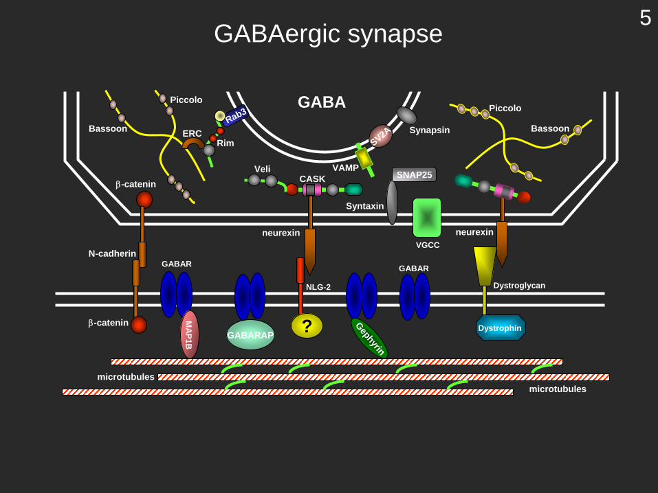
GABA
neurexin
SV2A
VAMP
Syntaxin
-catenin
Rab3
CASK SNAP25
Synapsin
VGCC
Bassoon
Piccolo
ERCRim
Veli
N-cadherin
NLG-2
-catenin
GABAR GABAR
Piccolo
Bassoon
? GephyrinDystroglycan
neurexin
DystrophinGABARAP
MA
P1B
microtubulesmicrotubules
GABAergic synapse5

from Kristen Harris and colleagues
Type 1 excitatorysynapses
Type 2 inhibitorysynapses
1.25 m1.25 m
Glutamate
SAP97
N-cadherin
mGluR
N-catenin
PP1Spin/Neu
drebrin
Kal-7
NMDAREpHR
OPH
F-actin
NLG
-PIX
Abp1
GKAP
Cort
SynGAP
Shank
PICK1
PKC
AMPAR
GRIP/ABP
SynCAM
IRSp53
Shank
Hom
er
IRSp53
neurexinephrin
SPAR
nNOSliprin
CamKII CamKII
CamKII PSD-95
TARP
integrins
SV2A
VAMP
Syntaxin
-catenin
Rab3
CASK CASKSNAP25
Synapsin
VGCC
Bassoon
Piccolo
ERCRim
Veli
Piccolo
Bassoon
talin-actinin
SER IP3R
profilin
F-actinF-actin
SER
TARP
PSD-95
6
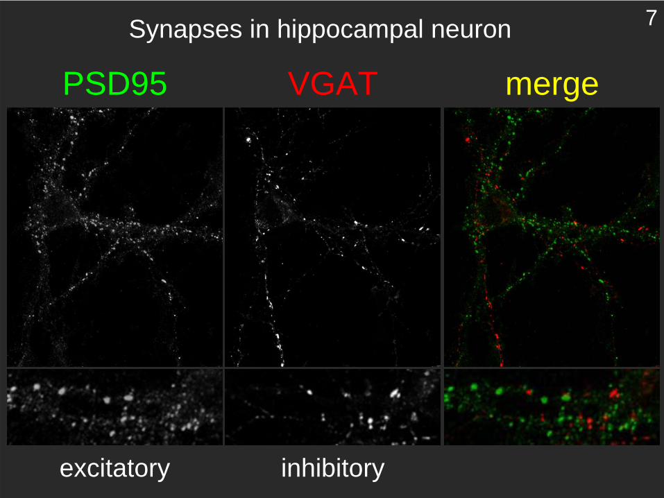
PSD95 VGAT merge
Synapses in hippocampal neuron
excitatory inhibitory
7

Brain and neurons need a correct balance between excitatory and inhibitory synapses to work well
Normal cognitive functions
8

Cognitive disfunctions
Possibly incorrect balance between excitatory (E) and inhibitory (I) synapses
9
Where is Shank in the synapses?
C. Sala- IN-CNR
10

Glutamate
SAP97
N-cadherin
mGluR
N-catenin
PP1Spin/Neu
drebrin
Kal-7
NMDAREpHR
OPH
F-actin
NLG
-PIX
Abp1
GKAP
Cort
SynGAP
Shank
PICK1
PKC
AMPAR
GRIP/ABP
SynCAM
IRSp53
Shank
Hom
er
IRSp53
neurexinephrin
SPAR
nNOSliprin
CamKII CamKII
CamKII PSD-95
TARP
integrins
SV2A
VAMP
Syntaxin
-catenin
Rab3
CASK CASKSNAP25
Synapsin
VGCC
Bassoon
Piccolo
ERCRim
Veli
Piccolo
Bassoon
talin-actinin
SER IP3R
profilin
F-actinF-actin
SER
TARP
PSD-95
11

Shank
GKAP
mGluR
Homer
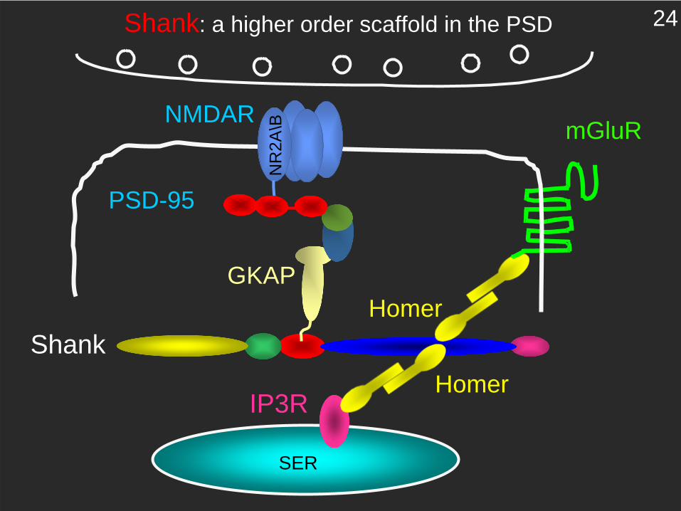
Shank: a higher order scaffold in the PSD
NMDAR
PSD-95
NR
2A\B
12

Shank: a higher order scaffold in the PSD
NMDAR
PSD-95
NR
2A\B
Shank
GKAP
IP3R
SER
Homer
13

Shank1 regulates dendritic spine morphology and synaptic function
Hayashi, M. K., Tang, C., Verpelli, C., Narayanan, R., Stearns, M. H., Xu, R. M., Li, H., Sala, C., and Hayashi, Y. (2009). The postsynaptic density proteins Homer and Shank form a polymeric network structure. Cell 137, 159- 171.
Hung, A. Y., Futai, K., Sala, C., Valtschanoff, J. G., Ryu, J., Woodworth, M. A., Kidd, F. L., Sung, C. C., Miyakawa, T., Bear, M. F., et al. (2008). Smaller dendritic spines, weaker synaptic transmission, but enhanced spatial learning in mice lacking Shank1. J Neurosci 28, 1697-1708.
Sala C, Roussignol G, Meldolesi J, Fagni L (2005) Key role of the PSD scaffold proteins, Shank and Homer, in the functional architecture of Ca2+ homeostasis at dendritic spines in hippocampal neurons. J Neurosci 25:4587-92
Roussignol G, Ango F, Romorini S, Tu JC, Sala C, Worley PF, Bockaert J, Fagni L (2005) Shank expression is sufficient to induce functional dendritic spine synapses in aspiny neurons. J Neurosci 25:3560-3570.
Romorini S, Piccoli G, Jiang M, Grossano P, Tonna N, Passafaro M, Zhang M, Sala C (2004) Multimerizationand interaction with PSD-95/GKAP regulate the association of Shank with synapses.J Neurosci 24:9391-404.
Sala C, Futai K, Yamamoto K, Worley PF, Hayashi Y, Sheng M (2003) Inhibition of dendritic spine morphogenesis and synaptic transmission by activity-inducible protein Homer1a. J Neurosci 23:6327-6337.
Sala C, Piech V, Wilson NR, Passafaro M, Liu G, Sheng M (2001) Regulation of dendritic spine morphology and synaptic function by Shank and Homer. Neuron 31:115-130.
14

0.15 s
50 pA
GFP
GFP+Shank1B
DIV 11 DIV 17
FM4-
64 P
unct
a/ μ
m2
DIV11 DIV170.00
0.04
0.08
0.12
0.16 *
**
Freq
uenc
y (H
z)
DIV11 DIV170
2
4
6
*
GFPGFP+Shank1B
Sala et al. Neuron (2001)
15

- dendritic spine density is reduced
from Hung and Sheng
16
In order to study the functions of Shank3we knocked down Shank3 expression
in neurons
17
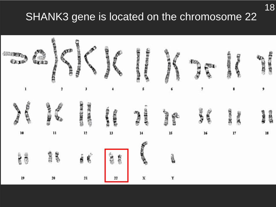
SHANK3 gene is located on the chromosome 2218
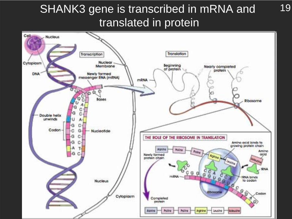
SHANK3 gene is transcribed in mRNA and translated in protein
19

Shank3
PSD-95
siRNS for Shank3 siRNA control
0
2
4
6
8
10
12
N. o
f clu
ster
s 10
m
siRNS for Shank3
siRNA control
We can block Shank3 protein production by degrading Shank3 mRNA with the siRNA
20

Shank3 siRNA can be transfer to neurons using lentivirus
We can block Shank3 expression in neurons for studying Shank3 function
21

What happen to dendritic spines and synapses in neurons knocked down for Shank3?
22

siRNAcontrol
siRNA forShank3
Number Width0
0,5
1
1,5
2
2,5
3
1,6
1,7
1,8
1,9
2
2,1
1
1,05
1,1
1,15
1,2
Spi
nes/
10m
m m
p < 0.003p < 0.003 p < 0.02
**** *
Length
GFP DsRED merge
GFP DsRed merge
Dendritic spines morphology in hippocampal neurons knocked down for Shank3:dendritic spines are reduced in dimendion and number
siRNA controlsiRNA for Shank3
23
Shank
GKAP
mGluR
Homer
Shank: a higher order scaffold in the PSD
NMDAR
PSD-95
NR
2A\B
IP3R
SER
Homer
24
MAPK
Syn
Abi1
IRSp53
eEF2
GluR2/3
mGluR5
NR1
Actin
PSD-95
PanShank
Shank1
Shank3
Homer
not infecte
d
siShank3
not infecte
d
siShank3
0
0,2
0,4
0,6
0,8
1
1,2
1,4
Pan
Sha
nk
Sha
nk3
Sha
nk1
0
0,2
0,4
0,6
0,8
1
1,2
1,4
actin
PS
D/9
5
Hom
er
Syn
eEF2
Abi
1
IRS
p53
MAP
K
mG
luR
5
Glu
R2/
3
NR
1Nor
mal
ized
inte
nsity
Nor
mal
ized
inte
nsity
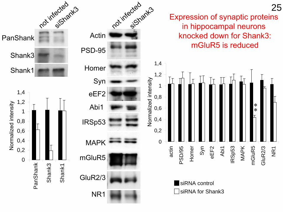
Expression of synaptic proteins in hippocampal neurons
knocked down for Shank3:mGluR5 is reduced
**
siRNA controlsiRNA for Shank3
25

siRNA for Shank3
pERK1/2
Actin
mGluR5 phopsphorilation of ERK1/2 is inhibited in neuronsknocked down for Shank3
DHPG 30’NT KClNMDA DHPG 30’NT KClNMDA
siRNA control
DH
PH p
ERK
1/2
leve
l
0
2
4
6
8
siRNS for Shank3
siRNA control
26

NM
DA
RAAAA
mGluR5
Dendritic spinesmaturation
mGluR dependent ERK1/2 activation
AAAA
Shank3
Synthesis of proteins that regulatedendritic spines
In presence of Shank3 27

NM
DA
RAAAA
mGluR5
Dendritic spinesmaturation
mGluR dependent ERK1/2 activation
AAAA
Shank3
Synthesis of proteins that regulatedendritic spines
In absence of Shank3 28

In absence of Shank3In presence of Shank3
In absence of Shank3 the ratio excitatory/inhibitory synapses might be altered
29
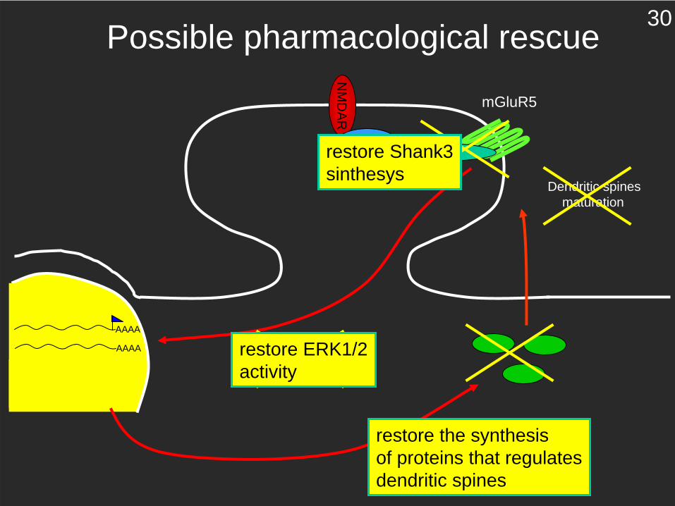
NM
DA
RAAAA
mGluR5
Dendritic spinesmaturation
mGluR dependent ERK1/2 activation
AAAA
Shank3
Synthesis of proteins that regulatedendritic spines
Possible pharmacological rescue
restore ERK1/2activity
restore the synthesisof proteins that regulatesdendritic spines
restore Shank3sinthesys
30
We plan to test the pharmacological rescue in two models:
-using rat neuronal cultures knocked down for Shank3-using mouse knock out for Shank3
-using induced pluripotent stem cells (iPS cells) derived from patients fibroblasts
31

From the pluripotent cells derives our organs32

We can take a unipotent cell (skin fibroblast) and transform the cell into a pluripotent cell
33

34
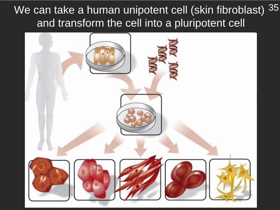
We can take a human unipotent cell (skin fibroblast) and transform the cell into a pluripotent cell
35
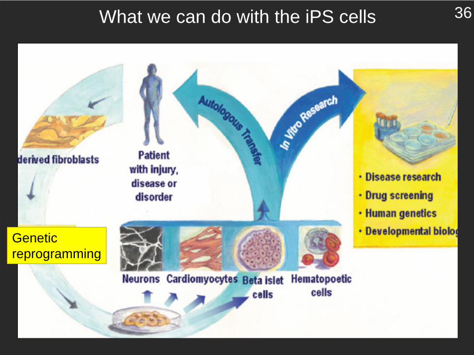
What we can do with the iPS cells
Geneticreprogramming
36
Chiara Verpelli
Monica Patti
Francesca Rossi
Hanako Tsushima
CNR Institute of NeuroscienceMilano, Italy
Antonella Gianfelice
Valentina Cea
Alice Zanchi
Clara BonagliaRobero GiordaLa Nostra Famiglia, Bosisio
Maria PassafaroDTI/CNR Milano
Morgan ShengMIT Cambridge MA
Albert Y. Hung
Univ. San RaffaeleVania Broccoli
37