The Role of Resurfacing Arthroplasty in the Knee · 2019-04-15 · • No statistically significant...
67
The Role of Resurfacing Arthroplasty in the Knee
Transcript of The Role of Resurfacing Arthroplasty in the Knee · 2019-04-15 · • No statistically significant...
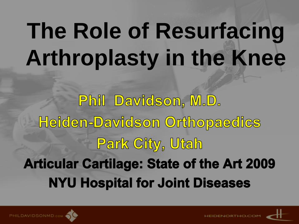
The Role of Resurfacing Arthroplasty in the Knee

OUTLINE• Continuum of Options• Introduction of Inlay
Arthroplasty Concept– Biomechanical Basis – FDA Trial
• Knee CAP– HemiCAP, UniCAP,PF– Indications– Technique– Cases
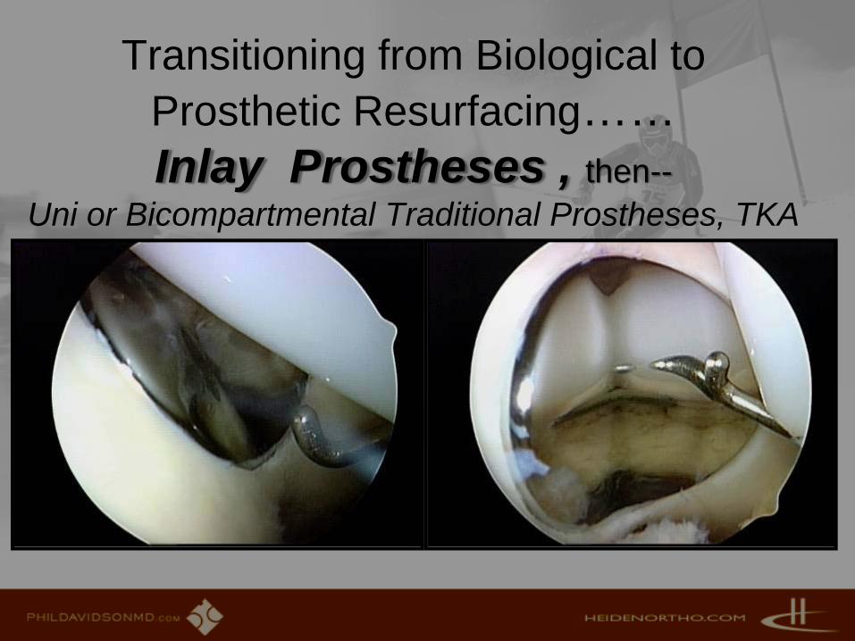
Transitioning from Biological to Prosthetic Resurfacing……Inlay Prostheses , then--
Uni or Bicompartmental Traditional Prostheses, TKA
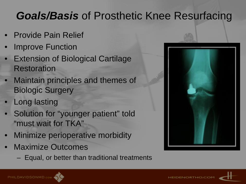
Goals/Basis of Prosthetic Knee Resurfacing
• Provide Pain Relief• Improve Function• Extension of Biological Cartilage
Restoration• Maintain principles and themes of
Biologic Surgery • Long lasting • Solution for “younger patient” told
“must wait for TKA”• Minimize perioperative morbidity• Maximize Outcomes
– Equal, or better than traditional treatments

ANATOMY is KEY• Concave and convex
geometric surfaces –complex curves
• Intraoperative articular mapping involves measuring/replicating complex geometric surface configurations
• Accounts for morphologic variability
• Implants are patient driven
FTG???
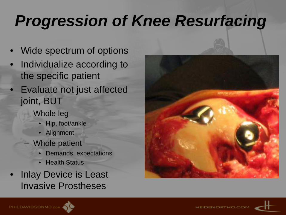
Progression of Knee Resurfacing
• Wide spectrum of options • Individualize according to
the specific patient • Evaluate not just affected
joint, BUT– Whole leg
• Hip, foot/ankle• Alignment
– Whole patient • Demands, expectations• Health Status
• Inlay Device is Least Invasive Prostheses
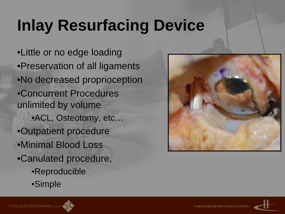
Inlay Resurfacing Device •Little or no edge loading•Preservation of all ligaments•No decreased proprioception•Concurrent Procedures unlimited by volume
•ACL, Osteotomy, etc… •Outpatient procedure•Minimal Blood Loss•Canulated procedure,
•Reproducible•Simple
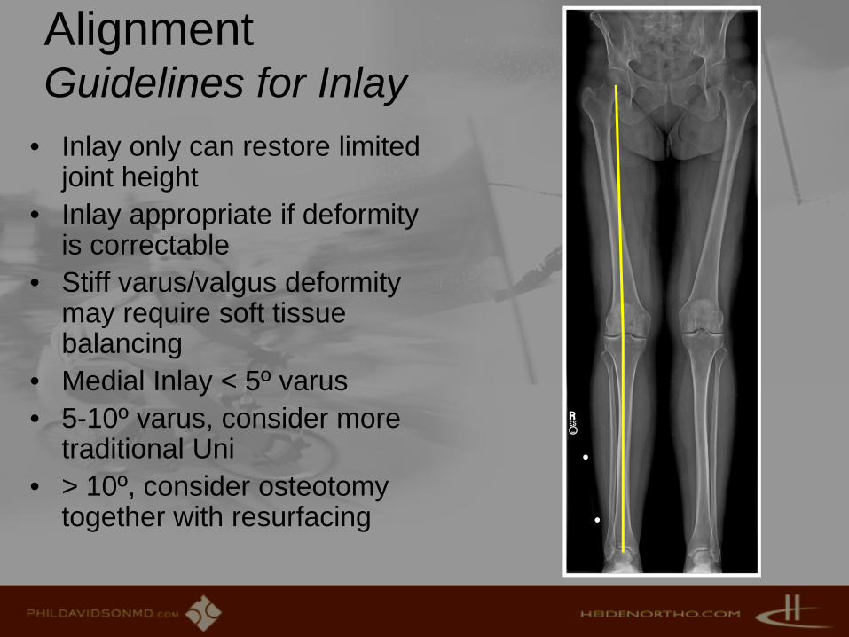
AlignmentGuidelines for Inlay
• Inlay only can restore limited joint height
• Inlay appropriate if deformity is correctable
• Stiff varus/valgus deformity may require soft tissue balancing
• Medial Inlay < 5º varus• 5-10º varus, consider more
traditional Uni• > 10º, consider osteotomy
together with resurfacing

Onlay ArthroplastyImplants generated from patient anatomy
• Implants manufactured from individual patient digital data (CT or MR)
• Less bone resection than TKA
• Can preserve ligaments• Allows greater angular and
height correction• More invasive than inlay• Another complimentary
option

Introduction of CAP(contoured articular prosthesis)
• Geometry based on patient’s native anatomy
• Intraoperative joint mapping (topo map)
• Account for complex asymmetrical geometry
• Extension of biological resurfacing

Knee Implants• HemiCAP (unipolar knee)
– Not currently FDA approved in US
• UniCAP• PF HemiCAP• PF XLT

1st question I asked:“what about the reciprocal surfaces”
www.phildavidsonmd.com

Becher C¹, Tibesku CO¹, Fuchs-Winkelmann S¹, Thermann H², Pässler HH²
²Center for Knee- & Foot SurgeryATOS CLINIC, Heidelberg
Tibiofemoral peak contact pressure in focal anatomic femoral resurfacing: A biomechanical study
Skrbensky G, Huber R
Biomechanic and Biomaterial Testing Laboratory
University of Vienna
¹Department for Orthopaedic Surgery,University of Marburg
Basic Science- Contact Pressure
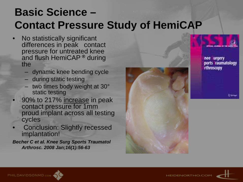
Basic Science –Contact Pressure Study of HemiCAP
• No statistically significant differences in peak contact pressure for untreated knee and flush HemiCAP ® during the – dynamic knee bending cycle– during static testing– two times body weight at 30°
static testing • 90% to 217% increase in peak
contact pressure for 1mm proud implant across all testing cycles
• Conclusion: Slightly recessed implantation!
Becher C et al. Knee Surg Sports TraumatolArthrosc. 2008 Jan;16(1):56-63
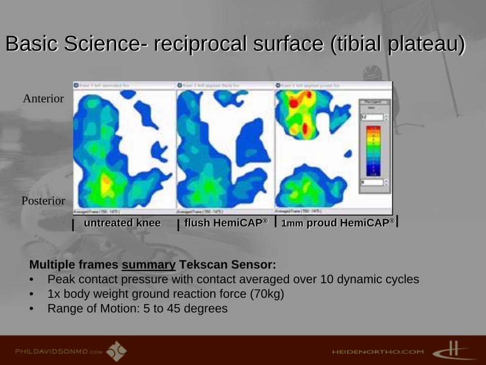
untreated knee flush HemiCAP® 1mm proud HemiCAP®
Multiple frames summary Tekscan Sensor: • Peak contact pressure with contact averaged over 10 dynamic cycles• 1x body weight ground reaction force (70kg)• Range of Motion: 5 to 45 degrees
Anterior
Posterior
Basic Science- reciprocal surface (tibial plateau)

Tibiofemoral peak contact pressure with a contoured articular prosthesis and a complete resection of the meniscus (posterior horn)
• Tibiofemoral peak contact pressure: – Untreated and flush demonstrate matching curves. – Significant increase with non-functional meniscus / radial tear
0
1
2
3
4
5
6
7
8
9
10
1 101 201 301 401 501 601 701 801 901 1001 1101 1201 1301 1401 1501
P (M
Pa)
time (tsec)
Peak contact pressure
Untreated Flush Meniscus tear
Basic Science- Relative Loading
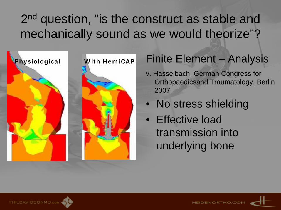
2nd question, “is the construct as stable and mechanically sound as we would theorize”?
Finite Element – Analysisv. Hasselbach, German Congress for
Orthopaedicsand Traumatology, Berlin 2007
• No stress shielding • Effective load
transmission into underlying bone
Physiological With HemiCAP
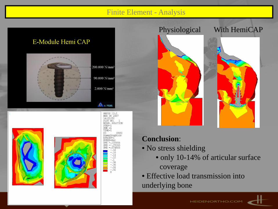
Finite Element - Analysis
Physiological With HemiCAP
Conclusion: • No stress shielding
• only 10-14% of articular surface coverage
• Effective load transmission into underlying bone
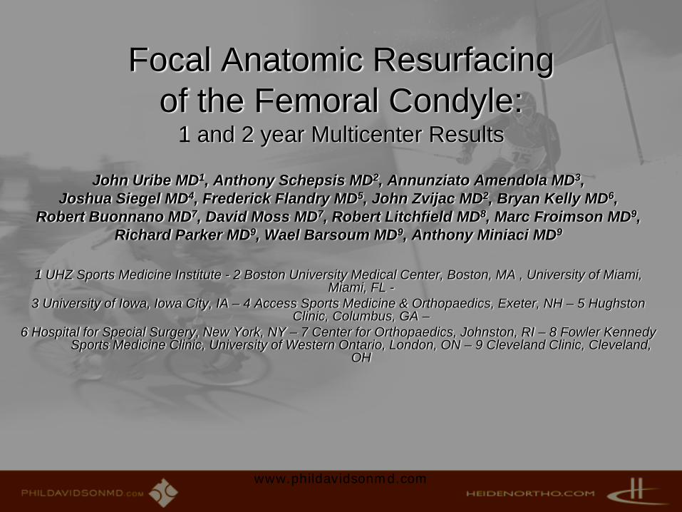
Focal Anatomic Resurfacing of the Femoral Condyle:
1 and 2 year Multicenter Results
John Uribe MD1, Anthony Schepsis MD2, Annunziato Amendola MD3, Joshua Siegel MD4, Frederick Flandry MD5, John Zvijac MD2, Bryan Kelly MD6,
Robert Buonnano MD7, David Moss MD7, Robert Litchfield MD8, Marc Froimson MD9, Richard Parker MD9, Wael Barsoum MD9, Anthony Miniaci MD9
1 UHZ Sports Medicine Institute - 2 Boston University Medical Center, Boston, MA , University of Miami, Miami, FL -
3 University of Iowa, Iowa City, IA – 4 Access Sports Medicine & Orthopaedics, Exeter, NH – 5 HughstonClinic, Columbus, GA –
6 Hospital for Special Surgery, New York, NY – 7 Center for Orthopaedics, Johnston, RI – 8 Fowler Kennedy Sports Medicine Clinic, University of Western Ontario, London, ON – 9 Cleveland Clinic, Cleveland,
OH
www.phildavidsonmd.com

Clinical Results- US FDA HemiCAP trialUS Multicenter Study: Study Population and Current Follow-up
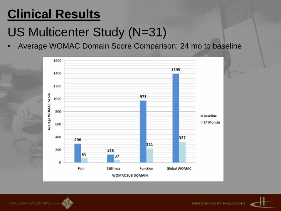
Clinical ResultsUS Multicenter Study (N=31)• Average WOMAC Domain Score Comparison: 24 mo to baseline
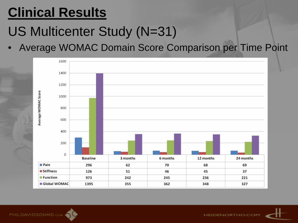
Clinical ResultsUS Multicenter Study (N=31)• Average WOMAC Domain Score Comparison per Time Point
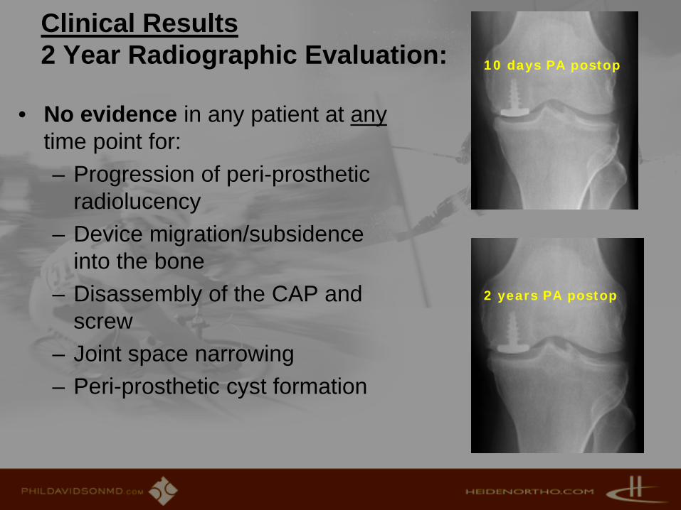
Clinical Results2 Year Radiographic Evaluation:
• No evidence in any patient at anytime point for:– Progression of peri-prosthetic
radiolucency– Device migration/subsidence
into the bone– Disassembly of the CAP and
screw– Joint space narrowing– Peri-prosthetic cyst formation
2 years PA postop
10 days PA postop

Clinical Results (HemiCAP for Femoral Condyle) C. v. Hasselbach, Essen, Presented at German Congress for Orthopaedics and Traumatology, Berlin 2007
• Patient Population: N = 121• Follow‐up: Mean 14 months (1‐25)• Patient Age: Mean 52,5 years (34‐67)• Gender: Female N = 13 (29,5%), Male N = 31 (70,5%)• Previous Cartilage Procedures: Mean N= 2.3 (0‐6)• Procedure Duration: 24 minutes• Postoperative Recovery until Return to Work: Mean 35.3 days (15‐82)• HSS Knee Scores improved from 85.2 preop to 95.3 postop• 17 Re‐look Arthroscopies: Contoured Implant Integration, No Deleterious
Cartilage Effects• Radiographic Examination: No peri‐Prosthetic Radiolucency, or Implant
Subsidence

Clinical Results - Australian National Knee Registry
• HemiCAP ® Resurfacing of Femur (initial experience)• N=90 implants in 81 patients reported over the
course of 4 years up until 12/31/07• Observed component years: 107 (~mean follow-up
around 1 year)• Male = Female• 8 revisions
– progression of disease (N=4) and – Continued pain (N=4)– 5/8 revised to unicondylar knee; 3/8 to TKA
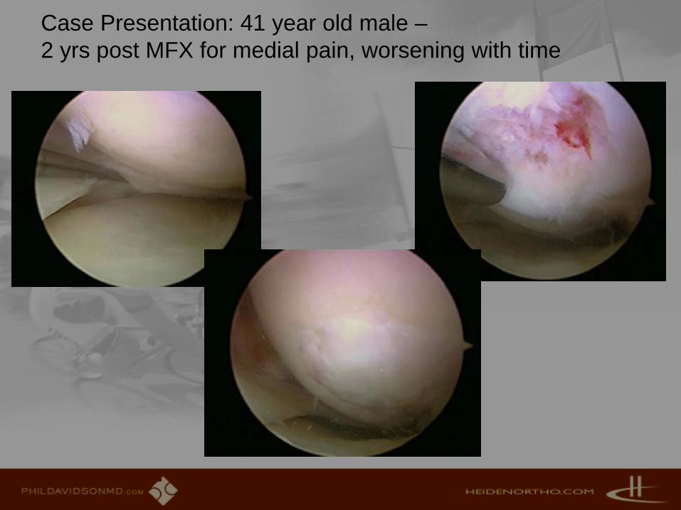
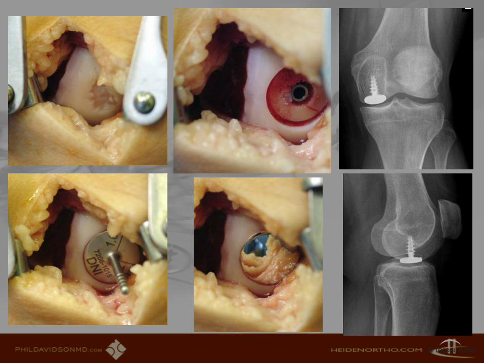
Case Presentation: 41 year old male –2 yrs post MFX for medial pain, worsening with time
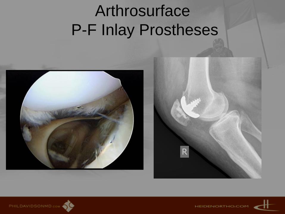
Arthrosurface P-F Inlay Prostheses

PF- PROSTHETIC RESURFACING• Vast difference between
traditional PFA and Inlay • Traditional prostheses
limited success and rarely used
• Inlay device allows for concurrent re-alignment
• Inlay device for younger patients
• Excellent new solution for vexing problem

Trochlear Implants Variety of Geometry
www.phildavidsonmd.com
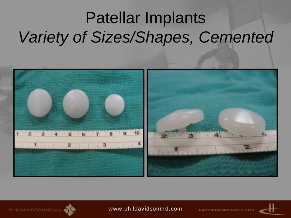
Patellar ImplantsVariety of Sizes/Shapes, Cemented
www.phildavidsonmd.com
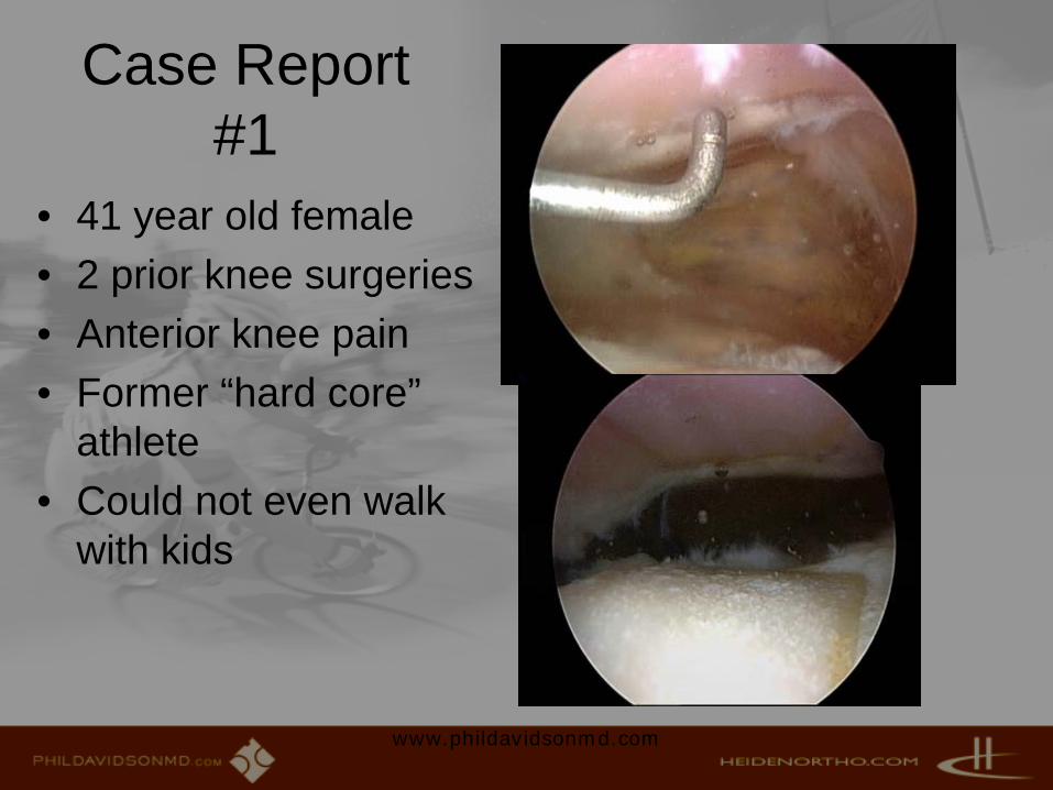
Case Report #1
• 41 year old female• 2 prior knee surgeries• Anterior knee pain• Former “hard core”
athlete • Could not even walk
with kids
www.phildavidsonmd.com
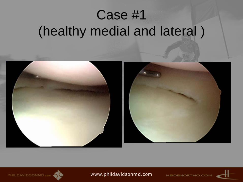
Case #1(healthy medial and lateral )
www.phildavidsonmd.com
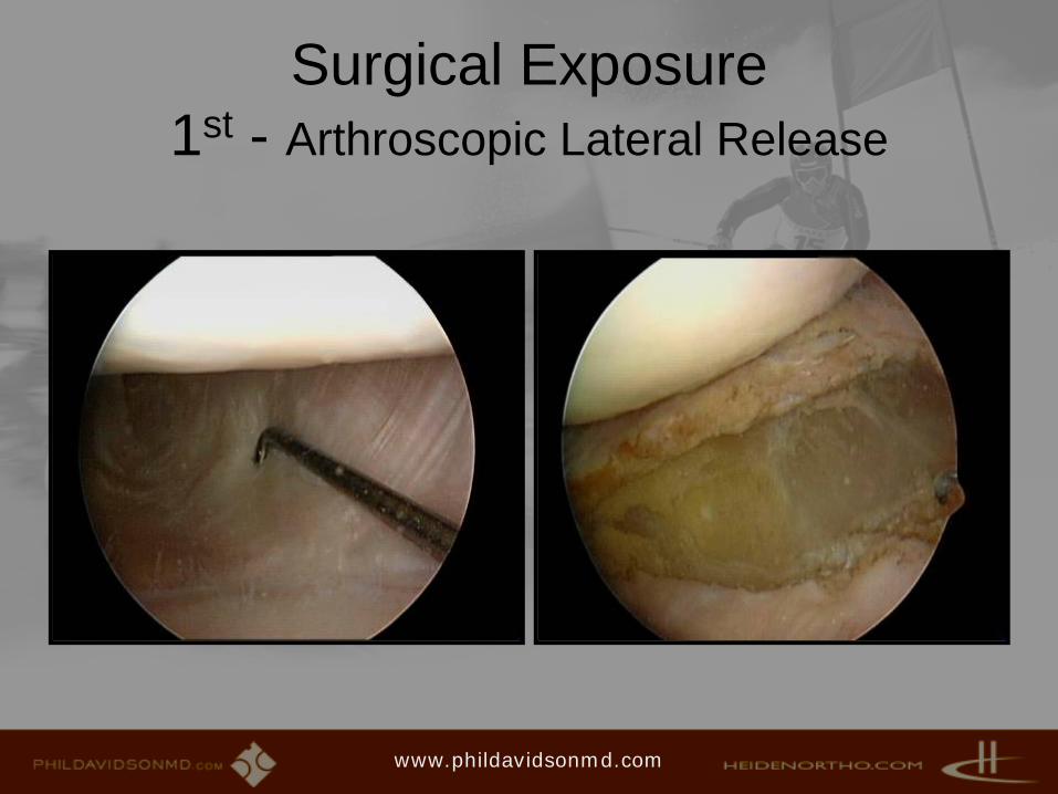
Surgical Exposure 1st - Arthroscopic Lateral Release
www.phildavidsonmd.com
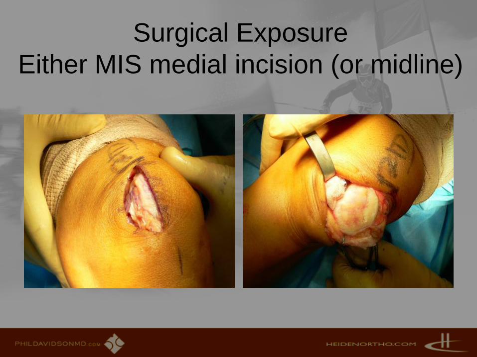
Surgical ExposureEither MIS medial incision (or midline)

Technique –• Guidewire key to
cannulated system
• Perpendicular placement– Careful attention to
this!!
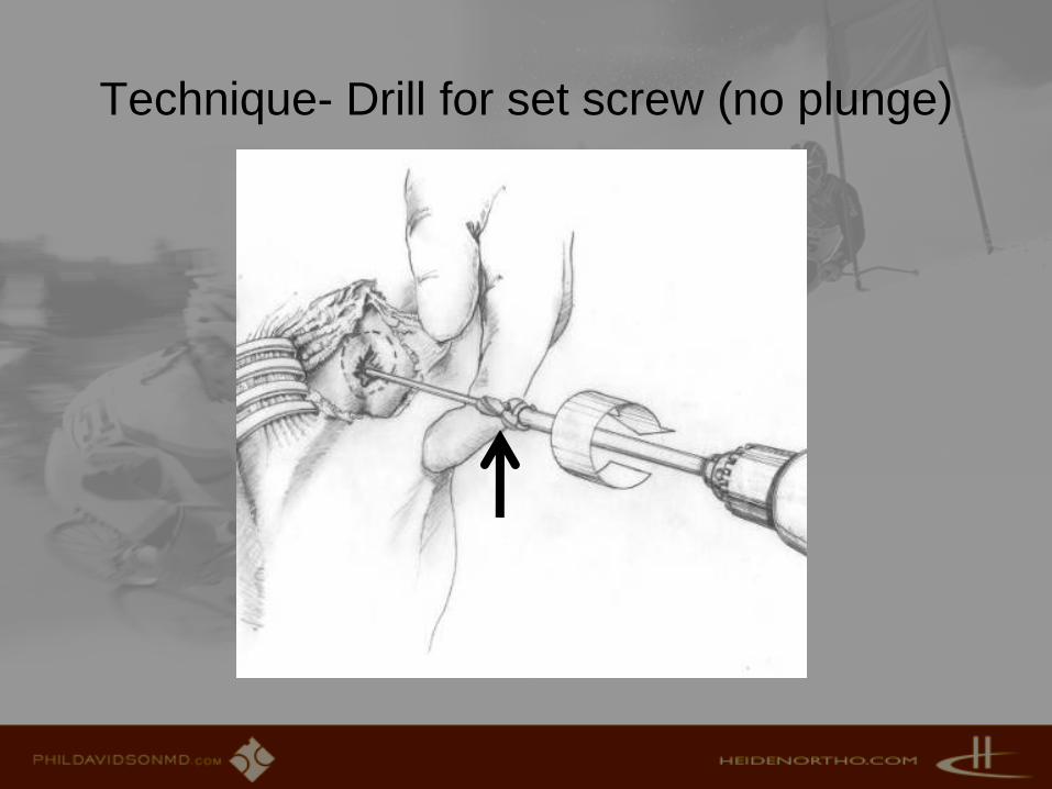
Technique- Drill for set screw (no plunge)
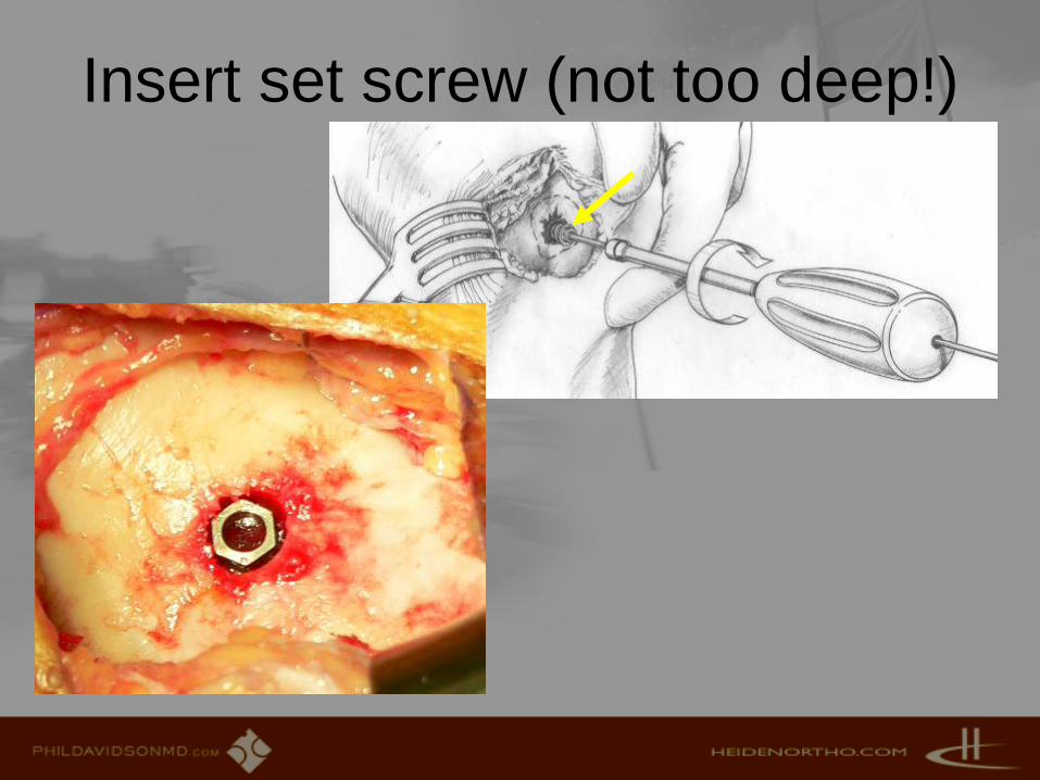
Insert set screw (not too deep!)

Height measuring cap

Articular Mapping…if measured values NOT on chart,
must consider WHY

Drilling for implant
• High speed drill• Do not use reamer• Cooling irrigation

Device Trial –can adjust/mark
rotation

Patellar Preparationbasically need patella “deep enough”
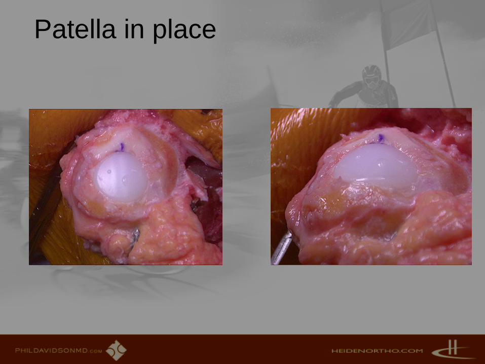
Patella in place
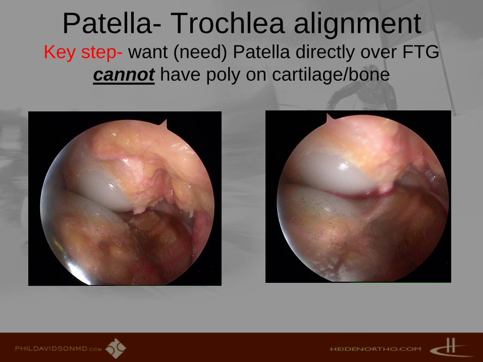
Patella- Trochlea alignment Key step- want (need) Patella directly over FTG
cannot have poly on cartilage/bone

Radiographs pre and post
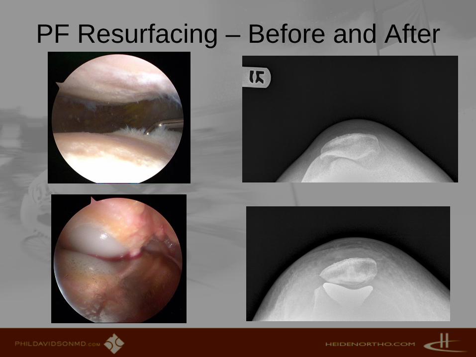
PF Resurfacing – Before and After

UniCAP™aka… inlay arthroplasty, scope
assisted Uni, AKR , etc..
UniCAP Advantages• UniCAP may prevent patello-femoral
complications/encroachment of conventional UKA through inlay resurfacing
• Revision to standard UKA may be possible due to shallow implant bed resurfacing technique• UniCAP avoids L-cutAmple room for ACL, osteotomy, soft tissue procedures
• UniCAP limitations are at the same time its advantages:• Meniscal sparing technology for patients with healthy,
functional meniscus
UniCAP Advantages• Knee biomechanics are left intact
through inlay resurfacing• Joint height, soft tissue tension are maintained• Conventional UKA are at risk of “overstuffing” the
joint• Patient selection remains critical:
• Proper joint stability, avoiding increased translational movement
• Monocompartmental degeneration, or concurrent multi-compartmental resurfacing, avoiding continuation of “referred pain”

UniCAP
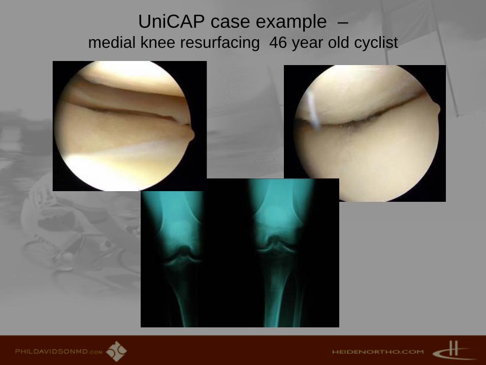
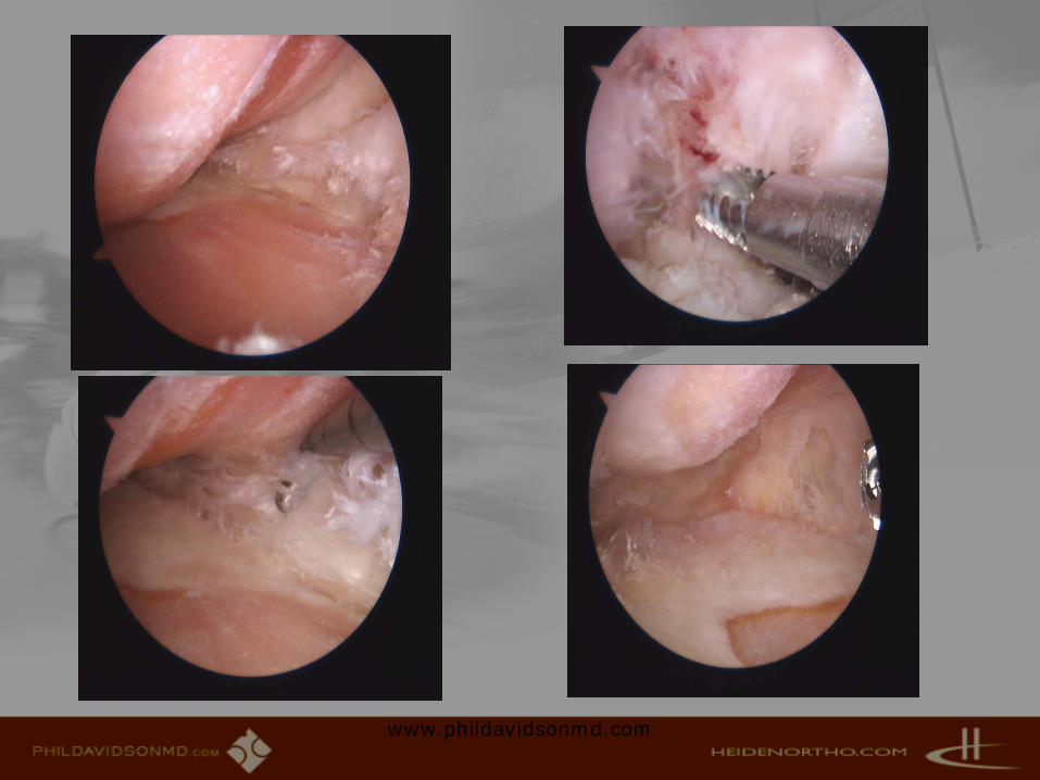
UniCAP case example –medial knee resurfacing 46 year old cyclist

UniCAP – medial knee resurfacing
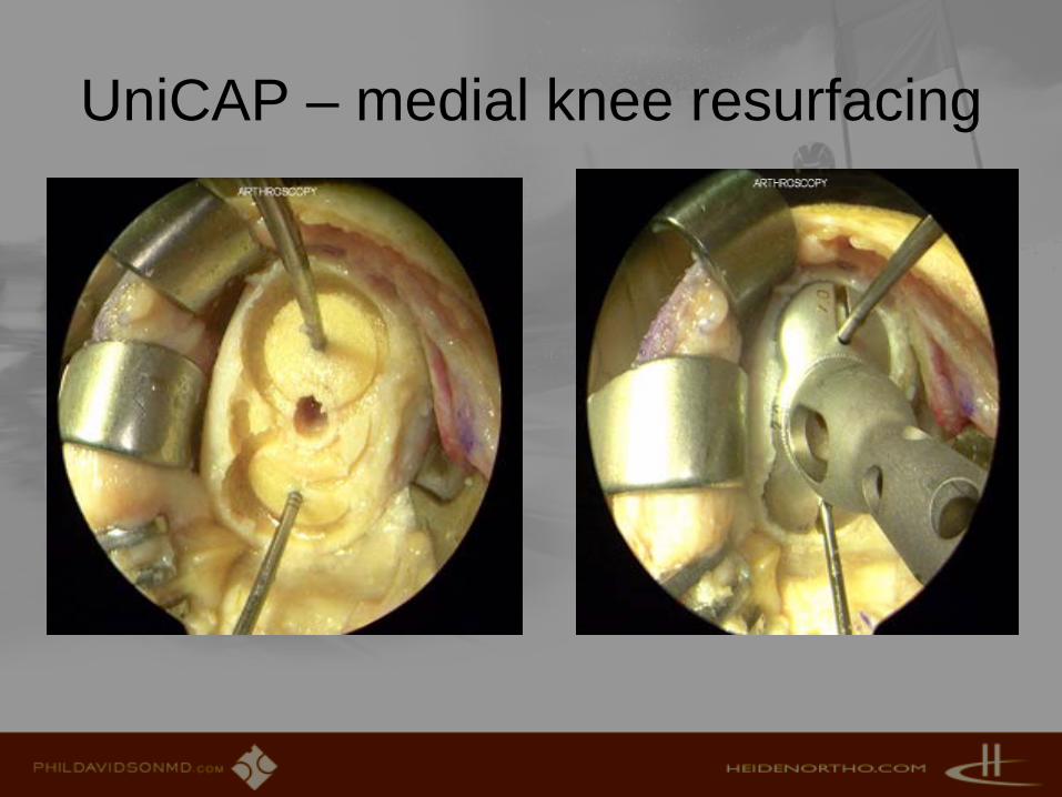
UniCAP – medial knee resurfacing

UniCAP – medial knee resurfacing

UniCAP – medial knee resurfacing
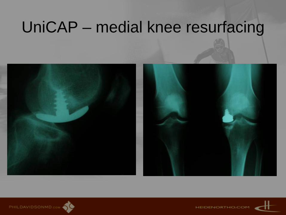
UniCAP – medial knee resurfacing
Case Report – 51 year old dancer
• Chronic knee pain and instability • Prior (30 yr ago) ACL reconstr• 5 degree varus• No Patellar nor lateral pain
www.phildavidsonmd.com


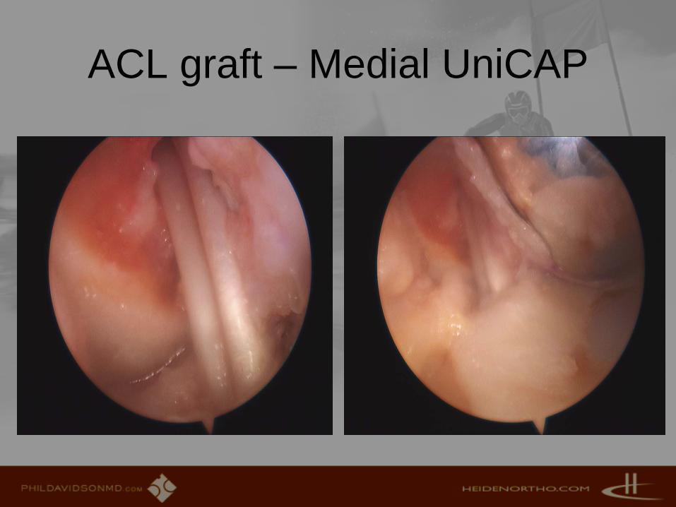
ACL graft – Medial UniCAP


“Ideal” First Patient for CAP
• 30-60 yrs (APPX)• Nearly normal align• Any comorbidity
mitigating against Biological solution
• Unicompartmentalmedial disease
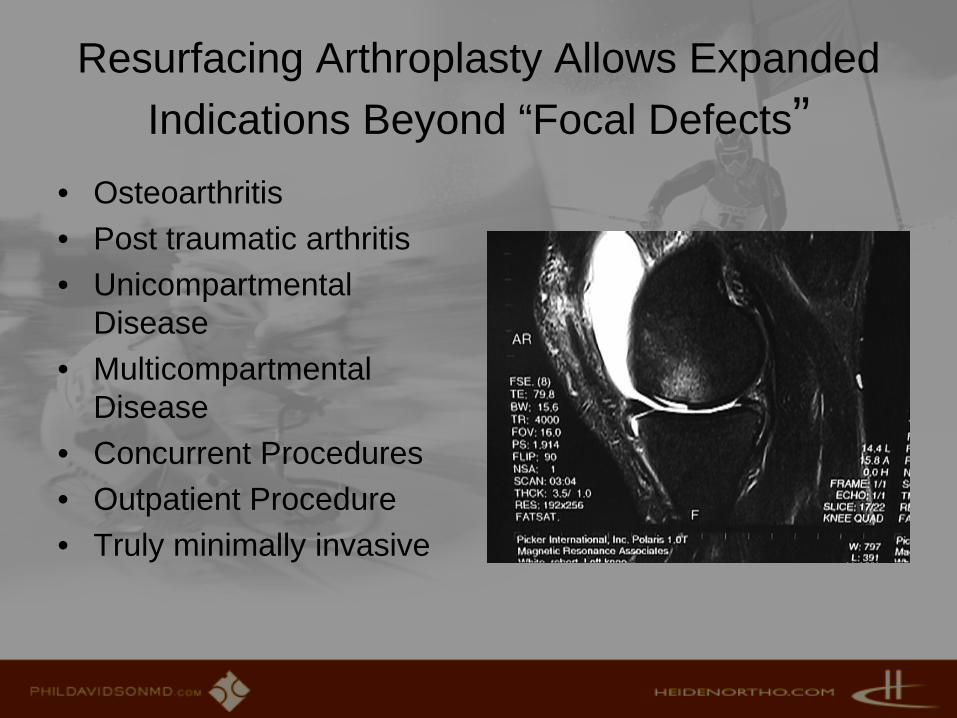
Resurfacing Arthroplasty Allows Expanded Indications Beyond “Focal Defects”
• Osteoarthritis• Post traumatic arthritis• Unicompartmental
Disease• Multicompartmental
Disease• Concurrent Procedures• Outpatient Procedure• Truly minimally invasive

Advantages of Inlay Resurfacing Arthroplasty
• Immediate, excellent pain relief
• Simple, canulated, reproducible, yet elegant surgery
• Very few soft tissue balancing challenges
• Minimally bone sacrificing • Minimal EBL, can be
outpatient• Can easily convert to
traditional arthroplasty
• Patient acceptance• Allows concurrent soft
tissue procedure • Maintain cartilage
restoration principles • Based on patient, or
ambient anatomy