The Role of Heterochromatin in the Expression of a - Genetics
16
Copyright 0 1993 by the Genetics Society of America The Role of Heterochromatin in the Expression of a Heterochromatic Gene, the rolled Locus of Drosophila melanogaster Daniel F. Eberl,’ Brenda J. Duyf and Arthur J. Hilliker2 Department of Molecular Biology and Genetics, University of Guelph, Guelph, Ontario, Canada NIG 2Wl Manuscript received August 5, 1992 Accepted for publication January 1 1, 1993 ABSTRACT Constitutive heterochromatic regions of chromosomes are those that remain condensed through most or all of the cell cycle. In Drosophila melanogaster, the constitutive heterochromatic regions, located around the centromere, contain a number of gene loci, but at a much lower density than euchromatin. In the autosomal heterochromatin, the gene loci appear to be unique sequence genes interspersed among blocks of highly repeated sequences. Euchromatic genes do not function well when brought into the vicinity of heterochromatin (position-effectvariegation). We test the possibility that the blocks of centromeric heterochromatin provide an environment essential for heterochromatic gene function. T o assay directly the functional requirement of autosomal heterochromatic genes to reside in heterochromatin, the rolled (rl) gene, whichis normally located deep in chromosome 2R heterochromatin, was relocated within small blocks of heterochromatin to a variety of euchromatic positions by successive series of chromosomal rearrangements. The function of the rl gene is severely affected in rearrangements in which the rl gene is isolated in a small block of heterochromatin, and these position effects can be reverted by rearrangements which bring the rl gene closer to any large block of autosomal or X chromosome heterochromatin. There is some evidence that five other 2R heterochromatic genes are also affected among these rearrangements. These findings demonstrate that the heterochromatic genes, in contrast to euchromatic genes whose function is inhibited by relocation to heterochromatin, require proximity to heterochromatin to function properly, and they argue strongly that a major function of the highly repeated satellite DNA, which comprises most of the heterochromatin, is to provide this heterochromatic environment. C ONSTITUTIVE heterochromatin refers to those regions of chromosomes which remain heteropyknotic through most or all of the cell cycle, and the remaining parts of chromosomes, those that disappear at telophase, are termed euchromatin (HEITZ 1928). Heterochromatin is phylogenetically a very widespread phenomenon, nearly ubiquitous among higher eukaryotes. In the Drosophila melano- gaster chromosomes, the subject of the present study, constitutive heterochromatin flanks the centromeres and is detectable cytologically at all stages of devel- opment except in the early embryonic cleavage divi- sions (HUETTNER 1933; RABINOWITZ 1941). In contrast, facultative heterochromatin (BROWN 1966) involves the heteropyknosis of an entire chro- mosome relative to its homolog. Facultative hetero- chromatin is typified by the heterochromatinized X chromosome (Barr body) in placental mammals (BARR and BERTRAM 1949; MITTWOCH 1964) and by the entireheterochromatinizedsetofchromosomes in mealy bugs (HUGHES-SCHRADER 1948). Facultative heterochromatinization of chromosomesis correlated ’ Current address: Department of Biochemical Pharmacology, State Uni- ’ Author to whom all correspondence should be addressed. versity of New York, Buffalo, New York 14260. Genetics 134: 277-292 (May, 1993) with genetic inactivation; however, the homologous chromosome is not affected. From this point the term “heterochromatin” will refer exclusively to “constitu- tive heterochromatin” and facultative heterochroma- tin will not be discussed further. The cytological discovery of constitutive hetero- chromatin immediately raised the question of its bio- logical significance. Three lines of evidence correlated heterochromatin with genetic inactivity. First, few genes mapped to heterochromatic regions in D. mel- anogaster (HEITZ 1933, MULLER and PAINTER 1932). Second, position-effect variegation showed that most euchromatic genes are partially suppressed if brought near heterochromatin by chromosome rearrangement (SCHULTZ 1936; DEMEREC 1941; SPOFFORD1976). Third, constitutive heterochromatin appeared to be composed largely of highly repeated DNA sequences (GALL, COHEN and POLAN 197 1; PEACOCK et al. 1973, 1977; STEFFENSEN, APPELS and PEACOCK 1981), which most likely lack coding functions. All such evidence argued that heterochromatin was a location, and indicator and an agent of genetic inactivity. In spite of this strong apparent correlation between heterochromatin and genetic inactivity, it is now clear, at least in D. melanogaster, that there are in fact active
Transcript of The Role of Heterochromatin in the Expression of a - Genetics
Copyright 0 1993 by the Genetics Society of America
The Role of Heterochromatin in the Expression of a Heterochromatic Gene, the rolled Locus of Drosophila melanogaster
Daniel F. Eberl,’ Brenda J. Duyf and Arthur J. Hilliker2 Department of Molecular Biology and Genetics, University of Guelph, Guelph, Ontario, Canada NIG 2Wl
Manuscript received August 5 , 1992 Accepted for publication January 1 1, 1993
ABSTRACT Constitutive heterochromatic regions of chromosomes are those that remain condensed through
most or all of the cell cycle. In Drosophila melanogaster, the constitutive heterochromatic regions, located around the centromere, contain a number of gene loci, but at a much lower density than euchromatin. In the autosomal heterochromatin, the gene loci appear to be unique sequence genes interspersed among blocks of highly repeated sequences. Euchromatic genes do not function well when brought into the vicinity of heterochromatin (position-effect variegation). We test the possibility that the blocks of centromeric heterochromatin provide an environment essential for heterochromatic gene function. To assay directly the functional requirement of autosomal heterochromatic genes to reside in heterochromatin, the rolled ( r l ) gene, which is normally located deep in chromosome 2R heterochromatin, was relocated within small blocks of heterochromatin to a variety of euchromatic positions by successive series of chromosomal rearrangements. The function of the rl gene is severely affected in rearrangements in which the rl gene is isolated in a small block of heterochromatin, and these position effects can be reverted by rearrangements which bring the rl gene closer to any large block of autosomal or X chromosome heterochromatin. There is some evidence that five other 2R heterochromatic genes are also affected among these rearrangements. These findings demonstrate that the heterochromatic genes, in contrast to euchromatic genes whose function is inhibited by relocation to heterochromatin, require proximity to heterochromatin to function properly, and they argue strongly that a major function of the highly repeated satellite DNA, which comprises most of the heterochromatin, is to provide this heterochromatic environment.
C ONSTITUTIVE heterochromatin refers to those regions of chromosomes which remain
heteropyknotic through most or all of the cell cycle, and the remaining parts of chromosomes, those that disappear at telophase, are termed euchromatin (HEITZ 1928). Heterochromatin is phylogenetically a very widespread phenomenon, nearly ubiquitous among higher eukaryotes. In the Drosophila melano- gaster chromosomes, the subject of the present study, constitutive heterochromatin flanks the centromeres and is detectable cytologically at all stages of devel- opment except in the early embryonic cleavage divi- sions (HUETTNER 1933; RABINOWITZ 1941).
In contrast, facultative heterochromatin (BROWN 1966) involves the heteropyknosis of an entire chro- mosome relative to its homolog. Facultative hetero- chromatin is typified by the heterochromatinized X chromosome (Barr body) in placental mammals (BARR and BERTRAM 1949; MITTWOCH 1964) and by the entire heterochromatinized set of chromosomes in mealy bugs (HUGHES-SCHRADER 1948). Facultative heterochromatinization of chromosomes is correlated
’ Current address: Department of Biochemical Pharmacology, State Uni-
’ Author to whom all correspondence should be addressed. versity of New York, Buffalo, New York 14260.
Genetics 134: 277-292 (May, 1993)
with genetic inactivation; however, the homologous chromosome is not affected. From this point the term “heterochromatin” will refer exclusively to “constitu- tive heterochromatin” and facultative heterochroma- tin will not be discussed further.
T h e cytological discovery of constitutive hetero- chromatin immediately raised the question of its bio- logical significance. Three lines of evidence correlated heterochromatin with genetic inactivity. First, few genes mapped to heterochromatic regions in D. mel- anogaster (HEITZ 1933, MULLER and PAINTER 1932). Second, position-effect variegation showed that most euchromatic genes are partially suppressed if brought near heterochromatin by chromosome rearrangement (SCHULTZ 1936; DEMEREC 1941; SPOFFORD 1976). Third, constitutive heterochromatin appeared to be composed largely of highly repeated DNA sequences (GALL, COHEN and POLAN 197 1; PEACOCK et al. 1973, 1977; STEFFENSEN, APPELS and PEACOCK 1981), which most likely lack coding functions. All such evidence argued that heterochromatin was a location, and indicator and an agent of genetic inactivity.
In spite of this strong apparent correlation between heterochromatin and genetic inactivity, it is now clear, at least in D. melanogaster, that there are in fact active
278 D. F. Eberl, B. J. Duyf and A. J. Hilliker
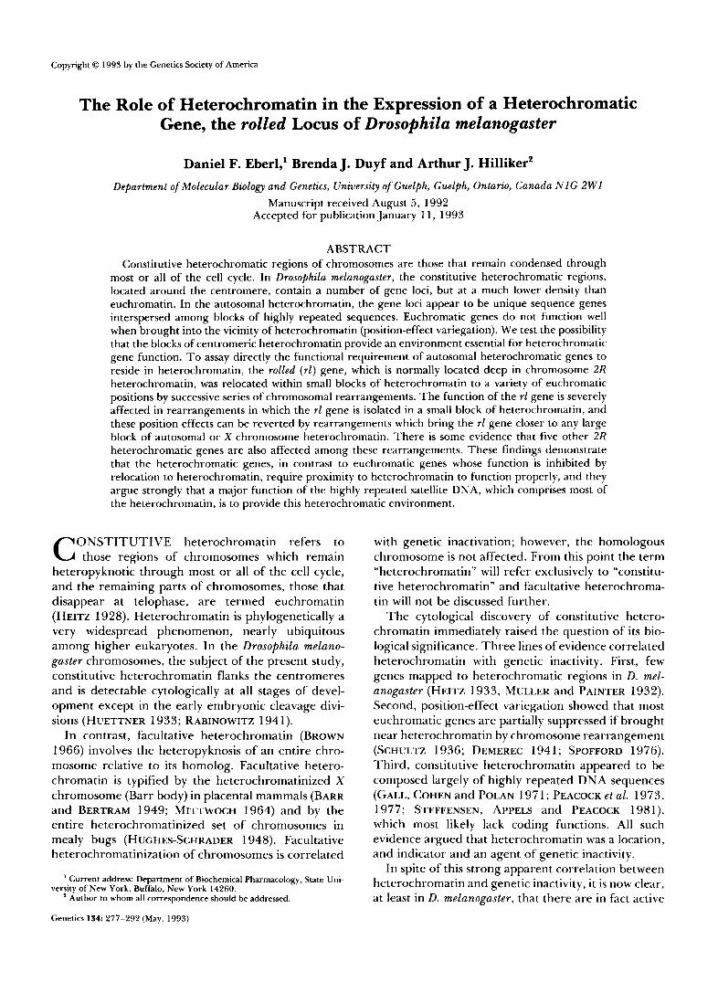
genes within heterochromatin, although these genes are present at a much lower density than genes in euchromatin [see HILLIKER and SHARP (1988) for a review]. The Y chromosome is completely heterochro- matic and carries a minimum of 12 genetic elements, including six male-fertility genes and an rDNA locus. The X chromosome heterochromatin (Xh) carries six genetic elements including the rDNA locus. There are 19 genes in chromosome 2 heterochromatin, as shown in Figure 1. Chromosome 3 heterochromatin carries at least 1 1 vital genes (MARCHANT and HOLM
How is it that some 50 genes are located in hetero- chromatin and are still able to function even though euchromatic genes cannot function when put into this environment? Are these heterochromatic genes able to function regardless of their location, or do they actually require a heterochromatic environment to function, as speculated by HILLIKER (1976)? This lat- ter question can be answered directly by assaying position effects on heterochromatic genes in chro- mosomal rearrangements which move these genes into euchromatic environments.
Position effects on heterochromatic genes have heretofore been most thoroughly studied on the light (It) locus, a gene in the heterochromatin of chromo- some arm 2L near the euchromatic boundary (HILLI- KER and HOLM 1975; DIMITRI 1991). Rearrangements which break anywhere between the It locus and the chromosome 2 centromere, and which relocate It and its associated heterochromatin to distal euchromatic positions, can show position effects on It (HESSLER 1958; HILLIKER 1980; WAKIMOTO and HEARN 1990). Evidence of position effects on other genes in 2L heterochromatin (2Lh) in rearrangements with posi- tion effects on It has also been found. SHARP (1988) saw viability effects on the group VI1 locus EMS 56-4 and on the group IX locus EMS 40-18 in translocations that variegate for It. WAKIMOTO and HEARN (1990) describe position effects on these genes and on the EMS 40-2, concertina (cta) and EMS 40-6 genes in 2Lh in It-variegating rearrangements. It is noteworthy that all these genes are located in distal heterochromatin (see Figure l) , close to the euchromatic boundary. This localization is based on detachment frequencies of compound autosomes (HILLIKER and HOLM 1975) and on cytological mapping (DIMITRI 1991). It has been suggested that the heterochromatin near the euchromatic boundary, P-heterochromatin, is quali- tatively different from the more proximal a-hetero- chromatin, which constitutes most of the mitotic het- erochromatin, and that It (and presumably also many of the other 2Lh genes) is located in P-heterochroma- tin (DEVLIN, BINGHAM and WAKIMOTO 1990; DEVLIN et al. 1990; WAKIMOTO and HEARN 1990). I t is pos- tulated that P-heterochromatin is quite unlike a-het-
1988).
A
2L rl 2R
Group IX Vlll VI1 VI I II Ill IV v
M(2R)MSZ lo
M(2R)PRF M(2R)PRB
M ( 2 R ) f J R ~ I
2Rh
' h38 h40 h43 h45
- I '1 AAGAG (42)
AAGAGAG (34) - AAGAC (1 98)
U rl
FIGURE 1 .-Genetic map of chromosome 2 heterochromatin. (A) The relative positions of the 14 vital genes (HILLIKER 1976), the two Segregation Distortion genes, Sdn and Rsp (GANETZKY 1977; SHARP, HILLIKER and HOLM 1985), and the two fertility genes (SHARP 1988; SCHUPBACH and WIESCHAUS 1989) are shown, based on the detachment frequencies of compound chromosomes (HIL- LIKER and HOLM 1975). The positions of a vital gene inferred from overlapping deletions (SHARP 1988) as well as the most proximal known euchromatic genes are also shown. The shaded regions denote heterochromatin, the thick solid lines are truncated repre- sentations of the euchromatic arms. The genes are grouped into intervals defined by deficiency breakpoints (HILLIKER and HOLM 1975; HILLIKER 1976). At the bottom, the genetic extents of 2Rh deficiencies used in complementation analysis are shown. (B) The positions of N bands (black blocks) in 2Rh are according to DIMITRI (1991). The hybridization sites of the cloned satellite DNA se- quences (clone identification numbers are in parentheses) and the positions of breakpoints of the original translocations relative to these satellites and to N bands were determined by A. J. HILLIKER and A. R. LOHE (unpublished). The r l gene is between the break- points of T(2;3)33 and T(2;3)127.
erochromatin: first, it replicates during polytene chro- mosome formation; second, it does not possess satellite (highly repeated) DNA sequences; third, it possesses a gene density comparable to that of euchromatin (whereas a-heterochromatin has a very low gene den- sity relative to euchromatin) (HEITZ 1934; GALL 1973; MIKLOS and COTSELL 1990).
N o position effects have been seen for those heter- ochromatic genes which are more proximal, deeper

Drosophila Autosomal Heterochromatin 279
in heterochromatin, even though several re- arrangements are known to break between these genes and their centromere. Many of the re- arrangements studied by WAKIMOTO and HEARN (1 990) are reported to have breaks proximal to EMS 40-5, the most proximal 2Lh gene, but fail to show a detectable position effect on this gene. SHARP (1 988) also found no position effects on EMS 40-5 among translocations that are known to have very proximal 2Lh breakpoints (A. J. HILLIKER and A. R. LOHE, unpublished observations). Furthermore, in testing many translocations with 2R heterochromatic breaks including some very proximal breaks, SHARP (1988) found no position effects on genes in 2R heterochro- matin (2Rh), which are fairly evenly dispersed in 2Rh (HILLIKER and HOLM 1975; DIMITRI 1991) rather than clustered near the euchromatic boundary as are the 2Lh genes. It is possible that the more proximal genes are somehow immune to position effects; that is, that they do not require a heterochromatic envi- ronment. Alternatively, it is possible that these genes are indeed subject to position effects but that these position effects are difficult to reveal.
Here we report studies designed to reveal position effects on the heterochromatic rolled gene ( r l ) . This gene was chosen for study for two reasons. First, it is located very deep in a-heterochromatin, near the middle of 2Rh as shown by detachment frequencies of compound autosomes (HILLIKER and HOLM 1975) or in the proximal third of 2Rh, specifically in the h40 or h41 blocks defined by cytological mapping (DIMI- TRI 1991) (see Figure 1). Second, the existence of both nonlethal mutations with visible phenotypes and lethal mutations makes it relatively easy to screen for both mutant and revertant flies in F1 rather than F2 screens. Since simple reciprocal translocations with a breakpoint proximal to rl do not produce detectable position effects on rl, it is possible that the large block of heterochromatin (most of 2Rh) which is translo- cated with rl provides a sufficient heterochromatic environment for function of the gene.
We hypothesized that position effects on rl can occur only if it is in a small block of heterochromatin isolated in distal euchromatin. This would require breaks on both sides of rl to excise it from the large heterochromatic block in addition to a t least one euchromatic break with which this fragment can as- sociate; such complex rearrangements with appropri- ate breakpoints would not be recovered at an appre- ciable frequency from mutagenesis of normal se- quence chromosomes (EBERL et al. 1989). However, if we start with translocations that already have a heterochromatic break near the rl gene, then only one additional heterochromatic break is required to isolate the gene in a small block of heterochromatin; this second step requires only a simple two-break
rearrangement. Thus, a complex rearrangement is constructed by a stepwise approach to sequester rl in a small block of heterochromatin some distance from another major block of heterochromatin. According to this model, position effects on rl in such re- arrangements can then be reverted by a further simple two-break rearrangement which places the small het- erochromatic block, containing the rl gene, in juxta- position to or close to a large heterochromatic block.
MATERIALS AND METHODS
Genetic strains: Heterochromatic deletions and lethal mutations used for complementation analysis were induced and characterized by HILLIKER and HOLM (1 975) and HIL- LIKER (1976) and are shown in Figure 1. The original translocations, T(2;3)33, T(2;3)127 and T(2;3)76, are de- scribed by HILLIKER and TRUSIS-COULTER (1987). Other mutations and stocks mentioned are described by LINDSLEY and GRELL (1968) and LINDSLEY and ZIMM (1992). All strains and crosses were maintained on a standard cornmeal- yeast-agar medium and were incubated at 25" unless oth- erwise indicated.
Cytology: Polytene chromosome preparations were made from larvae heterozygous for the rearrangement and cyto- logically normal chromosomes 2 and 3 derived from the red strain. Salivary glands were dissected in 45% acetic acid, transferred to a drop of aceto-lacto-orcein stain, squashed under a coverslip and examined using phase-contrast optics with a Zeiss Standard microscope. Breakpoints were deter- mined using the polytene-chromosome maps of C. B. and P. N . BRIDGES (BRIDGES and BRIDGES 1939; BRIDGES 1935, 1941a, b, 1942).
Diploid somatic chromosome preparations were made by dissecting brains from third-instar larvae in 0.7% sodium chloride and treating in one of two ways. For deciphering rearrangements, the brains were incubated in 0.5% sodium citrate for 15 min, then transferred into a drop of aceto- lacto-orcein stain and squashed after 5 min. This hypotonic treatment swells the neuroblasts and makes the chromo- somes appear longer, and somatic pairing is often main- tained. For visualizing discrete blocks of heterochromatin, the brains were incubated in 1% sodium citrate for 1 min before staining and squashing. This hypertonic treatment makes the chromosomes more compact and produces in- creased contrast between heterochromatic and euchromatic regions. These preparations were examined with phase- contrast optics.
Mutagenesis: To generate chromosomal rearrange- ments, males were mutagenized with y-rays. The flies were place in 8-dram glass vials (maximum of 50 per vial) con- taining standard medium and exposed to 2000 rad of y-rays from a 6oCo source at about 750 rad/min.
Complementation analysis: Crosses to test complemen- tation among lethals, deficiencies and new rearrangements were performed by placing three or four males of one balanced stock with three or four virgin females of the other in a vial at 25", discarding the parents after about 6 days and scoring the offspring after 16- 18 days.
RESULTS
Induction of position effects on the rl gene: Figure 1 shows the positions of heterochromatic breakpoints of translocations, relative to N bands and hybridiza-
280
A &'" 2000 rads y rays
D. F. Eberl, B. J. Duyf and A. J. Hilliker
T(2;3)' x Df(2L)PR3 1 . 2 b,k d; + 1 I SM1, It ' +
recognized by r/ phenotype
t balanced stock
B -. 2000 rads y rays
2 25"
T(2;3)' x Df(2L)PR31 . 2 L b k d ; + I I SM1,It ' +
recognized by d + phenotype
t balanced stock
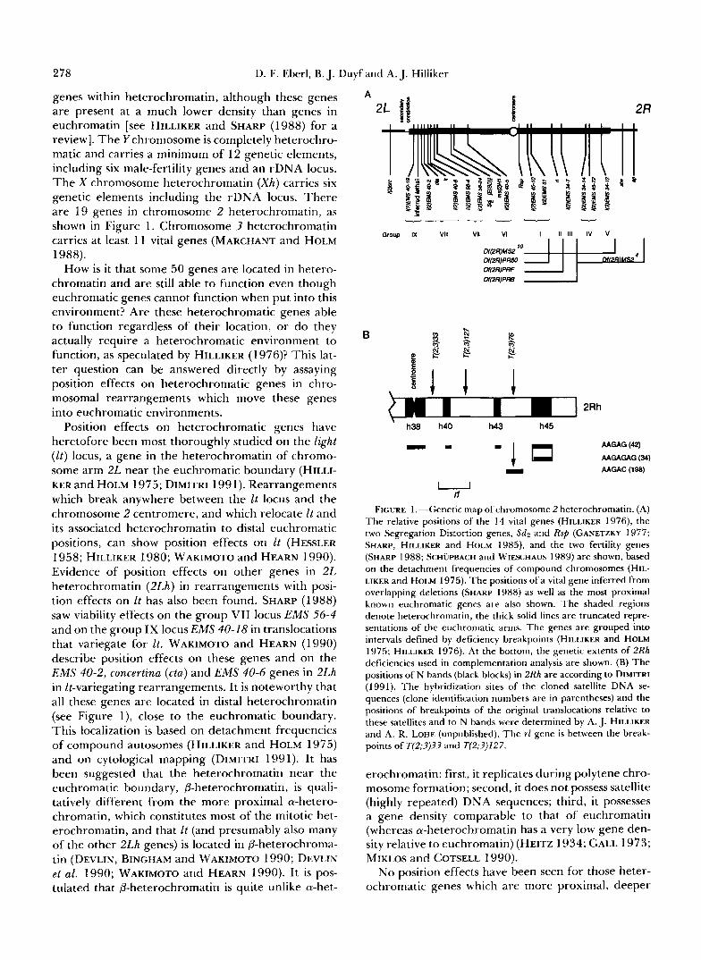
FIGURE 2.-Mating schemes for generating rl position effects and reversions. Crosses are shown for the generation and recovery of rearrangements in which the rl gene is position-affected (A) and rearrangements in which these position effects are reverted (B).
tion sites of cloned satellite DNA sequences, deter- mined by A. J. HILLIKER and A. R. LOHE (unpub- lished). The simple translocations, T(2;3)33, T(2;3)127 and T(2;3)76, have breakpoints at various locations in 2Rh. Since none of these shows a detectable position effect on rl, but all break the heterochromatin near r l , these simple translocations were used as starting points for further rearrangements. These three rl+ translocations will be designated the original translo- cations. By the cross outlined in Figure 2A, rl deriv- atives were selected from the progeny of irradiated flies carrying the original translocation. Each rl deriv- ative is called a first derivative and is numbered with a suffix after the number of the translocation from which it was derived. For example, first derivative number six derived from T(2;3)?3 is called T(2;3)33- 6.
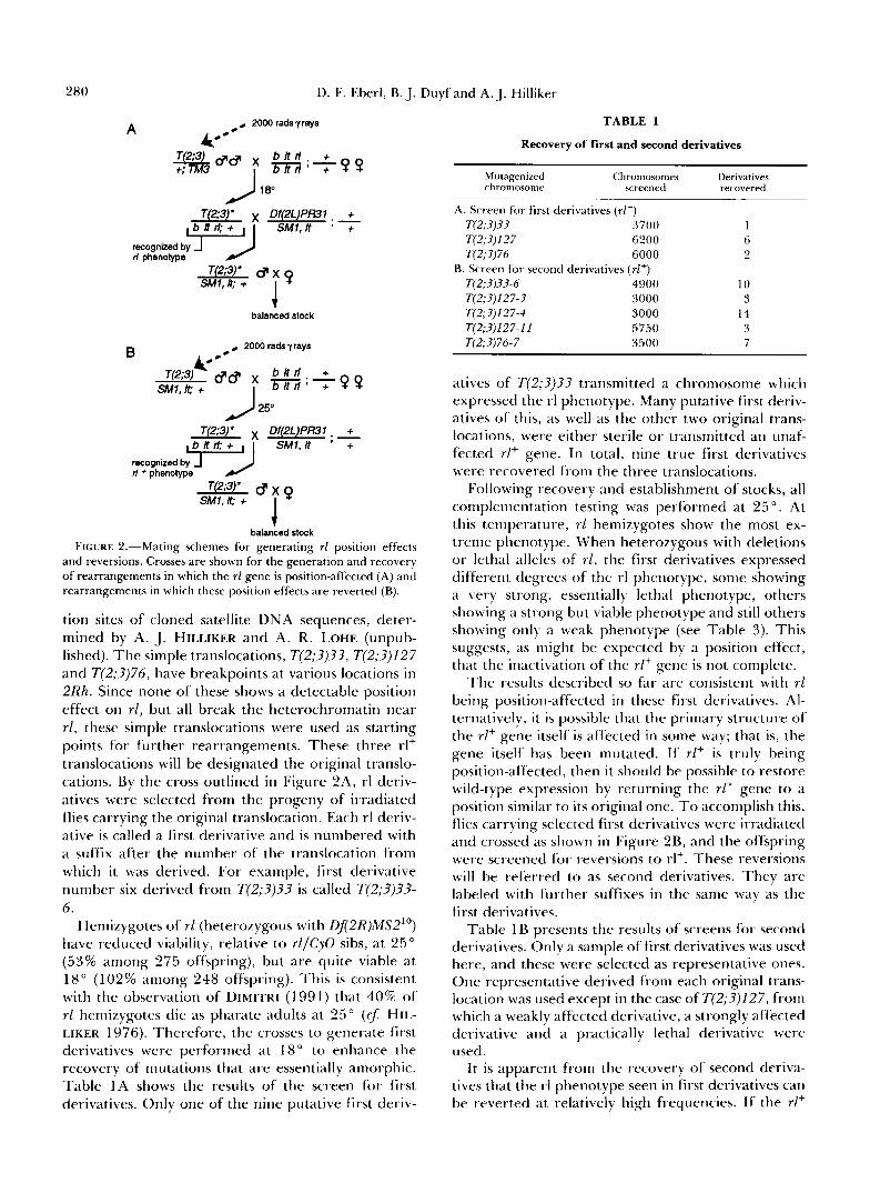
Henlizygotes of rl (heterozygous with Df2R)MS210) have reduced viability, relative to rl/CyO sibs, at 25" (53% among 275 offspring), but are quite viable at 18" (102% among 248 offspring). This is consistent with the observation of DIMITRI (1991) that 40% of rl hemizygotes die as pharate adults at 25" (CJ: HIL- LIKER 1976). Therefore, the crosses to generate first derivatives were performed at 18" to enhance the recovery of mutations that are essentially amorphic. Table 1A shows the results of the screen for first derivatives. Only one of the nine putative first deriv-
TABLE 1
Recovery of first and second derivatives
chromosome Mutagenized Chromosomes Derivatives
recovered screened
A. Screen for first derivatives ( r l - ) T(2;3)33 3700 1 T(2;3)127 6200 6 T(2;3)76 6000 2
T(2;3)33-6 4900 10 T(2;3)127-3 3000 3 T(2;3)127-4 3000 14 T(2;3)127-11 5750 3 T(2;3)76-7 3500 7
B. Screen for second derivatives (r l+)
atives of T(2;3)33 transmitted a chromosome which expressed the rl phenotype. Many putative first deriv- atives of this, as well as the other two original trans- locations, were either sterile or transmitted an unaf- fected rl+ gene. In total, nine true first derivatives were recovered from the three translocations.
Following recovery and establishment of stocks, all complementation testing was performed at 25 ". At this temperature, rl hemizygotes show the most ex- treme phenotype. When heterozygous with deletions or lethal alleles of rl, the first derivatives expressed different degrees of the rl phenotype, some showing a very strong, essentially lethal phenotype, others showing a strong but viable phenotype and still others showing only a weak phenotype (see Table 3). This suggests, as might be expected by a position effect, that the inactivation of the rlf gene is not complete.
The results described so far are consistent with rl being position-affected in these first derivatives. Al- ternatively, it is possible that the primary structure of the rl+ gene itself is affected in some way; that is, the gene itself has been mutated. If rl+ is truly being position-affected, then it should be possible to restore wild-type expression by returning the rlf gene to a position similar to its original one. To accomplish this, flies carrying selected first derivatives were irradiated and crossed as shown in Figure 2B, and the offspring were screened for reversions to rl+. These reversions will be referred to as second derivatives. They are labeled with further suffixes i n the same way as the first derivatives.
Table 1 B presents the results of screens for second derivatives. Only a sample of first derivatives was used here, and these were selected as representative ones. One representative derived from each original trans- location was used except in the case of T(2;?)127, from which a weakly affected derivative, a strongly affected derivative and a practically lethal derivative were used.
I t is apparent from the recovery of second dcriva- tives that the rl phenotype seen in first derivatives can be reverted at relatively high frequencies. If the rl+
Drosophila Autosomal Heterochromatin 28 1
gene had been mutated, a very specific reversion mutation would be required to restore function to the gene; this would be a very rare event. A position effect, however, should be readily reverted since any breakpoint sufficiently close to the rl+ gene has the potential to allow adequate rl+ function if the resulting rearrangement places the rl+ gene into or close to heterochromatin. Such rearrangements are expected to occur relatively frequently. The ease of recovery of rl+ revertants on our study favored the position- effect interpretation for these derivatives; this inter- pretation was then confirmed by cytological analysis.
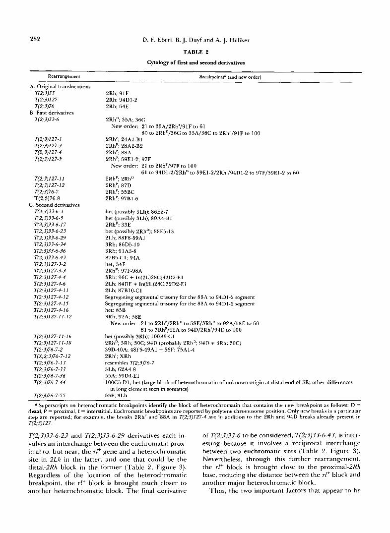
Cytological analysis of first (rl) and second (rl+) derivatives: The chromosomes of each of the nine first derivatives and most of the second derivatives were examined in heterozygous condition, both in salivary-gland polytene nuclei and in diploid brain-cell (somatic) nuclei. The results of this analysis are pre- sented in Table 2. It can be seen that, with few exceptions, the derivatives examined are associated with an additional chromosomal rearrangement, al- most always involving one heterochromatic and one euchromatic breakpoint. I t should be emphasized that in the polytene chromosomes the euchromatic break- points can be determined with great precision, but for the heterochromatic breakpoints this usually is not possible. However, the heterochromatic breakpoint can often be ascertained in somatic-chromosome prep- arations, though with relatively coarse resolution. Nevertheless, the complexity of some of the re- arrangements can hamper the interpretation of the breakpoints, and any consequent uncertainty is in- cluded in the listing of breakpoints in Table 2. Finally, a diagrammatical representation of each of the re- arrangements that has been elucidated is shown in Figures 3-5. These are drawn to scale, and in the cases of uncertainty listed in Table 2, the most likely con- figuration is given.
The individual rearrangements will now be dis- cussed in detail, beginning with the derivatives of T(2;3)33. T(2;3)33 itself was formed by a reciprocal interchange between 2Rh, at a position between the centromeric N band (h38) and the adjacent one (h40) (Figure l) , and the middle of the 3R euchromatic arm at polytene-chromosome location 91F (Table 2, Fig- ure 3). Since the rl+ gene lies in h40 or h41 (DIMITRI 199 l), it is moved in a large block of heterochromatin to a position almost 11 polytene sections from the nearest large heterochromatic region, namely 3Rh. Although the heterochromatin is removed from one side of the rl+ gene, this rearrangement does not produce a detectable rl phenotype. From this trans- location, we recovered only one first derivative that gives a rl phenotype, T(2;3)33-6. T(2;3)33-6 is derived from T(2;?)33 by a reciprocal interchange between the mid-proximal region of euchromatic 2L and the
distal block of 2Rh which contains the rl+ gene (Table 2, Figure 3). If this heterochromatic breakpoint is distal to the rl+ gene, then this rearrangement causes much of the heterochromatin to be removed from the other side of the rl+ gene (relative to the side of the gene from which the heterochromatin is stripped by the original translocation), leaving the gene stranded in a small block of heterochromatin in a euchromatic environment. In this arrangement, the rl+ gene does not function properly, as indicated by the visible rl phenotype.
Eight of the 10 second derivatives of T(2;3)33 that are derived from T(2;3)33-6 were examined cytologi- cally (Table 2), and seven were found to have a new breakpoint in the euchromatin of polytene sections 86-91, just proximal to the interstitial-2Rh block which contains the rl+ gene. The remaining re- arrangement has a new breakpoint just distal to this interstitiaL2Rh block, in polytene section 33. There- fore, each of the eight involves a breakpoint near the stranded rl+ gene in T(2;3)33-6 (Figure 3). This con- centration of breakpoints is strong confirmation that the rl+ gene is actually located in this interstitial block of 2Rh in T(2;3)33-6. Furthermore, it demonstrates that the rl phenotype seen in T(2;3)33-6 is really a position effect in that it can be reverted by re- arrangements with breakpoints near, but not in, the rl+ gene. It therefore constitutes proof that the rl+ gene has been affected by its position in the chromo- somes rather than by an alteration in the gene per se. Consider first the derivatives T(2;3)33-6-3, T(2;3)33- 6-5, T(2;3)33-6-34 and T(2;3)33-6-36. The first two are most likely pericentric inversions, superimposed on T(2;3)33-6, which result in the relocation of a large block of heterochromatin including proximal 3Lh and all of ?Rh closer to the rl+ block (Table 2, Figure 3); the latter two are paracentric inversions which move a block of 3Rh closer to the rl+ block. These deriva- tives revert the rl phenotype to rl+, indicating that the distance of the stranded rl+ block from another large heterochromatic block is an important factor in the position effects on the rl' gene. Next, consider the T(2;3)33-6-17 derivative. It is an approximate rever- sion of the T(2;3)33-6 interchange, so it returns much of distal 2Rh to close to its previous position distal to the rl+ gene, with only a small intervening segment of euchromatin (Table 2, Figure 3). This is a very inter- esting rearrangement since the rl+ block has recruited an additional heterochromatic block in a centromere- distal position, leaving the gene-centromere distance unaltered while increasing the local content of heter- ochromatin. The paracentric inversions T(2;3)33-6-34 and T(2;3)33-6-36 also leave the gene-centromere dis- tance unaltered. Therefore, the distance effect is not a centromere-distance effect, but rather an effect of distance from a major heterochromatic block. The
282 D. F. Eberl, B. J. Duyf and A. J. Hilliker
TABLE 2
Cytology of first and second derivatives
Rearrangement Breakpoints" (and new order)
A. Original translocations T(2;?)?? T(2;?) 1 2 7 T(2;?)76
B. First derivatives T(2;?)3?-6
T(2;?)127-1 T(2;?)127-? T(2;?)127-4 T(2; 3) I 2 7-5
T(2;?)127-11 T(2;?) 1 2 7- 1 2
T(2;3)76-8
T(2;?)??-6-?
T(2;?)76-7
C. Second derivatives
T(2;?)?3-6-5 T(2;?)??-6-17 T(2;?)3?-6-2? T(2;3)3?-6-29 T(2;?)??-6-?4 T(2;?)??-6-?6 T(2;3)??-6-4? T(2;?)127-3-2 T(2;3)127-?-? T(2;3)127-4-4 T(2;3)127-4-6
T(2;?)127-4-12
T(2;?)127-4-16
T(2;?)127-4-11
T(2;?)127-4-15
T(2;?)127-11-12
T(2;?)127-11-16 T(2;?)127-11-18 T(2;3)76-7-2
T(2;?)76-7-13 T(X;2;?)76-7-12
T(2;?)76-7-3? T(2;?)76-7-?6 T(2;?)76-7-44
T(2;?)76-7-55
2Rh; 91F 2Rh; 94Dl-2 2Rh; 64E
2RhD; 35A; 36C New order: 2 1 to 35A/2Rh1/9 1 F to 6 1
60 to 2RhD/36C to 35A/36C to 2RhP/91F to 100 2RhP; 24AI-Bl 2RhP; 28A2-B2 2RhP; 88A 2RhP; 59E1-2; 97F
New order: 21 to 2RhP/97F to 100 61 to 94D1-2/2RhD to 59E1-2/2Rh1/94D1-2 to 97F/59E1-2 to 60
2RhP; 2RhD 2RhP; 87D 2RhP; 55BC 2RhP; 97B1-6
het (possibly 3Lh); 86E2-7 het (possibly 3Lh); 89A4-Bl 2RhD; 33E het (possibly 2RhD); 88E5-13 2Lh; 88F8-89A1 3Rh; 86D5-10 3Rh; 9 1 A3-8
het; 34F 2RhD; 97F-98A 3Rh; 96C + In(2L)28C;32D2-E1 2Lh; 84DE + In(2L)28C;32D2-E1 2Lh; 87BlO-Cl Segregating segmental trisomy for the 88A to 94D1-2 segment Segregating segmental trisomy for the 88A to 94D1-2 segment het; 85B 3Rh; 92A; 58E
87B5-C 1; 94A
New order: 21 to 2RhP/2RhD to 58E/3RhD to 92A/58E to 60 61 to 3RhP/92A to 94D/2Rh1/94D to 100
het (possibly 3Rh); 100B5-Cl 2Rh'; 3Rh; 30C; 94D (probably 2RhD; 94D + 3Rh; 30C) 39D-40A; 48F5-49A1 + 56F; 75A1-4 2Rh'; XRh resembles T(2;?)76-7 3Lh; 62A4-9 55A; 59D4-El 100C5-DI; het (large block of heterochromatin of unknown origin at distal end of 3R; other differences
53F; 3Lh in long element seen in somatics)
" Superscripts on heterochromatic breakpoints identify the block of heterochromatin that contains the new breakpoint as follows: D = distal, P = proximal, I = interstitial. Euchromatic breakpoints are reported by polytene chromosome position. Only new breaks in a particular step are reported; for example, the breaks 2RhP and 88A in T(2;?)127-4 are in addition to the 2Rh and 94D breaks already present in T(2;?) I2 7 .
T(2;?)??-6-23 and T(2;3)33-6-29 derivatives each in- volves an interchange between the euchromatin prox- imal to, but near, the rZ+ gene and a heterochromatic site in 2Lh in the latter, and one that could be the distal-2Rh block in the former (Table 2, Figure 3). Regardless of the location of the heterochromatic breakpoint, the rl+ block is brought much closer to another heterochromatic block. The final derivative
of T(2;?)33-6 to be considered, T(2;3)?3-6-43, is inter- esting because it involves a reciprocal interchange between two euchromatic sites (Table 2, Figure 3). Nevertheless, through this further rearrangement, the rl+ block is brought close to the proximal-2Rh base, reducing the distance between the rl+ block and another major heterochromatic block.
Thus, the two important factors that appear to be
Drosophila Autosomal Heterochromatin
,,,Zp.- * . .*.. + 0 2
lsssnx.... t?
+ 10.9
283
+
T(2;3)33
T(2;3)33-6
T(2;3p+-3
T(2;3)33-55
T(2:3)33-& 17
T(2:3)336-23
T(2;3)33-529
T(2;3)33-&34
T(2;3)33-536
T(2:3)33-643
*,. ._ .... - I intannediale 10.9
5.2
2.7
1.5
2.8
3.0
5.3
0.7
6.8
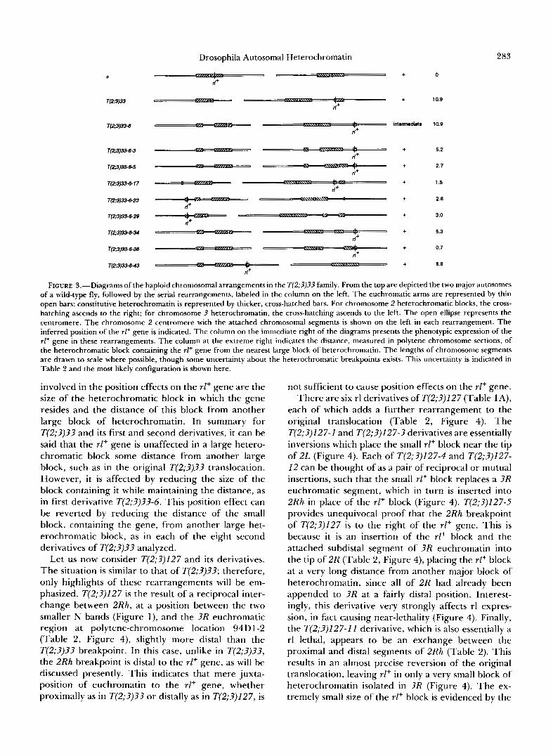
FIGURE 3,”Diagrams of the haploid chromosomal arrangements in the T(2;3)33 family. From the top are depicted the two major autosomes of a wild-type fly, followed by the serial rearrangements, labeled in the column on the left. The euchromatic arms are represented by thin open bars; constitutive heterochromatin is represented by thicker, cross-hatched bars. For chromosome 2 heterochromatic blocks, the cross- hatching ascends to the right; for chromosome 3 heterochromatin, the cross-hatching ascends to the left. The open ellipse represents the centromere. The chromosome 2 centromere with the attached chromosomal segments is shown on the left in each rearrangement. The inferred position of the rl+ gene is indicated. The column on the immediate right of the diagrams presents the phenotypic expression of the rl+ gene in these rearrangements. The column at the extreme right indicates the distance, measured in polytene chromosome sections, of the heterochromatic block containing the rl+ gene from the nearest large block of heterochromatin. The lengths of chromosome segments are drawn to scale where possible, though some uncertainty about the heterochromatic breakpoints exists. This uncertainty is indicated in
v
Table 2 and the most likely configuration is shown here.
involved in the position effects on the rl+ gene are the size of the heterochromatic block in which the gene resides and the distance of this block from another large block of heterochromatin. In summary for T(2;3)33 and its first and second derivatives, it can be said that the rl+ gene is unaffected in a large hetero- chromatic block some distance from another large block, such as in the original T(2;3)33 translocation. However, it is affected by reducing the size of the block containing it while maintaining the distance, as in first derivative T(2;3)3?-6. This position effect can be reverted by reducing the distance of the small block, containing the gene, from another large het- erochromatic block, as in each of the eight second derivatives of T(2;3)3? analyzed.
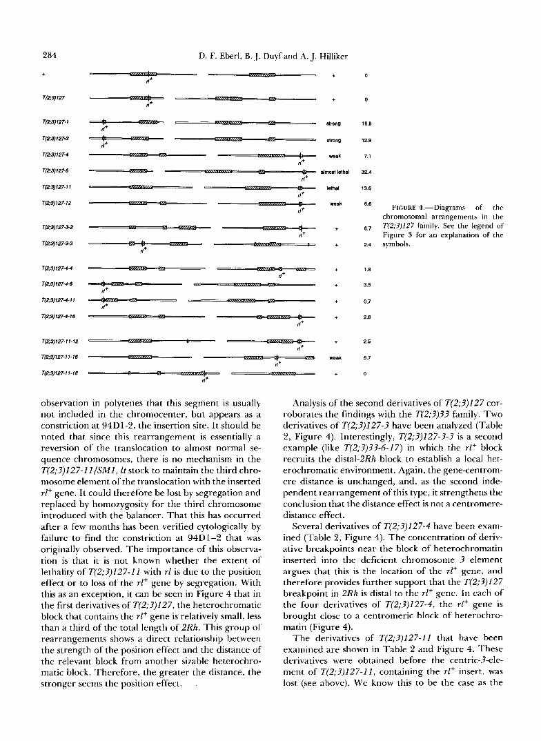
Let us now consider T(2;3)127 and its derivatives. The situation is similar to that of T(2;3)33; therefore, only highlights of these rearrangements will be em- phasized. T(2;3)127 is the result of a reciprocal inter- change between 2Rh, at a position between the two smaller N bands (Figure l), and the ?R euchromatic region at polytene-chromosome location 94D1-2 (Table 2, Figure 4), slightly more distal than the T(2;3)3? breakpoint. In this case, unlike in T(2;?)33, the 2Rh breakpoint is distal to the rl+ gene, as will be discussed presently. This indicates that mere juxta- position of euchromatin to the rl+ gene, whether proximally as in T(2;3)33 or distally as in T(2;?)127, is
not sufficient to cause position effects on the rl+ gene. There are six rl derivatives of T(2;3)127 (Table 1 A),
each of which adds a further rearrangement to the original translocation (Table 2, Figure 4). The T(2;3)127-1 and T(2;3)127-3 derivatives are essentially inversions which place the small rl+ block near the tip of 2L (Figure 4). Each of T(2;3)127-4 and T(2;3)127- 12 can be thought of as a pair of reciprocal or mutual insertions, such that the small rl+ block replaces a 3R euchromatic segment, which in turn is inserted into 2Rh in place of the rl+ block (Figure 4). T(2;3)127-5 provides unequivocal proof that the 2Rh breakpoint of T(2;?)127 is to the right of the rl+ gene. This is because it is an insertion of the rl+ block and the attached subdistal segment of ?R euchromatin into the tip of 2R (Table 2, Figure 4), placing the rl+ block at a very long distance from another major block of heterochromatin, since all of 2R had already been appended to ?R at a fairly distal position. Interest- ingly, this derivative very strongly affects rl expres- sion, in fact causing near-lethality (Figure 4). Finally, the T(2;3)127-1 I derivative, which is also essentially a rl lethal, appears to be an exchange between the proximal and distal segments of 2Rh (Table 2). This results in an almost precise reversion of the original translocation, leaving rl+ in only a very small block of heterochromatin isolated in ?R (Figure 4). The ex- tremely small size of the rl+ block is evidenced by the
284
+
T(2;3) 127
T(2;3) 127-1
T(2;3)127-3
T(Z3)127-4
T(23)127-5
T(2;3) 127- 1 1
T(2;3)127-12
T(2:3) 127-3-2
T(2;3)127-3-3
T(2;3)127-4-4
T(2;3)127-4-6
T(2:3)127-4-11
T(2:3)127-4-16
T(2;3)127-11-12
T(2;3) 127- 1 1 - 16
T(2;3)127-11-16
D. F. Eberl, B. J. Duyf and A. J. Hilliker
a%?m+- rl+
+
+
strong
strong
weak
almost lethal
lethal
weak
+
+
+
+
+
+
+
weak
+
0
0
16.9
12.9
7.1
32.4
13.6
6.6
6.7
2.4
1.8
3.5
0.7
2.8
2.5
5.7
0
FIGURE 4.-Diagrams of the chromosomal arrangements in the T(2;3)127 family. See the legend of Figure 3 for an explanation of the symbols.
observation in polytenes that this segment is usually not included in the chromocenter, but appears as a constriction at 94D1-2, the insertion site. It should be noted that since this rearrangement is essentially a reversion of the translocation to almost normal se- quence chromosomes, there is no mechanism in the T(2;?)127-II/SMI, It stock to maintain the third chro- mosome element of the translocation with the inserted rl+ gene. It could therefore be lost by segregation and replaced by homozygosity for the third chromosome introduced with the balancer. That this has occurred after a few months has been verified cytologically by failure to find the constriction at 94D1-2 that was originally observed. The importance of this observa- tion is that it is not known whether the extent of lethality of T(2;?)127-11 with rl is due to the position effect or to loss of the rl+ gene by segregation. With this as an exception, it can be seen in Figure 4 that in the first derivatives of T(2;?)127, the heterochromatic block that contains the rl+ gene is relatively small, less than a third of the total length of 2Rh. This group of rearrangements shows a direct relationship between the strength of the position effect and the distance of the relevant block from another sizable heterochro- matic block. Therefore, the greater the distance, the stronger seems the position effect.
Analysis of the second derivatives of T(2;?)127 cor- roborates the findings with the T(2;?)?3 family. Two derivatives of T(2;?)127-? have been analyzed (Table 2, Figure 4). Interestingly, T(2;3)127-?-? is a second example (like T(2;?)??-6-17) in which the rl+ block recruits the distal-2Rh block to establish a local het- erochromatic environment. Again, the gene-centrom- ere distance is unchanged, and, as the second inde- pendent rearrangement of this type, it strengthens the conclusion that the distance effect is not a centromere- distance effect.
Several derivatives of T(2;?)127-4 have been exam- ined (Table 2, Figure 4). The concentration of deriv- ative breakpoints near the block of heterochromatin inserted into the deficient chromosome 3 element argues that this is the location of the rl+ gene, and therefore provides further support that the T(2;?)127 breakpoint in 2Rh is distal to the rl+ gene. In each of the four derivatives of T(2;?)127-4, the rl+ gene is brought close to a centromeric block of heterochro- matin (Figure 4).
The derivatives of T(2;?)127-11 that have been examined are shown in Table 2 and Figure 4. These derivatives were obtained before the centric-?-ele- ment of T(2;3)127-11, containing the rl+ insert, was lost (see above). We know this to be the case as the
Drosophila Autosomal Heterochromatin 0 + +
d+
285
T(2;3)78 ” + 0 d+
n FIGURE 5.-Diagrams of the chromosomal arrangements in the ,
.I+
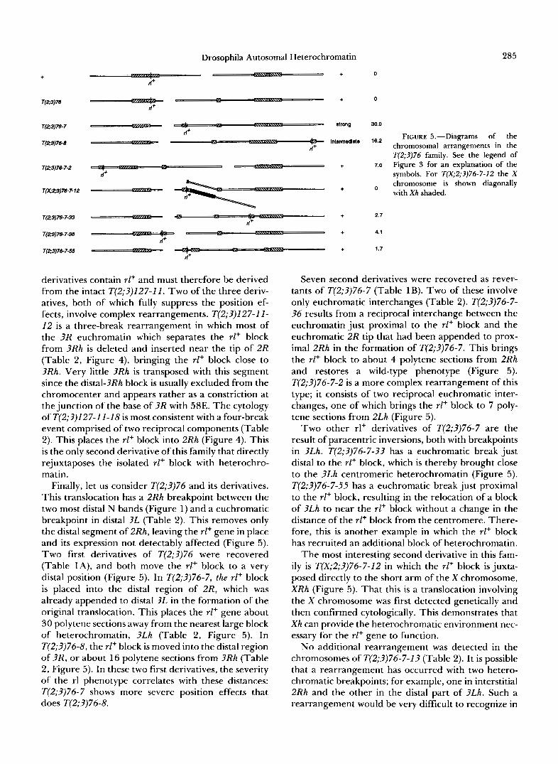
. intennedids 16.2 - T(2;3)76 family. See the legend of
~(23)m-7-2 .,-x..= + 7.0 Figure 3 for an explanation of the symbols. For T(X;2;3)76-7-12 the X
Tm2;3)78-7- 12 L
chromosome is shown diagonally with X h shaded.
, .-,, T(23)78-7-33 . %~ + 2.7
.e. + 4.1
2 T(23)78-7-38 -=“F -=
T(23)7&7-55 - + 1.7
derivatives contain rl+ and must therefore be derived from the intact T(2;3)127-11. Two of the three deriv- atives, both of which fully suppress the position ef- fects, involve complex rearrangements. T(2;3)127-11- 12 is a three-break rearrangement in which most of the 3R euchromatin which separates the rl+ block from 3Rh is deleted and inserted near the tip of 2R (Table 2, Figure 4), bringing the rl+ block close to 3Rh. Very little 3Rh is transposed with this segment since the distal-3Rh block is usually excluded from the chromocenter and appears rather as a constriction at the junction of the base of 3R with 58E. The cytology of T(2;3)127-11-18 is most consistent with a four-break event comprised of two reciprocal components (Table 2). This places the rl+ block into 2Rh (Figure 4). This is the only second derivative of this family that directly rejuxtaposes the isolated rl+ block with heterochro- matin.
Finally, let us consider T(2;3)76 and its derivatives. This translocation has a 2Rh breakpoint between the two most distal N bands (Figure 1) and a euchromatic breakpoint in distal 3L (Table 2). This removes only the distal segment of 2Rh, leaving the rl+ gene in place and its expression not detectably affected (Figure 5). Two first derivatives of T(2;3)76 were recovered (Table IA), and both move the rl+ block to a very distal position (Figure 5 ) . In T(2;3)76-7, the rl+ block is placed into the distal region of 2R, which was already appended to distal 3L in the formation of the original translocation. This places the rl+ gene about 30 polytene sections away from the nearest large block of heterochromatin, 3Lh (Table 2, Figure 5 ) . In T(2;3)76-8, the rl+ block is moved into the distal region of 3R, or about 16 polytene sections from 3Rh (Table 2, Figure 5) . In these two first derivatives, the severity of the rl phenotype correlates with these distances: T(2;3)76-7 shows more severe position effects that does T(2;3)76-8.
Seven second derivatives were recovered as rever- tants of T(2;3)76-7 (Table 1B). Two of these involve only euchromatic interchanges (Table 2). T(2;3)76-7- 36 results from a reciprocal interchange between the euchromatin just proximal to the rl+ block and the euchromatic 2R tip that had been appended to prox- imal 2Rh in the formation of T(2;3)76-7. This brings the rl+ block to about 4 polytene sections from 2Rh and restores a wild-type phenotype (Figure 5) . T(2;3)76-7-2 is a more complex rearrangement of this type; it consists of two reciprocal euchromatic inter- changes, one of which brings the rE+ block to 7 poly- tene sections from 2Lh (Figure 5 ) .
Two other rl+ derivatives of T(2;3)76-7 are the result of paracentric inversions, both with breakpoints in 3Lh. T(2;3)76-7-33 has a euchromatic break just distal to the rl+ block, which is thereby brought close to the 3Lh centromeric heterochromatin (Figure 5) . T(2;3)76-7-55 has a euchromatic break just proximal to the rl+ block, resulting in the relocation of a block of 3Lh to near the rl+ block without a change in the distance of the rl+ block from the centromere. There- fore, this is another example in which the rl+ block has recruited an additional block of heterochromatin.
The most interesting second derivative in this fam- ily is T(X;2;3)76-7-12 in which the rl+ block is juxta- posed directly to the short arm of the X chromosome, XRh (Figure 5). That this is a translocation involving the X chromosome was first detected genetically and then confirmed cytologically. This demonstrates that X h can provide the heterochromatic environment nec- essary for the rl+ gene to function.
No additional rearrangement was detected in the chromosomes of T(2;3)76-7-13 (Table 2). It is possible that a rearrangement has occurred with two hetero- chromatic breakpoints; for example, one in interstitial 2Rh and the other in the distal part of 3Lh. Such a rearrangement would be very difficult to recognize in
286 D. F. Eberl, B. J. Duyf and A. J. Hilliker
these chromosome preparations. Finally, T(2;3)76-7- 44 is a rearrangement with euchromatic and hetero- chromatic breakpoints (Table 2), but the nature of the rearrangement remains enigmatic.
This examination of the individual derivatives has shown that the distance between the heterochromatic block containing the rl+ gene and another large block of heterochromatin is a major factor in the production of position effects on the rl+ gene and that the rl+ block should be small. The discrepancy in absolute distance relative to the strength of the rl phenotype is most likely explained by differences in rl+ block size. Compare, for example, the T(2;3)76 derivatives with the T(2;3)127 derivatives with equivalent distances (Figures 4 and 5). The rl+ block is almost certainly larger in the T(2;3)76 derivatives because the original 2Rh breakpoint is more distal than that of T(2;3)127 (Figure 1) and because the second 2Rh breakpoint is required to be proximal to the rl+ gene in both cases. As summarized in Figures 4 and 5, T(2;3)76-7 differs from T(2;3)127-5 in having a larger rl+ block, but the block is roughly the same distance from another major heterochromatic block. T(2;3)127-5 shows a stronger rl phenotype, suggesting that a smaller block increases the position effect. The same is true of T(2;3)76-8 relative to T(2;3)127-1 and T(2;3))127-3 (Figures 4 and 5). Therefore, a generalization of these observations is that if the rl+ gene is in a larger heterochromatic block, then it needs to be farther from another major block to have the same magnitude of position effect. This raises the question of whether a rearrangement with only one break in 2Rh proximal to the rl+ locus could produce a position effect on rl+ if the large block is moved far enough away. No informative rearrangements exist, although it may be possible to derive one from T(2;3)33. It is probably not significant that none was recovered among the first derivatives of this translocation, first because of the small size of the experiment, but more importantly because a sim- ple rearrangement would make the relevant distance a maximum of about 30 polytene sections-perhaps not sufficient to produce a detectable effect. Perhaps a complex event which moved it 40 or 50 polytene sections away would be sufficient.
A third parameter, the identity of the euchromatic regions that separate the relevant heterochromatic blocks, has so far not been explicitly mentioned be- cause it does not have a major effect. This conclusion arises from the observation that any of the four major autosomal arms can be involved. In T(2;3)76-7, 3L and 2R are involved; in T(2;3)127-5, 3R and 2R are involved; T(2;3)127-1 and T(2;3)127-3 involve 2L; the remaining four first derivatives involve 3R (Table 2).
Consider now the characteristics of rearrangements which revert the position effects on the rl+ gene. As seen in Figures 3-5, the revertants invariably reduce
the distance of the heterochromatic block containing the rl+ gene from another major heterochromatic block. In one case, T(2;3)127-11-18, the rl+ block appears to be brought into direct juxtaposition with the original 2Rh (Figure 4). In another case, T(X;2;3)76-7-12, the rl+ block is juxtaposed directly with XRh (Figure 5) . In every other second derivative analyzed there is still some euchromatin in between.
What can be said about the length of this euchro- matin? In the first derivatives, it appears that about 7 polytene sections separating the blocks are required to produce at least a weak phenotype if the rl+ gene is in a small block (Figures 3-5). All second derivatives which have eliminated the position effect have re- duced the intervening distance to less than 7 polytene sections. While it is possible that there is a threshold of this kind, the fact that rl+ block size is also involved warns that the threshold is probably not absolute. T(2;3)127-11-16 is an example of a second derivative which does bring the rl+ block from about 13 to well within 7 polytene sections of another major hetero- chromatic block, yet fails to completely revert the position effect (Figure 4). A sister derivative, T(2;3)127-11-12, which reduces the distance to 2.5 polytene sections, does completely revert it (Figure 4). Another example with comparable distances is T(2;3)127-3 and its two derivatives. Here, even though the distances are almost identical to the T(2;3)127-1 I situation, both derivatives fully revert the position effect. While the trend is the same, the absolute values are different. Therefore, there are probably differ- ences between the sizes of blocks of heterochromatin, though these differences cannot be resolved in the somatic-chromosome preparations used. It has previ- ously been mentioned that the size of the rl+ block in T(2;3)127-11 and its derivatives is likely smaller than that in T(2;3)127-3 and its derivatives because in po- lytene nuclei of the T(2;3)127-3 family the rl+ block fuses with the chromocenter, while in the T(2;3)127- I I family it is usually free of the chromocenter and visible as a constriction. Similarly, the position effect in T(2;3)76-7-2, with a medium-sized rl+ block at 7 polytene sections from another large block, is fully reverted even though the distance is equivalent to those in T(2;3)127-4 and T(2;3)127-12.
An additional characteristic of the reversion events relates to the identity of the large heterochromatic block. There appears to be no requirement for prox- imity specifically to 2Rh. Rearrangements such as T(2;3)127-4-4 and T(2;3)127-11-12 clearly combine the rl+ block with 3Rh (Table 2, Figure 4). Similarly, T(2;3)127-4-6 and T(2;3)127-4-1 I appear to involve 2Lh. Furthermore, 3Lh can also provide a functional environment for the rl+ gene, as seen in T(2;3)76-7- 33 and T(2;3)76-7-55 (Table 2 , Figure 5). Finally, T(X;2;3)76-7-12 is a rearrangement involving the short
Drosophila Autosomal Heterochromatin 287
arm of the X chromosome. Interestingly, none of the second derivatives has been associated with a Y chro- mosome break. The reason for this may be that the Y chromosome heterochromatin is not sufficiently sim- ilar to autosomal heterochromatin to allow rl+ to function. Alternatively, the lack of Y chromosome rearrangements may be due to the disruption of fer- tility factors in the Y chromosome. Thus, many of the sterile second derivatives may represent Y chromo- some-associated reversions which are lost because their recovery relies on functional integrity of the Y chromosome. The only remaining heterochromatin, on chromosome 4 , though apparently not involved in any of the derivatives studied so far, cannot be ruled out at this point. In summary, the heterochromatic regions of at least 2L, 2R, 3L, 3R and X are effective in restoring rl+ function. Y and fourth chromosome heterochromatic regions have also not been ruled out.
These studies have shown that rl+ can indeed be position-affected in complex rearrangements which place the rl+ gene in a small block of heterochromatin that is some distance from another major heterochro- matic region. The magnitude of the position effect is directly related to the distance of the block containing rl+ from another large block of heterochromatin, and is inversely related to the size of the rl+ block. There is no evidence that the identity of the euchromatic region influences the position effect. The position effects can be reverted by moving the rl+ block close to any large block of heterochromatin that is derived from the major autosomes or from the X chromosome. The position effects are clearly independent of cen- tromere position.
Position effects on other 2R heterochromatic genes: These studies which characterize the position effects on the rl gene have produced a large number of rearrangements with breaks at many sites in 2Rh, rearrangements that subdivide and separate 2Rh into a variety of smaller fragments scattered throughout the genome. This collection of rearrangements pro- vides an excellent opportunity to test for position effects on other genes in 2Rh. Accordingly, comple- mentation crosses were performed between these re- arrangements and the representative 2Rh mutations, including deficiencies and lethals (Figure 1). The re- sults of this analysis are summarized in Table 3.
Evidence of position effects at the two group I vital genes can be seen in T(2;3)127-11 and its derivatives (Table 3). These observations must be examined cau- tiously because one element of the translocation has been lost by segregation, as already discussed. Lethal- ity of T(2;3)127-1 I with EMS 31 indicates that loss of this element very likely deleted the EMS 31 gene. However, the chromosomal arrangement, even be- fore this element was lost, may have shown a position effect on this gene because one of the second deriva-
tives, T(2;3)127-11-16, which shows a partial reversion of the rl position effect, does not fully restore the EMS 31 gene to wild-type expression (Table 3). The EMS 45-10 gene, conversely, is not deleted with the loss of the one element of T(2;3)127-11 because some heterozygotes do survive (Table 3), indicating partial function of the gene. That this semi-lethality is attrib- utable to a position effect, rather than to a hypo- morphic mutation of the gene itself, is supported by its reversion in the three second derivatives.
Aside from those on the rl gene, the most obvious effects of the rearrangements in this study are on the group 111 gene, EMS 34-7. A general observation is that this gene is quite easily position-affected, and the position effects are very difficult to revert. Such dif- ferences in sensitivity to position effects among het- erochromatic genes are not unusual, they have previ- ously been found among 2Lh genes (WAKIMOTO and HEARN 1990).
In the T(2;3)33 family, a position effect on EMS 34- 7 is first seen in the first derivative, T(2;3)33-6 (Table 3). This position effect appears to be quite strong, manifest as semi-lethality with survivors showing un- even, wavy wings and shortened, thickened and some- times terminally knarled legs, especially metathoracic ones. The second derivatives in this family, which revert the rl position effects, also show a partial rever- sion of the EMS 34-7 position effects, since they have normal viability but maintain the wing and leg abnor- malities (Table 3). These observations are consistent with location of both the rl and EMS 34-7 genes in the same heterochromatic block in these re- arrangements; this should be the case if T(2;3)33-6 breaks within the EMS 34-14 gene, as will be shown later. Failure of complete reversion probably reflects the high sensitivity of this gene to position effects.
There are clearly position effects on the EMS 34-7 gene in the T(2;3)127 family as well, but the nature of these position effects is difficult to explain. Position effects are seen not only in most of the derivatives, but in the original T(2;3)127 as well (Table 3). The position effect in T(2;3)127 probably indicates that the 2Rh breakpoint is proximal to EMS 34-7, consistent with its location between the two N bands that define h40 and h43 (Figure 1) and the mapping of EMS 34- 7 to h43 or h44 (DIMITRI 1991). Interestingly, homo- zygotes of T(2;3)127 are viable but do not exhibit the phenotypes associated with EMS 34-7.
The observations with the first and second deriva- tives in the T(2;3)127 family are more difficult to explain. The first derivatives have new heterochro- matic breaks proximal to rl . If EMS 34-7 is indeed distal to the T(2;3)127 breakpoint, then EMS 34-7 and rl are in different blocks of heterochromatin, and neither first derivatives causing position effects on rl nor second derivatives that revert the rl position ef-
288 D. F. Eberl, B. J. Duyf and A. J. Hilliker
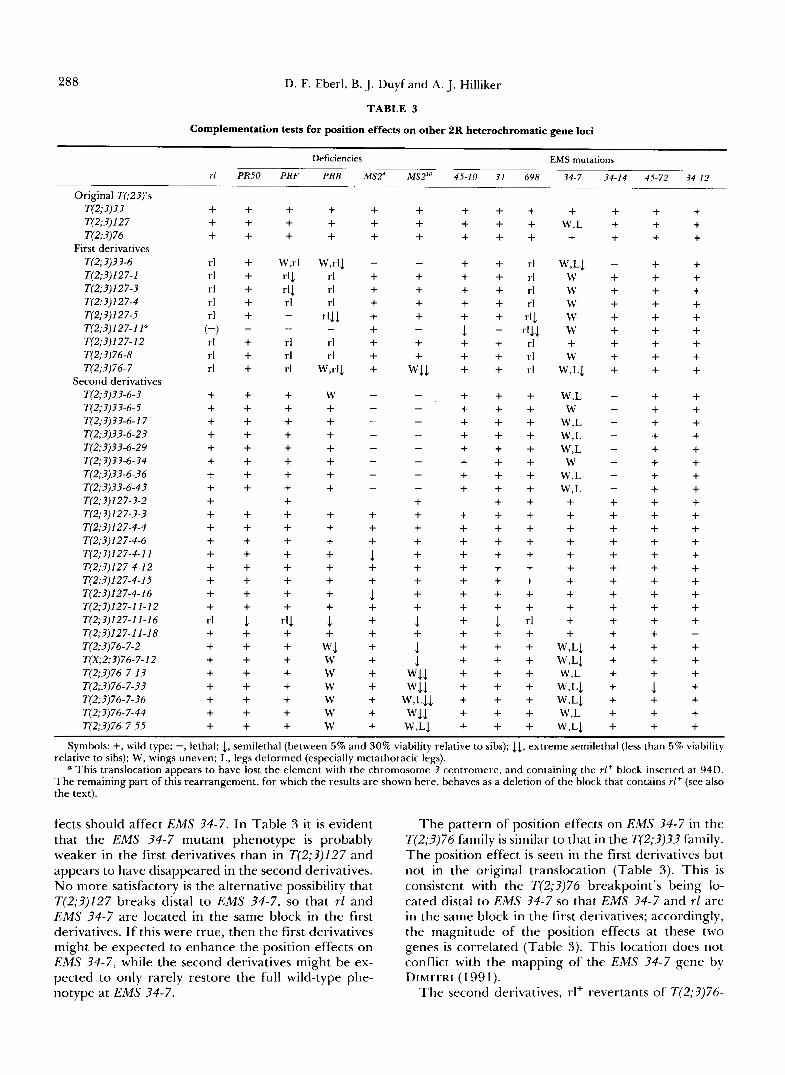
TABLE 3
Complementation tests for position effects on other 2R heterochromatic gene loci
Original T(;23)’s T(2;3)33 T(2;3)127 T(2;3)76
First derivatives T(2;3)33-6
T(2;3)127-3 T(2;3)127-4 T(2;3)127-5 T(2;3)127-11”
T(2;3)76-8 T(2;3)76-7
T(2;3) 1 2 7- 1
T(2;3)127-12
Second derivatives T(2;3)33-6-3 T(2;3)33-6-5 T(2;3)33-6-17 T(2;3)33-6-23 T(2;3)33-6-29 T(2;3)33-6-34
T(2;3)33-6-43 T(2;3)127-3-2 T(2;3)127-3-3 T(2;3)127-4-4
T(2;3)127-4-11 T(2;3)127-4-12 T(2;3)127-4-15
T(2;3)127-11-12 T(2;3)127-11-16 T(2;3)127-11-18 T(2;3)76-7-2 T(X;2;3)76-7-12
T(2;3)76-7-33 T(2;3)76-7-36 T(2;3)76-7-44 T(2;3)76-7-55
T(2;3)33-6-36
T(2;3)127-4-6
T(2;3)127-4-16
T(2;3)76-7-13
rl
+ + + rl rl rl rl rl
(-) rl rl rl
+ + + + + + + + + + + + + + + + + rl + + + + + + + +
~
PR50 ~
+ + + + + + + + - + + + + + + + + + + + + + + + + + + + 1 + + + + + + + +
~
PRF ~
+ + +
W,rl 4 r’l rl - - rl rl rl
+ + + + + + + + + + + + + + + + + rI.1 + + + + + + + +
Deficiencies EMS mutations
PRB
+ + +
W,rlJ rl rl rl
.I11 - rl rl
W d J
W + + + + + + + + + + + + + + + 1
W1 + W W W W W W
45-10 31 698
+ + + + + + + + + + + rl + + rl + + rl + + rl + + rI1 + + rl + + rl + + rl
1 - r11.1
+ + + + + + + + + + + + + + + + + + + + + + + +
+ + + + + + + + + + + + + + + + + + + + + + + + + + + 1 rl + + + + + + + + + + + + + + + + + + + + + + + +
34-7 34-14 45-72 34-12
+ + + + W,L + + + + + + +
W,LI - + + W + + + W + + + W + + + W + + + W + + + + + + + W + + +
W,LJ + + + W,L - + + W,L - + + W,L - + + W,L - + + W,L - + + W,L - + + + + + + + + + + + + + + + + + + + + + + + + + + + + + + + + + + + + + + + + + + + + +
W - + +
W - + +
W,L1 + W,L1 + + + W,L + + +
W,L1 + 1 + W,LI + + + W,L + + +
W,LI + + +
- + +
Symbols: +, wild type; -, lethal; 1, semilethal (between 5 % and 30% viability relative to sibs); 3.4, extreme semilethal (less than 5% viability relative to sibs); W, wings uneven; L, legs deformed (especially metathoracic legs).
a This translocation appears to have lost the element with the chromosome 3 centromere, and containing the rl+ block inserted at 94D. The remaining part of this rearrangement, for which the results are shown here, behaves as a deletion of the block that contains rl+ (see also the text).
fects should affect EMS 34-7. In Table 3 it is evident that the EMS 34-7 mutant phenotype is probably weaker in the first derivatives than in T(2;3)127 and appears to have disappeared in the second derivatives. N o more satisfactory is the alternative possibility that T(2;3)127 breaks distal to EMS 34-7, so that rl and EMS 34-7 are located in the same block in the first derivatives. If this were true, then the first derivatives might be expected to enhance the position effects on EMS 34-7, while the second derivatives might be ex- pected to only rarely restore the full wild-type phe- notype at EMS 34-7.
The pattern of position effects on EMS 34-7 in the T(2;3)76 family is similar to that in the T(2;3)33 family. The position effect is seen in the first derivatives but not in the original translocation (Table 3). This is consistent with the T(2;3)76 breakpoint’s being lo- cated distal to EMS 34-7 so that EMS 34-7 and rl are in the same block in the first derivatives; accordingly, the magnitude of the position effects at these two genes is correlated (Table 3). This location does not conflict with the mapping of the EMS 34-7 gene by DIMITRI (1 99 1).
The second derivatives, rl+ revertants of T(2;3)76-
Drosophila Autosomal Heterochromatin 289
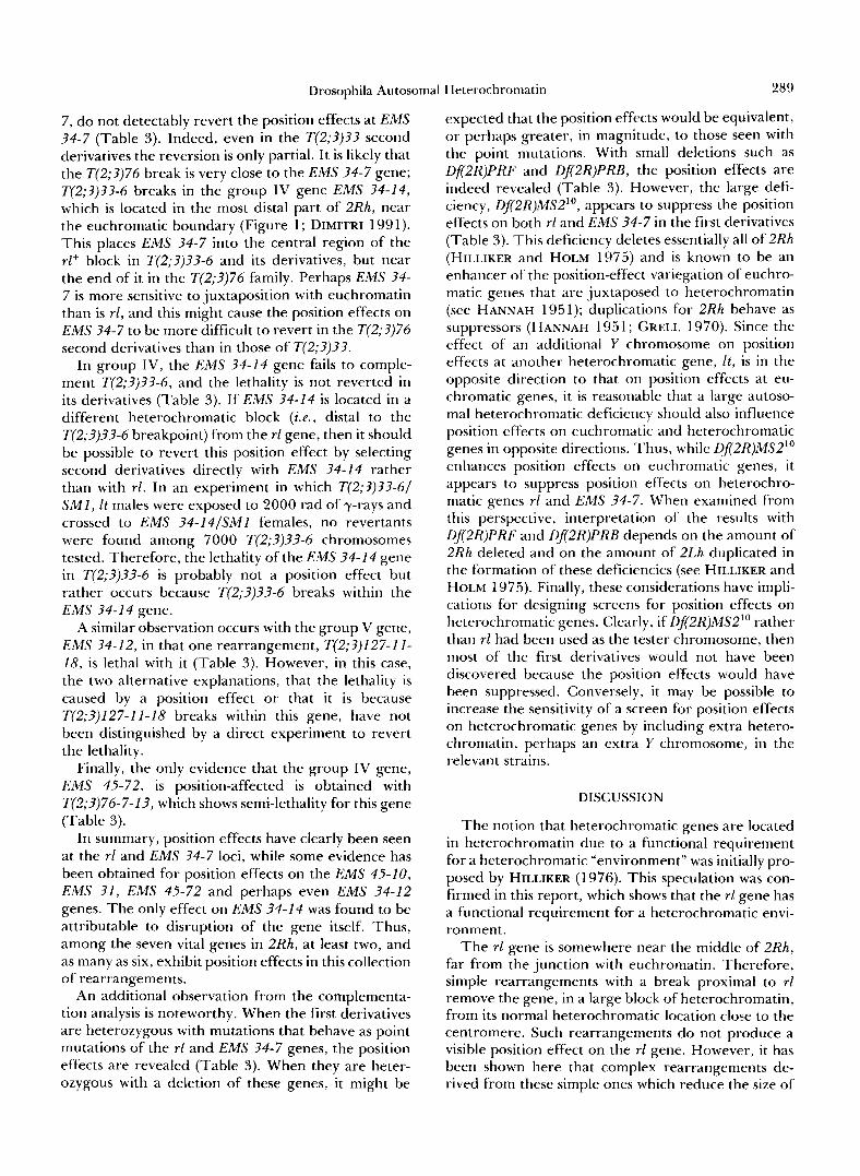
7 , do not detectably revert the position effects at EMS 34-7 (Table 3). Indeed, even in the T(2;?)33 second derivatives the reversion is only partial. It is likely that the T(2;3)76 break is very close to the EMS 34-7 gene; T(2;?)??-6 breaks in the group IV gene EMS 34-14, which is located in the most distal part of 2Rh, near the euchromatic boundary (Figure 1; DIMITRI 1991). This places EMS 34-7 into the central region of the rl+ block in T(2;3)3?-6 and its derivatives, but near the end of it in the T(2;3)76 family. Perhaps EMS 34- 7 is more sensitive to juxtaposition with euchromatin than is r l , and this might cause the position effects on EMS 34-7 to be more difficult to revert in the T(2;3)76 second derivatives than in those of T(2;3)??.
In group IV, the EMS 34-14 gene fails to comple- ment T(2;3)??-6, and the lethality is not reverted in its derivatives (Table 3). If EMS 34-14 is located in a different heterochromatic block (ie., distal to the T(2;?)?3-6 breakpoint) from the rl gene, then it should be possible to revert this position effect by selecting second derivatives directly with EMS 34-14 rather than with rl. In an experiment in which T(2;?)??-6/ SMI, It males were exposed to 2000 rad of y-rays and crossed to EMS ?4-14/SM1 females, no revertants were found among 7000 T(2;3)33-6 chromosomes tested. Therefore, the lethality of the EMS 34-14 gene in T(2;?)??-6 is probably not a position effect but rather occurs because T(2;?)??-6 breaks within the EMS 34-14 gene.
A similar observation occurs with the group V gene, EMS 34-12, in that one rearrangement, T(2;?)127-11- 18, is lethal with it (Table 3 ) . However, in this case, the two alternative explanations, that the lethality is caused by a position effect or that it is because T(2;?)127-11-18 breaks within this gene, have not been distinguished by a direct experiment to revert the lethality.
Finally, the only evidence that the group IV gene, EMS 45-72, is position-affected is obtained with T(2;?)76-7-1?, which shows semi-lethality for this gene (Table 3 ) .
In summary, position effects have clearly been seen at the rl and EMS 34-7 loci, while some evidence has been obtained for position effects on the EMS 45-10, EMS 31, EMS 45-72 and perhaps even EMS 34-12 genes. The only effect on EMS 34-14 was found to be attributable to disruption of the gene itself. Thus, among the seven vital genes in 2Rh, at least two, and as many as six, exhibit position effects in this collection of rearrangements.
An additional observation from the complementa- tion analysis is noteworthy. When the first derivatives are heterozygous with mutations that behave as point mutations of the rl and EMS ?4-7 genes, the position effects are revealed (Table 3). When they are heter- ozygous with a deletion of these genes, it might be
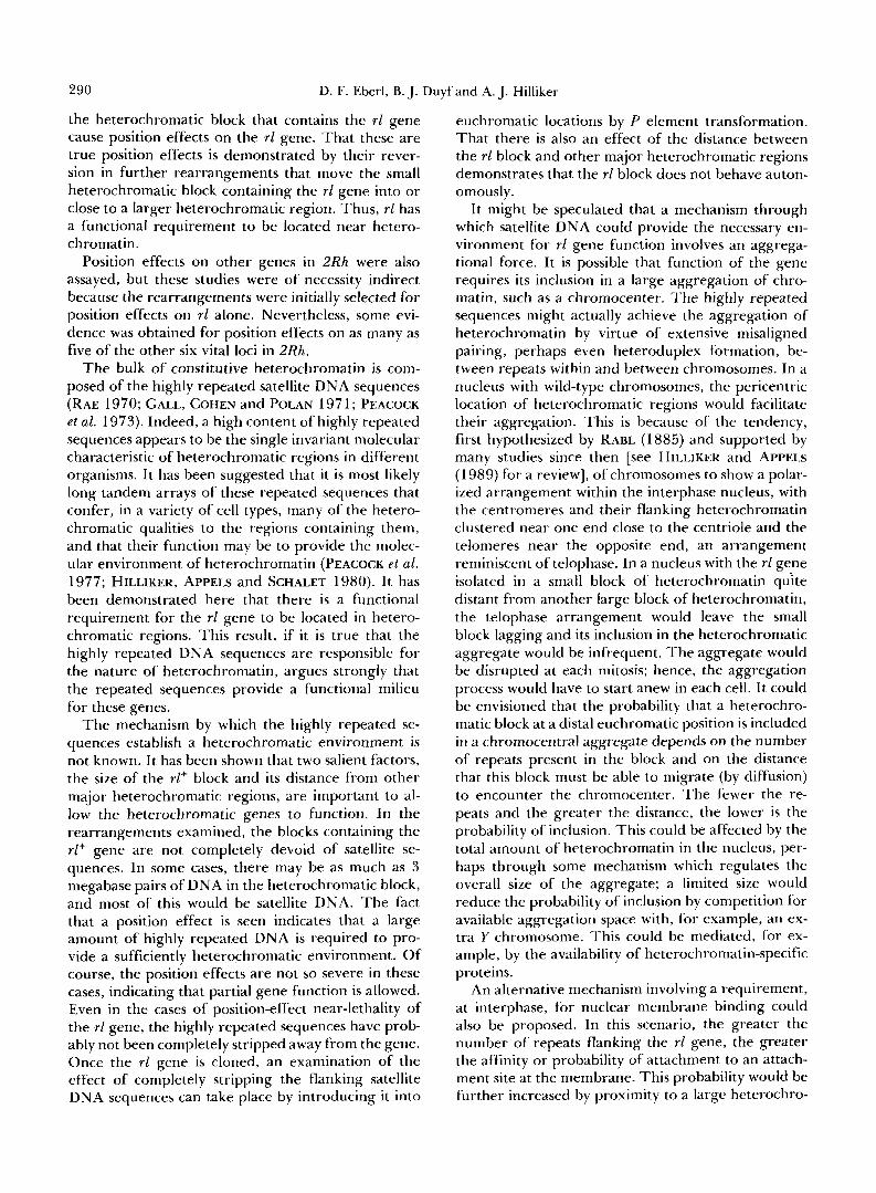
expected that the position effects would be equivalent, or perhaps greater, in magnitude, to those seen with the point mutations. With small deletions such as Df2R)PRF and Df(2R)PRB, the position effects are indeed revealed (Table 3 ) . However, the large defi- ciency, Df(2R)MS2", appears to suppress the position effects on both rl and EMS 34-7 in the first derivatives (Table 3). This deficiency deletes essentially all of 2Rh (HILLIKER and HOLM 1975) and is known to be an enhancer of the position-effect variegation of euchro- matic genes that are juxtaposed to heterochromatin (see HANNAH 1951); duplications for 2Rh behave as suppressors (HANNAH 195 1; GRELL 1970). Since the effect of an additional Y chromosome on position effects at another heterochromatic gene, It, is in the opposite direction to that on position effects at eu- chromatic genes, it is reasonable that a large autoso- mal heterochromatic deficiency should also influence position effects on euchromatic and heterochromatic genes in opposite directions. Thus, while Df2R)MS21° enhances position effects on euchromatic genes, it appears to suppress position effects on heterochro- matic genes rl and EMS 34-7. When examined from this perspective, interpretation of the results with Df2R)PRF and Df2R)PRB depends on the amount of 2Rh deleted and on the amount of 2Lh duplicated in the formation of these deficiencies (see HILLIKER and HOLM 1975). Finally, these considerations have impli- cations for designing screens for position effects on heterochromatic genes. Clearly, if Df2R)MS2l0 rather than rl had been used as the tester chromosome, then most of the first derivatives would not have been discovered because the position effects would have been suppressed. Conversely, it may be possible to increase the sensitivity of a screen for position effects on heterochromatic genes by including extra hetero- chromatin, perhaps an extra Y chromosome, in the relevant strains.
DISCUSSION
The notion that heterochromatic genes are located in heterochromatin due to a functional requirement for a heterochromatic "environment" was initially pro- posed by HILLIKER (1976). This speculation was con- firmed in this report, which shows that the rl gene has a functional requirement for a heterochromatic envi- ronment.
The rl gene is somewhere near the middle of 2Rh, far from the junction with euchromatin. Therefore, simple rearrangements with a break proximal to rl remove the gene, in a large block of heterochromatin, from its normal heterochromatic location clo:e to the centromere. Such rearrangements do not produce a visible position effect on the rl gene. However, it has been shown here that complex rearrangements de- rived from these simple ones which reduce the size of
290 D. F. Eberl, B. J. Duyf and A. J. Hilliker
the heterochromatic block that contains the rl gene cause position effects on the rl gene. That these are true position effects is demonstrated by their rever- sion in further rearrangements that move the small heterochromatic block containing the rl gene into or close to a larger heterochromatic region. Thus, rl has a functional requirement to be located near hetero- chromatin.
Position effects on other genes in 2Rh were also assayed, but these studies were of necessity indirect because the rearrangements were initially selected for position effects on rl alone. Nevertheless, some evi- dence was obtained for position effects on as many as five of the other six vital loci in 2Rh.
The bulk of constitutive heterochromatin is com- posed of the highly repeated satellite DNA sequences (RAE 1970; GALL, COHEN and POLAN 1971; PEACOCK et al. 1973). Indeed, a high content of highly repeated sequences appears to be the single invariant molecular characteristic of heterochromatic regions in different organisms. I t has been suggested that it is most likely long tandem arrays of these repeated sequences that confer, in a variety of cell types, many of the hetero- chromatic qualities to the regions containing them, and that their function may be to provide the molec- ular environment of heterochromatin (PEACOCK e t al. 1977; HILLIKER, APPELS and SCHALET 1980). It has been demonstrated here that there is a functional requirement for the rl gene to be located in hetero- chromatic regions. This result, if it is true that the highly repeated DNA sequences are responsible for the nature of heterochromatin, argues strongly that the repeated sequences provide a functional milieu for these genes.
The mechanism by which the highly repeated se- quences establish a heterochromatic environment is not known. It has been shown that two salient factors, the size of the rl+ block and its distance from other major heterochromatic regions, are important to al- low the heterochromatic genes to function. In the rearrangements examined, the blocks containing the rl+ gene are not completely devoid of satellite se- quences. In some cases, there may be as much as 3 megabase pairs of DNA in the heterochromatic block, and most of this would be satellite DNA. The fact that a position effect is seen indicates that a large amount of highly repeated DNA is required to pro- vide a sufficiently heterochromatic environment. Of course, the position effects are not so severe in these cases, indicating that partial gene function is allowed. Even in the cases of position-effect near-lethality of the rl gene, the highly repeated sequences have prob- ably not been completely stripped away from the gene. Once the rl gene is cloned, an examination of the effect of completely stripping the flanking satellite DNA sequences can take place by introducing it into
euchromatic locations by P element transformation. That there is also an effect of the distance between the rl block and other major heterochromatic regions demonstrates that the rl block does not behave auton- omously.
It might be speculated that a mechanism through which satellite DNA could provide the necessary en- vironment for rl gene function involves an aggrega- tional force. It is possible that function of the gene requires its inclusion in a large aggregation of chro- matin, such as a chromocenter. The highly repeated sequences might actually achieve the aggregation of heterochromatin by virtue of extensive misaligned pairing, perhaps even heteroduplex formation, be- tween repeats within and between chromosomes. In a nucleus with wild-type chromosomes, the pericentric location of heterochromatic regions would facilitate their aggregation. This is because of the tendency, first hypothesized by RABL (1 885) and supported by many studies since then [see HILLIKER and APPELS (1 989) for a review], of chromosomes to show a polar- ized arrangement within the interphase nucleus, with the centromeres and their flanking heterochromatin clustered near one end close to the centriole and the telomeres near the opposite end, an arrangement reminiscent of telophase. In a nucleus with the rl gene isolated in a small block of heterochromatin q d t e distant from another large block of heterochromatin, the telophase arrangement would leave the small block lagging and its inclusion in the heterochromatic aggregate would be infrequent. The aggregate would be disrupted at each mitosis; hence, the aggregation process would have to start anew in each cell. I t could be envisioned that the probability that a heterochro- matic block at a distal euchromatic position is included in a chromocentral aggregate depends on the number of repeats present in the block and on the distance that this block must be able to migrate (by diffusion) to encounter the chromocenter. The fewer the re- peats and the greater the distance, the lower is the probability of inclusion. This could be affected by the total amount of heterochromatin in the nucleus, per- haps through some mechanism which regulates the overall size of the aggregate; a limited size would reduce the probability of inclusion by competition for available aggregation space with, for example, an ex- tra Y chromosome. This could be mediated, for ex- ample, by the availability of heterochromatin-specific proteins.
An alternative mechanism involving a requirement, at interphase, for nuclear membrane binding could also be proposed. In this scenario, the greater the number of repeats flanking the rl gene, the greater the affinity or probability of attachment to an attach- ment site at the membrane. This probability would be further increased by proximity to a large heterochro-
Drosophila Autosomal Heterochromatin 29 1
matic region which will itself be ensured an attach- ment site, and therefore will “anchor” the rl block near the periphery where it is more likely to contact an attachment site. An extra Y chromosome in the nucleus would compete for such attachment sites, thereby decreasing the probability of attachment of the rl block to the nuclear membrane.
I t should be noted that both of these models could accommodate either variegated or uniform position effects. In the aggregation hypothesis, a variegated phenotype would result from inclusion of the gene in a major aggregate in some cells but not others, while a uniform phenotype would involve the strength of the association of the rl block with the aggregate. In the membrane-binding hypothesis, variegation could result from attachment in some cells but not others, while uniformity could result from a reduced equilib- rium of binding.
The role of the highly repeated DNA has been the subject of speculation for a long time but now there is strong experimental evidence that it has a very important role in the expression and proper function of heterochromatic genes. We have very clearly dem- onstrated that, to function properly, rl and other heterochromatic genes require proximity to hetero- chromatin, which consists largely of highly repeated DNA sequences.
This work was supported by a Natural Sciences and Engineering Research Council of Canada grant to A.J.H.
LITERATURE CITED
BARR, M. L., and E. G . BERTRAM, 1949 A morphological distri- bution between neurones of the male and female and the behavior of the nucleolar satellite during accelerated nuclear protein synthesis. Nature 163: 676-677.
BRIDGES, C. B. 1935 Salivary chromosome maps with a key to the banding of the chromosomes of Drosophila melanogaster. J. Hered. 26: 60-64.
BRIDGES, C. B., and P. N. BRIDGES, 1939 A new map of the second chromosome: a revised map of the right limb of the second chromosome of Drosophila melanogaster. J. Hered. 3 0 475- 476.
BRIDGES, P. N., 194 la A revised map of the left limb of the third chromosome of Drosophila melanogaster. J. Hered. 32: 64-65.
BRIDGES, P. N., 1941b A revision of the salivary gland 3R-chro- mosome map of Drosophila melanogaster. J. Hered. 32: 299- 300.
BRIDGES, P. N., 1942 A new map of the salivary gland 2L chro- mosome ofDrosophila melanogaster. J. Hered. 33: 403-408.
BROWN, S. W., 1966 Heterochromatin. Science 151: 418-425. DEMEREC, M., 1941 The nature of changes on the white-Notch
region of the X-chromosome of Drosophila melanogaster. h o c . Int. Congr. Genet. 7: 99-103.
DEVLIN, R. H., B. BINGHAM and B. T. WAKIMOTO, 1990 The organization and expression of the light gene, a heterochro- matic gene of Drosophila melanogaster. Genetics 125: 129-140.
DEVLIN, R. H., D. G. HOLM, K. MORIN and B. HONDA, 1990 Identifying a single-copy DNA sequence associated with the expression of a heterochromatic gene, the light locus of Drosophila melanogaster. Genome 33: 405-4 15.
DIMITRI, P., 199 1 Cytogenetic analysis of the second chromosome
heterochromatin of Drosophila melanogaster. Genetics 127: 553-564.
EBERL, D. F., A. J. HILLIKER, C. B. SHARP and S. N. TRUSIS- COULTER, 1989 Further observations on the nature of radia- tion-induced chromosomal interchanges recovered from Dro- sophila sperm. Genome 32: 847-855.
GALL, J. G . , 1973 Repetitive DNA in Drosophila, pp. 59-74 in Molecular Cytogenetics, edited by B. A. HAMKALO and J. PAPA- CINSTANTINOU. Plenum Press, New York.
GALL, J. G. , E. H. COHEN and M. L. POLAN, 1971 Repetitive DNA sequences in Drosophila. Chromosoma 33: 319-344.
GANETZKY, B., 1977 On the components of Segregation Distor- tion in Drosophila melanogaster. Genetics 86: 321-355.
GRELL, R. F. , 1970 The time of initiation of segregation pairing between nonhomologues in Drosophila melanogaster: a re-ex- amination of wm4. Genetics 64: 337-365.
HANNAH, A,, 1951 Localization and function of heterochromatin in Drosophila melanogaster. Adv. Genet. 4: 87-125.
HEITZ, E., 1928 Das heterochromatin der Moose. I. Jahrb. Wiss. Botanik 69: 762-81 8.
HEITZ, E., 1933 Die somatische Heteropyknose bei Drosophila melanogaster und ihre genetische Bedeutung. 2. Zellforsch. 2 0 237-287.
HEITZ, E., 1934 Uber a- and P-Heterochromatin sowie Kontstanz und Bau der Chromomeren bei Drosophila. Biol. Zentralbe.
HESSLER, A. Y . , 1958 V-type position effects at the light locus in Drosophila melanogaster. Genetics 43: 395-403.
HILLIKER, A. J., 1976 Genetic analysis of the centromeric heter- ochromatin of chromosome 2 of Drosophila melanogaster: defi- ciency mapping of EMS-induced lethal complementation groups. Genetics 83: 765-782.
HILLIKER, A. J., 1980 Variegation of a heterochromatic gene in Drosophila melanogaster-the light locus. Genetics 94: s44.
HILLIKER, A. J., and R. APPELS, 1989 The arrangement of inter- phase chromosomes: structural and functional aspects. Exp. Cell Res. 185: 297-318.
HILLIKER, A. J., R. APPELS and A. SCHALET, 1980 The genetic analysis of D. melanogaster heterochromatin. Cell 21: 607-619.
HILLIKER, A. J., and D. G. HOLM, 1975 Genetic analysis of the proximal region of chromosome 2 of Drosophila melanogaster. I. Detachment products of compound autosomes. Genetics 81: 705-721.
HILLIKER, A. J., and C. B. SHARP, 1988 New perspectives on the genetics and molecular biology of constitutive heterochroma- tin. Stadler Genet. Symp. 18: 9 I - 1 15.
HILLIKER, A. J., and S. N. TRUSIS-COULTER, 1987 Analysis of the functional significance of linkage group conservation in Dro- sophila. Genetics 117: 233-244.
HUETTNER, A. F., 1933 Continuity of the centrioles in Drosophila melanogaster. 2. Zellforsch Mikrosk. Anat. 19 119-134.
HUGHES-SCHRADER, S., 1948 Cytology of coccids (Coccoidea- Homoptera). Adv. Genet. 2: 127-203.
LINDSLEY, D. L., and E. H. GRELL, 1968 Genetic Variations of Drosophila melanogaster. Carnegie Inst. Washington Publ. 627.
LINDSLEY, D. L., and G . G . ZIMM, 1992 The Genome of Drosophila melanogaster. Academic Press, San Diego.
MARCHANT, G. E., and D. G. HOLM, 1988 Genetic analysis of the heterochromatin of chromosome 3 in Drosophila melanogaster. 11. Vital loci identified through EMS mutagenesis. Genetics
MIKLOS, G . L. G.,andJ. N. COTSELL, 1990 Chromosomestructure at the interfaces between two major chromatin types: alpha- and beta-heterochromatin. BioEssays 12: 1-6.
MITTWOCH, U., 1964 Sex chromosomes and sex chromatin. Na- ture 204: 1032-1034.
MULLER, H. J., and T. PAINTER, 1932 The differentiation of the sex chromosomes of Drosophila into genetically active and inert
54: 588-609.
120 519-532.

292 D. F. Eberl, B. J. Duyf and A. J. Hilliker
regions. Z. Indukt. Abstammungs- Vererbungsl. 62: 316-365. PEACOCK, W. J., D. L. BRUTLAG, E. GOLDRING, R. APPELS, C. W.
HINTON and D. L. LINDSLEY, 1973 The organization of highly repeated DNA sequences in Drosophila melanogaster chromosomes. Cold Spring Harbor Symp. Quant. Biol. 38:
PEACOCK, W. J., A. R. LOHE, W. L. GERLACH, P . DUNSMUIR, E. S. DENNIS and R. APPELS, 1977 Fine structure and evolution of DNA in heterochromatin. Cold Spring Harbor Symp. Quant. Biol. 42: 1121-1135.
RABINOWITZ, M., 1941 Studies on the cytology and early embryol- ogy of the egg of Drosophila melanogaster. J. Morphol. 6 9 1- 49.
405-416.
RABL, C., 1885 Uber Zellteilung. Morphol. Jahrb. 10: 214-330. RAE, P. M. M., 1970 Chromosomal distribution of rapidly rean-
nealing DNA in Drosophila melanogaster. Proc. Natl. Acad. Sci.
SCHULTZ, J., 1936 Variegation in Drosophila and the inert chro- mosome regions. Proc. Natl. Acad. Sci. USA 22: 27-33.
SCHUPBACH, T., and E. WIESCHAUS, 1989 Female sterile muta- tions on the second chromosome of Drosophila melanogaster. I
USA 67: 1018-1025.
Maternal effect mutations. Genetics 121: 101-1 17. SHARP, C. B., 1988 Biometrical and genetic studies of segregation
distortion in Drosophila melanogaster. Ph.D. Dissertation, Uni- versity of Guelph, Guelph, Ontario, Canada.
SHARP, C. B., A. J. HILLIKER and D. G. HOLM, 1985 Further characterization of genetic elements associated with the segre- gation distortion phenomenon in Drosophila melanogaster. Ge- netics 110: 671-688.
SPOFFORD, J., 1976 Position-effect variegation in Drosophila, pp. 955-1 018 in The Genetics and Biology of Drosophila, Vol. I C , edited by M. ASHBURNER and E. NOVITSKI. Academic Press, New York.
STEFFENSEN, D. L., R. APPELS and W. J. PEACOCK, 1981 The distribution of two highly repeated DNA sequences within Drosophila melanogaster chromosomes. Chromosoma 82: 525- 541.
WAKIMOTO, B. T., and M. G. HEARN, 1990 The effects of chro- mosome rearrangements on the expression of heterochromatic genes in chromosome 2L of Drosophila melanogaster. Genetics 125: 141-154.
Communicating editor: A. CHOVNICK