THE ROLE OF CTLA-4 IN HEALTH AND AUTOIMMUNE DISEASE
55
From the Department of Medical Biochemistry and Biophysics Karolinska Institutet, Stockholm, Sweden THE ROLE OF CTLA-4 IN HEALTH AND AUTOIMMUNE DISEASE Katrin Klocke Stockholm 2017
Transcript of THE ROLE OF CTLA-4 IN HEALTH AND AUTOIMMUNE DISEASE

From the Department of Medical Biochemistry and Biophysics Karolinska Institutet, Stockholm, Sweden
THE ROLE OF CTLA-4 IN HEALTH AND AUTOIMMUNE DISEASE
Katrin Klocke
Stockholm 2017

All previously published papers were reproduced with permission from the publisher. Published by Karolinska Institutet. Printed by E-Print AB 2017 © Katrin Klocke, 2017 ISBN 978-91-7676-673-6
The role of CTLA-4 in health and autoimmune disease
THESIS FOR DOCTORAL DEGREE (Ph.D.)
By
Katrin Klocke
Principal Supervisor: Assistant Professor Kajsa Wing Karolinska Institutet Department of Medical Biochemistry and Biophysics Division of Medical Inflammation Research Co-supervisor(s): Professor Rikard Holmdahl Karolinska Institutet Department of Medical Biochemistry and Biophysics Division of Medical Inflammation Research
Opponent: Professor David Sansom University College London Department of Immunity and Transplantation Division of Transplant Immunology Examination Board: Susanne Gabrielsson Karolinska Institutet Department of Medicine Division of Immunology and Allergy Helen Kaipe Karolinska Institutet Department of Laboratory Medicine Division of Therapeutic Immunology Sara Mangsbo Uppsala Universitet Department of Immunology, Genetics and Pathology Division of Clinical Immunology


“Science, my lad, is made up of mistakes but they are mistakes which it is useful to make, because they lead little by little to the truth.”
Jules Verne


ABSTRACT FOXP3+ regulatory T (Treg) cells are powerful regulators of the immune system, as shown by the development of multi-organ autoimmunity in mice and men upon loss or dysfunction of these cells. Whilst Treg cells are vital to control normal immune responses, they are also involved in the development of autoimmunity and the failure to combat cancer. Cytotoxic T lymphocyte antigen-4 (CTLA-4) is an important regulator of adaptive immune responses and vital for the function of Treg cells, as demonstrated by loss of function studies that result in similar autoimmunity as that caused by loss of Treg cells.
The broad aim of this thesis was to gain a deeper understanding of how CTLA-4 mediated tolerance operates both in health and in autoimmune disease, as its role in the adult immune system remains ill-defined. Therefore, in study I, we developed a novel system that allows for the inducible depletion of CTLA-4. Loss of CTLA-4 in adulthood unleashed T cell, but especially Treg cell homeostasis, resulting in multi-organ lymphocytic infiltrations that however did not progress to fatal autoimmunity and affected different organs than congenic CTLA-4 deficiency did, which implies a role for CTLA-4 in thymic T cell selection. Furthermore, we could show that loss of CTLA-4 can have both disease promoting as well as protective effects, depending on the type of autoimmunity as well as the inducing agent, depicting the need for context specific studies. Thus, in study II, we focused on collagen-induced arthritis (CIA) and found that CTLA-4 regulated all stages of disease as well as disease spreading, albeit without affecting heterogeneous T or B cell responses. Autologous responses were however controlled by CTLA-4 and loss of CTLA-4 could break pre-established T cell tolerance to type-II collagen (CII). Furthermore, we found that CTLA-4 on conventional T cells limited the priming of autoreactive T cells, whereas Treg cell specific CTLA-4 was necessary to control the inflammatory tissue attack. In study III, we analyzed CII-specific T cells and found that CTLA-4 could induce tolerance in individuals with an inherent T cell predisposition to joint reactivity. These tolerized, CII-reactive T cells were found in high frequencies in the liver and transfer of liver lymphocytes could induce tolerance in otherwise susceptible hosts. Further studies are needed to elucidate if tolerance is induced in the liver or whether tolerized T cells accumulate in this organ, but it opens exciting possibilities for research aiming to induce tolerance in arthritis patients. In study IV, we used mice defective in reactive oxygen species that suffer from an increased susceptibility to infections, to show that increased susceptibility to autoimmunity is not merely a consequence of these infections but is a separate phenomenon.
Taken together, our data reveal the complex nature of CTLA-4 mediated regulation and the necessity to study antigen-specific responses. They also demonstrate the immensely powerful tolerance inducing abilities of CTLA-4, even in the face of enhanced self-reactivity. This increased understanding will be of importance for finding novel treatment strategies for patients suffering from autoimmunity.

LIST OF SCIENTIFIC PAPERS
I. Induction of autoimmune disease by deletion of CTLA-4 in mice in adulthood. Katrin Klocke, Shimon Sakaguchi, Rikard Holmdahl and Kajsa Wing Proc Natl Acad Sci U S A. 2016 Apr 26;113(17):E2383-92.
II. CTLA-4 expressed by FOXP3+ Treg cells prevents inflammatory tissue attack and not T cell priming in arthritis. Katrin Klocke, Rikard Holmdahl and Kajsa Wing Immunology. 2017 (Accepted)
III. Collagen reactive T cells accumulate in the liver and protect from arthritis.
Katrin Klocke, Rikard Holmdahl and Kajsa Wing Manuscript
IV. Germ-free mice deficient of reactive oxygen species have increased arthritis susceptibility. Kajsa Wing, Katrin Klocke, Annika Samuelsson and Rikard Holmdahl Eur J Immunol. 2015 May;45(5):1348-53.

CONTENTS 1 The immune system in a nutshell ........................................................................ 7
1.1 The innate immune system .......................................................................... 7 1.1.1 Reactive oxygen species ................................................................. 8
1.2 The adaptive immune system ...................................................................... 8 1.3 Antigen presentation ................................................................................... 9 1.4 T cells .......................................................................................................... 9
1.4.1 T cell activation ............................................................................. 10 1.5 B cells ........................................................................................................ 10
2 Immunologic tolerance ...................................................................................... 11 2.1 Central tolerance ........................................................................................ 11 2.2 Peripheral tolerance ................................................................................... 11
2.2.1 Ignorance ....................................................................................... 12 2.2.2 Anergy and Exhaustion ................................................................. 12 2.2.3 Apoptosis ....................................................................................... 13 2.2.4 Dominant suppression ................................................................... 13
3 FOXP3+ regulatory T cells ................................................................................. 14 3.1 Development ............................................................................................. 14 3.2 Phenotype .................................................................................................. 16 3.3 Functions ................................................................................................... 16
3.3.1 Regulation of T cell function ........................................................ 17 3.3.2 Regulation of APC function .......................................................... 18
3.4 Plasticity and stability ............................................................................... 19 4 CTLA-4 .............................................................................................................. 20
4.1 Expression ................................................................................................. 20 4.2 Functions ................................................................................................... 20
4.2.1 T cell intrinsic or autonomous functions ...................................... 21 4.2.2 T cell extrinsic or non-autonomous functions .............................. 22 4.2.3 Models of CTLA-4 function ......................................................... 23
4.3 CTLA-4 Isoforms ...................................................................................... 24 4.4 CTLA-4 based therapy .............................................................................. 24
5 Autoimmune diseases ........................................................................................ 26 5.1 Rheumatoid arthritis .................................................................................. 26
5.1.1 Treatment of rheumatoid arthritis ................................................. 27 5.1.2 Regulatory T cells and CTLA-4 in rheumatoid arthritis .............. 28
5.2 Multiple sclerosis ...................................................................................... 28 6 Animal models for autoimmune diseases .......................................................... 29
6.1 Mouse models for rheumatoid arthritis ..................................................... 29 6.1.1 Collagen-induced arthritis ............................................................. 29
6.2 Mouse models for multiple sclerosis ........................................................ 30 6.2.1 Experimental autoimmune encephalomyelitis ............................. 30
7 Present Investigations ........................................................................................ 31
7.1 Paper I ........................................................................................................ 31 7.2 Paper II ...................................................................................................... 32 7.3 Paper III ..................................................................................................... 32 7.4 Paper IV ..................................................................................................... 33
8 Concluding remarks and future perspective ...................................................... 34 9 Acknowledgements ............................................................................................ 35 10 References .......................................................................................................... 39

LIST OF ABBREVIATIONS
ACPA Anti-citrullinated protein antibody
AICD Activation induced cell death
AIRE Autoimmune regulator
APCs
BCR
CFA
CGD
CIA
CII
CNS
CTL
CTLA-4
DCs
DMARDs
EAE
EULAR
FDA
FOXP3
FR4
ICAM-1
IDO
IFA
iKO
IPEX
iTreg
KO
Lag-3
li
LFA-1
MBP
Antigen-presenting cells
B cell receptor
Complete Freund’s adjuvant
Chronic granulomatous disease
Collagen-induced arthritis
Type-II collagen
Central nervous system
Cytotoxic T lymphocyte
Cytotoxic T lymphocyte antigen-4
Dendritic cells
Disease-modifying antirheumatic drugs
Experimental autoimmune encephalomyelitis
European League Against Rheumatism
Food and Drug Administration
Forkhead box P3
Folate receptor 4
Intercellular adhesion molecule 1
Indoleamine 2,3-dioxygenase
Incomplete Freund’s adjuvant
Induced knock-out
Immunodysregulation polyendocrinopathy X-linked
Induced Treg
Knock-out
Lymphocyte-activation gene 3
Ligand-independent
Lymphocyte function-associated antigen 1
Myelin basic protein
MHC
MHC-II
MMC
MOG
MS
MTX
Ncf1
NOX2
Nrp-1
nTreg
PAMPs
PD-1
PKC
PLP
PRRs
pTreg
RA
ROS
SPF
Tconv
TCR
Tfh
TSDR
tTreg
WT
Major histocompatibility complex
Major histocompatibility complex class II
Mouse mutated collagen
Myelin oligodendrocyte glycoprotein
Multiple Sclerosis
Methotrexate
Neutrophil cytosolic factor 1
NADPH oxidase 2
Neuropilin 1
Natural regulatory T cell
Pathogen-associated molecular patterns
Programmed cell death protein 1
Protein kinase C
Proteolipid protein
Pattern recognition receptors
Peripheral regulatory T cell
Rheumatoid arthritis
Reactive oxygen species
Specific-pathogen-free
Conventional T cell
T cell receptor
Follicular helper T cell
Treg specific demethylated region
Thymic derived regulatory T cell
Wild type
7
1 THE IMMUNE SYSTEM IN A NUTSHELL Our bodies are exposed to millions of potentially infective agents every day but we seldom get sick thanks to our powerful immune system that is well equipped for fighting off bacteria, fungi, viruses and parasites. However not all microorganisms are harmful, like the commensal bacteria of the gut and our immune system has learned to tolerate these beneficial microorganisms instead of fighting against them.
The immune system is a complex network of highly specialized cells, organs, tissues and molecules that all work together in protecting the human body against pathogens by discriminating self from non-self (1, 2) and dangerous from innocuous antigens (3). Herein, every immune response must fulfill four tasks: (I) invading harmful pathogens must be recognized, (II) the infection has to be cleared or at least limited to prevent spreading, (III) the established immune response has to be tightly regulated and shut down once the infection is cleared to avoid harm to the body by excessive inflammation and (IV) an immunological memory has to be generated, enabling the immune system to react faster to a subsequent infection caused by the same pathogen.
Our immune system consists of an innate and an adaptive arm. These two are inadvertently linked and a successful immune response usually requires both the speed of the innate as well as the specificity of the adaptive system, which is amongst others, controlled by cytotoxic T lymphocyte antigen-4 (CTLA-4), a powerful regulator of the adaptive immune response.
1.1 THE INNATE IMMUNE SYSTEM
The innate immune system is considered as the fast first line of defense after pathogens have crossed the body’s tissue barriers consisting of skin and mucosal epithelia. Cells of the innate immune system, such as macrophages, dendritic cells (DCs), neutrophils and mast cells cannot distinguish between different antigens but they can distinguish self from non-self with the help of pathogen recognition receptors (PRRs) that recognize pathogen-associated molecular patterns (PAMPS), found on pathogens but not on own cells. Innate cells fulfill three important functions: (I) pathogens are phagocytosed and destroyed, (II) cytokines and chemokines are produced that increase vascular permeability and attract other immune cells, amongst them adaptive cells, to the site of infection and (III) antigens of engulfed pathogens are presented via major histocompatibility complex (MHC) molecules to adaptive cells that lack PRRs to alert them to the presence of pathogens.
Molecular components of the innate immune system are, amongst others, antimicrobial peptides and the complement proteins. Antimicrobial peptides are small molecules that damage and kill microbes either by attaching themselves to and creating pores in bacterial membranes or by binding to molecules inside of bacteria and interfering with bacterial replication. The complement system consists of a number of small proteins that attach themselves to cell membranes, starting a cascade of cleavages that finally results in the creation of a pore in the cell membrane. Apart from lysing cells, the complement cascade releases powerful

8
chemotactic molecules that alert and recruit other immune cells to the site of infection. The complement system can either be activated spontaneously or by cross-linking of antibodies. Spontaneous activation can in principle occur on any membrane but is usually prevented on the body’s own cells by inhibitory molecules.
Often, the innate immune system is sufficient to combat an infection but since it does not possess a memory function it cannot prevent an infection from reoccurring and sometimes all the innate system can do is to hold an overwhelming infection at bay until the more specific and potent yet slow to start adaptive immune system has gotten in gear.
1.1.1 Reactive oxygen species
Biological processes are subject to redox regulation, through a system of oxidases and reductases. For example, cysteines in thiol-containing proteins can be oxidized and subsequently form covalent disulfide bridges which render a protein’s 3D confirmation more rigid. Furthermore, cysteine oxidation has been shown to change antigenicity of peptides (4). Reactive oxygen species (ROS) are formed in mitochondria as part of the cellular metabolism of oxygen or in phagocytes upon stimulation of the NADPH oxidase 2 (NOX2) complex, a process called phagocytic burst that is crucial for host defense against bacteria. The NOX2 complex can be found both at the plasma membrane as well as at phagosomal membranes, releasing ROS both intra- and extracellular. Compared to other ROS species, H2O2 has a long half-life and can freely diffuse through lipid membranes, opening the possibility to affect cells other than the ROS producing one. A naturally occurring polymorphism in the neutrophil cytosolic factor 1 (Ncf1) subunit of the NOX2 complex, which results in a truncated Ncf1 protein and absence of NOX2 derived ROS, renders rats and mice more susceptible to experimental arthritis, rebutting the dogma that ROS is always pro-inflammatory and pathogenic (5, 6). Instead, it has been shown that ROS mediated oxidation of the surface of T cells renders them less arthritogenic (7) and that absence of ROS can break both pre-established T- and B cell tolerance to type-II collagen in mice (8, 9).
1.2 THE ADAPTIVE IMMUNE SYSTEM
Cells of the adaptive immune system have specialized cell surface receptors enabling them to distinguish between pathogen types and strains. Their effector functions are therefore much more targeted and thus can be more powerful without causing tissue damage. The adaptive immune system consists of T- and B cells that are derived from the same multipotent hematopoietic stem cell in the bone marrow. T cell progenitors travel from the bone marrow to the thymus where they develop into T cells, which undergo an intricate selection process that leaves only very few T cells to exit into the periphery. The main function of T cells involves cell-mediated immune responses and will be explained in more detail in section 1.4. B cell progenitors also undergo a differentiation and selection process, but unlike T cells, this happens in the bone marrow. The main function of B cells involves humoral responses, i.e. the production of antibodies. Both T and B cells can develop a memory, a quality that is utilized in the vaccination of healthy people against common pathogens.

9
1.3 ANTIGEN PRESENTATION
T- and B cells cannot be activated by the pathogen itself but require presentation of pathogen associated antigens on MHC molecules of innate cells. Every nucleated body cell is capable of presenting endogenous antigens on MHC class-I molecules to cytotoxic CD8+ T cells. The majority of endogenous antigens are self-antigens but can also stem from bacteria and viruses that hide inside host cells. On the contrary, only so-called professional antigen-presenting cells (APCs), such as dendritic cells, macrophages and B cells, are capable of taking up and presenting exogenous antigen on MHC class-II (MHC-II) molecules to CD4+ T cells.
As already mentioned, the function of MHC molecules is to bind and present peptides to T cells. The MHC is polygenic, consisting of more than 200 genes in humans. Every MHC molecule has a unique peptide-binding groove that is capable of binding different peptides with various affinities, creating a tremendous number of peptides that can be recognized by any individual. Furthermore, the MHC is also polymorphic, which means that there are many variants of the same gene found amongst a population. This ensures that there will always be some individuals who have the right MHC variant to recognize any given pathogen and this ensures the survival of the population as a whole.
APCs that have taken up pathogens upregulate MHC-II and costimulatory molecules, enabling them to activate and prime naïve T cells in secondary lymphoid organs such as spleen and lymph nodes.
1.4 T CELLS
T cells recognize peptides presented on MHC by their highly specific T cell receptor (TCR). Similar to MHC molecules being able to bind a huge variety of different peptides, TCRs are able to recognize huge varieties of presented peptides. A TCR is a heterodimer consisting of an α- and a β-chain that are generated through random somatic combination of three gene segments that each exist in multiple copies. This combinatorial diversity is even further increased by the many possible different combinations of α- and β-chains resulting in a unique TCR repertoire of approximately 25 million specificities in any individual (10).
T cells can be divided into CD8+ and CD4+ subtypes. CD8+ cytotoxic T-lymphocytes (CTL) recognize peptides presented on MHC class I that are mostly derived from endogenous pathogens that infect cells. CTLs have the ability to kill these infected cells with preformed granules containing cytotoxic molecules, such as granzymes, to prevent systemic propagation of the pathogen.
CD4+ T cells recognize peptides from endogenous pathogens that were phagocytosed and are then presented on MHC-II. They are also called T helper cells since they help infected macrophages to increase their phagocytic activity and B cells to produce antibodies. T helper cells can be further divided into various subtypes such as Th1, Th2 and Th17 that differ in their effector functions and cytokine production. A subtype of CD4+ T cells, forkhead box P3+ (FOXP3) regulatory T cells, will be described in detail in section 3.
10
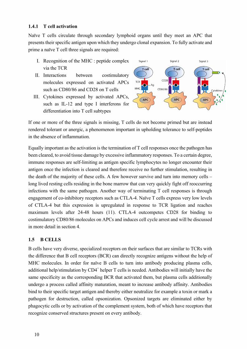
1.4.1 T cell activation
Naïve T cells circulate through secondary lymphoid organs until they meet an APC that presents their specific antigen upon which they undergo clonal expansion. To fully activate and prime a naïve T cell three signals are required:
I. Recognition of the MHC : peptide complex via the TCR
II. Interactions between costimulatory molecules expressed on activated APCs such as CD80/86 and CD28 on T cells
III. Cytokines expressed by activated APCs, such as IL-12 and type I interferons for differentiation into T cell subtypes
If one or more of the three signals is missing, T cells do not become primed but are instead rendered tolerant or anergic, a phenomenon important in upholding tolerance to self-peptides in the absence of inflammation.
Equally important as the activation is the termination of T cell responses once the pathogen has been cleared, to avoid tissue damage by excessive inflammatory responses. To a certain degree, immune responses are self-limiting as antigen specific lymphocytes no longer encounter their antigen once the infection is cleared and therefore receive no further stimulation, resulting in the death of the majority of these cells. A few however survive and turn into memory cells – long lived resting cells residing in the bone marrow that can very quickly fight off reoccurring infections with the same pathogen. Another way of terminating T cell responses is through engagement of co-inhibitory receptors such as CTLA-4. Naïve T cells express very low levels of CTLA-4 but this expression is upregulated in response to TCR ligation and reaches maximum levels after 24-48 hours (11). CTLA-4 outcompetes CD28 for binding to costimulatory CD80/86 molecules on APCs and induces cell cycle arrest and will be discussed in more detail in section 4.
1.5 B CELLS
B cells have very diverse, specialized receptors on their surfaces that are similar to TCRs with the difference that B cell receptors (BCR) can directly recognize antigens without the help of MHC molecules. In order for naïve B cells to turn into antibody producing plasma cells, additional help/stimulation by CD4+ helper T cells is needed. Antibodies will initially have the same specificity as the corresponding BCR that activated them, but plasma cells additionally undergo a process called affinity maturation, meant to increase antibody affinity. Antibodies bind to their specific target antigen and thereby either neutralize for example a toxin or mark a pathogen for destruction, called opsonization. Opsonized targets are eliminated either by phagocytic cells or by activation of the complement system, both of which have receptors that recognize conserved structures present on every antibody.
11
2 IMMUNOLOGIC TOLERANCE Every immune response comprises the release of tissue damaging factors and death of cells, resulting in cell debris and inflammation. Therefore, it is important to locally contain the immune response, to terminate it once the pathogen has been eradicated and to ensure that the response is limited to harmful microorganisms. Intricate control mechanisms have evolved both during lymphocyte development and also in the periphery to tailor the immune system to only respond to pathogens. Yet sometimes these control mechanisms fail and the body’s own structures are attacked, resulting in loss of tolerance to self and the development of autoimmune diseases such as rheumatoid arthritis, multiple sclerosis or type I diabetes.
2.1 CENTRAL TOLERANCE
T cell precursors travel from the bone marrow to the thymus where they undergo a process of positive and negative selection with the aim to select only those T cells that recognize self-MHC:non-self peptide complexes.
Positive selection of CD4CD8 double positive T cells occurs in the thymic cortex and is mediated by MHC expressing thymic cortical epithelial cells with the goal to select only those T cells that have rearranged their TCR chains in a fashion that allows recognition of self-MHC. Depending on whether MHC-I or -II is recognized, T cells will continue to express only CD8 or CD4 respectively. T cells that recognize neither class of MHC undergo apoptosis due to the lack of survival signals (12).
Negative selection of CD4 and CD8 single positive T cells occurs in the thymic medulla and is mediated by medullary thymic epithelial cells and DCs with the goal to eliminate those T cells that recognize self-peptides with high affinity. Medullary thymic epithelial cells express the autoimmune regulator (AIRE) gene, which enables them to express most peripheral tissue-restricted self-antigens. Only those T cells that have intermediate TCR affinities for self-peptides are allowed to leave the thymus and form the peripheral TCR repertoire, whereas those with absent or too high affinities undergo apoptosis (12). However, not all T cells with a high affinity for self-peptides are negatively selected but some turn into FOXP3+ regulatory T cells that will be discussed in more detail in section 3.
2.2 PERIPHERAL TOLERANCE
Thymic negative selection should in principle get rid of all high affinity self-reactive T cells. However, since every TCR harbors a certain degree of cross-reactivity to other antigens, a too stringent negative selection would dangerously limit the TCR repertoire that is able to respond to pathogens. Therefore, negative selection is not complete and low-affinity auto-reactive T cells can be found in healthy individuals (13). It is thus necessary to implement a second layer of tolerance to control these auto-reactive T cells in the periphery. Several different pathways of peripheral tolerance exist that are not mutually exclusive, as several tolerant states can exist in parallel in vivo depending on the dose (14) and location (15) of antigen.
12
2.2.1 Ignorance
Ignorance entails that self-antigens are either expressed in such small amounts that they are below the threshold for T cell activation, are expressed in immunoprivileged organs that are not surveyed by the immune system, such as the testes and anterior chamber of the eye, or are separated from T cells through physical barriers such as the blood-brain-barrier around the central nervous system (CNS). This entails however that these self-reactive cells are not centrally deleted and that they can cause autoimmunity if activated elsewhere, as is the case in multiple sclerosis (16).
2.2.2 Anergy and Exhaustion
Anergy, or non-responsiveness, implies the functional inactivation of T cells after encounter with antigen. Anergic T cells are refractory to subsequent stimulation with antigen but survive for extended periods of time (17). Anergy can be divided into two classes. (I) Clonal anergy that is induced in naïve T cells that meet their cognate antigen in the absence of CD28 costimulatory signals (18) and (II) division arrest anergy (also called adaptive tolerance or in vivo anergy) that is induced in activated T cells by stimulation through co-inhibitory CTLA-4, resulting in a failure of cell cycle progression (19, 20). Clonal but not division arrest anergy can be reversed by addition of IL-2 (21). Anergic T cells can be identified by increased surface expression of CD73 and folate receptor 4 (FR4) (22) but should not be confused with regulatory T cells, that constitutively express high levels of both CD73 (23) and FR4 (24). Of special interest in this context is that anergic T cells have been shown to generate precursors for peripheral regulatory T cells (25), further reinforcing the tolerogenic concept of anergy.
Another form of T cell non-responsiveness, called exhaustion, occurs in chronic infections with persistent antigen. At first sight, T cell exhaustion might not appear to be a tolerance mechanism but rather a sign of defeat. However, persistent immune responses in chronic infections that have no prospect of eradicating the pathogen cause extensive tissue damage and thus reaching a balanced state of co-existence might be the better option (26). High expression of programmed cell death protein 1 (PD-1) in combination with reduced proliferation is commonly used to identify exhausted T cells (27). PD-1 is a co-inhibitory receptor that, similarly to CTLA-4, is rapidly upregulated on activated T cells and targets TCR proximal Akt signaling, however by a different mechanism (28), leading to inhibition of T cell proliferation and cytokine production (29). Contrary to CTLA-4 ligands that are exclusively expressed on APCs, ligands for PD-1 have a much broader expression and can also be inducibly expressed in non-lymphoid tissues. Therefore it has been suggested that CTLA-4 might be more important for tolerance induction whereas PD-1 might be more important for the long-term maintenance of tolerance at the local tissue site (30). Of note, exhausted T cells are not inert but have residual function that can often be restored upon blockade of PD-1, especially in combination with other co-inhibitory blocking agents (31-33).
Even though anergy and exhaustion have different molecular signatures (34), the resulting T cell phenotypes are quite similar and can therefore be difficult to distinguish. Since most of

13
the knowledge about anergy stems from CD4+ T cells whereas exhaustion has been extensively studied in CD8+ T cells during viral infections, one tends to simply call T cell unresponsiveness anergy in CD4+ T cells and exhaustion in CD8+ T cells, without carefully distinguishing between the two.
2.2.3 Apoptosis
Unarguably, the safest way of controlling autoreactive T cells in the periphery is to simply delete them by inducing apoptosis. However, if all weakly self-reactive T cells were deleted, this would dangerously limit the TCR repertoire and therefore the ability to adequately respond to invading pathogens. Hence this mechanism needs to be tightly controlled. Peripheral deletion is most commonly seen in the retraction of antigen-specific T cell clones after an infection has been cleared and can be mimicked in vitro by TCR stimulation plus IL-2 followed by T cell reactivation several days later (35). This process is also called activation induced cell death (AICD), as it is the TCR mediated activation that sensitizes T cells for apoptosis, whereas naïve T cells are highly resistant (36). One pathway of apoptosis induction involves Fas:Fas ligand mediated activation of caspases. Activated lymphocytes express both Fas and Fas ligand and can thus kill each other or be killed in Fas ligand expressing tissues (37). The latter has been shown to be an important mechanism of tolerance in immunoprivileged organs, especially the eye (38).
2.2.4 Dominant suppression
Another powerful way of harnessing immune responses is dominant suppression, which entails active suppression by peripheral regulatory cells. Dominant suppression can involve the production of anti-inflammatory cytokines, expression of regulatory molecules, cytokine deprivation or reduced APC costimulatory function. In principle can any lymphocyte have regulatory functions in certain circumstances and this has been described for CD8+ T cells, B cells, DCs and macrophages but most often dominant suppression is associated with FOXP3+ Treg cells. However, at least two more peripherally induced CD4+ regulatory T cells have been described: IL-10 secreting Tr1 cells and TGF-β secreting Th3 cells that play important roles in mucosal and oral tolerance (39, 40).
14
3 FOXP3+ REGULATORY T CELLS The idea of regulatory T cells goes back to 1969 and the classical day 3 thymectomy experiments by Nishizuka and colleagues that resulted in autoimmunity (41). A year later, Gershon et al. showed that these suppressor cells originated from the thymus (42). In the following years however, the field crashed as the putative suppressive molecule I-J was shown to be non-existent (43) and hence for the next decades the field was discredited as “no realm of immunology has less credibility than that of suppressor T cells” (44). Only 25 years later, in 1995, were suppressor cells rediscovered by Sakaguchi who identified them as being CD4+CD25+ and renamed them regulatory T cells (45). In 2001, mutations in the X-linked Foxp3 gene were shown to be the underlying cause for autoimmunity in scurfy mice (46) and in immunodysregulation polyendocrinopathy enteropathy X-linked syndrome (IPEX) patients (47) and in 2003 FOXP3 was identified as the transcriptional master regulator of regulatory T cell (Treg) development in mice, overexpression of which could render naïve CD4+ T cells suppressive (48, 49).
3.1 DEVELOPMENT
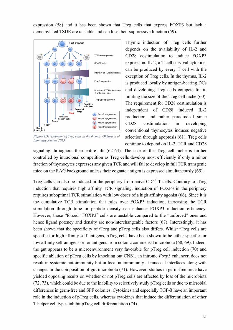
FOXP3+ Treg cells can be selected in the thymus (tTreg; often also referred to as natural nTreg) or be peripherally induced from naïve CD4+ T cells in vivo (pTreg) or in vitro (iTreg).
tTreg cells are selected in the thymus starting from the CD4CD8 double positive but mostly in the CD4 single positive T cell stage. Unlike conventional T cells, tTreg cells are selected based on high affinity to self-peptides and the strength of the TCR signal has been shown to correlate with the degree of FOXP3 induction with the exception for the very highest affinities that result in negative selection (50-52). Thus, Treg cells express TCRs with affinities that fall between those of positive and negative selection (53). Despite the view of FOXP3 being the master regulator of Treg lineage specification, FOXP3 expression alone is in fact not sufficient to confer a full Treg phenotype as illustrated by the fact that activated human conventional T cells (Tconv) transiently express FOXP3 without adopting a suppressive phenotype and that retroviral overexpression of FOXP3 in Tconv cells only reproduced one third of the Treg cell signature genes (54). Indeed, it has been shown very recently, that FOXP3 is in fact not required for the induction of most Treg cell signature genes, but that it augments a pre-established pattern of gene expression and repression. Instead, patterns of Treg cell specific super-enhancers that are established by the genome organizer Satb1 and potentially others, are active before the expression of FOXP3 and might be a key-determinant in Treg cell lineage specification (55). Induction of a full Treg phenotype additionally requires the establishment of a Treg specific demethylated region (TSDR). The TSDR is an evolutionary conserved CpG rich element within the Foxp3 locus (56), whereas the nTreg-Me additionally includes Treg signature genes such as CTLA-4 (57). tTreg cell-type CpG hypomethylation occurs gradually, starting in the thymus and being finished in the periphery. It requires TCR ligation with self-ligands but is established independently of FOXP3 expression as illustrated by retroviral overexpression of FOXP3 in Tconv cells as well as FOXP3 expressing iTreg cells that both lack a fully demethylated TSDR (54, 57). A demethylated TSDR is crucial for stable FOXP3
15
expression (58) and it has been shown that Treg cells that express FOXP3 but lack a demethylated TSDR are unstable and can lose their suppressive function (59).
Thymic induction of Treg cells further depends on the availability of IL-2 and CD28 costimulation to induce FOXP3 expression. IL-2, a T cell survival cytokine, can be produced by every T cell with the exception of Treg cells. In the thymus, IL-2 is produced locally by antigen-bearing DCs and developing Treg cells compete for it, limiting the size of the Treg cell niche (60). The requirement for CD28 costimulation is independent of CD28 induced IL-2 production and rather paradoxical since CD28 costimulation in developing conventional thymocytes induces negative selection through apoptosis (61). Treg cells continue to depend on IL-2, TCR and CD28
signaling throughout their entire life (62-64). The size of the Treg cell niche is further controlled by intraclonal competition as Treg cells develop most efficiently if only a minor fraction of thymocytes expresses any given TCR and will fail to develop in full TCR transgenic mice on the RAG background unless their cognate antigen is expressed simultaneously (65).
Treg cells can also be induced in the periphery from naïve CD4+ T cells. Contrary to tTreg induction that requires high affinity TCR signaling, induction of FOXP3 in the periphery requires suboptimal TCR stimulation with low doses of a high affinity agonist (66). Since it is the cumulative TCR stimulation that rules over FOXP3 induction, increasing the TCR stimulation through time or peptide density can enhance FOXP3 induction efficiency. However, those “forced” FOXP3+ cells are unstable compared to the “unforced” ones and hence ligand potency and density are non-interchangeable factors (67). Interestingly, it has been shown that the specificity of tTreg and pTreg cells also differs. Whilst tTreg cells are specific for high affinity self-antigens, pTreg cells have been shown to be either specific for low affinity self-antigens or for antigens from colonic commensal microbiota (68, 69). Indeed, the gut appears to be a microenvironment very favorable for pTreg cell induction (70) and specific ablation of pTreg cells by knocking out CNS1, an intronic Foxp3 enhancer, does not result in systemic autoimmunity but in local autoimmunity at mucosal interfaces along with changes in the composition of gut microbiota (71). However, studies in germ-free mice have yielded opposing results on whether or not pTreg cells are affected by loss of the microbiota (72, 73), which could be due to the inability to selectively study pTreg cells or due to microbial differences in germ-free and SPF colonies. Cytokines and especially TGF-b have an important role in the induction of pTreg cells, whereas cytokines that induce the differentiation of other T helper cell types inhibit pTreg cell differentiation (74).
Figure 1Development of Treg cells in the thymus. Ohkura et al. Immunity Review 2013
16
In vitro, naïve CD4+ T cells can be induced to become FOXP3 expressing iTreg cells by stimulation through the TCR in the presence of TGF-b (75) and IL-2 (76). Addition of retinoic acid can further increase the efficiency of iTreg cell generation (77, 78). Furthermore, expression of high amounts of FOXP3 in iTreg cells requires upregulation of CTLA-4 (79). There is significant debate on whether iTreg cells suppress equally well as nTreg and pTreg cells and their therapeutic use is further hindered by their demonstrated instability caused by only partial demethylation of the TSDR (56, 59).
3.2 PHENOTYPE
Whilst there are many cells that can acquire suppressive abilities in the immune system, FOXP3+ tTreg cells are unique because they are developmentally specialized for just this purpose. FOXP3+ T cells commonly express molecules that are associated with anergy in Tconv cells, like CTLA-4, PD-1, FR4 and CD73 and do not proliferate upon stimulation via the TCR in vitro in the absence of other cells.
Mouse Treg cells can be specifically identified by their expression of FOXP3 or if viable cells are needed based on CD4+CD25hi expression. Attempts to distinguish between tTreg and pTreg cells based on higher expression of Helios and Neuropilin-1 (Nrp-1) on tTreg cells (80-82) were soon questioned, as both Helios and Nrp-1 can be induced in peripheral Treg cells (83, 84) and both Helios+ and Helios- tTreg cells coexist in humans (85). Despite studies showing that neither Helios nor Nrp-1 can distinguish between tTreg and pTreg cells (86), no consensus has been reached and due to the lack of better alternatives, many studies continue to use these markers. LAP, a component of latent TGF-b, is bound to tTreg cell membranes through the anchoring protein GARP and has also been discussed as a tTreg marker but at least in mice, GARP can also be expressed by pTreg and iTreg cells (87, 88).
In humans, Treg cells cannot be identified solely by the expression of FOXP3 or CD25, since activated human Tconv cells can transiently express FOXP3 and a large percentage expresses high levels of CD25, whereas naïve Treg cells can have lower CD25 expression. Instead, a combination of various cell surface markers, such as CD62L, CD127, GITR, CD73, CD39 CD45RA, CD45RO, ICOS and HLA-DR is used to identify different subpopulations of human Treg cells (89).
3.3 FUNCTIONS
Treg cells are a double-edged sword. They are of major importance to control normal immune responses and to uphold tolerance to self but are also involved in autoimmunity and failure to combat cancer. For example, Treg cells have been shown to accumulate or be induced at tumor sites and depletion of Treg cells rescues mice from succumbing to cancer (90).
Just like any other T cell, Treg cells need to be activated through their TCR. However, once activated, they can suppress in an antigen non-specific manner. Treg cells have numerous ways of suppressing target cells, as elaborated below and the exact impact of each pathway remains
17
to be elucidated. However, it appears that the location, the state of the immune system and the subtype of Treg cell all influence which pathway predominates in a given situation (91).
3.3.1 Regulation of T cell function
Treg cells can directly inhibit other T cells through various mechanisms, some of which are listed below.
3.3.1.1 IL-2 Inhibition/Deprivation
Treg cells do not produce the T cell survival factor IL-2 themselves due to FOXP3 mediated gene repression and are thus dependent on IL-2 produced by other T cells for their survival (92). Since Treg cells constitutively express high levels of the high-affinity IL-2 receptor a-chain CD25, they can effectively take up IL-2 from the periphery and this IL-2 consumption has been suggested as a suppressive mechanism of Treg cells (93). Furthermore, Treg cells have been shown to inhibit production of IL-2 mRNA in responder Tconv cells (94).
3.3.1.2 ATP degradation
Treg cells co-express high surface levels of the ectonucleotidases CD73 and CD39 that are involved in the catabolism of extracellular nucleotides such as ATP to immunosuppressive adenosine. Treg cells can thus increase the amount of pericellular adenosine which suppresses effector functions of activated Tconv cells that express the high affinity adenosine receptor A2A (23, 95).
3.3.1.3 Production of anti-inflammatory cytokines
A number of studies have implicated an important contribution of anti-inflammatory cytokines such as IL-10 and TGF-b to Treg cell mediated suppression in vivo (96). However, since physical separation of Treg cells from responder T cells abrogates suppression in vitro, the magnitude of influence of anti-inflammatory cytokines to Treg cell function remains debated (94). This is further illustrated by the lack of spontaneous autoimmunity in mice deficient for Treg cell specific TGF-b (97), whereas mice deficient for Treg cell specific IL-10 suffer from inflammation that is restricted to environmental surfaces such as the skin, lung and colon (98).
IL-35, a relatively new inhibitory cytokine of the IL-12 heterodimeric cytokine family, consisting of the Ebi3 and Il12a chains, is constitutively secreted by Treg cells and secretion is further potentiated during active suppression of Tconv cells. Treg cells from either Ebi3 knock-out (KO) or Il12a KO mice are less suppressive in vitro and fail to control homeostatic expansion or cure inflammatory bowel disease in vivo. Ectopic expression of IL-35 confers suppressive capacity on Tconv cells and recombinant IL-35 can suppress T cell proliferation (99).
3.3.1.4 Cell cytotoxicity
Besides suppressing the activation of other immune cells, Treg cells can also directly kill target cells. Human Treg cells have been shown to express granzyme A and to kill target cells
18
in a perforin dependent fashion (100). Similarly, mouse Treg cells have been shown to express granzyme B and to kill target cells in a manner that can be either perforin dependent or independent (101, 102).
3.3.2 Regulation of APC function
Instead of directly suppressing T cells, Treg cells can suppress the costimulatory capacity of APCs and thereby indirectly inhibit the activation of Tconv cells.
3.3.2.1 CTLA-4
Treg cells can reduce the costimulatory capacity of APCs by reducing their expression of CD80/86 and can thus inhibit the activation of T cells. This two-step process requires an initial lymphocyte function-associated antigen 1 (LFA-1) dependent clustering of Treg cells around APCs, outcompeting access of Tconv cells to APCs, and a second LFA-1 and CTLA-4 dependent active down-regulation of CD80/86 (103). This CD80/86 downregulation is even seen in the presence of strong APC maturation stimuli (103, 104). In the case of CD80 but not CD86, reduced surface expression has been shown to be paralleled by reduced CD80 mRNA expression (104). An alternative mechanism for reduced expression of CD80/86 has been shown to involve trans-endocytosis. This process involves CTLA-4 mediated uptake of CD80/86 from the surface of APCs into CTLA-4+ cells, where they are degraded in lysosomes. This trans-endocytosis occurs within minutes after cell contact and is highly efficient as a ratio of 1:8 (CTLA-4:CD86) is sufficient to achieve functionally relevant depletion as seen in reduced APC costimulatory capacity (105).
3.3.2.2 IDO
Indoleamine 2,3-dioxygenase (IDO) catalyzes the first step in the catabolism of the essential amino acid tryptophan to kynurenine. Treg cells can induce production of IDO in DCs via interactions between CTLA-4 and CD80/86 (106). Induction of IDO depletes tryptophan from the microenvironment, thereby resulting in inhibition of T cell activation and proliferation (107). Furthermore, tryptophan metabolites have been shown to induce apoptosis in T cells (108).
3.3.2.3 Lag-3
Lymphocyte-activation gene 3 (Lag-3) is a CD4 homologue that can bind to MHC-II and induce an inhibitory signal that suppresses DC maturation and costimulatory capacity (109). Antibodies against Lag-3 can partially block Treg cell mediated suppression and Lag-3 deficient Treg cells are less efficient suppressors than their wild type (WT) counterparts. The degree of Lag-3 mediated suppression appears to depend on both the strength of T cell stimulation as well as the ratio of responder to suppressor cells (110). As activated human T cells can express MHC-II, a direct suppression of T cells is possible.

19
3.3.2.4 Nrp-1
Nrp-1, a cofactor for vascular endothelial growth factor, is preferentially expressed on Treg cells and one of the markers used to identify human Treg cells. Nrp-1 prolongs interactions between DCs and Treg cells (111). It remains unclear whether Nrp-1 merely marks a pool of activated effector Treg cells or whether Nrp-1 itself has suppressive capacity, as studies with blocking antibodies have led to conflicting results (111, 112). Studies with Nrp-1 deficient Treg cells have shown that Nrp-1 is dispensable for regulation of immune homeostasis but required for suppression of anti-tumor immune responses (113, 114).
3.4 PLASTICITY AND STABILITY
It has been suggested in recent years that Treg cells can become unstable under certain pathologic conditions and turn into pro-inflammatory IFNg and IL-17 producing ex-FOXP3 effector T cells (115, 116). However, these studies have assumed that FOXP3+ Treg cells are tTreg cells and not a mixture of tTreg and pTreg cells. A study by Rubtsov took this into account and refuted tTreg instability (117). Lack of reliable surface markers to distinguish tTreg from pTreg cells makes reliable studies on their differential stability difficult but as it has been shown that pTreg cell TSDR demethylation is incomplete compared to tTreg cells, it is very likely that the observed degree of plasticity stems from pTreg cells. This is especially likely due to the developmental relationship of Th17 and extrathymically induced Treg cells, at least in vitro. Induction of both cell types is dependent on TGF-b but induction of Th17 cells requires the additional stimulation with IL-6 in order to overcome the suppressive effect of FOXP3 on RORgt, the master transcription factor for Th17 cells (118). Today, the predominant view amongst experts is that fully committed tTreg cells indeed are a remarkably stable population but that Tconv cells can transiently express FOXP3 and that recently induced pTreg cells can lose FOXP3 expression and suppressive function under high inflammatory pressure (119, 120).
20
4 CTLA-4 Cytotoxic T-lymphocyte antigen 4 (CTLA-4), a member of the immunoglobulin superfamily, was first discovered in 1987, as a cDNA isolated from activated CD8+ cytotoxic T cells (121). In 1991, CTLA-4 was shown to share close sequence homology to CD28 and to bind to the same CD80/86 ligands on APCs, albeit with a ~20-fold higher affinity (122, 123). At that time, a debate started whether engagement of CTLA-4 transmitted a positive or negative signal as co-cultures with T cells, APCs and soluble CTLA-4 showed decreased T cell proliferation (123), which could have been due to CTLA-4 on T cells being needed as a positive stimulus or because the CD28:CD80/86 interaction was blocked. This dispute was settled in 1995 when two independent groups demonstrated that loss of CTLA-4 in mice resulted in a lymphoproliferative disorder and early lethality, confirming a negative role beyond doubt (124, 125).
4.1 EXPRESSION
CTLA-4 forms homodimers and its expression is rapidly induced on T cells in response to activation whereby accumulation in the immune synapse is proportional to the TCR signal strength (126). This rapid expression is possible due to the majority of CTLA-4 being stored in intracellular vesicles (127). Surface CTLA-4 is rapidly turned over through clathrin-dependent endocytosis followed by either re-expression or lysosomal degradation and this requires the highly conserved cytoplasmic tail of CTLA-4 (128). Recently, it has been shown that interaction of internalized CTLA-4 with lipopolysaccharide-responsive and beige-like anchor protein (LRBA) in recycling endosomes rescues CTLA-4 from lysosomal degradation and patients with homozygous loss of LRBA suffer from autoimmunity due to decreased surface CTLA-4 levels and thus impaired Treg cell function (129, 130).
Contrary to the induced expression on Tconv cells, Treg cells constitutively express high levels of surface CTLA-4 and this is vital for their suppressive function, as loss of CTLA-4 specifically on Treg cells results in similar, albeit delayed lethal autoimmunity as total loss of CTLA-4 (131).
Apart from T cells, a few studies have reported CTLA-4 to be expressed on subsets of B cells (132, 133) and DCs (134), however detection of these low-levels often requires special staining methods and the overall expression and function of CTLA-4 on these non-T cells requires further clarification.
4.2 FUNCTIONS
CTLA-4 is an important regulator of immune homeostasis and autoimmunity. Loss of CTLA-4 on all T cells leads to a lymphoproliferative disorder and early lethality (124, 125). FOXP3+ Treg cell specific loss of CTLA-4 results in a similar phenotype, albeit a bit delayed, depicting a vital role of CTLA-4 for Treg cell suppressive function (131). Furthermore, resting human T cells can be rendered suppressive through transfection with CTLA-4 (135). Recently, human CTLA-4 haploinsufficiency was identified as the underlying cause for an autosomal
21
dominant immune dysregulation syndrome with autoimmune infiltration, decreased Treg cell CTLA-4 expression and suppressive function accompanied by a decrease in circulating B cells in some but not all patients (136, 137).
CTLA-4 can function in various different ways that can be classified as either T cell intrinsic/autonomous or as T cell extrinisic/non-autonomous. Since CTLA-4 is expressed on both activated Tconv and Treg cells, both cell types can contribute to CTLA-4 mediated suppression, however Treg cells are more efficient/effective due to their constitutive high expression of CTLA-4.
4.2.1 T cell intrinsic or autonomous functions
T cell intrinsic functions of CTLA-4 (see figure below) include the competition with CD28 for access to CD80/86 ligands on the surface of APCs. Since the CTLA-4 homodimer has a ~20-fold higher affinity to these ligands, CTLA-4 can efficiently outcompete CD28 and thus deprive T cells from receiving costimulatory signals through CD28. Antibody-mediated crosslinking of CTLA-4 leads to inhibition of T cell proliferation and overexpression of ligand-independent CTLA-4 (see 4.3) can partially rescue the CTLA-4 KO phenotype, depicting a B7 independent function through transmission of inhibitory signals. Indeed, CTLA-4 has been shown to recruit various phosphatases and to reduce phosphorylation of components of the TCR signaling cascade (138). However, there is no consensus between studies as to which molecules are affected, the cytoplasmic tail of CTLA-4 lacks an ITIM motif, gene expression studies failed to detect a signature of active negative regulation in CTLA-4+ cells and to this day no cohesive inhibitory pathway has been identified (139). A conserved motif in the cytoplasmic tail of CTLA-4 interacts with the protein kinase C (PKC)-h and recruits it to the Treg cell immune synapse, whereas in the Tconv cell immune synapse, CD28 instead recruits PKC-q. In the absence of CTLA-4 mediated PKC-h recruitment, due to PKC-h deficiency, Treg cell development is normal but their suppressive activity is markedly impaired due to the defective assembly of a focal adhesion disassembly complex that increases cell motility and serial engagement of APCs (140).
TCR ligation reduces T cell motility which is necessary for stable T cell:APC interactions, the formation of the immunological synapse and the initiation of T cell priming. This process, called the “TCR stop signal”, can be reversed by CTLA-4 ligation, shortening the interaction time of T cells and APCs albeit only in Tconv but not in Treg cells (141). Additionally, CTLA-4 ligation induces the expression of LFA-1 on T cells, which binds to intercellular adhesion molecule 1 (ICAM-1) on APCs. This interaction is a prerequisite for T cell motility and migration through high endothelial venules and CTLA-4 thus increases T cell motility, further decreasing the chances of stable T cell APC interactions (138, 142).
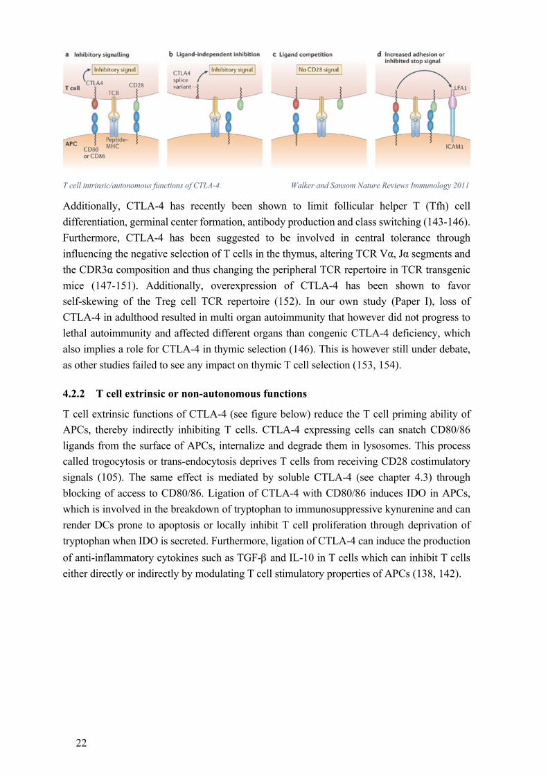
22
T cell intrinsic/autonomous functions of CTLA-4. Walker and Sansom Nature Reviews Immunology 2011
Additionally, CTLA-4 has recently been shown to limit follicular helper T (Tfh) cell differentiation, germinal center formation, antibody production and class switching (143-146). Furthermore, CTLA-4 has been suggested to be involved in central tolerance through influencing the negative selection of T cells in the thymus, altering TCR Vα, Jα segments and the CDR3α composition and thus changing the peripheral TCR repertoire in TCR transgenic mice (147-151). Additionally, overexpression of CTLA-4 has been shown to favor self-skewing of the Treg cell TCR repertoire (152). In our own study (Paper I), loss of CTLA-4 in adulthood resulted in multi organ autoimmunity that however did not progress to lethal autoimmunity and affected different organs than congenic CTLA-4 deficiency, which also implies a role for CTLA-4 in thymic selection (146). This is however still under debate, as other studies failed to see any impact on thymic T cell selection (153, 154).
4.2.2 T cell extrinsic or non-autonomous functions
T cell extrinsic functions of CTLA-4 (see figure below) reduce the T cell priming ability of APCs, thereby indirectly inhibiting T cells. CTLA-4 expressing cells can snatch CD80/86 ligands from the surface of APCs, internalize and degrade them in lysosomes. This process called trogocytosis or trans-endocytosis deprives T cells from receiving CD28 costimulatory signals (105). The same effect is mediated by soluble CTLA-4 (see chapter 4.3) through blocking of access to CD80/86. Ligation of CTLA-4 with CD80/86 induces IDO in APCs, which is involved in the breakdown of tryptophan to immunosuppressive kynurenine and can render DCs prone to apoptosis or locally inhibit T cell proliferation through deprivation of tryptophan when IDO is secreted. Furthermore, ligation of CTLA-4 can induce the production of anti-inflammatory cytokines such as TGF-b and IL-10 in T cells which can inhibit T cells either directly or indirectly by modulating T cell stimulatory properties of APCs (138, 142).

23
T cell extrinsic/non-autonomous functions of CTLA-4. Walker and Sansom Nature Reviews Immunology 2011
4.2.3 Models of CTLA-4 function
Combining the different mechanisms of action of CTLA-4 under steady-state or inflammatory conditions, two models of action become apparent that work in parallel to shape the quality and magnitude of the immune response.
Adapted from Egen et al. Nat. Immunol. 2002
In situations of limited CD28 costimulation, CTLA-4 regulates the activation threshold of T cells, i.e. the combined strength of TCR and CD28 stimulation that is necessary to enter the cell cycle. In the activation threshold model, CTLA-4 will inhibit activation of autoreactive T cells in the absence of inflammation. Under inflammatory situations, where CD28 costimulation is not the limiting factor, CTLA-4 becomes powerless to regulate the activation threshold but instead regulates the effector T cell’s capacity to divide by inhibiting several components of the cell cycle machinery. The degree of division regulation correlates with the strength of the TCR signal. This has two effects: (I) immune responses are terminated before

24
they get out of control and cause tissue damage and (II) the immune response is broadened by allowing more T cell clones to take part, instead of only those with the highest affinities that would otherwise outcompete weaker affinity clones for access to antigen bearing APCs and available costimulatory signals.
4.3 CTLA-4 ISOFORMS
The Ctla‑4 gene consists of four exons, of which exon 1 encodes the leader peptide, exon 2 the ligand binding domain, exon 3 the transmembrane region and exon 4 the short cytoplasmic tail. In addition to full length CTLA-4, three splice variants exist. (I) Soluble CTLA-4 that lacks the transmembrane domain is elevated in several autoimmune diseases and has been shown to reduce antigen-specific T cell proliferation and to modulate cytokine secretion (155). A SNP in the 3’ UTR of CTLA-4 is associated with increased risk of developing autoimmunity and had originally been linked to decreased levels of soluble CTLA-4, however this could not be reproduced in subsequent studies (155). (II) 1/4 CTLA-4 lacks the ligand binding and transmembrane domain and transgenic overexpression has been shown to enhance disease in two models of autoimmunity and to elevate levels of auto-antibodies and accumulation of memory T cells in aged mice, albeit without the massive lymphoproliferation seen in CTLA-4 KO mice (156, 157). (III) Ligand-independent (li) CTLA-4 lacks the extracellular domain and thus the ability to bind to CD80/86. Overexpression of this isoform in CTLA-4 KO mice inhibits in vitro T cell responses, prevents tissue infiltration and ameliorates but does not rescue from lymphoproliferative disease (158). Treg cells from NOD mice that genetically lack li-CTLA-4 but were engineered to re-express physiological levels of the li-CTLA-4 isoform only, were shown to confer comparable disease protection as WT Treg cells in a type I diabetes model, whereas activated Tconv cells were significantly reduced in these mice. Despite reduced effector functions, Tconv cells expressing only the li-CTLA-4 isoform, were more potent in transferring disease and protection from autoimmunity required the presence of full length CTLA-4. This indicates that li-CTLA-4 is important for regulating the tonic TCR signaling but that the ligand binding domain is required for full control of autoimmunity during stimulation with antigen (159). However, since ligand-independent CTLA-4 is only found in mice and not humans, it has not been studied extensively.
4.4 CTLA-4 BASED THERAPY
Antibodies that block CTLA-4 were the first successful therapeutics in the class of immune checkpoint therapy, which targets regulatory pathways in T cells with the aim to improve antitumor immune responses.
CTLA-4 isoforms. Adapted from Valk et al. Trends in Immunology 2008
25
Ipilimumab is a fully human IgG1 antibody that blocks CTLA-4 and has been FDA (food and drug administration) approved in 2011 for the treatment of unresectable or metastatic melanoma as well as in adjuvant treatment of patients with cutaneous melanoma that have undergone tumor resection. Treatment with ipilimumab leads to an enhanced overall survival of patients with metastatic melanoma (160) but failed to improve overall survival of prostate cancer patients despite antitumor activity in a subset of patients (161). The effects of blocking CTLA-4 appear to be rather slow, as those melanoma patients that achieved complete remission required a median of 30 months to do so. However, effects were very long lasting, as the vast majority of patients remained remission free for extended periods, the longest lasting >99 months (162, 163). 10-15% of metastatic melanoma patients treated with ipilimumab develop serious immune related adverse events that often manifest in bowel inflammation, severe skin problems and problems affecting the liver, nerves or endocrine system that can be life-threatening and require systemic treatment with high-dose corticosteroids. Furthermore, many patients do not respond to treatment and to this day there are no biomarkers available to predict which patients might benefit from anti-CTLA-4 therapy (162). Interestingly, the anti-cancer effects of anti-CTLA-4 therapy have been shown to be dependent on the gut microbiota, more specifically the outgrowth of Bacteroides fragilis upon treatment, which increased IL-12 dependent Th1 responses and thus controlled the tumor without interrupting intestinal integrity (164).
Abatacept is a fusion protein of the Fc part of human IgG1 fused to the extracellular domain of CTLA-4. Abatacept binds to CD80/86 molecules on APCs and thus deprives T cells from receiving CD28 costimulatory signals. Abatacept has been approved by the FDA for the treatment of rheumatoid arthritis and juvenile idiopathic arthritis in 2005. Abatacept can be used as a first line monotherapy but is more commonly used only in those patients that failed to respond to treatment with disease-modifying antirheumatic drugs (DMARDs), due to its high cost (165). Treatment significantly improves disease symptoms by directly or indirectly affecting T cells, myeloid DCs, B cells and macrophages. Furthermore, a bone protective effect through inhibition of osteoclast differentiation has been reported (166). Abatacept is generally well tolerated with the most common mild adverse events being headache, upper respiratory tract infections, nasopharyngitis and nausea but should not be used concomitantly with other biological agents as this significantly increases the incidence of serious infections and malignancies (167). Abatacept has undergone trials for various other indications but outcomes have been rather disappointing, with lack of efficacy in ulcerative colitis, multiple sclerosis, asthma and systemic lupus erythematosus (165).
Belatacept is a mutated version of abatacept, differing in two amino acids, which increases its binding avidity to CD80 and CD86. This makes belatacept a more effective blocker of CD28 costimulation and thus better suited to counteract transplant rejection caused by recipient effector T cells. In 2011 belatacept has been approved by the FDA for the treatment of kidney transplant recipients as treatment has been shown to significantly prolong both transplant and patient survival (168).
26
5 AUTOIMMUNE DISEASES Even though most autoreactive T cells are removed during thymic selection or kept under control in the periphery, individual autoreactive T cells escape from these control mechanisms and can mount an immune response against self. An immune response against self is called autoimmune disease and can be classified into organ specific such as type I diabetes and Addison’s disease and systemic such as systemic lupus erythematosus and rheumatoid arthritis. Around 7-9% of the world population is affected by autoimmune diseases (169) and almost all of them disproportionally affect women (170). Despite the relative rarity of autoimmune diseases, they cause high individual suffering, socioeconomic costs and are one of the most common causes for death in young to middle-aged women in the USA (171).
5.1 RHEUMATOID ARTHRITIS
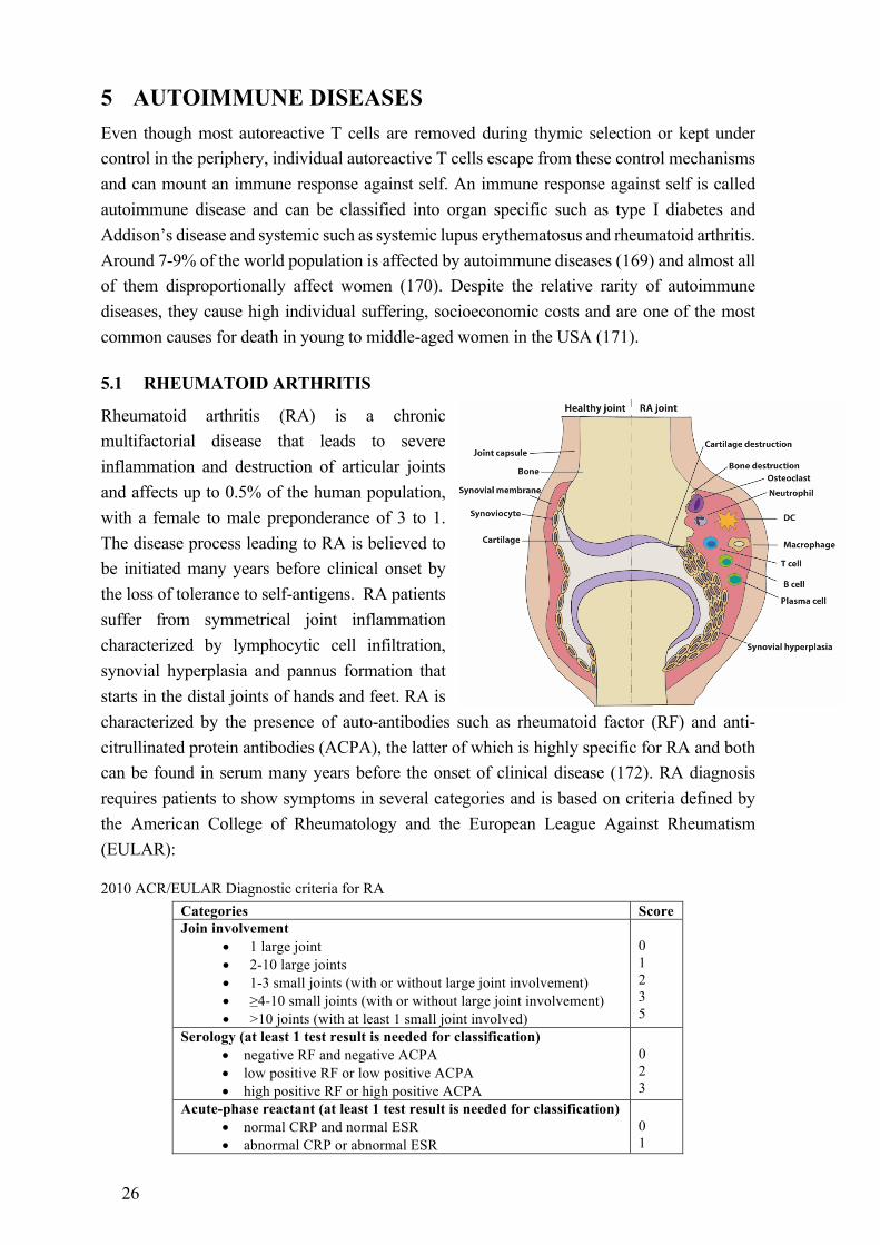
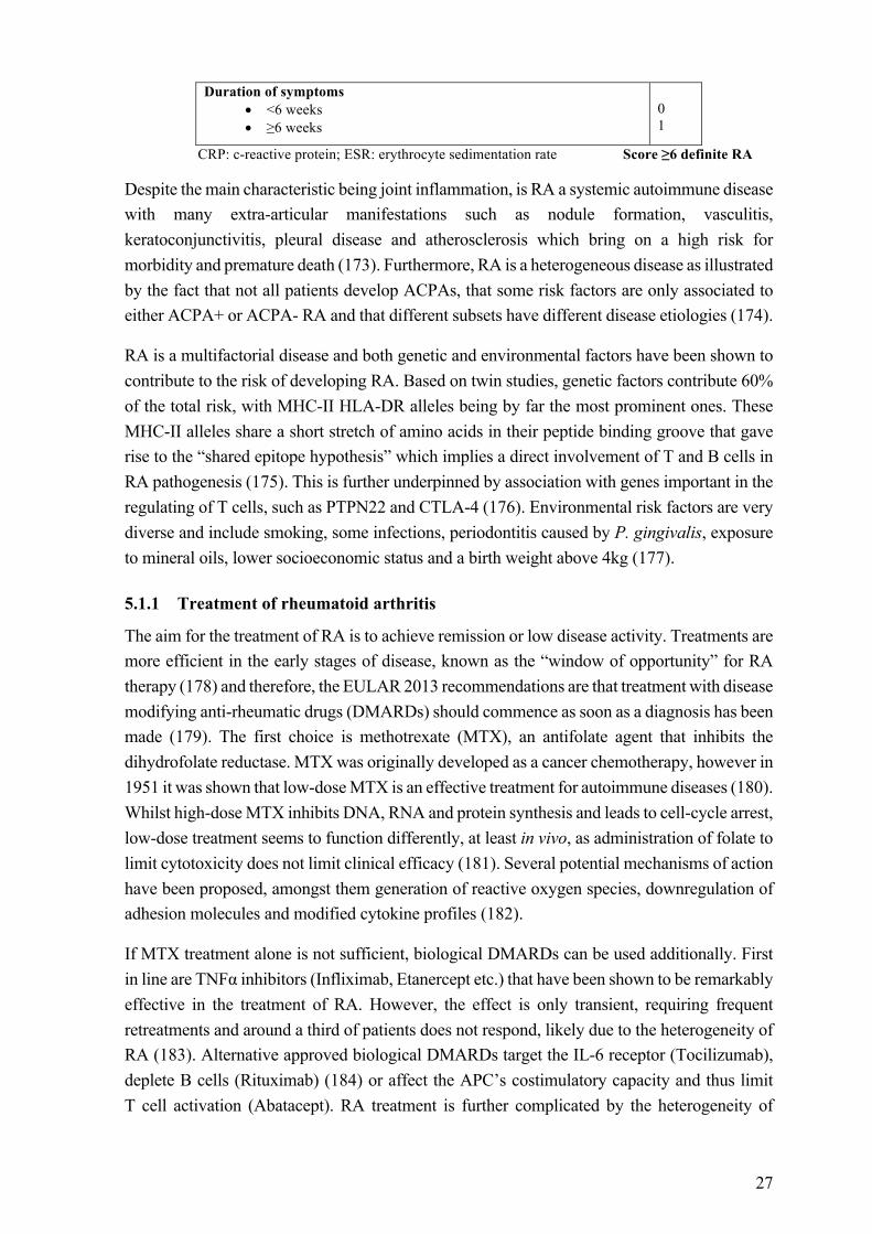
Rheumatoid arthritis (RA) is a chronic multifactorial disease that leads to severe inflammation and destruction of articular joints and affects up to 0.5% of the human population, with a female to male preponderance of 3 to 1. The disease process leading to RA is believed to be initiated many years before clinical onset by the loss of tolerance to self-antigens. RA patients suffer from symmetrical joint inflammation characterized by lymphocytic cell infiltration, synovial hyperplasia and pannus formation that starts in the distal joints of hands and feet. RA is characterized by the presence of auto-antibodies such as rheumatoid factor (RF) and anti-citrullinated protein antibodies (ACPA), the latter of which is highly specific for RA and both can be found in serum many years before the onset of clinical disease (172). RA diagnosis requires patients to show symptoms in several categories and is based on criteria defined by the American College of Rheumatology and the European League Against Rheumatism (EULAR):
2010 ACR/EULAR Diagnostic criteria for RA Categories Score Join involvement
• 1 large joint • 2-10 large joints • 1-3 small joints (with or without large joint involvement) • ≥4-10 small joints (with or without large joint involvement) • >10 joints (with at least 1 small joint involved)
0 1 2 3 5
Serology (at least 1 test result is needed for classification) • negative RF and negative ACPA • low positive RF or low positive ACPA • high positive RF or high positive ACPA
0 2 3
Acute-phase reactant (at least 1 test result is needed for classification) • normal CRP and normal ESR • abnormal CRP or abnormal ESR
0 1
27
Duration of symptoms • <6 weeks • ≥6 weeks
0 1
CRP: c-reactive protein; ESR: erythrocyte sedimentation rate Score ≥6 definite RA
Despite the main characteristic being joint inflammation, is RA a systemic autoimmune disease with many extra-articular manifestations such as nodule formation, vasculitis, keratoconjunctivitis, pleural disease and atherosclerosis which bring on a high risk for morbidity and premature death (173). Furthermore, RA is a heterogeneous disease as illustrated by the fact that not all patients develop ACPAs, that some risk factors are only associated to either ACPA+ or ACPA- RA and that different subsets have different disease etiologies (174).
RA is a multifactorial disease and both genetic and environmental factors have been shown to contribute to the risk of developing RA. Based on twin studies, genetic factors contribute 60% of the total risk, with MHC-II HLA-DR alleles being by far the most prominent ones. These MHC-II alleles share a short stretch of amino acids in their peptide binding groove that gave rise to the “shared epitope hypothesis” which implies a direct involvement of T and B cells in RA pathogenesis (175). This is further underpinned by association with genes important in the regulating of T cells, such as PTPN22 and CTLA-4 (176). Environmental risk factors are very diverse and include smoking, some infections, periodontitis caused by P. gingivalis, exposure to mineral oils, lower socioeconomic status and a birth weight above 4kg (177).
5.1.1 Treatment of rheumatoid arthritis
The aim for the treatment of RA is to achieve remission or low disease activity. Treatments are more efficient in the early stages of disease, known as the “window of opportunity” for RA therapy (178) and therefore, the EULAR 2013 recommendations are that treatment with disease modifying anti-rheumatic drugs (DMARDs) should commence as soon as a diagnosis has been made (179). The first choice is methotrexate (MTX), an antifolate agent that inhibits the dihydrofolate reductase. MTX was originally developed as a cancer chemotherapy, however in 1951 it was shown that low-dose MTX is an effective treatment for autoimmune diseases (180). Whilst high-dose MTX inhibits DNA, RNA and protein synthesis and leads to cell-cycle arrest, low-dose treatment seems to function differently, at least in vivo, as administration of folate to limit cytotoxicity does not limit clinical efficacy (181). Several potential mechanisms of action have been proposed, amongst them generation of reactive oxygen species, downregulation of adhesion molecules and modified cytokine profiles (182).
If MTX treatment alone is not sufficient, biological DMARDs can be used additionally. First in line are TNFα inhibitors (Infliximab, Etanercept etc.) that have been shown to be remarkably effective in the treatment of RA. However, the effect is only transient, requiring frequent retreatments and around a third of patients does not respond, likely due to the heterogeneity of RA (183). Alternative approved biological DMARDs target the IL-6 receptor (Tocilizumab), deplete B cells (Rituximab) (184) or affect the APC’s costimulatory capacity and thus limit T cell activation (Abatacept). RA treatment is further complicated by the heterogeneity of
28
disease as it has been shown that ACPA+ and ACPA- disease require different treatment regimens (185).
5.1.2 Regulatory T cells and CTLA-4 in rheumatoid arthritis
Treg cells have been shown to modulate arthritis in mice as depletion enhances disease whereas transfer ameliorates disease (186-188). In RA patients however, the role of Treg cells is not as clear. While there is no consensus on frequencies of peripheral blood Treg cells in RA patients, most studies report increased frequencies in synovial fluid (189). Yet it remains controversial whether Treg cells at the site of inflammation are fully functional (190-194) or if it is the synovial effector T cells that have become resistant to suppression (195). TNFa, a key inflammatory cytokine in the pathogenesis of RA, inhibits Treg cells via dephosphorylation of FOXP3 and treatment with anti-TNFa restores Treg cell function, thereby potentially explaining some of the success of this therapy in RA (191).
Defects in CTLA-4 regulation have been reported to contribute to the abnormal Treg cell function observed in synovial fluid of RA patients (196, 197) and certain polymorphisms in the Ctla‑4 gene are associated with an increased risk of developing RA (198). Its importance in RA is further underlined by the successful therapeutic treatment with CTLA-4Ig fusion protein (Abatacept) (179, 199).
5.2 MULTIPLE SCLEROSIS
Multiple sclerosis (MS) is a complex chronic autoimmune disease of the CNS with progressive neuronal demyelination. There is a female to male preponderance of two to one and disease can be either relapsing-remitting or progressive. Symptoms cover a wide variety of neurological disorders such as difficulty of speech, ataxia and paralysis depending on in which CNS area inflammatory infiltrates are located. The exact cause of MS is unknown but myelin-specific CD4+ Th1 and Th17 cells have been shown to play a role in the initiation of neuropathology and both genetic and environmental factors are believed to be involved. Currently, there is no cure for MS and treatment aims at reducing disease activity and slowing disease progression (200).
29
6 ANIMAL MODELS FOR AUTOIMMUNE DISEASES Due to the complex nature of most autoimmune diseases, with contributions from many genes and environmental factors, finding the underlying disease mechanism in patients is very difficult. This is why animal models are often used as a tool since the genetic as well as the environmental background can be controlled. Due to the complexity of most autoimmune diseases, no single animal model can recapitulate the entire spectrum of heterogeneous human diseases but each model is suitable to study one or more specific features of the corresponding human disease. Animal models are especially valuable in diseases where patient biopsies can only be obtained postmortem, as for example brain samples of MS patients.
6.1 MOUSE MODELS FOR RHEUMATOID ARTHRITIS
There are many different mouse models for RA and they can be divided into spontaneous and induced models. Spontaneous models include TNFa overproducing transgenic mice, the TCR transgenic K/BxN model and the SKG model, although arthritis development in this model often requires an i.p. injection of mannan. Examples of induced RA models include glucose-6-phosphate isomerase (GPI)-induced arthritis, collagen-antibody-induced arthritis and the most commonly used and best studied, collagen-induced arthritis. All of these models mimic one or more hallmarks of RA, making them very well suited to study these specific aspects but one should be aware that no animal model mimics RA to 100%. For example, ACPAs, one of the most prominent features of RA, have so far not been found in any animal model.
6.1.1 Collagen-induced arthritis
Collagen-induced arthritis (CIA) is induced in susceptible mouse strains by the intradermal injection of heterologous type-II collagen (CII), the predominant component of articular joint cartilage, in Complete Freund’s adjuvant (CFA) often followed by a booster immunization with CII in Incomplete Freund’s adjuvant (IFA) 21-35 days later. CIA closely resembles RA, exhibiting erythema, synovial hyperplasia, influx of inflammatory cells, pannus formation and bone erosion. The major genetic factor determining arthritis susceptibility is MHC-II, HLA-DR4 molecules in RA and H2-q,r and b haplotypes in CIA (201). These H2 molecules have been shown to bind the galactosylated immunodominant T cell epitope CII256-270 on rat CII with high affinity, whereas the endogenous mouse CII epitope that only differs by a single amino acid shows much weaker binding (202). CIA is T- and B cell dependent and both cell types reactive to glycosylated CII can be found in RA patients (203-206). Pro-inflammatory cytokines, and especially IFNg, IL-17 and TNFa are involved in CIA pathogenicity (207), whereas Treg cells have a mitigating effect (186-188). In our own studies, we could show that CTLA-4 is an important regulator of all stages of CIA and that CTLA-4 expressed by Tconv cells and Treg cells regulates the priming and inflammatory tissue attack respectively (Paper II). Furthermore, loss of CTLA-4 can break established T cell tolerance in mice (Paper II), whereas high CTLA-4 expression can confer a protective phenotype on T cells with increased inherent joint reactivity (Paper III).
30
6.2 MOUSE MODELS FOR MULTIPLE SCLEROSIS
Just like for RA, multiple mouse models exist for MS and they can also be roughly divided into spontaneous and induced models. Spontaneous MS models are based on mice that express TCRs or BCRs transgenic for CNS proteins, such as the 2D2 or IgHMOG mice that have myelin oligodendrocyte glycoprotein (MOG) specific T or B cells respectively. Induced models can be initiated by infection with a virus, such as Theiler’s murine encephalomyelitis virus, through treatment with toxins such as cuprizone or through the immunization of susceptible mice with self-CNS proteins in CFA. The latter is termed experimental autoimmune encephalomyelitis and will be described in more detail below. A major advantage of MS models compared to murine RA models is that disease can be transferred by activated CNS specific T cells (208).
6.2.1 Experimental autoimmune encephalomyelitis
Experimental autoimmune encephalomyelitis (EAE) is the most widely used model for MS. EAE is induced in susceptible mouse strains by the intradermal injection of self-CNS proteins such as MOG, myelin basic protein (MBP), proteolipid protein (PLP) or spinal cord homogenate in CFA. An additional injection of pertussis toxin is often required. Paralysis typically starts in the tail tip and progresses from there in a caudal to cranial fashion. Depending on the genetic background, EAE is either monophasic (C57BL) or relapsing-remitting (SJL/J). Pro-inflammatory cytokines like IFNg, IL-23, IL-17 and GM-CSF are crucial for disease development in EAE and both Th1 and Th17 cells from immunized mice can transfer disease to naïve recipients (208). The dependency on B cells is less stringent, as MOG1-125 protein induced EAE is dependent on B cells, whereas B cells are not required but can modulate disease severity in MOG peptide induced EAE (209, 210).

31
7 PRESENT INVESTIGATIONS This thesis comprises four studies, all of which deal with various aspects of regulation of autoimmunity. Paper I focuses on the spontaneous development of systemic autoimmunity in adult mice upon loss of CTLA-4 and that this loss can have disease promoting as well as protecting effects in different models of induced autoimmunity. Paper II looks more specifically into the role of CTLA-4 in the regulation of CIA, at which stage of disease and on which cell types CTLA-4 is important. Furthermore, the differential role of CTLA-4 in response to heterologous and autologous CII is studied. In Paper III, we study a novel model of tolerance induction in mice with increased inherent CII reactivity and the role of CTLA-4 on CII specific T cells. Paper IV deals with the more general question of whether bacteria in the gut microbiota have an impact on arthritis susceptibility in ROS deficient Ncf1 mice that have a predisposition to infections, inflammation and CIA.
7.1 PAPER I
Induction of autoimmune disease by deletion of CTLA-4 in mice in adulthood.
In this study, we focus on the role of CTLA-4 in the regulation of the adult immune system since most of our knowledge about CTLA-4 stems from KO mice that succumb to autoimmunity after a few weeks of life. We established a tamoxifen induced, Cre driven CTLA-4 depletion in adult mice, termed induced KO (iKO) mice. iKO mice developed profound lymphoproliferation, multi-organ lymphocytic cell infiltrations and hypergammaglobulinemia, reminiscent of germline and congenital CTLA-4 deficiency, albeit without reaching lethality. We showed that organ infiltration was accompanied by production of specific autoantibodies, a functional deficit in affected organs and that Treg and Tconv cells infiltrated into tissues simultaneously. Kinetic studies further revealed that Treg cells were more sensitive to loss of CTLA-4 and proliferated before levels of CTLA-4 where completely diminished, whereas Tconv cell numbers only started to increase upon complete loss. This led to an increased Treg:Tconv cell ratio, which together with the simultaneous tissue infiltration, could be the explanation why induced loss of CTLA-4 does not progress to fatal autoimmunity. Furthermore, T cell transfer studies into T cell deficient hosts revealed that thymic and peripheral T cells from iKO donors caused different pathologies, indicating a role for CTLA-4 in thymic selection and the shaping of the TCR repertoire. Finally, with the help of different models of autoimmunity, we could show that there is no default pathway of CTLA-4 regulation but that loss of CTLA-4 instead can have disease promoting or inhibiting effects, likely depending on the local milieu and the type of cells involved in the disease specific immune response.
32
7.2 PAPER II
CTLA-4 expressed by FOXP3+ Treg cells prevents inflammatory tissue attack and not T cell priming in arthritis.
In this study, we performed an in-depth analysis of how CTLA-4 regulates CIA. In a first set of experiments we studied the different disease stages of CIA and could show that CTLA-4 is not involved in T cell priming but rather in the effector phase of disease and also in the spreading of disease to previously unaffected limbs in the chronic phase. Despite the disease enhancing effect of loss of CTLA-4, effects on CII specific T and B cell responses were minor to non-existent. Since CIA is induced with heterologous rat CII, not all aspects of tolerance towards a true self-antigen are reflected and this could influence CTLA-4 mediated regulation. Therefore, iKO mice were crossed to mouse mutated collagen (MMC) mice, that express rat CII in a joint restricted fashion and are thus protected from CIA due to their T cells being naturally tolerized towards the immunodominant rat CII epitope. Loss of CTLA-4 completely broke tolerance in MMC+ mice, independent of depletion occurring before priming or at disease onset. Contrary to the heterologous setting however, loss of CTLA-4 in the autologous MMC setting led to elevated autoreactive T cell responses. Hence CTLA-4 appears to regulate the priming of autoreactive but not heteroreactive T cells, whereas the onset of arthritis is regulated in both cases. In MMC+ mice, loss of CTLA-4 specifically at the onset of disease led to elevated antibody titers against CII, which might be due to enhanced germinal center reactions caused by loss of CTLA-4 in T follicular regulatory cells. With the help of inducible Foxp3-Cre mice, we could further distinguish between cell type specific effects of CTLA-4, since it is expressed on both Tconv as well as Treg cells. We could show that loss of CTLA-4 on Tconv cells is responsible for enhanced arthritis susceptibility during the priming of autoreactive T cells, whereas Treg cell specific CTLA-4 is crucial for maintaining tolerance at the secondary encounter with antigen and to protect the joints from being attacked by a destructive inflammatory response.
7.3 PAPER III
Tolerized collagen-reactive T cells are enriched in the liver and protect from collagen-induced arthritis.
CTLA-4 is a major regulator of both the naïve immune system as well as autoimmunity and has been implicated in the impaired Treg cell function and bone erosion observed in RA patients. However, defining the exact role of CTLA-4 in RA has been hampered by the difficulty to identify and isolate extremely rare antigen-specific T cells. Hence, in this study, we employed single TCR b-chain transgenic Vb12 mice whose T cells are specific to the galactosylated immunodominant epitope of rat CII in case of recombination with endogenous TCR Va16 with a specific CDR3 region. We crossed Vb12 mice to iKO mice and used the previously generated clonotypic antibody B22a.1 to identify CII-reactive T cells and to discriminate the effect of loss of CTLA-4 on antigen-specific vs. total T cells. The inherent T cell predisposition to joint reactivity made Vb12 mice highly susceptible to CIA, independent

33
of CTLA-4 status. In a subimmunogenic approach consisting of CII emulsified in IFA however, Vb12+ mice were completely protected from disease and this was dependent on CTLA-4, which was highly expressed first by CII-specific Tconv cells and later on by all T cells. Despite this resistance to clinical arthritis, T cells from Vb12+ mice were functionally primed in vivo and had full effector functions ex vivo. B cells in protected Vb12+ mice however failed to produce CII-specific antibodies but a general defect in antibody production could be excluded. Surprisingly, CII-specific T cells were found to be enriched in the liver and adoptive transfer of total liver lymphocytes from protected Vb12+ donors could suppress disease development in otherwise susceptible Vb12- recipients. Further studies will be needed to elucidate whether CII-reactive T cells are expanded and tolerized locally in the liver or whether peripherally tolerized T cells accumulate in the liver due to cross-reactivity to an unknown liver antigen.
7.4 PAPER IV
Germ-free mice deficient of reactive oxygen species have increased arthritis susceptibility.
Both patients suffering from chronic granulomatous disease (CGD) and ROS deficient Ncf1 mice have an increased susceptibility to microbial flora provoked inflammation and are also more likely to develop autoimmune diseases. This paper deals with the longstanding and unresolved question whether the increased inflammation in the above individuals is causative for the increased development of autoimmune diseases, or whether these are two parallel phenomena. We therefore generated germ-free Ncf1 mice and studied their susceptibility to CIA in comparison to the same mice reared in specific pathogen-free (SPF) conditions. Additionally, SPF Ncf1 mice were treated with antibiotics to partially deplete their microbial flora. We could show that the absence of commensal bacteria had no effect on CIA development in germ-free Ncf1 mice and neither did the partial reduction in SPF Ncf1 mice treated with antibiotics. All Ncf1 mice retained their high susceptibility to CIA compared to WT littermates and had similar T- and B cell responses to CII, independent of their commensal status. This is a strong indication that increased susceptibility to autoimmunity in ROS deficient Ncf1 mice is not secondary to defective host defense against bacteria but rather due to a specific effect on adaptive responses to CII. Since ROS production in CD68+ macrophages reduces CIA susceptibility down to WT levels and CIA has been shown to be T cell dependent but T cells themselves do not produce ROS, it is likely that T cells are exposed to and influenced by ROS during antigen presentation.

34
8 CONCLUDING REMARKS AND FUTURE PERSPECTIVE Even though immunology researchers today are very familiar with the term Treg cells and consider them a well-established member of the T cell family, we should not forget that the field of FOXP3+ Treg cell biology has in fact been discovered less than 20 years ago. And even though major advances have been made in those scarce 20 years, we are still far from fully understanding the development and function of this cell population, especially on the individual cell level. Even though the field of autoimmunity and RA is much older in comparison, also here our knowledge is far from complete. We still do not know what initiates RA and why the disease course is so heterogeneous. Furthermore, current treatment of RA is not preventive/curative, requires life-long intake of medicine and a large proportion of patients does not respond satisfactorily. The treatment goal for every autoimmune disease would be of course to reinstall tolerance to self and Treg cells are the most promising candidate for this and CTLA-4 especially, since it has been shown to be vital for the prevention of autoimmunity. Reinstallation of tolerance requires however a more detailed understanding of Treg cells both in health and in autoimmunity, in order to decipher how they normally function and why they do not function properly in autoimmunity. The four papers presented in this thesis aim to increase this knowledge by dealing with various aspects of CTLA-4 mediated regulation of Treg cells. They add to existing knowledge bringing us one step closer to the goal. However, for every two steps forwards, one step backwards is taken as new questions are thrown up. For example, recent years have shown that Treg cells are not a homologous population but that there are tissue specific Treg cells that can look and behave very differently. Additionally, autoimmune diseases are very different even though they are believed to share some common genes and pathways. Throw these two together and the complexity will increase exponentially. We have shown this impressively with the observation that loss of CTLA-4 enhances susceptibility to arthritis but in fact protects from experimental encephalomyelitis. Not only the disease as such but also the initiating factors seem to influence the effect of CTLA-4, as mice were protected from peptide but not protein induced EAE. Likewise intriguing is our finding that the age at loss of CTLA-4 is in fact the difference between life and death. Whereas mice born with CTLA-4 deficiency succumb to fatal autoimmunity shortly after birth, adult deficiency has a much milder phenotype that is not lethal.
Hence, in the future we will need to consider many aspects when studying CTLA-4 as a modulator of tolerance and autoimmune diseases: the initiating factor, the place of action, the different cell types involved, how these interact, the local milieu and probably many more. This seems to be a truly overwhelming task but if we slowly add piece by piece to the puzzle even if we do not know how they are connected at the moment, eventually we will be able to piece them together to see a complete picture!

35
9 ACKNOWLEDGEMENTS Doing a PhD is a long journey, maybe longer than I first bargained for. And yet it is an adventure. But you better like rollercoaster rides! But you don’t have to do it on your own. All of this would not have been possible without many travel companions that accompanied me throughout the years. Some of them only for a short stretch, some of some for the entire journey but everyone contributed in one way or another and I would like to thank all of you!
Kajsa, you have been with me on this ride from day one and I couldn’t have asked for a better main supervisor! Thank you for all your trust, faith, guidance, support and patience. You allowed me to develop my own ideas, to try them out and you build up my confidence and spirit after failures and misfortunes. Thank you for always having an open ear, for lending extra hands when I needed them, for enduring my first attempts at scientific writing (I still think we should write about failed experiments!), for red currants and for the best Christmas speech.
Rikard, my co-supervisor, thank you for your enthusiasm, never ending flow of ideas and suggestions and support. Thank you for treating us as equals, for always engaging in retreat games and for being able to laugh about imitations of yourself in all those spexes.
Beatrix, my diploma supervisor and mentor. Without you I might never have ended up in immunology. Thank you for teaching me all the basics, for becoming a friend who kept in touch all those years, listens to my tirades about academia life and for visiting me to celebrate milestones.
Ulle, they say it is really difficult for foreigners get invited into a Swedish home. Well maybe they have never met a Skåning! Thank you for your friendship, for the hours we spent at pottery, for creating an outlet for my building and pyssel needs with all your projects, for believing I can do anything and for trusting me with your wonderful son (thank you also to Björn).
Danielle, thank you for your outspokenness, for hours of talking and many nice days spent together. For your spontaneousness for all sorts of activities, for an amazing time in Australia, for stroopwafels and for agreeing that pepper is the best spice in the world! Thank you for starting “Dani’s fitness club” and for forcing us to work out whilst enduring all our complaints :D
My office: Cecilia for a great weekend at your family’s cottage in Åland, for sharing my love for chocolate, for helping me move. I’m sorry I never made it to Åbo. Jaime, for bringing in some fresh spirit into the office. For awesome brownies, love of spicy food, amazing singing skills and for always being available for a chat. Gonzalo, for your German approach to work even though you claim to be Argentinian! Dongmei, for Chinese remedies against colds and coughs, for feeding me with yummy Chinese homemade food and for your awesome remarks ever once in a while. Bibo, for sharing your cookies and laughter. Naru, for your gentleness and faith.
36
The office next door. Min, for daring to work with my crazy mice, for nice discussions and sharing of ideas and your GPI expertise. Vilma, for not letting me forget my German, for shared interest in tolerance and shared despair about Vb12 mice. You are a gentle and quiet soul but immensely knowledgeable! Erik, for always making me feel like my desk/bench really does not look that bad. For kladdkaka muffins, barbecues, countless briefings about anything and everything that you have read in some study and for your stress-free approach to life. Emma, for never doing my histology but for teaching me how to do it myself and for sharing all your little tips and tricks. Changrong, for your interest in everyone’s work and your ability to ask valuable questions no matter the topic.
The last office for making me feel like a true early bird. Amit, for believing in your ideas and pursuing them even everyone else is skeptical. For all those adaptive meetings where it was just you and me, your advice, especially on EAE, chocolate volcano cake and your fondness of the word shit :D. Clara, for also suffering after training, for the best sangria and for lending me your apartment! Anthony, for also walking fast in the lab, for being a great bench neighbor that never complained about me occupying your space or helping myself to things from your drawer stocks. Mike, for taking good care of our Diva and your love for provocative discussions. John, for being a working bee. Lina, the sole MIR satellite in Lund, for your impressive perseverance.
The first office. Liselotte, for all your help with mystical computer and server issues that drove me insane but not you. You will keep going until your work is perfect. Johan, for your expertise on MMC and all aspects of collagen. For not despairing about me coming up with ever new strange findings in the Vb12. Bingze, for sharing the early hours in the lab and for always being in a good mood! Outi, for a great time in Keystone and for sharing my love for the outdoors. Biborka, for ensuring that we have ethical permits and for fighting for our needs in various committees.
Kristina and Carlos for taking care of all the mice, for always cutting me some slack when I was late with separating them, for quickly printing last minute orders and for early morning talks in the kitchen. Tomek, Evelina, Lina, Craig, Lilu, Jinlian, Liang, for taking good care of the mice over the years. MIR would not be MIR without our very own Angel! Your name says it all. You bring us together at various celebrations, always strive for the perfect organization and decoration and care about the wellbeing of every single one of us. You always find time to listen to work or personal related issues. Thank you!
Thank you to my collaborators from the other side: Sang, Christina and John. Finally, someone that is also working on Tregs and CTLA-4! Sang for complaining with me, Christina, for also liking Treg cartoons (I hope you enjoyed my cover) and John for your easygoing personality. Shimon, for lots of help with the iKO paper despite your incredibly busy schedule. Katarina Johansson, for nice chats on the staircase and making the hours teaching the amylase lab enjoyable

37
Working in an international environment means that many people have come and gone over the years and I would like to thank all of you for the time spent together. Susann, for your friendship, working attitude, immense knowledge of immunology, proofreading, many nice days spent together and especially for coming to my defense – it means so much to me! Ida, for your friendship, all those apples from your garden, great days in Umeå with hopefully many more to follow and your truly gentle and caring personality. Susanne, for your enthusiasm for tackling and fixing problems. For your efforts at organizing things, for always lending a hand and for showing us that age is no limitation for anything. Sara, for calling me a tönt which got me to start speaking Swedish and for correcting me ever since :D and for radiating happiness and joy. Ingrid, for being the best neighbor I’ve had (even though you moved away), for your generosity in lending us your apartment and car and for showing me that you can have a job and three small kids at the same time! The Boston gang: Sabrina, Jonatan and Bruno, for giving me the feeling that I always knew who to ask about anything work related, for sharing your knowledge with me and for creating high work standards. Jonatan, for teaching me about the difference between the two arrows in illustrator, saving me from going insane. Mika, for being such an easy person to talk to and for acquiring a Canadian accent. Flo, for sledging in Hagaparken, were we had more fun than any of the kids around even though none of us had a proper sledge. Martina, for your vibrant, positive personality. Ia and Dorota for letting me dive into the spex culture and to develop my movie making skills. Nan, for showing me there are people more stubborn than me. Simon for ice-skating and barbecues. Angela, for a nice reencounter in Melbourne. Diana, for awesome computer skills.
A big thank you also to friends that stayed in touch all those years and came for visits. Janina, we started together and we finished (almost) together! Thank you for your friendship, all those hours spent together, Christmas cookie baking days and amazing skills of sending birthday cards to arrive on exactly the right day. Christoph, for never losing faith in academia, for several visits and always having something to discuss. Allison, my beloved, slightly crazy roommate from Wales! For the best letters written on just about anything but normal paper, for introducing me to samosas and your cooking adventures. Azamat and Madina, for surprise phone calls, airport shuttle services and enjoyable meetups in Bonn (hopefully to be continued in Düsseldorf). Marina and Anders for fika, dinners and sharing precious memories like your wedding and Edwin’s baptism with us. Theresa, Tom, Viktoria, Martin and Jana, the German speaking community. Thank you for nice days spent together and many talks about baking with some of you ;) . Ashley, Shannon and Kierstin, for staying in touch all those years and still feeling close at heart, even though we live on different continents.
And of course, who would I be without family! I feel incredibly lucky to call people in Canada, Norway and Germany my family. The Ruder family, thank you for taking me in for a year, for treating me like one of yours and for showing me that I can feel at home anywhere in the world! I am so happy we are still in touch even though I am always like half a year behind the calendar! Ellen and Per, for showing me what real working life means, for letting me put my ideas into practice, for always having a bed for us no matter how spontaneous the visit and for many visits in Stockholm. Peder for letting me experience a lot of things, converting me from
38
skier to snowboarder and for being the brother I never had. Last but not least, my German family. Frau Geisen, for looking after us when we were small, making the best lasagna and still spoiling us at every occasion. Evamarie, for being the best godmother on earth, supplying me with hand-knitted woolen socks and making sure we are the best equipped household with knives in Sweden. Oma, for precious handwritten letters and for keeping me updated about the rest of the family and vice versa. Mone, my big (small) sister. For making sure I never run out of shower gel or candy and for many Skype calls. Mama and Papa, I know one cannot choose one’s parents but even if I could, I would not have chosen anyone else! You are always supportive, let me try out new things, learn from my own mistakes and always let me know that you will be there for me no matter what! Thank you for sparing us the Stockholm apartment move marathon! Mama, for you attitude of “you can throw eggs around if you are really mad as long as you clean up after yourself”. Papa for teaching me all about fixing things and for helping me in putting my projects into action.
Last but not least, an immense thank you to Moritz who has accompanied me for more than 10 years and who is there to catch me when I most need it. Your love, support and understanding makes anything possible. Thank you for challenging my comfort zone from time to time, for trekking adventures, shared love of being outdoors, surprising me with small gifts, for knowing exactly what I love and need, for your ability to be as childish as myself. I love you and cannot wait to discover small aspects of you in our little one!
39
10 REFERENCES
1. Crist E, Tauber AI (2000) Selfhood, immunity, and the biological imagination: The thought of Frank Macfarlane Burnet. Biology & Philosophy 15(4):509–533.
2. Janeway CA (1992) The Immune-System Evolved to Discriminate Infectious Nonself From Noninfectious Self. Immunol Today 13(1):11–16.
3. Matzinger P (1994) Tolerance, danger, and the extended family. Annu Rev Immunol 12:991–1045.
4. Weiskopf D, et al. (2010) Oxidative stress can alter the antigenicity of immunodominant peptides. J Leukoc Biol 87(1):165–172.
5. Olofsson P, et al. (2003) Positional identification of Ncf1 as a gene that regulates arthritis severity in rats. Nat Genet 33(1):25–32.
6. Hultqvist M, et al. (2004) Enhanced autoimmunity, arthritis, and encephalomyelitis in mice with a reduced oxidative burst due to a mutation in the Ncf1 gene. Proc Natl Acad Sci USA 101(34):12646–12651.
7. Gelderman KA, Hultqvist M, Holmberg J, Olofsson P, Holmdahl R (2006) T cell surface redox levels determine T cell reactivity and arthritis susceptibility. Proc Natl Acad Sci USA 103(34):12831–12836.
8. Hultqvist M, Bäcklund J, Bauer K, Gelderman KA, Holmdahl R (2007) Lack of reactive oxygen species breaks T cell tolerance to collagen type II and allows development of arthritis in mice. J Immunol 179(3):1431–1437.
9. Khmaladze I, Saxena A, Nandakumar KS, Holmdahl R (2015) B-cell epitope spreading and inflammation in a mouse model of arthritis is associated with a deficiency in reactive oxygen species production. Eur J Immunol 45(8):2243–2251.
10. Arstila TP, et al. (1999) A direct estimate of the human alpha beta T cell receptor diversity. Science 286(5441):958–961.
11. Walunas TL, et al. (1994) CTLA-4 can function as a negative regulator of T cell activation. Immunity 1(5):405–413.
12. Hogquist KA, Baldwin TA, Jameson SC (2005) Central tolerance: learning self-control in the thymus. Nature Reviews Immunology 5(10):772–782.
13. Danke NA, Koelle DM, Yee C, Beheray S, Kwok WW (2004) Autoreactive T cells in healthy individuals. J Immunol 172(10):5967–5972.
14. Ferber I, et al. (1994) Levels of Peripheral T-Cell Tolerance Induced by Different Doses of Tolerogen. Science 263(5147):674–676.
15. Legoux FP, et al. (2015) CD4+ T Cell Tolerance to Tissue-Restricted Self Antigens Is Mediated by Antigen-Specific Regulatory T Cells Rather Than Deletion. Immunity 43(5):896–908.
16. Walker L, Abbas AK (2002) The enemy within: Keeping self-reactive T cells at bay in the periphery. Nature Reviews Immunology 2(1):11–19.
17. Schwartz RH (2003) T cell anergy. Annu Rev Immunol 21:305–334. 18. Schwartz RH (1990) A Cell-Culture Model for Lymphocyte-T Clonal Anergy.
Science 248(4961):1349–1356. 19. Perez VL, et al. (1997) Induction of peripheral T cell tolerance in vivo requires
CTLA-4 engagement. Immunity 6(4):411–417. 20. Walunas TL, Bluestone JA (1998) CTLA-4 regulates tolerance induction and T cell
differentiation in vivo. J Immunol 160(8):3855–3860. 21. Wells AD, Walsh MC, Bluestone JA, Turka LA (2001) Signaling through CD28 and
CTLA-4 controls two distinct forms of T cell anergy. J Clin Invest 108(6):895–903. 22. Martinez RJ, et al. (2012) Arthritogenic self-reactive CD4+ T cells acquire an
FR4hiCD73hi anergic state in the presence of Foxp3+ regulatory T cells. The Journal of Immunology 188(1):170–181.

40
23. Kobie JJ, et al. (2006) T regulatory and primed uncommitted CD4 T cells express CD73, which suppresses effector CD4 T cells by converting 5 '-adenosine monophosphate to adenosine. J Immunol 177(10):6780–6786.
24. Yamaguchi T, et al. (2007) Control of immune responses by antigen-specific regulatory T cells expressing the folate receptor. Immunity 27(1):145–159.
25. Kalekar LA, et al. (2016) CD4(+) T cell anergy prevents autoimmunity and generates regulatory T cell precursors. Nat Immunol 17(3):304–314.
26. Wherry EJ, Kurachi M (2015) Molecular and cellular insights into T cell exhaustion. Nat Rev Immunol 15(8):486–499.
27. Wherry EJ (2011) T cell exhaustion. Nat Immunol 131(6):492–499. 28. Parry RV, et al. (2005) CTLA-4 and PD-1 receptors inhibit T-cell activation by
distinct mechanisms. Mol Cell Biol 25(21):9543–9553. 29. Freeman GJ, et al. (2000) Engagement of the PD-1 immunoinhibitory receptor by a
novel B7 family member leads to negative regulation of lymphocyte activation. J Exp Med 192(7):1027–1034.
30. Fife BT, Bluestone JA (2008) Control of peripheral T-cell tolerance and autoimmunity via the CTLA-4 and PD-1 pathways. Immunol Rev 224:166–182.
31. Kaufmann DE, et al. (2007) Upregulation of CTLA-4 by HIV-specific CD4+ T cells correlates with disease progression and defines a reversible immune dysfunction. Nat Immunol 8(11):1246–1254.
32. Nakamoto N, et al. (2009) Synergistic reversal of intrahepatic HCV-specific CD8 T cell exhaustion by combined PD-1/CTLA-4 blockade. PLoS Pathog 5(2):e1000313.
33. Duraiswamy J, Kaluza KM, Freeman GJ, Coukos G (2013) Dual blockade of PD-1 and CTLA-4 combined with tumor vaccine effectively restores T-cell rejection function in tumors. Cancer Res 73(12):3591–3603.
34. Wherry EJ, et al. (2007) Molecular signature of CD8(+) T cell exhaustion during chronic viral infection. Immunity 27(4):670–684.
35. LENARDO MJ (1991) Interleukin-2 Programs Mouse Alpha-Beta-Lymphocytes-T for Apoptosis. Nature 353(6347):858–861.
36. KABELITZ D, POHL T, PECHHOLD K (1993) Activation-Induced Cell-Death (Apoptosis) of Mature Peripheral T-Lymphocytes. Immunol Today 14(7):338–338.
37. Green DR, Droin N, Pinkoski M (2003) Activation-induced cell death in T cells. Immunological Reviews 193:70–81.
38. Ferguson TA, Griffith TS (2006) A vision of cell death: Fas ligand and immune privilege 10 years later. Immunological Reviews 213:228–238.
39. Zeng H, Zhang R, Jin B, Chen L (2015) Type 1 regulatory T cells: a new mechanism of peripheral immune tolerance. Cell Mol Immunol 12(5):566–571.
40. Gol-Ara M, Jadidi-Niaragh F, Sadria R, Azizi G, Mirshafiey A (2012) The role of different subsets of regulatory T cells in immunopathogenesis of rheumatoid arthritis. Arthritis 2012:805875.
41. Nishizuka Y, Sakakura T (1969) Thymus and reproduction: sex-linked dysgenesia of the gonad after neonatal thymectomy in mice. Science 166(3906):753–755.
42. Gershon RK, Kondo K (1970) Cell interactions in the induction of tolerance: the role of thymic lymphocytes. Immunology 18(5):723–737.
43. KRONENBERG M, et al. (1983) Rna Transcripts for I-J Polypeptides Are Apparently Not Encoded Between the I-a and I-E Subregions of the Murine Major Histocompatibility Complex. Proc Natl Acad Sci USA 80(18):5704–5708.
44. BLOOM BR, SALGAME P, DIAMOND B (1992) Revisiting and Revising Suppressor T-Cells. Immunol Today 13(4):131–136.
45. Sakaguchi S, Sakaguchi N, ASANO M, ITOH M, TODA M (1995) Immunological Self-Tolerance Maintained by Activated T-Cells Expressing Il-2 Receptor Alpha-Chains (Cd25) - Breakdown of a Single Mechanism of Self-Tolerance Causes
41
Various Autoimmune-Diseases. J Immunol 155(3):1151–1164. 46. Brunkow ME, et al. (2001) Disruption of a new forkhead/winged-helix protein,
scurfin, results in the fatal lymphoproliferative disorder of the scurfy mouse. Nat Genet 27(1):68–73.
47. Bennett CL, et al. (2001) The immune dysregulation, polyendocrinopathy, enteropathy, X-linked syndrome (IPEX) is caused by mutations of FOXP3. Nat Genet 27(1):20–21.
48. Hori S, Nomura T, Sakaguchi S (2003) Control of regulatory T cell development by the transcription factor Foxp3. Science 299(5609):1057–1061.
49. Fontenot JD, Gavin MA, Rudensky AY (2003) Foxp3 programs the development and function of CD4(+)CD25(+) regulatory T cells. Nat Immunol 4(4):330–336.
50. Jordan MS, et al. (2001) Thymic selection of CD4+CD25+ regulatory T cells induced by an agonist self-peptide. Nat Immunol 2(4):301–306.
51. Cabarrocas J, et al. (2006) Foxp3+ CD25+ regulatory T cells specific for a neo-self-antigen develop at the double-positive thymic stage. Proc Natl Acad Sci USA 103(22):8453–8458.
52. Lee H-M, Bautista JL, Scott-Browne J, Mohan JF, Hsieh C-S (2012) A Broad Range of Self-Reactivity Drives Thymic Regulatory T Cell Selection to Limit Responses to Self. Immunity 37(3):475–486.
53. Hsieh C-S, Zheng Y, Liang Y, Fontenot JD, Rudensky AY (2006) An intersection between the self-reactive regulatory and nonregulatory T cell receptor repertoires. Nat Immunol 7(4):401–410.
54. Hill JA, et al. (2007) Foxp3 transcription-factor-dependent and -independent regulation of the regulatory T cell transcriptional signature. Immunity 27(5):786–800.
55. Kitagawa Y, et al. (2017) Guidance of regulatory T cell development by Satb1-dependent super-enhancer establishment. Nat Immunol 18(2):173–183.
56. Floess S, et al. (2007) Epigenetic control of the foxp3 locus in regulatory T cells. PLoS Biol 5(2):e38.
57. Ohkura N, et al. (2012) T cell receptor stimulation-induced epigenetic changes and Foxp3 expression are independent and complementary events required for Treg cell development. Immunity 37(5):785–799.
58. Zheng Y, et al. (2010) Role of conserved non-coding DNA elements in the Foxp3 gene in regulatory T-cell fate. Nature 463(7282):808–812.
59. Huehn J, Polansky JK, Hamann A (2009) Epigenetic control of FOXP3 expression: the key to a stable regulatory T-cell lineage? Nature Reviews Immunology 9(2):83–89.
60. Weist BM, Kurd N, Boussier J, Chan SW, Robey EA (2015) Thymic regulatory T cell niche size is dictated by limiting IL-2 from antigen-bearing dendritic cells and feedback competition. Nat Immunol 16(6):635–641.
61. Tai X, Cowan M, Feigenbaum L, Singer A (2005) CD28 costimulation of developing thymocytes induces Foxp3 expression and regulatory T cell differentiation independently of interleukin 2. Nat Immunol 6(2):152–162.
62. Barron L, et al. (2010) Cutting Edge: Mechanisms of IL-2-Dependent Maintenance of Functional Regulatory T Cells. J Immunol 185(11):6426–6430.
63. Levine AG, Arvey A, Jin W, Rudensky AY (2014) Continuous requirement for the TCR in regulatory T cell function. Nat Immunol 15(11):1070–1078.
64. Zhang R, et al. (2013) An obligate cell-intrinsic function for CD28 in Tregs. J Clin Invest. doi:10.1172/JCI65013.
65. Bautista JL, et al. (2009) Intraclonal competition limits the fate determination of regulatory T cells in the thymus. Nat Immunol 10(6):610–617.
66. Kretschmer K, et al. (2005) Inducing and expanding regulatory T cell populations
42
by foreign antigen. Nat Immunol 6(12):1219–1227. 67. Gottschalk RA, Corse E, Allison JP (2010) TCR ligand density and affinity
determine peripheral induction of Foxp3 in vivo. Journal of Experimental Medicine 207(8):1701–1711.
68. Lathrop SK, et al. (2011) Peripheral education of the immune system by colonic commensal microbiota. Nature 478(7368):250–254.
69. Round JL, Mazmanian SK (2010) Inducible Foxp3+ regulatory T-cell development by a commensal bacterium of the intestinal microbiota. Proc Natl Acad Sci USA 107(27):12204–12209.
70. Pasztoi M, Pezoldt J, Huehn J (2015) Microenvironment Matters: Unique Conditions Within Gut-Draining Lymph Nodes Favor Efficient De Novo Induction of Regulatory T Cells. Prog Mol Biol Transl Sci 136:35–56.
71. Josefowicz SZ, et al. (2012) Extrathymically generated regulatory T cells control mucosal TH2 inflammation. Nature 482(7385):395–399.
72. Strauch UG, et al. (2005) Influence of intestinal bacteria on induction of regulatory T cells: lessons from a transfer model of colitis. Gut 54(11):1546–1552.
73. Gad M, Pedersen AE, Kristensen NN, Claesson MH (2004) Demonstration of strong enterobacterial reactivity of CD4(+)CD25(-) T cells from conventional and germfree mice which is counter-regulated by CD4(+)CD25(+) T cells. Eur J Immunol 34(3):695–704.
74. Zhou L, Chong MMW, Littman DR (2009) Plasticity of CD4+ T cell lineage differentiation. Immunity 30(5):646–655.
75. Chen WJ, et al. (2003) Conversion of peripheral CD4(+)CD25(-) naive T cells to CD4(+)CD25(+) regulatory T cells by TGF-beta induction of transcription factor Foxp3. Journal of Experimental Medicine 198(12):1875–1886.
76. Zheng SG, Wang J, Wang P, Gray JD, Horwitz DA (2007) IL-2 is essential for TGF-beta to convert naive CD4(+)CD25(-) cells to CD25(+)Foxp3(+) regulatory T cells and for expansion of these cells. J Immunol 178(4):2018–2027.
77. Benson MJ, Pino-Lagos K, Rosemblatt M, Noelle RJ (2007) All-trans retinoic acid mediates enhanced T reg cell growth, differentiation, and gut homing in the face of high levels of co-stimulation. Journal of Experimental Medicine 204(8):1765–1774.
78. Hill JA, et al. (2008) Retinoic acid enhances Foxp3 induction indirectly by relieving inhibition from CD4+CD44hi Cells. Immunity 29(5):758–770.
79. Zheng SG, et al. (2006) TGF-beta requires CTLA-4 early after T cell activation to induce FoxP3 and generate adaptive CD4+CD25+ regulatory cells. J Immunol 176(6):3321–3329.
80. Thornton AM, et al. (2010) Expression of Helios, an Ikaros transcription factor family member, differentiates thymic-derived from peripherally induced Foxp3+ T regulatory cells. The Journal of Immunology 184(7):3433–3441.
81. Yadav M, et al. (2012) Neuropilin-1 distinguishes natural and inducible regulatory T cells among regulatory T cell subsets in vivo. J Exp Med 209(10):1713–22– S1–19.
82. Weiss JM, et al. (2012) Neuropilin 1 is expressed on thymus-derived natural regulatory T cells, but not mucosa-generated induced Foxp3+ T reg cells. J Exp Med 209(10):1723–42– S1.
83. Gottschalk RA, Corse E, Allison JP (2012) Expression of Helios in peripherally induced Foxp3+ regulatory T cells. The Journal of Immunology 188(3):976–980.
84. Schliesser U, et al. (2013) Generation of highly effective and stable murine alloreactive Treg cells by combined anti-CD4 mAb, TGF-β, and RA treatment. Eur J Immunol 43(12):3291–3305.
85. Himmel ME, MacDonald KG, Garcia RV, Steiner TS, Levings MK (2013) Helios+ and Helios- cells coexist within the natural FOXP3+ T regulatory cell subset in

43
humans. The Journal of Immunology 190(5):2001–2008. 86. Szurek E, et al. (2015) Differences in Expression Level of Helios and Neuropilin-1
Do Not Distinguish Thymus-Derived from Extrathymically-Induced CD4+Foxp3+ Regulatory T Cells. PLoS ONE 10(10):e0141161.
87. Edwards JP, et al. (2013) Regulation of the Expression of GARP/Latent TGF- 1 Complexes on Mouse T Cells and Their Role in Regulatory T Cell and Th17 Differentiation. J Immunol 190(11):5506–5515.
88. Wang R, et al. (2009) Expression of GARP selectively identifies activated human FOXP3+ regulatory T cells. Proceedings of the National Academy of Sciences 106(32):13439–13444.
89. Sakaguchi S, Miyara M, Costantino CM, Hafler DA (2010) FOXP3+ regulatory T cells in the human immune system. Nat Rev Immunol 10(7):490–500.
90. Beyer M, Schultze JL (2006) Regulatory T cells in cancer. Blood 108(3):804–811. 91. Yamaguchi T, Wing JB, Sakaguchi S (2011) Two modes of immune suppression by
Foxp3(+) regulatory T cells under inflammatory or non-inflammatory conditions. Semin Immunol 23(6):424–430.
92. Yu A, Zhu L, Altman NH, Malek TR (2009) A Low Interleukin-2 Receptor Signaling Threshold Supports the Development and Homeostasis of T Regulatory Cells. Immunity 30(2):204–217.
93. Pandiyan P, Zheng L, Ishihara S, Reed J, Lenardo MJ (2007) CD4(+) CD25(+) Foxp3(+) regulatory T cells induce cytokine deprivation -mediated apoptosis of effector CD4(+) T cells. Nat Immunol 8(12):1353–1362.
94. Thornton AM, Shevach EM (1998) CD4(+)CD25(+) immunoregulatory T cells suppress polyclonal T cell activation in vitro by inhibiting interleukin 2 production. Journal of Experimental Medicine 188(2):287–296.
95. Deaglio S, et al. (2007) Adenosine generation catalyzed by CD39 and CD73 expressed on regulatory T cells mediates immune suppression. J Exp Med 204(6):1257–1265.
96. Boehmer von H (2005) Mechanisms of suppression by suppressor T cells. Nat Immunol 6(4):338–344.
97. Gutcher I, et al. (2011) Autocrine Transforming Growth Factor-beta 1 Promotes In Vivo Th17 Cell Differentiation. Immunity 34(3):396–408.
98. Rubtsov YP, et al. (2008) Reaulatory T cell-derived interleukin-10 limits inflammation at environmental interfaces. Immunity 28(4):546–558.
99. Collison LW, et al. (2007) The inhibitory cytokine IL-35 contributes to regulatory T-cell function. Nature 450(7169):566–569.
100. Grossman WJ, et al. (2004) Human T regulatory cells can use the perforin pathway to cause autologous target cell death. Immunity 21(4):589–601.
101. Cao X, et al. (2007) Granzyme B and perforin are important for regulatory T cell-mediated suppression of tumor clearance. Immunity 27(4):635–646.
102. Gondek DC, Lu LF, Quezada SA, Sakaguchi S, Noelle RJ (2005) Cutting edge: Contact-mediated suppression by CD4(+)-CD25(+) regulatory cells involves a granzyme B-dependent, perforin-independent mechanism. J Immunol 174(4):1783–1786.
103. Onishi Y, Fehervari Z, Yamaguchi T, Sakaguchi S (2008) Foxp3+ natural regulatory T cells preferentially form aggregates on dendritic cells in vitro and actively inhibit their maturation. Proceedings of the National Academy of Sciences 105(29):10113–10118.
104. Cederbom L, Hall H, Ivars F (2000) CD4+CD25+ regulatory T cells down-regulate co-stimulatory molecules on antigen-presenting cells. Eur J Immunol 30(6):1538–1543.
105. Qureshi OS, et al. (2011) Trans-endocytosis of CD80 and CD86: a molecular basis

44
for the cell-extrinsic function of CTLA-4. Science 332(6029):600–603. 106. Boasso A, Herbeuval J-P, Hardy AW, Winkler C, Shearer GM (2005) Regulation of
indoleamine 2,3-dioxygenase and tryptophanyl-tRNA-synthetase by CTLA-4-Fc in human CD4+ T cells. Blood 105(4):1574–1581.
107. Mellor AL, et al. (2003) Cutting edge: induced indoleamine 2,3 dioxygenase expression in dendritic cell subsets suppresses T cell clonal expansion. J Immunol 171(4):1652–1655.
108. Fallarino F, et al. (2002) T cell apoptosis by tryptophan catabolism. Cell Death Differ 9(10):1069–1077.
109. Liang B, et al. (2008) Regulatory T cells inhibit dendritic cells by lymphocyte activation gene-3 engagement of MHC class II. J Immunol 180(9):5916–5926.
110. Huang C-T, et al. (2004) Role of LAG-3 in regulatory T cells. Immunity 21(4):503–513.
111. Sarris M, Andersen KG, Randow F, Mayr L, Betz AG (2008) Neuropilin-1 expression on regulatory T cells enhances their interactions with dendritic cells during antigen recognition. Immunity 28(3):402–413.
112. Battaglia A, et al. (2008) Neuropilin-1 expression identifies a subset of regulatory T cells in human lymph nodes that is modulated by preoperative chemoradiation therapy in cervical cancer. Immunology 123(1):129–138.
113. Delgoffe GM, et al. (2013) Stability and function of regulatory T cells is maintained by a neuropilin-1-semaphorin-4a axis. Nature 501(7466):252–.
114. Hansen W, et al. (2012) Neuropilin 1 deficiency on CD4+Foxp3+ regulatory T cells impairs mouse melanoma growth. J Exp Med 209(11):2001–2016.
115. Zhou X, et al. (2009) Instability of the transcription factor Foxp3 leads to the generation of pathogenic memory T cells in vivo. Nat Immunol 10(9):1000–U104.
116. Komatsu N, et al. (2014) Pathogenic conversion of Foxp3(+) T cells into TH17 cells in autoimmune arthritis. Nat Med 20(1):62–68.
117. Rubtsov YP, et al. (2010) Stability of the Regulatory T Cell Lineage in Vivo. Science 329(5999):1667–1671.
118. Bettelli E, et al. (2006) Reciprocal developmental pathways for the generation of pathogenic effector TH17 and regulatory T cells. Nature 441(7090):235–238.
119. Sakaguchi S, Vignali DAA, Rudensky AY, Niec RE, Waldmann H (2013) The plasticity and stability of regulatory T cells. Nature Reviews Immunology 13(6):461–467.
120. Miyao T, et al. (2012) Plasticity of Foxp3(+) T cells reflects promiscuous Foxp3 expression in conventional T cells but not reprogramming of regulatory T cells. Immunity 36(2):262–275.
121. Brunet J-F, et al. (1987) A new member of the immunoglobulin superfamily—CTLA-4. Nature 328(6127):267–270.
122. Linsley PS, et al. (1991) Binding of the B cell activation antigen B7 to CD28 costimulates T cell proliferation and interleukin 2 mRNA accumulation. Journal of Experimental Medicine 173(3):721–730.
123. Linsley PS, et al. (1991) CTLA-4 is a second receptor for the B cell activation antigen B7. Journal of Experimental Medicine 174(3):561–569.
124. Tivol EA, et al. (1995) Loss of CTLA-4 leads to massive lymphoproliferation and fatal multiorgan tissue destruction, revealing a critical negative regulatory role of CTLA-4. Immunity 3(5):541–547.
125. Waterhouse P, et al. (1995) Lymphoproliferative disorders with early lethality in mice deficient in Ctla-4. Science 270(5238):985–988.
126. Egen JG, Allison JP (2002) Cytotoxic T Lymphocyte Antigen-4 Accumulation in the Immunological Synapse Is Regulated by TCR Signal Strength. Immunity 16(1):23–35.

45
127. Leung HT, Bradshaw J, Cleaveland JS, Linsley PS (1995) Cytotoxic T Lymphocyte-associated Molecule-4, a High Avidity Receptor for CD80 and CD86, Contains an Intracellular Localization Motif in Its Cytoplasmic Tail. Journal of Biological Chemistry 270(42):25107–25114.
128. Valk E, Rudd CE, Schneider H (2008) CTLA-4 trafficking and surface expression. Trends in Immunology 29(6):272–279.
129. Lo B, et al. (2015) AUTOIMMUNE DISEASE. Patients with LRBA deficiency show CTLA4 loss and immune dysregulation responsive to abatacept therapy. Science 349(6246):436–440.
130. Hou TZ, et al. (2017) Identifying functional defects in patients with immune dysregulation due to LRBA and CTLA-4 mutations. Blood 129(11):1458–1468.
131. Wing K, et al. (2008) CTLA-4 control over Foxp3+ regulatory T cell function. Science 322(5899):271–275.
132. Pioli C, Gatta L, Ubaldi V, Doria G (2000) Inhibition of IgG1 and IgE production by stimulation of the B cell CTLA-4 receptor. J Immunol 165(10):5530–5536.
133. Quandt D, Hoff H, Rudolph M, Fillatreau S, Brunner-Weinzierl MC (2007) A new role of CTLA-4 on B cells in thymus-dependent immune responses in vivo. J Immunol 179(11):7316–7324.
134. Wang XB, et al. (2011) CTLA4 is expressed on mature dendritic cells derived from human monocytes and influences their maturation and antigen presentation. BMC Immunol 12(1):21.
135. Zheng Y, et al. (2008) Acquisition of Suppressive Function by Activated Human CD4+CD25− T Cells Is Associated with the Expression of CTLA-4 Not FoxP3. J Immunol 181(3):1683–1691.
136. Schubert D, et al. (2014) Autosomal dominant immune dysregulation syndrome in humans with CTLA4 mutations. Nat Med 20(12):1410–1416.
137. Kuehn HS, et al. (2014) Immune dysregulation in human subjects with heterozygous germline mutations in CTLA4. Science. doi:10.1126/science.1255904.
138. Walker LSK, Sansom DM (2011) The emerging role of CTLA4 as a cell-extrinsic regulator of T cell responses. Nat Rev Immunol 11(12):852–863.
139. Walker LSK, Sansom DM (2015) Confusing signals: Recent progress in CTLA-4 biology. Trends Immunol 36(2):63–70.
140. Kong K-F, et al. (2014) Protein kinase C-η controls CTLA-4-mediated regulatory T cell function. Nat Immunol 15(5):465–472.
141. Lu Y, Schneider H, Rudd CE (2012) Murine regulatory T cells differ from conventional T cells in resisting the CTLA-4 reversal of TCR stop-signal. Blood 120(23):4560–4570.
142. Wing K, Yamaguchi T, Sakaguchi S (2011) Cell-autonomous and -non-autonomous roles of CTLA-4 in immune regulation. Trends in Immunology 32(9):428–433.
143. Sage PT, Paterson AM, Lovitch SB, Sharpe AH (2014) The coinhibitory receptor ctla-4 controls B cell responses by modulating T follicular helper, T follicular regulatory, and T regulatory cells. Immunity 41(6):1026–1039.
144. Wang CJ, et al. (2014) CTLA-4 controls follicular helper T-cell differentiation by regulating the strength of CD28 engagement. Proceedings of the National Academy of Sciences. doi:10.1073/pnas.1414576112.
145. Wing JB, Ise W, Kurosaki T, Sakaguchi S (2014) Regulatory T Cells Control Antigen-Specific Expansion of Tfh Cell Number and Humoral Immune Responses via the Coreceptor CTLA-4. Immunity 41(6):1013–1025.
146. Klocke K, Sakaguchi S, Holmdahl R, Wing K (2016) Induction of autoimmune disease by deletion of CTLA-4 in mice in adulthood. Proceedings of the National Academy of Sciences. doi:10.1073/pnas.1603892113.
147. Takahashi S, et al. (2005) In vivo overexpression of CTLA-4 suppresses
46
lymphoproliferative diseases and thymic negative selection. Eur J Immunol 35(2):399–407.
148. Wagner DH, et al. (1996) Rescue of thymocytes from glucocorticoid-induced cell death mediated by CD28/CTLA-4 costimulatory interactions with B7-1/B7-2. J Exp Med 184(5):1631–1638.
149. Cilio CM, Daws MR, Malashicheva A, Sentman CL, Holmberg D (1998) Cytotoxic T lymphocyte antigen 4 is induced in the thymus upon in vivo activation and its blockade prevents anti-CD3-mediated depletion of thymocytes. J Exp Med 188(7):1239–1246.
150. Buhlmann JE, Elkin SK, Sharpe AH (2003) A role for the B7-1/B7-2:CD28/CTLA-4 pathway during negative selection. J Immunol 170(11):5421–5428.
151. Verhagen J, et al. (2012) CTLA-4 controls the thymic development of both conventional and regulatory T cells through modulation of the TCR repertoire. Proceedings of the National Academy of Sciences. doi:10.1073/pnas.1208573110.
152. Yamaguchi T, et al. (2013) Construction of self-recognizing regulatory T cells from conventional T cells by controlling CTLA-4 and IL-2 expression. Proceedings of the National Academy of Sciences. doi:10.1073/pnas.1307185110.
153. Schmidt EM, et al. (2009) Ctla-4 controls regulatory T cell peripheral homeostasis and is required for suppression of pancreatic islet autoimmunity. The Journal of Immunology 182(1):274–282.
154. Chambers CA, Cado D, Truong T, Allison JP (1997) Thymocyte development is normal in CTLA-4-deficient mice. Proc Natl Acad Sci USA 94(17):9296–9301.
155. Ward FJ, Dahal LN, Khanolkar RC, Shankar SP, Barker RN (2014) Targeting the alternatively spliced soluble isoform of CTLA-4: prospects for immunotherapy? Immunotherapy 6(10):1073–1084.
156. Liu SM, et al. Overexpression of the CTLA-4 Isoform Lacking Exons 2 and 3 Causes Autoimmunity. The Journal of Immunology. doi:10.4049/jimmunol.1102042.
157. Ichinose K, et al. (2012) Increased expression of a short splice variant of CTLA-4 exacerbates lupus in MRL/lpr mice. Arthritis Rheum. doi:10.1002/art.37790.
158. Chikuma S, Abbas AK, Bluestone JA (2005) B7-independent inhibition of T cells by CTLA-4. J Immunol 175(1):177–181.
159. Stumpf M, Zhou X, Bluestone JA (2013) The B7-Independent Isoform of CTLA-4 Functions To Regulate Autoimmune Diabetes. The Journal of Immunology. doi:10.4049/jimmunol.1201362.
160. Hodi FS, et al. (2010) Improved survival with ipilimumab in patients with metastatic melanoma. N Engl J Med 363(8):711–723.
161. Beer TM, et al. (2017) Randomized, Double-Blind, Phase III Trial of Ipilimumab Versus Placebo in Asymptomatic or Minimally Symptomatic Patients With Metastatic Chemotherapy-Naive Castration-Resistant Prostate Cancer. J Clin Oncol 35(1):40–47.
162. Callahan MK, Wolchok JD (2013) At the bedside: CTLA-4- and PD-1-blocking antibodies in cancer immunotherapy. J Leukoc Biol 94(1):41–53.
163. Prieto PA, et al. (2012) CTLA-4 Blockade with Ipilimumab: Long-term Follow-up of 177 Patients with Metastatic Melanoma. Clin Cancer Res 18(7):2039–2047.
164. Vétizou M, et al. (2015) Anticancer immunotherapy by CTLA-4 blockade relies on the gut microbiota. Science 350(6264):1079–1084.
165. Adams AB, Ford ML, Larsen CP (2016) Costimulation Blockade in Autoimmunity and Transplantation: The CD28 Pathway. The Journal of Immunology 197(6):2045–2050.
166. Cutolo M, Sulli A, Paolino S, Pizzorni C (2016) CTLA-4 blockade in the treatment of rheumatoid arthritis: an update. Expert Rev Clin Immunol:1–9.
167. Vital EM, Emery P (2006) Abatacept in the treatment of rheumatoid arthritis. Ther

47
Clin Risk Manag 2(4):365–375. 168. Vincenti F, et al. (2016) Belatacept and Long-Term Outcomes in Kidney
Transplantation. N Engl J Med 374(4):333–343. 169. Cooper GS, Bynum MLK, Somers EC (2009) Recent insights in the epidemiology
of autoimmune diseases: improved prevalence estimates and understanding of clustering of diseases. J Autoimmun 33(3-4):197–207.
170. Cooper GS, Stroehla BC (2003) The epidemiology of autoimmune diseases. Autoimmunity Reviews 2(3):119–125.
171. Walsh SJ, Rau LM (2000) Autoimmune diseases: a leading cause of death among young and middle-aged women in the United States. Am J Public Health 90(9):1463–1466.
172. McInnes IB, Schett G (2011) The pathogenesis of rheumatoid arthritis. N Engl J Med 365(23):2205–2219.
173. Cojocaru M, Cojocaru IM, Silosi I, Vrabie CD, Tanasescu R (2010) Extra-articular Manifestations in Rheumatoid Arthritis. Maedica (Buchar) 5(4):286–291.
174. Padyukov L, et al. (2011) A genome-wide association study suggests contrasting associations in ACPA-positive versus ACPA-negative rheumatoid arthritis. Ann Rheum Dis 70(2):259–265.
175. Gregersen PK, Silver J, Winchester RJ (1987) The shared epitope hypothesis. An approach to understanding the molecular genetics of susceptibility to rheumatoid arthritis. Arthritis Rheum 30(11):1205–1213.
176. Kurkó J, et al. (2013) Genetics of rheumatoid arthritis - a comprehensive review. Clin Rev Allergy Immunol 45(2):170–179.
177. Liao KP, Alfredsson L, Karlson EW (2009) Environmental influences on risk for rheumatoid arthritis. Curr Opin Rheumatol 21(3):279–283.
178. Quinn MA, Emery P (2003) Window of opportunity in early rheumatoid arthritis: possibility of altering the disease process with early intervention. Clin Exp Rheumatol 21(5 Suppl 31):S154–7.
179. Smolen JS, et al. (2013) EULAR recommendations for the management of rheumatoid arthritis with synthetic and biological disease-modifying antirheumatic drugs: 2013 update. Ann Rheum Dis 73(3):492–509.
180. GUBNER R, AUGUST S, GINSBERG V (1951) Therapeutic suppression of tissue reactivity. II. Effect of aminopterin in rheumatoid arthritis and psoriasis. Am J Med Sci 221(2):176–182.
181. Whittle SL, Hughes RA (2004) Folate supplementation and methotrexate treatment in rheumatoid arthritis: a review. Rheumatology (Oxford) 43(3):267–271.
182. Brown PM, Pratt AG, Isaacs JD (2016) Mechanism of action of methotrexate in rheumatoid arthritis, and the search for biomarkers. Nat Rev Rheumatol 12(12):731–742.
183. Suryaprasad AG, Prindiville T (2003) The biology of TNF blockade. Autoimmunity Reviews 2(6):346–357.
184. Cohen MD, Keystone E (2015) Rituximab for Rheumatoid Arthritis. Rheumatol Ther 2(2):99–111.
185. Seegobin SD, et al. (2014) ACPA-positive and ACPA-negative rheumatoid arthritis differ in their requirements for combination DMARDs and corticosteroids: secondary analysis of a randomized controlled trial. Arthritis Res Ther 16(1):R13.
186. Morgan ME, et al. (2003) CD25+ cell depletion hastens the onset of severe disease in collagen-induced arthritis. Arthritis Rheum 48(5):1452–1460.
187. Morgan ME, et al. (2005) Effective treatment of collagen-induced arthritis by adoptive transfer of CD25+ regulatory T cells. Arthritis Rheum 52(7):2212–2221.
188. Nguyen LT, Jacobs J, Mathis D, Benoist C (2007) Where FoxP3-dependent regulatory T cells impinge on the development of inflammatory arthritis. Arthritis

48
Rheum 56(2):509–520. 189. Miyara M, et al. (2011) Human FoxP3+ regulatory T cells in systemic autoimmune
diseases. Autoimmun Rev 10(12):744–755. 190. Kelchtermans H, et al. (2005) Defective CD4+CD25+ regulatory T cell functioning
in collagen-induced arthritis: an important factor in pathogenesis, counter-regulated by endogenous IFN-gamma. Arthritis Res Ther 7(2):R402–15.
191. Nie H, et al. (2013) Phosphorylation of FOXP3 controls regulatory T cell function and is inhibited by TNF-α in rheumatoid arthritis. Nat Med 19(3):322–328.
192. Xiao H, Wang S, Miao R, Kan W (2011) TRAIL is associated with impaired regulation of CD4+CD25- T cells by regulatory T cells in patients with rheumatoid arthritis. J Clin Immunol 31(6):1112–1119.
193. Ehrenstein MR, et al. (2004) Compromised function of regulatory T cells in rheumatoid arthritis and reversal by anti-TNF alpha therapy. J Exp Med 200(3):277–285.
194. Mottonen M, et al. (2005) CD4(+) CD25(+) T cells with the phenotypic and functional characteristics of regulatory T cells are enriched in the synovial fluid of patients with rheumatoid arthritis. Clin Exp Immunol 140(2):360–367.
195. Wehrens EJ, et al. (2011) Functional human regulatory T cells fail to control autoimmune inflammation due to PKB/c-akt hyperactivation in effector cells. Blood 118(13):3538–3548.
196. Cribbs AP, et al. (2014) Regulatory T cell function in rheumatoid arthritis is compromised by CTLA-4 promoter methylation resulting in a failure to activate the IDO pathway. Arthritis Rheumatol. doi:10.1002/art.38715.
197. Flores-Borja F, Jury EC, Mauri C, Ehrenstein MR (2008) Defects in CTLA-4 are associated with abnormal regulatory T cell function in rheumatoid arthritis. Proceedings of the National Academy of Sciences 105(49):19396–19401.
198. Stahl EA, et al. (2010) Genome-wide association study meta-analysis identifies seven new rheumatoid arthritis risk loci. Nat Genet 42(6):508–514.
199. Kempis von J, et al. (2012) Use of abatacept in rheumatoid arthritis. Swiss Med Wkly 142:w13581.
200. Dendrou CA, Fugger L, Friese MA (2015) Immunopathology of multiple sclerosis. Nat Rev Immunol 15(9):545–558.
201. Holmdahl R, Bockermann R, Bäcklund J, Yamada H (2002) The molecular pathogenesis of collagen-induced arthritis in mice--a model for rheumatoid arthritis. Ageing Res Rev 1(1):135–147.
202. Michaëlsson E, ANDERSSON M, Engström A, Holmdahl R (1992) Identification of an immunodominant type-II collagen peptide recognized by T cells in H-2q mice: self tolerance at the level of determinant selection. Eur J Immunol 22(7):1819–1825.
203. Bäcklund J, et al. (2002) Glycosylation of type II collagen is of major importance for T cell tolerance and pathology in collagen-induced arthritis. Eur J Immunol 32(12):3776–3784.
204. CLAGUE RB, MORGAN K, REYNOLDS I, WILLIAMS HJ (1994) The Prevalence of Serum Igg Antibodies to Type-Ii Collagen in American Patients with Rheumatoid-Arthritis. Br J Rheumatol 33(4):336–338.
205. Kim WU, et al. (2000) IgG antibodies to type II collagen reflect inflammatory activity in patients with rheumatoid arthritis. J Rheumatol 27(3):575–581.
206. Mullazehi M, Mathsson L, Lampa J, Ronnelid J (2007) High anti-collagen type-II antibody levels and induction of proinflammatory cytokines by anti-collagen antibodycontaining immune complexes in vitro characterise a distinct rheumatoid arthritis phenotype associated with acute inflammation at the time of disease onset. Ann Rheum Dis 66(4):537–541.
207. Lubberts E, van den Berg WB (2003) Cytokines in the pathogenesis of rheumatoid
49
arthritis and collagen-induced arthritis. Adv Exp Med Biol 520:194–202. 208. Procaccini C, De Rosa V, Pucino V, Formisano L, Matarese G (2015) Animal
models of Multiple Sclerosis. Eur J Pharmacol 759:182–191. 209. Lyons JA, San M, Happ MP, Cross AH (1999) B cells are critical to induction of
experimental allergic encephalomyelitis by protein but not by a short encephalitogenic peptide. Eur J Immunol 29(11):3432–3439.
210. Svensson L, et al. (2002) A comparative analysis of B cell-mediated myelin oligodendrocyte glycoprotein-experimental autoimmune encephalomyelitis pathogenesis in B cell-deficient mice reveals an effect on demyelination. Eur J Immunol 32(7):1939–1946.