The role of CMR in the evaluation of athletes and...
49
The role of CMR in the evaluation of athletes and cardiomyopathies Rory O’ Hanlon April 15 th 2011
Transcript of The role of CMR in the evaluation of athletes and...

The role of CMR in the evaluation of athletes and cardiomyopathies
Rory O’ Hanlon
April 15th 2011

Difficult Scenarios
• Athlete
– Abnormal ECG
– LVH on echo-grey zone 12-15mm
• Differentiation of physiology from HCM and HCM phenocopies
– Dilated chambers • ?early DCM
• Diagnosis of suspected cardiomyopathies
– Abnormal ecg with suboptimal or normal echo
– Syncope evaluation
– Chest pain/SOB
– Family screening and follow up

When to consider CMR
• Not as initial screening strategy
• Concerning symptoms in young patient or an athlete
– Chest pain, palpitations, syncope
• Abnormal ECG with “normal” or inconclusive echocardiogram
• Answers with single imaging modality


Causes of SCD in the young
So can a single imaging modality reliably in a
single scan setting screen for these
conditions and potentially provide markers of
risk stratification?

LVH
SarcomericProtein Disease
Amyloid
Sarcoid
The Athletic Heart
Mitochondrial Disease
LysosomalStorage Disease
Glycogen Storage Disease
HTN Heart Disease

What is CMR
Siemens sonata 1.5T
(Bo)
Liquid helium,
superconducting coil,
far less energy
wastage, continuous
uniform field within ~
50cm sphere at centre
of bore
Require fast, high strength gradients for
switching during acquisition, for CMR.2 x 2 x 1.5m

STIRS
T2* Iron
Flow Mapping Late Gadolinium
Proximal Coronaries
Perfusion
TaggingCine Imaging

CMR Quantification
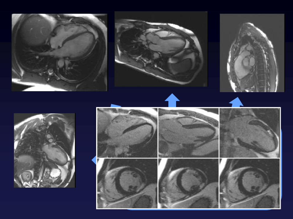
Base
Apex

CMR Structure and Function

Maceira AM. EHJ 2006.
Maceira AM. JCMR 2006.

Accurate Reproducible Volumes and Mass
• Superior reproducibility allows for reliable follow up of patients– Response to detraining
– Accurate visualisation of and assessment of wall thickness
• Basal anteroseptal wall
• Apical LV
– RV imaging• ?ARVC
Bottini. For a 10g difference in LV mass with a novel ant-
HTN therapy: Need 505 patients for TTE but only 14
using CMR (90% power, p value 0.05)

Tissue characterisation
• Intrinsic contrast properties
– Cine
– T1 weighted (FAT)
– T2 weighted (WATER)
• Extrinsic contrast properties
– Early gadolinium
– Late gadolinium

A
B
P<0.0001

-0,01
0,01
0,03
0,05
0,07
0,09
0,11
Pre Immediately-Post 6 hours-Post
cT
nI (u
g/L
)
cTnI (ug/L)
0
20
40
60
80
100
120
140
160
Pre Immediately-Post 6 hours-Post
NT
pro
BN
P (
ng
/L)
NTproBNP (ng/L)
O’ Hanlon, Whyte et al. JCMR 2010
STIR rGE LGE

Participant
Number
Age
(yr)
% of total LGE
Mass (g)
LGE Pattern Interpretation
1 67
18.9 CAD
Probable Dual
Infarction
2 50
8 Non-CAD
Probable
myocarditis
3
66 3 Non-CAD Nonspecific
4 60 3 Non-CAD Nonspecific
5 50 1 Non-CAD Nonspecific
6 51 1 Non-CAD Nonspecific

Contrast Enhancement
NSTEMI
DCM
STEMI
AMYLOID
MYOCARDITIS
HCM

Pathological Validation
O’ Hanlon R. JACC 2010Moon JC. JACC 2004

Strijack. JCMR 2008
• 35 yr old female
• Fam Hx HCM
• Normal ECG and TTE
• Normal wall thickness and function.
• LGE
Mis-sense mutation (275G>A) TNNT2 gene

High level triathlete with episodic palpitations and frequent bigeminyand trigeminy on holterNormal echo
Wilson M, O’ Hanlon R, et al. BMJ Case Reports 2009
32 yr old male
Combined endurance and weight training
Possible (denied) anabolic steroid use
Admitted with TnI positive ACS
Minor LAD disease on angio
Possible recent hx of flu-like illness
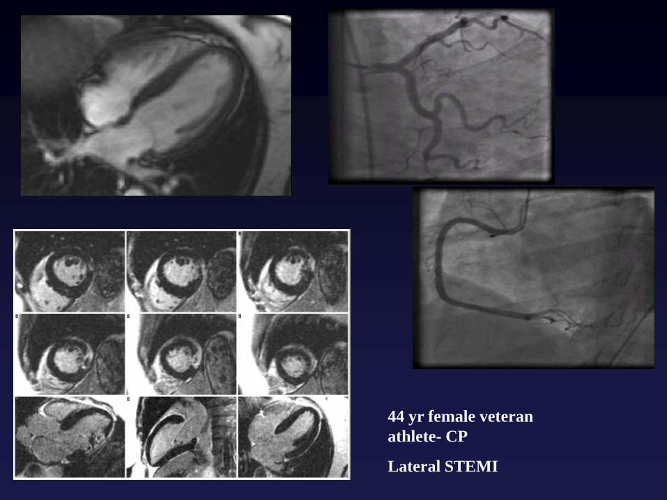
44 yr female veteran
athlete- CP
Lateral STEMI

TTE with agitated saline

Syncope in a 32 yr old


HCM Diagnosis
Rickers. Circulation 2005.
Moon. Heart 2004

Phenocopies

MORE COMPLETE RISK STRATIFICATION


Perfusion and LGE
• 42 yr old asymptomatic
• No risk factors for SCD
• Normal angiogram
– Significant microvascular ischaemia
– Minimal diffuse fibrosis
• How would one treat him?
Two patients (19 and 21 yrs) with
HCM in their 20. Both have NSVT
on holter (6 beats).
No family hx of SCD
Nil else of note in RF profile.
Who has LGE?
What to do from now?

2008 scan 2004 scan

30 yr old female. Tennis and squash.Palpitations and murmur.No Fam Hx

SSFP
T2
T1
LGEEGE
Miller C, O’ Hanlon R.
JCMR 2011. In Press.

LGE and Non-Ischaemic Cardiomyopathy
What do we know?

Gd-CMR and Sudden Death in HCM
≤1 risk factor for sudden death
≥2 risk factors for sudden death
35
30
25
20
15
10
5
0
16.3%*
2.5%
*p=0.03
>=2rfsd
<=1rfsd
Moon JC. JACC 2003; 41: 1561-7

LGE and LV Mass
Olivotto et al. JACC 2008Rudolph et al. JACC 2009
128+/-62 g/m2 vs 99+/-38g/m2


24 year oldUnexplained syncopeNo other risk factors
ICD?Follow up?

•61 year old male
•HCM diagnosis aged 49
•Asymptomatic•Annual follow up and risk stratification
•22mm septum and LVOTO 35mmHg at rest
•ICD?

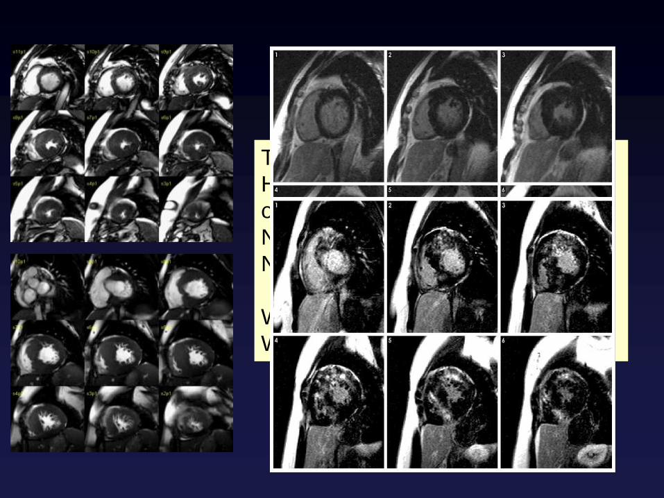
Athletes Heart vs DCM

Fibrosis in DCM explanted heart

DCM
Death/ hospitalization
p<0.001
Assomull. JACC2006; 48:
1977
HR
3.4
SCD/ VT
Assomull. JACC2006;
48: 1977
McCrohon Circulation 2003; 108: 54-9.
Wu KC. JACC 2008.

Abnormal ECG +/- ventricular ectopy

ARVC

CMR Sequences
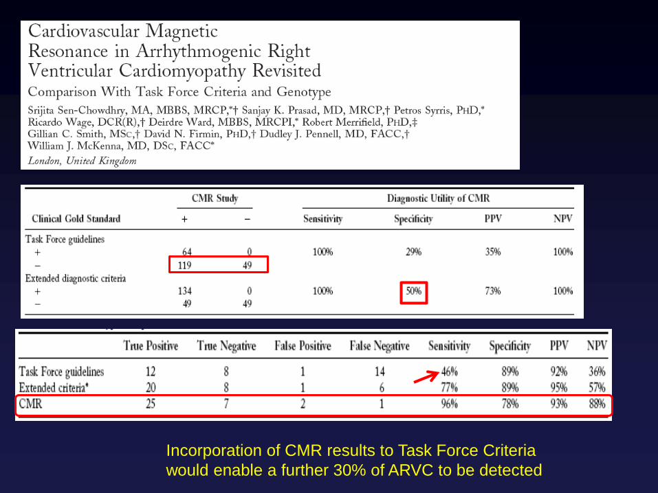
Incorporation of CMR results to Task Force Criteria
would enable a further 30% of ARVC to be detected

In Conclusion
• SCD in athletes rare
• CMR offers a unique imaging solution to screen for multiple congenital and acquired pathologies
– Inconclusive imaging
– Concerning symptoms
– High clinical suspicion
• Not 1st line test however