THE ROLE OF AUTOPHAGY IN OXIDATIVE STRESS-MEDIATED ...
150
1 THE ROLE OF AUTOPHAGY IN OXIDATIVE STRESS-MEDIATED DYSFUNCTION IN CARDIAC CELLS AND RODENT HEARTS By DEBAPRIYA DUTTA A DISSERTATION PRESENTED TO THE GRADUATE SCHOOL OF THE UNIVERSITY OF FLORIDA IN PARTIAL FULFILLMENT OF THE REQUIREMENTS FOR THE DEGREE OF DOCTOR OF PHILOSOPHY UNIVERSITY OF FLORIDA 2012
Transcript of THE ROLE OF AUTOPHAGY IN OXIDATIVE STRESS-MEDIATED ...

1
THE ROLE OF AUTOPHAGY IN OXIDATIVE STRESS-MEDIATED DYSFUNCTION IN CARDIAC CELLS AND RODENT HEARTS
By
DEBAPRIYA DUTTA
A DISSERTATION PRESENTED TO THE GRADUATE SCHOOL OF THE UNIVERSITY OF FLORIDA IN PARTIAL FULFILLMENT
OF THE REQUIREMENTS FOR THE DEGREE OF DOCTOR OF PHILOSOPHY
UNIVERSITY OF FLORIDA
2012

2
© 2012 Debapriya Dutta

3
To my family for their unconditional love

4
ACKNOWLEDGMENTS
I thank my parents, Pradip Narayan Dutta and Kanika Dutta for their constant
support and words of advice, all throughout my life. I thank my mentor, Dr.
Leeuwenburgh for his endless encouragement and able guidance throughout my PhD
life. He is one of the most positive people I have ever met and he always had my best
interests in mind. I thank Drs. William A. Dunn Jr. and Jae-Sung Kim from whom I have
learnt so much about the field of autophagy, learnt how to best design experiments and
how to critically evaluate scientific work. I am grateful to Drs. John P. Aris and Robert J.
Cousins for their constructive evaluation of my work and providing me with suggestions
throughout my time at graduate school. I also thank the current and past members of
the Leeuwenburgh lab: Jinze Xu, Marvin Dirain, Anna Maria Joseph, Brian Bouverat,
Gauthami Balagopal, Christopher Scoma and Arnold Seo for making the lab a fun place
to be. I especially thank Jinze for her help with the planning and execution of the animal
study, it would not have been possible without her. I thank Debra Akin and Kimberly
Hodges (Department of Anatomy and Cell Biology) and Louise Perras (Department of
Aging and Geriatrics) for constantly answering my queries and helping me out in every
possible way. I thank my husband, Manav for his continued words of advice, for
teaching me how to make the best out of every situation, for lifting up my spirits every
time I needed a boost and for constantly reminding me that PhD life is never easy, so
it’s important to keep trying. I thank my friends outside the lab, for making Gainesville a
second home. Finally, I thank the IDP program, the American Heart Association and the
Department of Anatomy and Cell Biology for supporting me financially. This has been
an incredible journey.

5
TABLE OF CONTENTS page
ACKNOWLEDGMENTS .................................................................................................. 4
LIST OF TABLES ............................................................................................................ 8
LIST OF FIGURES .......................................................................................................... 9
LIST OF ABBREVIATIONS ........................................................................................... 11
ABSTRACT ................................................................................................................... 15
CHAPTER
1 INTRODUCTION .................................................................................................... 17
Oxidative Stress and Mitochondria in Cardiovascular Diseases ............................. 17
Mechanisms of Cardiac Mitochondrial Dysfunction ................................................ 18 Consequences of Abnormal Mitochondrial Free Radical Generation in the Heart .. 20 Autophagic Machinery and Cardiomyocyte Homeostasis ....................................... 22
Types of Autophagy ......................................................................................... 22 Autophagic Process and Molecular Regulation ................................................ 24
Mitophagy: A Specialized Form of Autophagy .................................................. 26 Cross-talk between Autophagy and the Ubiquitin-Proteasomal System for
Protein Quality Control......................................................................................... 28 Impact of Impaired Autophagy: Accumulation of Dysfunctional Mitochondria and
Lipofuscin ............................................................................................................ 30
Left Ventricular Hypertrophy ............................................................................. 33 Heart Attack ...................................................................................................... 33
Diabetic Cardiomyopathy ................................................................................. 34 Autophagy as a Therapeutic Target against Cardiac Pathology ............................. 35 Summary and Project Goals ................................................................................... 38
2 UPREGULATED AUTOPHAGY PROTECTS CARDIOMYOCYTES FROM OXIDATIVE STRESS-INDUCED CYTOTOXICITY ................................................ 45
Introduction ............................................................................................................. 45 Materials and Methods............................................................................................ 48
Cell Culture Conditions ..................................................................................... 48 Chemical Reagents .......................................................................................... 48 Flow Cytometric Determination of Mitochondrial O2
•− Generation .................... 48 Confocal Imaging of Mitochondrial O2
•− Generation ......................................... 49 Flow Cytometric Determination of Mitochondrial Membrane Potential ............. 49
Confocal Imaging of Mitochondrial Membrane Potential .................................. 50 Determination of Cell Viability ........................................................................... 50
Determination of DNA / RNA Oxidation ............................................................ 51

6
Induction and Assessment of Autophagy ......................................................... 51
Epifluorescence Imaging and Quantification of Autophagy .............................. 52 Confocal Imaging ............................................................................................. 53
Western Blotting ............................................................................................... 53 Assessment of Cellular Respiration .................................................................. 54 Statistical Analysis ............................................................................................ 54
Results .................................................................................................................... 55 AMA Increases Mitochondrial O2
•− Generation and Decreases Mitochondrial Membrane Potential (Δψm) ............................................................................ 55
AMA Treatment Induces Nuclear DNA Oxidation and Decreases Cell Viability .......................................................................................................... 56
Treatment with AMA does not Induce Autophagy in HL-1 Cells ....................... 56 Treatment with Rapamycin Induces Autophagy in HL-1 Cells .......................... 57
Rapamycin Plays a Protective Role against AMA-Induced Toxicity ................. 58 Rapamycin Prevents AMA-Induced Accumulation of Ubiquitinated Proteins ... 59
Rapamycin Protects against AMA-Induced Mitochondrial Dysfunction ............ 59
Inhibition of Autophagy Blocks the Cytoprotective Effect of Rapamycin against AMA Toxicity ..................................................................................... 60
Discussion .............................................................................................................. 60
3 CALORIE RESTRICTION COMBINED WITH RESVERATROL INDUCES AUTOPHAGY AND OFFERS PROTECTION FROM DOXORUBICIN-INDUCED TOXICITY IN 26-MONTH-OLD RAT HEARTS ....................................................... 80
Introduction ............................................................................................................. 80
Materials and Methods............................................................................................ 83 Animals and Experimental Design .................................................................... 83 Preparation of Tissue for Histological Staining ................................................. 84
Body Composition Analysis .............................................................................. 85 Preparation of Tissue Extracts ......................................................................... 85
Western Blotting ............................................................................................... 86 Plasma Resveratrol Concentration Analysis..................................................... 87 Blood Glucose Analysis .................................................................................... 87
Apoptotic Analysis ............................................................................................ 87 Creatine Kinase (CK) Assay ............................................................................. 87 Lactate Dehydrogenase (LDH) Assay .............................................................. 88 Statistical Analysis ............................................................................................ 88
Results .................................................................................................................... 88 CR Diet were Associated with Lower Body Weights ........................................ 88 Six weeks of CR and Resveratrol Interventions or Administration of
Doxorubicin does not Change Myocardial Morphology ................................. 89 CR Diet alone or in Combination with Resveratrol, Attenuates Body
Composition Changes over Time .................................................................. 89 Plasma Resveratrol Concentrations Increase in a Dose-Dependent Manner
in Resveratrol-Fed Rats ................................................................................ 89 Doxorubicin Administration Increases Blood Glucose Levels ........................... 90

7
CR + 50 mg/kg/day Resveratrol Increases Autophagic Flux in the Left Ventricle ........................................................................................................ 90
LC3B .......................................................................................................... 90
p62 ............................................................................................................. 91 Beclin-1 ...................................................................................................... 91 Atg5-Atg12 ................................................................................................. 92
CR + 50 mg/kg/day Resveratrol Attenuates Doxorubicin-Mediated Increases in Cardiac Apoptotic Levels .......................................................... 92
CR + 50 mg/kg/day Resveratrol Attenuates Doxorubicin-Mediated Increases in Serum CK Levels ...................................................................... 92
CR + Resveratrol Attenuates Doxorubicin-Mediated Increases in Serum LDH Activity ................................................................................................... 93
Discussion .............................................................................................................. 93
4 CONCLUSION ...................................................................................................... 113
Summary of Results and Therapeutic Relevance ................................................. 113 Future Directions .................................................................................................. 120
LIST OF REFERENCES ............................................................................................. 126
BIOGRAHICAL SKETCH ............................................................................................ 150

8
LIST OF TABLES
Table page 2-1 List of reagents used for in vitro study ................................................................ 78
2-2 List of primary antibodies used for in vitro study ................................................. 79
3-1 Ingredient composition for AL and 20% CR purified diet. ................................. 107
3-2 List of reagents used for in vivo study. ............................................................. 108
3-3 List of primary antibodies used for in vivo study. .............................................. 109
3-4 Body weight (BW) and heart weight (HW) analysis. ......................................... 110
3-5 Plasma resveratrol concentrations analysis. .................................................... 111
3-6 Blood glucose analysis in saline and doxorubicin-treated rats ......................... 112

9
LIST OF FIGURES
Figure page 1-1 Schematic representation of the distinct steps of the autophagic process. ........ 40
1-2 Schematic overview of the molecular regulation of autophagy in the context of mitochondrial homeostasis ............................................................................. 41
1-3 Schematic showing the contribution of mitochondria-derived ROS in the formation of lipofuscin in cells ............................................................................. 43
1-4 Schematic showing the beneficial effect of autophagy against oxidative stress-mediated dysfunction. .............................................................................. 44
2-1 Treatment with AMA leads to enhanced oxidative stress and cytotoxicity in HL-1 cardiomyocytes. ......................................................................................... 66
2-2 Treatment with AMA does not induce autophagy in HL-1 cardiomyocytes. ........ 68
2-3 Treatment with rapamycin induces autophagy in HL-1 cardiomyocytes ............. 70
2-4 Rapamycin induces autophagy in the presence of AMA in HL-1 cardiomyocytes ................................................................................................... 71
2-5 Rapamycin protects against the cytotoxic effects of AMA .................................. 73
2-6 Rapamycin enhances the loss of ubiquitinated proteins induced by AMA in HL-1 cardiomyocytes .......................................................................................... 74
2-7 Rapamycin protects against AMA-induced mitochondrial depolarization and respiration dysfunction in HL-1 cardiomyocytes. ................................................ 75
2-8 Inhibition of autophagy attenuates rapamycin-mediated protection against AMA toxicity in cardiomyocytes .......................................................................... 76
2-9 Schematic representation of the protective effects of rapamycin against AMA toxicity. ............................................................................................................... 77
3-1 CR + Resv-5 and CR + Resv-50 interventions for 6 weeks or doxorubicin administration for 24h does not change heart morphology ............................... 101
3-2 CR diet alone or in combination with resveratrol, attenuates body composition changes over time ........................................................................ 102
3-3 CR + Resv-50 decreases p62 abundance and increases Beclin-1 levels in left ventricular homogenates............................................................................. 103

10
3-4 Doxorubicin increases LC3-II/LC3-I ratio and p62 accumulation in left ventricular homogenates .................................................................................. 104
3-5 CR + Resv-50 attenuates doxorubicin-mediated increases in cardiac damage markers ............................................................................................................ 105
3-6 CR + Resv-50 attenuates doxorubicin-mediated increases in serum LDH activity. ............................................................................................................. 106
4-1 Regulation of autophagy by rapamycin, CR and resveratrol. ........................... 123
4-2 Chronic rapamycin administration induces autophagy in C57BL/J6 mice hearts ............................................................................................................... 124
4-3 Schematic of overall observations from in vitro and in vivo studies. ................. 125

11
LIST OF ABBREVIATIONS
2DG 2-deoxy-d-glucose
3-MA 3-Methyladenine
8-oxo-dGuo 8-oxo-7,8-dihydro-2’-deoxyguanosine
8-oxo-Guo 8-oxo-7,8-dihydroguanosine
AMA Antimycin A
AMP Adenosine monophosphate
AMPK AMP-activated protein kinase
BafA1 Bafilomycin A1
Bcl-2 B cell leukemia-2
Bcl-XL B cell leukemia-X long
Bnip3 Bcl-2 and adenovirus E1B 19 kDa-interacting protein-3
CAD coronary artery disease
CK creatine kinase
CMA chaperone-mediated autophagy
CR calorie restriction
CuZnSOD copper-zinc-containing superoxide dismutase
CVD cardiovascular disease
CypD cyclophilin D
DFOM deferoxamine mesylate
EGTA ethylene glycol tetraacetic acid
ETC electron transport chain
FBN Fischer 344 x Brown Norway
FIP200 Focal adhesion kinase family interacting protein of 200 kD
FSC forward scatter
12
Gpx glutathione peroxidase
GTC guanidium thiocyanate
HEPES 4-(2-hydroxyethyl)-1-piperazineethanesulfonic acid
HIF-1α hypoxia-inducible factor-1α
HPLC-ECD HPLC coupled to electrochemical detection
I/R ischemia/reperfusion
IGF-1 insulin-like growth factor-1
IP3 inositol triphosphate
IP3R inositol triphosphate receptor
LAMP-1 lysosomal membrane-associated protein-1
LAMP-2 lysosomal membrane-associated protein-2
LC3 light chain-3
LDH lactate dehydrogenase
LVH left ventricular hypertrophy
MAPKs mitogen activated protein kinases
mCAT catalase targeted to the mitochondrial matrix
MnSOD manganese-containing superoxide dismutase
mPTP mitochondrial permeability transition pore
mtDNA mitochondrial DNA
mTOR mammalian target of rapamycin
mTORC1 mammalian target of rapamycin complex 1
MTT 3-(4, 5-dimethylthiazol-2-yl)-2,5-diphenyltetrazolium bromide\
NADH nicotinamide adenine dinucleuotide (reduced)
NAPDH nicotinamide adenine dinucleotide phosphate
nDNA nuclear DNA
13
NF-κB nuclear factor κB
Nix Nip3-like protein X
NRF-1 nuclear respiratory factor-1
O2•− superoxide radical
OXPHOS oxidative phosphorylation
PARL presenilins-associated rhomboid-like
pCAT peroxisomal catalase
PE phosphatidylethanolamine
PGC-1α peroxisome proliferator-activated receptor- coactivator-1α
PI3K phosphatidylinositol-3-kinase
PKB/Akt protein kinase B
PolG mtDNA polymerase γ
Prx peroxiredoxin
Resv Resveratrol
RHEB Ras homolog enriched in brain
ROS reactive oxygen species
SIRT1 Sirtuin 1
SNARE Soluble N-ethylmaleimide-sensitive factor Attachment protein Receptor
SQSTM1 Sequestosome 1
SSC side scatter
TD-NMR time domain – nuclear magnetic resonance
TFAM mitochondrial transcription factor A
TMRM Tetramethyl Rhodamine Methyl Ester
TNF-α tumor necrosis factor α
TSC1/TSC2 tuberous sclerosis complex ½

14
ULK1 unc-51-like kinase 1
UPS ubiquitin proteasomal system
UBA ubiquitin associated
UVRAG UV radiation resistance-associated gene
VDAC voltage-dependent anion channel
Vps vacuolar protein sorting
Δψm mitochondria membrane potential

15
Abstract of Dissertation Presented to the Graduate School of the University of Florida in Partial Fulfillment of the Requirements for the Degree of Doctor of Philosophy
THE ROLE OF AUTOPHAGY IN OXIDATIVE STRESS-MEDIATED DYSFUNCTION IN
CARDIAC CELLS AND RODENT HEARTS
By
Debapriya Dutta
August 2012
Chair: Christiaan Leeuwenburgh Co-chair: William A Dunn, Jr. Major: Medical Sciences – Molecular Cell Biology
Cardiovascular disease (CVD) is the leading cause of mortality and morbidity in
the developing world. While long-term exposure to cardiovascular risk factors plays a
major role in the etiopathogenesis of CVD, oxidative stress-induced cardiac damage
enhances the susceptibility to develop heart pathologies in late life. The enhanced
generation of reactive oxygen species, especially by damaged mitochondria, is
considered to be a major contributing mechanism. Hence, the removal of oxidatively
modified cellular components and dysfunctional mitochondria is critical for maintenance
of cellular homeostasis.
Autophagy is a self-digestion process which can degrade damaged proteins and
organelles and is also the only known mechanism targeting dysfunctional mitochondria
for removal. In our studies, we have investigated whether pharmacological or nutritional
enhancement of autophagy offers protection against oxidative stress-mediated
dysfunction in a cardiomyocyte cell line (HL-1) and in late middle-aged rat hearts. In HL-
1 cells, we mimicked mitochondria-mediated oxidative stress by treating cells with
Antimycin A (AMA). AMA increased mitochondrial superoxide generation, decreased

16
mitochondrial membrane potential, increased nuclear DNA oxidation and decreased
cellular respiration. Treatment of HL-1 cells with the mTOR inhibitor rapamycin lead to a
strong induction of autophagy and mitophagy and protected against the cytotoxic effects
of AMA, assessed by cell survival and apoptotic signaling analysis. Rapamycin also
prevented AMA-mediated increases in ubiquitinated protein aggregates. Autophagy
inhibition attenuated the cytoprotective effects of rapamycin. For our in vivo
experiments, we investigated whether a late-life intervention of moderate calorie
restriction (CR, 20%) alone, or in combination with the plant polyphenol resveratrol can
induce autophagy in the hearts of late middle-aged rats. We further investigated
whether such an induction of autophagy is protective against the cardiotoxic effects of
the chemotherapeutic drug doxorubicin, a known oxidative stressor. Our observed that
CR and resveratrol, only when combined, stimulated autophagic flux in the left
ventricular tissue of late middle-aged rat hearts. Additionally, autophagy induction
protected against the cytotoxic effects of doxorubicin, as assessed by apoptotic analysis
in the myocardium and cardiac damage markers in serum. Our studies therefore
suggest that interventions aimed at enhancing basal autophagy may offer protection
against oxidative stress-mediated dysfunction in cardiomyocytes.

17
CHAPTER 1 INTRODUCTION
Oxidative Stress and Mitochondria in Cardiovascular Diseases
The worldwide trend in cardiovascular diseases (CVDs) has been growing at an
alarming rate and it is estimated that by the year 2020, up to 40% cases of deaths
would be due to cardiovascular complications (Braunwald, 1997). In the United States
alone, mortality data from 2008 shows that 1 of every 3 deaths results from CVD (Roger
et al., 2012). CVD comprises any pathologic changes affecting the heart itself or the
vessels carrying blood to and from the heart. CVD includes, but is not limited to heart
failure, ischemic heart disease, left ventricular hypertrophy (LVH), coronary artery
disease (CAD), hypertensive cardiomyopathy and diabetic cardiomyopathy. Notably, the
prevalence of such cardiac pathologies increases with age. The disproportionate
occurrence of CVD at advanced age is largely attributable to long-term exposure to
cardiovascular risk factors such as hypertension, dyslipidemia, diabetes mellitus,
physical inactivity, etc (Kovacic et al., 2012). In addition, oxidative stress-mediated
damage observed during intrinsic cardiac aging can cause structural and functional
alterations in the heart and render it more vulnerable to various stressors, ultimately
favoring the development of CVD (Lakatta, 2001). Reactive oxygen species (ROS) are
constantly generated within cells by several enzymatic reactions, including those
catalyzed by cyclooxygenases, nicotinamide adenine dinucleotide phosphate (NADPH)
oxidase and xanthine oxidase (Paravicini & Touyz, 2008). However, the bulk of ROS
production occurs as a byproduct of mitochondrial oxidative phosphorylation
(OXPHOS). Experimental evidence indicates that mitochondrial function decreases over
the course of aging, resulting in increased ROS generation, enhanced free radical-

18
inflicted damage and further mitochondrial decay (Balaban et al., 2005). In this scenario,
the removal of dysfunctional mitochondria and oxidatively damaged components
generated thereof, through the self-digestion process of autophagy is critical for the
maintenance of cell viability (Levine & Klionsky, 2004). The efficiency of this process
declines with advancing age which may be critically involved in heart senescence as
well as in age-related CVD (Inuzuka et al., 2009; Taneike et al., 2010)
In this chapter, we will highlight the role played by oxidative stress, especially
those generated by the mitochondria, in causing cardiac senescence and predisposing
it to age-related CVD. This chapter will also illustrate the putative mechanisms whereby
dysfunctional autophagy can cause pathologic changes in the heart and play a role in
the pathogenesis of specific heart diseases especially prevalent in late life. Interventions
proposed to counter cardiac oxidative stress through improvements in autophagy are
also presented, along with a general discussion of our research goals of using
autophagy as a therapeutic intervention against CVD.
Mechanisms of Cardiac Mitochondrial Dysfunction
Mitochondria are essential for cardiomyocyte function and viability. Indeed, the
myocardium is a highly energy demanding tissue, with mitochondria supplying over 90%
of ATP. Free radicals are constantly generated during mitochondrial respiration. Under
physiological conditions, 0.2-2% of oxygen is converted into superoxide anion (O2•−)
mainly at complex I and III of the electron transport chain (ETC) (Boveris & Chance,
1973). To counteract the burden of ROS production, the mitochondrion is equipped with
a multileveled defense network comprising detoxifying enzymes and non-enzymatic
antioxidants (Andreyev et al., 2005a). Within the mitochondrial matrix, manganese-
containing superoxide dismutase (MnSOD, SOD2) converts O2•− into hydrogen peroxide
19
(H2O2), which is further detoxified into O2 and H2O by glutathione peroxidase (Gpx-I)
and peroxiredoxine (Prx-III). Alternatively, O2•− can be released in the mitochondrial
intermembrane space where it is converted to H2O2 by copper-zinc-containing SOD
(CuZnSOD, SOD1). In addition, O2•− leaked in the intermembrane space can be
scavenged by cytochrome c (Pasdois et al., 2011).
Once merely considered unwanted byproducts of OXPHOS, small amounts of
oxidants have now been shown to function as essential signaling molecules, necessary
for the induction of endogenous defense mechanisms that culminate in increased stress
resistance (Finkel, 2011). In contrast, the generation of excessive ROS by dysfunctional
mitochondria, often coupled with a defective oxidant scavenging, have been implicated
in the aging process as well as in the pathogenesis of several chronic degenerative
diseases, including CVD (Judge & Leeuwenburgh, 2007).
Damaged cardiac mitochondria can release up to 10-fold more H2O2 than intact
organelles (Grivennikova et al., 2010). Furthermore, in the presence of non-protein-
bound redox cycling metals (e.g., iron and copper), H2O2 can be converted into the
highly reactive hydroxyl radical (•OH), through Fenton’s and Haber-Weiss’ reactions. In
such circumstances, the mitochondrion is exposed to a high burden of oxidative stress,
resulting in the primary damage to its own constituents. It is worth mentioning that the
mitochondrial iron content increases with aging in rodent post-mitotic tissues, including
the myocardium, which may exacerbate the extent of oxidative damage in late life (Xu et
al., 2010).
The mitochondrial DNA (mtDNA) is especially prone to oxidative damage due to its
proximity to the ETC, the lack of protective histones and a less efficient repair system
20
compared with nuclear DNA (nDNA) (Yakes & Van, 1997). As a result, the level of
oxidatively-modified bases in mtDNA is several-fold higher than that in nDNA (Yakes &
Van, 1997). Moreover, due to the compactness of the mitochondrial genome (i.e., lack
of introns), each mutation is likely to affect gene integrity and hence, mitochondrially
encoded protein, mRNA and tRNA function (Wei & Lee, 2002). It follows that mtDNA
mutations can lead to the synthesis of defective ETC components, resulting in
impairment of OXPHOS, decreased ATP production and further ROS generation
(Linnane et al., 1989). The vicious cycle originating from ROS-inflicted mtDNA damage
represents the main tenet of the mitochondrial free radical theory of aging and is also
believed to play a central role in the pathogenesis of age-associated degenerative
diseases, including CVD (Linnane et al., 1989).
Consequences of Abnormal Mitochondrial Free Radical Generation in the Heart
Elevated levels of oxidative damage to mitochondrial proteins, lipids and nucleic
acids have been detected in the failing myocardium of old rodents (Barja & Herrero,
2000; Judge et al., 2005b; Leeuwenburgh et al., 1997). The frequency of mtDNA point
mutations and deletions is ∼3-fold higher in the aged mouse heart compared with young
adult controls (Dai & Rabinovitch, 2009). Similarly, the frequency of the common 4977
bp deletion of mtDNA increases during aging in the human heart, and is 5 to 15-fold
higher in persons over 40 years of age than in younger individuals (Liu et al., 1998;
Mohamed et al., 2006). However, the proof of principle that the accumulation of mtDNA
damage and subsequent mitochondrial dysfunction may be causative to cardiac
senescence has been provided by the characterization of mice expressing a
proofreading-deficient mtDNA polymerase γ (PolG) (Kujoth et al., 2005; Trifunovic et al.,
2004). These mutants accumulate a high load of mtDNA mutations and deletions, and

21
are characterized by the early appearance of cardiac pathologic changes such as
cardiac enlargement. Heart mitochondria of PolG mice exhibit abnormal ETC with
depressed activity of complex I and IV, reduced ATP production, and accumulation of
enlarged, irregularly shaped mitochondria (Trifunovic et al., 2004). Furthermore, levels
of protein carbonyls are increased in cardiac mitochondria from mtDNA mutator mice
compared with wild-type rodents (Dai et al., 2010). PolG mice die prematurely with
dilated cardiomyopathy. Severe cardiomyopathy has also been observed in mice
expressing a heart-specific proofreading-deficient mtDNA polymerase (Zhang et al.,
2003). Remarkably, the PolG heart phenotype, the cardiac mtDNA mutation load and
the extent of mitochondrial protein oxidation are partially rescued by the overexpression
of catalase targeted to the mitochondrial matrix (mCAT) (Dai et al., 2010). In addition,
mCAT overexpression extends mean and maximum lifespan and delays the
development of cardiac pathology in mice (Dai et al., 2009a; Schriner et al., 2005). The
extent of mitochondrial oxidative damage, including mtDNA deletions, and the rate of
H2O2 generation, are significantly attenuated in the heart of old mCAT mice compared
with age-matched wild-type littermates (Dai et al., 2009a; Schriner et al., 2005).
In addition to increased ROS generation, dysfunctional mitochondria could lead to
the induction of apoptosis through the release of pro-apoptotic factors (Hamacher-Brady
et al., 2006; Sybers et al., 1976). Indeed, mitochondria are a major check-point for the
integration of apoptotic stimuli. Notably, cardiomyocyte removal through apoptosis
increases with advancing age, which, combined with insufficient replenishment by
cardiac stem cells, may contribute to the age-related heart remodeling (Marzetti et al.,
2009). However, whether the increased severity of apoptosis suffered by the aged
22
myocardium is directly attributable to autophagic failure is yet to be established. It is
therefore reasonable to say that, the efficient removal of damaged and potentially
harmful mitochondria along with other oxidatively damaged proteins are vital for the
preservation of cardiomyocyte homeostasis.
Autophagic Machinery and Cardiomyocyte Homeostasis
Regardless of the mechanism(s) primarily responsible for enhanced oxidative
stress and mitochondrial dysfunction, cellular quality control is essential for the
preservation of cardiomyocyte homeostasis. This task is accomplished through the
complex coordination of several processes (reviewed by (Tatsuta & Langer, 2008). For
instance, oxidatively damaged and misfolded proteins are marked by ubiquitination, to
be subsequently removed by the ubiquitin-proteasomal system (Shang & Taylor, 2011).
With regard to the mitochondria, an intramitochondrial proteolytic system selectively
removes damaged mitochondrial proteins. A second line of defense is provided by the
dynamic nature of the mitochondrial population, i.e. the functionality of damaged
mitochondria can be restored by their fusion with neighboring, intact organelles (Chen &
Chan, 2009). Finally, severely damaged mitochondria and oxidatively modified protein
aggregates can be eliminated through autophagy.
Types of Autophagy
Autophagy is a self-eating process through which cells degrade their own
components, recycling amino acids and other building blocks that can be eventually
reutilized (Yorimitsu & Klionsky, 2005). Such degradation is performed by lysosomal
acid hydrolases. Depending on the pathway cellular components are delivered to
lysosomes, three types of autophagy can be distinguished: macroautophagy,
microautophagy and chaperone-mediated autophagy (CMA). Macroautophagy
23
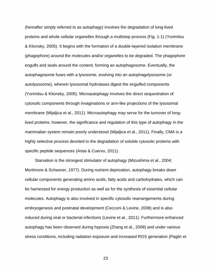
(hereafter simply referred to as autophagy) involves the degradation of long-lived
proteins and whole cellular organelles through a multistep process (Fig. 1-1) (Yorimitsu
& Klionsky, 2005). It begins with the formation of a double-layered isolation membrane
(phagophore) around the molecules and/or organelles to be degraded. The phagophore
engulfs and seals around the content, forming an autophagosome. Eventually, the
autophagosome fuses with a lysosome, evolving into an autophagolysosome (or
autolysosome), wherein lysosomal hydrolases digest the engulfed components
(Yorimitsu & Klionsky, 2005). Microautophagy involves the direct sequestration of
cytosolic components through invaginations or arm-like projections of the lysosomal
membrane (Mijaljica et al., 2011). Microautophagy may serve for the turnover of long-
lived proteins; however, the significance and regulation of this type of autophagy in the
mammalian system remain poorly understood (Mijaljica et al., 2011). Finally, CMA is a
highly selective process devoted to the degradation of soluble cytosolic proteins with
specific peptide sequences (Arias & Cuervo, 2011).
Starvation is the strongest stimulator of autophagy (Mizushima et al., 2004;
Mortimore & Schworer, 1977). During nutrient deprivation, autophagy breaks down
cellular components generating amino acids, fatty acids and carbohydrates, which can
be harnessed for energy production as well as for the synthesis of essential cellular
molecules. Autophagy is also involved in specific cytosolic rearrangements during
embryogenesis and postnatal development (Cecconi & Levine, 2008) and is also
induced during viral or bacterial infections (Levine et al., 2011). Furthermore enhanced
autophagy has been observed during hypoxia (Zhang et al., 2008) and under various
stress conditions, including radiation exposure and increased ROS generation (Paglin et

24
al., 2001; Scherz-Shouval et al., 2007b). In these circumstances, autophagy is essential
for the maintenance of cell homeostasis by promoting the removal of damaged
components (Levine & Klionsky, 2004; Levine & Kroemer, 2008). Indeed, impairments
in autophagy induce premature aging and shorten the lifespan in several organisms
(Hars et al., 2007; Matecic et al., 2010). Conversely, up-regulation of autophagy is
proposed to be a major mechanism underlying the lifespan-extending properties of
calorie restriction (CR) (Morselli et al., 2010; Toth et al., 2008).
The execution of autophagy involves the coordination of a complex molecular
machinery which is briefly described in the next subsection.
Autophagic Process and Molecular Regulation
To date, over 35 AuTophaGy-related (Atg) proteins have been identified in yeasts
and mammals; however, the precise role each Atg protein plays during autophagy is not
fully established (Chen & Klionsky, 2011). As illustrated in Fig. 1-1, the process of
autophagy can be divided into discrete steps, namely, induction and nucleation,
expansion, fusion, and degradation. The induction phase is mediated by the ULK1-
Atg13-FIP200 kinase complex (Xie & Klionsky, 2007). The regulation of the nucleation
stage, which consists in the recruitment of Atg proteins to the phagophore assembly
site, is not yet completely understood. However, the vacuolar protein sorting-34
(Vps34), a class III phosphatidylinositol-3-kinase (PI3K), is required for this step (Suzuki
et al., 2001). Vps34 associates with Beclin1, the mammalian homologue of yeast Atg6,
and subsequently recruits Atg14 and Vps15 (p150) to the pre-autophagosomal structure
(Suzuki et al., 2001). The elongation and expansion of the phagophore membrane
require two ubiquitin-like conjugation systems involving Atg12 (conjugated to Atg5) and
Atg8/light chain-3 (LC3, conjugated to phosphatidyl ethanolamine), along with other Atg

25
proteins such as Atg9 and Atg16 (Xie & Klionsky, 2007). The fusion of the
autophagosome with a lysosome relies on the canonical cellular fusion machinery
consisting of the Rab-SNARE (Soluble N-ethylmaleimide-sensitive factor Attachment
protein REceptor) system and requires the presence of lysosomal membrane-
associated protein-2 (LAMP-2) and the UV radiation resistance-associated gene
(UVRAG) (Nair et al., 2011; Tong et al., 2010). Finally, the digestion of the cargo is
carried out by lysosomal hydrolases, followed by the transportation of degraded
components into the cytoplasm by lysosomal efflux transporters such as Atg22 (Tong et
al., 2010).
With regard to the regulation of autophagy, the mammalian target of rapamycin
(mTOR) is considered to be a major player, linking the cellular nutritional state with the
level of ongoing autophagy (Fig. 1-2) (Kim et al., 2011). Under nutrient rich conditions,
mTOR is active and inhibits the ULK1-Atg13-FIP200 complex required for the induction
of autophagy (Jung et al., 2009a). Energy deprivation leads to mTOR inactivation,
thereby inducing autophagy (Kim et al., 2011; Talloczy et al., 2002). In addition,
starvation causes an increase in the cellular AMP:ATP ratio, leading to the activation of
AMP-activated protein kinase (AMPK). AMPK in turn promotes autophagy by directly
activating ULK1 as well as by relieving the mTOR-mediated inhibition of autophagy (Kim
et al., 2011) (Fig.1-2). It is worth mentioning here that in addition to stimulating
mitochondrial removal through autophagy, AMPK enhances the activity of sirtuin-1
(SIRT1) and its downstream target PGC-1α, resulting in the stimulation of mitochondrial
biogenesis (Canto et al., 2009). Hence, it is believed that through the activity of AMPK,

26
mitophagy and mitochondrial biogenesis are coordinately regulated, maintaining a
healthy and functional pool of mitochondria in the cell (Fig. 1-2).
Although autophagy might seem to be a random, bulk digestion process, evidence
is accumulating that intracellular components can be selectively targeted for
degradation (Kadandale & Kiger, 2010). For instance, autophagy can be specifically
directed towards the removal of peroxisomes (pexophagy), endoplasmic reticulum
(reticulophagy), and ribosomes (ribophagy) (Kadandale & Kiger, 2010). Likewise,
mitochondria can be selectively targeted for degradation via mitophagy (Wang &
Klionsky, 2011). The currently known molecular machinery and the regulation of this
cellular pathway are outlined in the next subsection.
Mitophagy: A Specialized Form of Autophagy
Mitophagy is a highly selective process that can promote the elimination of
dysfunctional or unnecessary mitochondria (Wang & Klionsky, 2011). The loss of
mitochondrial membrane potential (m) represents a major trigger of mitophagy (Wang
& Klionsky, 2011). Indeed, laser-induced photo-damage of selected mitochondria inside
living hepatocytes results in the rapid dissipation of m, followed by the quick removal
of depolarized mitochondria through mitophagy (Kim & Lemasters, 2011). In addition,
oxidative damage can lead to the formation of asymmetric daughter mitochondria
characterized by different m, with autophagy specifically targeting mitochondria with
lower m (Twig et al., 2008). Apart from the degradation of damaged mitochondria
under stress conditions, mitophagy is essential for mitochondrial turnover in the basal
state as well as during cell differentiation, such as the maturation of reticulocytes into

27
mature red blood cells (Tal et al., 2007). The occurrence of selective mitophagy in
cardiomyocytes has not yet been conclusively demonstrated.
Investigations into the molecular regulation of mitophagy have unveiled several
mitophagy-specific proteins (Green et al., 2011). Parkin and Pink1 are believed to play
an important role in the selective degradation of damaged mitochondria, at least under
certain circumstances (Narendra et al., 2008). Parkin is a cytosolic E3-ubiquitin ligase
which is selectively recruited to dysfunctional mitochondria, and assists in their removal
by mitophagy (Narendra et al., 2008). Pink1 is imported into healthy mitochondria
through a m-dependent process and is degraded by the presenilins-associated
rhomboid-like (PARL) protease (Matsuda et al., 2010). The dissipation of m results in
the accumulation of Pink1 on the mitochondrial surface, leading to the recruitment of
Parkin, which ubiquitinates outer membrane proteins including the voltage-dependent
anion channel (VDAC) (Geisler et al., 2010). It is proposed that ubiquitin-tagged
mitochondria are directly targeted to autophagic vacuoles through the interaction of
ubiquitinated proteins with the autophagosomal marker LC3, mediated by the adapter
protein p62 (Geisler et al., 2010). In addition, Parkin can ubiquitinate B cell leukemia-2
(Bcl-2), therefore derepressing Beclin1 and activating autophagy (Chen et al., 2010).
Recent evidence also suggests that the opening of the mitochondrial permeability
transition pore (mPTP) may be required for the selective removal of damaged
mitochondria (Carreira et al., 2010; Elmore et al., 2001). Opening of the mPTP causes a
sudden increase of the inner membrane permeability to solutes with molecular weight
up to 1,500 Da (Weiss et al., 2003). This results in mitochondrial depolarization,
activation of the mitochondrial F0F1 ATPase (i.e., ATP synthase operating in reverse),

28
and swelling and rupture of the outer membrane (Weiss et al., 2003). The loss of m
subsequent to permeability transition targets individual mitochondria for degradation
(Carreira et al., 2010). Notably, in cultured cardiomyocytes, starvation-induced
autophagy is preceded by mitochondrial depolarization (Carreira et al., 2010). The loss
of m and the activation of autophagy are prevented by cyclosporin A, an inhibitor of
the mPTP component cyclophilin D (CypD) (Carreira et al., 2010). Furthermore,
starvation fails to induce autophagy in CypD-deficient murine cardiomyocytes, whereas
in cardiac cells from mice overexpressing CypD autophagy is enhanced even under fed
conditions (Carreira et al., 2010). The NAD-dependent deacetylase sirtuin-3 (SIRT3)
appears to be critically involved in the control of mPTP by modulating CypD (Hafner et
al., 2010). Indeed, in transgenic mice, the loss of SIRT3 activity leads to increased
activation of the mPTP in cardiac mitochondria in response to Ca2+ increases and
hemodynamic stress, the latter commonly observed in CVD (Hafner et al., 2010).
Similar to the mPTP, the apoptotic proteins Bnip3 (Bcl-2 and adenovirus E1B 19
kDa-interacting protein-3) and Nix (Nip3-like protein X) are thought to trigger selective
mitophagy through mitochondrial depolarization (Zhang & Ney, 2009). Moreover, Bnip3
may induce mitophagy by competitively disrupting the inhibitory interaction between Bcl-
2 and Beclin-1(Zhang & Ney, 2009). Finally, Nix associates with mitochondrial
membranes and directly interacts with LC3 (Novak et al., 2010).
Cross-talk between Autophagy and the Ubiquitin-Proteasomal System for Protein Quality Control
The ubiquitin-proteasomal system (UPS) provides to the selective elimination of
short-lived and misfolded proteins, small enough to enter the narrow barrel-shaped
proteasome for digestion (Schrader et al., 2009). Proteins are targeted for degradation

29
by covalent binding to ubiquitin chains. Individual ubiquitin molecules are activated by
ubiquitin-activating enzymes (E1s) and transferred to ubiquitin-conjugating enzymes
(E2s). Afterwards, ubiquitin is transferred to the substrate, which is recognized by
ubiquitin-protein ligases (E3s). Polyubiquitin chains function as recognition motifs for
delivery to 26S proteasomes where the hydrolysis takes place (Schrader et al., 2009).
Alternatively, misfolded monomeric proteins may be degraded by CMA independent of
polyubiquitination (Arias & Cuervo, 2011). However, if the degradative capacity of the
UPS and CMA is overwhelmed, misfolded proteins accumulate, forming large
aggregates, known as inclusion bodies and aggresomes (Markossian & Kurganov,
2004). The formation of protein aggregates is promoted by the ubiquitin-binding protein
p62/SQSTM1 (sequestosome 1) and NBR1 (neighbor of BRCA1 gene 1). Such protein
aggregates are cleared through autophagy (Zheng et al., 2011). The existence of a
cross-talk between the UPS and autophagic system is witnessed by the observation
that inhibition of proteasomal degradation increases the appearance of protein
aggregates within autophagic vacuoles (Wojcik et al., 1996). Moreover, inhibition of
autophagy due to dynein mutations leads to the accumulation of cytosolic protein
aggregates in mouse neurons (Ravikumar et al., 2005). Importantly, p62/SQSTM1 is
required for the selective autophagic removal of protein aggregates. p62 binds to
ubiquitinated proteins through its ubiquitin-associated (UBA) domain (Seibenhener et
al., 2004) and interacts with Atg8/LC3 through the LC3 recognition sequence (LRS)
(Noda et al., 2008). This establishes a physical link between the protein aggregates and
the autophagosomal system, thereby mediating their clearance.

30
Impact of Impaired Autophagy: Accumulation of Dysfunctional Mitochondria and Lipofuscin
Cardiomyocytes are terminally differentiated, post-mitotic cells with a lifespan of
several decades. Hence, the maintenance of a healthy pool of mitochondria and the
efficient removal of damaged and potentially harmful organelles are vital for the
preservation of cardiomyocyte homeostasis. A decrease in autophagic flux may result in
the accumulation within cardiomyocytes of dysfunctional mitochondria that are
bioenergetically inefficient and prone to ROS leakage (Terman et al., 2010). Importantly,
autophagic flux has been shown to be decreased with age (Inuzuka et al., 2009;
Taneike et al., 2010) and the ultrastructural analysis of myocardium from aged rodents
has revealed the presence of enlarged mitochondria, characterized by swelling, loss of
cristae and matrix derangement (Sachs et al., 1977b; Vanneste & van den Bosch de,
1981). Biochemically, these senescent mitochondria exhibit reduced ATP production
and increased ROS generation (Karbowski et al., 1999). It is hypothesized that giant
mitochondria may progressively displace functional ones (Brunk & Terman, 2002),
attributed to a replicative advantage of damaged mitochondria, secondary to their
partially deleted genome (Arnheim & Cortopassi, 1992). Alternatively, giant
mitochondria may benefit from a survival advantage, being less likely to be
autophagocytosed by virtue of their dimensions. Along these lines, the so-called survival
of the slowest (SOS) theory postulates that damaged mitochondria would suffer from
less ROS damage on their own membranes due to a reduced respiratory function, and
would consequently be less targeted for autophagy compared with intact mitochondria
(de Grey, 1997).

31
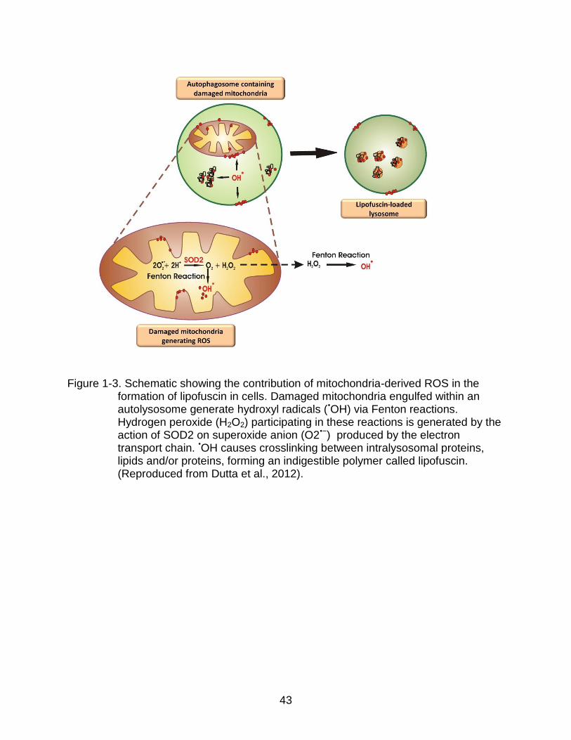
Importantly, oxidative stress is associated with the accumulation within post-mitotic
cells of a non-degradable, polymeric, toxic, yellow-brown pigment, called lipofuscin or
age pigment (Jung et al., 2007). Lipofuscigenesis results from peroxide-induced Fenton
reactions elicited by intralysosomal materials producing highly reactive hydroxyl radicals
(Fig. 1-3). ROS-derived modifications to proteins and lipids cause crosslinking inside
lysosomes/autolysosomes, generating lipofuscin (Jung et al., 2007). Peroxides involved
in these Fenton reactions can diffuse into lysosomes from cytosolic damaged
mitochondria or may originate from autophagocytosed, yet undegraded mitochondria
(Terman & Brunk, 2005). The accumulation of such intracellular garbage eventually
overburdens the autophagosomal-lysosomal degradative capacity, by acting as a sink
for lysosomal hydrolases (Terman, 2001). It follows that attempts to digest lipofuscin
eventually results in the incapacity of autophagy to keep up with the cell’s needs
(Terman, 2001). This series of events is thought to trigger a vicious cycle in which the
autophagic failure and the accumulation of damaged mitochondria perpetuate each
other, resulting in further oxidative stress and enhanced lipofuscinogenesis (Terman &
Brunk, 2005). The collapse of the catabolic machinery will eventually become
incompatible with the maintenance of cell homeostasis and survival. This assumption
also represents the basis of the mitochondrial-lysosomal axis theory of aging (Terman,
2001; Terman et al., 2010).
Mitochondrial dysfunction and oxidative stress is implicated in the pathogenesis of
a host of heart diseases highly prevalent at old age, including heart failure, LVH, heart
attack and diabetic cardiomyopathy (Lesnefsky et al., 2001). It is worth noting that an
altered regulation of autophagy has been shown to contribute to the pathogenesis of

32
these conditions, further supporting the relevance of the mitochondrial-lysosomal axis to
cardiac physiology. In the following sections, the role played by cardiomyocyte
mitochondrial dysfunction and abnormal regulation of autophagy in the above
mentioned heart diseases is discussed.
Heart Failure
Various stressors, as well as pressure overload may be responsible for
mitochondrial dysfunction and enhanced ROS generation during heart failure (Tsutsui et
al., 2009). Heart failure mitochondria generate larger amounts of O2•− compared with
normal mitochondria (Ide et al., 2001). This enhanced oxidant production has been
associated with increased levels of lipid peroxidation to mitochondrial membranes,
decreased mtDNA copy number, reduced abundance of mitochondrial RNA transcripts,
and impaired ATP generation capacity (Ide et al., 2001). In addition, oxidative stress
directly impacts cardiomyocyte structure and function by activating signaling pathways
involved in myocardial remodeling and heart failure (Ide et al., 2001; Spinale et al.,
1998). This suggests the existence of a pathogenic link between enhanced ROS
production, mitochondrial dysfunction and the development of heart failure. Recent
evidence also suggests that an altered regulation of cellular quality control by
autophagy, may contribute to the pathogenesis of heart failure. For instance,
cardiomyocytes isolated from autophagy deficient mice show an increased sensitivity to
β-adrenergic stress compared with wild-type cells (Nakai et al., 2007b). Indeed, stress
induced by 7-day isoproterenol treatment resulted in left ventricular dilation and cardiac
dysfunction in autophagy deficient mice, but not in wild-type controls (Nakai et al.,
2007b).

33
Left Ventricular Hypertrophy
Mitochondrial dysfunction is implicated in the pathogenesis of left ventricular
hypertrophy as well as in the transition from compensated left ventricular hypertrophy to
heart failure (Abel & Doenst, 2011). Mitochondrial misalignment and aggregation are
observed in adult mice with heart-specific deficiency of autophagy in response to
pressure overload induced by thoracic transverse aortic constriction (Nakai et al.,
2007b). These animals develop LVH, contractile dysfunction and heart dilation. Such
findings suggest that an abnormal regulation of autophagy may contribute to the
development of left ventricular hypertrophy, possibly through the accumulation of
dysfunctional mitochondria and subsequent increased ROS generation. In another
study, four weeks of 40% CR increased the activation of cardiomyocyte autophagy and
mitigated heart macroscopic and ultrastructural remodeling, while reducing mortality
(Finckenberg et al., 2012).
Heart Attack
Oxidative stress plays a central role in the pathogenesis of myocardial damage
during oxygen deprivation observed during heart attack. In this setting, mitochondria
contribute to cardiac dysfunction and cardiomyocyte injury via both a loss of metabolic
function and an increased generation of oxidants (Lesnefsky et al., 2001). Oxidative
stress is further exacerbated by the concomitant impairment of endogenous antioxidant
defenses (Becker, 2004). Autophagy is activated in response to myocardial oxygen
deprivation and promotes cardiomyocyte survival, likely via maintaining energy
production during acute nutrient deprivation (Matsui et al., 2007). The degradation of
proteins and organelles by autophagy generates amino acids and fatty acids, which can

34
be used to maintain mitochondrial ATP production and promote survival of cardiac cells
(Loos et al., 2011).
Diabetic Cardiomyopathy
The inefficient autophagic removal of damaged mitochondria may promote the
accumulation of dysfunctional organelles in the diabetic heart. Indeed, down-regulation
of cardiac autophagy, secondary to reduced AMPK activity, has been documented in
diabetic mice (Xie et al., 2011b). Ultrastructurally, hearts from these rodents display
several morphological aberrations, including aggregation of chaotically distributed
mitochondria (Xie et al., 2011b). Inhibition of AMPK by a cardiac-specific dominant
negative AMPK gene further reduced autophagy, exacerbated ultrastructural
aberrations, worsened cardiac dysfunction and increased mortality in diabetic mice (Xie
et al., 2011b). In contrast, treatment with the AMPK-activator metformin significantly
enhanced autophagy and ameliorated cardiomyocyte ultrastructural abnormalities, while
preserving cardiac function. Such benefits were not observed in rodents expressing a
dominant negative AMPK, indicating that cardioprotection by metformin is accomplished
through AMPK-mediated up-regulation of autophagy (Xie et al., 2011b).
Collectively, these findings suggest that depression of autophagy may promote the
accumulation of dysfunctional mitochondria in the diabetic myocardium, thereby
contributing to the development of diabetic cardiomyopathy. Therefore, interventions
that up-regulate autophagy appear as promising means to prevent and/or treat this
relevant complication of diabetes.
It is however worth remembering that in all the examples of cardiac pathology
above, the level of autophagy may be critical in determining whether autophagy will be
protective or detrimental. Indeed, the up-regulation of autophagy might represent an

35
attempt to cope with increased levels of mitochondrial dysfunction and cellular damage.
However, the possibility exists that an excessive up-regulation of autophagy is
detrimental, chewing up essential survival components in the failing myocardium and
leading to further toxicity.
Autophagy as a Therapeutic Target against Cardiac Pathology
The critical role postulated for mitochondria-driven oxidative damage in cardiac
aging and CVD would suggest that the administration of antioxidants might mitigate the
burden of cardiomyocyte injury. However, the efficacy of antioxidant supplementation is
still a matter of debate. Indeed, most clinical trials failed to show any positive effect of
antioxidants on cardiovascular outcomes (Bjelakovic et al., 2007). Chronic
administration of -carotene, vitamin A or vitamin E may even increase cardiovascular
mortality (Bjelakovic et al., 2007). Exploiting the ability of cells to repair or replace
oxidatively-damaged molecules and mitochondria represents an appealing alternative
against heart senescence and associated pathologies (Rubinsztein et al., 2011). A
schematic of the potential beneficial effects of autophagy against oxidative-stress
mediated cellular and mitochondrial dysfunction is shown in Figure 1-4. Interventions
aimed at improving mitochondrial turnover through the fine-tuning of autophagy (e.g.,
CR, resveratrol administration and sirtuin pathway activation) might be especially
relevant to prevent age-related CVD (Green et al., 2011).
CR, defined as a reduction in food intake without malnutrition, is the most robust
intervention for improving health, maintaining organ function and increasing mean and
maximum life span in a range of species (Omodei & Fontana, 2011). Wohlgemuth et
al.... showed that lifelong 40% CR increased the protein expression of Atg7, Atg9 and

36
lipidated LC3 (LC3-II) in the heart of old rats (Wohlgemuth et al., 2007). More recently,
Shinmura et al... demonstrated that a similar dietary regimen enhanced the autophagic
flux in the heart of aged rats through mTOR suppression (Shinmura et al., 2011). The
modulation of the autophagic response represents a primary mechanism underlying the
lifespan-extending properties of CR (Hars et al., 2007; Morselli et al., 2010; Toth et al.,
2008). Indeed, the inhibition of autophagy prevents the anti-aging effects of CR in lower
organisms (Jia & Levine, 2007). CR can induce autophagy through different pathways:
the insulin-like growth factor-1 (IGF-1) / insulin signaling pathway (Toth et al., 2008), the
sirtuin pathway (Morselli et al., 2010), the AMPK pathway (Egan et al., 2011) and the
mTOR pathway (Kim et al., 2011). These different pathways are intimately
interconnected and all play important roles in mediating different aspects of the
response. These adaptations were associated with reduced lipofuscin accumulation in
the myocardium, down-regulation of cardiomyocyte apoptosis, decreases in fiber cross-
sectional area, and preservation of left ventricular diastolic function (Shinmura et al.,
2011). Similar echocardiographic findings have been reported in late-middle aged
humans on long-term CR (Meyer et al., 2006). However, whether these effects were
linked with changes in autophagy activity was not investigated. Furthermore, it is
presently unclear if the cardioprotective effects of CR are primarily mediated by
improvements in autophagy.
Considerable effort has also been directed toward the discovery of drugs that
could mimic the effects of CR, without requiring food restriction and its detrimental
consequences (Ingram et al., 2006). The first CR-mimetic identified was 2-deoxy-D-
glucose (2DG), an analog of glucose, shown to extend both mean and maximum

37
lifespan in C. elegans (Schulz et al., 2007). However, a recent study demonstrated that
chronic 2DG administration to rats, although reproducing a CR-like phenotype, caused
cardiotoxicity and increased mortality (Minor et al., 2010). Promising CR-mimetics with
autophagy-inducing properties are those that intersect with the critical signaling
pathways identified above and include biguanides, such as metformin that targets the
AMPK and insulin signaling pathways (Xie et al., 2011a), resveratrol that affects sirtuin
activity (Morselli et al., 2010) and rapamycin that interacts with mTOR signaling
(Harrison et al., 2009).
Resveratrol has been shown to recapitulate the transcriptional profile and some
of the physiological changes that develop under CR (Barger et al., 2008a; Barger et al.,
2008b; Baur et al., 2006). In addition, resveratrol improved survival and reduced the
prevalence of cardiac pathology in mice fed a high-calorie diet (Baur et al., 2006).
Studies in rodents have also shown that resveratrol inhibits cardiomyocyte apoptosis,
protects the myocardium against I/R injury, prevents LVH, improves endothelial
function, inhibits platelet aggregation, and reduces inflammation (Petrovski et al., 2011).
SIRT1, which is directly or indirectly activated by resveratrol, exerts a similar hormetic
action on cardiomyocyte physiology (Alcendor et al., 2007). Low (2.5-fold) to moderate
(7.5-fold) transgenic overexpression of SIRT1 in the mouse heart attenuates age-
dependent hypertrophy and reduces the severity of apoptosis/fibrosis as well as cardiac
dysfunction (Alcendor et al., 2007). In contrast, high levels (12.5-fold) of SIRT1
expression increase the extent of cardiomyocyte apoptosis and the degree of
hypertrophy, while decreasing cardiac function, thereby inducing the development of
cardiomyopathy (Alcendor et al., 2007). Importantly, resveratrol has been shown to

38
induce autophagy in numerous cell lines from different tissues of origin (Jeong et al.,
2012; Lv & Zhou, 2012; Mauthe et al., 2011; Wu et al., 2011). Whether resveratrol also
induces autophagy in animal models is yet to be determined.
In conclusion, interventions aimed at modulating autophagy may represent a novel
strategy to prevent oxidative stress-associated CVD. However, a deeper understanding
of the role of autophagy in heart physiology is necessary to determine the “therapeutic
amount of stress” and the “hormetic window” that elicit an autophagic response within
the adaptive range.
Summary and Project Goals
Over their lifespan, cardiac cells suffer from a high burden of mitochondria-derived
oxidative damage, which cannot be diluted through cell proliferation. This implies that
the maintenance of a healthy pool of mitochondria and the removal of damaged
organelles are vital for the preservation of cardiomyocyte function and viability.
Autophagy serves this essential homeostatic function. In fact, the vital functions carried
out by autophagy in cardiac physiology suggest the possibility that therapeutic
interventions targeting this cellular pathway may represent effective means to counter
oxidative stress-induced cardiomyopathy. Hence, we report the use of both in vitro and
in vivo systems to investigate whether enhanced autophagy can provide protection
against oxidative stress-mediated dysfunction in cardiomyocytes. In vitro studies have
been conducted in mouse atrial HL-1 cardiomyocytes, which have phenotypic and
morphological features very similar to that of adult cardiomyocytes (Claycomb et al.,
1998). Oxidative stress has been induced in HL-1 cells by the mitochondrial stressor,
Antimycin A and autophagy has been induced by mTOR inhibitor rapamycin. The in
vitro study has been summarized in Chapter 2 of this dissertation. The in vivo study

39
comprises Chapter 3 and involves the use of the chemotherapeutic agent doxorubicin, a
known mitochondrial stressor, to induce oxidative stress in the hearts of 26-month-old
Fischer 344 x Brown Norway rats. CR and resveratrol, either alone or in combination,
has been tested as interventions to induce autophagy in the hearts of these rats. The
chapters provide novel results that further support the beneficial role of autophagy in the
context of oxidatively stressed cardiomyocytes. Finally, the overall conclusions of the
dissertation project as well as future directions will be discussed in Chapter 4. We
believe that the research questions investigated in this study will likely provide answers
to the role autophagy plays in alleviating oxidant-induced damage in cardiomyocytes.
40
Figure. 1-1. Schematic representation of the distinct steps of the autophagic process.
The process of autophagy can be dissected into several steps, comprising of induction and nucleation, expansion, fusion and degradation. Overall, autophagy begins with the formation of a double-layered isolation membrane (phagophore) around the molecules and/or organelles to be degraded. The phagophore grows in size and completely engulfs the cargo, forming an autophagosome. The autophagosome subsequently fuses with the lysosome evolving into an autophagolysosome wherein the cargo is digested. Some of the key molecular regulators controlling the autophagic process have also been shown. (Reproduced from Dutta et al., 2012).

41
Figure 1-2. Schematic overview of the molecular regulation of autophagy in the context of mitochondrial homeostasis. In the presence of nutrients and growth factors, the PKB/Akt pathway is activated, which blocks TSC1/TSC2 and relieving their inhibitory effect on RHEB. The latter, in turn, activates mTORC1. mTORC1 inhibits autophagy by blocking the ULK1-Atg13-FIP100 complex through inactivating phosphorylations on Atg1 and Atg13. Under conditions of energy depletion, the cellular AMP:ATP ratio increases and AMPK becomes activated, which in turn stimulates autophagy through multiple mechanisms. AMPK can phosphorylate and activate TSC1/TSC2, thereby relieving the mTORC1-mediated inhibition of autophagy. In addition, AMPK can phosphorylate Ulk1 at specific serine residues leading to the initiation of autophagy. AMPK can also inhibit the mTORC1 complex by phosphorylating Raptor (mTORC1 containing rapamycin-associated TOR protein), the binding partner required for mTORC1 activity. In addition, under nutrient starvation conditions, mTOR is inactivated and this removes the inactivating phosphorylation on Atg1 and Atg13, resulting in autophagy induction independent of AMPK. Once activated, autophagy serves multiple functions, including the clearance of damaged mitochondria. In addition AMPK induces mitochondrial biogenesis by activating PGC-1α either directly or through SIRT1. SIRT1 can also deacetylate and activate several autophagy-related proteins, such as Atg5, Atg7 and LC3, resulting in enhanced autophagic activity. The autophagic removal of damaged mitochondria coupled with mitochondrial biogenesis is essential for the maintenance of mitochondrial homeostasis. (Reproduced from Dutta et al., 2012).
(see figure on next page)

42
43
Figure 1-3. Schematic showing the contribution of mitochondria-derived ROS in the formation of lipofuscin in cells. Damaged mitochondria engulfed within an autolysosome generate hydroxyl radicals (•OH) via Fenton reactions. Hydrogen peroxide (H2O2) participating in these reactions is generated by the action of SOD2 on superoxide anion (O2•−) produced by the electron transport chain. •OH causes crosslinking between intralysosomal proteins, lipids and/or proteins, forming an indigestible polymer called lipofuscin. (Reproduced from Dutta et al., 2012).

44
Figure 1-4.Schematic showing the beneficial effect of autophagy against oxidative stress-mediated dysfunction. Oxidative stress induces the accumulation of dysfunctional mitochondria and aggregated proteins in cells. Damaged mitochondria show decreased membrane potential, generate increased amounts of ROS and enhance apoptotic induction. Induction of autophagy mediates the removal of such dysfunctional mitochondria and oxidatively damaged proteins, which when coupled with functional biogenesis, helps preserve cellular homeostasis.

45
CHAPTER 2 UPREGULATED AUTOPHAGY PROTECTS CARDIOMYOCYTES FROM OXIDATIVE
STRESS-INDUCED CYTOTOXICITY
Introduction
Progressive accumulation of mitochondrially-generated ROS has been proposed
to be a major player in the pathogenesis of many chronic degenerative diseases, such
as CVD (Balaban et al., 2005; Judge et al., 2005a). Experimental evidence indicates
that in cardiac cells, damaged and dysfunctional mitochondria can produce up to 10 fold
more hydrogen peroxide (H2O2) than intact organelles (Grivennikova et al., 2010).
Oxidants, in low amounts, can function as essential signaling molecules (Finkel, 2011).
In contrast, excessive ROS generation is detrimental, negatively affecting the function
and integrity of not only the mitochondria, but also of proteins, lipids and other
organelles in the cell, potentially leading to impaired cellular homeostasis and organ
function (Judge & Leeuwenburgh, 2007; Lin & Beal, 2006). In fact, mitochondrial
dysfunction and oxidative stress plays a major role in the pathogenesis of cardiac LVH
as well as in the progression from compensated LVH to heart failure ((Abel & Doenst,
2011). Mitochondria-generated oxidative stress also plays a critical role in the
pathogenesis of myocardial damage during ischemia/reperfusion, which eventually
progresses to cardiac dysfunction (Lesnefsky et al., 2001). Further evidences for the
involvement of mitochondrial derived oxidative stress in mammalian heart senescence
is provided by the observation that overexpressing the anti-oxidant enzyme catalase in
the mitochondria (mCAT) delays the development of cardiac pathology in mice (Dai et
al., 2009b). The rate of H2O2 generation and mitochondrial oxidative damage are
significantly decreased in the hearts of old mCAT mice compared with age-matched
wild-type controls (Dai et al., 2009b).

46
Apart from the intra-mitochondrial proteolytic system selectively removing
damaged proteins, the only known mechanism by which mitochondria can be cleared
from a cell is through autophagy. Autophagy is a self-digestion process whereby the cell
degrades long-lived proteins and organelles, generating amino acids and fatty acids that
can be reutilized by the cell (Mizushima & Levine, 2010). Growing evidence suggests
that autophagy could specifically target damaged and dysfunctional mitochondria for
removal, which might otherwise lead to apoptotic induction (Tait & Green, 2010).
Additionally, autophagy can also remove oxidatively modified and dysfunctional proteins
as a bulk digestion process, thereby maintaining a “cleaner” cell and avoiding potential
proteotoxicity arising from their unwanted accumulation (Cuervo et al., 2005). It is worth
noting here that oxidative stress per se has been shown to induce autophagy, by
specifically regulating the activity of Atg4 (Scherz-Shouval et al., 2007a). However,
recent studies show that the type of oxidant generated and its site of origin are crucial
factors in determining the extent and the result of such an autophagic induction
(Dewaele et al., 2010).
Rapamycin is an immunosuppressant antibiotic widely used as an inducer of
autophagy, acting through its inhibitory effect on mTOR (Ravikumar et al., 2004). In the
presence of nutrients and growth factors, mTOR is active, hyperphosphorylating and
inhibiting the ULK1 - Atg13 – FIP200 complex required for the induction of autophagy
(Jung et al., 2009b). Rapamycin therefore primarily acts to remove the inhibitory effect
of mTOR on autophagy. The cardioprotective properties of rapamycin have been
demonstrated in animal models of ischemia / reperfusion as well as in cardiac
hypertrophy (Khan et al., 2006; McMullen et al., 2004). In addition, rapamycin also
47
protects against neurodegeneration by mediating the removal of aggregated proteins
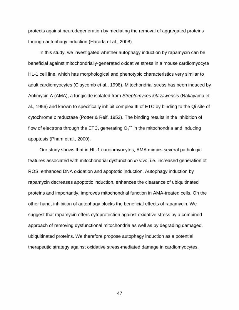
through autophagy induction (Harada et al., 2008).
In this study, we investigated whether autophagy induction by rapamycin can be
beneficial against mitochondrially-generated oxidative stress in a mouse cardiomyocyte
HL-1 cell line, which has morphological and phenotypic characteristics very similar to
adult cardiomyocytes (Claycomb et al., 1998). Mitochondrial stress has been induced by
Antimycin A (AMA), a fungicide isolated from Streptomyces kitazawensis (Nakayama et
al., 1956) and known to specifically inhibit complex III of ETC by binding to the Qi site of
cytochrome c reductase (Potter & Reif, 1952). The binding results in the inhibition of
flow of electrons through the ETC, generating O2•− in the mitochondria and inducing
apoptosis (Pham et al., 2000).
Our study shows that in HL-1 cardiomyocytes, AMA mimics several pathologic
features associated with mitochondrial dysfunction in vivo, i.e. increased generation of
ROS, enhanced DNA oxidation and apoptotic induction. Autophagy induction by
rapamycin decreases apoptotic induction, enhances the clearance of ubiquitinated
proteins and importantly, improves mitochondrial function in AMA-treated cells. On the
other hand, inhibition of autophagy blocks the beneficial effects of rapamycin. We
suggest that rapamycin offers cytoprotection against oxidative stress by a combined
approach of removing dysfunctional mitochondria as well as by degrading damaged,
ubiquitinated proteins. We therefore propose autophagy induction as a potential
therapeutic strategy against oxidative stress-mediated damage in cardiomyocytes.

48
Materials and Methods
Cell Culture Conditions
HL-1 mouse atrial cardiomyocytes were a kind gift of Dr. W Claycomb (Louisiana
State University Health Science Center, LA). Cells were cultured in Claycomb Media
(JRH Biosciences, KS) supplemented with 10% fetal bovine serum (Sigma, MO), 2 mM
L-glutamine (Life Technologies, MD), 0.1 mM norepinephrine (Sigma, MO), 100 U/mL
penicillin and 100 mg/mL streptomycin (Life Technologies, MD), on fibronectin-gelatin
coated plates at 37°C in a humidified atmosphere, with 5% CO2.
Chemical Reagents
AMA (Sigma, MO) was dissolved in ethanol at a concentration of 15 mM;
Rapamycin (Calbiochem, CA) was made up in ethanol at 5 mM; BafA1 (Sigma, MO)
was dissolved in dimethyl sulfoxide (DMSO) at a concentration of 25 µM and 3-MA
(Sigma, MO) was dissolved in deionized water at 180 mM. A more complete list of all
the reagents used in the study, their source and catalog numbers is given in table 2-1.
Flow Cytometric Determination of Mitochondrial O2•− Generation
O2•− generation in the mitochondria was analyzed using the fluorescent dye
MitoSOX Red (Invitrogen, CA). It is specifically targeted to bioenergetically active
mitochondria where it is rapidly and selectively oxidized by O2•− molecules. The oxidized
product binds to the mitochondrial DNA and fluoresces red.(Mukhopadhyay et al., 2007)
HL-1 cells were trypsinized and resuspended in fresh media at a density of 2x10⁶
cells/mL and MitoSOX Red was added to a final concentration of 3 μM. Cells were
allowed to load MitoSOX Red for a period of 30 mins at 37°C, washed twice with
Dulbecco’s Phosphate Buffered Saline (DPBS) and the media replaced with fresh
media. Cells were subsequently incubated with increasing concentrations of AMA or

49
vehicle control for 30 mins at 37°C, followed by Flow cytometric analysis of MitoSOX
Red fluorescence using FACSCalibur flow cytometer (BD Biosciences, CA). MitoSOX
Red was excited by laser at 488 nm and the data collected using forward scatter (FSC)
and side scatter (SSC) and FL2 (575 +/- 12.5 nm) channels. Cell debris is represented
by distinct low FSC and SSC and was gated out for analysis. The data shown
represents mean MitoSOX Red fluorescence intensity of 10,000 HL-1 cells treated with
the indicated concentrations of AMA.
Confocal Imaging of Mitochondrial O2•− Generation
Cells on No. 1.5 coverslip-glass bottom dishes (MatTek Corporation, MA) were
incubated with 3 μM MitoSOX Red and 1 µg/mL Hoechst 33342 (Invitrogen, CA) for a
period of 30 mins at 37°C, washed twice with DPBS and the media replaced with fresh
media. Cells were subsequently treated with 50 µM AMA or vehicle control for 30 mins
and imaged for MitoSOX Red fluorescence using Leica TCS SP2 Laser Scanning
Confocal Microscope (Leica Microsystems GmbH, Heidelberg, Germany) using an
excitation of 514 nm and emission of 560-600 nm. Hoechst 33342 was visualized using
405 nm excitation and 440-470 nm emission. The pictures were merged using Leica
LCS software (Leica Microsystems GmbH, Heidelberg, Germany).
Flow Cytometric Determination of Mitochondrial Membrane Potential
Δψm was analyzed using the cationic fluorescent dye TMRM (Invitrogen, CA),
which is specifically targeted to and accumulates in bioenergetically active mitochondria
(Scaduto & Grotyohann, 1999). The dye is lost when the mitochondrial membrane
potential (Δψm) is lost. HL-1 cells were trypsinized and resuspended in fresh media at a
density of 2x10⁶ cells/mL, after which TMRM was added to a final concentration of 50
nM. Cells were allowed to load TMRM for a period of 30 mins at 37°C, washed twice

50
with DPBS and the media replaced with fresh media. Cells were subsequently
incubated with increasing concentrations of AMA or vehicle control for 2h at 37°C,
followed by Flow cytometric analysis of TMRM fluorescence using the same excitation-
emission and gating protocol as that of MitoSOX Red.
Confocal Imaging of Mitochondrial Membrane Potential
Cells were incubated with 50 nM TMRM and 1 µg/mL Hoechst 33342 at 37°C for a
period of 30 mins, washed twice with DPBS and the media replaced with fresh media.
Cells were subsequently treated with 50 µM AMA or vehicle control for 2h and imaged
for TMRM fluorescence using Leica TCS SP2 Laser Scanning Confocal Microscope
(Leica Microsystems GmbH, Heidelberg, Germany) with an excitation of 543 nm and
emission of 560-600 nm. The pictures were merged using Leica LCS software (Leica
Microsystems GmbH, Heidelberg, Germany).
Determination of Cell Viability
Cell viability was determined by 3-(4, 5-dimethylthiazol-2-yl)-2,5-
diphenyltetrazolium bromide (MTT; Sigma, MO) and Trypan Blue dye exclusion assay
(Sigma, MO). To determine cell viability by MTT assay, cells in 24-well plates were
incubated with 0.5 mg/mL MTT for a period of 30 mins at 37ºC. The purple formazan
crystals formed were dissolved in DMSO at 37ºC for 10 mins and the absorbance of the
resulting solution measured at 540 nm using Biotek Synergy plate reader (Biotek, VT).
The background absorbance was subtracted at 630 nm. The values represented are
normalized to control vehicle-treated group. To determine cell viability by Trypan Blue
assay, cells were trypsinized and resuspended in fresh media containing equal volume
of 0.4% Trypan Blue (Sigma, MO) for 5 mins and the number of live (bright) or dead
(dark) cells counted using a hemocytometer.

51
Determination of DNA / RNA Oxidation
8-oxo-7,8-dihydro-2’-deoxyguanosine (8-oxo-dGuo; measure of DNA oxidation)
and 8-oxo-7,8-dihydroguanosine (8-oxo-Guo; measure of RNA oxidation)
concentrations were measured in cells using HPLC coupled to electrochemical
detection (HPLC-ECD) analysis (ESA Coulochem III electrochemical detector, MA)
according to protocol described before (Hofer et al., 2006). Briefly, cells were treated
with AMA or vehicle control for 2h and 24h later, they were trypsinized and
subsequently homogenized in 3 M guanidium thiocyanate lysis buffer (GTC, 0.2% w/v
N-lauroylsarcosinate, 20 mM Tris, pH 7.5) in the presence of the metal chelator
deferoxamine mesylate (DFOM; Sigma, MO) to prevent further oxidation. Cellular DNA /
RNA was isolated using phenol-chloroform method and precipitated in isopropanol. The
nucleic acid pellet was subsequently washed with ethanol, air-dried and dissolved in 10
mM DFOM followed by hydrolysis in 10 µL Nuclease P1 (stock of 0.4 U/µL in 300 mM
sodium acetate, 0.2 mM ZnCl2, pH 5.3; MP Biomedicals, OH) and 5 ul alkaline
phosphatase (1 U/µL; Sigma, MO) for a period of 60 mins at 50⁰C. The hydrolysate was
filtered to remove enzymes and subsequently loaded for HPLC-ECD analysis. For
quantification, HPLC peaks were compared to calibration curves of 8-oxo-dGuo and
dGuo standards (Sigma, MO). The amount of 8-oxo-Guo and 8-oxo-dGuo present was
normalized to the total amount of non-oxidized DNA and RNA respectively.
Induction and Assessment of Autophagy
Autophagic stimulation converts the cytosolic and unconjugated form of LC3 (LC3-
I) to the carboxy-terminal cleaved, phosphatidylethanolamine (PE) conjugated form
(LC3-II) and this causes a change in the molecular weight of the protein, allowing both
the forms to be resolved in an SDS-PAGE gel. The LC3-II/LC3-I ratio is therefore
52
indicative of the amount of ongoing autophagy. However, an increase in the LC3-II/LC3-
I ratio could be due to increased autophagic stimulation or due to a decreased
clearance of autophagic vacuoles in the lysosomes. Hence all autophagic responses
have been monitored in the absence or presence of the lysosomal inhibitor BafA1, to
determine steady state and cumulative autophagy (a.k.a autophagic flux), respectively.
To determine the optimum concentration of rapamycin for inducing autophagy in HL-1
cardiomyocytes, cells were treated with increasing concentrations of the drug for a
period of 16h in the presence or absence of 75 nM BafA1, added during the final 4h of
incubation. For subsequent experiments involving rapamycin, a concentration of 1 μM
was used.
Epifluorescence Imaging and Quantification of Autophagy
For assessment of autophagic activity using epifluorescence imaging, cells plated
on coverslips in 24-well plates were transfected with GFP-LC3 plasmid (kind gift of Dr T.
Yoshimori, National Institute of Genetics, Japan) (Kabeya et al., 2000) using
Lipofectamine 2000 (Invitrogen, CA) following manufacturer’s instructions. To prevent
the potential problem of GFP-LC3 aggregate formation(Kuma et al., 2007), transfection
was optimized with a low concentration of GFP-LC3 plasmid (0.8 μg for each well of a
24 well plate). Twenty-four hours after transfection, cells were incubated with 1 μM
rapamycin for 16h, in the absence or presence of 75 nM BafA1 added during the final
4h of incubation. Cells were subsequently by fixed in 4% paraformaldehyde and
mounted on slides with DAPI (Vector Laboratories, CA). Epifluorescence images were
taken using Leica DM IRBE epifluorescence microscope (Leica Microsystems, Rueil-
Malmaison, France) and images were processed using ImageJ software (NIH, MD).
Conversion of diffuse cytosolic GFP-LC3-I to punctate autophagic membrane-

53
associated GFP-LC3-II was monitored and considered indicative of autophagic activity
in cells. Specifically, the number of puncta in each cell was counted and cells with 30 or
more puncta were considered to have induced autophagy.
Confocal Imaging
Confocal microscopy was used for live-cell imaging of autophagy and mitophagy in
AMA and AMA plus rapamycin-treated cells. Cells were plated on 0.17 mm thick glass-
bottom Fluorodishes (World Precision Instruments, FL) and were transfected with GFP-
LC3 plasmid using Lipofectamine 2000 (Invitrogen, CA) following manufacturer’s
instructions. Twenty-four hours after transfection, cells were incubated with 1 μM
rapamycin or vehicle control for 16h. Cells were subsequently treated with 50 μM AMA
and the autophagic vacuoles visualized using Zeiss LSM 510 Meta laser scanning
microscope (Ziess, Germany). For visualization of mitophagy, cells were pre-loaded
with 50 nM TMRM for 30 mins at 37⁰C before the addition of 50 µM AMA. GFP
fluorescence was excited at 488 nm and emission was collected through a 500 - 530
nm band-pass filter. TMRM was excited at 545 nm and emission collected through a
565 – 619 nm band pass filter. Confocal images were processed using LSM Image
Browser software (Zeiss, Germany).
Western Blotting
Cells were lysed using RIPA buffer (Sigma, MO) and protein concentration
determined using Dc assay (BioRad, CA) (Bradford, 1976). Equal amounts of proteins
were then loaded in pre-cast Tris-HCl polyacrylamide gels (BioRad, CA). After
electrophoretic separation, proteins were transferred to polyvinylidene difluoride (PVDF)
membrane (BioRad, CA) and subsequently blocked for 1h in Starting Block (Thermo
Scientific, NJ), followed by overnight incubation with primary antibodies. Membranes

54
were subsequently washed three times with Tris buffered saline with 0.1% Tween 20
(TBST) and incubated with the appropriate secondary antibodies for 1h. Membranes
were washed again with TBST and chemiluminescent signals developed using ECL
Plus reagent (Amersham Biosciences, Buckinghamshire, UK). The signals were
captured using ChemiDoc XRS System (BioRad, CA) and digital images analyzed using
ImageLab software (BioRad, CA). A list of primary antibodies used in the study, their
source and catalog numbers is given in Table 2-2.
For detection of ubiquitinated proteins, cells were lysed and detergent soluble and
insoluble fractions separated by centrifugation at 8000g. The resulting pellets (insoluble
fraction) were resuspended in 2X SDS buffer and heated at 100⁰C for 10 mins. Equal
volumes of sample lysates were then loaded in each lane. The supernatant (soluble
fraction) was processed as mentioned above.
Assessment of Cellular Respiration
Cells were pre-treated with 1 µM rapamycin or vehicle control for 16h, followed by
incubation with 50 µM AMA or vehicle control for an additional 24h, in the presence of
rapamycin. Cells were subsequently trypsinized and resuspended in complete
Claycomb media. The O2 flux, as a measure of routine respiration was subsequently
measured in unpermeabilized cells using OROBOROS Oxygraph-2K (Oroboros
Instruments GmbH, Innsbruck, Austria) at 37⁰C in a constantly stirred chamber. The
rates of consumption are expressed as pmol / sec / 106 cells.
Statistical Analysis
All treatments were done in three or more independent experiments. All statistical
analyses were performed using GraphPad Prism 4 (GraphPad Software, Inc. San

55
Diego, CA). For statistical analysis, Student’s t-test and one-way ANOVA were
performed, wherever applicable. Statistical significance was set to p < 0.05 and the data
are represented as mean ± SE.
Results
AMA Increases Mitochondrial O2•− Generation and Decreases Mitochondrial
Membrane Potential (Δψm)
First, we established the concentration of AMA needed to increase ROS
generation in the mitochondria. Cells pre-labeled with MitoSOX Red, highly selective for
the detection of O2•− in the mitochondria (Mukhopadhyay et al., 2007), were treated with
increasing concentrations of AMA or vehicle control and the fluorescence intensity
analyzed using flow cytometry. In contrast to vehicle-treated cells which showed
minimal MitoSOX Red fluorescence, treatment with AMA resulted in a dose-dependent
increase in fluorescence intensity, with 50 µM being the lowest concentration required to
reach statistical significance (Fig. 2-1A; p < 0.05). To confirm flow cytometry results, we
performed confocal imaging on cells pre-labeled with MitoSOX Red and treated with 50
µM AMA. In contrast to vehicle-treated cells which showed minimal fluorescence,
treatment with 50 µM AMA resulted in a strong MitoSOX Red fluorescence originating
from the mitochondria (Fig. 2-1B).
Next, we determined the effects of AMA on Δψm. These studies were done in cells
pre-labeled with Tetramethyl Rhodamine Methyl Ester (TMRM), a cationic, fluorogenic
dye which specifically migrates to bioenergetically active mitochondria and fluoresces
red (Scaduto & Grotyohann, 1999). TMRM-loaded cells were treated with increasing
concentrations of AMA or vehicle control and the fluorescence intensity analyzed using
flow cytometry. Vehicle-treated cells showed a strong fluorescence, indicative of normal

56
membrane potential, whereas AMA treatment resulted in a dose dependent decrease in
TMRM fluorescence (Fig. 2-1C). We observed that 50 µM is the lowest concentration of
AMA required to reach statistically significant decreases in Δψm (p < 0.05). Confocal
imaging further confirmed that 50 µM AMA resulted in a drastic decrease in TMRM
fluorescence, compared to vehicle-treated cells (Fig. 2-1D).
AMA Treatment Induces Nuclear DNA Oxidation and Decreases Cell Viability
We next wanted to investigate whether AMA-induced ROS can leak out of the
mitochondria and have damaging effects on extra-mitochondrial components. To test
this, we measured the levels of 8-oxo-dGuo and 8-oxoGuo as an indicator of DNA and
RNA oxidation, respectively (Hofer et al., 2006). AMA significantly increased the level of
DNA oxidation in HL-1 cells at a concentration of 50 µM (Fig. 2-1E, p < 0.05). RNA
oxidation tended to increase with all concentrations, but changes were not statistically
significant (Fig. 2-1F; p < 0.10). Finally, the survival of HL-1 cells treated with
increasing concentrations of AMA, as determined by MTT assay at different time points,
showed a dose- and time-dependent decrease in viability (Fig. 2-1G). It is worth noting
here that in addition to being used as a viability marker, the MTT assay is also indicative
of mitochondrial activity, as most of the dehydrogenases required for the reduction of
MTT reside in the mitochondria.
Treatment with AMA does not Induce Autophagy in HL-1 Cells
Since oxidative stress has been shown to be an inducer of autophagy, we wanted
to determine whether treatment with AMA can lead to autophagy induction in HL-1 cells.
Cells were treated with increasing concentrations of AMA for either 4h or 24h in the
absence or presence of Bafilomycin A1 (BafA1), added during the final 4h or 2h of
incubation, respectively. At the 24h time point, cells were incubated with BafA1 for a

57
shorter period of time to avoid additional toxicity in stressed HL-1 cells. Immunoblot
analysis showed no significant increases in the LC3-II/LC3-I ratio at either time points
with any of the concentrations of AMA tested (Fig. 2-2A, C). Epifluorescence analysis of
GFP-LC3 puncta in AMA treated cells further confirmed no induction of autophagy by
AMA ( p > 0.05, Fig. 2-2B). In addition, we assessed the levels of other autophagy
proteins Atg5-Atg12, Beclin-1 and Atg7 and detected no difference in their expression
levels in AMA treated cells (Fig. 2-2D). We ruled out the possibility that this inability to
induce autophagy is due to an apparently high oxidative burden generated by the
concentrations of AMA used, as lower concentrations (50 nM) had no effect on the LC3-
II/LC3-I ratio as well (data not shown).
Treatment with Rapamycin Induces Autophagy in HL-1 Cells
Incubation of HL-1 cardiomyocytes with rapamycin caused an inhibition of mTOR
signaling, as shown by the disappearance of phospho-S6, one of the downstream
targets of mTOR kinase (Fig. 2-3A). Additionally, rapamycin caused a significant
increase in cumulative LC3-II/LC3-I ratio at a concentration of 1 μM (p < 0.01), with
lower concentrations not reaching statistical significance (Fig. 2-3B). As a further
confirmation of our immunoblotting data, we performed epifluorescence imaging on
GFP-LC3 expressing cells to determine steady state and cumulative autophagy (Fig. 2-
3C). For these experiments, cells were incubated with 1 μM rapamycin or vehicle
control for 16h, in the absence or presence of BafA1 present during the final 4h of
incubation. Quantification of cumulative GFP-LC3 puncta further confirmed induction of
autophagy by rapamycin (Fig. 2-3D) (p < 0.001). We further observed that treatment
with 1 µM rapamycin decreased mitochondrial content, as observed by the
disappearance of the mitochondrial outer membrane marker VDAC (Fig. 2-3E).

58
Additionally, we confirmed that rapamycin can induce autophagy in the presence of
AMA, both by immunoblotting (Fig. 2-4A) and confocal imaging in GFP-LC3 transfected
cells (Fig. 2-4B). Furthermore, rapamycin also led to the degradation of mitochondria in
AMA-treated cells (Fig. 2-4C). Importantly, the reported half-life of rapamycin is 63h
(Backman et al., 2002), confirming its chemical stability during the experimental period.
Rapamycin Plays a Protective Role against AMA-Induced Toxicity
We next investigated whether rapamycin can mediate a protective effect against
AMA-induced toxicity. For these experiments, cells were pre-treated with rapamycin for
16h, followed by addition of AMA in fresh media in the presence of rapamycin. Viability
analysis, both by MTT (Fig. 2-5A) and Trypan Blue exclusion assays (Fig. 2-5B)
revealed a drastic decrease (p < 0.01) in cell viability with AMA treatment, which was
significantly attenuated by treatment with rapamycin (p < 0.05). MTT assay revealed a
slight decrease in cell viability due to rapamycin treatment alone (Fig. 2-5A), but the
changes were not statistically significant and we believe this is to be due to the anti-
proliferative effect of the mTOR inhibitor per se, which led to a slightly decreased cell
number (as opposed to cell death) in comparison to vehicle-treated cells. This is further
supported by the observation that there was no difference in the number of live cells in
control and rapamycin-treated cells when viability was assessed by Trypan Blue dye
exclusion assay (Fig. 2-5B).
As compared to control or rapamycin-treated cells, AMA induced both caspase-3
and PARP-1 cleavage in HL-1 cells, and rapamycin treatment significantly attenuated
such proteolytic processing (Fig. 2-5C). Additionally, when observed by phase contrast
microscopy, AMA-treated cells showed intense vacuolization in the cytoplasm, a
phenomenon often observed with injury, toxicity and cell death (Henics & Wheatley,

59
1999) Treatment with rapamycin drastically decreased the appearance of such vacuoles
(Fig. 2-5D).
Rapamycin Prevents AMA-Induced Accumulation of Ubiquitinated Proteins
The accumulation of high molecular weight ubiquitinated proteins has been
observed under conditions of increased oxidative stress and can originate from an
inefficient clearance of damaged proteins through the proteasomal system (Dudek et
al., 2005; Shang & Taylor, 2011). Under such conditions, autophagy induction can
mediate their removal, thereby averting potential proteotoxicity. We found that treatment
with AMA induced the accumulation of ubiquitinated proteins in the detergent-insoluble
fraction and treatment with rapamycin drastically prevented such an accumulation (Fig.
2-6). Interestingly, in comparison to control, we observed a decreased accumulation of
ubiquitinated proteins in cells treated with rapamycin alone. This could be attributed to
the autophagic clearance of basal levels of uniquitinated proteins by rapamycin.
Rapamycin Protects against AMA-Induced Mitochondrial Dysfunction
We further wanted to investigate whether rapamycin provides any beneficial effect
on the mitochondrial polpulation in AMA-treated cells. In this respect, assessing cellular
respiration in non-permeabilized cells using polarographic oxygen sensors has been
shown to be an accurate measure of mitochondrial function (Gnaiger, 2008). We found
that AMA-treated cells significantly decreased routine respiration even 24h after
treatment (p < 0.01). This decline in respiration was ameliorated by rapamycin treatment
(p < 0.05) (Fig. 2-7A).
We also measured Δψm as an additional indicator of mitochondrial function. In
comparison to control or rapamycin-treated cells AMA-treated cells had mitochondria
with little or no Δψm even 24h after treatment (arrows, Fig. 2-7B). The few cells that

60
maintained Δψm showed drastically altered mitochondrial morphology, most of them
being highly swollen and drastically bigger in size (arrowheads, Fig. 2-7B inset).
Incubation with rapamycin restored Δψm in AMA-treated cells, as well as maintained
normal mitochondrial morphology (Fig. 2-7B).
Inhibition of Autophagy Blocks the Cytoprotective Effect of Rapamycin against AMA Toxicity
We next determined whether the cytoprotective effect of rapamycin against AMA
toxicity is specifically mediated through autophagy. For this, autophagy was inhibited by
co-incubating cells with 3-Methyladenine (3-MA), a widely used inhibitor of autophagy
(Seglen & Gordon, 1982; Stroikin et al., 2004). LC3-II/LC3-I ratios confirmed that 3-MA
inhibited rapamycin-induced autophagy (Fig. 2-8A). We subsequently performed MTT
assay to determine the viability of AMA-treated cells, co-incubated with rapamycin alone
or with rapamycin in the presence of 3-MA (Fig. 2-8B). The MTT assay revealed that 3-
MA attenuated the cytoprotective effects of rapamycin against AMA toxicity (Fig. 2-8B).
In addition, 3-MA inhibited rapamycin induced attenuation of PARP-1 caspase-3
cleavage in AMA-treated cells (Fig. 2-8C).
Discussion
Mitochondria from heart failure models generate larger amounts of O2•− in the
presence of NADH compared with normal mitochondria (Ide et al., 1999). Such
oxidative stress can activate signaling pathways involved in myocardial remodeling,
affecting cardiomyocyte structure and function (Ide et al., 1999). Our study shows that
the mitochondrial stressor AMA can mimic several features observed under
mitochondrial dysfunction-associated pathological conditions. For instance, AMA
treatment generated O2•− in the mitochondria and decreased Δψm in HL-1

61
cardiomyocytes. A decreased Δψm is indicative of defective oxidative phosphorylation
and a lower capacity of generating ATP (Huttemann et al., 2008); it is therefore used as
a marker of cellular and mitochondrial viability (Vayssier-Taussat et al., 2002).
Importantly, although AMA enhanced O2•− generation in the mitochondria, it is
worthwhile to note that under physiologically normal conditions, to counteract oxidative
stress, the mitochondria and the cytoplasm are equipped with detoxifying enzymes and
non-enzymatic anti-oxidants (Andreyev et al., 2005b). Therefore, oxidants generated by
the mitochondria could potentially be detoxified by cellular anti-oxidant defense
systems, preventing damage to cytoplasmic components (Andreyev et al., 2005b).
However, under conditions of stress and disease pathology, damaged mitochondria can
generate excessive amounts of ROS, overwhelm the defenses, and oxidatively modify
extra-mitochondrial components, such as the DNA and RNA. The DNA base oxidation
product, 8-oxo-dGuo, is potentially mutagenic (Cheng et al., 1992; Kuchino et al., 1987)
and is widely used for the estimation of oxidative stress in tissues and urine (Gedik &
Collins, 2005; Lin et al., 2004). We confirmed that ROS generated by AMA can increase
8-oxo-dGuo levels in HL-1 cardiomyocytes.
As regards the mitochondria, we observed that most cells treated with AMA had
little or no Δψm and the mitochondria were highly swollen. Such aberrant mitochondria,
often also characterized by loss of cristae and matrix derangements, has been
observed through ultrastructural analysis of the myocardium of aged rodents (Sachs et
al., 1977a) and in other models of mitochondrial injury (Bova et al., 2005), and is
considered a marker of mitochondrial toxicity (Kaufmann et al., 2006; Masubuchi et al.,
2006). Furthermore, AMA decreased cell viability and activated apoptotic markers, as

62
well as inhibited cellular respiration in HL-1 cardiomyocytes, further adding to
cytotoxicity (Fig. 2-9).
Some findings suggest that mitochondrial stress and ROS per se can act as
inducers of autophagy (Chen et al., 2009; Kiffin et al., 2006; Scherz-Shouval et al.,
2007a). Other studies have shown that mitochondrial dysfunction and depolarization
might not be sufficient to induce autophagy (Mendl et al., 2011). In our studies we could
not detect any induction of autophagy by treatment with AMA alone, but rapamycin in
the presence of AMA was capable of inducing autophagy. It is possible that additional
triggers might be necessary but missing in AMA induced toxicity (Mendl et al., 2011).
Furthermore, it has been proposed that the inhibition of ETC complex III can actually
have an inhibitory effect on autophagy induction, although the molecular mechanisms
are as of yet, unknown (Ma et al., 2011). Nevertheless, stimulated autophagy under
oxidative stress conditions could be hypothesized to potentially remove oxidized
macromolecules.
Since oxidative stress is postulated to be a major player in the pathogenesis of
cardiovascular diseases, one might argue that the administration of antioxidants will
offer protection against oxidant-mediated damage. However, clinical studies have been
controversial, with limited or no cardiac benefit reported with chronic administration of
anti-oxidants (Bjelakovic et al., 2007; Levonen et al., 2008; Tinkel et al.). Therefore
approaches to remove oxidatively modified macromolecules through autophagy
induction represent an appealing alternative. In our study, we observed that rapamycin
induced autophagy in HL-1 cardiomyocytes (Fig. 2-3). Rapamycin-stimulated autophagy
had a beneficial effect on AMA-treated cells, as assessed by the attenuation of

63
caspase-3 and PARP-1 cleavage, as well as by an improved survival of HL-1
cardiomyocytes (Figs. 2-5 and 2-9). Rapamycin treatment also inhibited the decrease in
Δψm and prevented the appearance of swollen morphology of the mitochondria (Figs. 2-
7 and 2-9). Additionally, respiration analysis in non-permeabilized HL-1 cells showed
that autophagy induction by rapamycin attenuated the decline in cellular respiration
induced by AMA (Figs. 2-7 and 2-9). We suggest that these protective effects of
rapamycin could be partly due to the result of autophagy-mediated removal of
dysfunctional mitochondria, which if not removed, can produce greater amounts of ROS
and / or induce apoptosis through the release of pro-apoptotic factors usually
sequestered in the intermembrane space (Tait & Green, 2010). Supporting this claim,
we have observed rapamycin-induced degradation of mitochondria (mitophagy) in AMA-
treated cells (Fig. 2-4C). The encapsulation and degradation of dysfunctional
mitochondria by rapamycin-induced autophagy has also been reported elsewhere (Pan
et al., 2009; Ravikumar et al., 2006). Additionally, mitochondrial clearance by rapamycin
has been confirmed in our study (Fig. 2-3E). The clearance of dysfunctional
mitochondria is further supported by our imaging analysis, where an accumulation of
dysfunctional (lacking Δψm) and swollen mitochondria was seen only in AMA-treated
cells; such dysfunctional mitochondria could not be detected in cells additionally treated
with rapamycin. Finally, we observed that respiration was inhibited to the same extent in
AMA and AMA plus rapamycin treated cells 2h after treatment (data not shown).
However, after 24 hours, respiration was significantly increased in AMA-treated cells
additionally exposed to rapamycin, suggesting that some of the dysfunctional
mitochondria might have been removed by autophagy during this time period. In this

64
context, fairly modest reductions in mitochondrial content can have significant effects in
improving cell viability, because of a major positive-feedback component involved in
apoptotic signaling by damaged mitochondria. Finally, in order to establish that the
protective effects of rapamycin against AMA toxicity is autophagy specific, we used 3-
MA (Seglen & Gordon, 1982);,(Stroikin et al., 2004), known to inhibit Class III PI3K,
required during the initial steps of autophagy (Petiot et al., 2000). We observed that 3-
MA not only inhibited rapamycin induced autophagy, but also attenuated the beneficial
effects of rapamycin against AMA toxicity.
Consistent with our observations, autophagy has been shown to play a protective
role against anoxia-regeneration in isolated cardiomyocytes (Dosenko et al., 2006), as
well as required for the pre-conditioning effect of adenosine (Cohen & Downey, 2008).
On the other hand, inhibition of autophagy through cardiac specific deletion of the
autophagy protein Atg5 results in mitochondrial misalignment and cardiac hypertrophy
in response to pressure overload induced by transverse aortic constriction. This
eventually progresses into left ventricular hypertrophy, contractile dysfunction and
cardiac dilatation (Nakai et al., 2007a). Inhibition of the autophagic clearance pathway
has also been implicated in the pathogenesis of Danon disease, which is characterized
by cardiomyopathy and heart failure (Kitsis et al., 2007).
Similar to mitochondria, an increase in the ‘dwell time’ of oxidatively modified
cytoplasmic proteins enhances their probability of being post-translationally modified,
which could further increase cellular toxicity (Gershon & Gershon, 1970; Miquel et al.,
1974). Damaged cellular components are marked by ubiquitination, to be subsequently
removed by the ubiquitin-proteasomal system (UPS) (Shang & Taylor, 2011). Growing

65
evidence suggest that under conditions of increased oxidative burden, accumulation of
high molecular weight ubiquitinated proteins is frequently observed, owing to their
inefficient removal through UPS (Dudek et al., 2005; Iwai et al., 1998; Shang & Taylor,
2011). Autophagy induction under these circumstances has been shown to mediate the
removal of such accumulated proteins. In our studies, rapamycin-induced autophagy
attenuated the accumulation of ubiquitinated proteins that resulted from AMA-induced
exposure. Such an accumulation was specifically detected in the insoluble fraction,
suggesting that they might be aggregated in nature. Notably, our observation is
consistent with studies showing that autophagy induction alleviates toxicity in cells
expressing mutant aggregate prone proteins such as α-synuclein (Ravikumar et al.,
2004) and huntingtin (Ravikumar et al., 2004; Webb et al., 2003).
In summary, it is important to note that cardiomyocytes are terminally
differentiated; therefore any damages accumulated during its lifespan of several
decades cannot be diluted by cell proliferation. Furthermore, the heart is highly
metabolic and hence challenged with a considerable amount of oxidative stress during
its lifetime. Hence, the maintenance of a functional pool of mitochondria, coupled with
the removal of aggregated and potentially harmful cytoplasmic proteins are vital for the
preservation of cardiomyocyte homeostasis. Our study conclusively proves that
autophagy induction by rapamycin can mediate the removal of dysfunctional
mitochondria as well as aggregated, ubiquitinated proteins in cardiomyocytes, leading to
a protective effect of autophagy induction against oxidative stress (Fig. 2-9). We
therefore propose rapamycin as a potential therapeutic strategy against cardiovascular
disorders associated with chronic generation of oxidation stress.

66
Figure 2-1. Treatment with AMA leads to enhanced oxidative stress and cytotoxicity in HL-1 cardiomyocytes. (A) HL-1 cells were trypsinized and resuspended in fresh media followed by staining with 3 µM MitoSOX Red. Cells were subsequently incubated with increasing concentrations of AMA or vehicle control for 30 mins followed by flow cytometric analysis of MitoSOX Red fluorescence. (B) Cells were incubated with 1 µg/uL Hoechst 33342 (blue) and 3 µM MitoSOX Red and subsequently treated with 50 µM AMA or vehicle control for 30 mins, followed by confocal imaging. (C) Cells were trypsinized and resuspended in fresh media followed by staining with 50 nM TMRM (red) and were subsequently incubated with increasing concentrations of AMA or vehicle control for 2h followed by flow cytometric analysis of TMRM fluorescence. (D) Cells were incubated with 1 µg/ul Hoechst 33342 and 50 nM TMRM and subsequently treated with 50 µM AMA or vehicle control for 2h, followed by confocal imaging. (E) Cells were incubated with increasing concentrations of AMA for 2h and 24h later, trypsinized and processed for HPLC analysis of DNA (E) and RNA (F) oxidation. Data has been normalized to vehicle-treated control. (G) Cells were incubated with increasing concentrations of AMA and cell viability determined using MTT assay after the indicated time points. Data has been normalized to vehicle-treated control. *p < 0.05; **p < 0.01, ***p < 0.001 vs. control. Data represents mean ± SE and is derived from three independent experiments.
(see figure on next page)
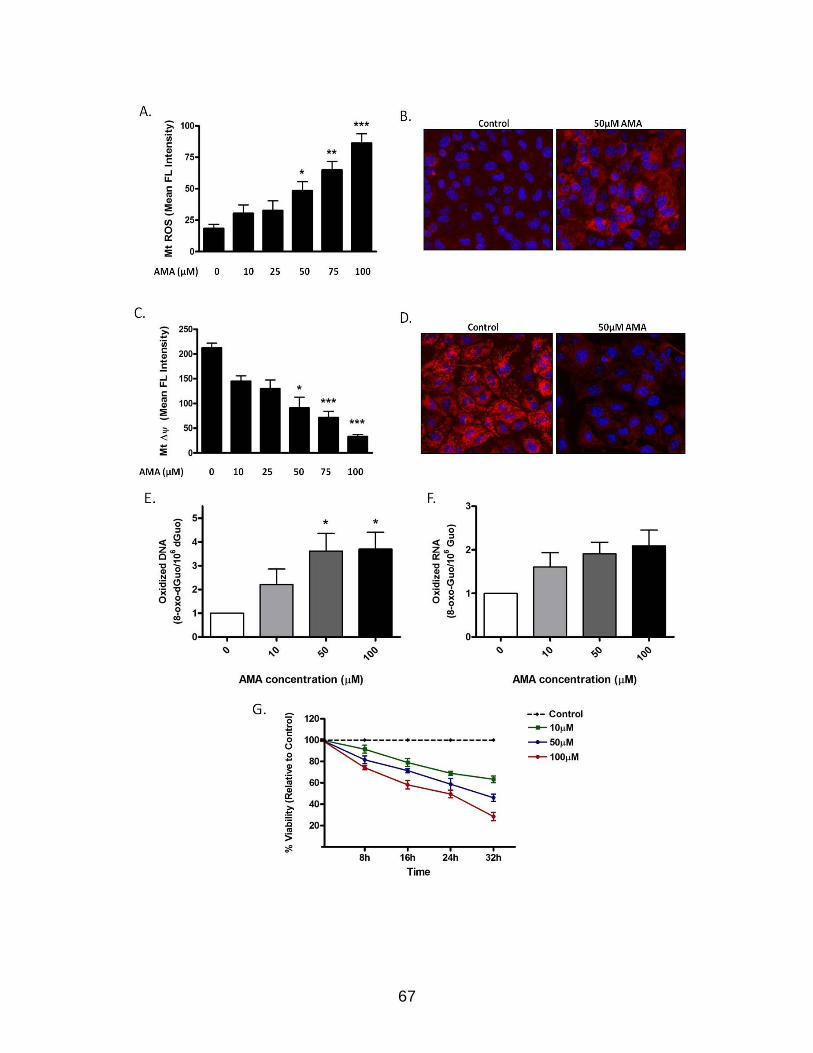
67

68
Figure 2-2. Treatment with AMA does not induce autophagy in HL-1 cardiomyocytes. (A) Cells were incubated with increasing concentrations of AMA or vehicle control for 4h, in the absence or presence of 75 nM BafA1. Cells were subsequently lysed, immunoblotted for LC3B and LC3-II/LC3-I ratio calculated to determine steady state or cumulative autophagy. GAPDH is used as a loading control. (B) Cells were transfected with GFP-LC3 plasmid and 24h after transfection, treated with 50 µM AMA or vehicle control for 4h, in the absence or presence of BafA1. Cells were subsequently fixed in 4% paraformaldehyde, stained with DAPI and images taken using epifluoresence microscope. Representative images of steady state and cumulative autophagy are shown (C) Cells were incubated with increasing concentrations of AMA or vehicle control for 24h, in the absence or presence of 75 nM BafA1 added during the final 2h of incubation. Cells were subsequently lysed, immunoblotted for LC3B and LC3-II/LC3-I ratio calculated to determine steady state or cumulative autophagy. GAPDH is used as a loading control (D) Cells were treated with the indicated concentrations of AMA for 2h or 24h and were subsequently lysed and immunoblotted for Atg5-Atg12, Beclin-1 and Atg7. GAPDH is used as a loading control. Data represents mean ± SE and is derived from three independent experiments.
(see figure on next page)

69

70
Figure 2-3. Treatment with rapamycin induces autophagy in HL-1 cardiomyocytes. (A) Cells were incubated with increasing concentrations of rapamycin or vehicle control for 16h and subsequently lysed and immunoblotted for phospho-S6 and total S6 levels. (B) Cells were incubated with increasing concentrations of rapamycin or vehicle control for 16h, in the absence or presence of 75 nM BafA1 added during the final 4h of incubation. Cells were subsequently lysed, immunoblotted for LC3B and LC3-II/LC3-I levels calculated to determine steady state or cumulative autophagy. β-tubulin is used as a loading control. ** p < 0.01 vs. control. (C) Cells were transfected with GFP-LC3 plasmid and 24h after transfection, treated with 1 µM rapamycin or vehicle control for 16h, in the absence or presence of BafA1 added during the final 4h of incubation. Cells were subsequently fixed in 4% paraformaldehyde, stained with DAPI and images taken using epifluoresence microscope. Representative images of steady state and cumulative autophagy are shown. (D) The number of GFP-LC3 puncta for cumulative autophagy in (C) were counted in each cell to determine the percentage of cells with stimulated autophagy *** p < 0.001 vs. control. (E). Cells were treated with vehicle control or 1 µM rapamycin for 48h. Cell lysates were subsequently collected and immunoblotted for VDAC. GAPDH is used as a loading control. Rapa, rapamycin. Data represents mean ± SE and is derived from three independent experiments.

71
Figure 2-4. Rapamycin induces autophagy in the presence of AMA in HL-1 cardiomyocytes. (A) Cells were treated with 50 µM AMA or 1 µM Rapamycin plus 50 µM AMA for 16h in the absence or presence of 75 nM BafA1 added during the final 4h of incubation. Cells were subsequently lysed, immunoblotted for LC3B and LC3-II/LC3-I ratio calculated to determine steady state or cumulative autophagy. GAPDH is used as a loading control.**p < 0.01 vs. AMA + Baf. (B) Cells were transfected with GFP-LC3 plasmid and 24h after transfection, treated with 1 µM rapamycin or vehicle control for 16h. Cells were subsequently treated with 50 μM AMA for a period of 2h and imaged using confocal microscopy. (C) Cells were transfected with GFP-LC3 plasmid and 24h after transfection, treated with 1 µM rapamycin or vehicle control for 16h. Cells were subsequently incubated with 50 nM TMRM for 30 mins. Cells were finally treated with 50 μM AMA and imaged using confocal microscopy. Arrow indicates mitochondria surrounded by a growing autophagic vacuole. Rapa, Rapamycin. Data represents mean ± SE and is derived from three independent experiments.
(see figure on next page)

72

73
Figure 2-5. Rapamycin protects against the cytotoxic effects of AMA. (A-C) Cells were pre-treated with 1 µM Rapamycin for 16h, followed by incubation with 50 µM AMA or vehicle control for an additional 32h, in the absence or presence of rapamycin. Cell viability was subsequently determined using MTT assay (A) and Trypan Blue exclusion assay (B) or cell lysates were collected and immunoblotted for cleaved PARP-1 and caspase-3 protein expression (C). For (A) and (B), data represents mean ± SE and is derived from three independent experiments. GAPDH is used as a loading control for (C). *p < 0.05; **p < 0.01 vs.control. (D) Cells were pre-treated with 1 µM rapamycin for 16h, followed by incubation with 50 µM AMA or vehicle control for an additional 24h, in the presence of rapamycin. Representative phase contrast images of cells are shown. Rapa, rapamycin.
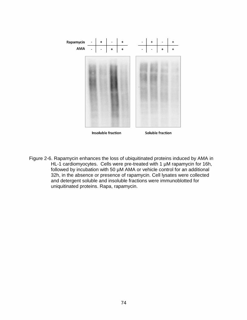
74
Figure 2-6. Rapamycin enhances the loss of ubiquitinated proteins induced by AMA in HL-1 cardiomyocytes. Cells were pre-treated with 1 µM rapamycin for 16h, followed by incubation with 50 µM AMA or vehicle control for an additional 32h, in the absence or presence of rapamycin. Cell lysates were collected and detergent soluble and insoluble fractions were immunoblotted for uniquitinated proteins. Rapa, rapamycin.

75
Figure 2-7. Rapamycin protects against AMA-induced mitochondrial depolarization and respiration dysfunction in HL-1 cardiomyocytes. (A) Cells were pre-treated with 1 µM rapamycin for 16h, followed by treatment with 50 µM AMA or vehicle control for an additional 24h, in the absence or presence of rapamycin. Cells were subsequently trypsinized, resuspended in Claycomb media and routine respiration analyzed. *p < 0.05; **p < 0.01 vs.control. Data represents mean ± SE and is derived from three independent experiments. (B) Cells were pre-treated with 1 µM rapamycin for 16h, followed by incubation with 50 µM AMA or vehicle control for an additional 24h, in the presence of rapamycin. Cells were subsequently stained with 50 nM TMRM (red) and 1ug/ml Hoechst 33342 (blue) and imaged using epifluorescence microscopy. Arrows represent cells with no Δψm. Arrowheads represent swollen mitochondria. Rapa, rapamycin.

76
Figure 2-8. Inhibition of autophagy attenuates rapamycin-mediated protection against AMA toxicity in cardiomyocytes. (A) Cells were treated with 1 µM rapamycin in the absence or presence 5 mM 3-MA for a period of 16h and with or without 75 nM BafA1 added during the final 4h of incubation. Cells were subsequently lysed, immunoblotted for LC3B and LC3-II/LC3-I levels calculated to determine steady state or cumulative autophagy. (B) Cells were pre-treated with 1 µM rapamycin in the absence or presence of 5 mM 3-MA for 16h, followed by treatment with 50 µM AMA or vehicle control for an additional 32h, in the presence of rapamycin and 5mM 3-MA. Cell viability was subsequently assessed using MTT assay. (C) Cells were treated as in (B), cell lysates collected and immunoblotted for PARP-1 and caspase-3 protein expression. GAPDH is used as a loading control. Data represents mean ± SE and is derived from three independent experiments.

77
Figure 2-9. Schematic representation of the protective effects of rapamycin against AMA toxicity. AMA induces PARP-1 cleavage and caspase-3 activation, decreases respiration, mitochondrial membrane potential as well as mediates the accumulation of ubiquitinated proteins, all of which culminates in apoptotic induction. Rapamycin treatment induces autophagy by removing the inhibitory effect of mTOR on autophagy induction. Stimulation of autophagy suppresses cytotoxic markers induced by AMA. Inhibition of rapamycin-induced autophagy by 3-MA blocks the cytoprotective effects.

78
Table 2-1. List of reagents used for in vitro study Reagent Name Source Catalog No.
3-MA Sigma, St. Louis, MO M9281
Alkaline phosphatase Sigma, St. Louis, MO P6774-10KU
Antimycin A Sigma, St. Louis, MO A8674
Bafilomycin A1(BafA1) Sigma, St. Louis, MO B1793
Claycomb Media JRH Biosciences, Lenexa, KS 51800C
DAPI Vector Laboratories,
Burlingame, CA
H-1200
Dc protein assay solution Biorad, Hercules, CA 500-0113
DFOM (deferoxamine mesylate) Sigma, St. Louis, MO D9533
dGuo standards Sigma, St. Louis, MO D7154
DMSO (Dimethyl sulfoxide) Sigma, St. Louis, MO D2650
ECL Plus reagent Amersham Biosciences,
Buckinghamshire, UK
RPN2132
FBS (Fetal bovine serum) Sigma, St. Louis, MO F2442
Fluorodish (0.17mm glass) World Precision Instruments,
Sarasota, FL
FD35-100
Hoechst 33342 Invitrogen, Carlsbad, CA H3570
L-glutamine Life Technologies, Gaithersburg,
MD
25030-081
Lipofectamine 2000 Invitrogen, Carlsbad, CA 11668-027
MitoSOX Red Invitrogen, Carlsbad, CA M36008
MTT Sigma, St. Louis, MO M5655
Norepinephrine Sigma, St. Louis, MO A0937
Nuclease P1 MP Biomedicals, Solon, OH N8630
Paraformaldehyde Electron Microscopy Sciences,
Fort Washington, PA
50-00-0
Penicillin / Streptomycin Life Technologies, Gaithersburg,
MD
15140-122
PVDF membrane Biorad, Hercules, CA 162-0177
Rapamycin Calbiochem, San Diego, CA 553211
Starting Block Thermo Scientific, Fair Lawn, NJ 37543
TMRM Invitrogen, Carlsbad, CA T668
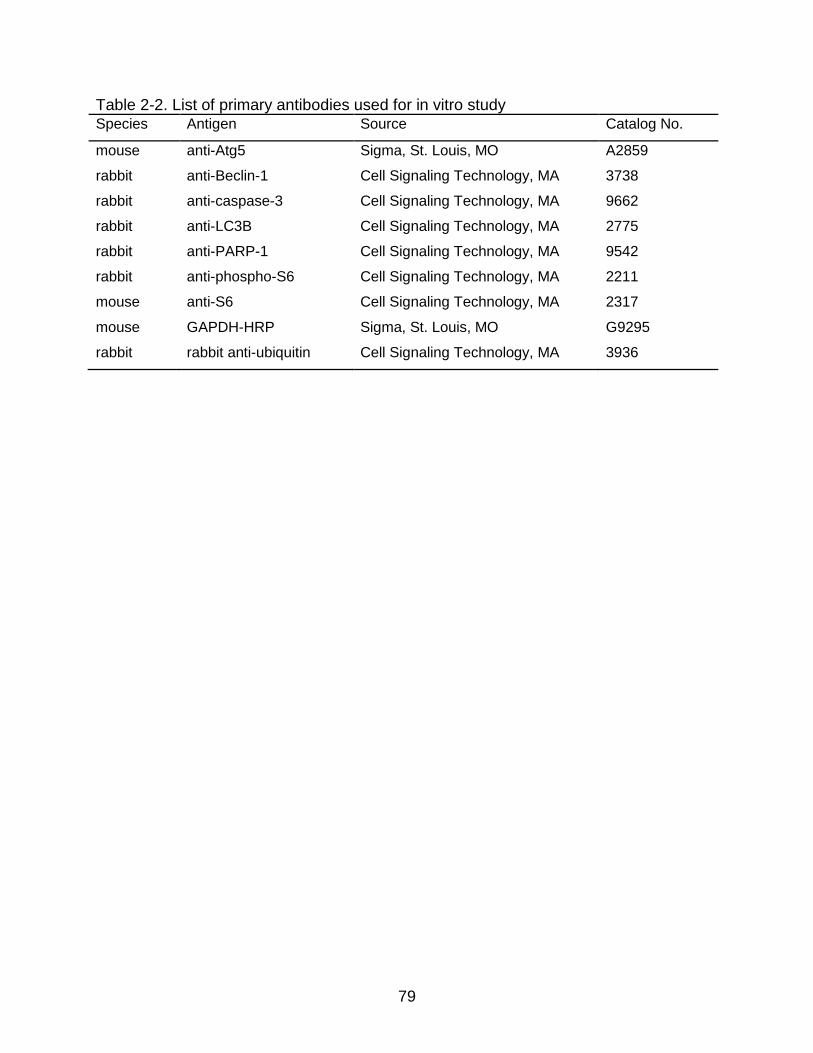
79
Table 2-2. List of primary antibodies used for in vitro study Species Antigen Source Catalog No.
mouse anti-Atg5 Sigma, St. Louis, MO A2859
rabbit anti-Beclin-1 Cell Signaling Technology, MA 3738
rabbit anti-caspase-3 Cell Signaling Technology, MA 9662
rabbit anti-LC3B Cell Signaling Technology, MA 2775
rabbit anti-PARP-1 Cell Signaling Technology, MA 9542
rabbit anti-phospho-S6 Cell Signaling Technology, MA 2211
mouse anti-S6 Cell Signaling Technology, MA 2317
mouse GAPDH-HRP Sigma, St. Louis, MO G9295
rabbit rabbit anti-ubiquitin Cell Signaling Technology, MA 3936

80
CHAPTER 3 CALORIE RESTRICTION COMBINED WITH RESVERATROL INDUCES
AUTOPHAGY AND OFFERS PROTECTION FROM DOXORUBICIN-INDUCED TOXICITY IN 26-MONTH-OLD RAT HEARTS
Introduction
The housekeeping process of autophagy has been shown to be required for
optimal cardiac functioning and survival (Nakai, 2007 and Taneiki, 2007). Given the post
mitotic nature of the heart, autophagy-mediated recycling of damaged cellular
components is essential for protein and organelle quality control. This is supported by
the observation that abrogation of the autophagic pathway in the adult heart by
conditional inactivation of Atg5 or Atg7 causes rapid onset cardiac abnormalities
characterized by cardiac hypertrophy, left ventricular dilatation and decreased cardiac
output (Nakai, 2007 and Taneiki, 2010). In addition, an age-related decline in the
efficiency of the autophagic process can lead to the accumulation of damaged cellular
components which can further lead to functional deterioration (Terman & Brunk, 2005).
Hence, interventions that induce autophagy in vivo, especially at an older age, are
highly desirable and potentially beneficial.
Although inducers of autophagy in cell culture conditions are increasingly being
reported (reviewed in Chapter 4), few studies have investigated interventions to induce
autophagy in vivo, especially, starting at an older age. As regards the heart, Kanamori
et al... reported that starving GFP-LC3 transgenic mice for 12h led to an increase in
fluorescent autophagic puncta, which was also confirmed by electron microscopic
visualization of autophagic vacuoles (Kanamori et al., 2009). Longer starvation periods
led to a more robust autophagic stimulation and enhanced the expression of
downstream lysosomal enzymes such as cathepsin D, suggesting an overall

81
enhancement of autophagic flux (Kanamori et al., 2009). Consistent with the
abovementioned study, our group has previously shown that lifelong 40% CR can
increase the appearance of autophagic vacuoles in the hearts of Fischer 344 rats and
enhance the expression of autophagy proteins Atg7 and Atg9 and lysosomal enzyme
procathepsin D (Wohlgemuth et al., 2007). Similar effects have also been observed in
the skeletal muscle of 40% CR animals (Wohlgemuth et al., 2010). However, in spite of
the autophagy enhancing capability of lifelong 40% CR, such a severe dietary restriction
is not feasible in humans. Additionally, it can lead to undesirable and potentially harmful
changes if started at a vulnerable stage of life cycle, such as in young adolescents, the
very old or in pregnant women. Hence in this present study, we investigated whether a
moderate CR (20%) regimen, beginning in late-middle-age, can induce autophagy in the
rat heart.
In addition to a mild CR regimen, we investigated the natural polyphenol
resveratrol as a potential inducer of autophagy. Resveratrol is produced in a wide
variety of plants such as grapes, berries and peanuts in response to environmental
stress and has been extensively investigated in a large number of clinical studies
(Mullin, 2011). Resveratrol has been implicated in the French paradox, an observation
of how the French people, in spite of consuming high amounts of saturated fats in their
diet, have decreased incidences of age-associated cardiovascular disorders. It is
believed that this protective effect is due to their high consumption of red wine, which
contains resveratrol. In experimental animals, resveratrol has shown numerous
beneficial effects, such as promoting vasodilation in models of coronary heart disease
and enhancing the expression of antioxidant enzymes (Bradamante et al., 2004).

82
Additionally, resveratrol can protect the heart against ischemia-reperfusion injury (Ray
et al., 1999), improve endothelial function (Li & Forstermann, 2009) and prevent platelet
aggregation (Ramprasath & Jones, 2010). Notably, resveratrol has been shown to be a
potent inducer of autophagy in several cell culture models, including cardiomyocytes
(Jeong et al., 2012; Lv & Zhou, 2012; Scarlatti et al., 2008; Wu et al., 2011). However,
studies investigating whether resveratrol can induce autophagy in animals are limited.
Our preliminary analysis on resveratrol-fed rats (5 and 50 mg/kg/day resveratrol for 6
weeks) did not show any changes in cardiac autophagic markers in 26-month-old
Fischer 344 x Brown Norway (FBN) rats (data not shown). Therefore, in the present
study, we investigated whether resveratrol combined with 20% CR is able to induce
autophagy in the hearts of 26-month-old FBN rats.
In addition to exploring interventions to induce autophagy in rodent hearts, we
further investigated whether such autophagy interventions can protect against cardiac
damages induced by the oxidative stressor, doxorubicin. Doxorubicin is a highly
effective chemotherapeutic agent used in the treatment of solid tumors and hematologic
malignancies (Octavia et al., 2012). However, the cardiotoxic effects of doxorubicin
severely limit its chemotherapeutic efficacy (Childs et al., 2002; Octavia et al., 2012).
Although a complete understanding of the mechanisms involved in doxorubicin’s toxicity
remain elusive, the generation of ROS in cells has been proposed to be a major player
(Mordente et al., 2012; Takemura & Fujiwara, 2007). Doxorubicin has been reported to
localize to the mitocohondria, induce ultrastructural changes and affect mitochondrial
function (Green & Leeuwenburgh, 2002; Montaigne et al., 2010). Doxorubicin contains
an unsubstituted quinone ring, which can act as an electron acceptor in reactions

83
mediated by oxidoreductase enzymes in the cell, such as by the NADH dehydrogenase
(Doroshow, 1983; Mukhopadhyay et al., 2009). The addition of free electrons converts
quinone to semiquinone, which can subsequently interact with molecular oxygen and
generate O2•−. The generation of ROS by doxorubicin can lead to cellular oxidative
damage and activation of proteases, resulting in cytotoxic effects (Kotamraju et al.,
2000).
Our study shows that 50 mg/kg/day resveratrol combined with 20% CR can
enhance autophagic flux in the hearts of 26-month-old FBN rats. In addition, such an
induction of autophagy has a beneficial effect on doxorubicin-mediated toxicity. We
therefore propose that induction of autophagy in late middle-aged rat hearts could be
developed as a therapeutic target for mitigating oxidative stress-induced damage.
Materials and Methods
Animals and Experimental Design
Male FBN rats at 25 months of age were purchased from the National Institute on
Aging (NIA) and were singly housed at the University of Florida animal facility in a
temperature (20 ± 2.5⁰C) and light-controlled (12:12 h light-dark cycle) environment with
unrestricted access to water. After arrival, animals were acclimated for a period of 3
weeks, during which period their baseline food consumption was measured. At the end
of the acclimation period, rats were randomly assigned into one of four experimental
groups: 1) Ad libitum (AL) 2) 20% CR (CR), 3) CR with 5 mg/kg/day resveratrol (CR +
Resv-5) and 4) CR with 50 mg/kg/day resveratrol (CR + Resv-50). They were
maintained on the interventions for a period of 6 weeks. Animals on the CR, CR + Resv-
5 and CR + Resv-50 groups received 20% less food from a 20% fortified diet so that
they were not compromised on the amino acid, vitamins and minerals intake. The diet

84
was based on AIN-93M chow, formulated for the maintenance of mature rodents and
have reduced levels of proteins and fats to help reduce the incidence of kidney stones
in older rodents (Reeves et al., 1993). The composition of the AL and the CR diet has
been summarized in Table 3-1. Resveratrol (Sigma, St. Louis, MO) was given as highly
palatable supplementation pellets, prepared by Custom Animal Diets (Bangor, PA)
utilizing an effective bacon flavor masking capability. To ensure that every rat received
the proper dose of resveratrol, the number of pellets given on each day was calculated
based on the body weight of the animal. Control supplementation tablets (without
resveratrol) was also given to AL rats. The supplementation tablets were similarly
formulated with AL or CR diet as the base chow, with or without resveratrol. Throughout
the entire study, the amount of food given to the CR rats was adjusted weekly based on
the food consumed by AL rats the week prior. Body weights of rats were recorded at
least once per week.
At the end of the intervention period, animals received a single i.p. injection of 10
mg/kg doxorubicin (Sigma, St. Louis, MO) or saline control. The dose was chosen
based on our previous observation that 10 mg/kg/day doxorubicin can activate caspase-
3 and induce apoptosis in the hearts of Fisher 344 rats (Jang et al., 2004). Twenty-four
hours after injection, animals were sacrificed by rapid-decapitation. Serum and plasma
was collected by trunk blood processing and stored at -80⁰C until further analysis. All
experiments and procedures were approved by the Institute on Animal Care and Use
Committee at the University of Florida.
Preparation of Tissue for Histological Staining
Upon sacrifice, the heart was removed, rinsed in DPBS, blotted dry, weighed and
dissected into individual compartments. Approximately 10 mg of the left ventricle was

85
further dissected out and placed immediately in O.C.T. compound (Fisher Scientific,
CA) in 15 x 15 x 5 mm cryomolds (Fisher Scientific, CA). The tissue was subsequently
snap frozen in liquid nitrogen and stored at -80⁰C until further analysis. Hematoxylin and
eosin (H&E) staining on O.C.T embedded tissue was performed at the Cell and Tissue
Analysis Core (CTAC) at the University of Florida and images were taken using Ziess
Axio Observer A1 microscope (Zeiss, Germany) at 20X objective magnification.
Body Composition Analysis
Fat and lean mass percentages of rats were determined at baseline levels (before
the start of interventions) and after 6 weeks of CR and resveratrol interventions using
time domain-nuclear magnetic resonance (TD-NMR) analyzer (Minispec, Bruker Optics,
TX). TD-NMR provides an accurate, fast, and easy-to-use method for determining fat
and lean tissue in rodents without the need for anesthesia. The validation of the TD-
NMR methodology has been provided elsewhere (Nixon et al., 2010).
Preparation of Tissue Extracts
For the preparation of tissue extracts, approximately 100 mg of the left ventricle
was pulverized in liquid nitrogen. The powder was subsequently resuspended in 1 mL
ice-cold buffer containing 20 mM HEPES, pH 7.4, 2 mM EGTA, 50 mM β-
glycerophosphate, 1 mM dithiothreitol, 1 mM Na3VO4, 10% glycerol, 1%Triton X-100
and Halts Protease Inhibitor (Thermo Scientific, NJ). Resuspended tissue was further
homogenized using a mechanically driven Potter-Elvehjem glass-glass homogenizer
with approximately 14 up and down strokes. The homogenates were subsequently
rotated for 1h at 4⁰C for efficient cell lysis. Finally, the homogenates were centrifuged at
10,000g for 10 minutes at 4⁰C and the supernatant was transferred to clean tubes.

86
Protein concentrations were determined using Bradford Assay (Bradford, 1976) and
aliquots were stored at -80⁰C until further analysis. A complete list of all the reagents
used in the study and their catalog numbers have been provided in Table 3-2.
Western Blotting
Protein samples were prepared in Laemmli buffer (62.5 mM Tris-HCl, 2% SDS,
25% Glycerol, 0.01% Bromophenol Blue, pH 6.8; BioRad, CA) with 5% β-
mercaptoethanol and were boiled at 95⁰C for 5 mins prior to loading in gels. Equal
amounts of proteins were loaded in pre-cast Tris-HCl polyacrylamide gels (Criterion
system, BioRad, Hercules, CA). After electrophoretic separation, proteins were
transferred to polyvinylidene difluoride (PVDF) membrane (BioRad, Hercules, CA) and
subsequently blocked for 1h in Starting Block (Thermo Scientific, Fair Lawn, NJ),
followed by overnight incubation with primary antibodies at 4⁰C. Membranes were
subsequently washed with Tris-buffered saline with 0.1% Tween 20 (TBST) and
incubated with the appropriate secondary antibodies in Starting Block for 1h.
Membranes were washed again with TBST, chemiluminescent signals developed using
ECL Plus reagent (Amersham Biosciences, Buckinghamshire, UK) and captured using
ChemiDoc XRS System (BioRad, CA). Digital images were analyzed for densitometry
using ImageLab software (BioRad, CA). The primary antibodies used for
immunoblotting analysis are as follows: rabbit anti-LC3B (1:1000), rabbit anti-p62
(1:1000) rabbit anti-Beclin-1 (1:1000) (all from Cell Signaling Technology, MA), mouse
anti-Atg5-Atg12 conjugate (1:1000; Sigma, MO) and GAPDH-HRP (1:35,000; Sigma,
MO). HRP conjugated secondary antibodies were anti-rabbit (1:10,000; GE-Amersham,

87
IL) or anti-mouse (1:10,000; GE-Amersham, IL). A complete list of primary antibodies
and their catalog number have been provided in Table 3-3.
Plasma Resveratrol Concentration Analysis
Trans-resveratrol was quantified in rat plasma using LC/MS/MS assay at the
Biomedical Mass Spectrometry Core at the University of Florida. Rat serum samples
(30 µL) were loaded onto an Accela Open Autosampler (Thermo Fisher Scientific, CA)
connected to an Accela 600 UPLC and LTQ Velos mass spectrometer (Thermo Fisher
Scientific, CA). Separation was achieved on a BetaBasic C18 HPLC column (150 x 2.1
mm, 5 um, ThermoFisher, CA).
Blood Glucose Analysis
Glucose levels were measured in freshly collected trunk blood using blood glucose
meter and test strips (both from Ascensia Contour; Bayer HealthCare LLC).
Apoptotic Analysis
Apoptotic analysis on left ventricular homogenates was performed using Cell
Death Detection Plus ELISA kit (Roche Diagnostics, IN) following manufacturer’s
instructions. The kit is designed to detect mono- and oligonucleosomes in the
cytoplasmic fraction of heart lysates and is based on a sandwich-enzyme immunoassay
principle using mouse monoclonal antibodies directed against DNA and histones.
Absorbance was read at 405 nm with a Biotek Synergy plate reader (Biotek, VT) and
the values were normalized to protein concentrations.
Creatine Kinase (CK) Assay
CK levels in serum were determined using Enzychrom Creatine Kinase Assay kit
(Bioassay Systems, CA) following manufacturer’s instructions. The kit utilizes an
enzyme-coupled reaction in which creatine phosphate and ADP is converted to creatine

88
and ATP by CK. The ATP generated in the process is used by hexokinase in
phosphorylating glucose to glucose-6-phosphate. The latter is subsequently oxidized by
NADP (in the presence of glucose-6-phosphate dehydrogenase) and produces NADPH
in the process. The produced NADPH is proportional to the CK activity in the samples
and is quantified by measuring the absorbance at 340 nm using Biotek Synergy plate
reader (Biotek, VT).
Lactate Dehydrogenase (LDH) Assay
LDH levels in serum were measured using Quantichrom Lactate Dehydrogenase
Assay kit (BioAssay Systems, CA) according to manufacturer’s instructions. The assay
is based on NADH catalyzed reduction of the tetrazolium salt MTT to a reduced form.
Absorbance is measured at 565 nm using Biotek plate reader (Biotek, VT) and the
values are directly proportional to the enzyme activity.
Statistical Analysis
Statistical analyses were performed using GraphPad Prism Version 4.0
(GraphPad Software, San Diego, CA). Student’s t-test and one-way ANOVA were
performed, wherever applicable. Whenever required, post-hoc analysis was performed
using Bonferroni multiple comparison test. Statistical significance was set to p < 0.05.
Results are expressed as mean ± SE.
Results
CR Diet were Associated with Lower Body Weights
Six weeks of 20% CR, with or without resveratrol, resulted in significantly lower
body weights in comparison to AL (p < 0.001 vs. AL, Table 3-4). Heart weights of rats in
CR + Resv-5 and CR + Resv-50 groups were significantly lower in comparison to that of

89
AL rats (p < 0.001 vs. AL). There was no statistical difference when heart weight was
normalized to body weight (Table 3-4).
Six weeks of CR and Resveratrol Interventions or Administration of Doxorubicin does not Change Myocardial Morphology
Since we observed decreased heart weights in animals fed CR + 5 mg/kg/day or
CR + 50 mg/kg/day resveratrol, we performed H&E staining on frozen left ventricular
tissue sections to determine whether the interventions affected cardiac morphology,
such as cardiac fibrosis or increased cardiomyocyte dropout. We observed no
differences between AL groups and the CR + Resv-5 or CR + Resv-50 groups by
histology analysis (Fig. 3-1, upper panel). Doxorubicin administration for 24h also did
not change tissue morphology (Fig. 3-1, lower panel).
CR Diet alone or in Combination with Resveratrol, Attenuates Body Composition
Changes over Time
Paired t-test analysis showed that fat mass % and fat / lean ratio significantly
increased (p < 0.01) and lean mass % decreased (p < 0.05) in animals on AL diet over
a 6-week period (Fig. 3-2). However, fat mass %, lean mass % and fat / lean ratio
remained unchanged in the CR, CR + Resv-5 and CR + Resv-50 groups before and
after the intervention period.
Plasma Resveratrol Concentrations Increase in a Dose-Dependent Manner in Resveratrol-Fed Rats
Plasma resveratrol concentrations determined by LC/MS/MS analysis showed a
dose-dependent increase in resveratrol concentrations (Table 3-5). The mean plasma
concentration of resveratrol in the oral administration doses of 5 and 50 mg/kg/day was
7.4 ± 3.7 and 41 ± 19 ng/mL, respectively. Resveratrol could not be detected in the
plasma of AL rats (Table 3-5).

90
Doxorubicin Administration Increases Blood Glucose Levels
Doxorubicin administration has been reported to increase blood glucose levels,
primarily due to damage to insulin-secreting beta cells in the pancreas (Deleers &
Goormaghtigh, 1985). We observed that blood glucose levels were increased in animals
treated with doxorubicin (main effect, p < 0.001 vs. saline, Table 3-6). None of the
interventions were able to attenuate the increase in blood glucose levels, although
animals in the CR + Resv-50 group had the lowest blood glucose levels (118 ± 30
mg/dL), close to that of AL rats (112 ± 17 ng/dL).
CR + 50 mg/kg/day Resveratrol Increases Autophagic Flux in the Left Ventricle
We analyzed the expression levels of key autophagy proteins (LC3B, p62, Beclin-
1 and Atg5-Atg12 conjugate) in the left ventricle of saline-treated rats, to determine
whether 20% CR by itself, or in combination with the two different doses of resveratrol
can induce cardiac autophagy in 26-month-old FBN rats. In addition, since oxidative
stress per se has been proposed to be an inducer of autophagy, we also determined
whether doxorubicin can affect the autophagic process. The results from autophagy
protein expression analysis in both saline and doxorubicin-treated groups are
summarized below:
LC3B
Autophagy induction converts cytosolic LC3-I to autophagosomal membrane-
associated and PE-conjugated LC3-II. LC3-II/LC3-I ratio is therefore a widely used
indicator of steady state autophagic levels. We could not detect an increase in LC3-
II/LC3-I ratio in any of the intervention groups (Fig. 3-3A). Treatment with doxorubicin
however, enhanced LC3-II /LC3-I ratio, in comparison to saline treated AL rats (p <
0.005, Fig. 3-4A).

91
p62
Although LC3-II /LC3-I ratio remained unchanged with the interventions, we
cannot rule out the possibility that an increase in the rate of their degradation in the
lysosomes, i.e. an enhancement in autophagic flux may have contributed to their
unaltered levels. To determine autophagic flux, we measured the abundance of p62 /
sequestosome 1 (SQSTM1) in left ventricular homogenates. p62 binds to ubiquitinated
proteins through its UBA domain and can interact with LC3 through its LC3 binding
motif, thereby directing the degradation of ubiquitinated proteins through the autophagic
pathway (Bjorkoy et al., 2009). In the process, p62 is degraded through autophagy;
therefore, a decrease in the abundance of p62 is considered indicative of enhanced
autophagic flux (Bjorkoy et al., 2009; Larsen et al., 2010). We observed a decrease in
p62 abundance in the left ventricle of CR + Resv-50 rats (p < 0.05 vs. AL, Fig. 3-3B),
indicating an increased autophagic flux. No significant changes were observed in the
CR or CR + Resv-5 groups in comparison to AL. Notably, we observed an increase in
p62 accumulation in the doxorubicin-treated hearts, suggesting an impairment of the
autophagic flux with doxorubicin treatment (p < 0.01 vs. saline, Fig. 4-4B).
Beclin-1
Beclin-1 (mammalian homolog of yeast Atg6) is required for the induction phase of
autophagy (Liang et al., 1999). Specifically, Beclin-1 associates with a pre-
autophagosomal complex comprising of Atg14, Class III PI3K/Vps34 and Vps15 and
this complex is required for the localization of autophagic proteins to the pre-
autophagosomal structure (Kang et al., 2011; Kihara et al., 2001; Liang et al., 1999).
Autophagic triggers have been shown to upregulate Beclin-1 (Wang, 2008). We
observed that the expression of Beclin-1 was increased in left ventricular homogenates
92
of CR + Resv-50 rats in comparison to AL (p< 0.05, Fig. 3-3C). There were no changes
in Beclin-1 protein levels between saline and doxorubicin treatment groups (Fig. 3-4C).
Atg5-Atg12
In addition to the Atg8-PE (LC3-PE) conjugation system, a second ubiquitin-like
conjugation system (comprising of Atg7, Atg12, Atg10) links the carboxy-terminal
glycine residue of Atg12 to a lysine of Atg5 (Mizushima et al., 1998). The Atg5–Atg12
conjugate further complexes with Atg16, which is associated with the growing
phagophore (Mizushima, 2007). We observed no differences in Atg12-Atg5 levels in left
ventricular homogenates between AL rats and the intervention groups (Fig. 3-3D).
CR + 50 mg/kg/day Resveratrol Attenuates Doxorubicin-Mediated Increases in Cardiac Apoptotic Levels
We next determined whether induction of autophagy has a beneficial effect against
doxorubicin mediated toxicity in the cardiac tissue. Apoptotic levels were determined by
measuring mono- and oligo-nucleosomes released in the cell cytoplasm. Doxorubicin
increased apoptotic index in the left ventricle of AL rats (p < 0.05 vs. saline, Fig. 3-5A)
and CR + Resv-50 attenuated the apoptotic induction (p < 0.05 vs. AL + doxorubicin).
No significant changes in cardiac apoptotic levels were observed between AL and CR
or CR + Resv-5 groups (Fig. 3-5A).
CR + 50 mg/kg/day Resveratrol Attenuates Doxorubicin-Mediated Increases in Serum CK Levels
CK, an enzyme predominantly expressed in muscles, is involved in the conversion
of creatine to phosphocreatine with the consumption of ATP (Brancaccio et al., 2010).
An elevated CK level in blood is a widely used indicator of muscle damage. We
observed that treatment with doxorubicin increased serum CK levels (p < 0.01 vs.

93
saline) and that CR + Resv-50 significantly ameliorated the increase (p < 0.05 vs. AL +
doxorubicin, Fig. 3-5B).
CR + Resveratrol Attenuates Doxorubicin-Mediated Increases in Serum LDH Activity
LDH is an oxidoreductase enzyme which catalyzes the inter-conversion of lactate
and pyruvate. It is usually sequestered within cells and is released into the bloodstream
under conditions of tissue injury. Therefore, serum LDH activity represents a marker of
general tissue damage (Glick, 1969). We observed that treatment with doxorubicin
significantly increased serum LDH levels (p < 0.01, Fig. 3-6A) and that both CR + Resv-
5 and CR + Resv-50 significantly ameliorated the increase (p < 0.05 vs. AL + Dox, Fig.
3-6A). No differences were observed in serum LDH activity among AL and intervention
groups in saline-treated rats (Fig. 3-6B).
Discussion
The self-cleanup process of autophagy recycles damaged and dysfunctional
cellular components, thereby contributing to organelle and protein quality control.
Upregulation of this process could therefore be hypothesized to be beneficial against
oxidative stress-induced damage. Despite the beneficial effects of autophagy, few
studies have focused on its stimulation in vivo. Although lifelong 40% CR has been
reported to enhance autophagy in different tissues (Wohlgemuth et al., 2007;
Wohlgemuth et al., 2010), it is likely that most humans will not be able to sustain drastic
food restrictions for the long term. Severe CR is associated with osteoporosis, cold
sensitivity, slower wound healing, and psychological conditions such as food obsession,
depression, and irritability (Marzetti et al., 2009). Furthermore, adolescents practicing
chronic severe CR may experience several adverse events such as undesired changes
94
in physical appearance, loss of strength and stamina, menstrual irregularities, infertility
and loss of libido (Marzetti et al., 2009). Hence a moderate CR, starting late in life could
offset some of the limitations associated with life-long CR. Additionally, combining a
mild CR regimen with a so-called CR mimetic has the potential of reinforcing the
beneficial effects of CR, without the potential harmful effects of a drastic food reduction.
Resveratrol is a known CR mimetic, which has shown to produce CR-like extension in
lifespan in lower organisms and has a proposed mechanism of action very similar to
that of CR (Dolinsky & Dyck, 2011; Howitz et al., 2003; Wood et al., 2004).
Although showing numerous CR-like effects, a major drawback of resveratrol is its
poor bioavailability. Resveratrol undergoes rapid metabolism to glucoronide and
sulphate conjugates in the liver and clearance by the kidney (Kapetanovic et al., 2010),
resulting in low bioavailability. For instance, in humans, a single oral dose of 25 mg
resveratrol resulted in plasma resveratrol concentrations of < 5 ng/mL (Walle et al.,
2004) and similar observations have been reported in rodents (Kapetanovic et al.,
2010). However, clinical trials have reported that increased concentrations of resveratrol
can be obtained with multiple dosing (Brown et al., 2010; la Porte et al., 2010). In the
present study, feeding rats with 50 mg/kg/day resveratrol for a period of 6 weeks
resulted in plasma resveratrol concentration of 41 ng/mL (Table 3-5). In this respect, a
previous study has reported plasma resveratrol concentrations of 176 ng/mL in rats,
given daily oral gavages of 50 mg/kg/day resveratrol for 2 weeks. The study showed
that peak plasma resveratrol concentrations were reached 25 mins after the last oral
dosing and continued to drop thereafter. The relatively lower plasma resveratrol
concentrations in 50 mg/kg/day resveratrol fed rats in the current study can be attributed

95
to the fact that blood was collected 24h after the last oral dosing. Additionally, we
observed dose-dependent increases in plasma resveratrol concentrations, which have
also been reported elsewhere (Boocock et al., 2007).
In the present study, we have shown that 20% CR in combination with 50
mg/kg/day resveratrol can enhance autophagic flux in the hearts of late-middle-aged
(26-month-old) FBN rats. FBN rats are an excellent model for gerontology research, due
to their reduced incidences of age-associated pathological lesions and a relatively
longer disease-free life span in comparison to other rat strains (Lipman et al., 1996;
Weindruch & Masoro, 1991). In the FBN rats, changes in cardiomyocyte volume density
occurs as early as 24 months of age and percentage fractional shortening (as a
measure of cardiac function) shows significant differences starting at 27 months of age
(Hacker et al., 2006; Walker et al., 2006). These observations suggest that for the FBN
rat strain, 26 month may represent an optimum life stage to start late-life interventions,
i.e. at a time when structural changes begin to appear in the heart, without drastic
functional consequences. Additionally, 26 months of age in FBN rats correspond to
roughly 60 years in humans, a stage in life when moderate dietary restriction can be
assumed to have little or no deleterious effects (Quinn, 2005).
Autophagy assessments in the study were done by measuring protein expression
levels of LC3B, p62, Beclin-1 and Atg5-Atg12 conjugate. We did not observe any
changes in the LC3-II /LC3-I ratio in the left ventricle of AL and any of the interventions,
which could either mean: 1) no autophagic stimulation by the treatments or 2) an
enhanced autophagic flux, such that an increased amount of LC3-II is rapidly degraded
in the lysosomes, resulting in unaltered LC3-II /LC3-I ratios. The ubiquitin binding

96
protein p62 / SQSTM1 targets cytosolic cargo for autophagic degradation, being
selectively incorporated into autophagosomes in the process and efficiently degraded in
the autolysosomes (Bjorkoy et al., 2005). Therefore, the cellular abundance of p62
inversely correlates with autophagic flux (Mizushima et al., 2010). In our study, CR +
Resv-50 rats showed a decrease in the abundance of p62 in the left ventricle,
suggesting an increase in autophagic flux in this intervention group. In addition, we
observed an increase in Beclin-1 levels in the same rat group, pointing to an enhanced
induction of autophagy. Finally, there were no differences in Atg5-Atg12 conjugate
levels between AL and any of the intervention groups. Although the conjugation of Atg5
to Atg12 is essential for autophagy, Atg5–Atg12 dissociates from the autophagosome
after it completely engulfs the cargo (Mizushima, 2007). Hence, the observation that the
Atg5-Atg12 levels remain unaltered is consistent with LC3-II/LC3-I ratios observed in
the study.
The exact mechanism by which the combined regimen of 20% CR and 50
mg/kg/day resveratrol (but not either intervention alone) can induce autophagy in rodent
hearts remains unknown. Resveratrol is a known CR mimetic and is proposed to have a
mechanism of action similar to that of CR (Dolinsky & Dyck, 2011; Korolchuk et al.,
2009). It is possible that when present together, they reinforce and activate certain
signal transduction pathways, which can lead to an induction of autophagy. For
example, the NAD+ dependent deacetylase SIRT1 can deacetylate essential autophagy
proteins Atg5, Atg7 and Atg8 and activate autophagy (Lee et al., 2008). Both CR and
resveratrol has been shown to activate Sir2 (homolog of mammalian SIRT1) in lower
organisms (Guarente & Picard, 2005; Howitz et al., 2003) and SIRT1 in mammals

97
(Chen et al., 2005; Lagouge et al., 2006). However, although activation of the SIRT1
pathway was originally proposed to be responsible for its lifespan extending properties
of resveratrol (Howitz et al., 2003; Wood et al., 2004), recent studies have shown
inconsistent results (Bass et al., 2007; Kaeberlein et al., 2005). In addition, we have
previously shown that oral administration of low-dose resveratrol (4.9 mg/kg/day)
starting at middle-age in mice, was unable to activate SIRT1 in any of the analyzed
tissues (heart, skeletal muscle, liver and brain) (Barger et al., 2008b), despite the fact
that both CR and resveratrol showed very similar gene expression profiles in the hearts
in the same study (Barger et al., 2008b; Edwards et al., 2010). Other common
mechanism of action include activation of the AMPK pathway (Dasgupta & Milbrandt,
2007), which has been shown to enhance autophagic induction by phosphorylating
ULK1 and activating the ULK1-Atg13- Atg17 complex (Kim et al., 2011). Finally, both
CR and resveratrol can inhibit the mTOR pathway, which has inhibitory effects on
autophagic induction (Hansen et al., 2007; Kapahi et al., 2004; Liu et al.).
Our results also suggest that doxorubicin per se can increase LC3-II/LC3-I ratio.
An increase in LC3-II/LC3-I ratio by doxorubicin treatment has previously been reported
(Manov et al., 2011; Pan et al., 2011; Smuder et al., 2011), however autophagic flux
analysis was not conducted in these studies. We observed that doxorubicin increased
p62 abundance in the left ventricle, suggesting that the increase in LC3-II/ LC3-I levels
is due to an impairment of autophagic flux. Consistent with this hypothesis, a decrease
in the activity of lysosomal enzyme cathepsin D have been reported in the hearts of
doxorubicin-treated animals (Gebbia et al., 1985). Additionally, it is possible that the
autophagic flux impairment is a characteristic of the relatively older population of rats

98
used in the study, as all previous studies with doxorubicin have been conducted in a
younger cohort (Smuder et al., 2011). For instance, it is possible that doxorubicin
induces autophagy as a protective mechanism in all age groups, but complete
degradation of autophagic cargo is affected / attenuated only in older rats.
In the clinical setting, a significant proportion of patients administered with
doxorubicin for long-term periods go on to develop cardiac pathologies; however, as of
yet, there is no specific treatment for preventing such chemotherapy-derived
cardiomyopathy (Tokarska-Schlattner et al., 2006). The development of
cardioprotective agents that can intervene against doxorubicin-mediated toxicity is
therefore highly desirable. Notably, in the present study, we observed that induction of
autophagy protects the myocardium from the anthracycline-mediated injury, confirmed
by the attenuation of doxorubicin-mediated increases in myocardial apoptotic index and
serum CK levels (Fig. 3-5). Although not a specific marker of myocardial damage, CK
levels are routinely measured in emergency patients with chest pain. While skeletal
muscle injury cannot be ruled out, we believe that the increases in CK levels in
doxorubicin-treated rats is primarily due to cardiac damage, since the heart is especially
vulnerable to anthracycline-mediated toxicity (Chatterjee et al., 2009). Additionally, CR +
50 mg/kg/day resveratrol also attenuated doxorubicin-mediated increases in serum LDH
levels, the extracellular appearance of which is considered a general marker of tissue
damage (Brancaccio et al., 2010; Glick, 1969). Importantly, we could not observe any
morphological changes usually associated with doxorubicin administration (such as
cytoplasmic vacuolization and myocardial fibrosis), in the left ventricle of animals treated
with doxorubicin. However, these changes have usually been reported in models of
99
chronic doxorubicin administration over a period of few weeks (Chatterjee et al., 2009;
Rahman et al., 1982; Working et al., 1999; Yi et al., 2006), in contrast to the acute
doxorubicin toxicity model used in the present study. Importantly, a previous study has
shown that a 35 % CR can be protective against doxorubicin cardiotixicity in male
Sprague-dawley rats (Mitra et al., 2007) which is in contrast to our study where no
protective effect could be observed by CR alone. This could be attributed to the
relatively mild CR regimen used in our study.
Finally, it is worth noting that our moderate, late-age onset CR is similar to most
clinical trials of dietary restriction in humans (Racette et al., 2006; Willcox et al., 2004),
where a high severity CR can be challenging and potentially deleterious. For example, a
36-year follow up study showed that mortality rate in healthy non-smoking Japanese-
American men was increased when 50% CR was practiced and that a moderate 15%
CR showed a trend for lower mortality (Willcox et al., 2004). In another study, a 12-
month 20% CR has been shown to improve glucose tolerance (Weiss et al., 2006) and
reduce DNA and RNA oxidation in the white blood cells of healthy normal and
overweight person aged 50 - 60 years (Hofer et al., 2008). Studies have shown that
such low intensity dietary restriction can also result in significant improvements in
traditional cardiovascular risk factors such as blood pressure, blood glucose, circulating
lipids and body composition, in both overweight (Racette et al., 2006; Sung et al., 2010;
Wing & Jeffery, 1995) and lean individuals (Fontana et al., 2008; Meyer et al., 2006).
With respect to resveratrol, although animal studies are plenty, human clinical trials
investigating the cardiovascular effects of resveratrol have been comparatively limited
and such trials have mostly investigated the pharmacodynamics and safety issues

100
associated with the drug (Smoliga et al., 2011). However, at least one clinical trial in
obese men has shown that short-term (30 days) supplementation with 150 mg/kg/day
resveratrol increased AMPK and SIRT1 levels in muscles and led to metabolic changes
in blood similar to that of CR (Timmers et al., 2011). In conclusion, both late age-onset,
mild CR regimen and the plant polyphenol resveratrol have shown numerous
cardioprotective effects in experimental animal models and in controlled human clinical
trials. It therefore follows that combining the two interventions would only offer additional
cardioprotective effects. Although their exact mechanism of action is still a matter of
debate, our results suggest that an induction of myocardial autophagy is at least
partially responsible for the beneficial effects.

101
Figure 3-1. CR + Resv-5 and CR + Resv-50 interventions for 6 weeks or doxorubicin
administration for 24h does not change heart morphology. Hematoxylin and eosin (H&E) histological staining was performed on 10 µm thick sections of the left ventricular tissue isolated from saline and doxorubicin-treated AL, CR + Resv-5 and CR + Resv-50 animals. n = 2 to 3. Representative images are shown.

102
Figure 3-2. CR diet alone or in combination with resveratrol, attenuates body composition changes over time. (A-C) Fat mass %, lean mass % and Fat / Lean ratio was measured using TD-NMR analysis at baseline levels and at the end of the 6-week intervention period in AL, CR, CR + Resv-5 and CR + Resv-50 animals. AL diet significantly increased fat mass % (A), decreased lean mass % (B) and increased and fat / lean ratio (C) over the 6-week intervention period. CR, CR + Resv-5 and CR + Resv-50 did not show any significant changes in body composition before and after intervention. *p < 0.05 vs. before intervention; **p < 0.01 vs. before intervention. n = 8 to10.

103
Figure 3-3. CR + Resv-50 decreases p62 abundance and increases Beclin-1 levels in left ventricular homogenates. (A-D) Protein expression levels of autophagy markers LC3B (A), p62 (B), Beclin-1 (C) and Atg5-Atg12 conjugate (D) were measured by immunoblotting in the left ventricular homogenates of saline-treated AL, CR, CR + Resv-5 and CR + Resv-50 animals. GAPDH is used as a loading control. *p < 0.05 vs. AL. n = 4 to 12.
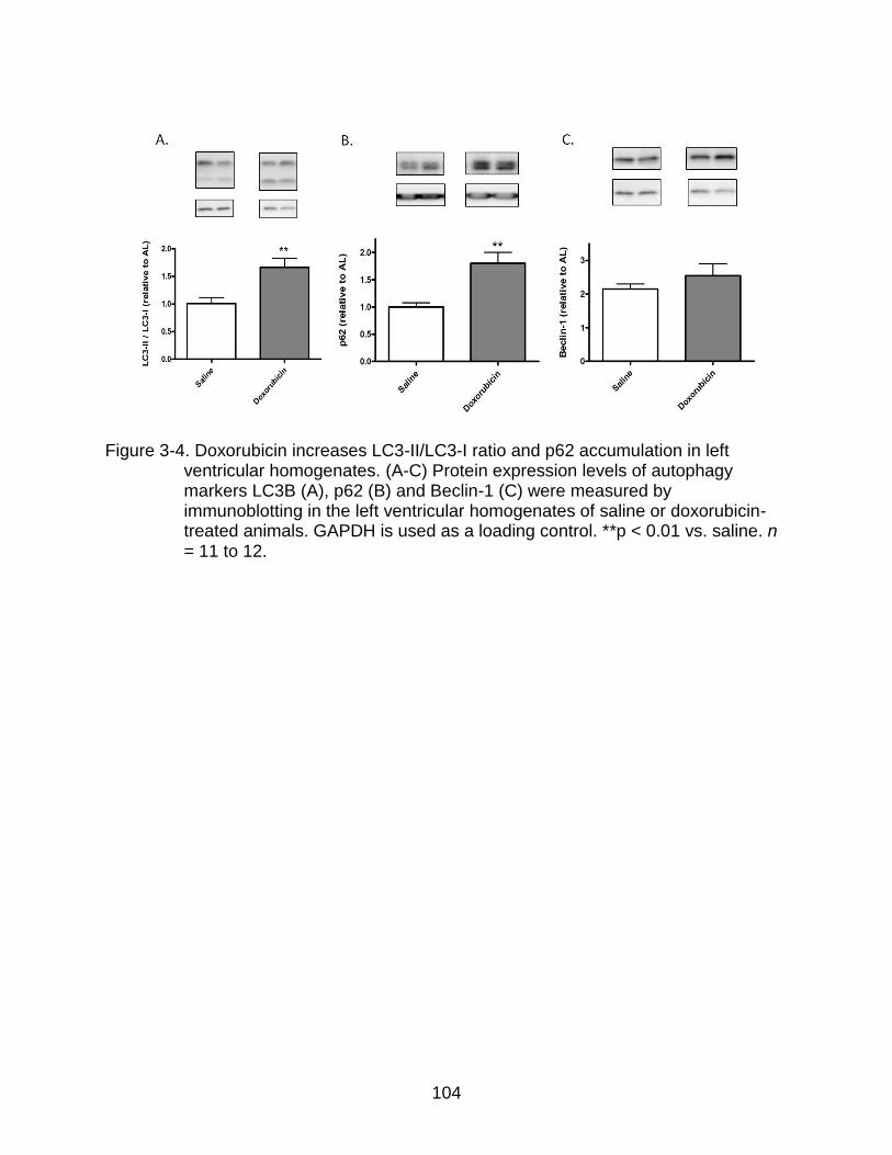
104
Figure 3-4. Doxorubicin increases LC3-II/LC3-I ratio and p62 accumulation in left ventricular homogenates. (A-C) Protein expression levels of autophagy markers LC3B (A), p62 (B) and Beclin-1 (C) were measured by immunoblotting in the left ventricular homogenates of saline or doxorubicin-treated animals. GAPDH is used as a loading control. **p < 0.01 vs. saline. n = 11 to 12.

105
Figure 3-5. CR + Resv-50 attenuates doxorubicin-mediated increases in cardiac damage markers. (A) Apoptotic induction in left ventricular homogenates of saline-treated AL rats and doxorubicin-treated AL, CR, CR + Resv-5 and CR + Resv-50 groups was determined by measuring the release of mono and oligo- nucleosomes in the cytosolic fraction. Results have been normalized to the total amount of protein used for the assay. *p < 0.05 vs. AL + saline; #p < 0.05 vs. AL + doxorubicin (B) Serum CK levels were measured in the same groups as in (A). **p < 0.01 vs. AL + saline, #p < 0.05 vs. AL + doxorubicin. n = 5 to 12.

106
Figure 3-6. CR + Resv-50 attenuates doxorubicin-mediated increases in serum LDH activity. (A) Serum LDH activity was determined in saline-treated AL and in doxorubicin-treated AL, CR, CR + Resv-5 and CR + Resv-50 animals. **p < 0.01 vs. AL + saline; #p < 0.05 vs. AL + doxorubicin. n = 5 to 12. (B) (A) Serum LDH activity was determined in saline-treated AL, CR, CR + Resv-5 and CR + Resv-50 animals. n = 4 to 12.

107
Table 3-1. Ingredient composition for AL and 20% CR purified diet. Composition AL 20% CR
Casein 140.00 175.00
Maltose Dextrin 230.00 230.00
Soybean Oil 40.00 50.00
Corn Starch 160.70 93.37
Cellulose 40.00 50.00
Sucrose 100.00 100.00
Dextrose 240.00 240.00
Salt mix, AIN-93MMX 35.00 43.75
Vitamin Mix, AIN-93 10.00 12.50
L-Cystine 1.80 2.25
Choline Bitartrate 2.50 3.13
Bacon Flavoring As needed
Flowing Agents As needed

108
Table 3-2. List of reagents used for in vivo study. Reagent Name Source Catalog No.
Bradford Reagent BioRad, Hercules, CA 500-0205
Cell Death Detection Plus ELISA kit Roche Molecular Diagnostics,
Indianapolis, IN
1-774-425
Dithiothreitol Sigma, St. Louis, MO D9779
ECL Plus reagent Amersham Biosciences,
Buckinghamshire, UK
RPN2132
EDTA Fisher Scientific, San Jose, CA BP120-500
Enzyhrom Creatine Kinase Assay kit Bioassay Systems, Hayward, CA ECPK - 100
Halt’s protease inhibitor Thermo Scientific, Fair Lawn, NJ 78439
HEPES Sigma, St. Louis, MO H3375
PVDF (polyvinylidene difluoride)
membrane
BioRad, Hercules, CA 162-0177
Quantichrom Lactate Dehydrogenase kit Bioassay Systems, Hayward, CA DLDH - 100
Resveratrol Sigma, St. Louis, MO R5010
Sodium orthovanadate (Na3VO4) Sigma, St. Louis, MO S6508
Starting Block Thermo Scientific, Fair Lawn, NJ 37543
Tris-HCl polyacrylamide gels BioRad, Hercules, CA
Triton X-100 Sigma, St. Louis, MO T9284
β-glycerophosphate Sigma, St. Louis, MO G9422

109
Table 3-3. List of primary antibodies used for in vivo study. Species Antigen Source Catalog No.
mouse anti-Atg5 Sigma, St. Louis, MO A2859
rabbit anti-Beclin-1 Cell Signaling Technology, Danvers, MA 3738
rabbit anti-LC3B Cell Signaling Technology, Danvers, MA 2775
rabbit anti-p62 Cell Signaling Technology, Danvers, MA 5114
mouse GAPDH-HRP Sigma, St. Louis, MO G9295

110
Table 3-4. Body weight (BW) and heart weight (HW) analysis. AL CR CR + Resv-5 CR + Resv-50
BW (g) 581 ± 39 529 ± 45** 519 ± 31*** 521 ± 35***
HW (g) 1.36 ± 0.08 1.33 ± 0.13 1.25 ± 0.06*** 1.24 ± 0.09***
HW/BW 2.34 ± 0.17 2.52 ± 0.18 2.43 ± 0.22 2.38 ± 0.16
RA (g) 0.041 ± 0.011 0.060 ± 0.028 0.050 ± 0.060 0.036 ± 0.009
RV (g) 0.205 ± 0.029 0.216 ± 0.029 0.187 ± 0.026 0.187 ± 0.034
LA (g) 0.040 ± 0.009 0.044 ± 0.019 0.034 ± 0.007 0.035 ± 0.007
LV (g) 0.523 ± 0.068 0.555 ± 0.053 0.505 ± 0.079 0.477 ± 0.058
RA, right atrium; RV, right ventricle; LA, left atrium; LV, left ventricle. **p < 0.01, ***p < 0.001 vs. AL. n = 9 to 23.

111
Table 3-5. Plasma resveratrol concentrations analysis.
Intervention groups Resveratrol concentration (ng/mL)
AL ND
20% CR + Resv-5 7.4 ± 3.7
20% CR + Resv-50 41 ± 19
ND, not detectable. n = 5 in each group.
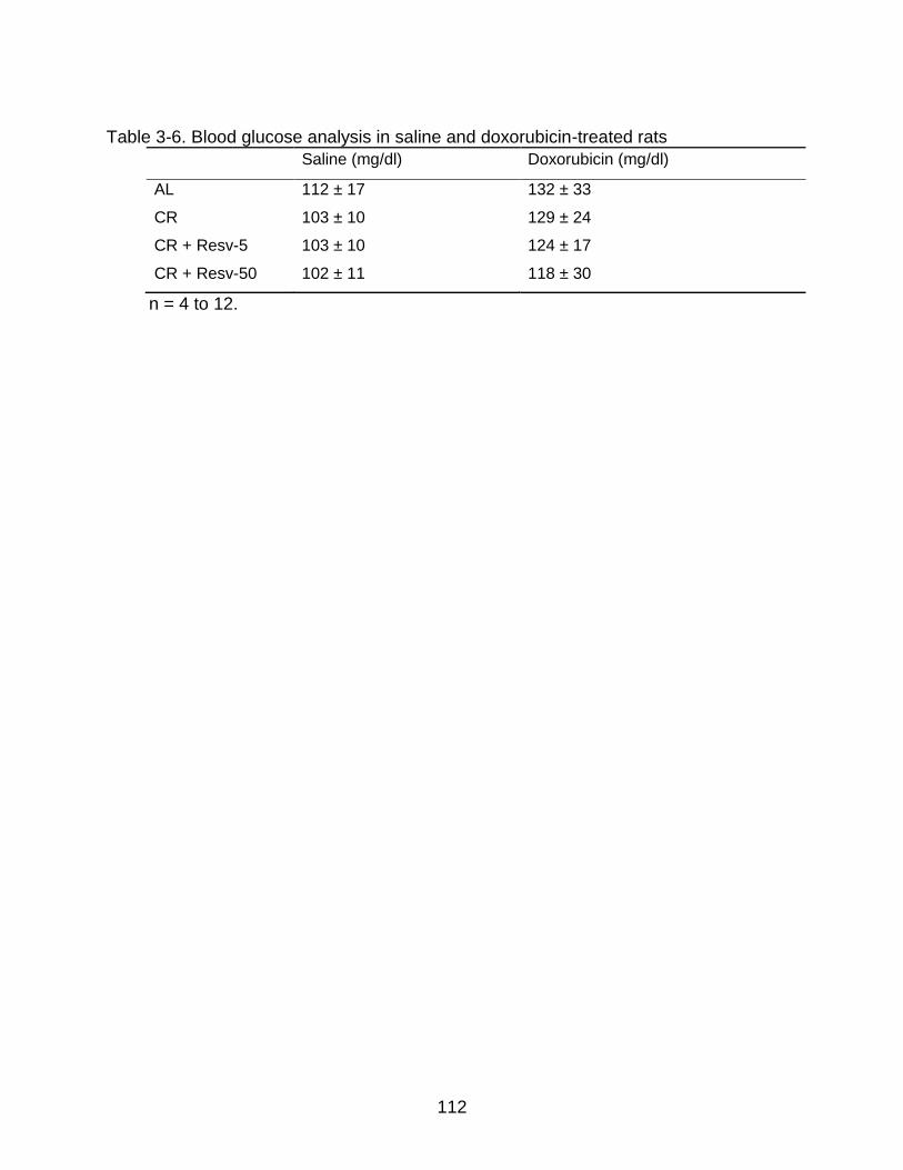
112
Table 3-6. Blood glucose analysis in saline and doxorubicin-treated rats
Saline (mg/dl) Doxorubicin (mg/dl)
AL 112 ± 17 132 ± 33
CR 103 ± 10 129 ± 24
CR + Resv-5 103 ± 10 124 ± 17
CR + Resv-50 102 ± 11 118 ± 30
n = 4 to 12.
113
CHAPTER 4
CONCLUSION
Summary of Results and Therapeutic Relevance
Age-related increases in oxidative damage have been well documented in the
heart and is implicated in age-associated decline of cardiac function (Marzetti et al.,
2009). Oxidative stress can damage cellular organelles, especially the mitochondria,
which further enhances oxidative burden by generating more ROS. Under such
circumstances, the removal of oxidatively damaged components and dysfunctional
mitochondria from cells would be highly desirable and potentially beneficial. This holds
true especially for cardiomyocytes, due to their post-mitotic nature and a lifespan of
decades. Autophagy serves this essential homeostatic clean-up function. The relevance
of the autophagy-lysosomal axis to cardiomyocyte homeostasis is witnessed by the
observation that abnormal oxidant generation, mitochondrial dysfunction and the
accumulation of intracellular waste material are observed under conditions of inhibited
autophagy (Nakai et al., 2007b). This suggests that the optimization of the
housekeeping function of autophagy may be harnessed as a therapeutic means against
oxidative damage and heart senescence. We hypothesized that enhancement of basal
autophagy in cardiomyocytes will protect against pathological consequences resulting
from enhanced generation of oxidative stress. We tested this hypothesis both in vitro
(HL-1 cardiomyocytes) and in vivo (26-month-old rat hearts).
For our in vitro study (Chapter 2), we investigated in detail, the role of
mitochondria-specific ROS generation in mediating cellular cytotoxicity and whether
autophagy enhancement could be protective under such circumstances. We observed
that treatment of HL-1 cardiomyocytes with the antibiotic compound AMA increased O2•−
114
generation, decreased m and inhibited cellular respiration (Fig. 2-1). In addition, AMA
augmented nuclear DNA oxidation and cell death (Fig. 2-1). Although oxidative stress
per se has been reported to be a potential inducer of autophagy, we did not observe
autophagic stimulation by AMA treatment (Fig. 2-2). Induction of autophagy and
mitophagy by the mTOR inhibitor rapamycin protected cardiomyocytes from the
cytotoxic effects of AMA, as assessed by viability assays and PARP-1 and caspase-3
activation analysis (Fig. 2-5). In addition to an overall effect on cell survival, rapamycin-
mediated autophagy improved mitochondrial function, as determined by respiration
analysis, m and mitochondrial morphology analysis (Fig. 2-7).
In addition to improving mitochondrial function, rapamycin suppressed the
accumulation of ubiquitinated proteins induced by AMA (Fig. 2-6). The ubiquitinated
proteins were observed only in the detergent insoluble fraction of AMA-treated cells,
indicating that these proteins could be aggregated in nature. Autophagy induction by
rapamycin was able to effectively mediate their clearance (Fig. 2-6). This observation is
consistent with previous studies illustrating a role of autophagy in removing protein
aggregates in cell culture models of aggregate-prone diseases (Harada et al., 2008;
Ravikumar et al., 2004).
In Chapter 3, we have summarized our findings from in vivo studies investigating
interventions targeted to induce autophagy in 26-month-old FBN rat hearts. Studies
investigating autophagic stimulation in late-middle-aged or old animals are very limited
and often the interventions proposed (such as lifelong 40% CR) are unfeasible in
humans. We have tested a moderate CR regimen (20% CR) alone, or in combination
with the plant polyphenol resveratrol (oral dosing) starting late in life and for a period of

115
6 weeks, as potential inducers of autophagy. Analysis of autophagy markers in the left
ventricle of heart tissue revealed that 20% CR in combination with 50 mg/kg/day
resveratrol can decrease the abundance of p62 (indicating enhanced autophagic flux)
(Fig. 3-3) and enhance the expression of autophagy protein Beclin-1 (indicating a
possible induction of autophagy) (Fig. 3-3). Our observation that a moderate CR
regimen and resveratrol, only when combined, stimulated autophagy indicates a
potential synergistic action between the two. The exact mechanism through which the
combined approach induces autophagy is currently unknown and can only be
speculated. Both CR and resveratrol have been shown to inhibit the mTOR pathway
and activate AMPK and SIRT1, all of which can induce autophagy through independent
ways (Fig. 4-1).
Furthermore, similar to in vitro studies, we investigated whether enhancing basal
levels of cardiac autophagy can be protective against oxidative stress-mediated
damage. Oxidative stress was generated in FBN rats by intraperitoneal administration of
doxorubicin, a widely used chemotherapeutic agent, reported to cause significant
cardiotoxicity. Our group has previously shown that doxorubicin increases cardiac levels
of 8-iso prostaglandin F2α, a known marker of lipid peroxidation (Childs et al., 2002). In
addition, it enhances cytochrome c release, caspase-3 activation and apoptotic levels
and in the myocardium (Childs et al., 2002). We observed that the induction of
autophagy by the CR + resveratrol regimen attenuated doxorubicin-mediated increases
in cardiac apoptotic levels (Fig. 3-5), serum CK (Fig. 3-5) and LDH levels (Fig. 3-5),
supporting the conclusion that enhanced autophagy serves to protect the myocardium
from doxorubicin-mediated damage. It is worth noting here that doxorubicin is mostly

116
administered to patients in late-middle-ages, at which stage the incidence of cancer is
among the highest. Our study was conducted in 26-month-old FBN rats which
correspond to about 60 years (late-middle-age) in humans (Quinn, 2005). Hence, the
study holds clinical and translational relevance.
Our study conclusively shows that the upregulation of autophagy is protective
against oxidative stress-mediated dysfunction in cardiomyocytes. Investigations into the
mechanisms of such a protective effect showed that at least in cell culture models,
autophagy induction leads to the clearance of aggregated, ubiquitinated proteins and
dysfunctional mitochondria. The protective effect of autophagy induction was also seen
in an animal model of oxidative stress-mediated injury, where autophagy was induced
by nutritional interventions such as CR and resveratrol. A mild CR regimen starting late
in life and the use of plant polyphenol resveratrol are feasible interventions that could be
used in humans. The 50 mg/kg/day oral dose of resveratrol used in the study (in
combination with CR) translates to a dose of approximately 4 mg/kg/day resveratrol in
humans (Reagan-Shaw et al., 2008). In other words, a human weighing 60 kg would
need a daily oral dose of 240 mg resveratrol. This dose of resveratrol is much lower
than what has been reported to cause side-effects in humans (Patel et al., 2011). No
clinical trial has investigated a combinatorial approach of a moderate CR regimen with
resveratrol supplementation, and therefore such a study would be highly desirable. Until
then, we propose that following a lifestyle of consuming fewer calories, combined with a
daily oral consumption of resveratrol may have potentially beneficial effects in humans.
Our in vitro and in vivo studies therefore illustrate the crucial role of autophagy in
attenuating cardiac oxidative damage. Others have demonstrated the contribution of

117
autophagy in preserving cardiomyocyte physiology in general (Nakai et al., 2007b). With
the beneficial roles of autophagy being increasingly reported, a lot of attention has also
been focused on identifying novel enhancers of autophagy. Autophagy inducers can be
broadly categorized to be acting through mTOR-dependent and -independent pathways.
Rapamycin and its homologs comprise mTOR-dependent inducers of autophagy, which
act by inhibiting mTOR, thereby removing its inhibitory effect on autophagy induction
(Ganley et al., 2009; Jung et al., 2009a). In collaboration with other investigators at the
University of Florida, we have observed that feeding orally 2.24 mg/kg/day rapamycin
for 8-10 months can enhance autophagy in the hearts of C57BL/J6 mice (Fig. 4-2).
Rapamycin used in the study was encased using an acrylic polymer spinning disk
enteric coating system to prevent digestion in the stomach and thereby increase the
bioavailability of rapamycin. We observed increased levels of LC3-II/LC3-I ratio (Fig. 4-
2A) in whole heart homogenates from these mice, along with decreases in the
abundance of p62 (Fig. 4-2B). No changes were observed in the protein expression
levels of Beclin-1 (Fig. 4-2C). Such observations complement our in vitro finding where
rapamycin significantly induced autophagy in mouse HL-1 and human AC16 cells. Other
researchers have shown that induction of autophagy by rapamycin removes mutant
huntingtin aggregates and offers protective effects in cell culture systems and in
transgenic Drosophila and mouse models of Huntington disease (Berger et al., 2006;
Ravikumar et al., 2002; Ravikumar et al., 2004). Apart from rapamycin, a recent
screening of a library of 3500 chemicals has identified four compounds: perhexiline,
niclosamide, amiodarone and rottlerin, which significantly induced autophagy through
the mTOR pathway (Balgi et al., 2009). Among these chemicals, rottlerin inhibited

118
mTORC1 by activating TSC2, while the rest acted in a TSC2-independent manner
(Balgi et al., 2009). Importantly, perhexiline (used for treating chest pain arising from an
ischemic heart), niclosamide (anti-helminthic agent) and amiodarone (used against
cardiac arrhythmias) are also FDA approved drugs.
The very first evidence of mTOR-independent induction of autophagy came from
the observation that a decrease in cellular levels of inositol 1,4,5-triphosphate (IP3) can
stimulate autophagy, with no effect on the mTORC1 or mTORC2 pathway (Sarkar et al.,
2005). IP3-mediated regulation of autophagy is most likely to be due to IP3 being
required for ER Ca2+ release. For instance, increased levels of cytosolic IP3 can bind to
IP3 receptors (IP3R) in the ER and trigger the release of ER-sequestered Ca2+ in the
cytoplasm (Berridge, 1993). Increased cytosolic calcium can inhibit Ca2+ dependent
cysteine proteases called calpains and this leads to an inhibition of autophagy
(Demarchi et al., 2007; Demarchi et al., 2006; Gordon et al., 1993). Accordingly, lithium
and other mood stabilizing agents (such as carbamazepine and sodium valproate),
which reduces intracellular inositol levels by inhibiting inositol monophosphatase,
enhanced autophagy and mediated the clearance of autophagy substrates (Berridge,
1993; Sarkar et al., 2005; Williams et al., 2002). Similarly, inhibition of IP3R by
xestospongin B also stimulated autophagy (Criollo et al., 2007). In another remarkable
study, a high-throughput screening of 50,729 compounds resulted in the identification of
three small molecule enhancers of autophagy, which increases autophagosome
formation and stimulates the clearance of autophagy substrates (Sarkar et al., 2007).
The molecules were found to act in an mTOR-independent manner, although the
precise cellular mechanism is yet to be known (Sarkar et al., 2007).

119
In spite of the identification of numerous autophagy inducers, it is important to note
that the majority of these experiments were conducted in cell culture models. Whether
the abovementioned compounds could also stimulate autophagy in vivo remains to be
established. Measuring autophagic flux in animal models is limited by the fact that
lysosomal inhibitors cannot be directly used in vivo. Although at least one paper
reported the use of chloroquine to inhibit lysosomal degradation in animals (Iwai-Kanai
et al., 2008), follow-up studies have not been conducted.The inhibition of lysosomal
degradation in vivo therefore remains extremely difficult and rare. Autophagy flux
determinations in animals can partly be accomplished by indirect methods such as by
analyzing p62 abundance or by measuring the clearance of mutant, aggregate-prone
proteins in mouse models of huntingin (Lin et al., 2001; White et al., 1997) or alpha-
synuclein aggregation (Chesselet, 2008).
Finally, while the self clean-up process of autophagy is critical for the maintenance
of cell homeostasis, nevertheless, an excessive activation of autophagy may be
maladaptive, chewing up essential survival components and leading to further toxicity.
Increased number of autophagic vacuoles have been observed under pathological
conditions, such as in the cardiac tissues of patients with heart failure (Kostin et al.,
2003), aortic valve stenosis (Hein et al., 2003), left ventricular hypertrophy (Yamamoto
et al., 2000) and the hibernating myocardium (Elsasser et al., 2004). However, it is
unclear whether these autophagic vacuoles were actually contributing to the
pathological conditions or whether it was upregulated in an attempt to protect against
cell death. For example, up-regulation of autophagy might represent an attempt to cope
with increased levels of mitochondrial dysfunction and cellular damage. Nevertheless, a

120
detrimental aspect of excessive autophagic stimulation has not been conclusively ruled
out. The level of autophagy may be critical in determining whether autophagy will be
protective or detrimental. Therapeutic interventions that exploit the homeostatic
properties of autophagy, without stimulating its maladaptive effects caused by an
excessive induction, is therefore highly desirable.
Future Directions
Although the induction of basal autophagy protects cardiomyocytes from
enhanced oxidative damage and despite the recent evidences supporting the
involvement of impaired autophagy in the accumulation of oxidative damage within old
cardiomyocytes, several research questions still remains to be addressed. First,
whether enhancing autophagy can attenuate cardiac senescence needs to be clearly
established. In this regard, Harrison and co-authors reported that oral administration of
rapamycin to mice, starting at 600 days of age, extended median lifespan in male and
female mice by 9% and 14% respectively (Harrison et al., 2009). Although the precise
molecular mechanisms leading to such an effect was not investigated in the study, it
can be hypothesized that autophagy induction was at least partially responsible for such
an effect. Additionally, studies investigating whether manipulation of autophagy rescues
the premature heart senescence phenotype observed in animal models characterized
by high loads of mtDNA mutation (and oxidative stress) would be highly desirable.
Second, autophagy is not just a random degradation process; rather, it is highly
regulated and potentially selective. Therefore, it is mandatory to understand whether
specific signal transduction pathways or the engulfment of particular autophagy
substrates are negatively affected under conditions of increased oxidative stress or
during senescence. With regard to the mitochondria, the identification of signaling

121
pathways linking mitochondrial dynamics and selective mitophagy is necessary for the
development of therapeutics that maximize the removal of damaged mitochondria, while
sparing functional ones. Third, it should be considered that most autophagy mediators
have multiple functions, meaning that their manipulation may produce unrelated and
perhaps undesirable effects. Therefore, a deeper understanding of the various actions
performed by autophagy-inducing compounds / interventions is warranted. Fourth,
whether the optimization of autophagy preserves cardiomyocyte mitochondrial function
and delays the development of CVD in humans remains to be established. Most data on
the effects of pharmacological or behavioral modulation of autophagy on cardiac aging
and physiology derive from model organisms in which the role of autophagy may differ
from what impacts human health, partly because of difficulties in modeling complex
human diseases and degenerative processes in experimental settings.
The current limitations make extremely challenging, the identification of specific
autophagy inducers and also the optimal window of autophagic activation to exploit the
cardioprotective effects of autophagy without any non-specific, undesirable or potentially
detrimental effects. Answering the above critical research questions will likely provide
cardiologists and geriatricians with novel therapeutic means to postpone the
degenerative fate of cardiomyocytes through the induction of autophagy and relieve the
burden associated with increased oxidative stress common at old age and in CVD
patients. In summary, the vital functions carried out by the autophagic process in
cardiac physiology suggest the possibility that therapeutic interventions targeting this
cellular pathway may represent effective means to counter heart senescence and age-
related damages due to oxidative stress. Untangling the complexity of autophagic

122
regulation and managing the dual nature of autophagy are major tasks the field of
geriatric cardiology is called to pursue.

123
Figure 4-1. Regulation of autophagy by rapamycin, CR and resveratrol. Rapamycin inhibits mTOR kinase and thereby removes mTOR-dependent inhibitory phosphorylation on the ULK1 – Atg13 – FIP200 complex. Removal of inhibitory phosphorylation results in the induction of autophagy. Both CR and resveratrol can activate autophagy by inhibiting the mTOR pathway and/or by activating of the AMPK and SIRT1 pathways. AMPK induces activating phosphorylation on the ULK1 – Atg13 – FIP200 complex, thereby stimulating autophagy. SIRT1 can decetylate and activate key autophagy genes Atg5, Atg7 and LC3, thereby inducing autophagy.

124
Figure 4-2. Chronic rapamycin administration induces autophagy in C57BL/J6 mice hearts. Protein expression levels of autophagy markers LC3B (A), p62 (B) and Beclin-1 (C) were measured by immunoblotting in whole heart homogenates of placebo or rapamycin-treated animals. Rapa, Rapamycin. *p < 0.05, *** p < 0.001 vs. placebo.

125
Figure 4-3. Schematic of overall observations from in vitro and in vivo studies. Oxidative stressors such as AMA and doxorubicin can cause damage to cellular components, affecting cell viability. Induction of autophagy by rapamycin in vitro (HL-1 cells) and by a combined intervention of CR and resveratrol in vivo (FBN rat hearts) can improve cell survival and homeostasis by potentially removing oxidatively modified components.
126
LIST OF REFERENCES
Abel ED, Doenst T (2011) Mitochondrial adaptations to physiological vs. pathological cardiac hypertrophy. Cardiovasc Res 90(2): 234-242 Alcendor RR, Gao S, Zhai P, Zablocki D, Holle E, Yu X, Tian B, Wagner T, Vatner SF, Sadoshima J (2007) Sirt1 regulates aging and resistance to oxidative stress in the heart. CircRes 100(10): 1512-1521 Andreyev AY, Kushnareva YE, Starkov AA (2005a) Mitochondrial metabolism of reactive oxygen species. Biochemistry (Mosc) 70(2): 200-214 Arias E, Cuervo AM (2011) Chaperone-mediated autophagy in protein quality control. CurrOpinCell Biol 23(2): 184-189 Arnheim N, Cortopassi G (1992) Deleterious mitochondrial DNA mutations accumulate in aging human tissues. MutatRes 275(3-6): 157-167 Backman L, Kreis H, Morales JM, Wilczek H, Taylor R, Burke JT (2002) Sirolimus steady-state trough concentrations are not affected by bolus methylprednisolone therapy in renal allograft recipients. Br J Clin Pharmacol 54(1): 65-68 Balaban RS, Nemoto S, Finkel T (2005) Mitochondria, oxidants, and aging. Cell 120(4): 483-495 Balgi AD, Fonseca BD, Donohue E, Tsang TC, Lajoie P, Proud CG, Nabi IR, Roberge M (2009) Screen for chemical modulators of autophagy reveals novel therapeutic inhibitors of mTORC1 signaling. PLoS One 4(9): e7124 Barger JL, Kayo T, Pugh TD, Prolla TA, Weindruch R (2008a) Short-term consumption of a resveratrol-containing nutraceutical mixture mimics gene expression of long-term caloric restriction in mouse heart. ExpGerontol 43(9): 859-866 Barger JL, Kayo T, Vann JM, Arias EB, Wang J, Hacker TA, Wang Y, Raederstorff D, Morrow JD, Leeuwenburgh C, Allison DB, Saupe KW, Cartee GD, Weindruch R, Prolla TA (2008b) A low dose of dietary resveratrol partially mimics caloric restriction and retards aging parameters in mice. PLoSOne 3(6): e2264 Barja G, Herrero A (2000) Oxidative damage to mitochondrial DNA is inversely related to maximum life span in the heart and brain of mammals. FASEB J 14(2): 312-318 Bass TM, Weinkove D, Houthoofd K, Gems D, Partridge L (2007) Effects of resveratrol on lifespan in Drosophila melanogaster and Caenorhabditis elegans. Mech Ageing Dev 128(10): 546-552

127
Baur JA, Pearson KJ, Price NL, Jamieson HA, Lerin C, Kalra A, Prabhu VV, Allard JS, Lopez-Lluch G, Lewis K, Pistell PJ, Poosala S, Becker KG, Boss O, Gwinn D, Wang M, Ramaswamy S, Fishbein KW, Spencer RG, Lakatta EG, Le CD, Shaw RJ, Navas P, Puigserver P, Ingram DK, de CR, Sinclair DA (2006) Resveratrol improves health and survival of mice on a high-calorie diet. Nature 444(7117): 337-342 Becker LB (2004) New concepts in reactive oxygen species and cardiovascular reperfusion physiology. Cardiovasc Res 61(3): 461-470 Berger Z, Ravikumar B, Menzies FM, Oroz LG, Underwood BR, Pangalos MN, Schmitt I, Wullner U, Evert BO, O'Kane CJ, Rubinsztein DC (2006) Rapamycin alleviates toxicity of different aggregate-prone proteins. HumMolGenet 15(3): 433-442 Berridge MJ (1993) Inositol trisphosphate and calcium signalling. Nature 361(6410): 315-325 Bjelakovic G, Nikolova D, Gluud LL, Simonetti RG, Gluud C (2007) Mortality in randomized trials of antioxidant supplements for primary and secondary prevention: systematic review and meta-analysis. JAMA 297(8): 842-857 Bjorkoy G, Lamark T, Brech A, Outzen H, Perander M, Overvatn A, Stenmark H, Johansen T (2005) p62/SQSTM1 forms protein aggregates degraded by autophagy and has a protective effect on huntingtin-induced cell death. J Cell Biol 171(4): 603-614 Bjorkoy G, Lamark T, Pankiv S, Overvatn A, Brech A, Johansen T (2009) Monitoring autophagic degradation of p62/SQSTM1. Methods Enzymol 452: 181-197 Boocock DJ, Faust GE, Patel KR, Schinas AM, Brown VA, Ducharme MP, Booth TD, Crowell JA, Perloff M, Gescher AJ, Steward WP, Brenner DE (2007) Phase I dose escalation pharmacokinetic study in healthy volunteers of resveratrol, a potential cancer chemopreventive agent. Cancer Epidemiol Biomarkers Prev 16(6): 1246-1252 Bova MP, Tam D, McMahon G, Mattson MN (2005) Troglitazone induces a rapid drop of mitochondrial membrane potential in liver HepG2 cells. Toxicol Lett 155(1): 41-50 Boveris A, Chance B (1973) The mitochondrial generation of hydrogen peroxide. General properties and effect of hyperbaric oxygen. BiochemJ 134(3): 707-716 Bradamante S, Barenghi L, Villa A (2004) Cardiovascular protective effects of resveratrol. Cardiovasc Drug Rev 22(3): 169-188 Bradford MM (1976) A rapid and sensitive method for the quantitation of microgram quantities of protein utilizing the principle of protein-dye binding. Anal Biochem 72: 248-254
128
Brancaccio P, Lippi G, Maffulli N (2010) Biochemical markers of muscular damage. Clin Chem Lab Med 48(6): 757-767 Braunwald E (1997) Shattuck lecture--cardiovascular medicine at the turn of the millennium: triumphs, concerns, and opportunities. N Engl J Med 337(19): 1360-1369 Brown VA, Patel KR, Viskaduraki M, Crowell JA, Perloff M, Booth TD, Vasilinin G, Sen A, Schinas AM, Piccirilli G, Brown K, Steward WP, Gescher AJ, Brenner DE (2010) Repeat dose study of the cancer chemopreventive agent resveratrol in healthy volunteers: safety, pharmacokinetics, and effect on the insulin-like growth factor axis. Cancer Res 70(22): 9003-9011 Brunk UT, Terman A (2002) The mitochondrial-lysosomal axis theory of aging: accumulation of damaged mitochondria as a result of imperfect autophagocytosis. EurJBiochem 269(8): 1996-2002 Canto C, Gerhart-Hines Z, Feige JN, Lagouge M, Noriega L, Milne JC, Elliott PJ, Puigserver P, Auwerx J (2009) AMPK regulates energy expenditure by modulating NAD+ metabolism and SIRT1 activity. Nature 458(7241): 1056-1060 Carreira RS, Lee Y, Ghochani M, Gustafsson AB, Gottlieb RA (2010) Cyclophilin D is required for mitochondrial removal by autophagy in cardiac cells. Autophagy 6(4) Cecconi F, Levine B (2008) The role of autophagy in mammalian development: cell makeover rather than cell death. DevCell 15(3): 344-357 Chatterjee K, Zhang J, Honbo N, Karliner JS (2009) Doxorubicin cardiomyopathy. Cardiology 115(2): 155-162 Chen D, Gao F, Li B, Wang H, Xu Y, Zhu C, Wang G (2010) Parkin mono-ubiquitinates Bcl-2 and regulates autophagy. J Biol Chem 285(49): 38214-38223 Chen D, Steele AD, Lindquist S, Guarente L (2005) Increase in activity during calorie restriction requires Sirt1. Science 310(5754): 1641 Chen H, Chan DC (2009) Mitochondrial dynamics--fusion, fission, movement, and mitophagy--in neurodegenerative diseases. Hum Mol Genet 18(R2): R169-176 Chen Y, Azad MB, Gibson SB (2009) Superoxide is the major reactive oxygen species regulating autophagy. Cell Death Differ 16(7): 1040-1052 Chen Y, Klionsky DJ (2011) The regulation of autophagy - unanswered questions. JCell Sci 124(Pt 2): 161-170
129
Cheng KC, Cahill DS, Kasai H, Nishimura S, Loeb LA (1992) 8-Hydroxyguanine, an abundant form of oxidative DNA damage, causes G----T and A----C substitutions. J Biol Chem 267(1): 166-172 Chesselet MF (2008) In vivo alpha-synuclein overexpression in rodents: a useful model of Parkinson's disease? Exp Neurol 209(1): 22-27 Childs AC, Phaneuf SL, Dirks AJ, Phillips T, Leeuwenburgh C (2002) Doxorubicin treatment in vivo causes cytochrome C release and cardiomyocyte apoptosis, as well as increased mitochondrial efficiency, superoxide dismutase activity, and Bcl-2:Bax ratio. Cancer Res 62(16): 4592-4598 Claycomb WC, Lanson NA, Jr., Stallworth BS, Egeland DB, Delcarpio JB, Bahinski A, Izzo NJ, Jr. (1998) HL-1 cells: a cardiac muscle cell line that contracts and retains phenotypic characteristics of the adult cardiomyocyte. Proc Natl Acad Sci U S A 95(6): 2979-2984 Cohen MV, Downey JM (2008) Adenosine: trigger and mediator of cardioprotection. Basic Res Cardiol 103(3): 203-215 Criollo A, Maiuri MC, Tasdemir E, Vitale I, Fiebig AA, Andrews D, Molgo J, Diaz J, Lavandero S, Harper F, Pierron G, di Stefano D, Rizzuto R, Szabadkai G, Kroemer G (2007) Regulation of autophagy by the inositol trisphosphate receptor. Cell Death Differ 14(5): 1029-1039 Cuervo AM, Bergamini E, Brunk UT, Droge W, Ffrench M, Terman A (2005) Autophagy and aging: the importance of maintaining "clean" cells. Autophagy 1(3): 131-140 Dai DF, Chen T, Wanagat J, Laflamme M, Marcinek DJ, Emond MJ, Ngo CP, Prolla TA, Rabinovitch PS (2010) Age-dependent cardiomyopathy in mitochondrial mutator mice is attenuated by overexpression of catalase targeted to mitochondria. Aging Cell 9(4): 536-544 Dai DF, Rabinovitch PS (2009) Cardiac aging in mice and humans: the role of mitochondrial oxidative stress. Trends CardiovascMed 19(7): 213-220 Dai DF, Santana LF, Vermulst M, Tomazela DM, Emond MJ, MacCoss MJ, Gollahon K, Martin GM, Loeb LA, Ladiges WC, Rabinovitch PS (2009a) Overexpression of catalase targeted to mitochondria attenuates murine cardiac aging. Circulation 119(21): 2789-2797 Dasgupta B, Milbrandt J (2007) Resveratrol stimulates AMP kinase activity in neurons. Proc Natl Acad Sci U S A 104(17): 7217-7222 de Grey AD (1997) A proposed refinement of the mitochondrial free radical theory of aging. Bioessays 19(2): 161-166

130
Deleers M, Goormaghtigh E (1985) Adriamycin effects on insulin secretion, Ca2+ movements and glucose oxidation in pancreatic islet cells. Pharmacol Res Commun 17(3): 227-232 Demarchi F, Bertoli C, Copetti T, Eskelinen EL, Schneider C (2007) Calpain as a novel regulator of autophagosome formation. Autophagy 3(3): 235-237 Demarchi F, Bertoli C, Copetti T, Tanida I, Brancolini C, Eskelinen EL, Schneider C (2006) Calpain is required for macroautophagy in mammalian cells. J Cell Biol 175(4): 595-605 Dewaele M, Maes H, Agostinis P (2010) ROS-mediated mechanisms of autophagy stimulation and their relevance in cancer therapy. Autophagy 6(7): 838-854 Dolinsky VW, Dyck JR (2011) Calorie restriction and resveratrol in cardiovascular health and disease. Biochim Biophys Acta 1812(11): 1477-1489 Doroshow JH (1983) Effect of anthracycline antibiotics on oxygen radical formation in rat heart. Cancer Res 43(2): 460-472 Dosenko VE, Nagibin VS, Tumanovska LV, Moibenko AA (2006) Protective effect of autophagy in anoxia-reoxygenation of isolated cardiomyocyte? Autophagy 2(4): 305-306 Dudek EJ, Shang F, Valverde P, Liu Q, Hobbs M, Taylor A (2005) Selectivity of the ubiquitin pathway for oxidatively modified proteins: relevance to protein precipitation diseases. FASEB J 19(12): 1707-1709 Dutta D, Calvani R, Bernabei R, Leeuwenburgh C, Marzetti E (2012) Contribution of impaired mitochondrial autophagy to cardiac aging: mechanisms and therapeutic opportunities. Circ Res 110(8): 1125-1138 Edwards AG, Donato AJ, Lesniewski LA, Gioscia RA, Seals DR, Moore RL (2010) Life-long caloric restriction elicits pronounced protection of the aged myocardium: a role for AMPK. Mech Ageing Dev 131(11-12): 739-742 Egan DF, Shackelford DB, Mihaylova MM, Gelino S, Kohnz RA, Mair W, Vasquez DS, Joshi A, Gwinn DM, Taylor R, Asara JM, Fitzpatrick J, Dillin A, Viollet B, Kundu M, Hansen M, Shaw RJ (2011) Phosphorylation of ULK1 (hATG1) by AMP-activated protein kinase connects energy sensing to mitophagy. Science 331(6016): 456-461 Elmore SP, Qian T, Grissom SF, Lemasters JJ (2001) The mitochondrial permeability transition initiates autophagy in rat hepatocytes. FASEB J 15(12): 2286-2287
131
Elsasser A, Vogt AM, Nef H, Kostin S, Mollmann H, Skwara W, Bode C, Hamm C, Schaper J (2004) Human hibernating myocardium is jeopardized by apoptotic and autophagic cell death. JAmCollCardiol 43(12): 2191-2199 Finckenberg P, Eriksson O, Baumann M, Merasto S, Lalowski MM, Levijoki J, Haasio K, Kyto V, Muller DN, Luft FC, Oresic M, Mervaala E (2012) Caloric restriction ameliorates angiotensin II-induced mitochondrial remodeling and cardiac hypertrophy. Hypertension 59(1): 76-84 Finkel T (2011) Signal Transduction by Mitochondrial Oxidants. JBiolChem Fontana L, Weiss EP, Villareal DT, Klein S, Holloszy JO (2008) Long-term effects of calorie or protein restriction on serum IGF-1 and IGFBP-3 concentration in humans. Aging Cell 7(5): 681-687 Ganley IG, Lam du H, Wang J, Ding X, Chen S, Jiang X (2009) ULK1.ATG13.FIP200 complex mediates mTOR signaling and is essential for autophagy. J Biol Chem 284(18): 12297-12305 Gebbia N, Leto G, Gagliano M, Tumminello FM, Rausa L (1985) Lysosomal alterations in heart and liver of mice treated with doxorubicin. Cancer Chemother Pharmacol 15(1): 26-30 Gedik CM, Collins A (2005) Establishing the background level of base oxidation in human lymphocyte DNA: results of an interlaboratory validation study. FASEB J 19(1): 82-84 Geisler S, Holmstrom KM, Skujat D, Fiesel FC, Rothfuss OC, Kahle PJ, Springer W (2010) PINK1/Parkin-mediated mitophagy is dependent on VDAC1 and p62/SQSTM1. Nat Cell Biol 12(2): 119-131 Gershon H, Gershon D (1970) Detection of inactive enzyme molecules in ageing organisms. Nature 227(5264): 1214-1217 Glick JH, Jr. (1969) Serum lactate dehydrogenase isoenzyme and total lactate dehydrogenase values in health and disease, and clinical evaluation of these tests by means of discriminant analysis. Am J Clin Pathol 52(3): 320-328 Gnaiger E (2008) Polarographic oxygen sensors, the oxygraph, and high-resolution respirometry to assess mitochondrial function. In Drug-Induced Mitochondrial Dysfunction, Dykens; JA, Will; Y e (eds). Hoboken, NJ, USA: John Wiley & Sons, Inc Gordon PB, Holen I, Fosse M, Rotnes JS, Seglen PO (1993) Dependence of hepatocytic autophagy on intracellularly sequestered calcium. J Biol Chem 268(35): 26107-26112
132
Green DR, Galluzzi L, Kroemer G (2011) Mitochondria and the Autophagy-Inflammation-Cell Death Axis in Organismal Aging. Science 333(6046): 1109-1112 Green PS, Leeuwenburgh C (2002) Mitochondrial dysfunction is an early indicator of doxorubicin-induced apoptosis. Biochim Biophys Acta 1588(1): 94-101 Grivennikova VG, Kareyeva AV, Vinogradov AD (2010) What are the sources of hydrogen peroxide production by heart mitochondria? BiochimBiophysActa 1797(6-7): 939-944 Guarente L, Picard F (2005) Calorie restriction--the SIR2 connection. Cell 120(4): 473-482 Hacker TA, McKiernan SH, Douglas PS, Wanagat J, Aiken JM (2006) Age-related changes in cardiac structure and function in Fischer 344 x Brown Norway hybrid rats. Am J Physiol Heart Circ Physiol 290(1): H304-311 Hafner AV, Dai J, Gomes AP, Xiao CY, Palmeira CM, Rosenzweig A, Sinclair DA (2010) Regulation of the mPTP by SIRT3-mediated deacetylation of CypD at lysine 166 suppresses age-related cardiac hypertrophy. Aging (Albany NY) 2(12): 914-923 Hamacher-Brady A, Brady NR, Gottlieb RA (2006) Enhancing macroautophagy protects against ischemia/reperfusion injury in cardiac myocytes. JBiolChem 281(40): 29776-29787 Hansen M, Taubert S, Crawford D, Libina N, Lee SJ, Kenyon C (2007) Lifespan extension by conditions that inhibit translation in Caenorhabditis elegans. Aging Cell 6(1): 95-110 Harada M, Hanada S, Toivola DM, Ghori N, Omary MB (2008) Autophagy activation by rapamycin eliminates mouse Mallory-Denk bodies and blocks their proteasome inhibitor-mediated formation. Hepatology 47(6): 2026-2035 Harrison DE, Strong R, Sharp ZD, Nelson JF, Astle CM, Flurkey K, Nadon NL, Wilkinson JE, Frenkel K, Carter CS, Pahor M, Javors MA, Fernandez E, Miller RA (2009) Rapamycin fed late in life extends lifespan in genetically heterogeneous mice. Nature 460(7253): 392-395 Hars ES, Qi H, Ryazanov AG, Jin S, Cai L, Hu C, Liu LF (2007) Autophagy regulates ageing in C. elegans. Autophagy 3(2): 93-95 Hein S, Arnon E, Kostin S, Schonburg M, Elsasser A, Polyakova V, Bauer EP, Klovekorn WP, Schaper J (2003) Progression from compensated hypertrophy to failure in the pressure-overloaded human heart: structural deterioration and compensatory mechanisms. Circulation 107(7): 984-991
133
Henics T, Wheatley DN (1999) Cytoplasmic vacuolation, adaptation and cell death: a view on new perspectives and features. Biol Cell 91(7): 485-498 Hofer T, Fontana L, Anton SD, Weiss EP, Villareal D, Malayappan B, Leeuwenburgh C (2008) Long-term effects of caloric restriction or exercise on DNA and RNA oxidation levels in white blood cells and urine in humans. Rejuvenation Res 11(4): 793-799 Hofer T, Seo AY, Prudencio M, Leeuwenburgh C (2006) A method to determine RNA and DNA oxidation simultaneously by HPLC-ECD: greater RNA than DNA oxidation in rat liver after doxorubicin administration. Biol Chem 387(1): 103-111 Howitz KT, Bitterman KJ, Cohen HY, Lamming DW, Lavu S, Wood JG, Zipkin RE, Chung P, Kisielewski A, Zhang LL, Scherer B, Sinclair DA (2003) Small molecule activators of sirtuins extend Saccharomyces cerevisiae lifespan. Nature 425(6954): 191-196 Huttemann M, Lee I, Pecinova A, Pecina P, Przyklenk K, Doan JW (2008) Regulation of oxidative phosphorylation, the mitochondrial membrane potential, and their role in human disease. J Bioenerg Biomembr 40(5): 445-456 Ide T, Tsutsui H, Hayashidani S, Kang D, Suematsu N, Nakamura K, Utsumi H, Hamasaki N, Takeshita A (2001) Mitochondrial DNA damage and dysfunction associated with oxidative stress in failing hearts after myocardial infarction. CircRes 88(5): 529-535 Ide T, Tsutsui H, Kinugawa S, Utsumi H, Kang D, Hattori N, Uchida K, Arimura K, Egashira K, Takeshita A (1999) Mitochondrial electron transport complex I is a potential source of oxygen free radicals in the failing myocardium. Circ Res 85(4): 357-363 Ingram DK, Zhu M, Mamczarz J, Zou S, Lane MA, Roth GS, deCabo R (2006) Calorie restriction mimetics: an emerging research field. Aging Cell 5(2): 97-108 Inuzuka Y, Okuda J, Kawashima T, Kato T, Niizuma S, Tamaki Y, Iwanaga Y, Yoshida Y, Kosugi R, Watanabe-Maeda K, Machida Y, Tsuji S, Aburatani H, Izumi T, Kita T, Shioi T (2009) Suppression of phosphoinositide 3-kinase prevents cardiac aging in mice. Circulation 120(17): 1695-1703 Iwai-Kanai E, Yuan H, Huang C, Sayen MR, Perry-Garza CN, Kim L, Gottlieb RA (2008) A method to measure cardiac autophagic flux in vivo. Autophagy 4(3): 322-329 Iwai K, Drake SK, Wehr NB, Weissman AM, LaVaute T, Minato N, Klausner RD, Levine RL, Rouault TA (1998) Iron-dependent oxidation, ubiquitination, and degradation of iron regulatory protein 2: implications for degradation of oxidized proteins. Proc Natl Acad Sci U S A 95(9): 4924-4928

134
Jang YM, Kendaiah S, Drew B, Phillips T, Selman C, Julian D, Leeuwenburgh C (2004) Doxorubicin treatment in vivo activates caspase-12 mediated cardiac apoptosis in both male and female rats. FEBS Lett 577(3): 483-490 Jeong JK, Moon MH, Bae BC, Lee YJ, Seol JW, Kang HS, Kim JS, Kang SJ, Park SY (2012) Autophagy induced by resveratrol prevents human prion protein-mediated neurotoxicity. Neurosci Res 73(2): 99-105 Jia K, Levine B (2007) Autophagy is required for dietary restriction-mediated life span extension in C. elegans. Autophagy 3(6): 597-599 Judge S, Jang YM, Smith A, Hagen T, Leeuwenburgh C (2005a) Age-associated increases in oxidative stress and antioxidant enzyme activities in cardiac interfibrillar mitochondria: implications for the mitochondrial theory of aging. Faseb J 19(3): 419-421 Judge S, Leeuwenburgh C (2007) Cardiac mitochondrial bioenergetics, oxidative stress, and aging. Am J Physiol Cell Physiol 292(6): C1983-1992 Jung CH, Jun CB, Ro SH, Kim YM, Otto NM, Cao J, Kundu M, Kim DH (2009a) ULK-Atg13-FIP200 complexes mediate mTOR signaling to the autophagy machinery. MolBiolCell 20(7): 1992-2003 Jung CH, Jun CB, Ro SH, Kim YM, Otto NM, Cao J, Kundu M, Kim DH (2009b) ULK-Atg13-FIP200 complexes mediate mTOR signaling to the autophagy machinery. Mol Biol Cell 20(7): 1992-2003 Jung T, Bader N, Grune T (2007) Lipofuscin: formation, distribution, and metabolic consequences. AnnNYAcadSci 1119: 97-111 Kabeya Y, Mizushima N, Ueno T, Yamamoto A, Kirisako T, Noda T, Kominami E, Ohsumi Y, Yoshimori T (2000) LC3, a mammalian homologue of yeast Apg8p, is localized in autophagosome membranes after processing. EMBO J 19(21): 5720-5728 Kadandale P, Kiger AA (2010) Role of selective autophagy in cellular remodeling: "self-eating" into shape. Autophagy 6(8): 1194-1195 Kaeberlein M, McDonagh T, Heltweg B, Hixon J, Westman EA, Caldwell SD, Napper A, Curtis R, DiStefano PS, Fields S, Bedalov A, Kennedy BK (2005) Substrate-specific activation of sirtuins by resveratrol. J Biol Chem 280(17): 17038-17045 Kanamori H, Takemura G, Maruyama R, Goto K, Tsujimoto A, Ogino A, Li L, Kawamura I, Takeyama T, Kawaguchi T, Nagashima K, Fujiwara T, Fujiwara H, Seishima M, Minatoguchi S (2009) Functional significance and morphological characterization of starvation-induced autophagy in the adult heart. AmJPathol 174(5): 1705-1714

135
Kang R, Zeh HJ, Lotze MT, Tang D (2011) The Beclin 1 network regulates autophagy and apoptosis. Cell Death Differ 18(4): 571-580 Kapahi P, Zid BM, Harper T, Koslover D, Sapin V, Benzer S (2004) Regulation of lifespan in Drosophila by modulation of genes in the TOR signaling pathway. Curr Biol 14(10): 885-890 Kapetanovic IM, Muzzio M, Huang Z, Thompson TN, McCormick DL (2010) Pharmacokinetics, oral bioavailability, and metabolic profile of resveratrol and its dimethylether analog, pterostilbene, in rats. Cancer Chemother Pharmacol 68(3): 593-601 Karbowski M, Kurono C, Wozniak M, Ostrowski M, Teranishi M, Nishizawa Y, Usukura J, Soji T, Wakabayashi T (1999) Free radical-induced megamitochondria formation and apoptosis. Free RadicBiolMed 26(3-4): 396-409 Kaufmann P, Torok M, Zahno A, Waldhauser KM, Brecht K, Krahenbuhl S (2006) Toxicity of statins on rat skeletal muscle mitochondria. Cell Mol Life Sci 63(19-20): 2415-2425 Khan S, Salloum F, Das A, Xi L, Vetrovec GW, Kukreja RC (2006) Rapamycin confers preconditioning-like protection against ischemia-reperfusion injury in isolated mouse heart and cardiomyocytes. J Mol Cell Cardiol 41(2): 256-264 Kiffin R, Bandyopadhyay U, Cuervo AM (2006) Oxidative stress and autophagy. Antioxid Redox Signal 8(1-2): 152-162 Kihara A, Kabeya Y, Ohsumi Y, Yoshimori T (2001) Beclin-phosphatidylinositol 3-kinase complex functions at the trans-Golgi network. EMBO Rep 2(4): 330-335 Kim I, Lemasters JJ (2011) Mitophagy selectively degrades individual damaged mitochondria after photoirradiation. AntioxidRedoxSignal 14(10): 1919-1928 Kim J, Kundu M, Viollet B, Guan KL (2011) AMPK and mTOR regulate autophagy through direct phosphorylation of Ulk1. NatCell Biol 13(2): 132-141 Kitsis RN, Peng CF, Cuervo AM (2007) Eat your heart out. Nat Med 13(5): 539-541 Korolchuk VI, Mansilla A, Menzies FM, Rubinsztein DC (2009) Autophagy inhibition compromises degradation of ubiquitin-proteasome pathway substrates. Mol Cell 33(4): 517-527 Kostin S, Pool L, Elsasser A, Hein S, Drexler HC, Arnon E, Hayakawa Y, Zimmermann R, Bauer E, Klovekorn WP, Schaper J (2003) Myocytes die by multiple mechanisms in failing human hearts. CircRes 92(7): 715-724
136
Kotamraju S, Konorev EA, Joseph J, Kalyanaraman B (2000) Doxorubicin-induced apoptosis in endothelial cells and cardiomyocytes is ameliorated by nitrone spin traps and ebselen. Role of reactive oxygen and nitrogen species. J Biol Chem 275(43): 33585-33592 Kovacic JC, Castellano JM, Fuster V (2012) Cardiovascular defense challenges at the basic, clinical, and population levels. Ann N Y Acad Sci 1254(1): 1-6 Kuchino Y, Mori F, Kasai H, Inoue H, Iwai S, Miura K, Ohtsuka E, Nishimura S (1987) Misreading of DNA templates containing 8-hydroxydeoxyguanosine at the modified base and at adjacent residues. Nature 327(6117): 77-79 Kujoth GC, Hiona A, Pugh TD, Someya S, Panzer K, Wohlgemuth SE, Hofer T, Seo AY, Sullivan R, Jobling WA, Morrow JD, Van RH, Sedivy JM, Yamasoba T, Tanokura M, Weindruch R, Leeuwenburgh C, Prolla TA (2005) Mitochondrial DNA mutations, oxidative stress, and apoptosis in mammalian aging. Science 309(5733): 481-484 Kuma A, Matsui M, Mizushima N (2007) LC3, an autophagosome marker, can be incorporated into protein aggregates independent of autophagy: caution in the interpretation of LC3 localization. Autophagy 3(4): 323-328 la Porte C, Voduc N, Zhang G, Seguin I, Tardiff D, Singhal N, Cameron DW (2010) Steady-State pharmacokinetics and tolerability of trans-resveratrol 2000 mg twice daily with food, quercetin and alcohol (ethanol) in healthy human subjects. Clin Pharmacokinet 49(7): 449-454 Lagouge M, Argmann C, Gerhart-Hines Z, Meziane H, Lerin C, Daussin F, Messadeq N, Milne J, Lambert P, Elliott P, Geny B, Laakso M, Puigserver P, Auwerx J (2006) Resveratrol improves mitochondrial function and protects against metabolic disease by activating SIRT1 and PGC-1alpha. Cell 127(6): 1109-1122 Lakatta EG (2001) Heart aging: a fly in the ointment? CircRes 88(10): 984-986 Larsen KB, Lamark T, Overvatn A, Harneshaug I, Johansen T, Bjorkoy G (2010) A reporter cell system to monitor autophagy based on p62/SQSTM1. Autophagy 6(6): 784-793 Lee IH, Cao L, Mostoslavsky R, Lombard DB, Liu J, Bruns NE, Tsokos M, Alt FW, Finkel T (2008) A role for the NAD-dependent deacetylase Sirt1 in the regulation of autophagy. Proc Natl Acad Sci U S A 105(9): 3374-3379 Leeuwenburgh C, Wagner P, Holloszy JO, Sohal RS, Heinecke JW (1997) Caloric restriction attenuates dityrosine cross-linking of cardiac and skeletal muscle proteins in aging mice. ArchBiochemBiophys 346(1): 74-80

137
Lesnefsky EJ, Moghaddas S, Tandler B, Kerner J, Hoppel CL (2001) Mitochondrial dysfunction in cardiac disease: ischemia--reperfusion, aging, and heart failure. JMolCell Cardiol 33(6): 1065-1089 Levine B, Klionsky DJ (2004) Development by self-digestion: molecular mechanisms and biological functions of autophagy. DevCell 6(4): 463-477 Levine B, Kroemer G (2008) Autophagy in the pathogenesis of disease. Cell 132(1): 27-42 Levine B, Mizushima N, Virgin HW (2011) Autophagy in immunity and inflammation. Nature 469(7330): 323-335 Levonen AL, Vahakangas E, Koponen JK, Yla-Herttuala S (2008) Antioxidant gene therapy for cardiovascular disease: current status and future perspectives. Circulation 117(16): 2142-2150 Li H, Forstermann U (2009) Resveratrol: a multifunctional compound improving endothelial function. Editorial to: "Resveratrol supplementation gender independently improves endothelial reactivity and suppresses superoxide production in healthy rats" by S. Soylemez et al.. Cardiovasc Drugs Ther 23(6): 425-429 Liang XH, Jackson S, Seaman M, Brown K, Kempkes B, Hibshoosh H, Levine B (1999) Induction of autophagy and inhibition of tumorigenesis by beclin 1. Nature 402(6762): 672-676 Lin CH, Tallaksen-Greene S, Chien WM, Cearley JA, Jackson WS, Crouse AB, Ren S, Li XJ, Albin RL, Detloff PJ (2001) Neurological abnormalities in a knock-in mouse model of Huntington's disease. Hum Mol Genet 10(2): 137-144 Lin HS, Jenner AM, Ong CN, Huang SH, Whiteman M, Halliwell B (2004) A high-throughput and sensitive methodology for the quantification of urinary 8-hydroxy-2'-deoxyguanosine: measurement with gas chromatography-mass spectrometry after single solid-phase extraction. Biochem J 380(Pt 2): 541-548 Lin MT, Beal MF (2006) Mitochondrial dysfunction and oxidative stress in neurodegenerative diseases. Nature 443(7113): 787-795 Linnane AW, Marzuki S, Ozawa T, Tanaka M (1989) Mitochondrial DNA mutations as an important contributor to ageing and degenerative diseases. Lancet 1(8639): 642-645 Lipman RD, Chrisp CE, Hazzard DG, Bronson RT (1996) Pathologic characterization of brown Norway, brown Norway x Fischer 344, and Fischer 344 x brown Norway rats with relation to age. J Gerontol A Biol Sci Med Sci 51(1): B54-59

138
Liu M, Wilk SA, Wang A, Zhou L, Wang RH, Ogawa W, Deng C, Dong LQ, Liu F Resveratrol inhibits mTOR signaling by promoting the interaction between mTOR and DEPTOR. J Biol Chem 285(47): 36387-36394 Liu VW, Zhang C, Nagley P (1998) Mutations in mitochondrial DNA accumulate differentially in three different human tissues during ageing. Nucleic Acids Res 26(5): 1268-1275 Loos B, Lochner A, Engelbrecht AM (2011) Autophagy in heart disease: a strong hypothesis for an untouched metabolic reserve. MedHypotheses 77(1): 52-57 Lv XC, Zhou HY (2012) Resveratrol protects H9c2 embryonic rat heart derived cells from oxidative stress by inducing autophagy: role of p38 mitogen-activated protein kinase. Can J Physiol Pharmacol Ma X, Jin M, Cai Y, Xia H, Long K, Liu J, Yu Q, Yuan J (2011) Mitochondrial electron transport chain complex III is required for antimycin A to inhibit autophagy. Chem Biol 18(11): 1474-1481 Manov I, Pollak Y, Broneshter R, Iancu TC (2011) Inhibition of doxorubicin-induced autophagy in hepatocellular carcinoma Hep3B cells by sorafenib--the role of extracellular signal-regulated kinase counteraction. FEBS J 278(18): 3494-3507 Markossian KA, Kurganov BI (2004) Protein folding, misfolding, and aggregation. Formation of inclusion bodies and aggresomes. Biochemistry (Mosc) 69(9): 971-984 Marzetti E, Wohlgemuth SE, Anton SD, Bernabei R, Carter CS, Leeuwenburgh C (2009) Cellular mechanisms of cardioprotection by calorie restriction: state of the science and future perspectives. ClinGeriatrMed 25(4): 715-732, ix Masubuchi Y, Kano S, Horie T (2006) Mitochondrial permeability transition as a potential determinant of hepatotoxicity of antidiabetic thiazolidinediones. Toxicology 222(3): 233-239 Masubuchi Y, Kano S, Horie T (2006) Mitochondrial permeability transition as a potential determinant of hepatotoxicity of antidiabetic thiazolidinediones. Toxicology 222(3): 233-239 Matecic M, Smith DL, Pan X, Maqani N, Bekiranov S, Boeke JD, Smith JS (2010) A microarray-based genetic screen for yeast chronological aging factors. PLoSGenet 6(4): e1000921 Matsuda N, Sato S, Shiba K, Okatsu K, Saisho K, Gautier CA, Sou YS, Saiki S, Kawajiri S, Sato F, Kimura M, Komatsu M, Hattori N, Tanaka K (2010) PINK1 stabilized by mitochondrial depolarization recruits Parkin to damaged mitochondria and activates latent Parkin for mitophagy. JCell Biol 189(2): 211-221

139
Matsui Y, Takagi H, Qu X, Abdellatif M, Sakoda H, Asano T, Levine B, Sadoshima J (2007) Distinct roles of autophagy in the heart during ischemia and reperfusion: roles of AMP-activated protein kinase and Beclin 1 in mediating autophagy. CircRes 100(6): 914-922 Mauthe M, Jacob A, Freiberger S, Hentschel K, Stierhof YD, Codogno P, Proikas-Cezanne T (2011) Resveratrol-mediated autophagy requires WIPI-1-regulated LC3 lipidation in the absence of induced phagophore formation. Autophagy 7(12): 1448-1461 McMullen JR, Sherwood MC, Tarnavski O, Zhang L, Dorfman AL, Shioi T, Izumo S (2004) Inhibition of mTOR signaling with rapamycin regresses established cardiac hypertrophy induced by pressure overload. Circulation 109(24): 3050-3055 Mendl N, Occhipinti A, Muller M, Wild P, Dikic I, Reichert AS (2011) Mitophagy in yeast is independent of mitochondrial fission and requires the stress response gene WHI2. J Cell Sci 124(Pt 8): 1339-1350 Meyer TE, Kovacs SJ, Ehsani AA, Klein S, Holloszy JO, Fontana L (2006) Long-term caloric restriction ameliorates the decline in diastolic function in humans. JAmCollCardiol 47(2): 398-402 Mijaljica D, Prescott M, Devenish RJ (2011) Microautophagy in mammalian cells: Revisiting a 40-year-old conundrum. Autophagy 7(7): 673-682 Minor RK, Smith DL, Jr., Sossong AM, Kaushik S, Poosala S, Spangler EL, Roth GS, Lane M, Allison DB, de CR, Ingram DK, Mattison JA (2010) Chronic ingestion of 2-deoxy-D-glucose induces cardiac vacuolization and increases mortality in rats. ToxicolApplPharmacol 243(3): 332-339 Miquel J, Tappel AL, Dillard CJ, Herman MM, Bensch KG (1974) Fluorescent products and lysosomal components in aging Drosophila melanogaster. J Gerontol 29(6): 622-637 Mitra MS, Donthamsetty S, White B, Latendresse JR, Mehendale HM (2007) Mechanism of protection of moderately diet restricted rats against doxorubicin-induced acute cardiotoxicity. Toxicol Appl Pharmacol 225(1): 90-101 Mizushima N (2007) Autophagy: process and function. Genes Dev 21(22): 2861-2873 Mizushima N, Levine B (2010) Autophagy in mammalian development and differentiation. Nat Cell Biol 12(9): 823-830 Mizushima N, Noda T, Yoshimori T, Tanaka Y, Ishii T, George MD, Klionsky DJ, Ohsumi M, Ohsumi Y (1998) A protein conjugation system essential for autophagy. Nature 395(6700): 395-398

140
Mizushima N, Yamamoto A, Matsui M, Yoshimori T, Ohsumi Y (2004) In vivo analysis of autophagy in response to nutrient starvation using transgenic mice expressing a fluorescent autophagosome marker. MolBiolCell 15(3): 1101-1111 Mizushima N, Yoshimori T, Levine B (2010) Methods in mammalian autophagy research. Cell 140(3): 313-326 Mohamed SA, Hanke T, Erasmi AW, Bechtel MJ, Scharfschwerdt M, Meissner C, Sievers HH, Gosslau A (2006) Mitochondrial DNA deletions and the aging heart. ExpGerontol 41(5): 508-517 Montaigne D, Marechal X, Baccouch R, Modine T, Preau S, Zannis K, Marchetti P, Lancel S, Neviere R (2010) Stabilization of mitochondrial membrane potential prevents doxorubicin-induced cardiotoxicity in isolated rat heart. Toxicol Appl Pharmacol 244(3): 300-307 Mordente A, Meucci E, Silvestrini A, Martorana GE, Giardina B (2012) Anthracyclines and mitochondria. Adv Exp Med Biol 942: 385-419 Morselli E, Maiuri MC, Markaki M, Megalou E, Pasparaki A, Palikaras K, Criollo A, Galluzzi L, Malik SA, Vitale I, Michaud M, Madeo F, Tavernarakis N, Kroemer G (2010) Caloric restriction and resveratrol promote longevity through the Sirtuin-1-dependent induction of autophagy. Cell DeathDis 1: e10 Mortimore GE, Schworer CM (1977) Induction of autophagy by amino-acid deprivation in perfused rat liver. Nature 270(5633): 174-176 Mukhopadhyay P, Rajesh M, Batkai S, Kashiwaya Y, Hasko G, Liaudet L, Szabo C, Pacher P (2009) Role of superoxide, nitric oxide, and peroxynitrite in doxorubicin-induced cell death in vivo and in vitro. Am J Physiol Heart Circ Physiol 296(5): H1466-1483 Mukhopadhyay P, Rajesh M, Hasko G, Hawkins BJ, Madesh M, Pacher P (2007) Simultaneous detection of apoptosis and mitochondrial superoxide production in live cells by flow cytometry and confocal microscopy. Nat Protoc 2(9): 2295-2301 Mullin GE (2011) Red wine, grapes, and better health--resveratrol. Nutr Clin Pract 26(6): 722-723 Nair U, Jotwani A, Geng J, Gammoh N, Richerson D, Yen WL, Griffith J, Nag S, Wang K, Moss T, Baba M, McNew JA, Jiang X, Reggiori F, Melia TJ, Klionsky DJ (2011) SNARE Proteins Are Required for Macroautophagy. Cell 146(2): 290-302
141
Nakai A, Yamaguchi O, Takeda T, Higuchi Y, Hikoso S, Taniike M, Omiya S, Mizote I, Matsumura Y, Asahi M, Nishida K, Hori M, Mizushima N, Otsu K (2007a) The role of autophagy in cardiomyocytes in the basal state and in response to hemodynamic stress. Nat Med 13(5): 619-624 Nakayama K, Okamoto F, Harada Y (1956) Antimycin A: isolation from a new Streptomyces and activity against rice plant blast fungi. J Antibiot (Tokyo) 9(2): 63-66 Narendra D, Tanaka A, Suen DF, Youle RJ (2008) Parkin is recruited selectively to impaired mitochondria and promotes their autophagy. JCell Biol 183(5): 795-803 Nixon JP, Zhang M, Wang C, Kuskowski MA, Novak CM, Levine JA, Billington CJ, Kotz CM (2010) Evaluation of a quantitative magnetic resonance imaging system for whole body composition analysis in rodents. Obesity (Silver Spring) 18(8): 1652-1659 Noda NN, Kumeta H, Nakatogawa H, Satoo K, Adachi W, Ishii J, Fujioka Y, Ohsumi Y, Inagaki F (2008) Structural basis of target recognition by Atg8/LC3 during selective autophagy. Genes Cells 13(12): 1211-1218 Novak I, Kirkin V, McEwan DG, Zhang J, Wild P, Rozenknop A, Rogov V, Lohr F, Popovic D, Occhipinti A, Reichert AS, Terzic J, Dotsch V, Ney PA, Dikic I (2010) Nix is a selective autophagy receptor for mitochondrial clearance. EMBO Rep 11(1): 45-51 Octavia Y, Tocchetti CG, Gabrielson KL, Janssens S, Crijns HJ, Moens AL (2012) Doxorubicin-induced cardiomyopathy: From molecular mechanisms to therapeutic strategies. J Mol Cell Cardiol 52(6): 1213-1225 Omodei D, Fontana L (2011) Calorie restriction and prevention of age-associated chronic disease. FEBS Lett 585(11): 1537-1542 Paglin S, Hollister T, Delohery T, Hackett N, McMahill M, Sphicas E, Domingo D, Yahalom J (2001) A novel response of cancer cells to radiation involves autophagy and formation of acidic vesicles. Cancer Res 61(2): 439-444 Pan T, Rawal P, Wu Y, Xie W, Jankovic J, Le W (2009) Rapamycin protects against rotenone-induced apoptosis through autophagy induction. Neuroscience 164(2): 541-551 Pan Y, Gao Y, Chen L, Gao G, Dong H, Yang Y, Dong B, Chen X (2011) Targeting autophagy augments in vitro and in vivo antimyeloma activity of DNA-damaging chemotherapy. Clin Cancer Res 17(10): 3248-3258 Paravicini TM, Touyz RM (2008) NADPH oxidases, reactive oxygen species, and hypertension: clinical implications and therapeutic possibilities. Diabetes Care 31 Suppl 2: S170-180

142
Pasdois P, Parker JE, Griffiths EJ, Halestrap AP (2011) The role of oxidized cytochrome c in regulating mitochondrial reactive oxygen species production and its perturbation in ischaemia. BiochemJ 436(2): 493-505 Patel KR, Scott E, Brown VA, Gescher AJ, Steward WP, Brown K (2011) Clinical trials of resveratrol. Ann N Y Acad Sci 1215: 161-169 Petiot A, Ogier-Denis E, Blommaart EF, Meijer AJ, Codogno P (2000) Distinct classes of phosphatidylinositol 3'-kinases are involved in signaling pathways that control macroautophagy in HT-29 cells. The Journal of biological chemistry 275(2): 992-998 Petrovski G, Gurusamy N, Das DK (2011) Resveratrol in cardiovascular health and disease. AnnNYAcadSci 1215: 22-33 Pham NA, Robinson BH, Hedley DW (2000) Simultaneous detection of mitochondrial respiratory chain activity and reactive oxygen in digitonin-permeabilized cells using flow cytometry. Cytometry 41(4): 245-251 Potter VR, Reif AE (1952) Inhibition of an electron transport component by antimycin A. J Biol Chem 194(1): 287-297 Quinn R (2005) Comparing rat's to human's age: how old is my rat in people years? Nutrition 21(6): 775-777 Racette SB, Weiss EP, Villareal DT, Arif H, Steger-May K, Schechtman KB, Fontana L, Klein S, Holloszy JO (2006) One year of caloric restriction in humans: feasibility and effects on body composition and abdominal adipose tissue. J Gerontol A Biol Sci Med Sci 61(9): 943-950 Rahman A, More N, Schein PS (1982) Doxorubicin-induced chronic cardiotoxicity and its protection by liposomal administration. Cancer Res 42(5): 1817-1825 Ramprasath VR, Jones PJ (2010) Anti-atherogenic effects of resveratrol. Eur J Clin Nutr 64(7): 660-668 Ravikumar B, Berger Z, Vacher C, O'Kane CJ, Rubinsztein DC (2006) Rapamycin pre-treatment protects against apoptosis. Hum Mol Genet 15(7): 1209-1216 Ravikumar B, cevedo-Arozena A, Imarisio S, Berger Z, Vacher C, O'Kane CJ, Brown SD, Rubinsztein DC (2005) Dynein mutations impair autophagic clearance of aggregate-prone proteins. NatGenet 37(7): 771-776 Ravikumar B, Duden R, Rubinsztein DC (2002) Aggregate-prone proteins with polyglutamine and polyalanine expansions are degraded by autophagy. Hum Mol Genet 11(9): 1107-1117
143
Ravikumar B, Vacher C, Berger Z, Davies JE, Luo S, Oroz LG, Scaravilli F, Easton DF, Duden R, O'Kane CJ, Rubinsztein DC (2004) Inhibition of mTOR induces autophagy and reduces toxicity of polyglutamine expansions in fly and mouse models of Huntington disease. Nat Genet 36(6): 585-595 Ray PS, Maulik G, Cordis GA, Bertelli AA, Bertelli A, Das DK (1999) The red wine antioxidant resveratrol protects isolated rat hearts from ischemia reperfusion injury. Free Radic Biol Med 27(1-2): 160-169 Reagan-Shaw S, Nihal M, Ahmad N (2008) Dose translation from animal to human studies revisited. FASEB J 22(3): 659-661 Reeves PG, Nielsen FH, Fahey GC, Jr. (1993) AIN-93 purified diets for laboratory rodents: final report of the American Institute of Nutrition ad hoc writing committee on the reformulation of the AIN-76A rodent diet. J Nutr 123(11): 1939-1951 Roger VL, Go AS, Lloyd-Jones DM, Benjamin EJ, Berry JD, Borden WB, Bravata DM, Dai S, Ford ES, Fox CS, Fullerton HJ, Gillespie C, Hailpern SM, Heit JA, Howard VJ, Kissela BM, Kittner SJ, Lackland DT, Lichtman JH, Lisabeth LD, Makuc DM, Marcus GM, Marelli A, Matchar DB, Moy CS, Mozaffarian D, Mussolino ME, Nichol G, Paynter NP, Soliman EZ, Sorlie PD, Sotoodehnia N, Turan TN, Virani SS, Wong ND, Woo D, Turner MB (2012) Executive summary: heart disease and stroke statistics--2012 update: a report from the American Heart Association. Circulation 125(1): 188-197 Rubinsztein DC, Marino G, Kroemer G (2011) Autophagy and aging. Cell 146(5): 682-695 Sachs HG, Colgan JA, Lazarus ML (1977a) Ultrastructure of the aging myocardium: a morphometric approach. Am J Anat 150(1): 63-71 Sachs HG, Colgan JA, Lazarus ML (1977b) Ultrastructure of the aging myocardium: a morphometric approach. AmJAnat 150(1): 63-71 Sarkar S, Floto RA, Berger Z, Imarisio S, Cordenier A, Pasco M, Cook LJ, Rubinsztein DC (2005) Lithium induces autophagy by inhibiting inositol monophosphatase. J Cell Biol 170(7): 1101-1111 Sarkar S, Perlstein EO, Imarisio S, Pineau S, Cordenier A, Maglathlin RL, Webster JA, Lewis TA, O'Kane CJ, Schreiber SL, Rubinsztein DC (2007) Small molecules enhance autophagy and reduce toxicity in Huntington's disease models. Nat Chem Biol 3(6): 331-338 Scaduto RC, Jr., Grotyohann LW (1999) Measurement of mitochondrial membrane potential using fluorescent rhodamine derivatives. Biophys J 76(1 Pt 1): 469-477

144
Scarlatti F, Maffei R, Beau I, Ghidoni R, Codogno P (2008) Non-canonical autophagy: an exception or an underestimated form of autophagy? Autophagy 4(8): 1083-1085 Scherz-Shouval R, Shvets E, Fass E, Shorer H, Gil L, Elazar Z (2007a) Reactive oxygen species are essential for autophagy and specifically regulate the activity of Atg4. EMBO J 26(7): 1749-1760 Schrader EK, Harstad KG, Matouschek A (2009) Targeting proteins for degradation. NatChemBiol 5(11): 815-822 Schriner SE, Linford NJ, Martin GM, Treuting P, Ogburn CE, Emond M, Coskun PE, Ladiges W, Wolf N, Van RH, Wallace DC, Rabinovitch PS (2005) Extension of murine life span by overexpression of catalase targeted to mitochondria. Science 308(5730): 1909-1911 Schulz TJ, Zarse K, Voigt A, Urban N, Birringer M, Ristow M (2007) Glucose restriction extends Caenorhabditis elegans life span by inducing mitochondrial respiration and increasing oxidative stress. Cell Metab 6(4): 280-293 Seglen PO, Gordon PB (1982) 3-Methyladenine: specific inhibitor of autophagic/lysosomal protein degradation in isolated rat hepatocytes. Proc Natl Acad Sci U S A 79(6): 1889-1892 Seibenhener ML, Babu JR, Geetha T, Wong HC, Krishna NR, Wooten MW (2004) Sequestosome 1/p62 is a polyubiquitin chain binding protein involved in ubiquitin proteasome degradation. MolCell Biol 24(18): 8055-8068 Shang F, Taylor A (2011) Ubiquitin-proteasome pathway and cellular responses to oxidative stress. Free Radic Biol Med 51(1): 5-16 Shinmura K, Tamaki K, Sano M, Murata M, Yamakawa H, Ishida H, Fukuda K (2011) Impact of long-term caloric restriction on cardiac senescence: caloric restriction ameliorates cardiac diastolic dysfunction associated with aging. JMolCell Cardiol 50(1): 117-127 Smoliga JM, Baur JA, Hausenblas HA (2011) Resveratrol and health--a comprehensive review of human clinical trials. Mol Nutr Food Res 55(8): 1129-1141 Smuder AJ, Kavazis AN, Min K, Powers SK (2011) Exercise protects against doxorubicin-induced markers of autophagy signaling in skeletal muscle. J Appl Physiol 111(4): 1190-1198 Spinale FG, Coker ML, Thomas CV, Walker JD, Mukherjee R, Hebbar L (1998) Time-dependent changes in matrix metalloproteinase activity and expression during the progression of congestive heart failure: relation to ventricular and myocyte function. Circ Res 82(4): 482-495

145
Stroikin Y, Dalen H, Loof S, Terman A (2004) Inhibition of autophagy with 3-methyladenine results in impaired turnover of lysosomes and accumulation of lipofuscin-like material. Eur J Cell Biol 83(10): 583-590 Sung MM, Soltys CL, Masson G, Boisvenue JJ, Dyck JR (2010) Improved cardiac metabolism and activation of the RISK pathway contributes to improved post-ischemic recovery in calorie restricted mice. J Mol Med (Berl) 89(3): 291-302 Suzuki K, Kirisako T, Kamada Y, Mizushima N, Noda T, Ohsumi Y (2001) The pre-autophagosomal structure organized by concerted functions of APG genes is essential for autophagosome formation. EMBO J 20(21): 5971-5981 Sybers HD, Ingwall J, DeLuca M (1976) Autophagy in cardiac myocytes. Recent Adv Stud Cardiac Struct Metab 12: 453-463 Tait SW, Green DR (2010) Mitochondria and cell death: outer membrane permeabilization and beyond. Nat Rev Mol Cell Biol 11(9): 621-632 Takemura G, Fujiwara H (2007) Doxorubicin-induced cardiomyopathy from the cardiotoxic mechanisms to management. Prog Cardiovasc Dis 49(5): 330-352 Tal R, Winter G, Ecker N, Klionsky DJ, Abeliovich H (2007) Aup1p, a yeast mitochondrial protein phosphatase homolog, is required for efficient stationary phase mitophagy and cell survival. JBiolChem 282(8): 5617-5624 Talloczy Z, Jiang W, Virgin HW, Leib DA, Scheuner D, Kaufman RJ, Eskelinen EL, Levine B (2002) Regulation of starvation- and virus-induced autophagy by the eIF2alpha kinase signaling pathway. ProcNatlAcadSciUSA 99(1): 190-195 Taneike M, Yamaguchi O, Nakai A, Hikoso S, Takeda T, Mizote I, Oka T, Tamai T, Oyabu J, Murakawa T, Nishida K, Shimizu T, Hori M, Komuro I, Shirasawa T, Mizushima N, Otsu K (2010) Inhibition of autophagy in the heart induces age-related cardiomyopathy. Autophagy 6(5) Tatsuta T, Langer T (2008) Quality control of mitochondria: protection against neurodegeneration and ageing. EMBO J 27(2): 306-314 Terman A (2001) Garbage catastrophe theory of aging: imperfect removal of oxidative damage? RedoxRep 6(1): 15-26 Terman A, Brunk UT (2005) The aging myocardium: roles of mitochondrial damage and lysosomal degradation. Heart Lung Circ 14(2): 107-114

146
Terman A, Kurz T, Navratil M, Arriaga EA, Brunk UT (2010) Mitochondrial turnover and aging of long-lived postmitotic cells: the mitochondrial-lysosomal axis theory of aging. AntioxidRedoxSignal 12(4): 503-535 Timmers S, Konings E, Bilet L, Houtkooper RH, van de Weijer T, Goossens GH, Hoeks J, van der Krieken S, Ryu D, Kersten S, Moonen-Kornips E, Hesselink MK, Kunz I, Schrauwen-Hinderling VB, Blaak EE, Auwerx J, Schrauwen P (2011) Calorie restriction-like effects of 30 days of resveratrol supplementation on energy metabolism and metabolic profile in obese humans. Cell Metab 14(5): 612-622 Tinkel J, Hassanain H, Khouri SJ Cardiovascular antioxidant therapy: a review of supplements, pharmacotherapies, and mechanisms. Cardiol Rev 20(2): 77-83 Tokarska-Schlattner M, Zaugg M, Zuppinger C, Wallimann T, Schlattner U (2006) New insights into doxorubicin-induced cardiotoxicity: the critical role of cellular energetics. J Mol Cell Cardiol 41(3): 389-405 Tong J, Yan X, Yu L (2010) The late stage of autophagy: cellular events and molecular regulation. Protein Cell 1(10): 907-915 Toth ML, Sigmond T, Borsos E, Barna J, Erdelyi P, Takacs-Vellai K, Orosz L, Kovacs AL, Csikos G, Sass M, Vellai T (2008) Longevity pathways converge on autophagy genes to regulate life span in Caenorhabditis elegans. Autophagy 4(3): 330-338 Trifunovic A, Wredenberg A, Falkenberg M, Spelbrink JN, Rovio AT, Bruder CE, Bohlooly Y, Gidlof S, Oldfors A, Wibom R, Tornell J, Jacobs HT, Larsson NG (2004) Premature ageing in mice expressing defective mitochondrial DNA polymerase. Nature 429(6990): 417-423 Tsutsui H, Kinugawa S, Matsushima S (2009) Mitochondrial oxidative stress and dysfunction in myocardial remodelling. Cardiovasc Res 81(3): 449-456 Twig G, Elorza A, Molina AJ, Mohamed H, Wikstrom JD, Walzer G, Stiles L, Haigh SE, Katz S, Las G, Alroy J, Wu M, Py BF, Yuan J, Deeney JT, Corkey BE, Shirihai OS (2008) Fission and selective fusion govern mitochondrial segregation and elimination by autophagy. EMBO J 27(2): 433-446 Vanneste J, van den Bosch de A (1981) Mitochondrial alterations in the spinal ganglion neurons in ageing rats. Acta Neuropathol 54(1): 83-87 Vayssier-Taussat M, Kreps SE, Adrie C, Dall'Ava J, Christiani D, Polla BS (2002) Mitochondrial membrane potential: a novel biomarker of oxidative environmental stress. Environ Health Perspect 110(3): 301-305

147
Walker EM, Jr., Nillas MS, Mangiarua EI, Cansino S, Morrison RG, Perdue RR, Triest WE, Wright GL, Studeny M, Wehner P, Rice KM, Blough ER (2006) Age-associated changes in hearts of male Fischer 344/Brown Norway F1 rats. Ann Clin Lab Sci 36(4): 427-438 Walle T, Hsieh F, DeLegge MH, Oatis JE, Jr., Walle UK (2004) High absorption but very low bioavailability of oral resveratrol in humans. Drug Metab Dispos 32(12): 1377-1382 Wang J (2008) Beclin 1 bridges autophagy, apoptosis and differentiation. Autophagy 4(7): 947-948 Wang K, Klionsky DJ (2011) Mitochondria removal by autophagy. Autophagy 7(3): 297-300 Webb JL, Ravikumar B, Atkins J, Skepper JN, Rubinsztein DC (2003) Alpha-Synuclein is degraded by both autophagy and the proteasome. The Journal of biological chemistry 278(27): 25009-25013 Wei YH, Lee HC (2002) Oxidative stress, mitochondrial DNA mutation, and impairment of antioxidant enzymes in aging. ExpBiolMed(Maywood) 227(9): 671-682 Weindruch R, Masoro EJ (1991) Concerns about rodent models for aging research. J Gerontol 46(3): B87-88 Weiss EP, Racette SB, Villareal DT, Fontana L, Steger-May K, Schechtman KB, Klein S, Holloszy JO (2006) Improvements in glucose tolerance and insulin action induced by increasing energy expenditure or decreasing energy intake: a randomized controlled trial. Am J Clin Nutr 84(5): 1033-1042 Weiss JN, Korge P, Honda HM, Ping P (2003) Role of the mitochondrial permeability transition in myocardial disease. CircRes 93(4): 292-301 White JK, Auerbach W, Duyao MP, Vonsattel JP, Gusella JF, Joyner AL, MacDonald ME (1997) Huntingtin is required for neurogenesis and is not impaired by the Huntington's disease CAG expansion. Nat Genet 17(4): 404-410 Willcox BJ, Yano K, Chen R, Willcox DC, Rodriguez BL, Masaki KH, Donlon T, Tanaka B, Curb JD (2004) How much should we eat? The association between energy intake and mortality in a 36-year follow-up study of Japanese-American men. J Gerontol A Biol Sci Med Sci 59(8): 789-795 Williams RS, Cheng L, Mudge AW, Harwood AJ (2002) A common mechanism of action for three mood-stabilizing drugs. Nature 417(6886): 292-295

148
Wing RR, Jeffery RW (1995) Effect of modest weight loss on changes in cardiovascular risk factors: are there differences between men and women or between weight loss and maintenance? Int J Obes Relat Metab Disord 19(1): 67-73 Wohlgemuth SE, Julian D, Akin DE, Fried J, Toscano K, Leeuwenburgh C, Dunn WA, Jr. (2007) Autophagy in the heart and liver during normal aging and calorie restriction. RejuvenationRes 10(3): 281-292 Wohlgemuth SE, Seo AY, Marzetti E, Lees HA, Leeuwenburgh C (2010) Skeletal muscle autophagy and apoptosis during aging: effects of calorie restriction and life-long exercise. ExpGerontol 45(2): 138-148 Wojcik C, Schroeter D, Wilk S, Lamprecht J, Paweletz N (1996) Ubiquitin-mediated proteolysis centers in HeLa cells: indication from studies of an inhibitor of the chymotrypsin-like activity of the proteasome. EurJCell Biol 71(3): 311-318 Wood JG, Rogina B, Lavu S, Howitz K, Helfand SL, Tatar M, Sinclair D (2004) Sirtuin activators mimic caloric restriction and delay ageing in metazoans. Nature 430(7000): 686-689 Working PK, Newman MS, Sullivan T, Yarrington J (1999) Reduction of the cardiotoxicity of doxorubicin in rabbits and dogs by encapsulation in long-circulating, pegylated liposomes. J Pharmacol Exp Ther 289(2): 1128-1133 Wu Y, Li X, Zhu JX, Xie W, Le W, Fan Z, Jankovic J, Pan T (2011) Resveratrol-activated AMPK/SIRT1/autophagy in cellular models of Parkinson's disease. Neurosignals 19(3): 163-174 Xie Z, He C, Zou MH (2011a) AMP-activated protein kinase modulates cardiac autophagy in diabetic cardiomyopathy. Autophagy 7(10): 1254-1255 Xie Z, Klionsky DJ (2007) Autophagosome formation: core machinery and adaptations. NatCell Biol 9(10): 1102-1109 Xie Z, Lau K, Eby B, Lozano P, He C, Pennington B, Li H, Rathi S, Dong Y, Tian R, Kem D, Zou MH (2011b) Improvement of cardiac functions by chronic metformin treatment is associated with enhanced cardiac autophagy in diabetic OVE26 mice. Diabetes 60(6): 1770-1778 Xu J, Marzetti E, Seo AY, Kim JS, Prolla TA, Leeuwenburgh C (2010) The emerging role of iron dyshomeostasis in the mitochondrial decay of aging. MechAgeing Dev 131(7-8): 487-493 Yakes FM, Van HB (1997) Mitochondrial DNA damage is more extensive and persists longer than nuclear DNA damage in human cells following oxidative stress. ProcNatlAcadSciUSA 94(2): 514-519

149
Yamamoto S, Sawada K, Shimomura H, Kawamura K, James TN (2000) On the nature of cell death during remodeling of hypertrophied human myocardium. J Mol Cell Cardiol 32(1): 161-175 Yi X, Bekeredjian R, DeFilippis NJ, Siddiquee Z, Fernandez E, Shohet RV (2006) Transcriptional analysis of doxorubicin-induced cardiotoxicity. Am J Physiol Heart Circ Physiol 290(3): H1098-1102 Yorimitsu T, Klionsky DJ (2005) Autophagy: molecular machinery for self-eating. Cell DeathDiffer 12 Suppl 2: 1542-1552 Zhang D, Mott JL, Farrar P, Ryerse JS, Chang SW, Stevens M, Denniger G, Zassenhaus HP (2003) Mitochondrial DNA mutations activate the mitochondrial apoptotic pathway and cause dilated cardiomyopathy. CardiovascRes 57(1): 147-157 Zhang H, Bosch-Marce M, Shimoda LA, Tan YS, Baek JH, Wesley JB, Gonzalez FJ, Semenza GL (2008) Mitochondrial autophagy is an HIF-1-dependent adaptive metabolic response to hypoxia. JBiolChem 283(16): 10892-10903 Zhang J, Ney PA (2009) Role of BNIP3 and NIX in cell death, autophagy, and mitophagy. Cell DeathDiffer 16(7): 939-946 Zheng Q, Su H, Ranek MJ, Wang X (2011) Autophagy and p62 in Cardiac Proteinopathy. CircRes 109(3): 296-308

150
BIOGRAHICAL SKETCH
Debapriya was born in Howrah, India, to Pradip Narayan Dutta and Kanika Dutta. She
obtained her primary education from St. Thomas High School, Howrah, and then from
Shri Shikshayatan, Kolkata. After completing high school, Debapriya joined Bengal
College of Engineering and Technology, affiliated to West Bengal University of
Technology (WBUT), India, to pursue a Bachelor of Engineering degree in
Biotechnology. She graduated from WBUT in 2005 and after completing an internship at
Rajabazar Science College (affiliated to Calcutta University, India), Debapriya joined the
Interdisciplinary Program (IDP) in Biomedical Sciences at the University of Florida in
2006. Debapriya joined Dr. Christiaan Leeuwenburgh’s lab in 2008, and was co-
mentored with Dr. William A. Dunn, Jr. Her research was primarily focused on
investigating the role of autophagy in alleviating oxidative stress-induced damage in
cardiomyocytes. Debapriya graduated in summer, 2012.
![Bacopa monnieri (Brahmi) induced Autophagy Inhibit …ethesis.nitrkl.ac.in/5625/1/412LS2044.pdf · “Bacopa monnieri (Brahmi) induced Autophagy Inhibit Benzo[a]pyrene mediated cytotoxicity”](https://static.fdocuments.in/doc/165x107/5b4e40e37f8b9af2438b898f/bacopa-monnieri-brahmi-induced-autophagy-inhibit-bacopa-monnieri-brahmi.jpg)


![Glycyrrhizic Acid Prevents Oxidative Stress Mediated DNA ... · genomic stress [10]. Natural products, including plant-based agents, have been shown to induce autophagy in human HDFs](https://static.fdocuments.in/doc/165x107/602cc09c8806f032c6103aa8/glycyrrhizic-acid-prevents-oxidative-stress-mediated-dna-genomic-stress-10.jpg)