The Respiratory System - Scottsbluff Public Schools System Organization The Nose, ... • Ducts lead...
85
Essentials of Anatomy & Physiology, 4th Edition Martini / Bartholomew PowerPoint ® Lecture Outlines prepared by Alan Magid, Duke University The Respiratory System The Respiratory System Copyright © 2007 Pearson Education, Inc., publishing as Benjamin Cummings Slides 1 to 85
-
Upload
truonglien -
Category
Documents
-
view
219 -
download
2
Transcript of The Respiratory System - Scottsbluff Public Schools System Organization The Nose, ... • Ducts lead...
Essentials of Anatomy & Physiology, 4th EditionMartini /Bartholomew
PowerPoint® Lecture Outlines prepared by Alan Magid, Duke University
The RespiratorySystemThe RespiratorySystem
Copyright © 2007 Pearson Education, Inc., publishing as Benjamin Cummings
Slides 1 to 85

Respiratory System Functions
Functions of Respiratory System• Gas exchange between blood and air• Move air to and from exchange
surfaces• Protect exchange surfaces from
environmental variations and pathogens
• Produce sound• Detect olfactory stimuli
Copyright © 2007 Pearson Education, Inc., publishing as Benjamin Cummings
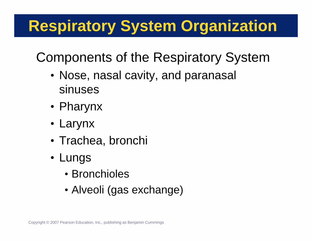
Respiratory System Organization
Components of the Respiratory System• Nose, nasal cavity, and paranasal
sinuses• Pharynx• Larynx• Trachea, bronchi• Lungs
• Bronchioles• Alveoli (gas exchange)
Copyright © 2007 Pearson Education, Inc., publishing as Benjamin Cummings

Respiratory System OrganizationThe Components of the Respiratory System
Figure 15-1

Respiratory System Organization
The Respiratory Tract• Conducting portion
• Conduct the air movement• From nares to small bronchioles
• Respiratory portion• Gas exchange region• Respiratory bronchioles and alveoli
The Respiratory TractCopyright © 2007 Pearson Education, Inc., publishing as Benjamin Cummings
PLAY

Respiratory System Organization
The Nose• External nares (nostrils) admit air
• Nasal vestibule lined with hairs to filter air• Vestibule opens into nasal cavity
• Hard palate separates nasal and oral cavities• Cavity continues through internal nares to
nasopharynx• Soft palate underlies nasopharynx
• Respiratory epithelium lines the airways
Copyright © 2007 Pearson Education, Inc., publishing as Benjamin Cummings
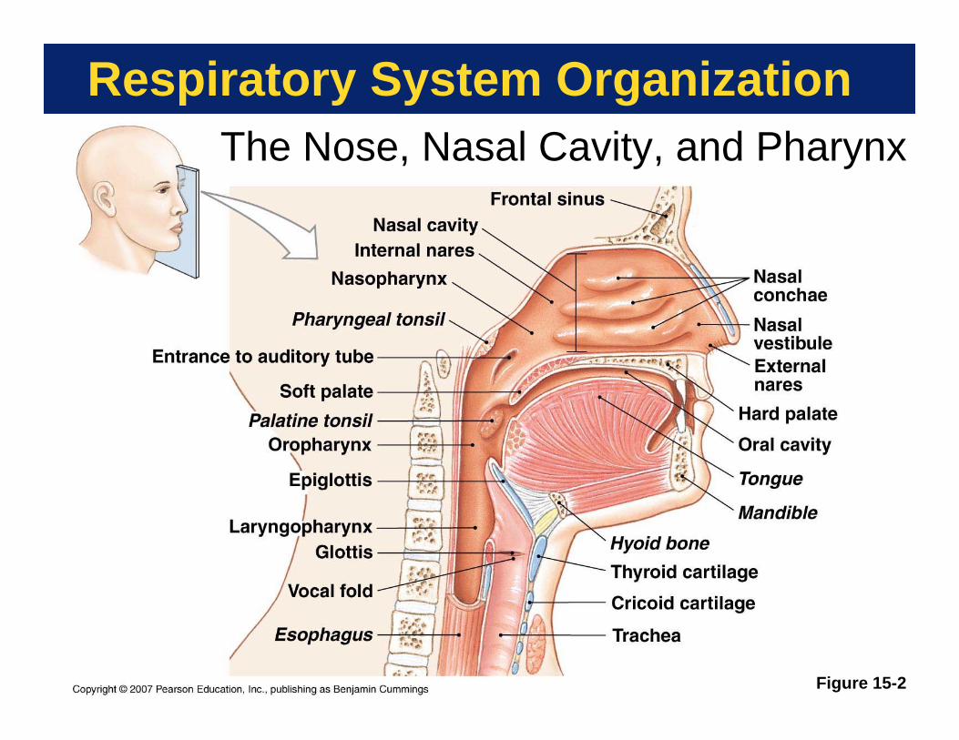
Respiratory System OrganizationThe Nose, Nasal Cavity, and Pharynx
Figure 15-2

Respiratory System Organization
Respiratory Mucosa• Respiratory epithelium plus supporting
connective tissue with mucous glands• Lines nasal cavity and most of airways• Goblet and gland cells secrete mucus• Mucus traps inhaled dirt, pathogens, etc.• Ciliated cells sweep the mucus out of
the airways into pharynx• Irritants stimulate secretion
• Causes “runny nose”
Copyright © 2007 Pearson Education, Inc., publishing as Benjamin Cummings
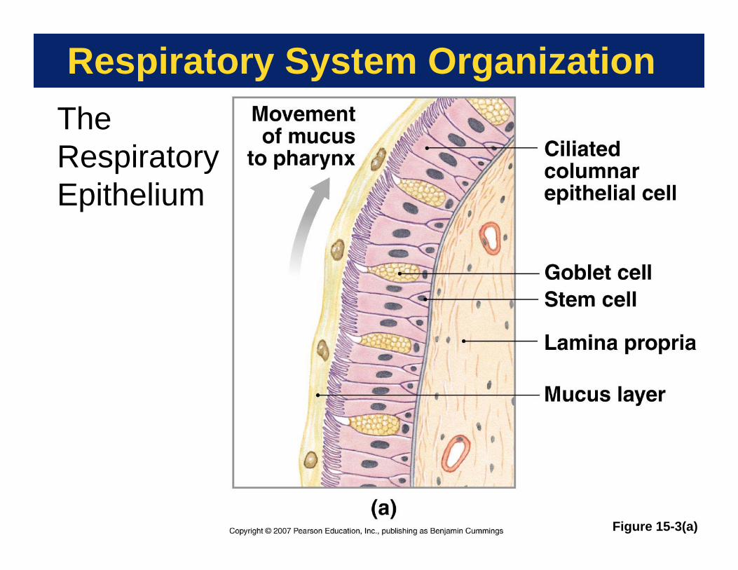
Respiratory System OrganizationThe Respiratory Epithelium
Figure 15-3(a)

Respiratory System Organization
Figure 15-3(b)
The Respiratory Epithelium

Respiratory System Organization
Three Regions of the Pharynx (Throat)• Respiratory system only
• Nasopharynx• Shared with digestive system
• Oropharynx• Opens into both esophagus
and larynx• Laryngopharynx
Copyright © 2007 Pearson Education, Inc., publishing as Benjamin Cummings

Respiratory System Organization
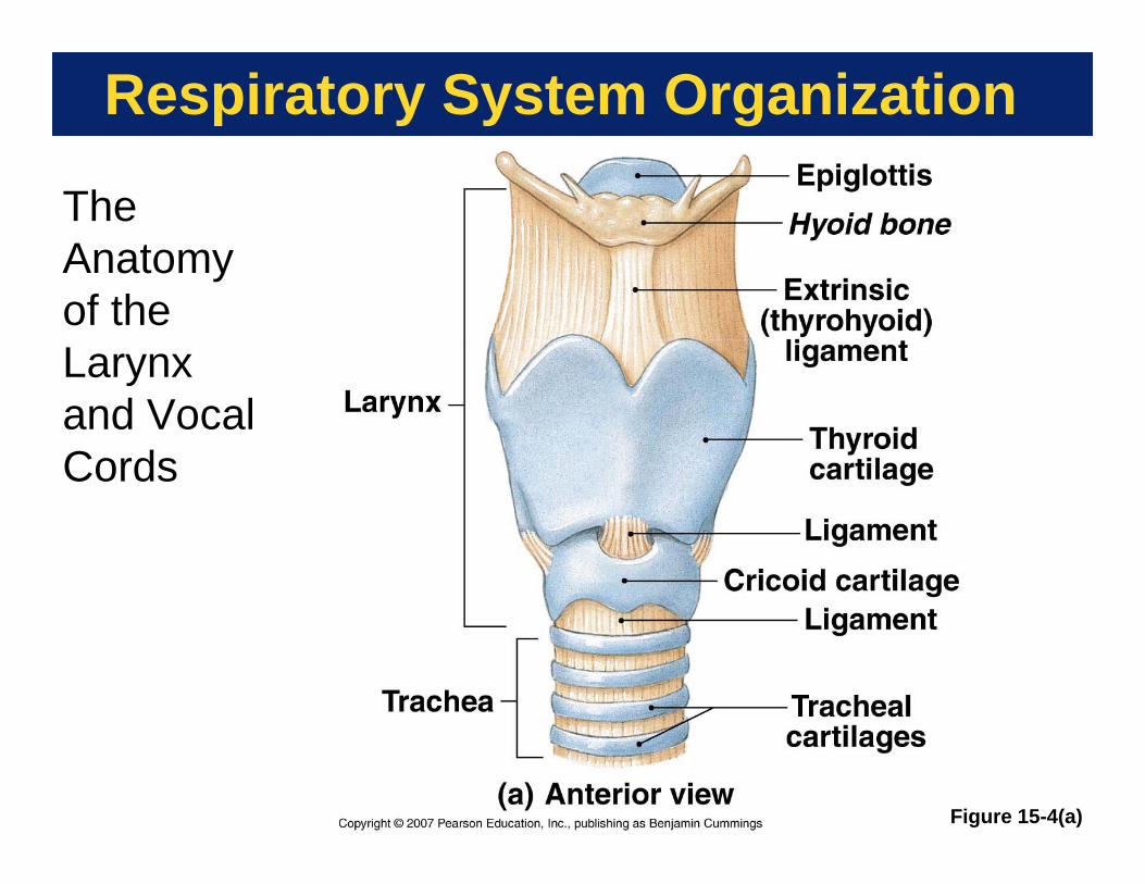
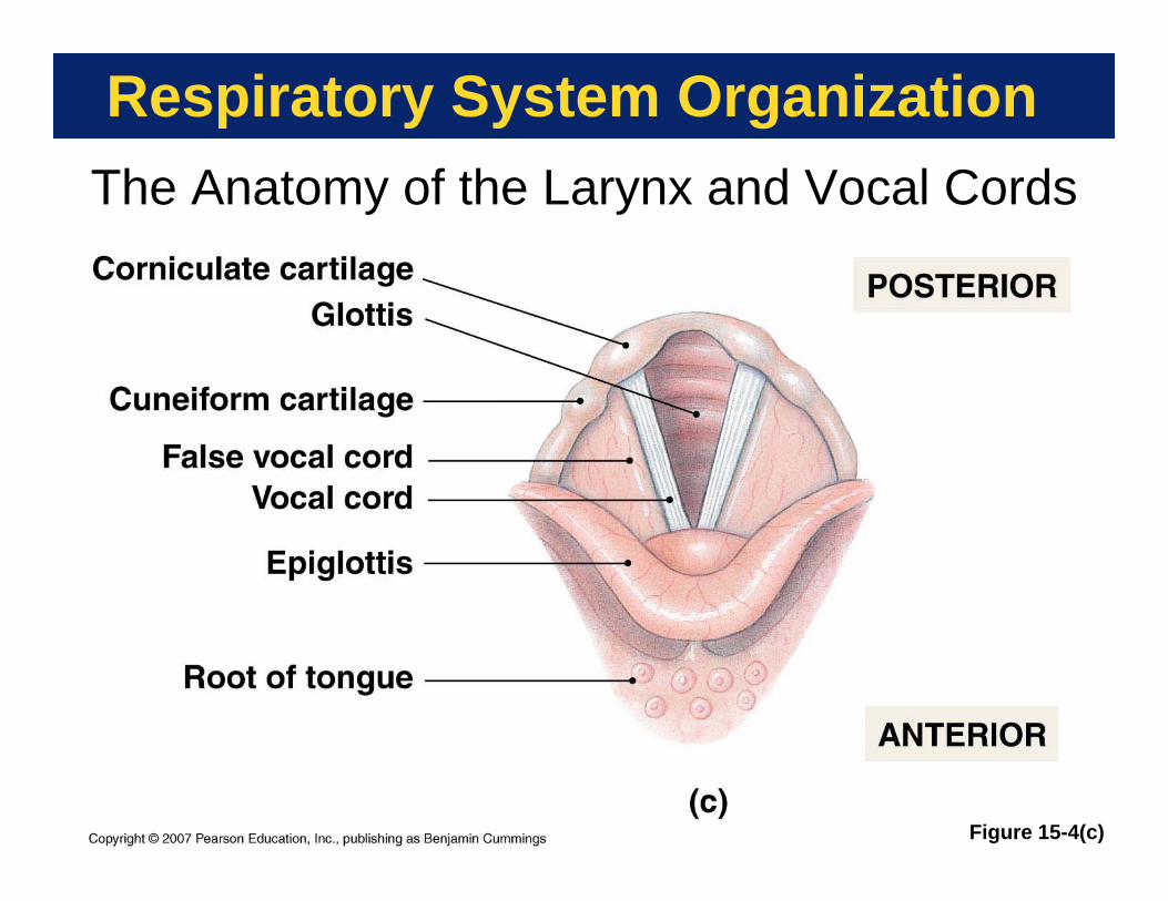
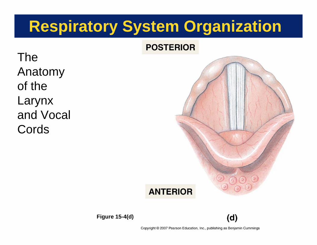
The Larynx• Also called, “voice box”• Made of nine cartilages• Air passes through glottis• Covered by epiglottis during swallowing
• Keeps solids, liquids out of airways• Made of elastic cartilage
• Supports true vocal cords• Exhaled air vibrates them to make sound
Copyright © 2007 Pearson Education, Inc., publishing as Benjamin Cummings
Respiratory System Organization
Figure 15-4(a)
The Anatomy of the Larynx and Vocal Cords

Respiratory System Organization
Figure 15-4(b)
The Anatomy of the Larynx and Vocal Cords
Respiratory System Organization
Figure 15-4(c)
The Anatomy of the Larynx and Vocal Cords
Respiratory System Organization
Figure 15-4(d)
The Anatomy of the Larynx and Vocal Cords

Respiratory System Organization
Figure 15-4(e)
The Anatomy of the Larynx and Vocal Cords

Respiratory System Organization
The Trachea• Also called “windpipe”• Stiffened by C-shaped cartilage rings• Esophagus stuck to posterior surface
• Cartilage missing there• Trachea distorted by balls of food as
they pass down esophagus to stomach
Copyright © 2007 Pearson Education, Inc., publishing as Benjamin Cummings
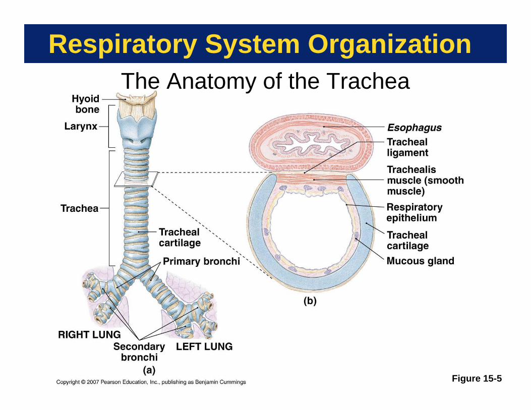
Respiratory System OrganizationThe Anatomy of the Trachea
Figure 15-5
Respiratory System Organization
The Bronchi• Trachea forms two branches
• Right and left primary bronchi• Primary bronchi branch
• Form secondary bronchi• Each ventilates a lobe
• Secondary bronchi branch• Form tertiary bronchi
• Tertiary bronchi branch repeatedly• Cartilage decreases, smooth
muscle increases
Copyright © 2007 Pearson Education, Inc., publishing as Benjamin Cummings
Respiratory System Organization
The Bronchioles• Cartilage absent• Diameter < 1.0 mm• Terminal bronchioles deliver air to a
single lobule• Smooth muscle in wall controlled by ANS
• Sympathetic causes bronchodilation• Parasympathetic causes
bronchoconstriction• Excess bronchoconstriction is asthma
Copyright © 2007 Pearson Education, Inc., publishing as Benjamin Cummings

Respiratory System Organization
The Bronchial Tree
Figure 15-6(a)

Respiratory System Organization
The Alveolar Ducts and Alveoli• Gas exchange regions of lung• Respiratory bronchioles lead into
alveolar ducts• Ducts lead into alveolar sacs• Sacs are clusters of
interconnected alveoli• Gives lung an open, spongy look• About 150 million/lung
Copyright © 2007 Pearson Education, Inc., publishing as Benjamin Cummings

Respiratory System OrganizationThe Lobules of the Lung
Figure 15-6(b)

Respiratory System OrganizationAlveolar Organization
Figure 15-7(a)

Respiratory System OrganizationAlveolar Organization
Figure 15-7(b)

Respiratory System Organization
Anatomy of the Alveolus Respiratory membrane
• Simple squamous epithelium• Capillary endothelium • Shared basement membrane
• Septal cells• Produce surfactant to reduce collapse
• Alveolar macrophages• Engulf foreign particles
Copyright © 2007 Pearson Education, Inc., publishing as Benjamin Cummings
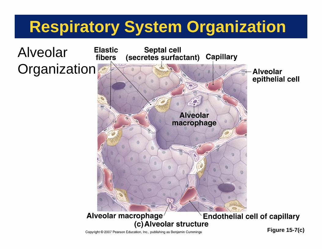
Respiratory System OrganizationAlveolar Organization
Figure 15-7(c)

Respiratory System Organization
Figure 15-7(d)
Alveolar Organization

Respiratory System Organization
Lung Gross Anatomy• Lungs comprise five lobes
• Separated by deep fissures• three lobes on right, two on left
• Apex extends above first rib• Base rests on diaphragm• Covered by a serous visceral pleura• Lie with pleural cavities
• Lined by a serous parietal pleura
Copyright © 2007 Pearson Education, Inc., publishing as Benjamin Cummings

Respiratory System OrganizationThe Gross Anatomy of the Lungs
Figure 15-8

Respiratory System Organization
PLAY
Anatomical Relationships in the Thoracic Cavity
Respiratory Movie Figure 15-9
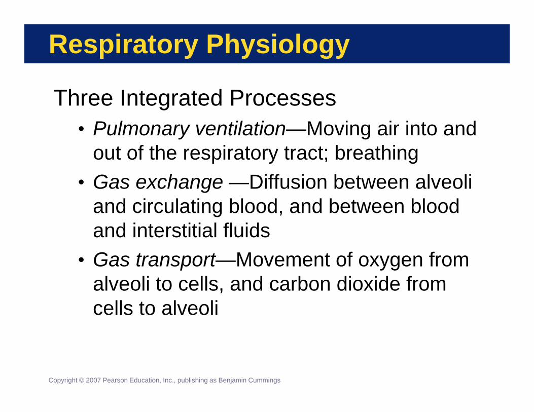
Respiratory Physiology
Three Integrated Processes• Pulmonary ventilation—Moving air into and
out of the respiratory tract; breathing• Gas exchange —Diffusion between alveoli
and circulating blood, and between blood and interstitial fluids
• Gas transport—Movement of oxygen from alveoli to cells, and carbon dioxide from cells to alveoli
Copyright © 2007 Pearson Education, Inc., publishing as Benjamin Cummings

Respiratory Physiology
Pulmonary Ventilation• Respiratory cycle—A single breath
consisting of inspiration (inhalation) and expiration (exhalation)
• Respiratory rate—Number of cycles per minute• Adult normal rate 12 to 18 breaths/minute• Child normal rate 18 to 20 breaths/minute
• Alveolar ventilation—Movement of air into and out of the alveoli
Copyright © 2007 Pearson Education, Inc., publishing as Benjamin Cummings

Respiratory Physiology
Key NoteThe direction of air flow is determined by the relationship of atmospheric pressure and pressure inside the respiratory tract. Flow is always from higher to lower pressure.
Copyright © 2007 Pearson Education, Inc., publishing as Benjamin Cummings

Respiratory Physiology
Quiet versus Forced Breathing• Quiet breathing—Diaphragm and external
intercostals are involved. Expiration is passive.
• Forced breathing—Accessory muscles become active during the entire breathing cycle. Expiration is active.
Copyright © 2007 Pearson Education, Inc., publishing as Benjamin Cummings

Respiratory PhysiologyPressure and Volume Relationships in the Lungs
Figure 15-10(a)

Copyright © 2007 Pearson Education, Inc., publishing as Benjamin CummingsFigure 15-10(b)1 of 4
Pleuralspace
Mediastinum
Diaphragm
Pressure outside andinside are equal, so no
movement occursPo = Pi
Volume increasesPressure inside falls,
and air flows inPo > Pi
Volume decreasesPressure inside rises,
so air flows outPo < Pi
Diaphragm
Externalintercostal
Serratus anterior
Pectoralis minor
Scalene muscles
Sternocleido-mastoid
TransversusthoracisInternalintercostals
Rectalabdominis(otherabdominalmusclesnot shown)
EXHALATIONINHALATIONAT REST

Copyright © 2007 Pearson Education, Inc., publishing as Benjamin CummingsFigure 15-10(b)2 of 4
Pleuralspace
Mediastinum
Diaphragm
Pressure outside andinside are equal, so no
movement occursPo = Pi
AT REST

Copyright © 2007 Pearson Education, Inc., publishing as Benjamin CummingsFigure 15-10(b)3 of 4
Pleuralspace
Mediastinum
Diaphragm
Pressure outside andinside are equal, so no
movement occursPo = Pi
Volume increasesPressure inside falls,
and air flows inPo > Pi
Diaphragm
Externalintercostal
Serratus anterior
Pectoralis minor
Scalene muscles
Sternocleido-mastoid
INHALATIONAT REST

Copyright © 2007 Pearson Education, Inc., publishing as Benjamin CummingsFigure 15-10(b)4 of 4
Pleuralspace
Mediastinum
Diaphragm
Pressure outside andinside are equal, so no
movement occursPo = Pi
Volume increasesPressure inside falls,
and air flows inPo > Pi
Volume decreasesPressure inside rises,
so air flows outPo < Pi
Diaphragm
Externalintercostal
Serratus anterior
Pectoralis minor
Scalene muscles
Sternocleido-mastoid
TransversusthoracisInternalintercostals
Rectalabdominis(otherabdominalmusclesnot shown)
EXHALATIONINHALATIONAT REST
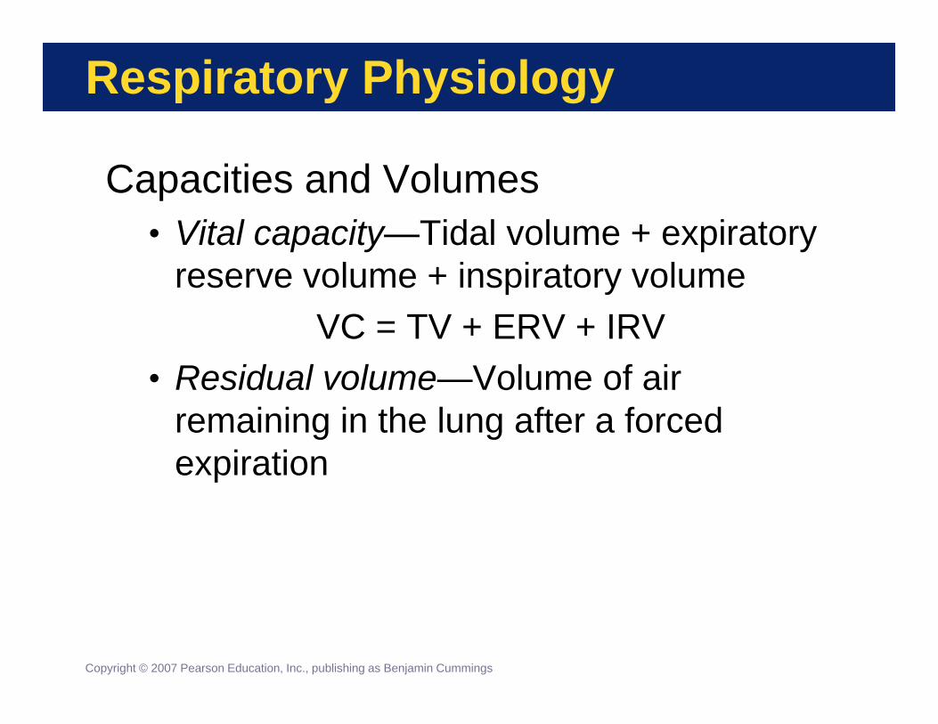
Respiratory Physiology
Capacities and Volumes• Vital capacity—Tidal volume + expiratory
reserve volume + inspiratory volumeVC = TV + ERV + IRV
• Residual volume—Volume of air remaining in the lung after a forced expiration
Copyright © 2007 Pearson Education, Inc., publishing as Benjamin Cummings
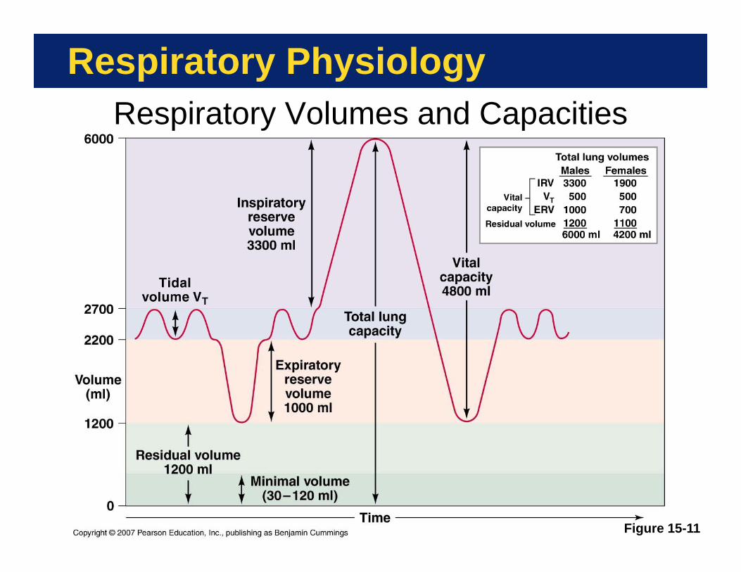
Respiratory PhysiologyRespiratory Volumes and Capacities
Figure 15-11

Respiratory Physiology
Gas Exchange• External respiration—Diffusion of gases
between alveolar air and pulmonary capillary blood across the respiratory membrane
• Internal respiration—Diffusion of gases between blood and interstitial fluids across the capillary endothelium
Copyright © 2007 Pearson Education, Inc., publishing as Benjamin Cummings
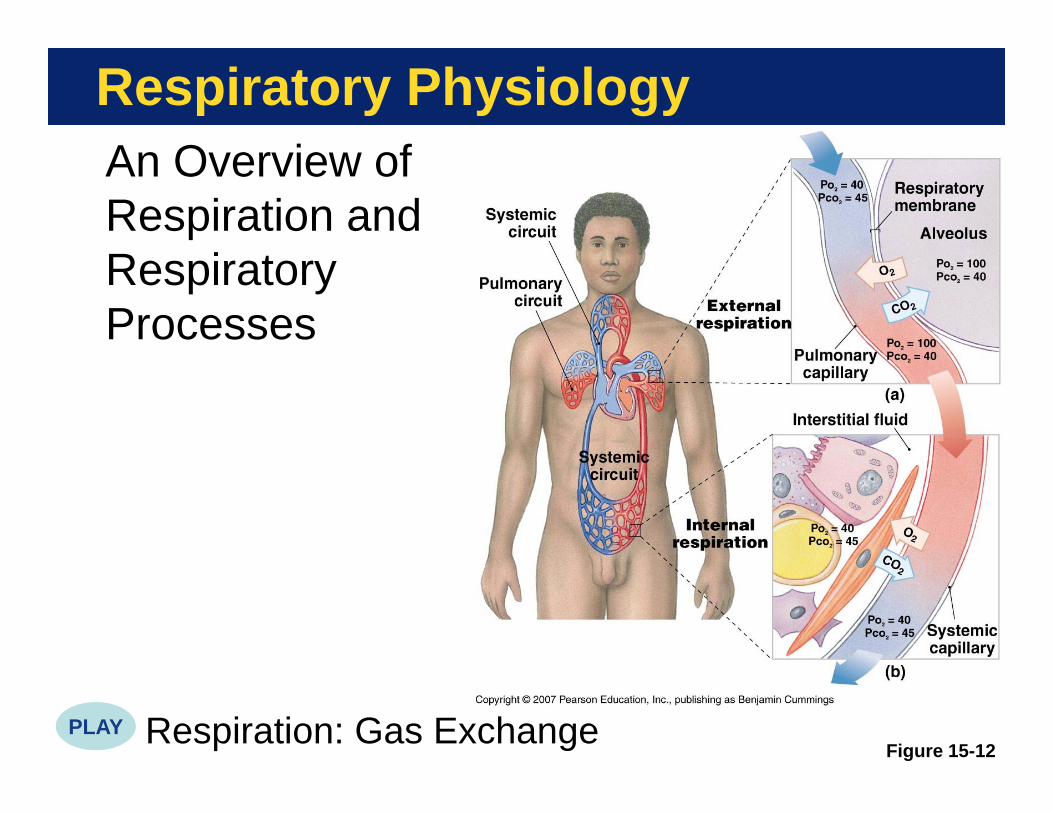
Respiratory PhysiologyAn Overview of Respiration and Respiratory Processes
Respiration: Gas ExchangeFigure 15-12
PLAY

Respiratory Physiology

Respiratory Physiology
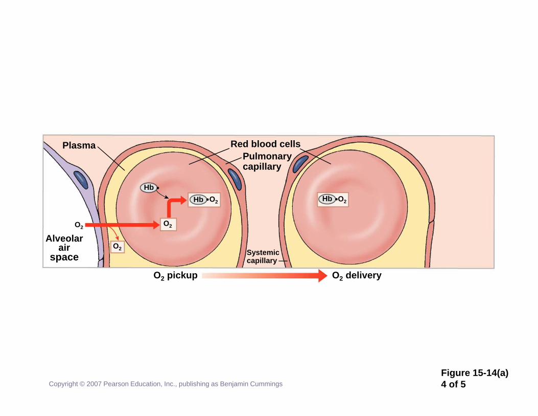
Gas Transport• Arterial blood entering peripheral
capillaries delivers oxygen and removes carbon dioxide
• Gas reactions with blood are completely reversible
• In general, a small change in plasma PO2 causes a large change in how much oxygen is bound to hemoglobin
Copyright © 2007 Pearson Education, Inc., publishing as Benjamin Cummings

Respiratory PhysiologyKey Note
Hemoglobin binds most of the oxygen in the bloodstream. If the PO2 in plasma increases, hemoglobin binds more oxygen; if PO2 decreases, hemoglobin releases oxygen. At a given PO2hemoglobin will release additional oxygen if the pH falls or the temperature rises.
Respiration: Carbon Dioxide and Oxygen ExchangeCopyright © 2007 Pearson Education, Inc., publishing as Benjamin Cummings
PLAY

Respiratory Physiology
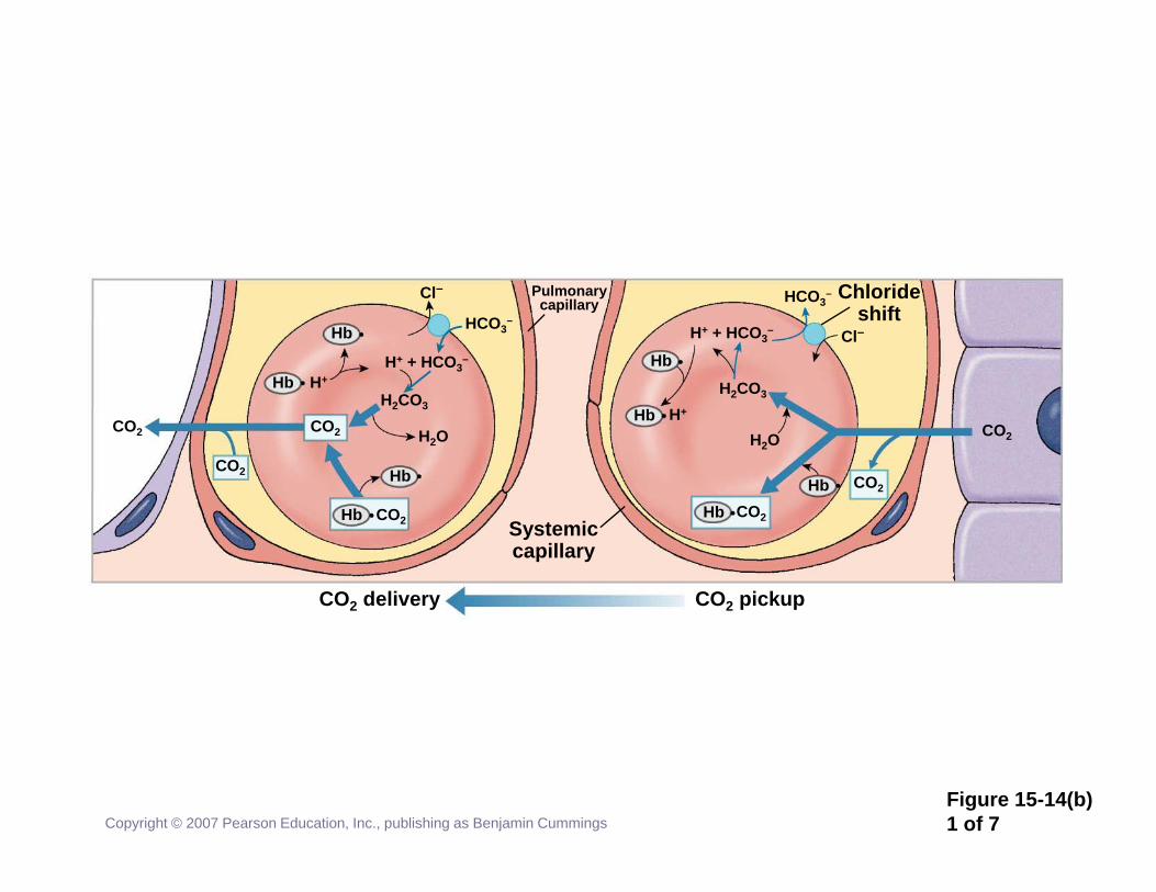
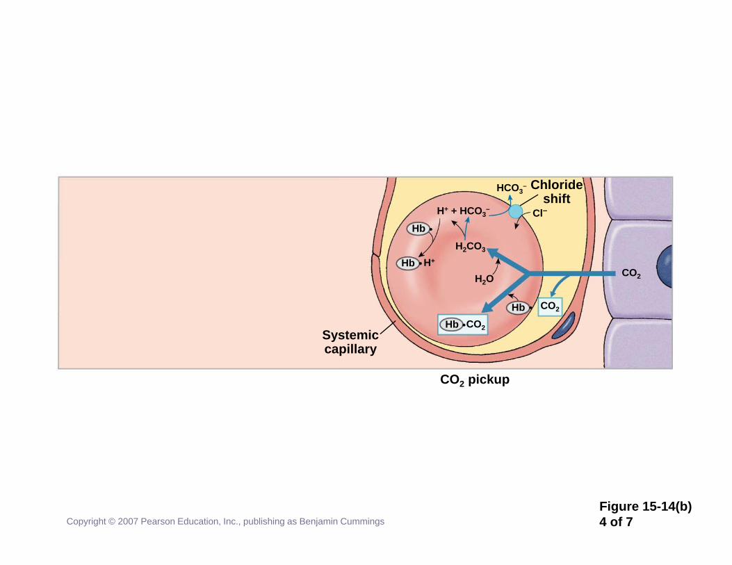
Carbon Dioxide Transport• Aerobic metabolism produces CO2
• 7% travels dissolved in plasma• 23% travels bound to hemoglobin
• Called carbaminohemoglobin• 70% is converted to H2CO3 in RBCs
• Catalyzed by carbonic anhydrase• Dissociates to H+ and HCO3
-
• HCO3- enters plasma from RBC
Copyright © 2007 Pearson Education, Inc., publishing as Benjamin Cummings
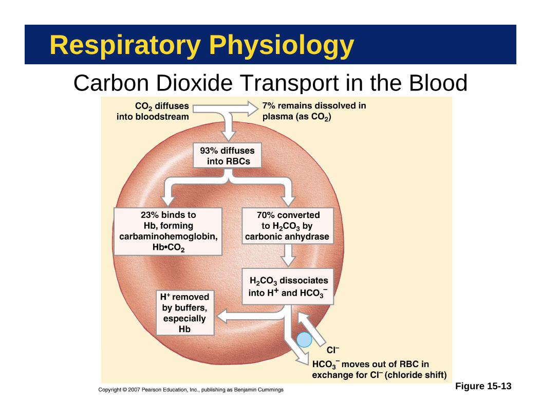
Respiratory PhysiologyCarbon Dioxide Transport in the Blood
Figure 15-13

Respiratory PhysiologyKey Note
Carbon dioxide (CO2) primarily travels in the bloodstream as bicarbonate ions (HCO3
-), which form through dissociation of the carbonic acid (H2CO3) produced by carbonic anhydrase inside RBCs. Lesser amounts of CO2 are bound to hemoglobin or dissolved in plasma.
Respiration: Pressure GradientsCopyright © 2007 Pearson Education, Inc., publishing as Benjamin Cummings
PLAY

Copyright © 2007 Pearson Education, Inc., publishing as Benjamin CummingsFigure 15-14(a)1 of 5
Plasma
Alveolarair
space
O2 pickup O2 delivery
Systemiccapillary
Red blood cellsPulmonarycapillary
Cells inperipheraltissues
O2
O2
O2
O2HbHb
Hb O2 O2
Hb O2
O2

Copyright © 2007 Pearson Education, Inc., publishing as Benjamin CummingsFigure 15-14(a)2 of 5
Plasma
Alveolarair
space
Red blood cellPulmonarycapillary
O2
O2
O2

Copyright © 2007 Pearson Education, Inc., publishing as Benjamin CummingsFigure 15-14(a)3 of 5
Plasma
Alveolarair
space
Red blood cellPulmonarycapillary
O2
O2
O2
O2HbHb
O2 pickup
Copyright © 2007 Pearson Education, Inc., publishing as Benjamin CummingsFigure 15-14(a)4 of 5
Plasma
Alveolarair
space
O2 pickup O2 delivery
Systemiccapillary
Red blood cellsPulmonarycapillary
O2
O2
O2
O2HbHb
Hb O2

Copyright © 2007 Pearson Education, Inc., publishing as Benjamin CummingsFigure 15-14(a)5 of 5
Plasma
Alveolarair
space
O2 pickup O2 delivery
Systemiccapillary
Red blood cellsPulmonarycapillary
Cells inperipheraltissues
O2
O2
O2
O2HbHb
Hb O2 O2
Hb O2
O2
Copyright © 2007 Pearson Education, Inc., publishing as Benjamin CummingsFigure 15-14(b)1 of 7
CO2 delivery
H+
Hb
CO2 pickup
Systemiccapillary
Pulmonarycapillary
Chlorideshift
CO2 CO2
CO2
CO2
Hb
Hb
Hb
H+ + HCO3–
HCO3–
Cl–
H2CO3
H2O
Hb
HCO3–
Cl–H+ + HCO3–
H2CO3
H2OH+Hb
CO2HbHb CO2
CO2

Copyright © 2007 Pearson Education, Inc., publishing as Benjamin CummingsFigure 15-14(b)2 of 7
CO2 pickup
Systemiccapillary
CO2
CO2

Copyright © 2007 Pearson Education, Inc., publishing as Benjamin CummingsFigure 15-14(b)3 of 7
CO2 pickup
Systemiccapillary
H2CO3
H2O
CO2HbHb CO2
CO2
Copyright © 2007 Pearson Education, Inc., publishing as Benjamin CummingsFigure 15-14(b)4 of 7
CO2 pickup
Systemiccapillary
Chlorideshift
Hb
HCO3–
Cl–H+ + HCO3–
H2CO3
H2OH+Hb
CO2HbHb CO2
CO2

Copyright © 2007 Pearson Education, Inc., publishing as Benjamin CummingsFigure 15-14(b)5 of 7
CO2 delivery CO2 pickup
Systemiccapillary
Pulmonarycapillary
Chlorideshift
CO2
CO2Hb
HCO3–
Cl–
Hb
HCO3–
Cl–H+ + HCO3–
H2CO3
H2OH+Hb
CO2HbHb CO2
CO2

Copyright © 2007 Pearson Education, Inc., publishing as Benjamin CummingsFigure 15-14(b)6 of 7
CO2 delivery
H+
Hb
CO2 pickup
Systemiccapillary
Pulmonarycapillary
Chlorideshift
CO2 CO2
CO2
CO2
Hb
Hb
H+ + HCO3–
HCO3–
Cl–
H2CO3
H2O
Hb
HCO3–
Cl–H+ + HCO3–
H2CO3
H2OH+Hb
CO2HbHb CO2
CO2

Copyright © 2007 Pearson Education, Inc., publishing as Benjamin CummingsFigure 15-14(b)7 of 7
CO2 delivery
H+
Hb
CO2 pickup
Systemiccapillary
Pulmonarycapillary
Chlorideshift
CO2 CO2
CO2
CO2
Hb
Hb
Hb
H+ + HCO3–
HCO3–
Cl–
H2CO3
H2O
Hb
HCO3–
Cl–H+ + HCO3–
H2CO3
H2OH+Hb
CO2HbHb CO2
CO2

The Control of Respiration
Meeting the Changing Demand for Oxygen• Requires integration cardiovascular and
respiratory responses• Depends on both:
• Local control of respiration• Control by brain respiratory centers
Copyright © 2007 Pearson Education, Inc., publishing as Benjamin Cummings
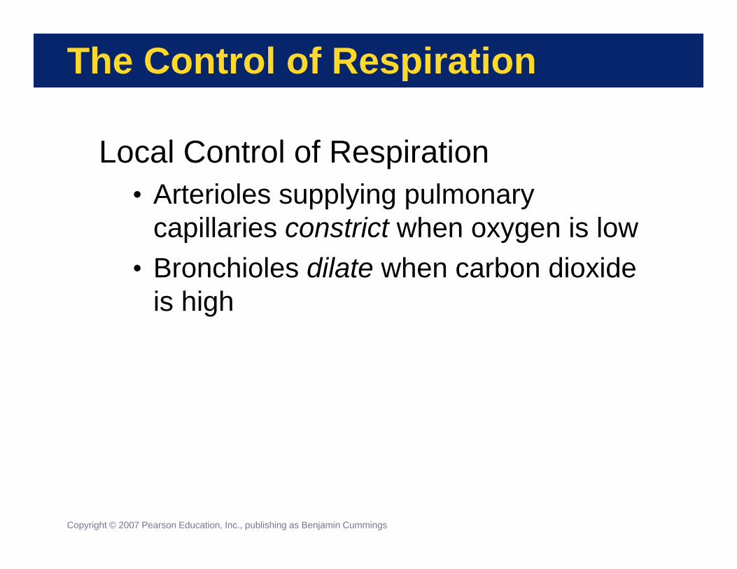
The Control of Respiration
Local Control of Respiration• Arterioles supplying pulmonary
capillaries constrict when oxygen is low• Bronchioles dilate when carbon dioxide
is high
Copyright © 2007 Pearson Education, Inc., publishing as Benjamin Cummings

The Control of Respiration
Control by Brain Respiratory Centers• Respiratory centers in brainstem
• Three pairs of nuclei• Two pairs in pons• One pair in medulla oblongata
• Control respiratory muscles• Set rate and depth of ventilation• Respiratory rhythmicity center in medulla
• Sets basic rhythm of breathing
Copyright © 2007 Pearson Education, Inc., publishing as Benjamin Cummings
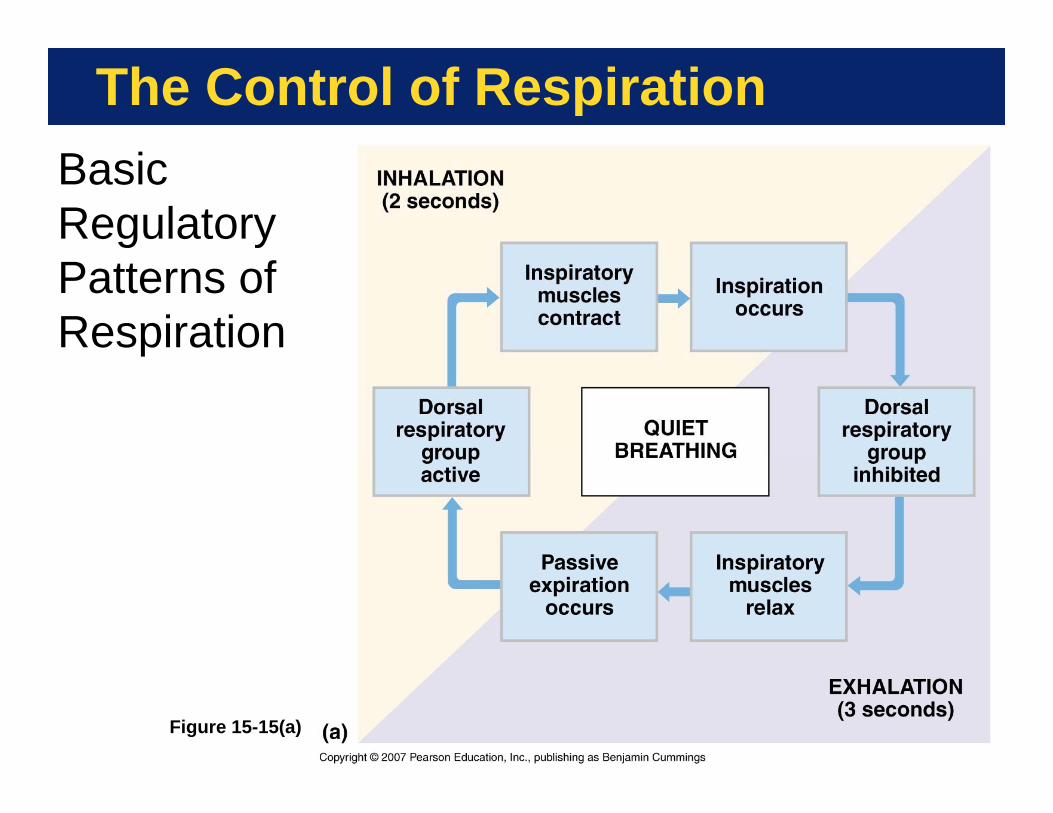
The Control of Respiration
Figure 15-15(a)
Basic Regulatory Patterns of Respiration

The Control of Respiration
Figure 15-15(b)
Basic Regulatory Patterns of Respiration
The Control of Respiration
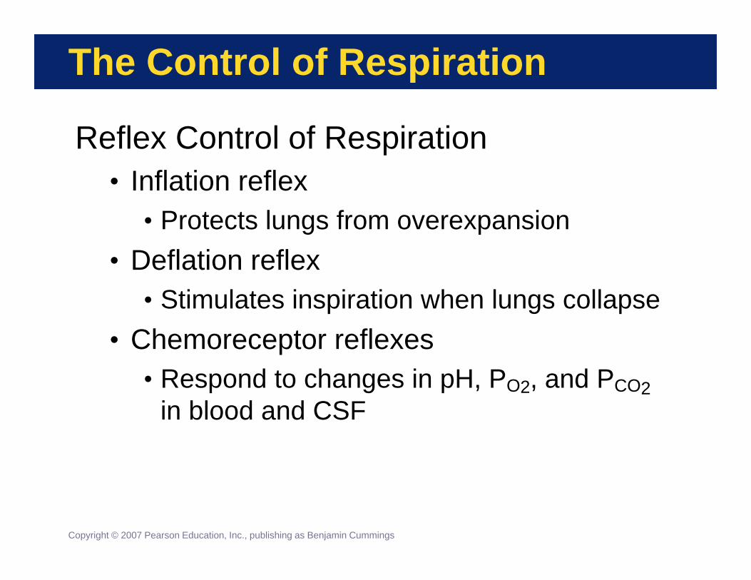
Reflex Control of Respiration• Inflation reflex
• Protects lungs from overexpansion• Deflation reflex
• Stimulates inspiration when lungs collapse• Chemoreceptor reflexes
• Respond to changes in pH, PO2, and PCO2in blood and CSF
Copyright © 2007 Pearson Education, Inc., publishing as Benjamin Cummings

The Control of Respiration
Control by Higher Centers• Exert effects on pons or on
respiratory motorneurons• Voluntary actions
• Speech, singing• Involuntary actions through the limbic
system• Rage, eating, sexual arousal
Copyright © 2007 Pearson Education, Inc., publishing as Benjamin Cummings
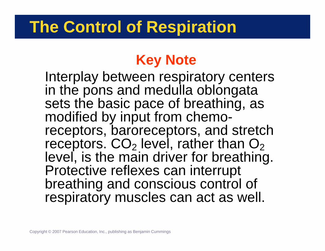
The Control of Respiration
Key NoteInterplay between respiratory centers in the pons and medulla oblongata sets the basic pace of breathing, as modified by input from chemo-receptors, baroreceptors, and stretch receptors. CO2 level, rather than O2level, is the main driver for breathing. Protective reflexes can interrupt breathing and conscious control of respiratory muscles can act as well.
Copyright © 2007 Pearson Education, Inc., publishing as Benjamin Cummings

The Control of Respiration
Figure 15-16
The Control of Respiration

Respiratory Changes at Birth
Conditions Before Birth• Pulmonary arterial resistance is high• Rib cage is compressed• Lungs are collapsed• Airways, alveoli are filled with fluid
Conditions After Birth• An heroic breath fills lungs with air,
displaces fluid, and opens alveoli• Surfactant stabilizes open alveoli
Copyright © 2007 Pearson Education, Inc., publishing as Benjamin Cummings
Respiratory System and Aging
Respiratory System Loses Efficiency• Elastic tissue deteriorates
• Lowers vital capacity• Rib cage movement restricted
• Arthritic changes• Costal cartilages loses flexibility
• Some emphysema usually appears
Copyright © 2007 Pearson Education, Inc., publishing as Benjamin Cummings

The Respiratory System in Perspective
FIGURE 15-17 Functional Relationships Between the Respiratory System and Other Systems
Figure 15-171 of 11Copyright © 2007 Pearson Education, Inc., publishing as Benjamin Cummings

Figure 15-172 of 11Copyright © 2007 Pearson Education, Inc., publishing as Benjamin Cummings
The Integumentary System
• Protects portions of upper respiratory tract; hairs guard entry to external nares
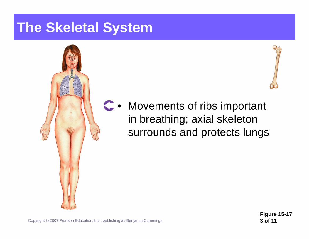
Figure 15-173 of 11Copyright © 2007 Pearson Education, Inc., publishing as Benjamin Cummings
The Skeletal System
• Movements of ribs important in breathing; axial skeleton surrounds and protects lungs
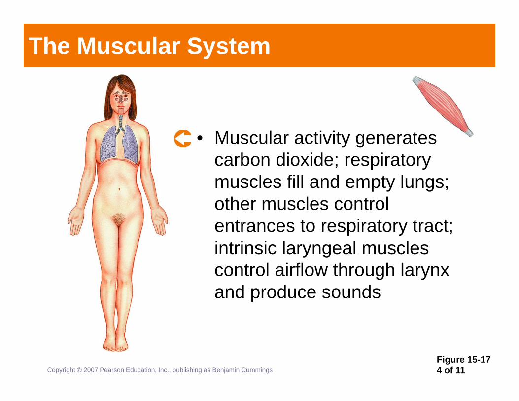
Figure 15-174 of 11Copyright © 2007 Pearson Education, Inc., publishing as Benjamin Cummings
The Muscular System
• Muscular activity generates carbon dioxide; respiratory muscles fill and empty lungs; other muscles control entrances to respiratory tract; intrinsic laryngeal muscles control airflow through larynx and produce sounds

Figure 15-175 of 11Copyright © 2007 Pearson Education, Inc., publishing as Benjamin Cummings
The Nervous System
• Monitors respiratory volume and blood gas levels; controls pace and depth of respiration

Figure 15-176 of 11Copyright © 2007 Pearson Education, Inc., publishing as Benjamin Cummings
The Endocrine System
• Epinephrine and norepinephrine stimulate respiratory activity and dilate respiratory passageways
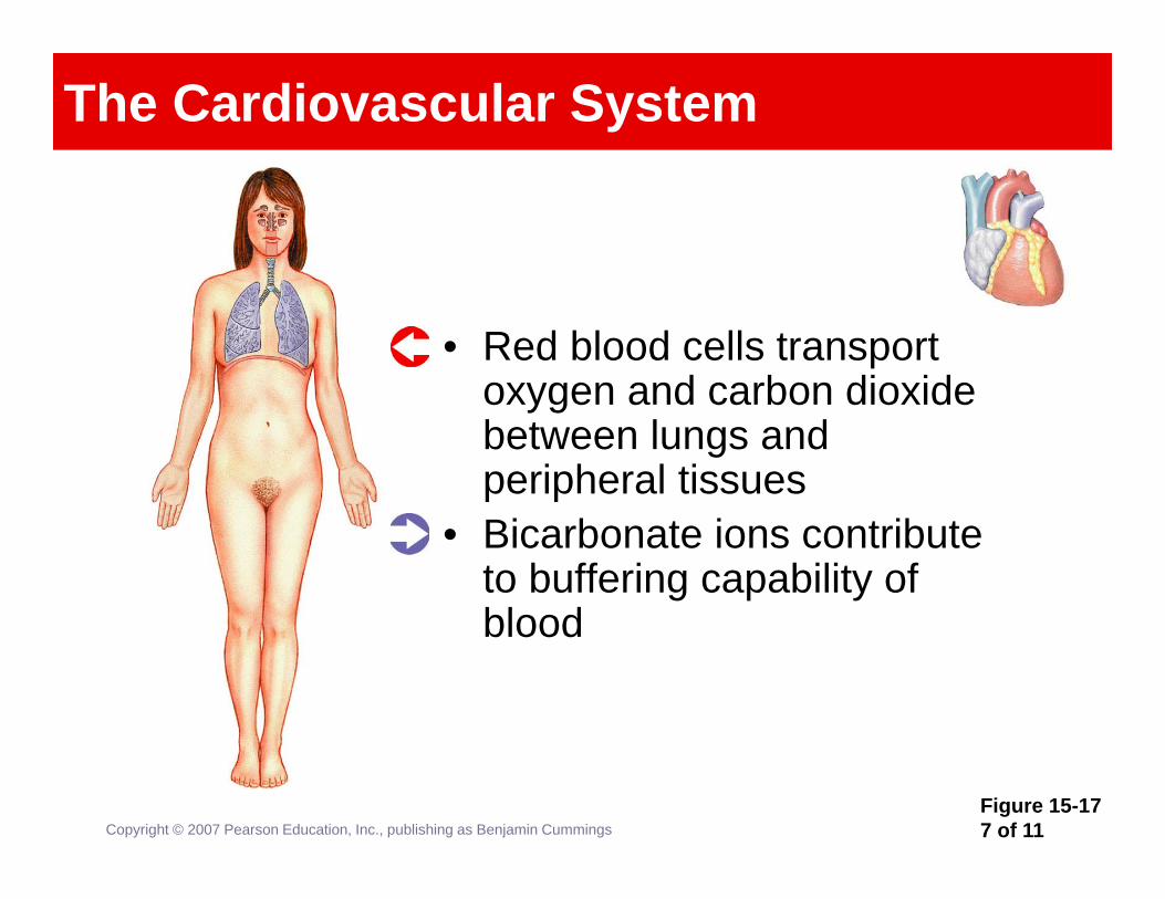
Figure 15-177 of 11Copyright © 2007 Pearson Education, Inc., publishing as Benjamin Cummings
The Cardiovascular System
• Red blood cells transport oxygen and carbon dioxide between lungs and peripheral tissues
• Bicarbonate ions contribute to buffering capability of blood

Figure 15-178 of 11Copyright © 2007 Pearson Education, Inc., publishing as Benjamin Cummings
The Lymphatic System
• Tonsils protect against infection at entrance to respiratory tract; lymphatic vessels monitor lymph drainage from lungs and mobilize specific defenses when infection occurs
• Alveolar phagocytes present antigens to trigger specific defenses; mucous membrane lining the nasal cavity and upper pharynx traps pathogens, protects deeper tissues

Figure 15-179 of 11Copyright © 2007 Pearson Education, Inc., publishing as Benjamin Cummings
The Digestive System
• Provides substrates, vitamins, water, and ions that are necessary to all cells of the respiratory system
• Increased thoracic and abdominal pressure through contraction of respiratory muscles can assist in defecation

Figure 15-1710 of 11Copyright © 2007 Pearson Education, Inc., publishing as Benjamin Cummings
The Urinary System
• Eliminates organic wastes generated by cells of the respiratory system; maintains normal fluid and ion balance in the blood
• Assists in the regulation of pH by eliminating carbon dioxide
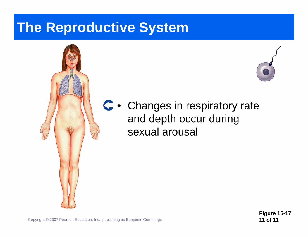
Figure 15-1711 of 11Copyright © 2007 Pearson Education, Inc., publishing as Benjamin Cummings
The Reproductive System
• Changes in respiratory rate and depth occur during sexual arousal