The receptor tyrosine kinase ErbB3 maintains the … · The receptor tyrosine kinase ErbB3...
6
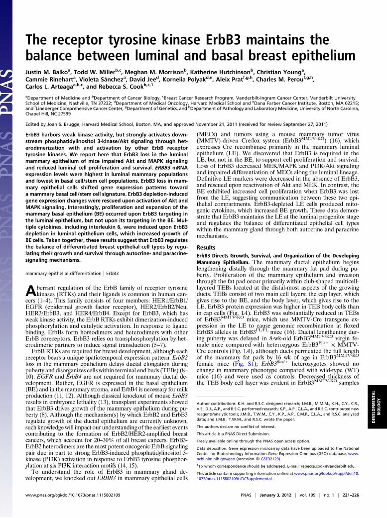
The receptor tyrosine kinase ErbB3 maintains the balance between luminal and basal breast epithelium Justin M. Balko a , Todd W. Miller b,c , Meghan M. Morrison b , Katherine Hutchinson b , Christian Young a , Cammie Rinehart a , Violeta Sánchez a , David Jee d , Kornelia Polyak d,e , Aleix Prat f,g,h , Charles M. Perou f,g,h , Carlos L. Arteaga a,b,c , and Rebecca S. Cook b,c,1 a Department of Medicine and b Department of Cancer Biology, c Breast Cancer Research Program, Vanderbilt-Ingram Cancer Center, Vanderbilt University School of Medicine, Nashville, TN 37232; d Department of Medical Oncology, Harvard Medical School and e Dana Farber Cancer Institute, Boston, MA 02215; and f Lineberger Comprehensive Cancer Center, g Department of Genetics, and h Department of Pathology and Laboratory Medicine, University of North Carolina, Chapel Hill, NC 27599 Edited by Joan S. Brugge, Harvard Medical School, Boston, MA, and approved November 21, 2011 (received for review September 27, 2011) ErbB3 harbors weak kinase activity, but strongly activates down- stream phosphatidylinositol 3-kinase/Akt signaling through het- erodimerization with and activation by other ErbB receptor tyrosine kinases. We report here that ErbB3 loss in the luminal mammary epithelium of mice impaired Akt and MAPK signaling and reduced luminal cell proliferation and survival. ERBB3 mRNA expression levels were highest in luminal mammary populations and lowest in basal cell/stem cell populations. ErbB3 loss in mam- mary epithelial cells shifted gene expression patterns toward a mammary basal cell/stem cell signature. ErbB3 depletion-induced gene expression changes were rescued upon activation of Akt and MAPK signaling. Interestingly, proliferation and expansion of the mammary basal epithelium (BE) occurred upon ErbB3 targeting in the luminal epithelium, but not upon its targeting in the BE. Mul- tiple cytokines, including interleukin 6, were induced upon ErbB3 depletion in luminal epithelium cells, which increased growth of BE cells. Taken together, these results suggest that ErbB3 regulates the balance of differentiated breast epithelial cell types by regu- lating their growth and survival through autocrine- and paracrine- signaling mechanisms. mammary epithelial differentiation | ErbB3 A berrant regulation of the ErbB family of receptor tyrosine kinases (RTKs) and their ligands is common in human can- cers (1–4). This family consists of four members: HER1/ErbB1/ EGFR (epidermal growth factor receptor), HER2/ErbB2/Neu, HER3/ErbB3, and HER4/ErbB4. Except for ErbB3, which has weak kinase activity, the ErbB RTKs exhibit dimerization-induced phosphorylation and catalytic activation. In response to ligand binding, ErbBs form homodimers and heterodimers with other ErbB coreceptors. ErbB3 relies on transphosphorylation by het- erodimeric partners to induce signal transduction (5–7). ErbB RTKs are required for breast development, although each receptor bears a unique spatiotemporal expression pattern. ErbB2 loss in the mammary epithelium delays ductal elongation during puberty and disorganizes cells within terminal end buds (TEBs) (8– 10). EGFR and ErbB4 are not required for mammary ductal de- velopment. Rather, EGFR is expressed in the basal epithelium (BE) and in the mammary stroma, and ErbB4 is necessary for milk production (11, 12). Although classical knockout of mouse ErbB3 results in embryonic lethality (13), transplant experiments showed that ErbB3 drives growth of the mammary epithelium during pu- berty (8). Although the mechanism(s) by which ErbB2 and ErbB3 regulate growth of the ductal epithelium are currently unknown, such knowledge will impact our understanding of the earliest events contributing to the formation of ErbB2/HER2-amplified breast cancers, which account for 20–30% of all breast cancers. ErbB3- ErbB2 heterodimers are the most potent oncogenic ErbB-signaling pair due in part to strong ErbB3-induced phosphatidylinositol 3- kinase (PI3K) activation in response to ErbB3 tyrosine phosphor- ylation at six PI3K interaction motifs (14, 15). To understand the role of ErbB3 in mammary gland de- velopment, we knocked out ERBB3 in mammary epithelial cells (MECs) and tumors using a mouse mammary tumor virus (MMTV)-driven Cre/lox system (ErbB3 MMTV-KO ) (16), which expresses Cre recombinase primarily in the mammary luminal epithelium (LE). We discovered that ErbB3 is required in the LE, but not in the BE, to support cell proliferation and survival. Loss of ErbB3 decreased MEK/MAPK and PI3K/Akt signaling and impaired differentiation of MECs along the luminal lineage. Definitive LE markers were decreased in the absence of ErbB3, and rescued upon reactivation of Akt and MEK. In contrast, the BE exhibited increased cell proliferation when ErbB3 was lost from the LE, suggesting communication between these two epi- thelial compartments. ErbB3-depleted LE cells produced mito- genic cytokines, which increased BE growth. These data demon- strate that ErbB3 maintains the LE at the luminal progenitor stage and regulates the balance of differentiated epithelial cell types within the mammary gland through both autocrine and paracrine mechanisms. Results ErbB3 Directs Growth, Survival, and Organization of the Developing Mammary Epithelium. The mammary ductal epithelium begins lengthening distally through the mammary fat pad during pu- berty. Proliferation of the mammary epithelium and invasion through the fat pad occur primarily within club-shaped multicell- layered TEBs located at the distal-most aspects of the growing ducts. TEBs consist of two main cell layers: the cap layer, which gives rise to the BE, and the body layer, which gives rise to the LE. ErbB3 protein expression was higher in TEB body cells than in cap cells (Fig. 1A). ErbB3 was substantially reduced in TEBs of ErbB3 MMTV-KO mice, which use MMTV-Cre transgene ex- pression in the LE to cause genomic recombination at floxed ErbB3 alleles in ErbB3 FL/FL mice (16). Ductal lengthening dur- ing puberty was delayed in 8-wk-old ErbB3 MMTV-KO virgin fe- male mice compared with heterozygous ErbB3 FL/+ × MMTV- Cre controls (Fig. 1A), although ducts permeated the full length of the mammary fat pads by 16 wk of age in ErbB3 MMTV-KO female mice (Fig. S1); ErbB3 flox/+ heterozygotes showed no change in mammary phenotype compared with wild-type (WT) mice (16) and were used as controls. Decreased thickness of the TEB body cell layer was evident in ErbB3 MMTV-KO samples Author contributions: K.H. and R.S.C. designed research; J.M.B., M.M.M., K.H., C.Y., C.R., V.S., D.J., A.P., and R.S.C. performed research; K.P., A.P., C.L.A., and R.S.C. contributed new reagents/analytic tools; J.M.B., T.W.M., C.Y., K.P., A.P., C.M.P., C.L.A., and R.S.C. analyzed data; and J.M.B., T.W.M., and R.S.C. wrote the paper. The authors declare no conflict of interest. This article is a PNAS Direct Submission. Freely available online through the PNAS open access option. Data deposition: Gene expression microarray data have been uploaded to the National Center for Biotechnology Information Gene Expression Omnibus (GEO) database, www. ncbi.nlm.nih.gov/geo (accession ID GSE32129). 1 To whom correspondence should be addressed. E-mail: [email protected]. This article contains supporting information online at www.pnas.org/lookup/suppl/doi:10. 1073/pnas.1115802109/-/DCSupplemental. www.pnas.org/cgi/doi/10.1073/pnas.1115802109 PNAS | January 3, 2012 | vol. 109 | no. 1 | 221–226 DEVELOPMENTAL BIOLOGY
Transcript of The receptor tyrosine kinase ErbB3 maintains the … · The receptor tyrosine kinase ErbB3...
The receptor tyrosine kinase ErbB3 maintains thebalance between luminal and basal breast epitheliumJustin M. Balkoa, Todd W. Millerb,c, Meghan M. Morrisonb, Katherine Hutchinsonb, Christian Younga,Cammie Rineharta, Violeta Sáncheza, David Jeed, Kornelia Polyakd,e, Aleix Pratf,g,h, Charles M. Perouf,g,h,Carlos L. Arteagaa,b,c, and Rebecca S. Cookb,c,1
aDepartment of Medicine and bDepartment of Cancer Biology, cBreast Cancer Research Program, Vanderbilt-Ingram Cancer Center, Vanderbilt UniversitySchool of Medicine, Nashville, TN 37232; dDepartment of Medical Oncology, Harvard Medical School and eDana Farber Cancer Institute, Boston, MA 02215;and fLineberger Comprehensive Cancer Center, gDepartment of Genetics, and hDepartment of Pathology and Laboratory Medicine, University of North Carolina,Chapel Hill, NC 27599
Edited by Joan S. Brugge, Harvard Medical School, Boston, MA, and approved November 21, 2011 (received for review September 27, 2011)
ErbB3 harbors weak kinase activity, but strongly activates down-stream phosphatidylinositol 3-kinase/Akt signaling through het-erodimerization with and activation by other ErbB receptortyrosine kinases. We report here that ErbB3 loss in the luminalmammary epithelium of mice impaired Akt and MAPK signalingand reduced luminal cell proliferation and survival. ERBB3 mRNAexpression levels were highest in luminal mammary populationsand lowest in basal cell/stem cell populations. ErbB3 loss in mam-mary epithelial cells shifted gene expression patterns towarda mammary basal cell/stem cell signature. ErbB3 depletion-inducedgene expression changes were rescued upon activation of Akt andMAPK signaling. Interestingly, proliferation and expansion of themammary basal epithelium (BE) occurred upon ErbB3 targeting inthe luminal epithelium, but not upon its targeting in the BE. Mul-tiple cytokines, including interleukin 6, were induced upon ErbB3depletion in luminal epithelium cells, which increased growth ofBE cells. Taken together, these results suggest that ErbB3 regulatesthe balance of differentiated breast epithelial cell types by regu-lating their growth and survival through autocrine- and paracrine-signaling mechanisms.
mammary epithelial differentiation | ErbB3
Aberrant regulation of the ErbB family of receptor tyrosinekinases (RTKs) and their ligands is common in human can-
cers (1–4). This family consists of four members: HER1/ErbB1/EGFR (epidermal growth factor receptor), HER2/ErbB2/Neu,HER3/ErbB3, and HER4/ErbB4. Except for ErbB3, which hasweak kinase activity, the ErbB RTKs exhibit dimerization-inducedphosphorylation and catalytic activation. In response to ligandbinding, ErbBs form homodimers and heterodimers with otherErbB coreceptors. ErbB3 relies on transphosphorylation by het-erodimeric partners to induce signal transduction (5–7).ErbB RTKs are required for breast development, although each
receptor bears a unique spatiotemporal expression pattern. ErbB2loss in the mammary epithelium delays ductal elongation duringpuberty and disorganizes cells within terminal end buds (TEBs) (8–10). EGFR and ErbB4 are not required for mammary ductal de-velopment. Rather, EGFR is expressed in the basal epithelium(BE) and in the mammary stroma, and ErbB4 is necessary for milkproduction (11, 12). Although classical knockout of mouse ErbB3results in embryonic lethality (13), transplant experiments showedthat ErbB3 drives growth of the mammary epithelium during pu-berty (8). Although the mechanism(s) by which ErbB2 and ErbB3regulate growth of the ductal epithelium are currently unknown,such knowledgewill impact our understanding of the earliest eventscontributing to the formation of ErbB2/HER2-amplified breastcancers, which account for 20–30% of all breast cancers. ErbB3-ErbB2 heterodimers are the most potent oncogenic ErbB-signalingpair due in part to strong ErbB3-induced phosphatidylinositol 3-kinase (PI3K) activation in response to ErbB3 tyrosine phosphor-ylation at six PI3K interaction motifs (14, 15).To understand the role of ErbB3 in mammary gland de-
velopment, we knocked out ERBB3 in mammary epithelial cells
(MECs) and tumors using a mouse mammary tumor virus(MMTV)-driven Cre/lox system (ErbB3MMTV-KO) (16), whichexpresses Cre recombinase primarily in the mammary luminalepithelium (LE). We discovered that ErbB3 is required in theLE, but not in the BE, to support cell proliferation and survival.Loss of ErbB3 decreased MEK/MAPK and PI3K/Akt signalingand impaired differentiation of MECs along the luminal lineage.Definitive LE markers were decreased in the absence of ErbB3,and rescued upon reactivation of Akt and MEK. In contrast, theBE exhibited increased cell proliferation when ErbB3 was lostfrom the LE, suggesting communication between these two epi-thelial compartments. ErbB3-depleted LE cells produced mito-genic cytokines, which increased BE growth. These data demon-strate that ErbB3 maintains the LE at the luminal progenitor stageand regulates the balance of differentiated epithelial cell typeswithin the mammary gland through both autocrine and paracrinemechanisms.
ResultsErbB3 Directs Growth, Survival, and Organization of the DevelopingMammary Epithelium. The mammary ductal epithelium beginslengthening distally through the mammary fat pad during pu-berty. Proliferation of the mammary epithelium and invasionthrough the fat pad occur primarily within club-shaped multicell-layered TEBs located at the distal-most aspects of the growingducts. TEBs consist of two main cell layers: the cap layer, whichgives rise to the BE, and the body layer, which gives rise to theLE. ErbB3 protein expression was higher in TEB body cells thanin cap cells (Fig. 1A). ErbB3 was substantially reduced in TEBsof ErbB3MMTV-KO mice, which use MMTV-Cre transgene ex-pression in the LE to cause genomic recombination at floxedErbB3 alleles in ErbB3FL/FL mice (16). Ductal lengthening dur-ing puberty was delayed in 8-wk-old ErbB3MMTV-KO virgin fe-male mice compared with heterozygous ErbB3FL/+ × MMTV-Cre controls (Fig. 1A), although ducts permeated the full lengthof the mammary fat pads by 16 wk of age in ErbB3MMTV-KO
female mice (Fig. S1); ErbB3flox/+ heterozygotes showed nochange in mammary phenotype compared with wild-type (WT)mice (16) and were used as controls. Decreased thickness ofthe TEB body cell layer was evident in ErbB3MMTV-KO samples
Author contributions: K.H. and R.S.C. designed research; J.M.B., M.M.M., K.H., C.Y., C.R.,V.S., D.J., A.P., and R.S.C. performed research; K.P., A.P., C.L.A., and R.S.C. contributed newreagents/analytic tools; J.M.B., T.W.M., C.Y., K.P., A.P., C.M.P., C.L.A., and R.S.C. analyzeddata; and J.M.B., T.W.M., and R.S.C. wrote the paper.
The authors declare no conflict of interest.
This article is a PNAS Direct Submission.
Freely available online through the PNAS open access option.
Data deposition: Gene expression microarray data have been uploaded to the NationalCenter for Biotechnology Information Gene Expression Omnibus (GEO) database, www.ncbi.nlm.nih.gov/geo (accession ID GSE32129).1To whom correspondence should be addressed. E-mail: [email protected].
This article contains supporting information online at www.pnas.org/lookup/suppl/doi:10.1073/pnas.1115802109/-/DCSupplemental.
www.pnas.org/cgi/doi/10.1073/pnas.1115802109 PNAS | January 3, 2012 | vol. 109 | no. 1 | 221–226
DEV
ELOPM
ENTA
LBIOLO
GY
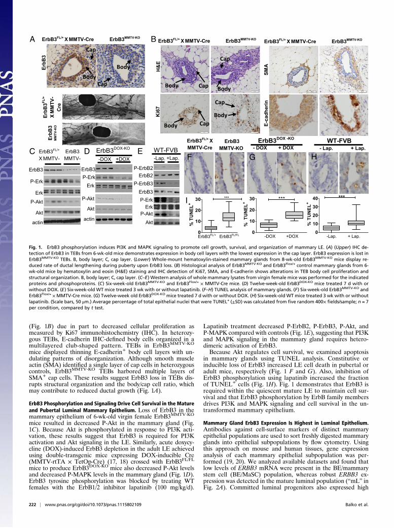
(Fig. 1B) due in part to decreased cellular proliferation asmeasured by Ki67 immunohistochemistry (IHC). In heterozy-gous TEBs, E-cadherin IHC-defined body cells organized in amultilayered club-shaped pattern. TEBs in ErbB3MMTV-KO
mice displayed thinning E-cadherin+ body cell layers with un-dulating patterns of disorganization. Although smooth muscleactin (SMA) identified a single layer of cap cells in heterozygouscontrols, ErbB3MMTV-KO TEBs harbored multiple layers ofSMA+ cap cells. These results suggest ErbB3 loss in TEBs dis-rupts structural organization and the body/cap cell ratio, whichmay contribute to reduced ductal growth (Fig. 1A).
ErbB3 Phosphorylation and Signaling Drive Cell Survival in the Matureand Pubertal Luminal Mammary Epithelium. Loss of ErbB3 in themammary epithelium of 6-wk-old virgin female ErbB3MMTV-KO
mice resulted in decreased P-Akt in the mammary gland (Fig.1C). Because Akt is phosphorylated in response to PI3K acti-vation, these results suggest that ErbB3 is required for PI3Kactivation and Akt signaling in the LE. Similarly, acute doxycy-cline (DOX)-induced ErbB3 depletion in the adult LE achievedusing double-transgenic mice expressing DOX-inducible Cre(MMTV-rtTA × TetOp-Cre) (17, 18) crossed with ErbB3FL/FL
mice to produce ErbB3DOX-KO mice also decreased P-Akt levelsand decreased P-MAPK levels in the mammary gland (Fig. 1D).ErbB3 tyrosine phosphorylation was blocked by treating WTfemales with the ErbB1/2 inhibitor lapatinib (100 mg/kg/d).
Lapatinib treatment decreased P-ErbB2, P-ErbB3, P-Akt, andP-MAPK compared with controls (Fig. 1E), suggesting that PI3Kand MAPK signaling in the mammary gland requires hetero-dimeric activation of ErbB3.Because Akt regulates cell survival, we examined apoptosis
in mammary glands using TUNEL analysis. Constitutive orinducible loss of ErbB3 increased LE cell death in pubertal oradult mice, respectively (Fig. 1 F and G). Also, inhibition ofErbB3 phosphorylation using lapatinib increased the fractionof TUNEL+ cells (Fig. 1H). Fig. 1 demonstrates that ErbB3 isrequired within the quiescent mature LE to maintain cell sur-vival and that ErbB3 phosphorylation by ErbB family membersdrives PI3K and MAPK signaling and cell survival in the un-transformed mammary epithelium.
Mammary Gland ErbB3 Expression Is Highest in Luminal Epithelium.Antibodies against cell-surface markers of distinct mammaryepithelial populations are used to sort freshly digested mammaryglands into epithelial subpopulations by flow cytometry. Usingthis approach on mouse and human tissues, gene expressionanalysis of each mammary epithelial subpopulation was per-formed (19, 20). We analyzed available datasets and found thatlow levels of ERBB3 mRNA were present in the BE/mammarystem cell (BE/MaSC) population, whereas robust ERBB3 ex-pression was detected in the mature luminal population (“mL” inFig. 2A). Committed luminal progenitors also expressed high
A BEr
bB3
MM
TV- K
O
ErbB
3FL/+
XM
MTV
-C
re
ErbB
3Cap
Body
ErbB3
P-Erk
Erk
P-Akt
Akt
actin
ErbB3 MMTV-
ErbB3FL/+
X MMTV- +DOX-DOX
ErbB3P-Erk
Erk
P-Akt
Akt
actin
ErbB3DOX-KOC D-Lap.
ErbB2
P-ErkErk
P-Akt
Akt
P-ErbB2
P-ErbB3ErbB3
WT-FVBE
0
10
20
30 ***
% T
UN
EL+
0
10
20
30 ***
% T
UNEL
+
+Lap.
+ DOX- DOX + Lap.- Lap.ErbB3FL/+ XMMTV-Cre
ErbB3 MMTV-KO
ErbB3DOX -KO WT-FVB
F G H
0
10
20
30
40 ***
% T
UN
EL+I
ErbB3FL/+ ErbB3FL/FL -DOX +DOX -Lap. + Lap.
Cap
Body
E-ca
dher
in
ErbB3FL/+ X MMTV-Cre ErbB3MMTV-KO
SMA
H&
EK
i67
Body Cap
Cap
Body
Body Cap
Cap
Body
ErbB3FL/+ X MMTV-Cre ErbB3MMTV-KOErbB3FL/+ X MMTV-Cre ErbB3MMTV-KO
Fig. 1. ErbB3 phosphorylation induces PI3K and MAPK signaling to promote cell growth, survival, and organization of mammary LE. (A) (Upper) IHC de-tection of ErbB3 in TEBs from 6-wk-old mice demonstrates expression in body cell layers with the lowest expression in the cap layer. ErbB3 expression is lost inErbB3MMTV-KO TEBs. B, body layer; C, cap layer. (Lower) Whole-mount hematoxylin-stained mammary glands from 8-wk-old ErbB3MMTV-KO mice display re-duced rate of ductal lengthening during puberty upon ErbB3 loss. (B) Histological analysis of ErbB3MMTV-KO and ErbB3Flox/+ control mammary glands from 6-wk-old mice by hematoxylin and eosin (H&E) staining and IHC detection of Ki67, SMA, and E-cadherin shows alterations in TEB body cell proliferation andstructural organization. B, body layer; C, cap layer. (C–E) Western analysis of whole mammary lysates from virgin female mice was performed for the indicatedproteins and phosphoproteins. (C) Six-week-old ErbB3MMTV-KO and ErbB3Flox/+ × MMTV-Cre mice. (D) Twelve-week-old ErbB3DOX-KO mice treated 7 d with orwithout DOX. (E) Six-week-old WT mice treated 3 wk with or without lapatinib. (F–H) TUNEL analysis of mammary glands. (F) Six-week-old ErbB3MMTV-KO andErbB3Flox/+ ×MMTV-Cre mice. (G) Twelve-week old ErbB3DOX-KO mice treated 7 d with or without DOX. (H) Six-week-old WT mice treated 3 wk with or withoutlapatinib. (Scale bars, 50 μm.) Average percentage of total epithelial nuclei that were TUNEL+ (±SD) was calculated from five random 400× fields/sample; n = 7per condition, compared by t test.
222 | www.pnas.org/cgi/doi/10.1073/pnas.1115802109 Balko et al.
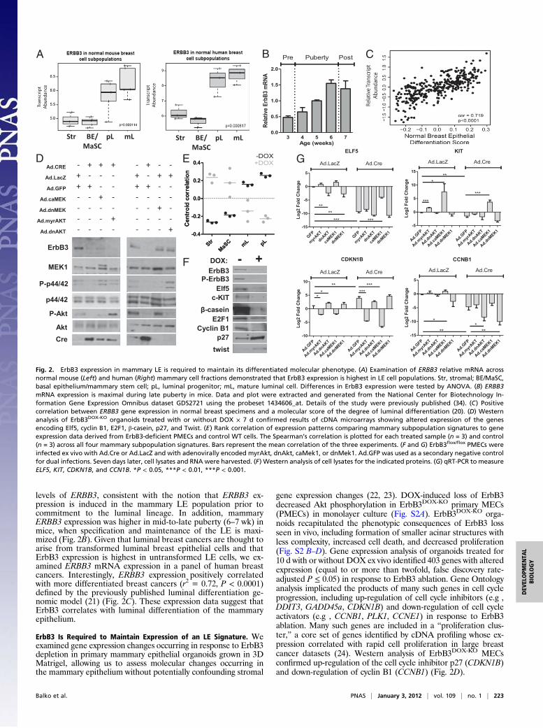
levels of ERBB3, consistent with the notion that ERBB3 ex-pression is induced in the mammary LE population prior tocommitment to the luminal lineage. In addition, mammaryERBB3 expression was higher in mid-to-late puberty (6–7 wk) inmice, when specification and maintenance of the LE is maxi-mized (Fig. 2B). Given that luminal breast cancers are thought toarise from transformed luminal breast epithelial cells and thatErbB3 expression is highest in untransformed LE cells, we ex-amined ERBB3 mRNA expression in a panel of human breastcancers. Interestingly, ERBB3 expression positively correlatedwith more differentiated breast cancers (r2 = 0.72, P < 0.0001)defined by the previously published luminal differentiation ge-nomic model (21) (Fig. 2C). These expression data suggest thatErbB3 correlates with luminal differentiation of the mammaryepithelium.
ErbB3 Is Required to Maintain Expression of an LE Signature. Weexamined gene expression changes occurring in response to ErbB3depletion in primary mammary epithelial organoids grown in 3DMatrigel, allowing us to assess molecular changes occurring inthe mammary epithelium without potentially confounding stromal
gene expression changes (22, 23). DOX-induced loss of ErbB3decreased Akt phosphorylation in ErbB3DOX-KO primary MECs(PMECs) in monolayer culture (Fig. S2A). ErbB3DOX-KO orga-noids recapitulated the phenotypic consequences of ErbB3 lossseen in vivo, including formation of smaller acinar structures withless complexity, increased cell death, and decreased proliferation(Fig. S2 B–D). Gene expression analysis of organoids treated for10 d with or without DOX ex vivo identified 403 genes with alteredexpression (equal to or more than twofold, false discovery rate-adjusted P ≤ 0.05) in response to ErbB3 ablation. Gene Ontologyanalysis implicated the products of many such genes in cell cycleprogression, including up-regulation of cell cycle inhibitors (e.g ,DDIT3, GADD45a, CDKN1B) and down-regulation of cell cycleactivators (e.g , CCNB1, PLK1, CCNE1) in response to ErbB3ablation. Many such genes are included in a “proliferation clus-ter,” a core set of genes identified by cDNA profiling whose ex-pression correlated with rapid cell proliferation in large breastcancer datasets (24). Western analysis of ErbB3DOX-KO MECsconfirmed up-regulation of the cell cycle inhibitor p27 (CDKN1B)and down-regulation of cyclin B1 (CCNB1) (Fig. 2D).
B C
GFP
myrAKT
dnAKT
caMEK1
dnMEK1GFP
myrAKT
dnAKT
caMEK1
dnMEK1-15
-10
-5
0
5
Ad.LacZ Ad.Cre
ELF5
Log2
Fol
d C
hang
e
****
*** ***
Ad.GFP
Ad.myrA
KT
Ad.dnAKT
Ad.caMEK1
Ad.dnMEK1
Ad.GFP
Ad.myrA
KT
Ad.dnAKT
Ad.caMEK1
Ad.dnMEK1-5
0
5
10
15
Ad.LacZ Ad.Cre
KIT
Log2
Fol
d C
hang
e
**
***
*
***
Ad.GFP
Ad.myrA
KT
Ad.dnAKT
Ad.caMEK1
Ad.dnMEK1
Ad.GFP
Ad.myrA
KT
Ad.dnAKT
Ad.caMEK1
Ad.dnMEK1-15
-10
-5
0
5
Ad.LacZ Ad.Cre
CCNB1
Log2
Fol
d C
hang
e
**
**
**
Ad.GFP
Ad.myrA
KT
Ad.dnAKT
Ad.caMEK1
Ad.dnMEK1
Ad.GFP
Ad.myrA
KT
Ad.dnAKT
Ad.caMEK1
Ad.dnMEK1-10
-5
0
5
10
Ad.LacZ Ad.Cre
CDKN1B
Log2
Fol
d C
hang
e
**
** ***
***
D
3 4 5 6 70.0
0.5
1.0
1.5
2.0
Pre PostPuberty
Age (weeks)
Relat
ive E
rbB3
mRN
A
A
BE/MaSC
pLStr mL
E
StrMaS
C mL pL-0.4
-0.2
0.0
0.2
0.4
Cen
troi
d co
rrel
atio
n
StrMaS
C mL pL-0.4
-0.2
0.0
0.2
0.4
Cen
troi
d co
rrel
atio
n
-DOX+DOX
DOX: - +FP-ErbB3
ErbB3
Elf5
β-casein
Cyclin B1E2F1
p27twist
c-KIT
BE/MaSC
pLStr mL
P-p44/42
ErbB3
MEK1
p44/42
P-Akt
AktCre
- + + +Ad.CRE - - -++ - - -Ad.LacZ + + +-+ + - -Ad.GFP + - -+- - + -Ad.caMEK - - --- - - -Ad.dnMEK - + --
Ad.myrAKT - - - + - - - -Ad.dnAKT - - - - - - - +
G
Fig. 2. ErbB3 expression in mammary LE is required to maintain its differentiated molecular phenotype. (A) Examination of ERBB3 relative mRNA acrossnormal mouse (Left) and human (Right) mammary cell fractions demonstrated that ErbB3 expression is highest in LE cell populations. Str, stromal; BE/MaSC,basal epithelium/mammary stem cell; pL, luminal progenitor; mL, mature luminal cell. Differences in ErbB3 expression were tested by ANOVA. (B) ERBB3mRNA expression is maximal during late puberty in mice. Data and plot were extracted and generated from the National Center for Biotechnology In-formation Gene Expression Omnibus dataset GDS2721 using the probeset 1434606_at. Details of the study were previously published (34). (C) Positivecorrelation between ERBB3 gene expression in normal breast specimens and a molecular score of the degree of luminal differentiation (20). (D) Westernanalysis of ErbB3DOX-KO organoids treated with or without DOX × 7 d confirmed results of cDNA microarrays showing altered expression of the genesencoding Elf5, cyclin B1, E2F1, β-casein, p27, and Twist. (E) Rank correlation of expression patterns comparing mammary subpopulation signatures to geneexpression data derived from ErbB3-deficient PMECs and control WT cells. The Spearman’s correlation is plotted for each treated sample (n = 3) and control(n = 3) across all four mammary subpopulation signatures. Bars represent the mean correlation of the three experiments. (F and G) ErbB3flox/flox PMECs wereinfected ex vivo with Ad.Cre or Ad.LacZ and with adenovirally encoded myrAkt, dnAkt, caMek1, or dnMek1. Ad.GFP was used as a secondary negative controlfor dual infections. Seven days later, cell lysates and RNA were harvested. (F) Western analysis of cell lysates for the indicated proteins. (G) qRT-PCR to measureELF5, KIT, CDKN1B, and CCN1B. *P < 0.05, ***P < 0.01, ***P < 0.001.
Balko et al. PNAS | January 3, 2012 | vol. 109 | no. 1 | 223
DEV
ELOPM
ENTA
LBIOLO
GY
Genes associated with luminal differentiation were also down-regulated in ErbB3-deficient mammary glands, including themilk protein β-casein, which was also down-regulated at theprotein level (Fig. 2D). Decreased gene expression of E74-likefactor 5 (ELF5), a transcription factor required for growth anddifferentiation of the luminal alveolar population (25–27), andthe RTK gene KIT were also observed. Elf5 and c-KIT haveemerged as definitive markers of the luminal progenitor pop-ulation (19). Elf5 and c-KIT down-regulation was confirmed byWestern blot (Fig. 2D).Next, we used the previously described gene expression sig-
natures for distinct cell types within the hierarchical model ofmammary epithelial differentiation (Fig. 2 A and B) to queryexpression data derived from organoid cultures expressing orlacking ErbB3. Untreated organoids retaining ErbB3 displayedexpression patterns that correlated positively with expressionsignatures from mature luminal cells and luminal progenitors(Fig. 2E) and negatively correlated with the BE/MaSC signature.However, DOX-induced loss of ErbB3 in organoids shifted geneexpression patterns, resulting in a negative correlation with lu-minal signatures, but a positive correlation with the BE/MaSCsignature.
ErbB3-PI3K and -MAPK Signaling Regulate Expression of LuminalMarkers. We investigated the signaling pathways downstream ofErbB3 that regulate expression of luminal molecular markers andproliferation cluster genes. ErbB3flox/flox PMECs were infected exvivo with adenoviral Cre (Ad.Cre) or lacZ control (Ad.LacZ) incombination with adenoviral constitutively active Akt (myrAkt),dominant-negative Akt (dnAkt), active MEK1 (caMEK1), ordominant-negative MEK1 (dnMEK1). Western analysis demon-strated that Cre-mediated loss of ErbB3 in Ad.Cre-infectedPMECs decreased P-Akt and P-MAPK (Fig. 2F). Expression ofmyrAkt1 and caMEK1 restored P-Akt and P-MAPK levels,
respectively. Conversely, P-Akt and P-MAPK were decreasedupon expression of dnAkt1 and dnMek1, respectively, even in thepresence of ErbB3.Expression of ELF5 and KIT were chosen as surrogate mark-
ers of luminal differentiation, as these are definitive markers ofluminal progenitors and were decreased upon ErbB3 depletion(Fig. 2D). Transcript levels of ELF5 and KIT were reduced uponErbB3 ablation in PMECs (Fig. 2G), but were partially rescuedupon expression of caMEK1 and myrAkt, although caMEK1produced a greater effect on ELF5 and KIT up-regulation com-pared with myrAkt. Conversely, ELF5 and KIT expression wereinhibited upon expression of dnAkt1 and dnMEK1 despite con-tinued expression of ErbB3.Down-regulation of CCNB1 and up-regulation of CDKN1B
were seen in ErbB3-depleted cells. Expression of caMEK1 ormyrAkt prevented down-regulation of CCNB1 and up-regulationCDKN1B in response to ErbB3 ablation. Conversely, dnMEK1and dnAkt increased expression of CDKN1B and decreasedCCNB1, despite continued expression of ErbB3. These resultshighlight the importance of ErbB3-PI3K signaling within the lu-minal lineage and establish an important role for ErbB3-MAPKsignaling in controlling proliferation in this compartment.
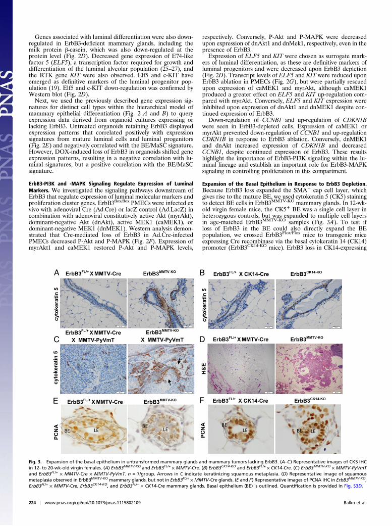
Expansion of the Basal Epithelium in Response to ErbB3 Depletion.Because ErbB3 loss expanded the SMA+ cap cell layer, whichgives rise to the mature BE, we used cytokeratin 5 (CK5) stainingto detect BE cells in ErbB3MMTV-KO mammary glands. In 12-wk-old virgin female mice, the CK5+ BE was a single cell layer inheterozygous controls, but was expanded to multiple cell layersin age-matched ErbB3MMTV-KO samples (Fig. 3A). To test ifloss of ErbB3 in the BE could also directly expand the BEpopulation, we crossed ErbB3Flox/Flox mice to transgenic miceexpressing Cre recombinase via the basal cytokeratin 14 (CK14)promoter (ErbB3CK14-KO mice). ErbB3 loss in CK14-expressing
ErbB3FL/+ X MMTV-Cre ErbB3MMTV-KO
H&
E
ErbB3FL/+ X MMTV-Cre ErbB3MMTV-KO
cyto
kera
tin 5
ErbB3FL/+ X CK14-Cre ErbB3CK14-KO
cyto
kera
tin 5
ErbB3FL/+ X MMTV-Cre X MMTV-PyVmT
cyto
kera
tin 5
ErbB3FL/+ X MMTV-Cre ErbB3MMTV-KO
PCN
A
ErbB3FL/+ X CK14-Cre ErbB3CK14-KO
PCN
A
A B
C
E
D
F
BE LEBE LE
BELE BE LE
ErbB3MMTV-KO
X MMTV-PyVmT
Fig. 3. Expansion of the basal epithelium in untransformed mammary glands and mammary tumors lacking ErbB3. (A–C) Representative images of CK5 IHCin 12- to 20-wk-old virgin females. (A) ErbB3MMTV-KO and ErbB3FL/+ × MMTV-Cre. (B) ErbB3CK14-KO and ErbB3FL/+ × CK14-Cre. (C) ErbB3MMTV-KO × MMTV-PyVmTand ErbB3FL/+ × MMTV-Cre × MMTV-PyVmT. n = 7/group. Arrows in C indicate keratinizing squamous metaplasia. (D) Representative image of squamousmetaplasia observed in ErbB3MMTV-KO mammary glands, but not in ErbB3FL/+ ×MMTV-Cre glands. (E and F) Representative images of PCNA IHC in ErbB3MMTV-KO,ErbB3FL/+ × MMTV-Cre, ErbB3CK14-KO, and ErbB3FL/+ × CK14-Cre mammary glands. Basal epithelium (BE) is outlined. Quantification is provided in Fig. S3D.
224 | www.pnas.org/cgi/doi/10.1073/pnas.1115802109 Balko et al.
basal cells did not disrupt mammary ductal elongation (Fig. S3A)and did not alter TEB cellular organization in ErbB3CK14-KOmice (Fig. S3B). Ki67 IHC did not reveal changes in cell pro-liferation due to BE knockout of ErbB3. Importantly, the SMA+
cap cell layer in developing TEBs appeared normal, and theCK5+ basal cell population was unaltered in ErbB3CK14-KO micecompared with heterozygous controls.We next examined CK5 expression in ErbB3-deficient
MMTV-PyVmT tumors (28). The CK5+ population exhibitedprofound expansion in ErbB3MMTV-KO × PyVmT tumors com-pared with heterozygous controls (Fig. 3C). Keratinizing squa-mous metaplasia was evident in 7/20 ErbB3-deficient MMTV-PyVmT tumors, but was not identified in ErbB3flox/+ × MMTV-PyVmT tumors (Fig. 3C). Similarly, keratinizing squamoustransdifferentiation of the mammary epithelium was seen in3/12 ErbB3MMTV-KO mice (Fig. 3D), but was not observed inErbB3CK14-KO samples (0/12). Therefore, loss of ErbB3 in theLE alters the balance of luminal and basal cells in both normaland transformed mammary epithelium.It is possible that ErbB3 loss in LE cells indirectly promotes
BE growth. In support of this idea, increased BE proliferationwas observed in ErbB3MMTV-KO mammary glands compared withheterozygous controls [assessed using proliferating cell nuclearantigen (PCNA) IHC] (Fig. 3E), but not in ErbB3CK14-KO glands(Fig. 3F). Fewer PCNA+ LE cells were seen in ErbB3MMTV-KO
mammary glands compared with heterozygous controls and withErbB3CK14-KO samples, consistent with the decreased body cellproliferation seen in ErbB3-deficient TEBs (Fig. 1B).
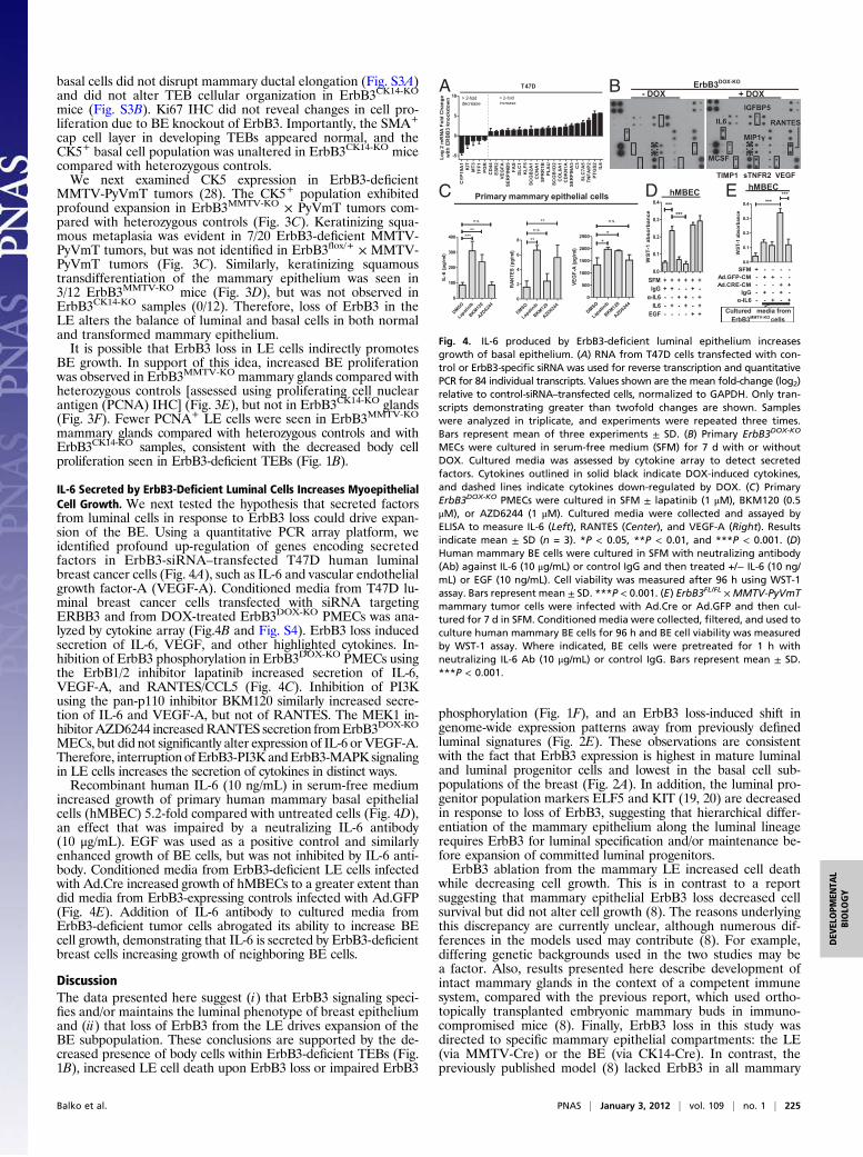
IL-6 Secreted by ErbB3-Deficient Luminal Cells Increases MyoepithelialCell Growth. We next tested the hypothesis that secreted factorsfrom luminal cells in response to ErbB3 loss could drive expan-sion of the BE. Using a quantitative PCR array platform, weidentified profound up-regulation of genes encoding secretedfactors in ErbB3-siRNA–transfected T47D human luminalbreast cancer cells (Fig. 4A), such as IL-6 and vascular endothelialgrowth factor-A (VEGF-A). Conditioned media from T47D lu-minal breast cancer cells transfected with siRNA targetingERBB3 and from DOX-treated ErbB3DOX-KO PMECs was ana-lyzed by cytokine array (Fig.4B and Fig. S4). ErbB3 loss inducedsecretion of IL-6, VEGF, and other highlighted cytokines. In-hibition of ErbB3 phosphorylation in ErbB3DOX-KO PMECs usingthe ErbB1/2 inhibitor lapatinib increased secretion of IL-6,VEGF-A, and RANTES/CCL5 (Fig. 4C). Inhibition of PI3Kusing the pan-p110 inhibitor BKM120 similarly increased secre-tion of IL-6 and VEGF-A, but not of RANTES. The MEK1 in-hibitorAZD6244 increasedRANTES secretion fromErbB3DOX-KO
MECs, but did not significantly alter expression of IL-6 or VEGF-A.Therefore, interruption ofErbB3-PI3K andErbB3-MAPK signalingin LE cells increases the secretion of cytokines in distinct ways.Recombinant human IL-6 (10 ng/mL) in serum-free medium
increased growth of primary human mammary basal epithelialcells (hMBEC) 5.2-fold compared with untreated cells (Fig. 4D),an effect that was impaired by a neutralizing IL-6 antibody(10 μg/mL). EGF was used as a positive control and similarlyenhanced growth of BE cells, but was not inhibited by IL-6 anti-body. Conditioned media from ErbB3-deficient LE cells infectedwith Ad.Cre increased growth of hMBECs to a greater extent thandid media from ErbB3-expressing controls infected with Ad.GFP(Fig. 4E). Addition of IL-6 antibody to cultured media fromErbB3-deficient tumor cells abrogated its ability to increase BEcell growth, demonstrating that IL-6 is secreted by ErbB3-deficientbreast cells increasing growth of neighboring BE cells.
DiscussionThe data presented here suggest (i) that ErbB3 signaling speci-fies and/or maintains the luminal phenotype of breast epitheliumand (ii) that loss of ErbB3 from the LE drives expansion of theBE subpopulation. These conclusions are supported by the de-creased presence of body cells within ErbB3-deficient TEBs (Fig.1B), increased LE cell death upon ErbB3 loss or impaired ErbB3
phosphorylation (Fig. 1F), and an ErbB3 loss-induced shift ingenome-wide expression patterns away from previously definedluminal signatures (Fig. 2E). These observations are consistentwith the fact that ErbB3 expression is highest in mature luminaland luminal progenitor cells and lowest in the basal cell sub-populations of the breast (Fig. 2A). In addition, the luminal pro-genitor population markers ELF5 and KIT (19, 20) are decreasedin response to loss of ErbB3, suggesting that hierarchical differ-entiation of the mammary epithelium along the luminal lineagerequires ErbB3 for luminal specification and/or maintenance be-fore expansion of committed luminal progenitors.ErbB3 ablation from the mammary LE increased cell death
while decreasing cell growth. This is in contrast to a reportsuggesting that mammary epithelial ErbB3 loss decreased cellsurvival but did not alter cell growth (8). The reasons underlyingthis discrepancy are currently unclear, although numerous dif-ferences in the models used may contribute (8). For example,differing genetic backgrounds used in the two studies may bea factor. Also, results presented here describe development ofintact mammary glands in the context of a competent immunesystem, compared with the previous report, which used ortho-topically transplanted embryonic mammary buds in immuno-compromised mice (8). Finally, ErbB3 loss in this study wasdirected to specific mammary epithelial compartments: the LE(via MMTV-Cre) or the BE (via CK14-Cre). In contrast, thepreviously published model (8) lacked ErbB3 in all mammary
0.0
0.1
0.2
0.3
0.4 ******
SFM + + + + + +IgG + + - - + -
α-IL6 - - + + - +IL6 - + - + - +
EGF - - - - + +
WS
T-1
abso
rban
ce
DMSO
Lapati
nib
BKM120
AZD6244
0
2
4
6
8
n.s.
**
**
RANT
ES (p
g/m
l)
DMSO
Lapati
nib
BKM120
AZD6244
0
500
1000
1500
2000
2500*
*
n.s.
VEG
F-A
(pg/
ml)
DMSO
Lapati
nib
BKM120
AZD6244
0
100
200
300
400
n.s.
*****
IL-6
(pg/
ml)
T47D
CY
P19
A1
KIT
MT3
TFF1
PG
RC
D44
ES
R2
VE
GFA
SE
RP
INB
5FA
SD
LC1
KLF
5S
CG
B2A
1C
CN
A1
SP
RR
1BP
LAU
SC
GB
1D2
CO
L6A
1C
DK
N1A
SE
RP
INA
3C
3S
LC7A
5TN
FAIP
2P
TGS
2IL
6
-5
0
5
10 > 2-folddecrease
> 2-foldincrease
Log
2 m
RN
A Fo
ld C
hang
ew
ith E
RB
B3
knoc
kdow
n
- DOX
TIMP1 VEGFsTNFR2
+ DOXA B
C DPrimary mammary epithelial cells hMBEC
ErbB3DOX-KO
0.0
0.1
0.2
0.3
0.4
******
SFM + - - - -Ad.GFP-CM - + + - -Ad.CRE-CM - - - + +
IgG - + - + -α-IL6 - - + - +
WST
-1 a
bsor
banc
e
E
Cultured media from ErbB3MMTV-KO cells
hMBEC
Fig. 4. IL-6 produced by ErbB3-deficient luminal epithelium increasesgrowth of basal epithelium. (A) RNA from T47D cells transfected with con-trol or ErbB3-specific siRNA was used for reverse transcription and quantitativePCR for 84 individual transcripts. Values shown are the mean fold-change (log2)relative to control-siRNA–transfected cells, normalized to GAPDH. Only tran-scripts demonstrating greater than twofold changes are shown. Sampleswere analyzed in triplicate, and experiments were repeated three times.Bars represent mean of three experiments ± SD. (B) Primary ErbB3DOX-KO
MECs were cultured in serum-free medium (SFM) for 7 d with or withoutDOX. Cultured media was assessed by cytokine array to detect secretedfactors. Cytokines outlined in solid black indicate DOX-induced cytokines,and dashed lines indicate cytokines down-regulated by DOX. (C) PrimaryErbB3DOX-KO PMECs were cultured in SFM ± lapatinib (1 μM), BKM120 (0.5μM), or AZD6244 (1 μM). Cultured media were collected and assayed byELISA to measure IL-6 (Left), RANTES (Center), and VEGF-A (Right). Resultsindicate mean ± SD (n = 3). *P < 0.05, **P < 0.01, and ***P < 0.001. (D)Human mammary BE cells were cultured in SFM with neutralizing antibody(Ab) against IL-6 (10 μg/mL) or control IgG and then treated +/− IL-6 (10 ng/mL) or EGF (10 ng/mL). Cell viability was measured after 96 h using WST-1assay. Bars represent mean ± SD. ***P < 0.001. (E) ErbB3FL/FL ×MMTV-PyVmTmammary tumor cells were infected with Ad.Cre or Ad.GFP and then cul-tured for 7 d in SFM. Conditioned media were collected, filtered, and used toculture human mammary BE cells for 96 h and BE cell viability was measuredby WST-1 assay. Where indicated, BE cells were pretreated for 1 h withneutralizing IL-6 Ab (10 μg/mL) or control IgG. Bars represent mean ± SD.***P < 0.001.
Balko et al. PNAS | January 3, 2012 | vol. 109 | no. 1 | 225
DEV
ELOPM
ENTA
LBIOLO
GY
epithelial populations. We have shown here that ErbB3 impactsdistinct mammary epithelial populations in profoundly differentways, potentially contributing to this phenotypic discrepancy.MMTV-PyVmT tumors express high levels of the luminal
cytokeratins 8 and 18 and low levels of CK5, a cytokeratin as-sociated with basal-like breast cancers, consistent with expressionanalyses clustering the MMTV-PyVmT tumor model with theluminal subtype of human breast cancers (29). Although loss ofErbB3 in the mammary epithelium decreases the rate of tumorformation in MMTV-PyVmT mice (28), ErbB3-deficient tumorseventually formed, exhibiting an increased CK5+ tumor cellpopulation (Fig. 3C). Because BE and LE cells arise fromcommon stem cells, it is possible that loss of ErbB3 preventsdifferentiation along the luminal lineage, forcing cells to differ-entiate into the basal lineage as a default. Our results do notdisprove this possibility, but strongly support an alternative sce-nario in which ErbB3 loss from the LE causes cytokine secretion,which causes growth of neighboring basal cells (Fig. 4).Interestingly, ErbB3 loss increased CK5+ cells in both un-
transformed mammary epithelia and mammary tumors (Fig. 3A),suggesting that ErbB3 directs cell fate decisions in cancers. Thiscould have important implications regarding molecular classi-fications of breast cancers. Advances in molecular analysis ofprimary tumorsmake clear that different subtypes of human breastcancer exist (30). Increasing evidence suggests that the molecularsubtype of a given breast cancer may be a reflection of the cell typefrom which that cancer originates (20, 31–33). Therefore, it iscritical to understand the signaling pathways that define the epi-
thelial ontogeny of the mammary gland and how these pathwaysmay be used within cancers that arise from each cell type. Ourresults demonstrate that ErbB3 is required within the luminallineages of the breast. Further study of how ErbB3 and otherdifferentiation signals may influence cell fate decisions withinpreneoplastic mammary glands will support our understanding ofhow tumors adopt specific molecular and clinical phenotypes, in-formation that may be used to treat or prevent breast cancer.
Materials and MethodsAll mouse experiments were approved by the Vanderbilt Institutional AnimalCare and Use Committee. All models used, including genetically engineeredmouse models and cell lines, are described in SI Materials and Methods.Detailed methods for Western analysis, RT-PCR, quantification of cellgrowth, and histological analyses can be found in SI Materials and Methods.Detailed materials and methods can be found in SI Materials and Methods.Additionally, Figs. S1–S4, associated legends, and references can also befound in Supporting Information.
ACKNOWLEDGMENTS. This work was supported by National Institutesof Health Grants R01CA143126 (to R.S.C.), R01CA80195 (to C.L.A.), andF32CA121900 and K99CA142899 (to T.W.M.); Breast Cancer SpecializedPrograms of Research Excellence Grants P50CA98131 (to Vanderbilt Univer-sity) and P50CA058223 (to University of North Carolina-Lineberger CancerCenter); Vanderbilt-Ingram Cancer Center Support Grant P30CA68485; SusanG. Komen for the Cure Grant KG100677 (to R.S.C.); American Cancer SocietyClinical Research Professorship CRP-07-234 (to C.L.A.); the Lee Jeans Trans-lational Breast Cancer Research Program (C.L.A.); and Stand Up to Cancer/American Association for Cancer Research Dream Team Translational CancerResearch Grant SU2C-AACR-DT0209 (to C.L.A.).
1. Abd El-Rehim DM, et al. (2004) Expression and co-expression of the members of theepidermal growth factor receptor (EGFR) family in invasive breast carcinoma. Br JCancer 91:1532–1542.
2. Citri A, Yarden Y (2006) EGF-ERBB signalling: Towards the systems level. Nat Rev MolCell Biol 7:505–516.
3. Bazley LA, Gullick WJ (2005) The epidermal growth factor receptor family. EndocrRelat Cancer 12(Suppl 1):S17–S27.
4. Holbro T, Civenni G, Hynes NE (2003) The ErbB receptors and their role in cancerprogression. Exp Cell Res 284:99–110.
5. Carraway KL, III, et al. (1994) The erbB3 gene product is a receptor for heregulin. J BiolChem 269:14303–14306.
6. Pinkas-Kramarski R, et al. (1996) Diversification of Neu differentiation factor andepidermal growth factor signaling by combinatorial receptor interactions. EMBO J 15:2452–2467.
7. Kim HH, Vijapurkar U, Hellyer NJ, Bravo D, Koland JG (1998) Signal transduction byepidermal growth factor and heregulin via the kinase-deficient ErbB3 protein. Bio-chem J 334:189–195.
8. Jackson-Fisher AJ, et al. (2008) ErbB3 is required for ductal morphogenesis in themouse mammary gland. Breast Cancer Res 10:R96.
9. Andrechek ER, White D, Muller WJ (2005) Targeted disruption of ErbB2/Neu in themammary epithelium results in impaired ductal outgrowth. Oncogene 24:932–937.
10. Jackson-Fisher AJ, et al. (2004) ErbB2 is required for ductal morphogenesis of themammary gland. Proc Natl Acad Sci USA 101:17138–17143.
11. Long W, et al. (2003) Impaired differentiation and lactational failure of Erbb4-deficient mammary glands identify ERBB4 as an obligate mediator of STAT5.Development 130:5257–5268.
12. Wiesen JF, Young P, Werb Z, Cunha GR (1999) Signaling through the stromal epi-dermal growth factor receptor is necessary for mammary ductal development.Development 126:335–344.
13. Erickson SL, et al. (1997) ErbB3 is required for normal cerebellar and cardiacdevelopment: A comparison with ErbB2-and heregulin-deficient mice. Development124:4999–5011.
14. Holbro T, et al. (2003) The ErbB2/ErbB3 heterodimer functions as an oncogenic unit:ErbB2 requires ErbB3 to drive breast tumor cell proliferation. Proc Natl Acad Sci USA100:8933–8938.
15. Wong KK, Engelman JA, Cantley LC (2010) Targeting the PI3K signaling pathway incancer. Curr Opin Genet Dev 20:87–90.
16. Qu S, et al. (2006) Gene targeting of ErbB3 using a Cre-mediated unidirectional DNAinversion strategy. Genesis 44:477–486.
17. Gunther EJ, et al. (2002) A novel doxycycline-inducible system for the transgenicanalysis of mammary gland biology. FASEB J 16:283–292.
18. Wagner KU, et al. (1997) Cre-mediated gene deletion in the mammary gland. NucleicAcids Res 25:4323–4330.
19. Lim E, et al. (2009) Aberrant luminal progenitors as the candidate target populationfor basal tumor development in BRCA1 mutation carriers. Nat Med 15:907–913.
20. Lim E, et al. (2010) Transcriptome analyses of mouse and human mammary cell sub-populations reveal multiple conserved genes and pathways. Breast Cancer Res 12:R21.
21. Prat A, et al. (2010) Phenotypic and molecular characterization of the claudin-lowintrinsic subtype of breast cancer. Breast Cancer Res 12:R68.
22. Fata JE, et al. (2007) The MAPK(ERK-1,2) pathway integrates distinct and antagonisticsignals from TGFalpha and FGF7 in morphogenesis of mouse mammary epithelium.Dev Biol 306:193–207.
23. Ewald AJ, Brenot A, Duong M, Chan BS, Werb Z (2008) Collective epithelial migrationand cell rearrangements drive mammary branching morphogenesis. Dev Cell 14:570–581.
24. Whitfield ML, George LK, Grant GD, Perou CM (2006) Common markers of pro-liferation. Nat Rev Cancer 6:99–106.
25. Harris J, et al. (2006) Socs2 and elf5 mediate prolactin-induced mammary glanddevelopment. Mol Endocrinol 20:1177–1187.
26. Oakes SR, et al. (2008) The Ets transcription factor Elf5 specifies mammary alveolarcell fate. Genes Dev 22:581–586.
27. Zhou J, et al. (2005) Elf5 is essential for early embryogenesis and mammary glanddevelopment during pregnancy and lactation. EMBO J 24:635–644.
28. Muraoka-Cook RS, et al. (2011) ErbB3 ablation impairs PI3K/Akt-dependent mammarytumorigenesis. Cancer Res 71:3941–3951.
29. Herschkowitz JI, et al. (2007) Identification of conserved gene expression featuresbetween murine mammary carcinoma models and human breast tumors. GenomeBiol 8:R76.
30. Sørlie T, et al. (2001) Gene expression patterns of breast carcinomas distinguish tumorsubclasses with clinical implications. Proc Natl Acad Sci USA 98:10869–10874.
31. Perou CM, et al. (2000) Molecular portraits of human breast tumours. Nature 406:747–752.
32. Asselin-Labat ML, et al. (2008) Delineating the epithelial hierarchy in the mousemammary gland. Cold Spring Harb Symp Quant Biol 73:469–478.
33. Lindeman GJ, Visvader JE (2010) Insights into the cell of origin in breast cancer andbreast cancer stem cells. Asia Pac J Clin Oncol 6:89–97.
34. McBryan J, Howlin J, Kenny PA, Shioda T, Martin F (2007) ERalpha-CITED1 co-regu-lated genes expressed during pubertal mammary gland development: implicationsfor breast cancer prognosis. Oncogene 26:6406–6419.
226 | www.pnas.org/cgi/doi/10.1073/pnas.1115802109 Balko et al.