The Proto-Oncogene c-myc Regulates Antibody Secretion and ... · The Proto-Oncogene c-myc Regulates...
11
of September 15, 2018. This information is current as Recombination Antibody Secretion and Ig Class Switch Regulates myc The Proto-Oncogene c- Alborán Arancha García, Dolores Martinez and Ignacio Moreno de David Fernández, Maitane Ortiz, Lorena Rodríguez, http://www.jimmunol.org/content/190/12/6135 doi: 10.4049/jimmunol.1300712 May 2013; 2013; 190:6135-6144; Prepublished online 20 J Immunol Material Supplementary 2.DC1 http://www.jimmunol.org/content/suppl/2013/05/21/jimmunol.130071 References http://www.jimmunol.org/content/190/12/6135.full#ref-list-1 , 12 of which you can access for free at: cites 52 articles This article average * 4 weeks from acceptance to publication Fast Publication! • Every submission reviewed by practicing scientists No Triage! • from submission to initial decision Rapid Reviews! 30 days* • Submit online. ? The JI Why Subscription http://jimmunol.org/subscription is online at: The Journal of Immunology Information about subscribing to Permissions http://www.aai.org/About/Publications/JI/copyright.html Submit copyright permission requests at: Email Alerts http://jimmunol.org/alerts Receive free email-alerts when new articles cite this article. Sign up at: Print ISSN: 0022-1767 Online ISSN: 1550-6606. Immunologists, Inc. All rights reserved. Copyright © 2013 by The American Association of 1451 Rockville Pike, Suite 650, Rockville, MD 20852 The American Association of Immunologists, Inc., is published twice each month by The Journal of Immunology by guest on September 15, 2018 http://www.jimmunol.org/ Downloaded from by guest on September 15, 2018 http://www.jimmunol.org/ Downloaded from
Transcript of The Proto-Oncogene c-myc Regulates Antibody Secretion and ... · The Proto-Oncogene c-myc Regulates...
of September 15, 2018.This information is current as
RecombinationAntibody Secretion and Ig Class Switch
RegulatesmycThe Proto-Oncogene c-
AlboránArancha García, Dolores Martinez and Ignacio Moreno de David Fernández, Maitane Ortiz, Lorena Rodríguez,
http://www.jimmunol.org/content/190/12/6135doi: 10.4049/jimmunol.1300712May 2013;
2013; 190:6135-6144; Prepublished online 20J Immunol
MaterialSupplementary
2.DC1http://www.jimmunol.org/content/suppl/2013/05/21/jimmunol.130071
Referenceshttp://www.jimmunol.org/content/190/12/6135.full#ref-list-1
, 12 of which you can access for free at: cites 52 articlesThis article
average*
4 weeks from acceptance to publicationFast Publication! •
Every submission reviewed by practicing scientistsNo Triage! •
from submission to initial decisionRapid Reviews! 30 days* •
Submit online. ?The JIWhy
Subscriptionhttp://jimmunol.org/subscription
is online at: The Journal of ImmunologyInformation about subscribing to
Permissionshttp://www.aai.org/About/Publications/JI/copyright.htmlSubmit copyright permission requests at:
Email Alertshttp://jimmunol.org/alertsReceive free email-alerts when new articles cite this article. Sign up at:
Print ISSN: 0022-1767 Online ISSN: 1550-6606. Immunologists, Inc. All rights reserved.Copyright © 2013 by The American Association of1451 Rockville Pike, Suite 650, Rockville, MD 20852The American Association of Immunologists, Inc.,
is published twice each month byThe Journal of Immunology
by guest on September 15, 2018
http://ww
w.jim
munol.org/
Dow
nloaded from
by guest on September 15, 2018
http://ww
w.jim
munol.org/
Dow
nloaded from

The Journal of Immunology
The Proto-Oncogene c-myc Regulates Antibody Secretion andIg Class Switch Recombination
David Fernandez,*,1 Maitane Ortiz,*,1 Lorena Rodrıguez,* Arancha Garcıa,†
Dolores Martinez,† and Ignacio Moreno de Alboran*
The immune response involves the generation of Ab-secreting cells and memory B cells through a process called terminal
B lymphocyte differentiation. This program requires the transcriptional repressor Blimp-1, which inhibits c-myc expression
and terminates proliferation. Although the role of c-Myc in cell proliferation is well characterized, it is not known whether it
has other functions in terminal differentiation. In this study, we show that c-Myc not only regulates cell proliferation, but it is also
essential for Ab-secreting cell function and differentiation in vivo. c-Myc–deficient B lymphocytes hypersecrete IgM and do not
undergo Ig class switch recombination (CSR). CSR has been previously linked to proliferation, and in this study we mechanis-
tically link class switching and proliferation via c-Myc. We observed that c-Myc regulates CSR by transcriptionally activating the
B cell–specific factor activation-induced cytidine deaminase. By linking cell proliferation and CSR, c-Myc is thus a critical
component for a potent immune response The Journal of Immunology, 2013, 190: 6135–6144.
Arobust humoral immune response to an Ag involves thegeneration of two populations of terminally differentiatedB lymphocytes, that is, Ab-secreting cells (ASC) and
memory B cells (1, 2). Mature naive B cells can give rise to short-lived ASC generated in response to thymus-independent (TI) or-dependent (TD) Ags in extrafollicular zones in secondary lym-phoid organs. Short-lived ASC have half-lives of 2–3 d, prolifer-ate, and secrete nonmutated IgM Abs. The immune responseagainst TD Ags can also induce specialized structures in secondarylymphoid organs, termed germinal centers (GC). After immuni-zation, GC generate long-lived ASC and memory B cells; theformer do not proliferate, produce high-affinity Abs by somatichypermutation (3), and undergo IgH class switch recombination(CSR) (4). Somatic hypermutation and CSR are dependent onexpression of activation-induced cytidine deaminase (AID; aicda)(5). Long-lived ASC are located preferentially in bone marrow.
Memory B cells express isotypes other than IgM after CSR, andthey differentiate to ASC following Ag rechallenge (6).The initial steps of terminal B cell differentiation are charac-
terized by the mutually exclusive expression of a zinc fingercontaining transcriptional repressors Bcl-6 and Blimp-1 (pdrm1)(7). Bcl-6 is expressed in GC B cells and is necessary for theirformation. Bcl-62/2 mice have more ASC in T cell–dependentresponses and secrete more Ig in T cell–independent responses,but they do not form GC structures (8). Bcl6 gene expression isregulated by the Irf4 transcription factor, a member of the IFNregulatory factor family, necessary for ASC differentiation and forCSR (9, 10). Irf4 is able to repress bcl6 expression by binding toits promoter region (11). Bcl-6 directly represses expression of thepdrm1 (Blimp-1) gene, at least in part by binding to a site in intron5 (12, 13). Blimp-1 is needed for full ASC differentiation bothin vitro and in vivo, although it is dispensable for the initial stepsof ASC differentiation and for memory B cell generation (14, 15).Blimp-1 is sufficient to induce terminal B cell differentiation bydirectly repressing genes responsible for cell proliferation andB cell identity such as c-myc and pax5, respectively (16–18).Downregulation of pax5 expression initiates ASC differentiationby activating genes such as xbp1 that is needed for full ASCdifferentiation, and it promotes a series of molecular events thatmediate Ig secretion (19, 20). Blimp-1 expression is normal inB cells lacking Xbp-1, suggesting that this factor acts downstreamof Blimp-1 (19). Inhibition of c-myc expression by Blimp-1 ter-minates cell proliferation in ASC. A basic model was proposedin which Blimp-1 acts by direct repression of c-myc and pax5,therefore promoting xbp1 expression and Ig secretion (21).The c-Myc protein is a member of a basic region/helix-loop-helix/
leucine zipper transcription factor family (N-, L-, and c-Myc) thatregulates essential biological functions such as cell proliferation,differentiation, and apoptosis (22). c-Myc is broadly expressed andits deregulation has an enormous impact on human health; it isestimated that 50% of human cancers show altered c-myc expres-sion (23). c-Myc is expressed in differentiating B lymphocytes andin mature B cells after activation (24). Transgenic mice that over-express c-myc in B lymphocytes show abnormal B cell differenti-ation and rapid lymphoma development, leading ultimately to death(25). Mature B lymphocytes that lack c-Myc in vivo show severely
*Departamento de Inmunologıa y Oncologıa, Centro Nacional de Biotecnologıa/Consejo Superior de Investigaciones Cientificas, Madrid E-28049, Spain; and†Centro Nacional de Investigaciones Oncologicas, Madrid E-28029, Spain
1D.F. and M.O. contributed equally to this work.
Received for publication March 15, 2013. Accepted for publication April 19, 2013.
This work was supported by grants from the Spanish Ministry of Science and Inno-vation, the Mutua Madrilena Foundation, the Concern Foundation, and the RegionalGovernment of Madrid (to I.M.d.A.). M.O. was supported by a fellowship from theRegional Government of Madrid and the Fondo Social Europeo. D.F. received a fel-lowship from the Spanish Ministry of Science and Innovation.
M.O. and D.F. designed, performed, and analyzed the in vitro and in vivo experi-ments; L.R., A.G., and D.M. assisted in the flow cytometry experiments; and I.M.d.A.supervised the project, analyzed the data, and wrote the paper.
Address correspondence and reprint requests to Dr. Ignacio Moreno de Alboran,Centro Nacional de Biotecnologıa/Consejo Superior de Investigaciones Cientificas,Darwin 3, Madrid E-28049, Spain. E-mail address: [email protected]
The online version of this article contains supplemental material.
Abbreviations used in this article: AID, activation-induced cytidine deaminase; ASC,Ab-secreting cell; CSR, class switch recombination; CT, circle transcript; FO, fol-licular; GC, germinal center; I1, intron-1; KLH, keyhole limpet hemocyanin; MZ,marginal zone; PNA, peanut agglutinin; qPCR, quantitative PCR; R1, region 1; R4,region 4; TD, thymus-dependent; TI, thymus-independent; TNP, 2,4,6-trinitrophenylhapten.
Copyright� 2013 by The American Association of Immunologists, Inc. 0022-1767/13/$16.00
www.jimmunol.org/cgi/doi/10.4049/jimmunol.1300712
by guest on September 15, 2018
http://ww
w.jim
munol.org/
Dow
nloaded from

impaired proliferation and resistance to apoptosis (26, 27). In-volvement of c-Myc in differentiation has been described for sev-eral cell types, including myeloid cells and B lymphocytes (18, 28–30). c-Myc downregulation is associated with cell cycle arrest andterminal differentiation. In a mature B lymphoma line (17), re-pression of c-Myc is necessary but not sufficient to induce terminalB lymphocyte differentiation. Despite numerous reports on c-Mycfunction, little is known of the precise role of this protein in ter-minal B cell differentiation.In this study, we analyzed c-Myc function in terminal B cell
differentiation in primary B lymphocytes in vivo. Our findingsshow that c-Myc is necessary for the generation of fully differ-entiated ASC, normal IgM secretion, and isotype switching.Moreover, c-Myc transcriptionally regulates the gene for AID(aicda) (5), linking cell proliferation and CSR. Finally, we foundthat the immune response to TI and TD Ags are largely Myc-dependent. Our results provide unexpected insights into c-Mycfunction in terminal B cell differentiation, and they show theimportance of this transcription factor as a key effector of thehumoral response.
Materials and MethodsGeneration of c-mycfl/fl mice
The generation of c-mycfl/fl (c-mycfl/fl;cd19cre/+;rosa26gfp/gfp) mice has beenreported (26, 27).
Immunization
Six- to 8-wk-old mice were immunized i.p. with 200mg 2,4,6-trinitrophenylhapten (TNP)–keyhole limpet hemocyanin (KLH) (Biosearch Technolo-gies) in aluminum hydroxide (Pierce) at a 1:1 ratio in 0.2 ml PBS. For thememory response, the same TNP-KLH dose was given 4 wk after theinitial dose, and mice were analyzed 12 d after secondary immunization.For TI responses, mice received i.v. injections of 3 mg LPS (Sigma-Aldrich) and were analyzed 3 d later. Mice were bled weekly to obtainserum.
Flow cytometry
Single-cell suspensions were incubated in NH4Cl buffer to lyse erythro-cytes and in Ab stained in PBS with 2% FBS. Abs used were fromeBioscience (anti–B220-PE-Cy7, GL-7-biotin) and BD Pharmingen(B220-Pacific Blue, IgG1-PE, IgG3-PE, IgG2b-PE, IgE-PE, Gr-1-allophycocyanin, F4/80-allophycocyanin, IgD-Alexa 647, CD38-Alexa700, CD23-PE, CD21-allophycocyanin, CD69-biotin, CD25-PE, CD138-biotin, and IgM-allophycocyanin). Biotinylated Abs were developed withstreptavidin-PE or streptavidin-PE-Cy7 (BD Pharmingen). To detect TNP-specific memory B cells by flow cytometry, we used TNP-BSA-biotin(Biosearch Technologies); for intracellular staining, we used an Intra-Prep kit (Beckman Coulter) and stained cells with anti–IgM-PE. Wemonitored cell proliferation with the CellTrace Violet cell proliferation kit(Invitrogen).
B cell purification and culture
Freshly isolated splenocytes were stained with anti–B220-PE-Cy7 in PBSwith 2% FBS. B220+GFP+ cells were purified (.97% purity) on a FAC-SAria IIu sorter (BD Biosciences, San Jose CA). Sorted B lymphocyteswere plated (105 cells/well) in RPMI 1640 medium with 15% FBS, 2-ME,and penicillin/streptomycin. TI-like stimulation was performed with 20mg/ml LPS (Sigma-Aldrich), and TD-like stimulation was performed with20 ng/ml IL-4 (R&D Systems) and 10 ng/ml anti-CD40 Ab (BD Phar-mingen). Activated cells and supernatants were harvested after 1, 3, or4 d in culture.
ELISA
Assays were performed following standard protocols using goat anti-mouseIgM Ab (SouthernBiotech). Anti–Igk-HRP (HRP clone 187.1; South-ernBiotech) secondary Ab was developed with Sigmafast OPD (Sigma-Aldrich). For IgG1 and IgG2b, plates were coated with purified anti-Igkand developed with specific biotinylated Abs for each isotype (BD Phar-mingen). For IgE, plates were coated with purified anti-IgE, followed bybiotinylated anti-IgE (SouthernBiotech). Plates were developed using
streptavidin-HRP (BD Pharmingen). For detection of TNP-specific Abs,plates were coated with TNP-BSA (10 mg/ml; Biosearch Technologies)in PBS. Biotinylated anti-IgM (SouthernBiotech), IgG2a, or IgG1 (BDPharmingen) were used as secondary Abs. For TNP-specific IgE, plateswere coated with purified anti-IgE, and samples were incubated with TNP-BSA-biotin (Biosearch Technologies).
ELISPOT
Plates were coated with goat anti-mouse IgM (BD Pharmingen), washed,and triplicates of 3-fold serially diluted B220+GFP+ spleen cells wereplated in RPMI 1640 with 1% FBS. Cells were incubated (37˚C, over-night), then lysed, incubated (60 min, 37˚C) with anti–Igk-alkaline phos-phatase (BD Pharmingen), and developed with 5-bromo-4-chloro-3-indolylphosphate/AMP buffer (Sigma-Aldrich) in 3% low-melt agarose. Thenumber of spots was counted from triplicates corresponding to two serialdilutions (six wells per sample) and the surface was measured with an AIDELISPOT reader and AID ELISPOT software (Autoimmun Diagnostika).
Immunoprecipitation and Western blot
Pooled supernatant from five wells (1 ml medium) was harvested 3 or4 d after activation, and total secreted IgM was immunoprecipitated withgoat anti-mouse IgM (BD Pharmingen). Remaining supernatant wasimmunoprecipitated again as a control. Ab-bound IgM was precipitatedwith Protein A/G PLUS-Agarose beads (Santa Cruz Biotechnology),washed in PBS with proteinase inhibitors, and bound protein was eluted(95˚C). Supernatants were resolved in 10% SDS-PAGE and transferred topolyvinylidene difluoride membranes. Blots were incubated with HRP goatanti-mouse IgM (Sigma-Aldrich) and developed with ChemiluminiscenceReagent Plus (PerkinElmer).
Immunofluorescence
For spleen cryosections, mice were sacrificed 10 d after the second im-munization. One-half of the spleen was snap-frozen in isopentane precooledin liquid nitrogen and stored at280˚C. Sections (10 mm) were prepared ona cryostat and acetone-fixed (room temperature, 10 min) before staining.For paraffin sections, half-spleens were fixed in 4% paraformaldehyde inPBS (room temperature, 2–3 h), followed by 70% ethanol (4˚C, overnight).Sections (5 mm) were deparaffinized before H&E staining. Cryosectionswere blocked for endogenous peroxidase activity (70% methanol with0.3% H2O2), for endogenous biotin (avidin/biotin blocking kit; Dako), andfor nonspecific Ab binding (10% goat serum). Sections were stained withrabbit anti-GFP (ab290), rabbit anti-Cre Ab (Covance), monoclonal ratanti-mouse B220 (SouthernBiotech), biotinylated anti-MOMA1 (Dianova),and/or peanut agglutinin (PNA)-biotin (Vector Laboratories). Goat anti-rabbit IgG-HRP (Santa Cruz Biotechnology) and Cy5 mouse anti-ratIgG were used as secondary Abs. Sections were stained with tyramide-FITC (for HRP; PerkinElmer) and streptavidin-Cyn3 (Jackson Immu-noResearch). Slides were mounted in Vectashield with DAPI (VectorLaboratories) and images were acquired on a Olympus confocal micro-scope.
Gene expression analysis
Anti-CD40 plus IL-4–stimulated B220+syndecan-12GFP+ and B220+syn-decan-1+GFP+ cells were purified by cell sorting. Total RNAwas extractedusing the RNeasy Micro kit (Qiagen) and cDNAwas generated with avianmyeloblastosis virus reverse transcriptase using random hexamers(Promega). cDNA was used for real-time PCR with specific primers andinternal probes for each gene in custom-designed TaqMan Express plates(Applied Biosystems). PCR was performed in an ABI Prism 7700 andresults were analyzed with SDS software version 1.9. Relative gene ex-pression was normalized to b-actin.
Luciferase activity assay
Murine aicda regions were amplified by PCR using the following primers:for region 1 (R1), forward, 59-GAAGATCTTGTGTCTCAGTATGTCAT-TCC-39, reverse, 59-ATGCCATGGGGAGCACATGCACAAGCAGAT-39;for region 4 (R4), forward, 59-GACGCGTGGAACTCTCTGTAACCTC-GGGC-39, reverse, 59-CGACGCGTGCAAGGCAGCGAGGACAGAG-39;for intron-1 (I1) region, forward, 59-CGCGGATCCGACAGTGGAGAG-ACACAG-39, reverse, 59-CCGCTCGAGGGTAAGGAGGACTTTGCTA-G-39 cloned into the pGL3-control vector (Promega), up- or downstream ofthe luciferase gene, to generate the vectors pGL3-R1, pGL3-R1-R4, orpGL3-R1-R4-I1. All constructs were sequenced. For luciferase assays,M12 cells were cotransfected with 10 mg reporter or control vector, 2 mgpRL-TK.Renilla, and pRV-IRES-c-Myc-GFP expression vector. Activity
6136 c-Myc IN TERMINAL B LYMPHOCYTE DIFFERENTIATION
by guest on September 15, 2018
http://ww
w.jim
munol.org/
Dow
nloaded from

was normalized to Renilla luciferase. At 18 h after transfection, firefly andRenilla activities were measured using the Dual-Luciferase reporter assay(Promega). For luciferase reporter assays in the P493.6 human B cell line,c-Myc was inhibited by adding tetracycline (1 mg/ml) after transfection.
Chromatin immunoprecipitation assays
Experiments were performed according to the chromatin immunoprecipi-tation assay kit protocol (ActiveMotif). M12 cells were crosslinked with 1%formaldehyde and incubated (room temperature, 10 min). Rabbit polyclonalanti-c-Myc N262 Ab (sc-764; Santa Cruz Biotechnology) or preimmuneserum was used to precipitate chromatin from 106 cells. Immunoprecipi-tated DNA and input samples were analyzed with the SYBR Green RT-PCR kit (Applied Biosystems), and percentage enrichment relative to theamount of input chromatin was determined as 2(Ct input 2 Ct Ab). Primersflanking E-box1 were AID.EB1, forward, 59-GAGACCGCCAGCCCC-TCA-39, reverse, 59-GCTGAGTTTTGGACATCTGG-39.
Primers for circle transcripts. Primers for circle transcripts (CT) were: Ig1,forward, 59-TCGAGAAGCCTGAGGAATGT-39, and Cm, reverse, 59-TGGTGCTGGGCAGGAAGT-39.
Statistical analysis
Statistical significance was determined with a two-tailed Student t test.
Resultsc-Myc is necessary for in vitro differentiation to B220lo
syndecan-1+ cells
To study the role of c-Myc in the generation of ASC and memoryB lymphocytes in vivo, we used the c-mycfl/fl;cd19cre/+;rosa26gfp/gfp
conditional mouse model (c-mycfl/fl hereafter). Briefly, c-mycfl/fl
expresses the Cre recombinase under the cd19 endogenous pro-moter, deletes c-myc, and specifically activates the GFP reporter inB cells (26, 27).Terminal B lymphocyte differentiation can be mimicked in vitro
by activation with TI or TD stimuli such as LPS or anti-CD40 plusIL-4, respectively. To determinewhether c-Myc is necessary for thegeneration of terminally differentiated B cells, we isolated B220+
GFP+ cells from spleens of c-mycfl/fl and control c-mycfl/+;cd19cre/+;rosa26gfp/gfp mice (c-mycfl/+) by cell sorting. Plasmablasts generatedin vitro can be identified by the B220losyndecan-1+ surface phenotype(31). Flow cytometry analysis showed that after in vitro activationwith LPS or anti-CD40 plus IL-4, c-Myc–deficient B lymphocytesdid not generate B220losyndecan-1+ cells (c-mycfl/+ versus c-mycfl/fl,7.4 versus 0.4% and 2.9 versus 0.1%, respectively; Fig. 1). Surfacelevels of the CD69 activation marker were similar in c-Myc–defi-cient B lymphocytes (GFP+) and in control cells (SupplementalFig. 1A).c-Myc–deficient B lymphocytes show severely impaired cell
proliferation as well as apoptosis resistance (26, 27). We observedconsistent lack of proliferation in c-Myc–deficient B lymphocytesafter in vitro stimulation with anti-CD40 plus IL-4 (SupplementalFig. 1B). We concluded that these lymphocytes neither proliferatenor differentiate into B220losyndecan-1+ cells.
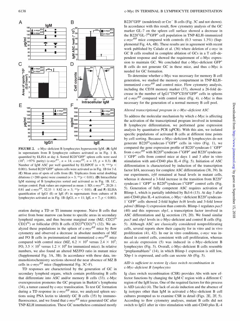
c-Myc–deficient B lymphocytes hypersecrete IgM
Our data showed that c-Myc–deficient mature B lymphocytes wereunable to generate the typical B220losyndecan-1+ cells. However,to determine whether functional ASC with an atypical surfacephenotype may be generated upon activation, we measured theamount of IgM by ELISA in the supernatants of cultured cells;IgM levels were similar for mutant and control cells (Fig. 2A).ELISPOT quantification of ASC number in these cultures indi-cated an ∼6-fold decrease in total ASC per well for c-Myc–deficientB lymphocytes compared with controls (697.9 6 335.6 versus4396.4 6 1006.2 ASC/well, respectively; Fig. 2B). c-Myc–deficientB lymphocytes thus generate fewer, although functional, IgM-secreting cells than do controls.
To account for similar IgM levels with fewer ASC, mutant Blymphocytes must secrete more IgM per cell and/or begin se-creting earlier than control cells after stimulation. In ELISPOT, weobserved comparable secretion kinetics by mutant and control ASC(Supplemental Fig. 1C). To analyze whether mutant ASC secretedmore IgM per cell, we used several approaches. We measured thearea of spots generated in the ELISPOT assay and found a 1.5-foldincrease in spot area from c-Myc–deficient compared with controlASC (0.0502 6 0.0058 versus 0.0286 6 0.0004 mm2; Fig. 2C,Supplemental Fig. 1D). As c-Myc–deficient B lymphocyte cul-tures had 6-fold fewer ASC than did controls, and Ig levels inELISA were similar in both cultures, average IgM secretion perASC was 3-fold higher in mutant than in control cells (661.0 655.9 versus 219.9 6 19.7 mg/105 ASC; Supplemental Fig. 1E).Consistent with these results, immunoprecipitation experimentsindicated similar amounts of IgM in c-Myc–deficient and controlB lymphocyte supernatants (Supplemental Fig. 1F). Finally, in-tracellular staining showed increased IgM in c-Myc–deficientcompared with control B cells (Fig. 2D). Ig hypersecretion wasrestricted to IgM, as we detected no IgG1 or IgE in mutant cellculture supernatants by ELISA (Fig. 2E, 2F). Based on these data,we concluded that after in vitro stimulation, c-Myc–deficient ASChypersecrete IgM and do not secrete IgG1 or IgE.
c-Myc–deficient B cells do not generate GC
We next examined the immune response against TD and TI Ags inthe context of c-Myc deficiency. With this aim in mind, we im-munized c-mycfl/fl and control mice with TD (TNP-KLH) or TIAgs (LPS) and analyzed them by flow cytometry. In our model,the GFP reporter identifies c-myc–deleted (GFP+) B cells thatundergo Cre-mediated deletion. As observed in our in vitro results(Fig. 1), generation of spleen B220losyndecan-1+GFP+ ASC wasimpaired in c-mycfl/fl mice compared with controls after LPS (0.3versus 1.8%) or TNP-KLH challenge (0.4 versus 1.2%) (Fig. 3A,3B). As for spleen, c-mycfl/fl mouse bone marrow showed no long-lived ASC (Supplemental Fig. 2A, 2B). Serum anti-TNPAb levelswere similar in c-mycfl/fl and control mice, probably due to thepresence of nondeleted B lymphocytes in the mutant mice (Sup-plemental Fig. 2C). c-Myc is therefore necessary for ASC gen-
FIGURE 1. c-Myc is needed for terminal B cell differentiation. (A and
B) c-Myc is necessary for the generation of B220losyndecan-1+ cells
in vitro. Sorted B220+GFP+ spleen cells from c-mycfl/fl and c-mycfl/+ mice
were activated with LPS (A) or anti-CD40 plus IL-4 (B) and analyzed by
flow cytometry 4 d later. For (A) and (B), we analyzed 12 mice per ge-
notype, with similar results.
The Journal of Immunology 6137
by guest on September 15, 2018
http://ww
w.jim
munol.org/
Dow
nloaded from
eration during a TD or TI immune response. Naive B cells thatarrive from bone marrow can home to specific areas in secondarylymphoid organs, and thus become marginal zone (MZ; CD23lo
CD21hi) or follicular (FO) B cells (CD23hiCD21lo) (32). We an-alyzed these populations in the spleen of c-mycfl/fl mice by flowcytometry and observed a decrease in absolute numbers of MZand FO B cells in preimmunized and immunized c-mycfl/fl micecompared with control mice (MZ, 6.2 3 104 versus 2.4 3 105;FO, 3.5 3 105 versus 1.2 3 106 for immunized mice). In relativenumbers, we also found an altered MZ/FO ratio in mutant mice(Supplemental Fig. 3A, 3B). In accordance with these data, im-munohistochemistry sections showed the near absence of MZ Bcells in c-mycfl/fl mice (Supplemental Fig. 3C–F).TD responses are characterized by the generation of GC in
secondary lymphoid organs, which contain proliferating B cellsthat differentiate into ASC and/or memory B cells (33). c-Mycoverexpression promotes the GC program in Burkitt’s lymphoma(34), a tumor caused by c-myc translocation. To test GC formationduring a TD response in c-mycfl/fl mice, we analyzed spleen sec-tions using PNA lectin to identify GC B cells (35) by immuno-fluorescence, and we found that c-mycfl/fl mice generated GC afterTNP-KLH immunization. These GC nonetheless contained mostly
B220+GFP- (nondeleted) or Cre2 B cells (Fig. 3C and not shown).In accordance with this result, flow cytometry analysis of the GCmarker GL-7 on the spleen cell surface showed a decrease inthe B220+GL-7hiGFP+ cell population in TNP-KLH–immunizedc-mycfl/fl mice compared with controls (0.3 versus 1.3%) (Sup-plemental Fig. 4A, 4B). These results are in agreement with recentwork published by Calado et al. (36) where deletion of c-myc inGC B cells resulted in complete ablation of GCs in a T cell–de-pendent response and showed the requirement of c-Myc expres-sion to maintain GC. We concluded that c-Myc–deficient GFP+
B cells do not generate GC in these mice, and thus c-Myc isneeded for GC formation.To determine whether c-Myc was necessary for memory B cell
generation, we studied the memory compartment in TNP-KLH–immunized c-mycfl/fl and control mice. Flow cytometry analysis,including the CD38 memory marker (37), showed a 26-fold de-crease in the number of IgG1+TNP+CD38+GFP+ cells in spleensof c-mycfl/fl compared with control mice (Fig. 4). c-Myc is thusnecessary for the generation of a normal memory B cell pool.
Altered transcriptional program in c-Myc–deficient ASC
To address the molecular mechanism by which c-Myc is affectingthe activation of the transcriptional program involved in terminalB lymphocyte differentiation, we performed gene expressionanalysis by quantitative PCR (qPCR). With this aim, we isolatedspecific populations of activated B cells at different time pointsby cell sorting. Because c-Myc–deficient B lymphocytes do notgenerate B220losyndecan-1+GFP+ cells in vitro (Fig. 1), wecompared the gene expression profile of B220+syndecan-12GFP+
from c-mycfl/fl with B220losyndecan-1+GFP+ and B220+syndecan-12GFP+ cells from control mice at days 1 and 3 after in vitrostimulation with anti-CD40 plus IL-4 (Fig. 5). Initiation of ASCdifferentiation is characterized by induction of the transcriptionfactor Irf4, necessary for complete ASC differentiation (38, 39). Inour experiments, irf4 remained at basal levels in mutant cells,whereas it showed a 3-fold increase in the transition from B220+
syndecan-12GFP+ to B220losyndecan-1+GFP+ control cells (Fig.5). Generation of fully competent ASC requires activation ofBlimp-1, which is partially inhibited by Bcl-6 (13). At day 3 afteranti-CD40 plus IL-4 activation, c-Myc2deficient B220+syndecan-12GFP+ cells showed 2-fold higher bcl6 levels and 3-fold lowerpdrm1 (Blimp-1) expression than controls. Blimp-1 regulates pax5(40) and this represses xbp1, a transcription factor involved inASC differentiation and Ig secretion (19, 20). We found similarpax5 and xbp1 levels in c-Myc–deficient and control B cells (Fig.5). Although ASC are classically considered nonproliferatingcells, several reports show their capacity for in vitro and in vivoproliferation (41, 42). In our in vitro conditions, c-myc was in-duced in control cells, consistent with cell proliferation, whereasno aicda expression (5) was induced in c-Myc–deficient Blymphocytes (Fig. 5). Overall, c-Myc–deficient B cells resemble“preplasmablasts” (14), in which Blimp-1 expression is still low,Xbp-1 is expressed, and cells can secrete Ab (Fig. 5).
AID is sufficient to restore Ig class switch recombination inc-Myc–deficient B lymphocytes
Ig class switch recombination (CSR) provides Abs with new ef-fector functions by changing the IgM C region with a different Cregion of the IgH locus. One of the required factors for this processis AID (aicda) (4). The lack of aicda induction and the absence ofIg isotypes other than IgM in activated c-Myc–deficient B cellcultures prompted us to examine CSR in detail (Figs. 2E, 2F, 5).According to flow cytometry analyses, mutant B cells did notswitch to IgG1 after in vitro stimulation with anti-CD40 plus IL-4
FIGURE 2. c-Myc–deficient B lymphocytes hypersecrete IgM. (A) IgM
in supernatants from B lymphocyte cultures activated as in Fig. 1 B,
quantified by ELISA at day 4. Sorted B220+GFP+ spleen cells were used
(105; .97% purity) (c-mycfl/+, n = 14; c-mycfl/fl, n = 15; p = 0.3). (B)
Number of IgM ASC per well quantified by ELISPOT (n = 9; ***p ,0.001). Sorted B220+GFP+ spleen cells were activated as in Fig. 1B for 3 d.
(C) Mean area of spots of cells from (B). Triplicates from serial doubling
dilutions (.200 spots) were counted (n = 3; **p , 0.01). (D) Intracellular
IgM staining of B lymphocytes sorted and activated as in Fig. 1B. I.C.,
isotype control. Peak values are expressed as mean6 SD; c-mycfl/+, 20.2660.81 and c-mycfl/fl, 52.33 6 8.82 (n = 3; **p , 0.01). (E and F) ELISA
quantification of IgG1 (E) or IgE (F) in supernatants from cultures of B
lymphocytes activated as in Fig. 1B (IgG1, n = 11; IgE, n = 7; p , 0.001).
6138 c-Myc IN TERMINAL B LYMPHOCYTE DIFFERENTIATION
by guest on September 15, 2018
http://ww
w.jim
munol.org/
Dow
nloaded from

(Fig. 6A, 6B). Germline transcripts must be generated for normalCSR (4). The fact that Ig1-Cg1 germline transcripts wereexpressed at normal levels allowed us to test whether AID ex-pression could restore CSR to IgG1 in c-Myc–deficient B cells(Fig. 6C). We thus bred c-mycfl/fl with a transgenic mouse thatexpresses aicda under the Ig k regulatory elements (Igk-AID
mice) (43). The c-mycfl/fl;Igk-AID mice generated B lymphocytesthat delete c-myc and express ectopic aicda (Supplemental Fig.4C, 4D). Flow cytometry showed that AID expression in c-Myc–deficient B lymphocytes was sufficient for IgG1 switching afterin vitro anti-CD40 plus IL-4 treatment (Fig. 6A). CSR generatescircular DNAs that originate specific transcripts (CT), a hallmark
FIGURE 3. c-Myc–deficient B cells do not generate B220losyndecan-1+ cells or GC after immunization. (A) Flow cytometry analysis of B220losyndecan-
1+GFP+ spleen cells from c-mycfl/fl and control mice immunized with LPS or TNP-KLH. (B) Absolute numbers of B220losyndecan-1+GFP+ cells in (A).
Preimmune: c-mycfl/+, n = 6; c-mycfl/fl, n = 4 (*p , 0.05); LPS: c-mycfl/+, n = 6; c-mycfl/fl, n = 7 (***p , 0.001); TNP-KLH: c-mycfl/+ and c-mycfl/fl, n = 4
(*p, 0.05). (C) Immunofluorescence of spleen sections. PNA lectin was used to identify GC B cells and anti-GFP was used to detect c-Myc–deficient cells.
Four to six spleen sections were analyzed per mouse (n = 6). Original magnification 3200.
FIGURE 4. c-Myc–deficient B cells do not generate memory B cells. (A) In immunized mice 12 d after a second TNP-KLH immunization, TNP-specific
memory B cells from spleen were identified in flow cytometry. We excluded IgM+, IgD+, Gr-1+, F4/80+ cells (MIX) and stained for TNP-BSA and the
indicated markers. (B) Absolute number of memory B cells (MIX2GFP+CD38+IgG1+TNP+) in (A) (n = 4; ***p , 0.001).
The Journal of Immunology 6139
by guest on September 15, 2018
http://ww
w.jim
munol.org/
Dow
nloaded from

of active CSR (44). To confirm the flow cytometry results usinga complementary technique (qPCR) we tested whether CSR cor-related with the presence of g1 CT (Ig 2 Cm) in B cells cultured
from c-mycfl/fl;Igk-AID mice. We observed an ∼4-fold increase ing1 CT transcripts in c-mycfl/fl;Igk-AID compared with c-mycfl/fl
mouse B lymphocytes (Fig. 6D). ELISA also showed a 6.8-fold
FIGURE 5. Gene expression analysis of activated c-Myc–deficient B cells in vitro. Sorted B220+GFP+ spleen cells from c-myc fl/fl and c-mycfl/+ mice
were activated with anti-CD40 plus IL-4 and resorted at days 1 and 3 to isolate B220+syndecan-12GFP+ and/or B220losyndecan-1+GFP+ cells. Mutant cells
did not generate B220losyndecan-1+GFP+ cells. Controls (filled columns) and mutant cells (open columns) are shown. RNA was prepared and qPCR
performed (see Materials and Methods). Results were normalized to b-actin. (n = 6; ***p , 0.001, **p , 0.01, *p , 0.05).
6140 c-Myc IN TERMINAL B LYMPHOCYTE DIFFERENTIATION
by guest on September 15, 2018
http://ww
w.jim
munol.org/
Dow
nloaded from
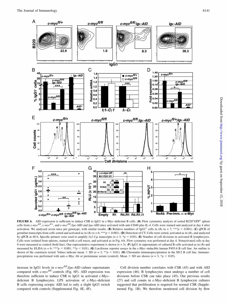
increase in IgG1 levels in c-mycfl/fl;Igk-AID culture supernatantscompared with c-mycfl/fl controls (Fig. 6F). AID expression wastherefore sufficient to induce CSR to IgG1 in activated c-Myc–deficient B lymphocytes. LPS activation of c-Myc–deficientB cells expressing ectopic AID led to only a slight IgG3 switchcompared with controls (Supplemental Fig. 4E, 4F).
Cell division number correlates with CSR (45) and with AIDexpression (46). B lymphocytes must undergo a number of celldivisions before CSR can take place (45). Our previous results(27) and cell counts in c-Myc–deficient B lymphocyte culturessuggested that proliferation is required for normal CSR (Supple-mental Fig. 1B). We therefore monitored cell division by flow
FIGURE 6. AID expression is sufficient to induce CSR to IgG1 in c-Myc–deficient B cells. (A) Flow cytometry analysis of sorted B220+GFP+ spleen
cells from c-mycfl/fl, c-mycfl/+, and c-mycfl/fl;Igk-AID and Igk-AID mice activated with anti-CD40 plus IL-4. Cells were stained and analyzed at day 4 after
activation. We analyzed seven mice per genotype, with similar results. (B) Relative numbers of IgG1+ cells in (A) (n = 7; ***p , 0.001). (C) qPCR of
germline transcripts from cells sorted and activated as in (A) (n = 6; ***p, 0.001). (D) Detection of CT. Cells were sorted, activated as in (A), and analyzed
by qPCR at 48 h. Specific primers were used to amplify Ig1-Cm transcripts (n = 3; *p , 0.05). (E) Number of cell divisions in activated B lymphocytes.
Cells were isolated from spleens, stained with a cell tracer, and activated as in Fig. 6A. Flow cytometry was performed at day 4. Nonactivated cells at day
0 were measured as control (bold line). One representative experiment is shown (n = 3). (F) IgG1 in supernatants of cultured B cells activated as in (A) and
measured by ELISA (n = 6; ***p , 0.001, **p , 0.01). (G) Luciferase reporter assays in the c-Myc–inducible human P493.6 B cell line. An outline is
shown of the constructs tested. Values indicate mean 6 SD (n = 3; **p , 0.01). (H) Chromatin immunoprecipitation in the M12 B cell line. Immuno-
precipitation was performed with anti–c-Myc Ab or preimmune serum (control). Mean 6 SD are shown (n = 3; *p , 0.05).
The Journal of Immunology 6141
by guest on September 15, 2018
http://ww
w.jim
munol.org/
Dow
nloaded from
cytometry and observed that in contrast to control cells, c-Myc–deficient B lymphocytes did not undergo even a single division(Fig. 6E). These results suggest that cell proliferation is requiredfor normal levels of CSR.
c-Myc transcriptionally regulates aicda expression
The fact that AID expression was sufficient to induce CSR, al-though at low levels, in c-Myc–deficient B cells led us to testwhether c-Myc transcriptionally regulates aicda. We screened theaicda genomic locus by computer analysis and identified a con-served putative c-Myc regulatory binding site (E-box) in bothhumans and mice within R4. R4 is located ∼10 kb upstream of theaicda promoter and has been defined as a cytokine response ele-ment that regulates aicda expression (47). Luciferase reporterassays in tetracycline-inducible, c-Myc-expressing human P493.6B cells and in the murine M12 B cell line indicated that the aicdapromoter alone (R1) was not induced by c-Myc. Nonetheless, useof a construct bearing R4 and R1 promoter regions led to c-Myc–dependent transactivation of aicda in both cell lines. A regionlocated in I1 known for B cell specificity and aicda repression (48)consistently abolished c-Myc–dependent transactivation (Fig. 6G,Supplemental Fig. 4G). To test whether c-Myc binds to a genomicfragment containing R4, we performed chromatin immunopre-cipitation and found 4-fold enrichment in amplification of geno-mic fragments containing R4 that were immunoprecipitated byanti–c-Myc Ab compared with preimmune sera (Fig. 6H). Weconclude that c-Myc binds to R4 and transcriptionally regulatesaicda.
DiscussionIn this study we analyzed c-Myc function in terminal B lymphocytedifferentiation of primary cells in vitro and in vivo by using ac-mycfl/fl mouse model that allows specific c-myc deletion in matureB lymphocytes. Additionally, this model allows GFP expression–based in vivo isolation and tracking of c-Myc–deficient B lym-phocytes. We tested the c-Myc contribution to terminal B lym-phocyte differentiation in different in vivo and in vitro settings. Ourdata identify c-Myc as a key regulator of not only the GC reactionbut also of terminal B cell differentiation, and therefore of thehumoral immune response.Our results show that c-Myc is needed for B lymphocyte dif-
ferentiation into B220losyndecan-1+ cells after LPS or anti-CD40plus IL-4 stimulation in vitro. c-Myc–deficient B cells did notdownregulate B220 or express the ASC marker syndecan-1+ at thecell surface. Despite this apparent role of c-Myc in terminal B celldifferentiation, the lack of cell proliferation in c-Myc–deficientB cells cannot rule out this process as the cause for this phenotype.Using various approaches, we found that despite this apparent
lack of ASC phenotype, c-Myc–deficient B lymphocytes hyper-secrete IgM. No significant changes were observed in expressionof the tested genes involved in the unfolded protein response (20).Many hyper-IgM syndromes show CSR defects, and some involvealtered AID expression (49). In our c-mycfl/fl mice, the lack ofaicda could contribute to IgM hypersecretion, although the geneexpression profile of c-Myc–deficient B cells argues for morecomplex c-Myc regulation. pdrm-1 (Blimp-1) is not induced atnormal levels in these mutants, and the functional interplay be-tween Irf4, Bcl-6, and Blimp-1 might be important in explainingthis phenotype. The low irf4 levels observed in c-Myc–deficientB cells might be insufficient to repress Bcl-6. This, as well as thelack Irf4-dependent activation of Blimp-1 (38), would prevent fullBlimp-1 induction and completion of the ASC differentiationprogram. Irf42/2 B cells do not become B220losyndecan-1+ anddo not undergo CSR or generate GD (10), as also observed in our
c-Myc–deficient B cells. Only ASC with very high Irf4 levels areable to induce Blimp-1 and fully differentiate into syndecan-1+
cells (39). We suggest a scenario in which c-Myc acts upstream ofIrf4 to trigger the terminal B cell differentiation program. Thispossibility is supported by an observation from Shaffer et al. (50),who showed that c-Myc binds to I1 of irf4 and activates its ex-pression in multiple myeloma and activated B cells. We examinedthe idea that c-myc repression is required but not sufficient forASC differentiation (17) by distinguishing which specific eventsthat lead to ASC generation can take place in the absence ofc-Myc. We observed that IgM secretion occurs without prolifera-tion, presumably due to normal expression of Xbp-1, whose levelsare not affected by the lack of c-Myc. Alternatively, the low levelsof Blimp-1 and Irf4 are likely contributing to create a “pre-plasmablast” (14) phenotype in secreting cells generated fromc-Myc–deficient B cells.Consistent with our in vitro results, both TD and TI responses
were severely compromised in c-mycfl/fl mice. c-Myc–deficientB lymphocytes have a severe impairment in the generation ofB220losyndecan-1+ cells in response to immunization with eitherTNP-KLH or LPS. These results correlated with the pattern ofexpression of c-myc during the formation of GC as shown byDominguez-Sola et al. (51).Serum analysis of immunized mice showed no significant dif-
ferences between mutant and control mice for the isotypes tested.The most likely interpretation of this result is the presence of cellsthat have not undergone Cre-mediated deletion (wild-type cells).The memory pool after TNP-KLH rechallenge is severely reducedin c-mycfl/fl mice. We cannot rule out a potential c-Myc role in thedifferentiation of these cells; however, c-myc deletion can takeplace at any time during in vivo differentiation. Additionally,memory cells have undergone CSR. These characteristics supportour idea that B cells have differentiated into memory B cells be-fore c-myc deletion and that the lack of expansion/proliferationresults almost in an undetectable number of cells.MZ B cells originate from an intermediate differentiation stage
(transitional cells) by a cell fate decision between follicular andMZcells (52). We observed a significant decrease in MZ c-Myc–de-ficient B cells, suggesting either dependence on proliferation toestablish a normal MZ pool and/or the implication of c-Myc inbalancing the MZ/FO ratio. In either case, c-Myc is needed fora normal MZ B cell compartment. This finding implicates c-Mycin the first stages of the B cell response to blood-borne encapsu-lated bacterial infections (innate-like immune response), beforedifferent isotypes other than IgM have been generated.Several reports suggest that CSR and cell proliferation are linked
by an unknown mechanism involving AID (45, 46). The absence ofcell proliferation and CSR in c-Myc–deficient B lymphocytesprovided an ideal context in which to test the relationship be-tween these two processes. Our data lead to several conclusions.First, in normal germline conditions, AID is sufficient to restoreCSR in c-Myc–deficient B lymphocytes. Second, normal levels ofCSR required cell proliferation, and thus give rise to only a smallpool of switched cells. The differences in the capacity to restoreswitching to different isotypes upon AID ectopic expression couldreflect additional c-Myc roles in B cells. These functions wouldaffect, for example, the generation of specific germline transcriptsand thus CSR. We next established direct transcriptional regula-tion of aicda and c-Myc in lymphoid cells by using reporter andchromatin immunoprecipitation assays; the induction kinetics ofc-myc and gene expression support such an interaction. Moreover,there is a direct correlation between c-Myc and AID expression inGC as shown by Dominguez-Sola et al. (51). c-Myc could act asa physiological link between cell proliferation and CSR, although,
6142 c-Myc IN TERMINAL B LYMPHOCYTE DIFFERENTIATION
by guest on September 15, 2018
http://ww
w.jim
munol.org/
Dow
nloaded from

as we have shown, these functions can be uncoupled mechanis-tically. We consider that the link between these two processesoffers a mechanism that promotes expansion of Ag-specificswitching cells and therefore a strong immune response. How-ever, c-Myc is not only regulating CSR through modulation ofAID expression. Our data strongly suggest that it is controllingother mechanisms that are essential for a full switching process.This points out the idea that AID does not need proliferation itselfto promote CSR, but highlights the necessity of studying otherc-Myc (in)direct target genes to explain the whole CSR processand its regulation.Using a number of criteria, we show that c-Myc regulates key
processes of terminal B lymphocyte differentiation in primaryB cells. Our data cannot rule out c-Myc–dependent cell prolifer-ation as a major player in terminal differentiation. However, it isdifficult to explain the phenotype relying on cell proliferationexclusively. The fact that c-Myc–deficient B cells generate IgMASC could suggest additional roles of c-Myc. The gene expressionprofile with key regulators of terminal differentiation affected bythe lack of c-Myc favor a more general role of this transcriptionfactor. Either in reference to cell proliferation or to more specificfunctions, c-Myc emerges as a principal player in the generationof a normal, potent immune response. Finally, identifying thecritical genes that control c-Myc–dependent proliferation is es-sential to characterize these functions in normal and pathologicalsituations in B lymphocytes.
AcknowledgmentsWe thank the Centro Nacional de Biotecnologıa animal facility, the
Departamento de Inmunologıa y Oncologıa, C. Mark for editorial assis-
tance, and J.M. Valpuesta for support. Igk-AID mice were provided by
M. Nussenzweig, and advice on ELISPOTs was provided by B. de Andres
and M.A.R. Marcos.
DisclosuresThe authors have no financial conflicts of interest.
References1. Calame, K. 2006. Transcription factors that regulate memory in humoral
responses. Immunol. Rev. 211: 269–279.2. Tarlinton, D., A. Radbruch, F. Hiepe, and T. Dorner. 2008. Plasma cell differ-
entiation and survival. Curr. Opin. Immunol. 20: 162–169.3. Neuberger, M. S. 2008. Antibody diversification by somatic mutation: from
Burnet onwards. Immunol. Cell Biol. 86: 124–132.4. Chaudhuri, J., U. Basu, A. Zarrin, C. Yan, S. Franco, T. Perlot, B. Vuong,
J. Wang, R. T. Phan, A. Datta, et al. 2007. Evolution of the immunoglobulinheavy chain class switch recombination mechanism. Adv. Immunol. 94: 157–214.
5. Muramatsu, M., K. Kinoshita, S. Fagarasan, S. Yamada, Y. Shinkai, and T. Honjo.2000. Class switch recombination and hypermutation require activation-inducedcytidine deaminase (AID), a potential RNA editing enzyme. Cell 102: 553–563.
6. Gourley, T. S., E. J. Wherry, D. Masopust, and R. Ahmed. 2004. Generation andmaintenance of immunological memory. Semin. Immunol. 16: 323–333.
7. Turner, C. A., Jr., D. H. Mack, and M. M. Davis. 1994. Blimp-1, a novel zincfinger-containing protein that can drive the maturation of B lymphocytes intoimmunoglobulin-secreting cells. Cell 77: 297–306.
8. Ye, B. H., G. Cattoretti, Q. Shen, J. Zhang, N. Hawe, R. de Waard, C. Leung,M. Nouri-Shirazi, A. Orazi, R. S. Chaganti, et al. 1997. The BCL-6 proto-oncogene controls germinal-centre formation and Th2-type inflammation. Nat.Genet. 16: 161–170.
9. Matsuyama, T., A. Grossman, H. W. Mittrucker, D. P. Siderovski, F. Kiefer,T. Kawakami, C. D. Richardson, T. Taniguchi, S. K. Yoshinaga, and T. W. Mak.1995. Molecular cloning of LSIRF, a lymphoid-specific member of the interferonregulatory factor family that binds the interferon-stimulated response element(ISRE). Nucleic Acids Res. 23: 2127–2136.
10. Mittrucker, H. W., T. Matsuyama, A. Grossman, T. M. Kundig, J. Potter,A. Shahinian, A. Wakeham, B. Patterson, P. S. Ohashi, and T. W. Mak. 1997.Requirement for the transcription factor LSIRF/IRF4 for mature B andT lymphocyte function. Science 275: 540–543.
11. Saito, M., J. Gao, K. Basso, Y. Kitagawa, P. M. Smith, G. Bhagat, A. Pernis,L. Pasqualucci, and R. Dalla-Favera. 2007. A signaling pathway mediatingdownregulation of BCL6 in germinal center B cells is blocked by BCL6 genealterations in B cell lymphoma. Cancer Cell 12: 280–292.
12. Shaffer, A. L., X. Yu, Y. He, J. Boldrick, E. P. Chan, and L. M. Staudt. 2000.BCL-6 represses genes that function in lymphocyte differentiation, inflamma-tion, and cell cycle control. Immunity 13: 199–212.
13. Tunyaplin, C., A. L. Shaffer, C. D. Angelin-Duclos, X. Yu, L. M. Staudt, andK. L. Calame. 2004. Direct repression of prdm1 by Bcl-6 inhibits plasmacyticdifferentiation. J. Immunol. 173: 1158–1165.
14. Kallies, A., J. Hasbold, K. Fairfax, C. Pridans, D. Emslie, B. S. McKenzie,A. M. Lew, L. M. Corcoran, P. D. Hodgkin, D. M. Tarlinton, and S. L. Nutt.2007. Initiation of plasma-cell differentiation is independent of the transcriptionfactor Blimp-1. Immunity 26: 555–566.
15. Shapiro-Shelef, M., K. I. Lin, L. J. McHeyzer-Williams, J. Liao,M. G. McHeyzer-Williams, and K. Calame. 2003. Blimp-1 is required for theformation of immunoglobulin secreting plasma cells and pre-plasma memoryB cells. Immunity 19: 607–620.
16. Lin, K. I., C. Angelin-Duclos, T. C. Kuo, and K. Calame. 2002. Blimp-1-dependent repression of Pax-5 is required for differentiation of B cells to im-munoglobulin M-secreting plasma cells. Mol. Cell. Biol. 22: 4771–4780.
17. Lin, K. I., Y. Lin, and K. Calame. 2000. Repression of c-myc is necessary but notsufficient for terminal differentiation of B lymphocytes in vitro. Mol. Cell. Biol.20: 8684–8695.
18. Lin, Y., K. Wong, and K. Calame. 1997. Repression of c-myc transcription byBlimp-1, an inducer of terminal B cell differentiation. Science 276: 596–599.
19. Reimold, A. M., N. N. Iwakoshi, J. Manis, P. Vallabhajosyula, E. Szomolanyi-Tsuda, E. M. Gravallese, D. Friend, M. J. Grusby, F. Alt, and L. H. Glimcher.2001. Plasma cell differentiation requires the transcription factor XBP-1. Nature412: 300–307.
20. Shaffer, A. L., M. Shapiro-Shelef, N. N. Iwakoshi, A. H. Lee, S. B. Qian,H. Zhao, X. Yu, L. Yang, B. K. Tan, A. Rosenwald, et al. 2004. XBP1, down-stream of Blimp-1, expands the secretory apparatus and other organelles, andincreases protein synthesis in plasma cell differentiation. Immunity 21: 81–93.
21. Oracki, S. A., J. A. Walker, M. L. Hibbs, L. M. Corcoran, and D. M. Tarlinton.2010. Plasma cell development and survival. Immunol. Rev. 237: 140–159.
22. Meyer, N., and L. Z. Penn. 2008. Reflecting on 25 years with MYC. Nat. Rev.Cancer 8: 976–990.
23. Nesbit, C. E., J. M. Tersak, and E. V. Prochownik. 1999. MYC oncogenes andhuman neoplastic disease. Oncogene 18: 3004–3016.
24. Zimmerman, K., and F. W. Alt. 1990. Expression and function of myc familygenes. Crit. Rev. Oncog. 2: 75–95.
25. Adams, J. M., A. W. Harris, C. A. Pinkert, L. M. Corcoran, W. S. Alexander,S. Cory, R. D. Palmiter, and R. L. Brinster. 1985. The c-myc oncogene driven byimmunoglobulin enhancers induces lymphoid malignancy in transgenic mice.Nature 318: 533–538.
26. de Alboran, I. M., E. Baena, and C. Martinez-A. 2004. c-Myc-deficientB lymphocytes are resistant to spontaneous and induced cell death. Cell DeathDiffer. 11: 61–68.
27. de Alboran, I. M., R. C. O’Hagan, F. Gartner, B. Malynn, L. Davidson,R. Rickert, K. Rajewsky, R. A. DePinho, and F. W. Alt. 2001. Analysis of C-MYC function in normal cells via conditional gene-targeted mutation. Immunity14: 45–55.
28. Hoffman, B., A. Amanullah, M. Shafarenko, and D. A. Liebermann. 2002. Theproto-oncogene c-myc in hematopoietic development and leukemogenesis. On-cogene 21: 3414–3421.
29. Vallespinos, M., D. Fernandez, L. Rodrıguez, J. Alvaro-Blanco, E. Baena,M. Ortiz, D. Dukovska, D. Martınez, A. Rojas, M. R. Campanero, and I. Morenode Alboran. 2011. B Lymphocyte commitment program is driven by the proto-oncogene c-Myc. J. Immunol. 186: 6726–6736.
30. Wu, S., C. Cetinkaya, M. J. Munoz-Alonso, N. von der Lehr, F. Bahram,V. Beuger, M. Eilers, J. Leon, and L. G. Larsson. 2003. Myc repressesdifferentiation-induced p21CIP1 expression via Miz-1-dependent interactionwith the p21 core promoter. Oncogene 22: 351–360.
31. Sanderson, R. D., P. Lalor, and M. Bernfield. 1989. B lymphocytes express andlose syndecan at specific stages of differentiation. Cell Regul. 1: 27–35.
32. Pillai, S., and A. Cariappa. 2009. The follicular versus marginal zoneB lymphocyte cell fate decision. Nat. Rev. Immunol. 9: 767–777.
33. McHeyzer-Williams, L. J., D. J. Driver, and M. G. McHeyzer-Williams. 2001.Germinal center reaction. Curr. Opin. Hematol. 8: 52–59.
34. Scheller, H., S. Tobollik, A. Kutzera, M. Eder, J. Unterlehberg, I. Pfeil, andB. Jungnickel. 2010. c-Myc overexpression promotes a germinal center-likeprogram in Burkitt’s lymphoma. Oncogene 29: 888–897.
35. Rose, M. L., M. S. Birbeck, V. J. Wallis, J. A. Forrester, and A. J. Davies. 1980.Peanut lectin binding properties of germinal centres of mouse lymphoid tissue.Nature 284: 364–366.
36. Calado, D. P., Y. Sasaki, S. A. Godinho, A. Pellerin, K. Kochert, B. P. Sleckman,I. M. de Alboran, M. Janz, S. Rodig, and K. Rajewsky. 2012. The cell-cycleregulator c-Myc is essential for the formation and maintenance of germinalcenters. Nat. Immunol. 13: 1092–1100.
37. Ridderstad, A., and D. M. Tarlinton. 1998. Kinetics of establishing the memoryB cell population as revealed by CD38 expression. J. Immunol. 160: 4688–4695.
38. Klein, U., S. Casola, G. Cattoretti, Q. Shen, M. Lia, T. Mo, T. Ludwig,K. Rajewsky, and R. Dalla-Favera. 2006. Transcription factor IRF4 controls plasmacell differentiation and class-switch recombination. Nat. Immunol. 7: 773–782.
39. Sciammas, R., A. L. Shaffer, J. H. Schatz, H. Zhao, L. M. Staudt, and H. Singh.2006. Graded expression of interferon regulatory factor-4 coordinates isotypeswitching with plasma cell differentiation. Immunity 25: 225–236.
40. Delogu, A., A. Schebesta, Q. Sun, K. Aschenbrenner, T. Perlot, andM. Busslinger. 2006. Gene repression by Pax5 in B cells is essential for bloodcell homeostasis and is reversed in plasma cells. Immunity 24: 269–281.
The Journal of Immunology 6143
by guest on September 15, 2018
http://ww
w.jim
munol.org/
Dow
nloaded from
41. Hasbold, J., L. M. Corcoran, D. M. Tarlinton, S. G. Tangye, and P. D. Hodgkin.2004. Evidence from the generation of immunoglobulin G-secreting cells thatstochastic mechanisms regulate lymphocyte differentiation. Nat. Immunol. 5:55–63.
42. Jego, G., N. Robillard, D. Puthier, M. Amiot, F. Accard, D. Pineau,J. L. Harousseau, R. Bataille, and C. Pellat-Deceunynck. 1999. Reactive plas-macytoses are expansions of plasmablasts retaining the capacity to differentiateinto plasma cells. Blood 94: 701–712.
43. Robbiani, D. F., A. Bothmer, E. Callen, B. Reina-San-Martin, Y. Dorsett,S. Difilippantonio, D. J. Bolland, H. T. Chen, A. E. Corcoran, A. Nussenzweig,and M. C. Nussenzweig. 2008. AID is required for the chromosomal breaks inc-myc that lead to c-myc/IgH translocations. Cell 135: 1028–1038.
44. Kinoshita, K., M. Harigai, S. Fagarasan, M. Muramatsu, and T. Honjo. 2001. Ahallmark of active class switch recombination: transcripts directed by I pro-moters on looped-out circular DNAs. Proc. Natl. Acad. Sci. USA 98: 12620–12623.
45. Hodgkin, P. D., J. H. Lee, and A. B. Lyons. 1996. B cell differentiation andisotype switching is related to division cycle number. J. Exp. Med. 184: 277–281.
46. Rush, J. S., M. Liu, V. H. Odegard, S. Unniraman, and D. G. Schatz. 2005.Expression of activation-induced cytidine deaminase is regulated by cell divi-
sion, providing a mechanistic basis for division-linked class switch recombina-tion. Proc. Natl. Acad. Sci. USA 102: 13242–13247.
47. Tran, T. H., M. Nakata, K. Suzuki, N. A. Begum, R. Shinkura, S. Fagarasan,T. Honjo, and H. Nagaoka. 2010. B cell-specific and stimulation-responsiveenhancers derepress Aicda by overcoming the effects of silencers. Nat. Immu-nol. 11: 148–154.
48. Gonda, H., M. Sugai, Y. Nambu, T. Katakai, Y. Agata, K. J. Mori, Y. Yokota, andA. Shimizu. 2003. The balance between Pax5 and Id2 activities is the key to AIDgene expression. J. Exp. Med. 198: 1427–1437.
49. Davies, E. G., and A. J. Thrasher. 2010. Update on the hyper immunoglobulin Msyndromes. Br. J. Haematol. 149: 167–180.
50. Shaffer, A. L., N. C. Emre, L. Lamy, V. N. Ngo, G. Wright, W. Xiao, J. Powell,S. Dave, X. Yu, H. Zhao, et al. 2008. IRF4 addiction in multiple myeloma.Nature 454: 226–231.
51. Dominguez-Sola, D., G. D. Victora, C. Y. Ying, R. T. Phan, M. Saito,M. C. Nussenzweig, and R. Dalla-Favera. 2012. The proto-oncogene MYC isrequired for selection in the germinal center and cyclic reentry. Nat. Immunol.13: 1083–1091.
52. Shapiro-Shelef, M., and K. Calame. 2005. Regulation of plasma-cell develop-ment. Nat. Rev. Immunol. 5: 230–242.
6144 c-Myc IN TERMINAL B LYMPHOCYTE DIFFERENTIATION
by guest on September 15, 2018
http://ww
w.jim
munol.org/
Dow
nloaded from


![Analyzing the effect of c-Myc oncogene and matrix ......expression of the c-Myc oncogene and matrix metolloproteninase-2 [MMP2] on the metastasis and prognosis of the malign melanoma](https://static.fdocuments.in/doc/165x107/60a7fab3d79f715ad65b87dd/analyzing-the-effect-of-c-myc-oncogene-and-matrix-expression-of-the-c-myc.jpg)