The Protein Folding Problem - Jay Ponder Lab Home...
28
The Protein Folding Problem Ken A. Dill, 1,2 S. Banu Ozkan, 3 M. Scott Shell, 4 and Thomas R. Weikl 5 1 Department of Pharmaceutical Chemistry, 2 Graduate Group in Biophysics, University of California, San Francisco, California 94143; email: [email protected] 3 Department of Physics, Arizona State University, Tempe, Arizona 85287; email: [email protected] 4 Department of Chemical Engineering, University of California, Santa Barbara, California 93106; email: [email protected] 5 Max Planck Institute of Colloids and Interfaces, Department of Theory and Bio-Systems, 14424 Potsdam, Germany; email: [email protected] Annu. Rev. Biophys. 2008. 37:289–316 The Annual Review of Biophysics is online at biophys.annualreviews.org This article’s doi: 10.1146/annurev.biophys.37.092707.153558 Copyright c 2008 by Annual Reviews. All rights reserved 1936-122X/08/0609-0289$20.00 Key Words structure prediction, funnel energy landscapes, CASP, folding code, folding kinetics Abstract The “protein folding problem” consists of three closely related puz- zles: (a) What is the folding code? (b) What is the folding mechanism? (c) Can we predict the native structure of a protein from its amino acid sequence? Once regarded as a grand challenge, protein fold- ing has seen great progress in recent years. Now, foldable proteins and nonbiological polymers are being designed routinely and mov- ing toward successful applications. The structures of small proteins are now often well predicted by computer methods. And, there is now a testable explanation for how a protein can fold so quickly: A protein solves its large global optimization problem as a series of smaller local optimization problems, growing and assembling the native structure from peptide fragments, local structures first. 289
Transcript of The Protein Folding Problem - Jay Ponder Lab Home...

The Protein FoldingProblemKen A. Dill,1,2 S. Banu Ozkan,3 M. Scott Shell,4
and Thomas R. Weikl51Department of Pharmaceutical Chemistry, 2Graduate Group in Biophysics,University of California, San Francisco, California 94143;email: [email protected] of Physics, Arizona State University, Tempe, Arizona 85287;email: [email protected] of Chemical Engineering, University of California, Santa Barbara,California 93106; email: [email protected] Planck Institute of Colloids and Interfaces, Department of Theory andBio-Systems, 14424 Potsdam, Germany; email: [email protected]
Annu. Rev. Biophys. 2008. 37:289–316
The Annual Review of Biophysics is online atbiophys.annualreviews.org
This article’s doi:10.1146/annurev.biophys.37.092707.153558
Copyright c© 2008 by Annual Reviews.All rights reserved
1936-122X/08/0609-0289$20.00
Key Words
structure prediction, funnel energy landscapes, CASP, foldingcode, folding kinetics
AbstractThe “protein folding problem” consists of three closely related puz-zles: (a) What is the folding code? (b) What is the folding mechanism?(c) Can we predict the native structure of a protein from its aminoacid sequence? Once regarded as a grand challenge, protein fold-ing has seen great progress in recent years. Now, foldable proteinsand nonbiological polymers are being designed routinely and mov-ing toward successful applications. The structures of small proteinsare now often well predicted by computer methods. And, there isnow a testable explanation for how a protein can fold so quickly: Aprotein solves its large global optimization problem as a series ofsmaller local optimization problems, growing and assembling thenative structure from peptide fragments, local structures first.
289

Contents
INTRODUCTION. . . . . . . . . . . . . . . . . 290THE FOLDING CODE: WHAT
BALANCE OF FORCESENCODES NATIVESTRUCTURES? . . . . . . . . . . . . . . . . 290Anfinsen’s Thermodynamic
Hypothesis . . . . . . . . . . . . . . . . . . . . 290One Dominant Driving Force
or Many Small Ones? . . . . . . . . . . 291Designing New Proteins and
Nonbiological Foldamers . . . . . . 292COMPUTATIONAL PROTEIN
STRUCTURE PREDICTIONIS INCREASINGLYSUCCESSFUL . . . . . . . . . . . . . . . . . . 293CASP: A Community-Wide
Experiment . . . . . . . . . . . . . . . . . . . 293ARE THERE MECHANISMS OF
PROTEIN FOLDING?. . . . . . . . . . 294There Have Been Big Advances in
Experimental and TheoreticalMethods . . . . . . . . . . . . . . . . . . . . . . 294
The PSB Plot: Folding SpeedCorrelates with the Localnessof Contacts in the NativeStructure. . . . . . . . . . . . . . . . . . . . . . 295
Proteins Fold on Funnel-ShapedEnergy Landscapes . . . . . . . . . . . . 296
The Zipping and AssemblyHypothesis for the FoldingRoutes . . . . . . . . . . . . . . . . . . . . . . . . 300
PHYSICS-BASED MODELING OFFOLDING AND STRUCTUREPREDICTION . . . . . . . . . . . . . . . . . . 301
SUMMARY. . . . . . . . . . . . . . . . . . . . . . . . . 303
INTRODUCTION
The protein folding problem is the question ofhow a protein’s amino acid sequence dictatesits three-dimensional atomic structure. Thenotion of a folding “problem” first emergedaround 1960, with the appearance of the firstatomic-resolution protein structures. Someform of internal crystalline regularity was pre-
viously expected (117), and α-helices had beenanticipated by Linus Pauling and colleagues(180, 181), but the first protein structures—ofthe globins—had helices that were packedtogether in unexpected irregular ways. Sincethen, the protein folding problem has cometo be regarded as three different problems:(a) the folding code: the thermodynamicquestion of what balance of interatomicforces dictates the structure of the protein,for a given amino acid sequence; (b) proteinstructure prediction: the computationalproblem of how to predict a protein’s nativestructure from its amino acid sequence; and(c) the folding process: the kinetics questionof what routes or pathways some proteinsuse to fold so quickly. We focus here onlyon soluble proteins and not on fibrous ormembrane proteins.
THE FOLDING CODE: WHATBALANCE OF FORCES ENCODESNATIVE STRUCTURES?
Anfinsen’s ThermodynamicHypothesis
A major milestone in protein science wasthe thermodynamic hypothesis of ChristianAnfinsen and colleagues (3, 92). From hisnow-famous experiments on ribonuclease,Anfinsen postulated that the native structureof a protein is the thermodynamically stablestructure; it depends only on the amino acidsequence and on the conditions of solution,and not on the kinetic folding route. It be-came widely appreciated that the native struc-ture does not depend on whether the proteinwas synthesized biologically on a ribosome orwith the help of chaperone molecules, or if,instead, the protein was simply refolded asan isolated molecule in a test tube. [Thereare rare exceptions, however, such as insulin,α-lytic protease (203), and the serpins (227),in which the biologically active form is ki-netically trapped.] Two powerful conclusionsfollowed from Anfinsen’s work. First, it en-abled the large research enterprise of in vitro
290 Dill et al.

protein folding that has come to understandnative structures by experiments inside testtubes rather than inside cells. Second, the An-finsen principle implies a sort of division oflabor: Evolution can act to change an aminoacid sequence, but the folding equilibrium andkinetics of a given sequence are then mattersof physical chemistry.
One Dominant Driving Forceor Many Small Ones?
Prior to the mid-1980s, the protein foldingcode was seen a sum of many different smallinteractions—such as hydrogen bonds, ionpairs, van der Waals attractions, and water-mediated hydrophobic interactions. A keyidea was that the primary sequence encodedsecondary structures, which then encodedtertiary structures (4). However, throughstatistical mechanical modeling, a differentview emerged in the 1980s, namely, that thereis a dominant component to the folding code,that it is the hydrophobic interaction, that thefolding code is distributed both locally andnonlocally in the sequence, and that a protein’ssecondary structure is as much a consequenceof the tertiary structure as a cause of it(48, 49).
Because native proteins are only 5–10 kcal/mol more stable than their denaturedstates, it is clear that no type of intermolec-ular force can be neglected in folding andstructure prediction (238). Although it re-mains challenging to separate in a clean andrigorous way some types of interactions fromothers, here are some of the main observa-tions. Folding is not likely to be dominated byelectrostatic interactions among charged sidechains because most proteins have relativelyfew charged residues; they are concentrated inhigh-dielectric regions on the protein surface.Protein stabilities tend to be independent ofpH (near neutral) and salt concentration, andcharge mutations typically lead to small effectson structure and stability. Hydrogen-bondinginteractions are important, because essentiallyall possible hydrogen-bonding interactions
are generally satisfied in native structures. Hy-drogen bonds among backbone amide andcarbonyl groups are key components of allsecondary structures, and studies of mutationsin different solvents estimate their strengthsto be around 1–4 kcal/mol (21, 72) or stronger(5, 46). Similarly, tight packing in proteins im-plies that van der Waals interactions are im-portant (28).
However, the question of the folding codeis whether there is a dominant factor thatexplains why any two proteins, for example,lysozyme and ribonuclease, have different na-tive structures. This code must be written inthe side chains, not in the backbone hydrogenbonding, because it is through the side chainsthat one protein differs from another. Thereis considerable evidence that hydrophobic in-teractions must play a major role in proteinfolding. (a) Proteins have hydrophobic cores,implying nonpolar amino acids are driven tobe sequestered from water. (b) Model com-pound studies show 1–2 kcal/mol for trans-ferring a hydrophobic side chain from wa-ter into oil-like media (234), and there aremany of them. (c) Proteins are readily de-natured in nonpolar solvents. (d ) Sequencesthat are jumbled and retain only their cor-rect hydrophobic and polar patterning fold totheir expected native states (39, 98, 112, 118),in the absence of efforts to design packing,charges, or hydrogen bonding. Hydrophobicand polar patterning also appears to be a keyto encoding of amyloid-like fibril structures(236).
What stabilizes secondary structures? Be-fore any protein structure was known, LinusPauling and colleagues (180, 181) inferredfrom hydrogen-bonding models that proteinsmight have α-helices. However, secondarystructures are seldom stable on their own insolution. Although different amino acids havedifferent energetic propensities to be in sec-ondary structures (6, 41, 55, 100), there arealso many “chameleon” sequences in naturalproteins, which are peptide segments that canassume either helical or β conformations de-pending on their tertiary context (158, 162).
www.annualreviews.org • The Protein Folding Problem 291
2°
str
uc
ture
([θ
] 22
2)
15
a b c
0 5 100
15
5
10
(radius)–3
Native fold
Partially folded structures
Figure 1(a) Binary code. Experiments show that a primarily binary hydrophobic-polar code is sufficient to foldhelix-bundle proteins (112). Reprinted from Reference 112 with permission from AAAS.(b) Compactness stabilizes secondary structure, in proteins, from lattice models. (c) Experimentssupporting panel b, showing that compactness correlates with secondary structure content in nonnativestates of many different proteins (218). Reprinted from Reference 218 with permission.
Three-helix bundle
[Alcohol]
FR
ET
eff
icie
ncy
0.0
1.0
20%0%
Q
FP
H
OR
N NH2
n
H
R
O
N NH2
n
a
b
Peptide
Peptoid
c
Methanol
Propanol
Ethanol
Designed molecule
Experimentally determinedstructure
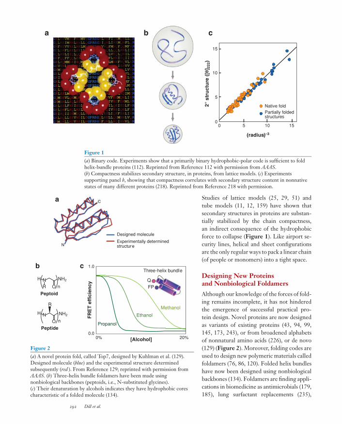
Figure 2(a) A novel protein fold, called Top7, designed by Kuhlman et al. (129).Designed molecule (blue) and the experimental structure determinedsubsequently (red ). From Reference 129; reprinted with permission fromAAAS. (b) Three-helix bundle foldamers have been made usingnonbiological backbones (peptoids, i.e., N-substituted glycines).(c) Their denaturation by alcohols indicates they have hydrophobic corescharacteristic of a folded molecule (134).
Studies of lattice models (25, 29, 51) andtube models (11, 12, 159) have shown thatsecondary structures in proteins are substan-tially stabilized by the chain compactness,an indirect consequence of the hydrophobicforce to collapse (Figure 1). Like airport se-curity lines, helical and sheet configurationsare the only regular ways to pack a linear chain(of people or monomers) into a tight space.
Designing New Proteinsand Nonbiological Foldamers
Although our knowledge of the forces of fold-ing remains incomplete, it has not hinderedthe emergence of successful practical pro-tein design. Novel proteins are now designedas variants of existing proteins (43, 94, 99,145, 173, 243), or from broadened alphabetsof nonnatural amino acids (226), or de novo(129) (Figure 2). Moreover, folding codes areused to design new polymeric materials calledfoldamers (76, 86, 120). Folded helix bundleshave now been designed using nonbiologicalbackbones (134). Foldamers are finding appli-cations in biomedicine as antimicrobials (179,185), lung surfactant replacements (235),
292 Dill et al.

cytomegalovirus inhibitors (62), and siRNAdelivery agents (217). Hence, questions ofdeep principle are no longer bottlenecks todesigning foldable polymers for practical ap-plications and new materials.
COMPUTATIONAL PROTEINSTRUCTURE PREDICTION ISINCREASINGLY SUCCESSFUL
A major goal of computational biology hasbeen to predict a protein’s three-dimensionalnative structure from its amino acid sequence.This could help to (a) accelerate drug dis-covery by replacing slow, expensive struc-tural biology experiments with faster, cheapercomputer simulations, and (b) annotate pro-tein function from genome sequences (9).With the rapid growth of experimentally de-termined structures available in the ProteinDatabank (PDB), protein structure predic-tion has become as much a problem of infer-ence and machine learning as it is of proteinphysics.
Among the earliest uses of proteindatabases to infer protein structures weresecondary structure prediction algorithms(33, 34, 190). In the mid-1980s, several groupsbegan using the methods of computationalphysics—atomic force fields plus Monte Carlosampling—to compute the structures of theMet-enkephalin, a five-residue peptide (95,141). The early 1990s saw significant stridesin using databases and homology detectionalgorithms to assemble structures from ho-mologous sequences (192) and to recognizefolds by threading unknown sequences ontothree-dimensional structures from a database(111). A key advance was the exploitation ofevolutionary relationships among sequencesthrough the development of robust sequencealignment methods (32, 64, 224).
CASP: A Community-WideExperiment
In 1994, John Moult invented CASP (CriticalAssessment of Techniques for Protein Struc-
ture Prediction) (165), a biennial, community-wide blind test to predict the unknown struc-tures of proteins. Organizers identify pro-teins likely to be solved or whose structureshave not yet been released, and predictorshave roughly 3–5 weeks to predict their nativestructures. CASP has grown from 35 predic-tor groups and 24 target sequences in CASP1in 1994 to over 200 groups and 100+ targetsin CASP7 in 2006.
Over the seven CASPs, two trends areclear (164, 219). First, although much re-mains to be done, there has been substantialimprovement in protein structure prediction.Web servers and software packages oftenpredict the native structure of small, single-domain proteins to within about 2–6 A oftheir experimental structures (8, 17, 242). Inaddition, fast-homology methods are com-puting approximate folds for whole genomes(182, 214). Figure 3 shows a quantitativeassessment of performance at the first fiveCASP meetings. The most significant gainshave occurred in the alignments of targetsto homologs, the detection of evolutionarilydistant homologs, and the generation ofreasonable models for difficult targets that do
CASP5
Target difficultyEasy Difficult
Pre
dic
tio
n s
uccess (
%)
100
0
CASP4
CASP3
CASP2
CASP1
Figure 3Progress in protein structure prediction in CASP1–5 (219). The y-axiscontains the GDT TS score, the percentage of model residues that can besuperimposed on the true native structure, averaged over four resolutionsfrom 1 to 8 A (100% is perfect). The x-axis is the ranked target difficulty,measured by sequence and structural similarities to proteins in the PDB atthe time of the respective CASP. This shows that protein structureprediction on easy targets is quite good and is improving for targets ofintermediate difficulty. Reprinted from Reference 219 with permission.
www.annualreviews.org • The Protein Folding Problem 293

not have templates (new folds). Since CASP5,predictions have also benefited from the useof metaservers, which solicit and establishconsensus among predictions from multiplealgorithms. Second, while most methodsrely on both physics and bioinformatics,the most successful methods currently drawheavily from knowledge contained in nativestructural databases. Bioinformatics methodshave benefited from the growth in size of thePDB (9, 219).
The following challenges remain (8, 164,219): (a) to refine homology models beyondthose of the best template structures; (b) toreduce errors to routinely better than 3 A,particularly for proteins that are large, havesignificant β content, are new folds, or havelow homology; (c) to handle large multido-main or domain-swapped proteins; (d ) to ad-dress membrane proteins; and (e) to predictprotein-protein interactions. Structural ge-nomics is likely to help here (87, 222). Inany case, the current successes in computer-based predictions of native protein struc-tures are far beyond what was expected thirtyyears ago, when structure prediction seemedimpossible.
ARE THERE MECHANISMSOF PROTEIN FOLDING?
In 1968, Cyrus Levinthal first noted the puz-zle that even though they have vast confor-mational spaces, proteins can search and con-verge quickly to native states, sometimes inmicroseconds. How do proteins find their na-tive states so quickly? It was postulated thatif we understood the physical mechanism ofprotein folding, it could lead to fast computeralgorithms to predict native structures fromtheir amino acid sequences. In its descriptionof the 125 most important unsolved problemsin science, Science magazine framed the prob-lem this way: “Can we predict how proteinswill fold? Out of a near infinitude of possibleways to fold, a protein picks one in just tens ofmicroseconds. The same task takes 30 yearsof computer time” (1).
The following questions of principle havedriven the field: How can all the denaturedmolecules in a beaker find the same na-tive structure, starting from different con-formations? What conformations are notsearched? Is folding hierarchical (10, 119)?Which comes first: secondary or tertiarystructure (80, 239)? Does the protein col-lapse to compact structures before struc-ture formation, or before the rate-limitingstep (RLS), or are they concurrent (7, 89,101, 195, 205, 213)? Are there folding nuclei(58, 152)?
Several models have emerged. In thediffusion-collision model, microdomainstructures form first and then diffuse andcollide to form larger structures (115, 116).The nucleation-condensation mechanism(70) proposes that a diffuse transition state en-semble (TSE) with some secondary structurenucleates tertiary contacts. Some proteins,such as helical bundles, appear to follow ahierarchical diffusion-collision model (155,169) in which secondary structure forms andassembles in a hierarchical order. In hierar-chic condensation (139), the chain searchesfor compact, contiguous structured units,which are then assembled into the foldedstate. Or, proteins may fold via the stepwiseassembly of structural subunits called foldons(22, 126), or they may search for topomers,which are largely unfolded states that havenative-like topologies (45, 150). Thesemodels are not mutually exclusive.
There Have Been BigAdvances in Experimentaland Theoretical Methods
The search for folding mechanisms has drivenmajor advances in experimental protein sci-ence. These include fast laser temperature-jump methods (22); mutational methodsthat give quantities called φ-values (71, 84,106) [now also used for ion-channel kinet-ics and other rate processes (42)] or ψ-values (204), which can identify those residuesmost important for folding speed; hydrogen
294 Dill et al.

exchange methods that give monomer-levelinformation about folding events (125, 149);and the extensive exploration of protein modelsystems, including cytochrome c, CI2, bar-nase, apomyoglobin, the src, α-spectrin, andfyn SH3 domains, proteins L and G, WW do-mains, trpzip, and trp cage (154). In addition,peptide model experimental test systems pro-vide insights into the fast early-folding events(14, 109, 124). Furthermore, single-moleculemethods are beginning to explore the con-formational heterogeneity of folding (23, 133,166, 194).
There have been corresponding advancesin theory and computation. Computer-basedmolecular minimization methods were firstapplied to protein structures in the 1960s(18, 79, 171), followed by molecular dynam-ics (140, 155), improved force fields (40) (re-viewed in Reference 114), weighted samplingand multi-temperature methods (130, 210),highly parallelized codes for supercomputers(2, 57), and distributed grid computing meth-ods such as Folding@home (198, 241). Mod-els having less atomic detail also emerged toaddress questions more global and less de-tailed in nature about protein conformationalspaces: (a) The Go model (82), which was in-tended to see if a computer could find thenative structure if native guiding constraintswere imposed, is now widely used to studyfolding kinetics of proteins having known na-tive structures (37, 38, 113, 197, 225). (b) Morephysical models, typically based on polymer-like lattices, are used to study the static anddynamic properties of conformational spaces(19, 50). (c) Master-equation approaches canexplore dynamics in heterogeneous systems(26, 36, 77, 138, 161, 176, 231, 232). Below,we describe some of what has been learnedfrom these studies.
The PSB Plot: Folding SpeedCorrelates with the Localness ofContacts in the Native Structure
One of the few universal features of proteinfolding kinetics was first observed by Plaxco,
0
14
8 22
R = 0.75
RCO (%)
ln k
f
Figure 4Folding rate versus relative contact order (ameasure of localness of contacts in the nativestructure) for the 48 two-state proteins given inReference 91, showing that proteins with the mostlocal contacts fold faster than proteins with morenonlocal contacts.
Simons, and Baker (PSB), namely, that thefolding speed of a protein is correlated witha topological property of its native struc-ture (88, 184). As shown in Figure 4, Plaxcoet al. found that the folding rates of two-stateproteins—now known to vary more than 8 or-ders of magnitude—correlate with the aver-age degree to which native contacts are localwithin the chain sequence: Fast-folders usu-ally have mostly local structure, such as he-lices and tight turns. Slow-folders usually havemore nonlocal structure, such as β-sheets(184), although there are exceptions (237).Folding rates have been subsequently foundto correlate well with other native topologi-cal parameters such as the protein’s effectivechain length (chain length minus the num-ber of amino acids in helices) (107), secondarystructure length (104), sequence-distant con-tacts per residue (90), the fraction of contactsthat are sequence distant (163), the total con-tact distance (245), and intrinsic propensities,for example, of α-helices (131). And, thereare now also methods that predict the foldingrate from the sequence (91, 186). It followsthat a protein typically forms smaller loopsand turns faster than it forms larger loopsand turns, consistent with the so-called zip-ping and assembly (ZA) mechanism, describedbelow, which postulates that search speed is
www.annualreviews.org • The Protein Folding Problem 295
governed by the effective loop sizes [the ef-fective contact order (ECO) (53, 73)] that thechain must search at any step.
Proteins Fold on Funnel-ShapedEnergy Landscapes
Why has folding been regarded as so chal-lenging? The issue is the astronomical num-ber of conformations a protein must searchto find its native state. Models arose in the1980s to study the nature of the conforma-tional space (19, 47), i.e., the shape of theenergy landscape, which is the mathemati-cal function F(φ, ϕ, X) that describes theintramolecular-plus-solvation free energy ofa given protein as a function of the micro-scopic degrees of freedom. A central goal hasbeen to quantify the statistical mechanical par-tition function, a key component of which isthe density of states (DOS), i.e., the number ofconformations at each energy level. In simplecases, the logarithm of the DOS is the con-formational entropy. Such entropies have notbeen determinable through all-atom model-ing, because that would require astronomi-cal amounts of computational sampling (al-though replica-exchange methods can now dothis for very small peptides). Hence under-standing a protein’s DOS and its entropieshas required simplified models, such as mean-field polymer and lattice treatments (51), spin-
glass theories (19, 47), or exact enumerationsin minimalist models (132).
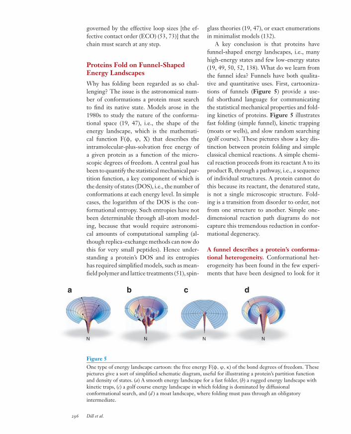
A key conclusion is that proteins havefunnel-shaped energy landscapes, i.e., manyhigh-energy states and few low-energy states(19, 49, 50, 52, 138). What do we learn fromthe funnel idea? Funnels have both qualita-tive and quantitative uses. First, cartooniza-tions of funnels (Figure 5) provide a use-ful shorthand language for communicatingthe statistical mechanical properties and fold-ing kinetics of proteins. Figure 5 illustratesfast folding (simple funnel), kinetic trapping(moats or wells), and slow random searching(golf course). These pictures show a key dis-tinction between protein folding and simpleclassical chemical reactions. A simple chemi-cal reaction proceeds from its reactant A to itsproduct B, through a pathway, i.e., a sequenceof individual structures. A protein cannot dothis because its reactant, the denatured state,is not a single microscopic structure. Fold-ing is a transition from disorder to order, notfrom one structure to another. Simple one-dimensional reaction path diagrams do notcapture this tremendous reduction in confor-mational degeneracy.
A funnel describes a protein’s conforma-tional heterogeneity. Conformational het-erogeneity has been found in the few experi-ments that have been designed to look for it
a b c d
N N N N
Figure 5One type of energy landscape cartoon: the free energy F(φ,ϕ, κ) of the bond degrees of freedom. Thesepictures give a sort of simplified schematic diagram, useful for illustrating a protein’s partition functionand density of states. (a) A smooth energy landscape for a fast folder, (b) a rugged energy landscape withkinetic traps, (c) a golf course energy landscape in which folding is dominated by diffusionalconformational search, and (d ) a moat landscape, where folding must pass through an obligatoryintermediate.
296 Dill et al.

(16, 143, 157, 206, 209, 244). For example, us-ing time-resolved FRET with four differentintramolecular distances, Sridevi et al. (206)found in Barstar that (a) that the chain entropyincreases as structures become less stable,(b) that there are multiple folding routes, and(c) that different routes dominate under differ-ent folding conditions. Moreover, changingthe denaturant can change the dominant path-way, implying heterogeneous kinetics (143).Figure 6 shows the funnel landscape thathas been determined by extensive mutationalanalysis of the seven ankyrin sequence repeatsof the Notch ankyrin repeat domain (16, 157,209).
A funnel describes a protein’s chainentropy. The funnel idea first arose to ex-plain denaturation, the balance between thechain entropy and the forces of folding (48).Proteins denature at high temperatures be-cause there are many states of high energyand fewer states of low energy, that is, thelandscape is funneled. For cold unfolding, theshape of the funnel changes with temperaturebecause of free-energy changes of the aqueoussolvent. When you can accurately compute a
-6
0
6
ΔG
kc
al/
mo
l
Figure 6The experimentally determined energy landscape of the seven ankyrinrepeats of the Notch receptor (16, 157, 209). The energy landscape isconstructed by measuring the stabilities of folded fragments for a series ofoverlapping modular repeats. Each horizontal tier presents the partiallyfolded fragments with the same number of repeats. Reprinted fromReference 157 with permission.
protein’s DOS, you can predict the protein’sfree energy of folding and its denaturation andcooperativity properties. Figure 7 shows anexample in which the DOS (set onto its sideto illustrate the funnel) was found by exten-sive lattice enumeration for F13W∗, a three-helix bundle, with predictions compared toexperiments (146).
Native
En
erg
y (
kc
al/
mo
l)
ln (conformation count)
–50
0 80
200
1
0.5
500 1000
1
0.5
4 6
Transitionstates
GuHCl (M)
Temperature (°C)
Fra
cti
on
na
tiv
eF
racti
on
nati
ve
Figure 7(Left) The density of states (DOS) cartoonized as an energy landscape for the three-helix bundle proteinF13W∗: DOS (x-axis) versus the energy (y-axis). (Right) Denaturation predictions versus experiments(146). The peak free energy (here, where the DOS is minimum), typically taken to be the transition state,is energetically very close to native.
www.annualreviews.org • The Protein Folding Problem 297

Funnels provide a microscopic frameworkfor folding kinetics. Folding kinetics is tra-ditionally described by simple mass actionmodels, such as D → I → N (on-path inter-mediate I between the denatured state D andnative state N) or X → D → N (off-path in-termediate X), where the symbol I or X rep-resents macrostates that are invoked for thepurpose of curve-fitting experimental kinet-ics data. In contrast, funnel models or master-equation models aim to explain the kineticsin terms of underlying physical forces. Theyaim to predict the microstate composition ofthose macrostates, for example. The states inmaster-equation models differ from those inmass-action models insofar as the former aremore numerous, more microscopic, are de-fined by structural or energetic criteria, andare arranged kinetically to reflect the underly-
ing funnel-like organization of the dynamicalflows.
For example, Figure 8b shows a master-equation model for the folding of SH3, illus-trating the apparently paradoxical result thatfolding can be serial and parallel at the sametime. The protein has multiple routes avail-able. However, one of the dominant seriespathways is U → B → BD → BDE → BCDE→ N. BD is the TSE because it is the dynam-ically least populated state. B precedes BDdiagrammatically in series in this pathway. Yet,the probability bucket labeled B does not firstfill up and then empty into BD; rather the lev-els in both buckets, B and BD, rise and fall to-gether and hence are dynamically in parallel.Such series and parallel steps are also seen incomputer simulations of CI2 (189), for exam-ple. Sometimes a chain contact A forms before
b
ABCE1.9
ACDE
BDE0.2
ABDE−1.3
B2.0
ABD2.9
ABCD2.4
BD2.4
A2.5
ACE2.9 −1.3
AC4.0
0.0
C1.5
BCD1.9
ABCDE−2.3
BCDE−0.3
BC3.5
0
a
R TS P
R
Time
10-4
10-2
1
0.1 10 1000
0
B
BDBDE
BCDE
ABCDE
Pi(t)
10-6
10-4
10-2
1
0.1 10 1000
Time
Pi(t)
TS
P
A
D
E
B
C
10 20 30 40 50
10
20
30
40
50β strand
Diverging turn DT 3-10 helix
Loop
Figure 8(a) A simple single-pathway system. R, reactant; TS, transition state; P, product. (b) Pathway diagram ofSH3 folding from a master-equation model. The native protein has five contact clusters: A = RT loop;B = β2β3; C = β3β4; D = RT loop-β4; and E = β1β5. Combined letters, such as BD, mean thatmultiple contact clusters have formed. Funneling occurs toward the right, because the symbols on the leftindicate large ensembles, whereas the symbols on the right are smaller ensembles. The numbers indicatefree energies relative to the denatured state. The arrows between the states are colored to indicatetransition times between states. The slowest steps are in red; the fastest steps in green. BD is thetransition state ensemble because it is the highest free energy along the dominant route. While B and BDwould seem to be obligatorily in series, the time evolutions of these states show that they actually rise andfall in parallel (232).
298 Dill et al.

another contact B in nearly all the simulationtrajectories (series-like). But another contactC may form before B in some trajectories andafter B in others (parallel-like). Some foldingis sequential, as in Fyn SH3 (123), cytochrome(66), T4 lysozyme (24), and Im7 (74), andsome folding is parallel, as in cytochrome C(83) and HEW lyzosyme (151).
Or, consider the traditional idea that a re-action’s RLS coincides with the point of thehighest free energy, �G‡, along the reactioncoordinate. As a matter of principle, the RLSneed not necessarily coincide with the free-energy barrier. There is evidence that theymay not coincide for some protein foldingprocesses (13, 15, 27). What’s the distinction?To find the RLS, you need a dynamical model.You would find the eigenvector correspondingto the slowest eigenvalue (in two-state kinet-ics). Microscopic master-equation modelingtypically finds that this eigenvector only iden-tifies the process U → N, with no finer pin-pointing of any special structures along theway. In contrast, �G‡ is a thermodynamicquantity—the maximum free energy along thefastest route, which usually does correspondto some specific ensemble of structures. Be-low, we describe why these matters of princi-ple are important.
How do we convert folding experimentsinto insights about molecular behavior?To interpret data, we must use models. φ-value experiments aim to identify RLSs infolding. But how we understand the molec-ular events causing a given φ-value dependson whether we interpret it by funnels or path-ways. A φ-value measures how a folding ratechanges when a protein is mutated (42, 67,71, 105, 144, 153, 154, 176, 178, 193) (seeReference 231 and references 1–24 therein). φequals the change in the logarithm of the fold-ing rate caused by the mutation, divided by thechange in the logarithm of the folding equi-librium constant. If we then seek a structuralinterpretation of φ, we need a model. Us-ing the Bronsted-Hammond pathway modelof chemical reactions, φ is often assumed to
indicate the position of the TSE along thefolding reaction coordinate: φ = 0 meansthe mutation site is denatured in the TSE;φ = 1 means the mutation site is native inthe TSE. In this pathway view, φ can neverlie outside the range from 0 to 1; in the fun-nel view, φ is not physically restricted to thisrange. For example, φ < 0 or φ > 1 has beenpredicted for mutations that stabilize a helixbut that destabilize the bundle’s tertiary struc-ture (231). Unfortunately, experiments are notyet definitive. While some φ-values are in-deed observed to be negative or greater than1 (44, 85, 176, 193), those values might be ex-perimental artifacts (193). Other challenges ininterpreting φ have also been noted (65, 188).To resolve the ambiguities in interpreting φ,we need to deepen our understanding beyondthe single-reaction-coordinate idea.
How do we convert computer simulationsinto insights about molecular behavior?Similarly, insights about folding events areoften sought from computer modeling. It ismuch easier to calculate structural or ener-getic quantities than kinetic quantities. Forexample, some modeling efforts compute φ-values by assuming some particular struc-ture for the TSE (78, 233) or some partic-ular reaction coordinate, such as the RMSDto native structure, radius of gyration, num-ber of hydrogen bonds, or number of na-tive contacts (196). Alternatively, a quantitycalled pfold (56), which defines a separatrix(a sort of continental divide between foldedand unfolded states), is sometimes computed.Although pfold predicts well the RLSs forsimple landscapes (147), it can give less in-sight into protein landscapes having multi-ple barriers or other complexity (30). To gobeyond classical assumptions, there has beenan extensive and growing effort to use master-equation approaches (13, 26, 31, 36, 60, 61, 77,110, 161, 172, 176, 201, 202, 208, 211, 212,231, 232) to explore underlying assumptionsabout reaction coordinates, pathways, transi-tion states, and RLSs.
www.annualreviews.org • The Protein Folding Problem 299

Funnel models can explain some non-canonical behaviors in ultrafast folding.More than a dozen proteins fold in microsec-onds (128). Some fold in hundreds of nanosec-onds (127, 237). Is there a state of proteinfolding that is so fast that there is no free-energy barrier at all (156)? This has some-times been called downhill folding (93, 122,128, 187). There is currently an intensivesearch for downhill folders—and much con-troversy about whether or not such foldinghas yet been observed, mainly in BBL, a40-residue helical protein. That controversyhinges on questions of experimental analysis(68, 69, 75, 103, 168, 170, 191): establishingproper baselines and ionization states to finddenaturation temperatures and to determinewhether the equilibrium is two-state, for ex-ample, which would imply a barrier betweenD and N.
Remarkably, all known ultrafast-foldershave anti-Arrhenius thermal kinetics. Thatis, heating those proteins at high tempera-tures slows down folding, the opposite of whatis expected from traditional activation barri-ers. Here too, any molecular interpretationrequires a model, and the common expecta-tion is based on the classical Arrhenius/Eyringpathway model. Is the Arrhenius model suf-ficient for funnels involving many fast pro-cesses? Ultrafast folding kinetics has recentlybeen explored in various models (59, 77, 122,148, 167). One funnel model (77) explainsthat the reason why increasing the tempera-ture leads to slower folding is because of ther-mal unfolding of the denatured chain, leadingto a larger conformational space that must besearched for the chain to find route to nativedownhill. It predicts that the ultimate speedlimit to protein folding, at temperatures thatwill disappear all other barriers, is the confor-mational search through the denatured basin.Near the speed limit of protein folding, theheterogeneity and searching that are intrinsicto funnels can be an important component ofthe folding physics. That model also explainsthat helical proteins fold faster than β-sheets,
on average, because helices have more paral-lel microscopic folding routes (because a he-lix can nucleate at many different points alongthe chain).
The Zipping and AssemblyHypothesis for the Folding Routes
Protein folding is a stochastic process: Oneprotein molecule in a beaker follows adifferent microscopic trajectory than anothermolecule because of thermal fluctuations.Hence, protein folding is often studied us-ing Monte Carlo or molecular dynamics sam-pling. However, computations seeking thenative state using purely physical modelsare prohibitively expensive, because this is achallenging needle-in-a-haystack global opti-mization problem (96, 132, 216). Since thebeginnings of experimental folding kinetics,there has been the view that the Levinthalparadox—of how a protein searches its con-formational space so quickly—might be ex-plained by a folding mechanism, i.e., by somehigher-level description (beyond the state-ment that it is stochastic) that clarifies howthe protein decides which structures to formand avoids searching vast stretches of the con-formational space in the first place.
Zipping and assembly (ZA) is a hypothesisfor a general folding mechanism. On fasttimescales, small fragments of the chain cansearch their conformations more completelythan larger fragments can (53, 73). There arecertain problems of global optimization—including the ZA mechanism of proteinfolding and the Cocke-Kasami-Youngermethod for parsing sentences (54, 102)—inwhich the globally optimal solution (nativestructure, in this case) can almost alwaysbe found (although not guaranteed) by adivide-and-conquer strategy, a fast processof cobbling together smaller locally optimaldecisions. Accordingly, in the earliest timesteps after folding is initiated (picoseconds tonanoseconds), each of the different peptidefragments of the chain searches for small
300 Dill et al.

local metastable structures, such as helicalturns, β-turns, or small loops. Each peptidesegment searches its own conformations, atthe same time that other segments are search-ing. Not stable on their own, a few of thoselocal structures are sufficiently metastableto survive to the next longer timescale,where they grow (or zip) into increasinglylarger and more stable structures. On stilllonger timescales, pairs or groups of thesesubstructures can assemble into structuresthat are still larger and more native-like, andmetastability gives way to stability (97, 102,103, 199, 215, 223, 228–230, 232).
The ZA mechanism shares much incommon with other mechanisms, such asdiffusion-collision, hierarchical, and foldonmodels. The last two mechanisms, however,are descriptors of experiments. They do notprescribe how to compute a protein’s fold-ing route from its amino acid sequence. Incontrast, the ZA mechanism is such a micro-scopic recipe, starting from the amino acidsequence and specifying a time series of en-sembles of conformations the chain searchesat each stage of folding. ZA is a funnel process:There are many parallel microscopic routes atthe beginning, and fewer and more sequentialroutes at the end. The ZA mechanism pro-vides a plausible answer to Levinthal’s para-dox of what vast stretches of conformationalspace the protein never bothers to search.For any compact native polymer structure,there are always routes to the native state thattake only small-conformational-entropy-losssteps. ZA otherwise explores very little of con-formational space. These few routes consti-tute the dominant folding processes in theZA mechanism. One test of this mechanism isthe prediction of the change of folding routes(229), measured by the change in φ-value dis-tributions (143), upon circular permutationof the chain. Proteins can be circularly per-muted if the chain termini are adjacent to eachother in the wild-type native structure. In suchcases, the ends are covalently linked and thechain is broken elsewhere. This alters the na-
tive topology (contact map) dramatically, andsometimes the folding routes, but does not ap-pear to substantially change the native struc-ture (20, 142, 143, 160, 221).
PHYSICS-BASED MODELINGOF FOLDING ANDSTRUCTURE PREDICTION
Computer simulations of purely physics-based models are becoming useful for struc-ture prediction and for studying foldingroutes. Here the metric of success is not purelyperformance in native structure prediction; itis to gain a deeper understanding of the forcesand dynamics that govern protein properties.When purely physical methods are success-ful, it will allow us to go beyond bioinfor-matics to (a) predict conformational changes,such as induced fit, important for computa-tional drug discovery; (b) understand proteinmechanisms of action, motions, folding pro-cesses, enzymatic catalysis, and other situa-tions that require more than just the staticnative structure; (c) understand how proteinsrespond to solvents, pH, salts, denaturants,and other factors; and (d ) design syntheticproteins having noncanonical amino acidsor foldameric polymers with nonbiologicalbackbones.
A key issue has been whether semiem-pirical atomic physical force fields are goodenough to fold up a protein in a computer.Physics-based methods are currently limitedby large computational requirements ow-ing to the formidable conformational searchproblem and, to a lesser extent, by weaknessesin force fields. Nevertheless, there have beennotable successes in the past decade enabledby the development of large supercomputerresources and distributed computing systems.The first milestone was a supercomputer sim-ulation by Duan and Kollman in 1998 ofthe folding of the 36-residue villin headpiecein explicit solvent, for nearly a microsec-ond of computed time, reaching a collapsedstate 4.5 A from the NMR structure (57).
www.annualreviews.org • The Protein Folding Problem 301
Another milestone was the development byPande and colleagues of Folding@home, adistributed grid computing system runningon the screensavers of volunteer comput-ers worldwide. Pande and colleagues (241)have studied the folding kinetics of villin.High-resolution structures of villin have re-cently been reached by Pande and colleagues(110) and Duan and colleagues (136, 137). Inaddition, three groups have folded the 20-residue Trp-cage peptide to ∼1 A: Simmer-ling et al. (200), the IBM Blue Gene groupof Pitera and Swope (183), and Duan andcolleagues (35). Recently, Lei & Duan (135)folded the albumin-binding domain, a 47-residue, three-helix bundle, to 2.0 A. Physics-based approaches are also folding small he-lices and β-hairpin peptides of up to ∼20residues that have stable secondary structures(63, 81, 108, 240, 246; M.S. Shell, R. Ritterson
MTYKLILNGKTLKGETTTEAVDAATAEKVFKQYANDNGVDGEWTYDDATKTFTVTE
8 15
6 1728 39
43 54
41 56
28 56
1 56
1 20
30 37 45 52
a
b
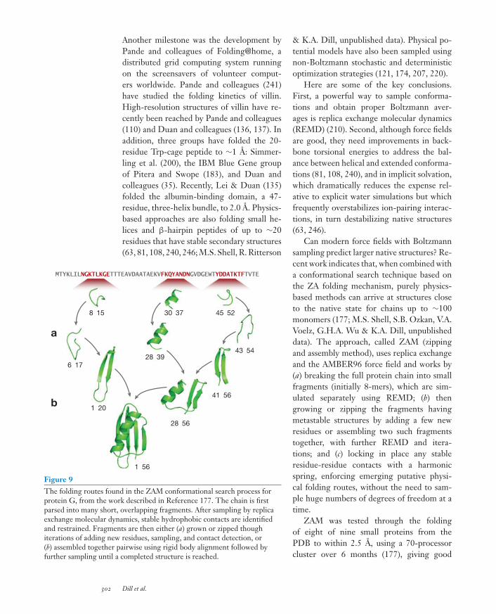
Figure 9The folding routes found in the ZAM conformational search process forprotein G, from the work described in Reference 177. The chain is firstparsed into many short, overlapping fragments. After sampling by replicaexchange molecular dynamics, stable hydrophobic contacts are identifiedand restrained. Fragments are then either (a) grown or zipped thoughiterations of adding new residues, sampling, and contact detection, or(b) assembled together pairwise using rigid body alignment followed byfurther sampling until a completed structure is reached.
& K.A. Dill, unpublished data). Physical po-tential models have also been sampled usingnon-Boltzmann stochastic and deterministicoptimization strategies (121, 174, 207, 220).
Here are some of the key conclusions.First, a powerful way to sample conforma-tions and obtain proper Boltzmann aver-ages is replica exchange molecular dynamics(REMD) (210). Second, although force fieldsare good, they need improvements in back-bone torsional energies to address the bal-ance between helical and extended conforma-tions (81, 108, 240), and in implicit solvation,which dramatically reduces the expense rel-ative to explicit water simulations but whichfrequently overstabilizes ion-pairing interac-tions, in turn destabilizing native structures(63, 246).
Can modern force fields with Boltzmannsampling predict larger native structures? Re-cent work indicates that, when combined witha conformational search technique based onthe ZA folding mechanism, purely physics-based methods can arrive at structures closeto the native state for chains up to ∼100monomers (177; M.S. Shell, S.B. Ozkan, V.A.Voelz, G.H.A. Wu & K.A. Dill, unpublisheddata). The approach, called ZAM (zippingand assembly method), uses replica exchangeand the AMBER96 force field and works by(a) breaking the full protein chain into smallfragments (initially 8-mers), which are sim-ulated separately using REMD; (b) thengrowing or zipping the fragments havingmetastable structures by adding a few newresidues or assembling two such fragmentstogether, with further REMD and itera-tions; and (c) locking in place any stableresidue-residue contacts with a harmonicspring, enforcing emerging putative physi-cal folding routes, without the need to sam-ple huge numbers of degrees of freedom at atime.
ZAM was tested through the foldingof eight of nine small proteins from thePDB to within 2.5 A, using a 70-processorcluster over 6 months (177), giving good
302 Dill et al.

agreement with the φ-values known forfour of them. Figure 9 shows the ZAMfolding process for one of these proteins, andFigure 10 shows the predicted versus experi-mental structures for all nine. In a more strin-gent test, ZAM was applied in CASP7 to thefolding of six small proteins from 76 to 112residues (M.S. Shell, S.B. Ozkan, V.A. Voelz,G.H.A. Wu & K.A. Dill, unpublished data).Of the four proteins attempted in CASP7 thatwere not domain-swapped, ZAM predictedroughly correct tertiary structures, segmentsof more than 40 residues with an averageRMSD of 5.9 angstroms, and secondary struc-tures with 73% accuracy. From these stud-ies it has been concluded that ZA routes canidentify limited-sampling routes to the nativestate from unfolded states, directed by all-atom force fields, and that the AMBER96 plusa generalized Born implicit solvent model is areasonable scoring function. Fragments thatadopt incorrect secondary structures early inthe simulations are frequently corrected inlater-stage folding because the emerging ter-tiary structure of the protein often will nottolerate them.
SUMMARY
The protein folding problem has seen enor-mous advances over the last fifty years.New experimental techniques have arisen, in-cluding hydrogen exchange, φ-value meth-ods that probe mutational effects on fold-ing rates, single-molecule methods that canexplore heterogeneity of folding and en-ergy landscapes, and fast temperature-jumpmethods. New theoretical and computa-tional approaches have emerged, includ-ing methods of bioinformatics, multiple-sequence alignments, structure-predictionWeb servers, physics-based force fields ofgood accuracy, physical models of energylandscapes, fast methods of conformationalsampling and searching, master-equationmethods to explore the physical mecha-
nisms of folding, parallel and distributedgrid-based computing, and the CASPcommunity-wide event for protein structureprediction.
Protein folding no longer appears to bethe insurmountable grand challenge that itonce appeared to be. Current knowledge offolding codes is sufficient to guide the suc-cessful designs of new proteins and foldamericmaterials. For the once seemingly intractableLevinthal puzzle, there is now a viable hy-pothesis: A protein can fold quickly andsolve its big global optimization puzzleby piecewise solutions of smaller compo-nent puzzles. Other matters of principleare now yielding to theory and physics-based modeling. And current computer algo-rithms are now predicting native structures ofsmall proteins remarkably accurately, promis-ing growing value in drug discovery andproteomics.
Protein A Albumin-binding domain protein α 3 D
Protein G α-spectrin SH3 src-SH3
YJQ8 WW domain (res 7–31) FBP28 WW domain (res 6–31) 35-mer unit of ubiquitin
Figure 10Ribbon diagrams of the predicted protein structures using the ZAM search al-gorithm ( purple) versus experimental PDB structures (orange). The backboneC-RMSDs with respect to PDB structures are protein A (1.9 A), albumindomain binding protein (2.4 A), alpha-3D [2.85 A (excluding loop residues)or 4.6 A], FBP26 WW domain (2.2 A), YJQ8 WW domain (2.0 A), 1–35residue fragment of Ubiquitin (2.0 A), protein G (1.6 A), and -spectrin SH3(2.2 A). ZAM fails to find the src-SH3 structure: Shown is a conformationthat is 6 A from the experimental structure. The problem in this case appearsto be overstabilization of nonnative ion pairs in the GB/SA implicit solvationmodel.
www.annualreviews.org • The Protein Folding Problem 303

SUMMARY POINTS
1. The protein folding code is mainly embodied in side chain solvation interactions.Novel protein folds and nonbiological foldamers are now being successfully designedand are moving toward practical applications.
2. Thanks to CASP, the growing PDB, and fast-homology and sequence alignmentmethods, computer methods now can often predict correct native structures of smallproteins.
3. The protein folding problem has both driven—and benefited from—big advances inexperimental and theoretical/computational methods.
4. Proteins fold on funnel-shaped energy landscapes, which describe the conformationalheterogeneity among the nonnative states. This heterogeneity is key to the entropythat opposes folding and thus to folding equilibria. This heterogeneity is also impor-tant for understanding folding kinetics at the level of the individual chain processes.
5. A protein can fold quickly to its native structure by ZA, making independent localdecisions first and then combining those substructures. In this way, a protein can avoidsearching most of its conformational space. ZA appears to be a useful search methodfor computational modeling.
DISCLOSURE STATEMENT
The authors are not aware of any biases that might be perceived as affecting the objectivity ofthis review.
ACKNOWLEDGMENTS
For very helpful comments and insights, both on this review and through ongoing discussionsover the years, we are deeply grateful to D. Wayne Bolen, Hue Sun Chan, John Chodera, YongDuan, Walter Englander, Frank Noe, Jose Onuchic, Vijay Pande, Jed Pitera, Kevin Plaxco,Adrian Roitberg, George Rose, Tobin Sosnick, Bill Swope, Dave Thirumalai, Vince Voelz,Peter G Wolynes, and Huan-Xiang Zhou. We owe particular thanks and appreciation to BuzzBaldwin, to whom this volume of the Annual Review of Biophysics is dedicated, not only for hisinterest and engagement with us on matters of protein folding over the many years, but alsofor his pioneering and founding leadership of the whole field. We appreciate the support fromNIH grant GM 34993, the Air Force, and the Sandler Foundation.
LITERATURE CITED
1. 2005. So much more to know. . . . Science 309:78–1022. Allen F, Coteus P, Crumley P, Curioni A, Denneau M, et al. 2001. Blue gene: a vision for
protein science using a petaflop supercomputer. IBM Syst. J. 40:310–273. Anfinsen CB. 1973. Principles that govern the folding of protein chains. Science 181:223–
304. Anfinsen CB, Scheraga HA. 1975. Experimental and theoretical aspects of protein folding.
Adv. Protein Chem. 29:205–300
304 Dill et al.

5. Auton M, Holthauzen LM, Bolen DW. 2007. Anatomy of energetic changes accompany-ing urea-induced protein denaturation. Proc. Natl. Acad. Sci. USA 104:15317–22
6. Avbelj F, Baldwin RL. 2002. Role of backbone solvation in determining thermodynamicβ propensities of the amino acids. Proc. Natl. Acad. Sci. USA 99:1309–13
7. Bachmann A, Kiefhaber T. 2001. Apparent two-state tendamistat folding is a sequentialprocess along a defined route. J. Mol. Biol. 306:375–86
8. Baker D. 2006. Prediction and design of macromolecular structures and interactions.Philos. Trans. R. Soc. B Biol. Sci. 361:459–63
9. Baker D, Sali A. 2001. Protein structure prediction and structural genomics. Science294:93–96
10. Baldwin RL, Rose GD. 1999. Is protein folding hierarchic? I. Local structure and peptidefolding. Trends Biochem. Sci. 24:26–33
11. Banavar JR, Maritan A. 2007. Physics of proteins. Annu. Rev. Biophys. Biomol. Struct.36:261–80
12. Banavar JR, Maritan A, Micheletti C, Trovato A. 2002. Geometry and physics of proteins.Proteins 47:315–22
13. Best RB, Hummer G. 2005. Reaction coordinates and rates from transition paths. Proc.Natl. Acad. Sci. USA 102:6732–37
14. Bieri O, Wirz J, Hellrung B, Schutkowski M, Drewello M, Kiefhaber T. 1999. The speedlimit for protein folding measured by triplet-triplet energy transfer. Proc. Natl. Acad. Sci.USA 96:9597–601
15. Bolhuis PG. 2005. Kinetic pathways of β-hairpin (un)folding in explicit solvent. Biophys.J. 88:50–61
16. Bradley CM, Barrick D. 2006. The Notch ankyrin domain folds via a discrete, centralizedpathway. Structure 14:1303–12
17. Bradley P, Misura KMS, Baker D. 2005. Toward high-resolution de novo structure pre-diction for small proteins. Science 309:1868–71
18. Brant DA, Flory PJ. 1965. The role of dipole interactions in determining polypeptideconfigurations. J. Am. Chem. Soc. 87:663–64
19. Bryngelson JD, Wolynes PG. 1987. Spin glasses and the statistical mechanics of proteinfolding. Proc. Natl. Acad. Sci. USA 84:7524–28
20. Bulaj G, Koehn RE, Goldenberg DP. 2004. Alteration of the disulfide-coupled foldingpathway of BPTI by circular permutation. Protein Sci. 13:1182–96
21. Byrne MP, Manuel RL, Lowe LG, Stites WE. 1995. Energetic contribution of sidechain hydrogen bonding to the stability of staphylococcal nuclease. Biochemistry 34:13949–60
22. Callender RH, Dyer RB, Gilmanshin R, Woodruff WH. 1998. Fast events in pro-tein folding: the time evolution of primary processes. Annu. Rev. Phys. Chem. 49:173–202
23. Cecconi C, Shank EA, Bustamante C, Marqusee S. 2005. Direct observation of the three-state folding of a single protein molecule. Science 309:2057–60
24. Cellitti J, Bernstein R, Marqusee S. 2007. Exploring subdomain cooperativity in T4lysozyme. II. Uncovering the C-terminal subdomain as a hidden intermediate in thekinetic folding pathway. Protein Sci. 16:852–62
25. Chan HS, Dill KA. 1990. Origins of structure in globular proteins. Proc. Natl. Acad. Sci.USA 87:6388–92
26. Chan HS, Dill KA. 1994. Transition states and folding dynamics of proteins and het-eropolymers. J. Chem. Phys. 100:9238–57
www.annualreviews.org • The Protein Folding Problem 305
27. Chekmarev SF, Krivov SV, Karplus M. 2005. Folding time distributions as an approachto protein folding kinetics. J. Phys. Chem. B 109:5312–30
28. Chen J, Stites WE. 2001. Packing is a key selection factor in the evolution of proteinhydrophobic cores. Biochemistry 40:15280–89
29. Chikenji G, Fujitsuka Y, Takada S. 2006. Shaping up the protein folding funnel by localinteraction: lesson from a structure prediction study. Proc. Natl. Acad. Sci. USA 103:3141–46
30. Cho SS, Levy Y, Wolynes PG. 2006. P versus Q: structural reaction coordinates captureprotein folding on smooth landscapes. Proc. Natl. Acad. Sci. USA 103:586–91
31. Chodera JD, Singhal N, Pande VS, Dill KA, Swope WC. 2007. Automatic discovery ofmetastable states for the construction of Markov models of macromolecular conforma-tional dynamics. J. Chem. Phys. 126:155101
32. Chothia C, Lesk AM. 1986. The relation between the divergence of sequence and structurein proteins. EMBO J. 5:823–26
33. Chou PY, Fasman GD. 1974. Prediction of protein conformation. Biochemistry 13:222–45
34. Chou PY, Fasman GD. 1978. Empirical predictions of protein conformation. Annu. Rev.Biochem. 47:251–76
35. Chowdhury S, Lee MC, Xiong G, Duan Y. 2003. Ab initio folding simulation of theTrp-cage mini-protein approaches NMR resolution. J. Mol. Biol. 327:711–17
36. Cieplak M, Henkel M, Karbowski J, Banavar JR. 1998. Master equation approach toprotein folding and kinetic traps. Phys. Rev. Lett. 80:3654–57
37. Cieplak M, Xuan Hoang T. 2000. Scaling of folding properties in Go models of proteins.J. Biol. Phys. 26:273–94
38. Clementi C, Nymeyer H, Onuchic JN. 2000. Topological and energetic factors: Whatdetermines the structural details of the transition state ensemble and “en-route” inter-mediates for protein folding? An investigation for small globular proteins. J. Mol. Biol.298:937–53
39. Cordes MHJ, Davidsont AR, Sauer RT. 1996. Sequence space, folding and protein design.Curr. Opin. Struct. Biol. 6:3–10
40. Cornell WD, Cieplak P, Bayly CI, Gould IR, Merz KM, et al. 1995. A second generationforce field for the simulation of proteins, nucleic acids, and organic molecules. J. Am.Chem. Soc. 117:5179–97
41. Creamer TP, Rose GD. 1994. A-helix-forming propensities in peptides and proteins.Proteins 19:85–97
42. Cymes GD, Grosman C, Auerbach A. 2002. Structure of the transition state of gat-ing in the acetylcholine receptor channel pore: a φ-value analysis. Biochemistry 41:5548–55
43. Dahiyat BI, Mayo SL. 1997. De novo protein design: fully automated sequence selection.Science 278:82–87
44. de Los Rios MA, Daneshi M, Plaxco KW. 2005. Experimental investigation of thefrequency and substitution dependence of negative φ-values in two-state proteins.Biochemistry 44:12160–67
45. Debe DA, Carlson MJ, Goddard WA. 1999. The topomer-sampling model of proteinfolding. Proc. Natl. Acad. Sci. USA 96:2596–601
46. Deechongkit S, Dawson PE, Kelly JW. 2004. Toward assessing the position-dependentcontributions of backbone hydrogen bonding to β-sheet folding thermodynamics em-ploying amide-to-ester perturbations. J. Am. Chem. Soc. 126:16762–71
306 Dill et al.

47. Dill KA. 1985. Theory for the folding and stability of globular proteins. Biochemistry24:1501–9
48. Dill KA. 1990. Dominant forces in protein folding. Biochemistry 29:7133–5549. Dill KA. 1999. Polymer principles and protein folding. Protein Sci. 8:1166–8050. Dill KA, Alonso DOV, Hutchinson K. 1989. Thermal stabilities of globular proteins.
Biochemistry 28:5439–4951. Dill KA, Bromberg S, Yue KZ, Fiebig KM, Yee DP, et al. 1995. Principles of protein
folding: a perspective from simple exact models. Protein Sci. 4:561–60252. Dill KA, Chan HS. 1997. From Levinthal to pathways to funnels. Nat. Struct. Biol. 4:10–
1953. Dill KA, Fiebig KM, Chan HS. 1993. Cooperativity in protein-folding kinetics. Proc. Natl.
Acad. Sci. USA 90:1942–4654. Dill KA, Lucas A, Hockenmaier J, Huang L, Chiang D, Joshi AK. 2007. Computational
linguistics: a new tool for exploring biopolymer structures and statistical mechanics. Poly-mer 48:4289–300
55. Drozdov AN, Grossfield A, Pappu RV. 2004. Role of solvent in determining conforma-tional preferences of alanine dipeptide in water. J. Am. Chem. Soc. 126:2574–81
56. Du R, Pande VS, Grosberg AY, Tanaka T, Shakhnovich ES. 1998. On the transitioncoordinate for protein folding. J. Chem. Phys. 108:334–50
57. Duan Y, Kollman PA. 1998. Pathways to a protein folding intermediate observed in a1-microsecond simulation in aqueous solution. Science 282:740–44
58. Dyson HJ, Wright PE, Scheraga HA. 2006. The role of hydrophobic interactions ininitiation and propagation of protein folding. Proc. Natl. Acad. Sci. USA 103:13057–61
59. Ellison PA, Cavagnero S. 2006. Role of unfolded state heterogeneity and en-route rugged-ness in protein folding kinetics. Protein Sci. 15:564–82
60. Elmer SP, Park S, Pande VS. 2005. Foldamer dynamics expressed via Markov statemodels. I. Explicit solvent molecular-dynamics simulations in acetonitrile, chloroform,methanol, and water. J. Chem. Phys. 123:114902
61. Elmer SP, Park S, Pande VS. 2005. Foldamer dynamics expressed via Markov state models.II. State space decomposition. J. Chem. Phys. 123:114903
62. English EP, Chumanov RS, Gellman SH, Compton T. 2006. Rational development ofβ-peptide inhibitors of human cytomegalovirus entry. J. Biol. Chem. 281:2661–67
63. Felts AK, Harano Y, Gallicchio E, Levy RM. 2004. Free energy surfaces of β-hairpin andα-helical peptides generated by replica exchange molecular dynamics with the AGBNPimplicit solvent model. Proteins 56:310–21
64. Feng DF, Doolittle RF. 1987. Progressive sequence alignment as a prerequisite to correctphylogenetic trees. J. Mol. Evol. 25:351–60
65. Feng H, Vu ND, Zhou Z, Bai Y. 2004. Structural examination of φ-value analysis inprotein folding. Biochemistry 43:14325–31
66. Feng H, Zhou Z, Bai Y. 2005. A protein folding pathway with multiple folding interme-diates at atomic resolution. Proc. Natl. Acad. Sci. USA 102:5026–31
67. Ferguson N, Sharpe TD, Johnson CM, Fersht AR. 2006. The transition state for foldingof a peripheral subunit-binding domain contains robust and ionic-strength dependentcharacteristics. J. Mol. Biol. 356:1237–47
68. Ferguson N, Sharpe TD, Johnson CM, Schartau PJ, Fersht AR. 2007. Structural biology:analysis of “downhill” protein folding. Nature 445:14–15
69. Ferguson N, Sharpe TD, Schartau PJ, Sato S, Allen MD, et al. 2005. Ultra-fast barrier-limited folding in the peripheral subunit-binding domain family. J. Mol. Biol. 353:427–46
www.annualreviews.org • The Protein Folding Problem 307
70. Fersht AR. 1997. Nucleation mechanisms in protein folding. Curr. Opin. Struct. Biol. 7:3–971. Fersht AR, Sato S. 2004. �-value analysis and the nature of protein-folding transition
states. Proc. Natl. Acad. Sci. USA 101:7976–8172. Fersht AR, Shi JP, Knill-Jones J, Lowe DM, Wilkinson AJ, et al. 1985. Hydrogen bonding
and biological specificity analysed by protein engineering. Nature 314:235–3873. Fiebig KM, Dill KA. 1993. Protein core assembly processes. J. Chem. Phys. 98:3475–8774. Friel CT, Beddard GS, Radford SE. 2004. Switching two-state to three-state kinetics
in the helical protein Im9 via the optimisation of stabilising non-native interactions bydesign. J. Mol. Biol. 342:261–73
75. Garcia-Mira MM, Sadqi M, Fischer N, Sanchez-Ruiz JM, Munoz V. 2002. Experimentalidentification of downhill protein folding. Science 298:2191–95
76. Gellman SH. 1998. Foldamers: a manifesto. Acc. Chem. Res. 31:173–8077. Ghosh K, Ozkan SB, Dill K. 2007. The ultimate speed limit to protein folding is confor-
mational searching. J. Am. Chem. Soc. 129:11920–2778. Gianni S, Guydosh NR, Khan F, Caldas TD, Mayor U, et al. 2003. Unifying features in
protein-folding mechanisms. Proc. Natl. Acad. Sci. USA 100:13286–9179. Gibson KD, Scheraga HA. 1967. Minimization of polypeptide energy I. Preliminary
structures of bovine pancreatic ribonuclease s-peptide. Proc. Natl. Acad. Sci. USA 58:420–27
80. Gilmanshin R, Williams S, Callender RH, Woodruff WH, Dyer RB. 1997. Fast events inprotein folding: relaxation dynamics of secondary and tertiary structure in native apomyo-globin. Proc. Natl. Acad. Sci. USA 94:3709–13
81. Gnanakaran S, Garcia AE. 2003. Validation of an all-atom protein force field: from dipep-tides to larger peptides. J. Phys. Chem. B 107:12555–57
82. Go N, Taketomi H. 1978. Respective roles of short- and long-range interactions in proteinfolding. Proc. Natl. Acad. Sci. USA 75:559–63
83. Goldbeck RA, Thomas YG, Chen E, Esquerra RM, Kliger DS. 1999. Multiple pathways ona protein-folding energy landscape: kinetic evidence. Proc. Natl. Acad. Sci. USA 96:2782–87
84. Goldenberg DP. 1988. Genetic studies of protein stability and mechanisms of folding.Annu. Rev. Biophys. Biomol. Struct. 17:481–507
85. Goldenberg DP. 1999. Finding the right fold. Nat. Struct. Biol. 6:987–9086. Goodman CM, Choi S, Shandler S, DeGrado WF. 2007. Foldamers as versatile frame-
works for the design and evolution of function. Nat. Chem. Biol. 3:252–6287. Grabowski M, Joachimiak A, Otwinowski Z, Wladek M. 2007. Structural genomics: keep-
ing up with expanding knowledge of the protein universe. Nucleic Acids Seq. Topol. 17:347–53
88. Grantcharova V, Alm EJ, Baker D, Horwich AL. 2001. Mechanisms of protein folding.Curr. Opin. Struct. Biol. 11:70–82
89. Grater F, Grubmuller H. 2007. Fluctuations of primary ubiquitin folding intermediatesin a force clamp. J. Struct. Biol. 157:557–69
90. Gromiha MM, Selvaraj S. 2001. Comparison between long-range interactions and contactorder in determining the folding rate of two-state proteins: application of long-range orderto folding rate prediction. J. Mol. Biol. 310:27–32
91. Gromiha MM, Thangakani AM, Selvaraj S. 2006. FOLD-RATE: prediction of proteinfolding rates from amino acid sequence. Nucleic Acids Res. 34:W70–74
92. Haber E, Anfinsen CB. 1962. Side-chain interactions governing the pairing of half-cystineresidues in ribonuclease. J. Biol. Chem. 237:1839–44
93. Hagen SJ. 2007. Probe-dependent and nonexponential relaxation kinetics: unreliable sig-natures of downhill protein folding. Proteins 68:205–17
308 Dill et al.

94. Handel T, Degrado WF. 1990. Denovo design of a Zn2+-binding protein. J. Am. Chem.Soc. 112:6710–11
95. Hansmann UHE, Okamoto Y. 1993. Prediction of peptide conformation by multicanoni-cal algorithm: new approach to the multiple-minima problem. J. Comput. Chem. 14:1333–38
96. Hart WE, Istrail S. 1997. Robust proofs of NP-hardness for protein folding: generallattices and energy potentials. J. Comput. Biol. 4:1–22
97. Haspel N, Tsai CJ, Wolfson H, Nussinov R. 2003. Reducing the computational complexityof protein folding via fragment folding and assembly. Protein Sci. 12:1177–87
98. Hecht MH, Das A, Go A, Bradley LH, Wei Y. 2004. De novo proteins from designedcombinatorial libraries. Protein Sci. 13:1711–23
99. Hecht MH, Richardson JS, Richardson DC, Ogden RC. 1990. De novo design, expression,and characterization of felix: a four-helix bundle protein of native-like sequence. Science249:884–91
100. Ho BK, Dill KA. 2006. Folding very short peptides using molecular dynamics. PLoSComput. Biol. 2:e27
101. Hoang L, Bedard S, Krishna MMG, Lin Y, Englander SW. 2002. Cytochrome c foldingpathway: kinetic native-state hydrogen exchange. Proc. Natl. Acad. Sci. USA 99:12173–78
102. Hockenmaier J, Joshi AK, Dill KA. 2006. Routes are trees: the parsing perspective onprotein folding. Proteins 66:1–15
103. Huang F, Sato S, Sharpe TD, Ying L, Fersht AR. 2007. Distinguishing between cooper-ative and unimodal downhill protein folding. Proc. Natl. Acad. Sci. USA 104:123–27
104. Huang JT, Cheng JP, Chen H. 2007. Secondary structure length as a determinant offolding rate of proteins with two- and three-state kinetics. Proteins 67:12–17
105. Hubner IA, Shimada J, Shakhnovich EI. 2003. � values and the folding transition state ofprotein G: utilization and interpretation of experimental data through simulation. Abstr.Pap. Am. Chem. Soc. 226:U450
106. Itzhaki LS, Otzen DE, Fersht AR. 1995. The structure of the transition state for foldingof chymotrypsin inhibitor 2 analysed by protein engineering methods: evidence for anucleation-condensation mechanism for protein folding. J. Mol. Biol. 254:260–88
107. Ivankov DN, Finkelstein AV. 2004. Prediction of protein folding rates from the aminoacid sequence-predicted secondary structure. Proc. Natl. Acad. Sci. USA 101:8942–44
108. Jang S, Kim E, Pak Y. 2007. Direct folding simulation of α-helices and β-hairpins basedon a single all-atom force field with an implicit solvation model. Proteins 66:53–60
109. Jas GS, Eaton WA, Hofrichter J. 2001. Effect of viscosity on the kinetics of α-helix andβ-hairpin formation. J. Phys. Chem. B 105:261–72
110. Jayachandran G, Vishal V, Pande VS. 2006. Using massively parallel simulation andMarkovian models to study protein folding: examining the dynamics of the villin head-piece. J. Chem. Phys. 124:164902
111. Jones DT, Taylor WR, Thornton JM. 1992. A new approach to protein fold recognition.Nature 358:86–89
112. Kamtekar S, Schiffer JM, Xiong H, Babik JM, Hecht MH. 1993. Protein design by binarypatterning of polar and nonpolar amino acids. Science 262:1680–85
113. Karanicolas J, Brooks CL. 2002. The origins of asymmetry in the folding transition statesof protein L and protein G. Protein Sci. 11:2351–61
114. Karplus M, Kuriyan J. 2005. Molecular dynamics and protein function. Proc. Natl. Acad.Sci. USA 102:6679–85
www.annualreviews.org • The Protein Folding Problem 309

115. Karplus M, Weaver DL. 1979. Diffusion-collision model for protein folding. Biopolymers18:1421–37
116. Karplus M, Weaver DL. 1994. Protein folding dynamics: the diffusion-collision modeland experimental data. Protein Sci. 3:650–68
117. Kendrew JC. 1961. The three-dimensional structure of a protein molecule. Sci. Am.205:96–110
118. Kim DE, Gu H, Baker D. 1998. The sequences of small proteins are not extensivelyoptimized for rapid folding by natural selection. Proc. Natl. Acad. Sci. USA 95:4982–86
119. Kim PS, Baldwin RL. 1982. Specific intermediates in the folding reactions of small proteinsand the mechanism of protein folding. Annu. Rev. Biochem. 51:459–89
120. Kirshenbaum K, Zuckermann RN, Dill KA. 1999. Designing polymers that mimicbiomolecules. Curr. Opin. Struct. Biol. 9:530–35
121. Klepeis JL, Floudas CA. 2003. ASTRO-FOLD: a combinatorial and global optimizationframework for ab initio prediction of three-dimensional structures of proteins from theamino acid sequence. Biophys. J. 85:2119–46
122. Knott M, Chan HS. 2006. Criteria for downhill protein folding: calorimetry, chevron plot,kinetic relaxation, and single-molecule radius of gyration in chain models with subdueddegrees of cooperativity. Proteins 65:373–91
123. Korzhnev DM, Salvatella X, Vendruscolo M, Di Nardo AA, Davidson AR, et al. 2004.Low-populated folding intermediates of Fyn SH3 characterized by relaxation dispersionNMR. Nature 430:586–90
124. Krieger F, Fierz B, Bieri O, Drewello M, Kiefhaber T. 2003. Dynamics of unfoldedpolypeptide chains as model for the earliest steps in protein folding. J. Mol. Biol. 332:265–74
125. Krishna MM, Hoang L, Lin Y, Englander SW. 2004. Hydrogen exchange methods tostudy protein folding. Methods 34:51–64
126. Krishna MMG, Maity H, Rumbley JN, Lin Y, Englander SW. 2006. Order of steps inthe cytochrome c folding pathway: evidence for a sequential stabilization mechanism. J.Mol. Biol. 359:1411–20
127. Kubelka J, Chiu TK, Davies DR, Eaton WA, Hofrichter J. 2006. Sub-microsecond proteinfolding. J. Mol. Biol. 359:546–53
128. Kubelka J, Hofrichter J, Eaton WA. 2004. The protein folding ‘speed limit’. Curr. Opin.Struct. Biol. 14:76–88
129. Kuhlman B, Dantas G, Ireton GC, Varani G, Stoddard BL, Baker D. 2003. Design of anovel globular protein fold with atomic-level accuracy. Science 302:1364–68
130. Kumar S, Bouzida D, Swendsen RH, Kollman PA, Rosenberg JM. 1992. The weightedhistogram analysis method for free-energy calculations on biomolecules. 1. The method.J. Comput. Chem. 13:1011–21
131. Kuznetsov IB, Rackovsky S. 2004. Class-specific correlations between protein folding rate,structure-derived, and sequence-derived descriptors. Proteins 54:333–41
132. Lau KF, Dill KA. 1989. A lattice statistical mechanics model of the conformational andsequence spaces of proteins. Macromolecules 22:3986–97
133. Laurence TA, Kong X, Jager M, Weiss S. 2005. Probing structural heterogeneities andfluctuations of nucleic acids and denatured proteins. Proc. Natl. Acad. Sci. USA 102:17348–53
134. Lee BC, Zuckermann RN, Dill KA. 2005. Folding a nonbiological polymer into a compactmultihelical structure. J. Am. Chem. Soc. 127:10999–1009
135. Lei H, Duan Y. 2007. Ab initio folding of albumin binding domain from all-atom molec-ular dynamics simulation. J. Phys. Chem. B 111:5458–63
310 Dill et al.

136. Lei H, Duan Y. 2007. Two-stage folding of Hp-35 from ab initio simulations. J. Mol. Biol.370:196–206
137. Lei H, Wu C, Liu H, Duan Y. 2007. Folding free-energy landscape of villin headpiecesubdomain from molecular dynamics simulations. Proc. Natl. Acad. Sci. USA 104:4925–30
138. Leopold PE, Montal M, Onuchic JN. 1992. Protein folding funnels: a kinetic approachto the sequence-structure relationship. Proc. Natl. Acad. Sci. USA 89:8721–25
139. Lesk AM, Rose GD. 1981. Folding units in globular proteins. Proc. Natl. Acad. Sci. USA78:4304–8
140. Levitt M. 1983. Molecular dynamics of native protein. I. Computer simulation of trajec-tories. J. Mol. Biol. 168:595–617
141. Li Z, Scheraga HA. 1987. Monte Carlo–minimization approach to the multiple-minimaproblem in protein folding. Proc. Natl. Acad. Sci. USA 84:6611–15
142. Lindberg MO, Haglund E, Hubner IA, Shakhnovich EI, Oliveberg M. 2006. Identificationof the minimal protein-folding nucleus through loop-entropy perturbations. Proc. Natl.Acad. Sci. USA 103:4083–88
143. Lindberg MO, Oliveberg M. 2007. Malleability of protein folding pathways: a simplereason for complex behaviour. Curr. Opin. Struct. Biol. 17:21–29
144. Lindorff-Larsen K, Paci E, Serrano L, Dobson CM, Vendruscolo M. 2003 . Calculation ofmutational free energy changes in transition states for protein folding. Biophys. J. 85:1207–14
145. Looger LL, Dwyer MA, Smith JJ, Hellinga HW. 2003. Computational design of receptorand sensor proteins with novel functions. Nature 423:185–90
146. Lucas A, Huang L, Joshi A, Dill KA. 2007. Statistical mechanics of helix bundles using adynamic programming approach. J. Am. Chem. Soc. 129:4272–81
147. Lucent D, Vishal V, Pande VS. 2007. Protein folding under confinement: a role for solvent.Proc. Natl. Acad. Sci. USA 104:10430–34
148. Ma H, Gruebele M. 2006. Low barrier kinetics: dependence on observables and freeenergy surface. J. Comput. Chem. 27:125–34
149. Maity H, Maity M, Krishna MM, Mayne L, Englander SW. 2005. Protein folding: thestepwise assembly of foldon units. Proc. Natl. Acad. Sci. USA 102:4741–46
150. Makarov DE, Keller CA, Plaxco KW, Metiu H. 2002. How the folding rate constant ofsimple, single-domain proteins depends on the number of native contacts. Proc. Natl. Acad.Sci. USA 99:3535–39
151. Matagne A, Radford SE, Dobson CM. 1997. Fast and slow tracks in lysozyme folding:insight into the role of domains in the folding process. J. Mol. Biol. 267:1068–74
152. Matheson RR Jr, Scheraga HA. 1978. A method for predicting nucleation sites for proteinfolding based on hydrophobic contacts. Macromolecules 11:819–29
153. Matouschek A, Kellis JT Jr, Serrano L, Fersht AR. 1989. Mapping the transition state andpathway of protein folding by protein engineering. Nature 340:122–26
154. Maxwell KL, Wildes D, Zarrine-Afsar A, De Los Rios MA, Brown AG, et al. 2005. Proteinfolding: defining a “standard” set of experimental conditions and a preliminary kinetic dataset of two-state proteins. Protein Sci. 14:602–16
155. McCammon JA, Gelin BR, Karplus M. 1977. Dynamics of folded proteins. Nature267:585–90
156. Meisner WK, Sosnick TR. 2004. Barrier-limited, microsecond folding of a stable proteinmeasured with hydrogen exchange: implications for downhill folding. Proc. Natl. Acad. Sci.USA 101:15639–44
157. Mello CC, Barrick D. 2004. An experimentally determined protein folding energy land-scape. Proc. Natl. Acad. Sci. USA 101:14102–7
www.annualreviews.org • The Protein Folding Problem 311
158. Mezei M. 1998. Chameleon sequences in the PDB. Protein Eng. 11:411–14159. Micheletti C, Banavar JR, Maritan A, Seno F. 1999. Protein structures and optimal folding
from a geometrical variational principle. Phys. Rev. Lett. 82:3372–75160. Miller EJ, Fischer KF, Marqusee S. 2002. Experimental evaluation of topological param-
eters determining protein-folding rates. Proc. Natl. Acad. Sci. USA 99:10359–63161. Miller R, Danko CA, Fasolka MJ, Balazs AC, Chan HS, Dill KA. 1992. Folding kinetics
of proteins and copolymers. J. Chem. Phys. 96:768–80162. Minor DL, Kim PS. 1996. Context-dependent secondary structure formation of a de-
signed protein sequence. Nature 380:730–34163. Mirny L, Shakhnovich E. 2001. Protein folding theory: from lattice to all-atom models.
Annu. Rev. Biophys. Biomol. Struct. 30:361–96164. Moult J. 2005. A decade of CASP: progress, bottlenecks and prognosis in protein
structure prediction. Curr. Opin. Struct. Biol. 15:285–89165. Moult J, Pedersen JT, Judson R, Fidelis K. 1995. A large-scale experiment to assess
protein structure prediction methods. Proteins 23:ii–iv166. Mukhopadhyay S, Krishnan R, Lemke EA, Lindquist S, Deniz AA. 2007. A natively
unfolded yeast prion monomer adopts an ensemble of collapsed and rapidly fluctuatingstructures. Proc. Natl. Acad. Sci. USA 104:2649–54
167. Munoz V. 2002. Thermodynamics and kinetics of downhill protein folding investigatedwith a simple statistical mechanical model. Int. J. Quant. Chem. 90:1522–28
168. Munoz V, Sanchez-Ruiz JM. 2004. Exploring protein-folding ensembles: a variable-barrier model for the analysis of equilibrium unfolding experiments. Proc. Natl. Acad.Sci. USA 101:17646–51
169. Myers JK, Oas TG. 2001. Preorganized secondary structure as an important determinantof fast protein folding. Nat. Struct. Biol. 8:552–58
170. Naganathan AN, Perez-Jimenez R, Sanchez-Ruiz JM, Munoz V. 2005. Robustness ofdownhill folding: guidelines for the analysis of equilibrium folding experiments on smallproteins. Biochemistry 44:7435–49
171. Nemethy G, Scheraga HA. 1965. Theoretical determination of sterically allowed confor-mations of a polypeptide chain by a computer method. Biopolymers 3:155–84
172. Noe F, Horenko I, Schutte C, Smith JC. 2007. Hierarchical analysis of conforma-tional dynamics in biomolecules: transition networks of metastable states. J. Chem. Phys.126:155102
173. O’Neil KT, Hoess RH, DeGrado WF. 1990. Design of DNA-binding peptides based onthe leucine zipper motif. Science 249:774–78
174. Oldziej S, Czaplewski C, Liwo A, Chinchio M, Nanias M, et al. 2005. Physics-basedprotein-structure prediction using a hierarchical protocol based on the UNRES forcefield: assessment in two blind tests. Proc. Natl. Acad. Sci. USA 102:7547–52
175. Onuchic JN, Wolynes PG. 1993. Energy landscapes, glass transitions, and chemical-reaction dynamics in biomolecular or solvent environment. J. Chem. Phys. 98:2218–24
176. Ozkan SB, Bahar I, Dill KA. 2001. Transition states and the meaning of φ-values in proteinfolding kinetics. Nat. Struct. Biol. 8:765–69
177. Ozkan SB, Wu GHA, Chodera JD, Dill KA. 2007. Protein folding by zipping and assem-bly. Proc. Natl. Acad. Sci. USA 104:11987–92
178. Paci E, Vendruscolo M, Dobson CM, Karplus M. 2002. Determination of a transitionstate at atomic resolution from protein engineering data. J. Mol. Biol. 324:151–63
179. Patch JA, Barron AE. 2003. Helical peptoid mimics of magainin-2 amide. J. Am. Chem.Soc. 125:12092–93
312 Dill et al.
180. Pauling L, Corey RB. 1951. Atomic coordinates and structure factors for two helicalconfigurations of polypeptide chains. Proc. Natl. Acad. Sci. USA 37:235–40
181. Pauling L, Corey RB, Branson HR. 1951. The structure of proteins: two hydrogen-bondedhelical configurations of the polypeptide chain. Proc. Natl. Acad. Sci. USA 37:205–11
182. Pieper U, Eswar N, Braberg H, Madhusudhan MS, Davis FP, et al. 2004. MODBASE,a database of annotated comparative protein structure models, and associated resources.Nucleic Acids Res. 32:D217–22
183. Pitera JW, Swope W. 2003. Understanding folding and design: replica-exchange simula-tions of “Trp-cage” miniproteins. Proc. Natl. Acad. Sci. USA 100:7587–92
184. Plaxco KW, Simons KT, Baker D. 1998. Contact order, transition state placement andthe refolding rates of single domain proteins. J. Mol. Biol. 277:985–94
185. Porter EA, Wang X, Lee HS, Weisblum B, Gellman SH. 2000. Non-haemolytic β-amino-acid oligomers. Nature 404:565
186. Punta M, Rost B. 2005. Protein folding rates estimated from contact predictions. J. Mol.Biol. 348:507–12
187. Qiu L, Hagen SJ. 2004. A limiting speed for protein folding at low solvent viscosity. J.Am. Chem. Soc. 126:3398–99
188. Raleigh DP, Plaxco KW. 2005. The protein folding transition state: What are φ-valuesreally telling us? Protein Pept. Lett. 12:117–22
189. Reich L, Weikl TR. 2006. Substructural cooperativity and parallel versus sequential eventsduring protein unfolding. Proteins 63:1052–58
190. Rost B, Eyrich VA. 2001. EVA: large-scale analysis of secondary structure prediction.Proteins 45:192–99
191. Sadqi M, Fushman D, Munoz V. 2006. Atom-by-atom analysis of global downhill proteinfolding. Nature 442:317–21
192. Sali A, Blundell TL. 1993. Comparative protein modelling by satisfaction of spatial re-straints. J. Mol. Biol. 234:779–815
193. Sanchez IE, Kiefhaber T. 2003. Origin of unusual φ-values in protein folding: evidenceagainst specific nucleation sites. J. Mol. Biol. 334:1077–85
194. Schuler B, Lipman EA, Eaton WA. 2002. Probing the free-energy surface for proteinfolding with single-molecule fluorescence spectroscopy. Nature 419:743–47
195. Segel DJ, Bachmann A, Hofrichter J, Hodgson KO, Doniach S, Kiefhaber T. 1999.Characterization of transient intermediates in lysozyme folding with time-resolved small-angle X-ray scattering. J. Mol. Biol. 288:489–99
196. Shea JE, Brooks CL III. 2001. From folding theories to folding proteins: a review andassessment of simulation studies of protein folding and unfolding. Annu. Rev. Phys. Chem.52:499–535
197. Shea JE, Onuchic JN, Brooks CL. 1999. Exploring the origins of topological frustration:design of a minimally frustrated model of fragment B of protein A. Proc. Natl. Acad. Sci.USA 96:12512–17
198. Shirts M, Pande VS. 2000. Computing: Screen savers of the world unite! Science 290:1903–4
199. Shmygelska A. 2005. Search for folding nuclei in native protein structures. Bioinformatics21:394–402
200. Simmerling C, Strockbine B, Roitberg AE. 2002. All-atom structure prediction and fold-ing simulations of a stable protein. J. Am. Chem. Soc. 124:11258–59
201. Singhal N, Snow CD, Pande VS. 2004. Using path sampling to build better Markovianstate models: predicting the folding rate and mechanism of a tryptophan zipper β hairpin.J. Chem. Phys. 121:415–25
www.annualreviews.org • The Protein Folding Problem 313

202. Snow CD, Rhee YM, Pande VS. 2006. Kinetic definition of protein folding transitionstate ensembles and reaction coordinates. Biophys. J. 91:14–24
203. Sohl JL, Jaswal SS, Agard DA. 1998. Unfolded conformations of α-lytic protease are morestable than its native state. Nature 395:817–19
204. Sosnick TR, Dothager RS, Krantz BA. 2004. Differences in the folding transition state ofubiquitin indicated by φ and ψ analyses. Proc. Natl. Acad. Sci. USA 101:17377–82
205. Sridevi K, Juneja J, Bhuyan AK, Krishnamoorthy G, Udgaonkar JB. 2000. The slow foldingreaction of barstar: The core tryptophan region attains tight packing before substantialsecondary and tertiary structure formation and final compaction of the polypeptide chain.J. Mol. Biol. 302:479–95
206. Sridevi K, Lakshmikanth GS, Krishnamoorthy G, Udgaonkar JB. 2004. Increasing sta-bility reduces conformational heterogeneity in a protein folding intermediate ensemble.J. Mol. Biol. 337:699–711
207. Srinivasan R, Rose GD. 2002. Ab initio prediction of protein structure using LINUS.Proteins 47:489–95
208. Sriraman S, Kevrekidis IG, Hummer G. 2005. Coarse master equation from Bayesiananalysis of replica molecular dynamics simulations. J. Phys. Chem. B 109:6479–84
209. Street TO, Bradley CM, Barrick D. 2007. Predicting coupling limits from an experimen-tally determined energy landscape. Proc. Natl. Acad. Sci. USA 104:4907–12
210. Sugita Y, Okamoto Y. 1999. Replica-exchange molecular dynamics method for proteinfolding. Chem. Phys. Lett. 314:141–51
211. Swope WC, Pitera JW, Suits F. 2004. Describing protein folding kinetics by moleculardynamics simulations. 1. Theory. J. Phys. Chem. B 108:6571–81
212. Swope WC, Pitera JW, Suits F, Pitman M, Eleftheriou M, et al. 2004. Describing proteinfolding kinetics by molecular dynamics simulations. 2. Example applications to alaninedipeptide and a B-hairpin peptide. J. Phys. Chem. B 108:6582–94
213. Travaglini-Allocatelli C, Cutruzzola F, Bigotti MG, Staniforth RA, Brunori M. 1999.Folding mechanism of Pseudomonas aeruginosa cytochrome c. J. Mol. Biol. 289:1459–67
214. Tress M, Ezkurdia I, Grana O, Lopez G, Valencia A. 2005. Assessment of predictionssubmitted for the CASP6 comparative modelling category. Proteins 61:27–45
215. Tsai CJ, Maizel JV Jr, Nussinov R. 2000. Anatomy of protein structures: visualizing how aone-dimensional protein chain folds into a three-dimensional shape. Proc. Natl. Acad. Sci.USA 97:12038–43
216. Unger R, Moult J. 1993. Finding the lowest free energy conformation of a protein is anNP-hard problem: proof and implications. Bull. Math. Biol. 55:1183–98
217. Utku Y, Dehan E, Ouerfelli O, Piano F, Zuckermann RN, et al. 2006. A peptidomimeticsiRNA transfection reagent for highly effective gene silencing. Mol. Biosyst. 2:312–17
218. Uversky VN, Fink AL. 2002. The chicken-egg scenario of protein folding revisited. FEBSLett. 515:79–83
219. Venclovas C, Zemla A, Fidelis K, Moult J. 2003. Assessment of progress over the CASPexperiments. Proteins 53:585–95
220. Verma A, Schug A, Lee KH, Wenzel W. 2006. Basin hopping simulations for all-atomprotein folding. J. Chem. Phys. 124:044515
221. Viguera AR, Serrano L, Wilmanns M. 1996. Different folding transition states may resultin the same native structure. Nat. Struct. Biol. 3:874–80
222. Vitkup D, Melamud E, Moult J, Sander C. 2001. Completeness in structural genomics.Nat. Struct. Biol. 8:559–66
314 Dill et al.

223. Voelz VA, Dill KA. 2006. Exploring zipping and assembly as a protein folding principle.Proteins 66:877–88
224. Wallace IM, Blackshields G, Higgins DG. 2005. Multiple sequence alignments. Curr.Opin. Struct. Biol. 15:261–66
225. Wallin S, Chan HS. 2006. Conformational entropic barriers in topology-dependent pro-tein folding: perspectives from a simple native-centric polymer model. J. Phys. Condens.Matter 18:S307–28
226. Wang L, Xie J, Schultz PG. 2006. Expanding the genetic code. Annu. Rev. Biophys. Biomol.Struct. 35:225–49
227. Wang Z, Mottonen J, Goldsmith EJ. 1996. Kinetically controlled folding of the serpinplasminogen activator inhibitor 1. Biochemistry 35:16443–48
228. Weikl TR. 2005. Loop-closure events during protein folding: rationalizing the shape ofφ-value distributions. Proteins 60:701–11
229. Weikl TR, Dill KA. 2003. Folding kinetics of two-state proteins: effect of circularization,permutation, and crosslinks. J. Mol. Biol. 332:953–63
230. Weikl TR, Dill KA. 2003. Folding rates and low-entropy-loss routes of two-state proteins.J. Mol. Biol. 329:585–98
231. Weikl TR, Dill KA. 2007. Transition-states in protein folding kinetics: the structuralinterpretation of φ values. J. Mol. Biol. 365:1578–86
232. Weikl TR, Palassini M, Dill KA. 2004. Cooperativity in two-state protein folding kinetics.Protein Sci. 13:822–29
233. White GWN, Gianni S, Grossmann JG, Jemth P, Fersht AR, Daggett V. 2005. Simulationand experiment conspire to reveal cryptic intermediates and a slide from the nucleation-condensation to framework mechanism of folding. J. Mol. Biol. 350:757–75
234. Wolfenden R. 2007. Experimental measures of amino acid hydrophobicity and the topol-ogy of transmembrane and globular proteins. J. Gen. Physiol. 129:357–62
235. Wu CW, Seurynck SL, Lee KY, Barron AE. 2003. Helical peptoid mimics of lung sur-factant protein C. Chem. Biol. 10:1057–63
236. Wurth C, Kim W, Hecht MH. 2006. Combinatorial approaches to probe the sequencedeterminants of protein aggregation and amyloidogenicity. Protein Pept. Lett. 13:279–86
237. Xu Y, Purkayastha P, Gai F. 2006. Nanosecond folding dynamics of a three-strandedβ-sheet. J. Am. Chem. Soc. 128:15836–42
238. Yang JS, Chen WW, Skolnick J, Shakhnovich EI. 2006. All-atom ab initio folding of adiverse set of proteins. Structure 15:53–63
239. Yeh SR, Rousseau DL. 2000. Hierarchical folding of cytochrome c. Nat. Struct. Biol. 7:443–45
240. Yoda T, Sugita Y, Okamoto Y. 2004. Secondary structure preferences of force fieldsfor proteins evaluated by generalized-ensemble simulations. Chem. Phys. 307:269–83
241. Zagrovic B, Snow CD, Shirts MR, Pande VS. 2002. Simulation of folding of a small α-helical protein in atomistic detail using worldwide-distributed computing. J. Mol. Biol.323:927–37
242. Zhang Y, Arakaki AK, Skolnick J. 2005. TASSER: an automated method for theprediction of protein tertiary structures in CASP6. Proteins 61:91–98
243. Zhao H, Giver L, Shao Z, Affholter JA, Arnold FH. 1998. Molecular evolution bystaggered extension process (StEP) in vitro recombination. Nat. Biotechnol. 16:258–61
www.annualreviews.org • The Protein Folding Problem 315

ANRV343-BB37-14 ARI 9 April 2008 11:34
244. Zhong S, Rousseau DL, Yeh SR. 2004. Modulation of the folding energy landscape ofcytochrome c with salt. J. Am. Chem. Soc. 126:13934–35
245. Zhou HY, Zhou YQ. 2002. Folding rate prediction using total contact distance. Biophys.J. 82:458–63
246. Zhou R. 2003. Free energy landscape of protein folding in water: explicit vs. implicitsolvent. Proteins 53:148–61
316 Dill et al.