Bmi-1 promotes the aggressiveness of glioma via activating the NF-kappaB/MMP-9 signaling
RESEARCH ARTICLE Open Access
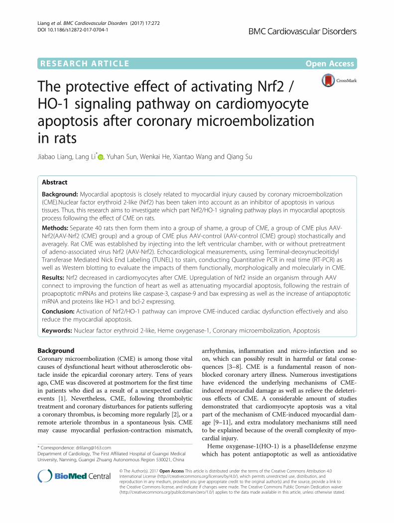
The protective effect of activating Nrf2 /HO-1 signaling pathway on cardiomyocyteapoptosis after coronary microembolizationin ratsJiabao Liang, Lang Li* , Yuhan Sun, Wenkai He, Xiantao Wang and Qiang Su
Abstract
Background: Myocardial apoptosis is closely related to myocardial injury caused by coronary microembolization(CME).Nuclear factor erythroid 2-like (Nrf2) has been taken into account as an inhibitor of apoptosis in varioustissues. Thus, this research aims to investigate which part Nrf2/HO-1 signaling pathway plays in myocardial apoptosisprocess following the effect of CME on rats.
Methods: Separate 40 rats then form them into a group of shame, a group of CME, a group of CME plus AAV-Nrf2(AAV-Nrf2 (CME) group) and a group of CME plus AAV-control (AAV-control (CME) group) stochastically andaveragely. Rat CME was established by injecting into the left ventricular chamber, with or without pretreatmentof adeno-associated virus Nrf2 (AAV-Nrf2). Echocardiological measurements, using Terminal-deoxynucleoitidylTransferase Mediated Nick End Labeling (TUNEL) to stain, conducting Quantitative PCR in real time (RT-PCR) aswell as Western blotting to evaluate the impacts of them functionally, morphologically and molecularly in CME.
Results: Nrf2 decreased in cardiomyocytes after CME. Upregulation of Nrf2 inside an organism through AAVconnect to improving the function of heart as well as attenuating myocardial apoptosis, following the restrain ofproapoptotic mRNAs and proteins like caspase-3, caspase-9 and bax expressing as well as the increase of antiapoptoticmRNA and proteins like HO-1 and bcl-2 expressing.
Conclusion: Activation of Nrf2/HO-1 pathway can improve CME-induced cardiac dysfunction effectively and alsoreduce the myocardial apoptosis.
Keywords: Nuclear factor erythroid 2-like, Heme oxygenase-1, Coronary microembolization, Apoptosis
BackgroundCoronary microembolization (CME) is among those vitalcauses of dysfunctional heart without atherosclerotic obs-tacle inside the epicardial coronary artery. Tens of yearsago, CME was discovered at postmortem for the first timein patients who died as a result of a unexpected cardiacevents [1]. Nevertheless, CME, following thrombolytictreatment and coronary disturbances for patients sufferinga coronary thrombus, is becoming more regularly [2], or aremote arteriole thrombus in a spontaneous lysis. CMEmay cause myocardial perfusion-contraction mismatch,
arrhythmias, inflammation and micro-infarction and soon, which can possibly result in harmful or fatal conse-quences [3–8]. CME is a fundamental reason of non-blocked coronary artery illness. Numerous investigationshave evidenced the underlying mechanisms of CME-induced myocardial damage as well as relieve the deleteri-ous effects of CME. A considerable amount of studiesdemonstrated that cardiomyocyte apoptosis was a vitalpart of the mechanism of CME-induced myocardial dam-age [9–11], and extra modulatory mechanisms still needto be explained because of the overall complexity of myo-cardial injury.Heme oxygenase-1(HO-1) is a phaseIIdefense enzyme
which has potent antiapoptotic as well as antioxidative* Correspondence: [email protected] of Cardiology, The First Affiliated Hospital of Guangxi MedicalUniversity, Nanning, Guangxi Zhuang Autonomous Region 530021, China
© The Author(s). 2017 Open Access This article is distributed under the terms of the Creative Commons Attribution 4.0International License (http://creativecommons.org/licenses/by/4.0/), which permits unrestricted use, distribution, andreproduction in any medium, provided you give appropriate credit to the original author(s) and the source, provide a link tothe Creative Commons license, and indicate if changes were made. The Creative Commons Public Domain Dedication waiver(http://creativecommons.org/publicdomain/zero/1.0/) applies to the data made available in this article, unless otherwise stated.
Liang et al. BMC Cardiovascular Disorders (2017) 17:272 DOI 10.1186/s12872-017-0704-1

stress effects. In those HT22 cells, HO-1 is overexpressed which confer antiapoptotic protective effectsagainst H/R [12]. Nuclear factor erythroid 2-like (Nrf2)is a primary element of transcription. It adjusts numer-ous ways of apoptosis in a cell [13]. Recent studies havepaid more attention to the Nrf2/HO-1 approach that isa key part in the apoptosis and that regulates and lowersthe cardiomyocyte apoptosis [14].The purpose of the current research was in order to
determine the part of Nrf2/HO-1 in CME-induced myo-cardial apoptosis. Here, we focused on the interactionsof Nrf2/HO-1 pathway with the bcl-2, bax, caspase3 andcaspase9 in CME-induced myocardial apoptosis. Thestudy suggested that Nrf2/HO-1 was a major part inCME-induced myocardial apoptosis and activation ofNrf2/HO-1 pathway could remarkably restrain apoptosisin CME-induced myocardial as well as enhance thefunction of heart. We supplied a possible CME-inducedmyocardial apoptosis mechanism.
MethodsAnimal preparationSprague-Dawley rats (250-300 g) were gained from theAnimal Center of the Guangxi Medical Collage which isin Nanning, China. During every phase of this experi-ment, the rats were raised with manipulated light status,moisture, with clean water and feed provided unlimited.All the procedures were authorized and supervised bythe Animal Experiment Ethics Committees of GuangxiMedical Collage.
Design, synthesis as well as transfection of AAV-Nrf2The adeno-associated virus Nrf2(AAV-Nrf2) gene se-quence was located in GeneBank (Gene ID: 83619) andcompounded by Hanbio Biotechnology Co (Shanghai,China). Control AAV was also bought from this com-pany. A dose of 10╳11E in all injected by tail veins wasmixed with saline for each rat.
Creation of a CME model and groupingCME model was generated as described by Li L et al.previously [15]. Forty rats were separated then formedinto 4 groups, containing a sham group, CME group,CME plus AAV-Nrf2 group (AAV-Nrf2(CME) group)and CME plus AAV-control group (AAV-control(CME) group), with 10 rats in each one of them.AAV-Nrf2(CME) group and AAV-control (CME)group both received 2-week transfection of AAV-Nrf2or AAV-control virus. And then, the two groups re-ceived the following operation: put simply, a sternot-omy in the middle the second and third intercostalwas undertaken. The pericardium was unfolded andthe increasing aorta was totally revealed. The plasticmicrospheres (3000 microspheres per rat, diameter of
42 μm, Biosphere Medical Inc., Rockland, USA) dis-solved in 0.1 mL NS were injected into the ventricu-lar (LV) chamber on the left and meanwhile clampingthe ascending aorta with the use of the hemostaticforceps during the 10 s. Rats in the control group re-ceived 0.1 ml normal saline (PubChem CID: 5234)merely as the sham group did.
Echocardiography studyThe left ventricle was detected applying echocardiog-raphy which was used to examine the left ventricle ofrats, which were lightly anesthetized by injecting 10%chloral hydrate (1-2 ml/kg) into an enterocoelia. In aword, an S12 transducer was used to be put on the leftside and anterior position of chest wall to gain the leftventricle end-diastolic dimension (LVEDd), left ventricleejection fraction (LVEF), cardiac output (CO) as well asfractional shortening (FS). A Philips Sonos 7500 system(Philips, Andover, USA) was used then all echocardiog-raphy was carried on by a veteran physician. Evaluatingcardiac function three times, and the average values ofthe three figures were taken.
Myocardial micro-infarct size measurementHBFP stain was applied to detect myocardial ischemia inan early stage or infarct area. The dyed yellow or brownmyocardial tissue was normal while the red tissue wasischemia or necrotic. A DMR-Q550 pathological pictureanalyzer (Lei, Germany) analyzed The HBFP-stained sec-tions before. In short, 10 optical areas in the microscope(magnification, ×100) were casually selected from everypart and Leica Qwin analysis software was used to ob-serve. The formula- ischemic area/total area × 100% wasused to calculate the relative ischemic area [16].
Using TUNEL assay to examining myocardial apoptosisThis process was grimly conformed to the instructionsof Kit specification. Apoptotic nuclei showed yellow-brown while the normal colour is light blue (TUNELpositive), and a whole amount of 10 non-overlappingareas (magnification,×400) from every slice which wasobserved stochastically [17]. The myocardial apoptoticratio = apoptotic number/entire cell number × 100%was to be used.
Quantitative Polymerase Chain Reaction in real time (RT-PCR)We extracted the entire RNA from the cardiac tissue ofrats with TRIzol® Reagent (Takara, Japan) 6 h after CMEoperation. Reverse transcription RNA (1 μg) was per-formed applying a reverse transcriptase kit (Takara,Japan) referring to the instructions provided by themanufacturer. Reactions involved 2 μL cDNA, 2 μLprimers and 12.5 μL SYBR Premix Ex Taq II, and put
Liang et al. BMC Cardiovascular Disorders (2017) 17:272 Page 2 of 9

into a ultimate volume of 20 μL of water. Real time-PCRwas undertaken the next phase situations: ten-min initialdenaturation at 95 C following 15-s 40 phases of amplifi-cation with 95 C as well as 1-min 60 °C. PCR in realtime was applied with the following primers: 5′-GCTGCCATTAGTCAGTCGCTCTC -3′ and 5′- ACCGTGCCTTCAGTGTGCTTC-3′ for the rat nuclear rela-tive factor 2-like (Nrf2), 5′- TTAAGCTGGTGATGGCCTCC -3′ and 5′- GTGGGGCATAGACTGGGTTC-3′for the rat heme oxygenase1(HO-1), and 5′-ACTGCTTCCCAGACCCACA-3′ and 5′-CGAGACCTTGGAACACAGAGAA-3′ for caspase9, and 5′-GCAGCAGCCTCAAATTGTTGAC-3′ and 5′-TGCTCCGGCTCAAACCATC-3′ for caspase3, and 5′-AGACACCTGACCTTGGA-3and 5′-TTGAAGTTGCCATCAGCAAACA-3 for bax,and 5′-AGACACCTGACCTTGGA-3 and 5′-TTGAAGTTGCCATCAGCAAACA-3 for bcl-2, and 5′-GAGATTACTGCCCTGGCTCCTA-3′ and 5′-CATCGTACTCCTGCTTGCTGAT-3′ for β-actin. The dissociation curve wasanalyzed to confirm specific amplification. The amplifica-tion of the overall cDNA samples were divided into 3 por-tions and standardized compared to a triplicate of β-actinin this dish. The expression of the data was 2–ΔΔCt.
Western blottingProteins were extracted from tissue samples from 6 h afterCME operation. Subsequently, the protein (50 μg) wasquantified using a BCA assay (Solarbio, Beijing, China)and was loaded onto a 10% SDS-PAGE gel for electro-phoresis and then transferred into a PVDF membrane(Millipore). Blocking with 5 % non-fat milk in TBS at thetemperature in a room for 1 h, following the incubation ofthe membranes with a 1:500-10,000 dilution of primaryantibody (anti-Nrf2, anti-HO-1, anti-caspase-3, anti-caspase-9, anti-bax, as well as anti-bcl-2; Abcam, UK) inbuffer overnight at 4 °C, after washing 3 times in TBS, the
incubation happened with a 1:10,000 dilution of fluores-cent anti-rabbit secondary antibody (Abcam, UK) in bufferat the temperature in the room for 2 h. The immunoreac-tive bands were observed by exposing the blots in anOdyssey infrared imaging system (LI-COR).
ELISATo measure the expression of cTnI in serum, blood sam-ples from each rat were collected for ELISA 6 h afterCME operation based on the instructions provided bythe manufacturer.
Preparation of the tissue samplesTen rats per group were killed under anesthesia 6 h afterCME operation. Blood samples were collected and usedfor enzyme linked immunoassay (ELISA). Heart tissue washarvested. Four tissue samples were fixed in 4% parafor-maldehyde (PubChem CID: 62081) for 24 h for prepar-ation of paraffin sections and the myocardial infarct sizewas detected by HE staining and hematoxylin-red com-plex picric acid (HBFP) staining. The apoptosis of myocar-dium was detected by TUNEL staining. Three hearts werepreparation for Real time-PCR. The remaining heart tissuewas used for the preparation of Western blotting. All tis-sue samples were prepared in an identical manner.
Table 1 The weights and heart rates of four groups beforeoperation. There were not any statistically significant differencesamong four groups
Groups Sham CME AAV-Nrf2 AAV-control
Weight (g) 251.2 ± 10.3 253.2 ± 12.2 249.9 ± 11.3 250.8 ± 9.3
Heart rate (bpm) 463 ± 20 460 ± 27 458 ± 32 462 ± 24
Data is shown as Mean ± SDNrf2 nuclear factor erythroid 2-like, AAV adeno-associated virusaP < 0.05 contrasted with shambP < 0.05 contrasted with CME or AAV control
A B
E
C DFig. 1 Microinfarction detected with HBFP staining (×100), bar = 100 μm. The normal myocardial staining was yellow, while the stain of ischemicmyocardium was reddish. Arrows reveal microinfarction focus. a Sham; b CME; c AAV-Nrf2(CME); d AAV-control (CME). e The percentage of microinfarctionarea. AAV-Nrf2(CME): rats injected Nrf2 undergone CME operation; AAV-control (CME): rats injected AAV-control undergone CME operation; CME: coronarymicroembolization; Nrf 2: nuclear factor erythroid 2-like; AAV: adeno-associated virus. Data is shown as Mean ± SD. a: P < 0.05 contrasted with sham; b:P < 0.05 contrasted with CME or AAV control (CME)
Liang et al. BMC Cardiovascular Disorders (2017) 17:272 Page 3 of 9
Statistical analysisData was analyzed by SPSS v18.0 (Chicago, Illinois,USA). Continuous variables are the mean ± SD. We ap-plied one-way ANOVA (Analysis of Variance) to con-trast differences among groups. Categorical variableswere reported as frequencies and percentages (%), anddifferences between groups were examined by Fisherexact examination or chi-square examination. P < 0.05was considered statistically remarkable.
ResultGroups of animalNo significant discrepancies statistically (P > 0.05) inweight of the body or rate of the heart were testifiedamong these four groups (Table 1).
HBFP staining and HE stainingSix hours after CME modeling, the HBFP stainingshowed that no obvious myocardial microinfarction inthe sham group. Meanwhile, it was remarkably dis-tinct in the CME group and AAV-control (CME)group (P < 0.05). As contrasted with CME group,
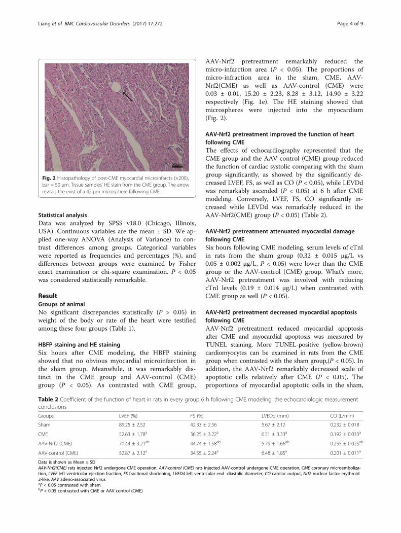
AAV-Nrf2 pretreatment remarkably reduced themicro-infarction area (P < 0.05). The proportions ofmicro-infraction area in the sham, CME, AAV-Nrf2(CME) as well as AAV-control (CME) were0.03 ± 0.01, 15.20 ± 2.23, 8.28 ± 3.12, 14.90 ± 3.22respectively (Fig. 1e). The HE staining showed thatmicrospheres were injected into the myocardium(Fig. 2).
AAV-Nrf2 pretreatment improved the function of heartfollowing CMEThe effects of echocardiography represented that theCME group and the AAV-control (CME) group reducedthe function of cardiac systolic comparing with the shamgroup significantly, as showed by the significantly de-creased LVEF, FS, as well as CO (P < 0.05), while LEVDdwas remarkably ascended (P < 0.05) at 6 h after CMEmodeling. Conversely, LVEF, FS, CO significantly in-creased while LEVDd was remarkably reduced in theAAV-Nrf2(CME) group (P < 0.05) (Table 2).
AAV-Nrf2 pretreatment attenuated myocardial damagefollowing CMESix hours following CME modeling, serum levels of cTnIin rats from the sham group (0.32 ± 0.015 μg/L vs0.05 ± 0.002 μg/L, P < 0.05) were lower than the CMEgroup or the AAV-control (CME) group. What’s more,AAV-Nrf2 pretreatment was involved with reducingcTnI levels (0.19 ± 0.014 μg/L) when contrasted withCME group as well (P < 0.05).
AAV-Nrf2 pretreatment decreased myocardial apoptosisfollowing CMEAAV-Nrf2 pretreatment reduced myocardial apoptosisafter CME and myocardial apoptosis was measured byTUNEL staining. More TUNEL-positive (yellow-brown)cardiomyocytes can be examined in rats from the CMEgroup when contrasted with the sham group,(P < 0.05). Inaddition, the AAV-Nrf2 remarkably decreased scale ofapoptotic cells relatively after CME (P < 0.05). Theproportions of myocardial apoptotic cells in the sham,
Fig. 2 Histopathology of post-CME myocardial microinfarcts (×200),bar = 50 μm. Tissue samples’ HE stain from the CME group. The arrowreveals the exist of a 42-μm microsphere following CME
Table 2 Coefficient of the function of heart in rats in every group 6 h following CME modeling: the echocardiologic measurementconclusions
Groups LVEF (%) FS (%) LVEDd (mm) CO (L/min)
Sham 89.25 ± 2.52 42.33 ± 2.56 5.67 ± 2.12 0.232 ± 0.018
CME 52.63 ± 1.78a 36.25 ± 3.22a 6.51 ± 3.33a 0.192 ± 0.033a
AAV-Nrf2 (CME) 70.44 ± 3.21ab 44.74 ± 1.58ab 5.79 ± 1.66ab 0.255 ± 0.025ab
AAV-control (CME) 52.87 ± 2.12a 34.55 ± 2.24a 6.48 ± 1.85a 0.201 ± 0.011a
Data is shown as Mean ± SDAAV-Nrf2(CME) rats injected Nrf2 undergone CME operation, AAV-control (CME) rats injected AAV-control undergone CME operation, CME coronary microemboliza-tion, LVEF left ventricular ejection fraction, FS fractional shortening, LVEDd left ventricular end -diastolic diameter, CO cardiac output, Nrf2 nuclear factor erythroid2-like, AAV adeno-associated virusaP < 0.05 contrasted with shambP < 0.05 contrasted with CME or AAV control (CME)
Liang et al. BMC Cardiovascular Disorders (2017) 17:272 Page 4 of 9
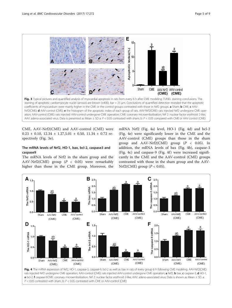
CME, AAV-Nrf2(CME) and AAV-control (CME) were0.23 ± 0.10, 12.34 ± 1.27,5.01 ± 0.50, 11.34 ± 0.72 re-spectively (Fig. 3e).
The mRNA levels of Nrf2, HO-1, bax, bcl-2, caspase3 andcaspase9The mRNA levels of Nrf2 in the sham group and theAAV-Nrf2(CME) group (P < 0.05) were remarkablyhigher than those in the CME group. Moreover, the
mRNA Nrf2 (Fig. 4a) level, HO-1 (Fig. 4d) and bcl-2(Fig. 4e) were significantly lower in the CME and theAAV-control (CME) groups than those in the shamgroup and AAV-Nrf2(CME) group (P < 0.05). Inaddition, the mRNA levels of bax (Fig. 4b), caspase-3(Fig. 4c) and caspase-9 (Fig. 4f ) were increased signifi-cantly in the CME and the AAV-control (CME) groupscontrasted with those in the sham group and the AAV-Nrf2(CME) group (P < 0.05).
A B
E
C DFig. 3 Typical pictures and quantified analysis of myocardial apoptosis in rats from every 6 h after CME modeling: TUNEL staining conclusions. Thestaining of apoptotic cardiomyocyte nuclei (arrows) are brown (×400), bar = 25 μm. Conclusions of quantified detection revealed that the apoptoticcoefficients of myocardium were mainly higher in the CME or the control groups contrasted with those in Nrf2 groups. a Sham; b CME; c AAV-Nrf2(CME); d AAV-control (CME); e the histogram of the apoptotic index of each group of rats. AAV-Nrf2(CME): rats injected Nrf2 undergone CME oper-ation; AAV-control (CME): rats injected AAV-control undergone CME operation; CME: coronary microembolization; Nrf 2: nuclear factor erythroid 2-like;AAV: adeno-associated virus. Data is presented as Mean ± SD a: P < 0.05 contrasted with sham; b: P < 0.05 compared with CME or AAV control (CME)
A B C
D E F
Fig. 4 The mRNA expression of Nrf2, HO-1, caspase-3, caspase-9, bcl-2 as well as bax in rats of every group 6 h following CME modeling. AAV-Nrf2(CME):rats injected Nrf2 undergone CME operation; AAV-control (CME): rats injected AAV-control undergone CME operation; a Nrf2, b bax, c caspase-3, d HO-1,e bcl-2, f caspase-9.CME: coronary microembolization; Nrf 2: nuclear factor erythroid 2-like; AAV: adeno-associated virus; Data is shown as Mean ± SD. a:P < 0.05 contrasted with sham; b: P < 0.05 contrasted with CME or AAV-control (CME)
Liang et al. BMC Cardiovascular Disorders (2017) 17:272 Page 5 of 9

AAV-Nrf2 pretreatment activated myocardial HO-1signaling in CMEWestern blotting exhibited significantly down-adjustmentof HO-1 in CME and AAV-control (CME) groups con-trasted with the sham group (P < 0.05). Nevertheless,AAV-Nrf2 pretreatment enhanced the expression of HO-1protein (Fig. 5g) and bcl-2 protein (Fig. 5h) contrastedwith the CME group or the AAV-control (CME) group(P < 0.05), and reduced the expression of cleave-caspase-3protein (Fig. 5f), caspase-9 protein (Fig. 5i) and bax pro-tein (Fig. 5f) (P < 0.05).
DiscussionIn our research, we testified that transfecting Nrf2 attenu-ated myocardial apoptosis and partially reversed CME,and that Nrf2/HO-1 signaling pathway was concluded inthe pathogenesis of CME-induced myocardial apoptosiswas saw. The results of this research emphasized the sig-nificant part that Nrf2/HO-1 plays in the pathogenesis ofCME-connected myocardial dysfunction as well as in theapoptosis. We initially illuminate the part of Nrf2/HO-1pathway plays in the CME model. These findings could
enable us to illustrate the mechanisms by which Nrf2/HO-1 mediates myocardial injury supports our hypothesisthat Nrf2 might represent a potential target for therapy.AAV is a small and nonpathogenic human virus that be-
longs to the parvovirus family and was originally discov-ered in the mid-1960s. In the preliminary experiment, wefound that there were no statistical difference of cleave-caspase-3 among the groups of rats transfected byAAV-Nrf2 and AAV-control of which rats were notundergone CME and the control rats (Fig. 6).AAV is as acontaminant of cell culture also infected with adenovirus.The high efficiency of in vivo transduction of postmitotictissues, including heart, brain, and retina, combined withits low immunogenicity, led to the widespread use of AAVas a transfer method for gene therapy in various organs[18–20]. The results and previous studies can be tendedto indicate that it has no effect on myocardial apoptosis.Therefore, the rats, which were not undergone CME, weretransfected by AAV-Nrf2 and AAV-control groups werenot included in further analyses.Microcirculation of acute dysfunction and myocardial
damage perfusion is a characteristic of CME. Once CME
A B C
D E F
G H I
Fig. 5 Coronary microembolization reduced cardiomyocyte apoptosis via Nrf2/HO-1 signaling approach. The impacts of CME and AAV-Nrf2 on theprotein levels of Nrf2 and HO-1 pathways as well as bax, bcl-2, caspase-3 and caspase-9 in the myocardial tissue in rat 6 h post-CME modeling are asabove: the conclusions of the Western blotting detection. AAV-Nrf2(CME): rats injected Nrf2 undergone CME operation; AAV-control (CME): rats injectedAAV-control undergone CME operation; a the bands of total-Nrf2,HO-1 and b-actin, b the bands of bax,bcl-2 and b-actin, c the bands of cleave-caspase-3,caspase-9 and b-actin, d Nrf2, e bax, f cleave-caspase-3, g HO-1, h bcl-2, i caspase-9. CME: coronary microembolization; Nrf 2: nuclear factorerythroid 2-like; HO-1: Heme oxygenase-1; AAV: adeno-associated virus. Data is shown as Mean ± SD. a: P < 0.05 contrasted with sham; b: P < 0 .05contrasted with CME or AAV-control (CME)
Liang et al. BMC Cardiovascular Disorders (2017) 17:272 Page 6 of 9
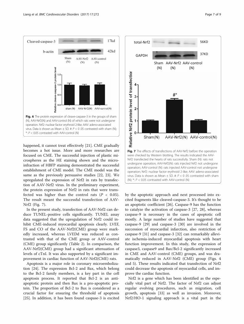
happened, it cannot treat effectively [21]. CME graduallybecomes a hot issue. More and more researches arefocused on CME. The successful injection of plastic mi-crospheres as the HE staining shown and the micro-infraction of HBFP staining demonstrated the successfulestablishment of CME model. The CME model was thesame as the previously persuasive studies [22, 23]. Weupregulated the expression of Nrf2 in rats by transfec-tion of AAV-Nrf2 virus. In the preliminary experiment,the protein expression of Nrf2 in rats that were trans-fected was higher than the control rats (P < 0.05).The result meant the successful transfection of AAV-Nrf2 (Fig. 7).In the present study, transfection of AAV-Nrf2 can de-
duce TUNEL-positive cells significantly. TUNEL assaydata suggested that the upregulation of Nrf2 could in-hibit CME-induced myocardial apoptosis clearly. LVEF,FS and CO of the AAV-Nrf2(CME) group were mark-edly increased, whereas LVEDd was reduced as con-trasted with that of the CME group or AAV-control(CME) group significantly (Table 2). In comparison, theAAV-Nrf2(CME) group had a significant attenuation oflevels of cTnI. It was also supported by a significant im-provement in cardiac function of AAV-Nrf2(CME) rats.Apoptosis is a major role in coronary microemboliza-
tion [24]. The expression Bcl-2 and Bax, which belongto the Bcl-2 family members, is a key part in the cellapoptosis process. It reported that Bcl-2 is an anti-apoptotic protein and then Bax is a pro-apoptotic pro-tein. The proportion of Bcl-2 to Bax is considered as acrucial factor for ensuring the threshold of apoptosis[25]. In addition, it has been found caspase-3 is excited
by the apoptotic approach and next processed into ex-cited fragments like cleaved-caspase-3. It’s thought to bean apoptotic coefficient [26]. Caspase-9 has the functionto catalyze the activation of caspase-3 [27, 28], whereascaspase-9 is necessary in the cases of apoptotic cellmostly. A large number of studies have suggested thatcaspase-9 [29] and caspase-3 [30] are involved in thesuccession of myocardial infarction, also restriction ofcaspase-9 [31] and caspase-3 [32] can remarkably allevi-ate ischemia-induced myocardial apoptosis with heartfunction improvement. In this study, the expression ofcaspase3, caspase9 and Bax/Bcl-2 significantly increasedin CME and AAV-control (CME) groups, and was dra-matically reduced in AAV-Nrf2 (CME) group (Figs. 4and 5). These results indicated that transfection of Nrf2could decrease the apoptosis of myocardial cells, and im-prove the cardiac function.Nrf2 is a gene which has been identified as the espe-
cially vital part of Nrf2. The factor of Nrf2 can adjustregular evolving procedures, such as migration, cellgrowth, apoptosis [33] as well as invasion. Moreover,Nrf2/HO-1 signaling approach is a vital part in the
Fig. 6 The protein expression of cleave-caspase-3 in the groups of sham(N), AAV-Nrf2(N) and AAV-control (N) of which rats were not undergoneoperation. Nrf2: nuclear factor erythroid 2-like; AAV: adeno-associatedvirus. Data is shown as Mean ± SD. #: P < 0 .05 contrasted with sham (N);*: P < 0.05 contrasted with AAV-control (N)
Fig. 7 The effects of transfections of AAV-Nrf2 before the operationwere checked by Western blotting. The results indicated the AAV-Nrf2 transfected the hearts of rats successfully. Sham (N): rats notundergone operation; AAV-Nrf2(N): rats injected Nrf2 not undergoneoperation; AAV-control (N): rats injected AAV-control not undergoneoperation; Nrf2: nuclear factor erythroid 2-like; AAV: adeno-associatedvirus. Data is shown as Mean ± SD. #: P < 0 .05 contrasted with sham(N); *: P < 0.05 contrasted with AAV-control (N)
Liang et al. BMC Cardiovascular Disorders (2017) 17:272 Page 7 of 9

process of apoptosis in the recent studies [34, 35]. Actu-ally, Nrf2 is a crucial modulator of the expression ofHO-1 [36].HO is a stress-responsive enzyme whichcould reduce highly deleterious free heme and produceanti-oxidants, anti-inflammatories, and regulators ofapoptosis as well as multiplication, therefore maintaininghomeostasis under pathological conditions [37, 38].There are two distinct mammalian HO isoforms: HO-1and HO-2. HO-1 is upregulated in the myocardium ofrats in response to ischemia-reperfusion injury. HO-1can also protect cells from apoptosis induced by light[39]. Some researches have clarified that HO-1 express-ing is adjusted primarily by Nrf2. Activated Nrf2 hasbeen considered to be a crucial contributor to theexpression of HO-1 [40]. Meanwhile, Ning et al. foundthat down-regulation of miR-142-5p attenuated ische-mia/reperfusion injury in hippocampal neurons by pro-moting Nrf2/HO-1 signaling approach [41]. Liu et al.found that apoptosis was negatively correlated with theNrf2 and HO-1 expressing, and ultimately affected theexpression of cleaved caspase-3 [42]. In our study,mRNA and protein levels (Figs. 4 and 5) of Nrf2 andHO-1 upregulated in AAV-Nrf2(CME) rats and the Nrf2could reduce the expression of caspase-3 as well ascaspase-9 and a ratio of bax/bcl-2 and apoptosis of myo-cardial cells (Fig. 3), and decrease levels of cTnI, demon-strating activation of Nrf2/HO-1 signaling can improvecardiac function in CME rats. The apoptotic pathwaysmainly include intrinsic and extrinsic pathways [43].Some studies have confirmed that Nrf2/HO-1 mainlyregulated apoptosis through intrinsic pathway, but rela-tive studies about extrinsic pathway of Nrf2/HO-1 werefew [44, 45].In our study, Nrf2/HO-1 could suppress theexpression of caspase-9 and caspase-3, and caspase-9 isthe key factor of intrinsic pathway and active caspase-3to induce apoptosis. Nrf2/HO-1 is probably concludedin the process of myocardial apoptosis after CMEthrough the intrinsic pathway. Whether Nrf2/HO-1 canregulate apoptosis through extrinsic pathway, furtherstudies are still needed.There are likewise some limitations in the current study:
Firstly, the conclusions were drawn from the model of ratCME and it was prepared by the injection of the plasticmicrospheres into the LV. Thus, the present outcomemight not be straightforward compared to the resultsachieved in the patient. Secondly, we only studied theshort-term protection of Nrf2. Therefore, how long thisprotective effect lasts is uncertain. With this in mind, fu-ture research needs to be explored in more CME models.
ConclusionsThese findings showed that activating Nrf2/HO-1 signal-ing can inhibit apoptosis to attenuate coronary micro-embolization, and also improve the cardiac function.
Nrf2/HO-1 signaling pathway is possibly a potentialmedical goal for treatment associated and it has achance to be worthy for sick people who are undergoneacute coronary syndrome or PCI and improve the long-term prognosis.
AbbreviationsAAV: Adeno-associated virus; ANOVA: Analysis of variance; CME: Coronarymicroembolization; HO-1: Heme oxygenase-1; Nrf2: Nuclear factor erythroid2-like; RT-PCR: Reverse Transcription-Polymerase Chain Reaction; TBST: Tris-buffered saline tween; TUNEL: TdT-mediated dUTP Nick-End labeling
AcknowledgementsWe would like to thank Yuanxi Lu and Wenqin Guo for their technicalsupport. In addition, we would like to thank all colleagues for contributingthe experiments.
FundingThis research received support from National Natural Science Foundation ofChina (Grant No.81600283) and Guangxi Natural Science Foundation (GrantNo. 2016GXNSFBA380022).
Availability of data and materialsThe unprocessed data from the current study can be obtained from thecorresponding author.
Authors’ contributionsJL, LL and YS conceived the idea of the project. JL, XW, and QS conductedthe experiments of real time-PCR and Western blotting and analyzed data.WH and YL collected samples. JL and LL wrote the manuscript. QS and YSparticipated in the revision of the important part of the manuscript. JL, XWand WG worked together to produce the tables and figures. All contributiveauthors of this original manuscript authorized the ultimate manuscript. Allauthors read and approved the final manuscript.
Ethics approval and consent to participateThis experiment got the Ethics Committee of Guangxi Medical University,China permission and was carried out according with the Norms on theAnimal Experiments. The animals were sampled and managed under theNational Institute of Health (NIH Publication NO. 85-23, revised 1996).
Consent for publicationNot applicable.
Competing interestsAll authors reach a consensus to consent for coming out in the journal. Thispaper has not been published elsewhere in whole or in part, which are noconflicts involved in the article.
Publisher’s NoteSpringer Nature remains neutral with regard to jurisdictional claims inpublished maps and institutional affiliations.
Received: 31 May 2017 Accepted: 13 October 2017
References1. Erbel R, Heusch G. Coronary microembolization—its role in acute coronary
syndromes and interventions. Herz. 1999;24(7):558–75.2. Otto S, Seeber M, Fujita B, et al. Microembolization and myonecrosis during
elective percutaneous coronary interventions in diabetic patients: anintracoronary Doppler ultrasound study with 2-year clinical follow-up. BasicRes Cardiol. 2012;107(5):1–12.
3. Heusch G, Kleinbongard P, Böse D, et al. Coronary microembolization.Circulation. 2009;120(18):1822–36.
4. Falk E, Thuesen L. Pathology of coronary microembolisation and no reflow.Heart. 2003;89(9):983–5.
5. Breuckmann F, Nassenstein K, Bucher C, et al. Systematic analysis offunctional and structural changes after coronary microembolization: a
Liang et al. BMC Cardiovascular Disorders (2017) 17:272 Page 8 of 9

cardiac magnetic resonance imaging study. J Am Coll Cardiol Img. 2009;2(2):121–30.
6. Dorge H, Neumann T, Behrends M, et al. Perfusion-contraction mismatchwith coronary microvascular obstruction: role of inflammation. Am J Physiol.2000;279(2):H2587–92.
7. Ferrari R, Balla C, Malagù M, et al. Reperfusion damage―a story of success,failure, and hope. Circ J. 2017;81(2):131–41.
8. Heusch G. The coronary circulation as a target of cardioprotection. Circ Res.2016;118(10):1643–58.
9. Li L, Su Q, Wang Y, et al. Effect of atorvastatin (Lipitor) on myocardialapoptosis and caspase-8 activation following coronary microembolization.Cell Biochem Biophys. 2011;61(2):399–406.
10. Liu T, Zhou Y, Wang JY, et al. Coronary microembolization inducescardiomyocyte apoptosis in swine by activating the LOX-1-dependentmitochondrial pathway and caspase-8-dependent pathway. J CardiovascPharmacol Ther. 2016;21(2):209–18.
11. Liu T, Zhou Y, Liu YC, et al. Coronary Microembolization inducesCardiomyocyte apoptosis through the LOX-1–dependent endoplasmicreticulum stress pathway involving JNK/P38 MAPK. Can J Cardiol. 2015;31(10):1272–81.
12. Jia Z, Dong A, Che H, et al. 17-DMAG protects against hypoxia−/Reoxygenation-induced cell injury in HT22 cells through Akt/Nrf2/HO-1 pathway. DNA Cell Biol. 2017;36(2):95–102.
13. Zhang JF, Su L, Ye Q, et al. Discovery of a novel Nrf2 inhibitor that inducesapoptosis of human acute myeloid leukemia cells. Oncotarget. 2017;8(5):7625.
14. Chumboatong W, Thummayot S, Govitrapong P, et al. Neuroprotection ofagomelatine against cerebral ischemia/reperfusion injury through anantiapoptotic pathway in rat. Neurochem Int. 2017;102:114–22.
15. Fu FY, Chen BY, Chen LL, et al. Improvement of the survival and therapeuticeffects of implanted mesenchymal stem cells in a rat model of coronarymicroembolization by rosuvastatin treatment. Eur Rev Med Pharmacol Sci.2016;20(11):2368–81.
16. Thielmann M, Dörge H, Martin C, et al. Myocardial dysfunction with coronarymicroembolization: signal transduction through a sequence of nitric oxide,tumor necrosis factor-alpha, and sphingosine. Circ Res. 2002;90(7):807–13.
17. Zhang QY, Ge JB, Chen JZ, et al. Mast cell contributes to cardiomyocyteapoptosis after coronary microembolization. J Histochem Cytochem. 2006;54(5):515–23.
18. Hoggan MD, Blacklow NR, Rowe WP. Studies of small DNA viruses found invarious adenovirus preparations: physical, biological, and immunologicalcharacteristics. Proc Natl Acad Sci. 1966;55(6):1467–74.
19. Atchison RW, Casto BC, Hammon WMD. Adenovirus-associated defectivevirus particles. Science. 1965;149(3685):754–5.
20. Zacchigna S, Zentilin L, Giacca M. Adeno-associated virus vectors astherapeutic and investigational tools in the cardiovascular system. Circ Res.2014;114(11):1827–46.
21. Fu FY, Chen BY, Chen LL, et al. Improvement of the survival and therapeuticeffects of implanted mesenchymal stem cells in a rat model of coronarymicroembolization by rosuvastatin treatment. Eur Rev Med Pharmacol Sci.2016;20(11):2368.
22. Wang X, Lu Y, Sun Y, et al. TAK-242 protects against apoptosis in coronarymicroembolization-induced myocardial injury in rats by suppressing TLR4/NF-κB signaling pathway. Cell Physiol Biochem. 2017;41(4):1675–83.
23. Sun Y, Su Q, Li L, et al. MiR-486 regulates cardiomyocyte apoptosis by p53-mediated BCL-2 associated mitochondrial apoptotic pathway. BMCCardiovasc Disord. 2017;17(1):119.
24. Liu YC, Li L, Su Q, et al. Trimetazidine pretreatment inhibits myocardialapoptosis and improves cardiac function in a swine model of coronarymicroembolization. Cardiology. 2015;130(2):130–6.
25. Hu T, Wei G, Xi M, et al. Synergistic cardioprotective effects of Danshensuand hydroxysafflor yellow A against myocardial ischemia-reperfusion injuryare mediated through the Akt/Nrf2/HO-1 pathway. Int J Mol Med. 2016;38(1):83–94.
26. Salvesen GS. Caspases and apoptosis. Essays Biochem. 2002;38:9–19.27. Cain K, Bratton SB, Cohen GM. The Apaf-1 apoptosome: a large caspase-
activating complex. Biochimie. 2002;84(2):203–14.28. Rodriguez J, Lazebnik Y. Caspase-9 and APAF-1 form an active holoenzyme.
Genes Dev. 1999;13(24):3179–84.29. Takatani T, Takahashi K, Uozumi Y, et al. Taurine inhibits apoptosis by
preventing formation of the Apaf-1/caspase-9 apoptosome. Am J Phys CellPhys. 2004;287(4):C949–53.
30. Balsam LB, Kofidis T, Robbins RC. Caspase-3 inhibition preserves myocardialgeometry and long-term function after infarction. J Surg Res. 2005;124(2):194–200.
31. Sodhi RK, Singh M, Singh N, et al. Protective effects of caspase-9 and poly(ADP-ribose) polymerase inhibitors on ischemia-reperfusion-inducedmyocardial injury. Arch Pharm Res. 2009;32(7):1037–43.
32. Liu Q. Lentivirus mediated interference of Caspase-3 expression amelioratesthe heart function on rats with acute myocardial infarction. Eur Rev MedPharmacol Sci. 2014;18(13):1852–8.
33. Li P, Su L, Li X, et al. Remote limb ischemic postconditioning protects mousebrain against cerebral ischemia/reperfusion injury via upregulating expressionof Nrf2, HO-1 and NQO-1 in mice. Int J Neurosci. 2016;126(6):552–9.
34. Lei X, Lei L, Zhang Z, et al. Neuroprotective effects of lycopene pretreatmenton transient global cerebral ischemia-reperfusion in rats: the role of the Nrf2/HO-1 signaling pathway. Mol Med Rep. 2016;13(1):412–8.
35. Zhang C, Wen C, Lin J, et al. Protective effect of pyrroloquinoline quinine onultraviolet A irradiation-induced human dermal fibroblast senescence in vitroproceeds via the anti-apoptotic sirtuin 1/nuclear factor-derived erythroid 2-related factor 2/heme oxygenase 1 pathway. Mol Med Rep. 2015;12(3):4382–8.
36. Sun GY, Li R, Cui J, et al. Withania somnifera and its withanolides attenuateoxidative and inflammatory responses and up-regulate antioxidantresponses in BV-2 microglial cells. NeuroMolecular Med. 2016;18(3):241–52.
37. Fan J, Xu G, Jiang T, et al. Pharmacologic induction of Heme Oxygenase-1plays a protective role in diabetic retinopathy in RatsHeme Oxygenase-1 indiabetic retinopathy in rats. Invest Ophthalmol Vis Sci. 2012;53(10):6541–56.
38. Soares MP, Bach FH. Heme oxygenase-1: from biology to therapeuticpotential. Trends Mol Med. 2009;15(2):50–8.
39. Sun MH, Pang JHS, Chen SL, et al. Photoreceptor protection against lightdamage by AAV-mediated overexpression of heme oxygenase-1. InvestOphthalmol Vis Sci. 2007;48(12):5699–707.
40. Zhang M, Wang S, Mao L, et al. Omega-3 fatty acids protect the brainagainst ischemic injury by activating Nrf2 and upregulating hemeoxygenase 1. J Neurosci. 2014;34(5):1903–15.
41. Wang N, Zhang L, Lu Y, et al. Down-regulation of microRNA-142-5pattenuates oxygen-glucose deprivation and reoxygenation-induced neuroninjury through up-regulating Nrf2/ARE signaling pathway. BiomedPharmacother. 2017;89:1187–95.
42. Liu C, Vojnovic D, Kochevar IE, et al. UV-A irradiation activates Nrf2-regulatedantioxidant defense and induces p53/Caspase3-dependent apoptosis incorneal endothelial CellsUV-A activates Nrf2 and induces p53 in cornealendothelial cells. Invest Ophthalmol Vis Sci. 2016;57(4):2319–27.
43. Hong S, Kim HY, Kim J, et al. Smad7 protein induces interferon regulatoryfactor 1-dependent transcriptional activation of caspase 8 to restore tumornecrosis factor-related apoptosis-inducing ligand (TRAIL)-mediatedapoptosis. J Biol Chem. 2013;288(5):3560–70.
44. Zhang X, Liang D, Lian X, et al. Berberine activates Nrf2 nucleartranslocation and inhibits apoptosis induced by high glucose in renaltubular epithelial cells through a phosphatidylinositol 3-kinase/Akt-dependent mechanism. Apoptosis. 2016;21(6):721–36.
45. Yan B, Ma Z, Shi S, et al. Sulforaphane prevents bleomycin-inducedpulmonary fibrosis in mice by inhibiting oxidative stress via nuclear factorerythroid 2-related factor-2 activation. Mol Med Rep. 2017;15(6):4005–14.
• We accept pre-submission inquiries
• Our selector tool helps you to find the most relevant journal
• We provide round the clock customer support
• Convenient online submission
• Thorough peer review
• Inclusion in PubMed and all major indexing services
• Maximum visibility for your research
Submit your manuscript atwww.biomedcentral.com/submit
Submit your next manuscript to BioMed Central and we will help you at every step:
Liang et al. BMC Cardiovascular Disorders (2017) 17:272 Page 9 of 9