The proportion–ratio on dipeptidyl aminopeptidase-4 (DP-4 ...
11
Full Terms & Conditions of access and use can be found at https://www.tandfonline.com/action/journalInformation?journalCode=ljii20 Journal of Immunoassay and Immunochemistry ISSN: 1532-1819 (Print) 1532-4230 (Online) Journal homepage: https://www.tandfonline.com/loi/ljii20 The proportion–ratio on dipeptidyl aminopeptidase-4 (DP-4) inhibition by gelatin compared to synthetic sitagliptin Yoni Atma, Hanifah Nuryani Lioe, Endang Prangdimurti, Hermawan Seftiono, Moh Taufik, Dita Fitriani & Apon Zaenal Mustopa To cite this article: Yoni Atma, Hanifah Nuryani Lioe, Endang Prangdimurti, Hermawan Seftiono, Moh Taufik, Dita Fitriani & Apon Zaenal Mustopa (2019) The proportion–ratio on dipeptidyl aminopeptidase-4 (DP-4) inhibition by gelatin compared to synthetic sitagliptin, Journal of Immunoassay and Immunochemistry, 40:4, 386-395, DOI: 10.1080/15321819.2019.1613243 To link to this article: https://doi.org/10.1080/15321819.2019.1613243 Published online: 09 May 2019. Submit your article to this journal Article views: 90 View Crossmark data
Transcript of The proportion–ratio on dipeptidyl aminopeptidase-4 (DP-4 ...
Full Terms & Conditions of access and use can be found athttps://www.tandfonline.com/action/journalInformation?journalCode=ljii20
Journal of Immunoassay and Immunochemistry
ISSN: 1532-1819 (Print) 1532-4230 (Online) Journal homepage: https://www.tandfonline.com/loi/ljii20
The proportion–ratio on dipeptidylaminopeptidase-4 (DP-4) inhibition by gelatincompared to synthetic sitagliptin
Yoni Atma, Hanifah Nuryani Lioe, Endang Prangdimurti, HermawanSeftiono, Moh Taufik, Dita Fitriani & Apon Zaenal Mustopa
To cite this article: Yoni Atma, Hanifah Nuryani Lioe, Endang Prangdimurti, Hermawan Seftiono,Moh Taufik, Dita Fitriani & Apon Zaenal Mustopa (2019) The proportion–ratio on dipeptidylaminopeptidase-4 (DP-4) inhibition by gelatin compared to synthetic sitagliptin, Journal ofImmunoassay and Immunochemistry, 40:4, 386-395, DOI: 10.1080/15321819.2019.1613243
To link to this article: https://doi.org/10.1080/15321819.2019.1613243
Published online: 09 May 2019.
Submit your article to this journal
Article views: 90
View Crossmark data
ORIGINAL ARTICLE
The proportion–ratio on dipeptidyl aminopeptidase-4 (DP-4)inhibition by gelatin compared to synthetic sitagliptinYoni Atma a, Hanifah Nuryani Lioe b, Endang Prangdimurtib,Hermawan Seftionoa, Moh Taufika, Dita Fitriania, and Apon Zaenal Mustopac
aDepartment of Food Science and Technology, Faculty of Bioindustry, Universitas Trilogi, Kalibata,Jakarta, Indonesia; bDepartment of Food Science and Technology, Faculty of Agricultural Engineeringand Technology, Bogor Agricultural University, Bogor, West Java, Indonesia; cDivision of MolecularBiology, Research Center for Biotechnology, Indonesian Institute of Science (LIPI), Cibinong, Bogor,West Java, Indonesia
ABSTRACTThe current study aims to determine the inhibition activity gelatinagainst dipeptidyl aminopeptidase 4 (DP-4). Two commercial gela-tins, i.e., bovine and fish skin gelatin and one extracted (in ourlaboratory) gelatin, i.e., fish bone gelatin were selected for analysis.Each gelatin have same protein pattern (75–245 kDa) on sodiumdodecyl sulfate polyacrylamide gel electrophoresis with mean ofprotein concentration of 1.72 mg/mL. The inhibition activity wasmeasured on the capacity to inhibit DP-4 by usingGly-Pro-p-nitroa-nilide as their substrate. The sitagliptin was used as standardcomparison. Based on the percent inhibition, gelatin has beenshown to be the prospective DP-4 inhibitor.
KEYWORDSDipeptidyl peptidase IV (DPP-4);gelatin; incretin; inhibitoryactivity; diabetes mellitustype 2
Introduction
Dipeptidyl aminopeptidase (DP-4) or dipeptidyl peptidase 4 (DPP-4) is a proteasethat degrades some hormones, including incretin. Incretin, which is formed byglucose-dependent insulinotropic polypeptide (GIP) and glucagon-like peptide-1(GLP-1), has a short half-life (only 1–2 min) because of DP-4 activity. Thedegradation of GIP and GLP-1 causes the loss of incretin.[1] Incretin, however,has an important role regarding blood glucose homeostasis, and it was promisingtherapeutic target in type 2 diabetic therapy.[2] Therefore, recently, many studieson seeking and characterizing of DP-4 inhibitor have been growing rapidly.
Some of DP-4 inhibitors has been patented and commercialized includingsynthetic substances such as sitagliptin, saxagliptin, and linagliptin.[3]
Unfortunately, the synthetic DP-4 inhibitors potentially have side effectsthat lead to other diseases.[4] So that, research in looking for DP-4 inhibitorfrom natural sources pay close attention. The natural compound has been
CONTACT Yoni Atma [email protected] Department of Food Science and Technology, Faculty ofBioindustry, Universitas Trilogi, Kalibata, Jakarta 12760, IndonesiaThis article has been republished with minor changes. These changes do not impact the academic content of thearticle.Color versions of one or more of the figures in the article can be found online at www.tandfonline.com/ljii.
JOURNAL OF IMMUNOASSAY AND IMMUNOCHEMISTRY2019, VOL. 40, NO. 4, 386–395https://doi.org/10.1080/15321819.2019.1613243
© 2019 Taylor & Francis
studied as DP-4 inhibitor including the substance of plant-based extract[5,6];however, it also dominated food-based protein such as milk protein, beef,cheese, egg protein as well as fish protein and gelatin.[7] Some studies weredone for investigating the inhibitory activity of gelatin and their hydrolysateagainst DP-4 covering porcine, bovine, and fish gelatin.[4,8,9] The applicationof porcine gelatin as source DP-4 inhibitor has been limited because thissource is prohibited in Islam and Judaism. Fortunately, most of bovine andfish gelatin which was analyzed as DP-4 inhibitor were derived from the by-product. Fish processing by-product has been most potential as alternativesource for gelatin extraction.[10] For instance, the research and applicationabout bovine and fish-based gelatin-related DP-4 inhibitor would be promis-ing in the future. Even less, gelatin has been widely used for a long time asa safe and unique ingredient in food and pharmaceutical industries.[11]
The aim of this research was to determine the percent inhibition of bovine,fish skin, and fish bone gelatin against DP-4 through their ratio inhibitioncompared to synthetic sitagliptin. In the previous studies, the quantifiedinhibitory activity of gelatin and another food-based protein against DP-4enzymes do not use sitagliptin as standard, but diprotin A.[4,8,12] Actually,sitagliptin is a well-known synthetic DP-4 inhibitor in the market.[3,13] Theseeking for DP-4 inhibitors from natural substances and gelatin is certainlyintended to replacing this synthetics medicine.
Experimental method
Gelatin preparation
Two commercial gelatins were used in this study, i.e., bovine gelatin (technicalgelatin, no brand) and fish skin gelatin (progel, Ho Chi Minh, Vietnam), whilefish bone gelatin extracted from the bone of Pangasius catfish by pretreatmentand main extraction was based on Pertiwi et al.[14] In the pretreatment, the bonewas soaked in 1% citric acid solution for 48 hr to obtain the leach bone (ossein).Then, the ossein was separated with the solvent through centrifugation (5000 ×g,10 min), then it was washed with aquadest until the pH became neutral. Later, themain extraction was carried out by soaking the ossien in aquadest for 5 hr at 75°C. The clear extract of gelatin was collected by filtration using filter paper. At last,the extract was dried by food dehydrator (Excalibur, Sacramento, USA) for 8 hrat 55°C. The gelatin was dissolved with distillated water in concentration of25 mg/mL (m/v), stored at 4°C, and thawed before further analysis.
Vertical electrophoresis
Vertical electrophoresis was used as confirmation test regarding of gelatinexistence in the solution. It was performed using sodium dodecyl sulfate
JOURNAL OF IMMUNOASSAY AND IMMUNOCHEMISTRY 387
polyacrylamide gel electrophoresis (SDS-PAGE). The SDS-PAGE was carriedout by discontinues Tris/HCl/glycine buffer system.[15] Each gelatin wasdiluted in buffer containing 0.5 M Tris-HCl pH 6.8 with 3% (w/v) sodiumdodecyl sulfate (SDS), 30% (v/v) glycerol, and 15% (v/v) β-mercaptoethanolat ratio 1:1 (v/v). A 12% separating gel and 3.9% stacking gel were used.Then, a 20 µL sample and protein marker (SMOBIO PM 2700, Hsinchu,Taiwan) was run in electrophoresis device (ATTO Page Ace, Tokyo, Japan)at voltage of 50 V for 90 min. After that, the gel from electrophoresis wasstained with Coomassie Brilliant Blue R-250 for 24 hr until a clear bluebackground gel was obtained.
DP-4 inhibition
DP-4 inhibitory activity was determined based on DP-4 activity for using theirsubstrate in the presence of sample. It is interpreted by the capacity of reactedsolution to absorb color at visible wavelength.[16] To measure DP-4 inhibition,first, gelatin was added to the solution of Gly-Pro-p-nitroanilide (Sigma, St.Louis, MO, USA) 1.59 mM in 100 mM buffer Tris pH 8 in a ratio 1:1 (v/v).This mixed solution was then incubated at 37°C for 10 min. Next, the DPP-4(Sigma, St. Louis, MO, USA) was diluted using 100 mM buffer Tris pH 8 andpipetted 40 μl DPP-IV (concentration of 0.01 U/mL) was mixed with 80 μlmixing solution that contains sample gelatin and Gly-Pro-p-nitroanilide, whichwas earlier prepared. After that, the solution was gently shaken and reacted at37°C for 60 min. Then, this reaction was stopped by adding 100 µl of sodiumacetate 1 M pH 4. Finally, the absorbance of solution measured using enzyme-linked immunosorbent assay (ELISA) Microplate Reader (Multiskan Ex,Champaign, IL, USA) at a wavelength of 405 nm. Sitagliptin (EuropeanPharmacopoeia, Strasbourg, France) with a concentration of 0.0001–0.01 ng/mL was used as standard comparison. The percent of inhibition calculated bysetting up absorbance from 0.1 ng/mL sitagliptin as 100% which is multipliedwith the another absorbance of inhibition reaction.
Protein quantification
The protein concentration from three sources of gelatin (bovine, fish skin,and fish bone) were quantified using bicinchoninic acid (BCA) assay kit(Thermo Fisher, Rockford, IL, USA) by the method described Atma andRamdhani.[17] Each gelatin solution was pipetting as many as 10 µL, thenputted into each well in 96-well plate. After that, 200 µL BCA solution thatcontains solution A (sodium carbonate, sodium bicarbonate, bicinchoninicacid, sodium tartrate in 0.1 M sodium hydroxide) and solution B (copper (II)sulfate) in ratio 50:1 (v/v) was added. Then, the mixture was mixed andincubated for 30 min at room temperature. The absorbance of sample inBCA solution was measured at 560 nm with ELISA Microplate Reader
388 Y. ATMA ET AL.

(Multiskan Ex, Champaign, IL, USA). In this quantification procedure, thebovine serum albumin in the range of 0–1 mg/mL was used as standardsolution.
Data analysis
Data were analyzed using one-way analysis of variance, followed by Tukey’sHonestly Significant Different test or Tukey’s range test to statistically deter-mine the level of significance difference (P < .05) between data.
Result
Gelatin confirmation test
In this study, all three types of gelatin, i.e., bovine, fish skin, and fish bone,appeared at the molecular weight range of 75–245 kDa. It was confirmed asgelatin with protein concentration of 2.19 ± 0.008, 1.70 ± 0.002, and 1.27 ±0.004 mg/mL, respectively. The electropherogram or SDS-PAGE of the gelatinis presented in Figure 1, while the protein concentration each of gelatin isshown in Table 1. Table 1 also demonstrated the value of percent inhibitiondivided by protein concentration where the gelatin obtained from the bone offish was higher compared to that of bovine and fish skin gelatin. Thehighest percent of inhibition from gelatin was used for determining this value.
Figure 1. Electroforegram bovine gelatin (B), fish skin (FS), and fish bone (FB)gelatin; M = marker protein.
JOURNAL OF IMMUNOASSAY AND IMMUNOCHEMISTRY 389

DP-4 inhibition
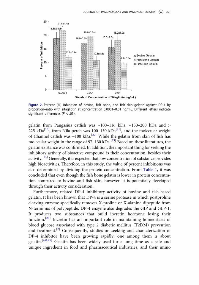
One of the most used procedures for determining the inhibition capacity ofgelatin against DP-4 is through the measurement of DP-4 activity by usingGly-Pro-p-nitroanilide as their substrate in the presence and absence ofgelatin. The increase in absorbance in the presence of gelatin at visiblewavelength on DP-4/substrate interaction increases the inhibitory activity.In this study, sitagliptin was used as positive control. The inhibitory activityperformed in percent (%) was quantified based on the absorbance samplegelatin compared to the absorbance of sitagliptin, and then multiplied by100%. There are three concentrations of sitagliptin that was used as controlin the range of 0.0001–0.01 ng/mL. Previously, Nongonierma and FitzGeraldwas used 0.006 and 0.03 ng/mL sitagliptin, regarding the study on inhibitoryproperties of whey protein against DP-4 enzyme.[18] Overall, the percent ofinhibition reduced by increasing the standard concentration of sitagliptin. Byusing 0.0001 ng/mL sitagliptin, the percent of inhibition reached its peak to21.8%. In this research, the gelatin derived from fish bone was higherin percent of inhibition against DP-4, while the gelatin from fish skin wasthe lowest (9.8–11.8%). In addition, the percent of inhibition bovine gelatinwas 15.6–18.8% where is no significant difference (P < .05) was found tocorrelate in fish bone gelatin, and significant difference was found in fish skingelatin. The inhibitory activity of the three gelatin sources in this studycompared to sitagliptin DP-4 inhibition is presented in Figure 2.
Discussion
SDS-PAGE was used to confirm the presence of gelatin in the prepared solution.The SDS-PAGE was used for protein identification based on their molecularweight. The gelatin is formed by different polypeptides categorized as α-chain, β-chain, and γ-chain gelatin. The α-chain gelatin hasmolecular weight in the rangeof 97–140 kDa, β-chain gelatin has molecular weight about 150–250 kDa, and γ-chain gelatin has molecular weight of >250 kDa.[19] Bovine gelatin has molecularweight in the range of 100–200 kDa.[20] The fish bone gelatin also has molecularweight range in 100–200 kDa. In more detail, the molecular weight of fish bone
Table 1. Protein concentration and value of percent inhibition divided protein concentration ofgelatin.Sources ofgelatin
Protein concentration(mg/mL)
Percent inhibition divided by protein concentration (% inhibitionper mg/mL protein)a
Bovine 2.19 ± 0.008a 8.58 ± 1.15b
Fish Bone 1.27 ± 0.004c 17.23 ± 1.21a
Fish Skin 1.70 ± 0.002b 6.96 ± 0.54b
Values in the same row followed by different letters are significant differents (P < 0.05)aThe highest percent inhibition from gelatin was used for determining the value of percent inhibitiondivided by protein concentration.
390 Y. ATMA ET AL.
gelatin from Pangasius catfish was ~100–116 kDa, ~150–200 kDa and >225 kDa[15], from Nila perch was 100–150 kDa[21], and the molecular weightof Channel catfish was ~100 kDa.[22] While the gelatin from skin of fish hasmolecular weight in the range of 97–130 kDa.[23] Based on these literatures, thegelatin existance was confirmed. In addition, the important thing for seeking theinhibitory activity of bioactive compound is their concentration, besides theiractivity.[24] Generally, it is expected that low concentration of substance provideshigh bioactivities. Therefore, in this study, the value of percent inhibitions wasalso determined by dividing the protein concentration. From Table 1, it wasconcluded that even though the fish bone gelatin is lower in protein concentra-tion compared to bovine and fish skin, however, it is potentially developedthrough their activity consideration.
Furthermore, related DP-4 inhibitory activity of bovine and fish-basedgelatin. It has been known that DP-4 is a serine protease in which postprolinecleaving enzyme specifically removes X-proline or X-alanine dipeptide fromN-terminus of polypeptide. DP-4 enzyme also degrades the GIP and GLP-1.It produces two substances that build incretin hormone losing theirfunction.[25] Incretin has an important role in maintaining homeostasis ofblood glucose associated with type 2 diabetic mellitus (T2DM) preventionand treatment.[2] Consequently, studies on seeking and characterization ofDP-4 inhibitor have been growing rapidly; one among them is aboutgelatin.[4,8,25] Gelatin has been widely used for a long time as a safe andunique ingredient in food and pharmaceutical industries, and their imino
Figure 2. Percent (%) inhibition of bovine, fish bone, and fish skin gelatin against DP-4 byproportion–ratio with sitagliptin at concentration 0.0001–0.01 ng/mL. Different letters indicatesignificant differences (P < .05).
JOURNAL OF IMMUNOASSAY AND IMMUNOCHEMISTRY 391
acid (proline+hydroxyproline) composition also makes this colloid fit forDPP-IV inhibition.[4,8,25,26]
The measurement of inhibitory activity against DP-4 enzyme uses sita-gliptin as positive control, which was used as inhibitor of DP-4 and used asmedicine for T2DM therapy as well as treatment manner. Some syntheticmedicines have been used as DP-4 inhibitors, including sitagliptin, saxaglip-tin, linagliptin, and vildagliptin.[27] Study by Tatosian et al.[3] concluded thattreatment of diabetic patient with sitagliptin trough DP-4 inhibition wassignificantly greater than another such as saxagliptin or vildagliptin adminis-tered once daily. Administration of once-daily sitagliptin is similar to thatprovided by vildagliptin administered twice daily. Sitagliptin categorized asnon-substrate like inhibitor for DP-4.[28] In our research paper, inhibitoryactivity of sitagliptin determined of 100% inhibition because it was standardDP-4 inhibitor.
Implementation of sitagliptin as standard was also carried out by Ekayantiet al. (2018), in which showed that DP-4 inhibitor was quantified from whitetea (Camellia sinensis (L.) Kuntze) extract. It was found that the white teaextracted using methanol has greatest DP-4 inhibition than hexane and ethylacetate. The percent inhibition of white tea fraction extracted using metha-nol, hexane, and ethyl acetate was 50.48, 32.42, and 36.54%, respectively.[5]
Another research regarding DP-4 inhibitor using sitagliptin as comparisonmedicine was also conducted by Riyanti et al. (2016) by using various plants.Based on this research, it was found that leaves of various plants have percentinhibition about 13.94 ± 4.08%–68.98 ± 1.95% against DP-4. The inhibitoryactivity of semen, herb, stem, and rhozime of herb plant against DP-4 was6.48 ± 0.32%–71.29 ± 0.33%, 33.52% – 37.03 ± 0.65%, 65.86 ± 1.02%, and17.12 ± 1.95% respectively.[6]
The percent of inhibition gelatin against DP-4 enzyme was also studiedpreviously. The porcine gelatin has percent inhibition about 9.2%,[8] whilethe gelatin from salmon skin was about 10%.[25] The gelatin from skin ofpacific hake, halibut, nila tipalia, and milkfish has percent inhibition lowerthan 10%.[4] In addition, fish protein from tuna sauce has inhibitory activityof 9.8%.[12] This inhibitory activity gelatin was increased after hydrolysis andultrafiltration.[4,8,25] All of those studies used diprotin A as DP-4 inhibitor asstandard comparison. The IC50 of diprotin A was 24.7 µM,[25] while the IC50
of sitagliptin was 18–20 nM.[29]
Regarding their mechanism in human intestinal tract, gelatin is likelyhydrolyzed by gastric and intestinal protease. Protein and/or gelatin ishydrolyzed in gastrointestinal route and then absorbed only in a form offree amino acid, di- and tripeptides[26] indeed, so it might become pro-spective and challeging for the future studies about the peptide derivedfrom gelatin hydrolysis in human digestion tract correlated with theirbioactivities as DP-4 inhibitor. It is highly potential if the gelatin
392 Y. ATMA ET AL.

hydrolyzed to become bioactive peptide which directly absorbed in humanintestinal ephitellium. Based on previous study was indicated that thecollagen-derived peptides have bioactivities after absorption from thedigestive tract by in vivo experiment, and several researches have beenfocused on the effect of oral intake in both animal and human models,which have expressed the excellent absorption and metabolism on Hyp-containing peptides.[30] However, if the peptide cannot be directlyabsorbed after passing through the human digestive tract, it will bea challege to discover a vehicle transporting the peptide lead up to thehuman systemic circulation.
Conclusion
Percent inhibition of bovine, fish skin, and fish bone gelatin against DP-4was 9.8–21.8% using sitagliptin as standard comparison. Each percent inhibi-tion of gelatin was increased if sitagliptin, used as standard comparison,decreased. Among the three gelatin sources, the inhibitory activity of gelatinfrom fish bone was the highest. It was not significant compared to bovinegelatin; however, it was significantly different when compared to fish skingelatin. The proportion–ratio of percent inhibition of gelatin in this studywas found to be higher when compared to another fish-based protein andgelatin.
Acknowledgments
This research was supported by Grant of Directorate General of Research and Developmentin Penelitian Kerjasama Antar Perguruan Tinggi (PKPT) scheme, Ministry ResearchTechnology and Higher Education of Indonesia
Funding
This work was supported by the Ministry of Research Technology and Higher Education ofIndonesia [grant number: 037/KM/PNT/2018].
ORCID
Yoni Atma http://orcid.org/0000-0002-0487-9601Hanifah Nuryani Lioe http://orcid.org/0000-0003-1364-0798
References
[1] Kazakos, K.;. Incretin Effect: GLP-1, GIP, DPP4. Diabetes Res. Clin. Pract. 2011, 93S,S32–36. DOI: 10.1016/S0168-8227(11)70011-0.
JOURNAL OF IMMUNOASSAY AND IMMUNOCHEMISTRY 393

[2] Phillips, L. K.; Prins, J. B. Update on Incretin Hormones. Ann. N Y Acad. Sci. 2015,1243, E55–74. DOI: 10.1111/j.1749-6632.2012.06491.x.
[3] Tatosian, D. A.; Guo, Y.; Schaeffer, K. A.; Gaibu, N.; Popa, S.; Stoch, A.; Langdon, R. B.;Kauh, E. A. Dipeptidyl Peptidase-4 Inhibition in Patients with Type 2 Diabetes Treatedwith Saxagliptin, Sitagliptin, or Vildagliptin. Diabetes Ther. 2013, 4(2), 431–442. DOI:10.1007/s13300-013-0045-8.
[4] Wang, T. Y.; Hsieh, C. H.; Hung, C. C.; Jao, C. L.; Chen, M. C.; Hsu, K. C. Fish SkinGelatin Hydrolysates As Dipeptidyl Peptidase IV Inhibitors and Glucagon-LikePeptide-1 Stimulators Improve Glycaemic Control in Diabetic Rats: A ComparisonBetween Warm- and Cold-Water Fish. J. Funct. Foods. 2015, 19, 330–340. DOI:10.1016/j.jff.2015.09.037.
[5] Ekayanti, M.; Sauriasari, R.; Elya, B. Dipeptidyl Peptidase IV Inhibitory Activity ofFraction from White Tea Ethanolic Extract (Camellia Sinensis (L.) Kuntze) Ex Vivo.Pharmacog. J. 2018, 10(1), 190–193. DOI: 10.5530/pj.2018.1.32.
[6] Riyanti, S.; Suganda, A. G.; Sukandar, E. Y. Dipeptidyl Peptidase-IV Inhibitory Activityof Some Indonesian Medicinal Plants. Asian J. Pharm. Clin. Res. 2016, 9(2), 375–377.
[7] Patil, P.; Mandal, S.; Tomar, S. K.; Anand, S. Food Protein-Derived Bioactive PeptidesIn Management of Type 2 Diabetes. Eur. J. Nutr. 2015, 54(6), 863–880. DOI: 10.1007/s00394-015-0974-2.
[8] Huang, S. L.; Hung, C. C.; Jao, C. L.; Tung, Y. S.; Hsu, K. C. Porcine Skin GelatinHydrolysate as a Dipeptidyl Peptidase IV Inhibitor Improves Glycemic Control inStreptozotocin-Induced Diabetic Rats. J. Funct. Foods. 2014, 11, 235–242. DOI:10.1016/j.jff.2014.09.010.
[9] Hatanaka, T.; Kawakami, K.; Uraji, M. Inhibitory Effect of Collagen-DerivedTripeptides on Dipeptidylpeptidase-IV Activity. J. Enzyme Inhib. Med. Chem. 2014,29(6), 823–828. DOI: 10.3109/14756366.2013.858143.
[10] Karim, A. A.; Bhat, R. Fish Gelatin: Properties, Challenges, and Prospects as anAlternative to Mammalian Gelatin. Food Hydrocoll. 2009, 23(3), 563–576. DOI:10.1016/j.foodhyd.2008.07.002.
[11] Mariod, A. A.; Fadul, A. H. Review: Gelatin, Source, Extraction and IndustrialApplications. Acta Sci. Pol. Technol. Aliment. 2013, 12(2), 135–147.
[12] Huang, S. L.; Jao, C. L.; Ho, K. P.; Hsu, K. C. Dipeptidyl-Peptidase IV InhibitoryActivity of Peptides Derived From Tuna Cooking Juice Hydrolysates. Peptides. 2012, 35(1), 114–121. DOI: 10.1016/j.peptides.2012.03.006.
[13] Engel, S. S.; Williams-Herman, D. E.; Golm, G. T.; Clay, R. J.; Machotka, S. V.;Kaufman, K. D.; Goldstein, B. J. Sitagliptin: Review of Preclinical and Clinical DataRegarding Incidence of Pancreatitis. Int. J. Clin. Pract. 2010, 64(7), 984–990. DOI:10.1111/j.1742-1241.2010.02382.x.
[14] Pertiwi, M.; Atma, Y.; Mustopa, A. Z.; Maisarah, R. Karakteristik Fisik dan KimiaGelatin dari Tulang Ikan Patin dengan Pre- Treatment Asam Sitrat. Jurnal AplikasiTeknologi Pangan. 2018, 7(2). DOI: 10.17728/jatp.2470.
[15] Mahmoodani, F.; Ardekani, V. S.; See, S. F.; Yusop, S. M.; Babji, A. S. Optimizationand Physical Properties of Gelatin Extracted from Pangasius Catfish (PangasiusSutchi) Bone. J. Food Sci. Technol. 2014, 51(11), 3104–3113. DOI: 10.1007/s13197-012-0816-7.
[16] Kojima, K.; Ham, T.; Kato, T. Rapid Chromatographic Purification of DipeptidylPeptidase IV in Human Submaxillary Gland. J. Chromatogr. A. 1980, 189(2), 233−240. DOI: 10.1016/S0021-9673(00)81523-X.
394 Y. ATMA ET AL.

[17] Atma, Y.; Ramdhani, H. Gelatin Extraction the Indegenous Pangasius Catfish BoneUsing Pineapple Liquid Waste. Indones. J. Biotechnol. 2017, 22(2), 86–91. DOI:10.22146/ijbiotech.32472.
[18] Nongonierma, A. B.; FitzGerald, R. J. Dipeptidyl Peptidase IV Inhibitory Properties ofa Whey Protein Hydrolysate: Influence of Fractionation, Stability to SimulatedGastrointestinal Digestion and Food Drug Interaction. Int. Dairy J. 2013, 32(1),33–39. DOI: 10.1016/j.idairyj.2013.03.005.
[19] Zhou, P.; Regenstein, J. M. Effects of Alkaline and Acid Pretreatments on AlaskaPollock Skin Gelatin Extraction. J. Food Sci. 2006, 70(6), 392–396. DOI: 10.1111/j.1365-2621.2005.tb11435.x.
[20] Hermanto, S.; Sumarlin, L. O.; Fatimah, W. Differentiation of Bovine and PorcineGelatin Based on Spectroscopic and Electrophoretic Analysis. J. Food Pharm. Sci. 2013,1(3), 68–73. DOI: 10.14499/jfps.
[21] Muyonga, J. H.; Colec, C. G. B.; Duodub, K. G. Extraction and PhysicochemicalCharacterisation of Nile Perch (Lates Niloticus) Skin and Bone Gelatin. FoodHydrocoll. 2004, 18(4), 581–592. DOI: 10.1016/j.foodhyd.2003.08.009.
[22] Liu, H. Y.; Han, J.; Guo, S. D. Characteristics of the Gelatin Extracted from ChannelCatfish (Ictalurus Punctatus) Head Bones. LWT - Food Sci. Technol. 2009, 42(2),540–544. DOI: 10.1016/j.lwt.2008.07.013.
[23] Taheri, A.; Abedian Kenari, A. M.; Gildberg, A.; Behnam, S. Extraction andPhysicochemical Characterization of Greater Lizardfish (Saurida Tumbil) Skin andBone Gelatin. J. Food Sci. 2009, 74(3), 160–165. DOI: 10.1111/j.1750-3841.2009.01106.x.
[24] Jao, C. L.; Hung, C. C.; Tung, Y. S.; Lin, P. Y.; Chen, M. C.; Hsu, K. C. TheDevelopment of Bioactive Peptides from Dietary Proteins as a Dipeptidyl PeptidaseIV Inhibitor for the Management of Type 2 Diabetes. Biomedicine. 2015, 5(3), 9–15.DOI: 10.7603/s40681-015-0014-9.
[25] Li-Chan, E. C. Y.; Hunag, S. L.; Jao, C. L.; Ho, K. P.; Hsu, K. C. Peptides Derived FromAtlantic Salmon Skin Gelatin as Dipeptidyl-Peptidase IV Inhibitors. J. Agric. FoodChem. 2012, 60(4), 973–978. DOI: 10.1021/jf204720q.
[26] Miner-Williams, W. M.; Stevens, B. R.; Moughan, P. J. Are Intact Peptides Absorbedfrom the Healthy Gut in the Adult Human? Nutr. Res. Rev. 2014, 27, 308–329. DOI:10.1017/S0954422414000225.
[27] Ahren, B.;. Vildagliptin: A DPP-4 Inhibitor for the Treatment of Type 2 Diabetes.Diabetes Manage. 2012, 2(5), 453–464. DOI: 10.2217/dmt.12.40.
[28] Monika, G.; Sarbjot, S.; Punam, G. Dipeptidyl Peptidase-4 Inhibitors: A New Approachin Diabetes Treatment. Int. J. Drug Dev. Res. 2009, 1(1), 146–156.
[29] European Medicines Agency (EMA). Assessment Report for Januvia InternationalNonproprietary Name: Sitagliptin. https://www.ema.europa.eu/documents/scientific-discussion/januvia-epar-scientific-discussion_en.pdf (accessed Oct 4, 2018).
[30] Aleman, A.; Martinez-Alvarez, O. Marine Collagen as a Source of Bioactive Molecules.A Review. Nat. Prod. J. 2013, 3(2). DOI: 10.2174/2210315511303020005.
JOURNAL OF IMMUNOASSAY AND IMMUNOCHEMISTRY 395