The Prognostic Value of Decreased KLF4 in Digestive System ...
12
Research Article The Prognostic Value of Decreased KLF4 in Digestive System Cancers: A Meta-Analysis from 17 Studies Jianpei Hu, 1 Huipu Li, 2 Chunyu Wu, 1 Xueying Zhao, 1 and Chaodong Liu 1 1 Department of Urology, The First Affiliated Hospital of Chongqing Medical University, Chongqing, China 2 Department of Gastroenterology, The First Affiliated Hospital of Chongqing Medical University, Chongqing, China Correspondence should be addressed to Chaodong Liu; [email protected] Received 10 April 2017; Revised 19 July 2017; Accepted 20 August 2017; Published 14 September 2017 Academic Editor: Benoit Dugue Copyright © 2017 Jianpei Hu et al. This is an open access article distributed under the Creative Commons Attribution License, which permits unrestricted use, distribution, and reproduction in any medium, provided the original work is properly cited. Background. The prognostic value of loss of Krüppel-like factor 4 (KLF4) expression in digestive system cancers has not reached a consensus. This study aimed for a comprehensive investigation of the internal associations between KLF4 expression loss and prognostic implications in patients with digestive system cancers. Methods. We searched for all relevant literatures in the electronic databases until February 1, 2017. The degree of association between KLF4 and prognosis was evaluated by pooled hazard ratios (HRs) as well as relevant 95% confidence intervals (95% CIs). Results. Seventeen eligible studies with 2118 patients revealed that loss of KLF4 expression was connected with poor prognosis, with the pooled HRs of 1.61 (95% CI: 1.17–2.20, P =0 003) for the overall survival (OS) and 1.99 (95% CI: 1.12–3.52, P =0 001) for the disease-free survival (DFS)/recurrence-free survival (RFS)/metastasis-free survival (MFS). Additionally, loss of KLF4 expression was also related to a worse disease-special survival (DSS) yielding a pooled HR of 1.73 (95% CI: 1.08–2.77, P =0 022). Conclusion. Our findings suggest that loss of KLF4 expression is correlated with a bad outcome in most digestive system cancers, apart from esophagus squamous cell carcinoma (ESCC). 1. Introduction Digestive system cancers generally refer to these cancers that arise from the esophagus, stomach, liver, gallbladder, biliary tract, colon, rectum, and anus, and all of them are common types of carcinomas around the world. Of note, colorectal, gastric, and liver cancer are the leading causes of cancer- related deaths which therefore confer a heavy burden on the society worldwide [1]. In the United States, there are approximately 310,440 new cases diagnosed with digestive system cancers with an estimated 157,700 deaths in 2017 [2]. On the one hand, despite a vast number of progresses have been made for the etiology, diagnosis and therapy of digestive system malignancies, the prognoses of these patients are still poor and unsatisfied; on the other hand, the advent of the molecular-targeted therapy era provides new choices of cancer therapy with a promising prospect [3, 4]. Hence, much more efforts should be made by researchers to identify those ideal molecular markers that represent both therapeutic value and predictive value for prognosis, then contributing to risk stratification and optimal choice of treatment for patients. Krüppel-like factor 4 (KLF4) can also be referred to as gut-enriched KLF (GKLF) or epithelial zinc finger protein (EZF) which mainly expresses in epithelial tissues of the mammals, including the intestine, skin, thymus, and lung. As a complicated transcription factor, KLF4 contains a highly conserved C-terminal DNA-binding domain with three zinc fingers. In physiological condition, upon binding to the spe- cific sequences, including CACCC boxes and GC boxes, KLF4 can exert multiple functions through regulating many cellular processes, such as cell proliferation, development, apoptosis, and homeostasis [5, 6]. Furthermore, in the context of most malignancies, KLF4 is necessary for the sup- pression of tumorigenesis and progression, basing on its inhibition of epithelial-mesenchymal transition (EMT), cell proliferation, and migration [7–10]. However, it has also been reported that KLF4 may be an oncogene in a few types Hindawi Disease Markers Volume 2017, Article ID 3064246, 11 pages https://doi.org/10.1155/2017/3064246
Transcript of The Prognostic Value of Decreased KLF4 in Digestive System ...

Research ArticleThe Prognostic Value of Decreased KLF4 in Digestive SystemCancers: A Meta-Analysis from 17 Studies
Jianpei Hu,1 Huipu Li,2 Chunyu Wu,1 Xueying Zhao,1 and Chaodong Liu1
1Department of Urology, The First Affiliated Hospital of Chongqing Medical University, Chongqing, China2Department of Gastroenterology, The First Affiliated Hospital of Chongqing Medical University, Chongqing, China
Correspondence should be addressed to Chaodong Liu; [email protected]
Received 10 April 2017; Revised 19 July 2017; Accepted 20 August 2017; Published 14 September 2017
Academic Editor: Benoit Dugue
Copyright © 2017 Jianpei Hu et al. This is an open access article distributed under the Creative Commons AttributionLicense, which permits unrestricted use, distribution, and reproduction in any medium, provided the original work isproperly cited.
Background. The prognostic value of loss of Krüppel-like factor 4 (KLF4) expression in digestive system cancers has not reached aconsensus. This study aimed for a comprehensive investigation of the internal associations between KLF4 expression loss andprognostic implications in patients with digestive system cancers. Methods. We searched for all relevant literatures in theelectronic databases until February 1, 2017. The degree of association between KLF4 and prognosis was evaluated by pooledhazard ratios (HRs) as well as relevant 95% confidence intervals (95% CIs). Results. Seventeen eligible studies with 2118patients revealed that loss of KLF4 expression was connected with poor prognosis, with the pooled HRs of 1.61 (95% CI:1.17–2.20, P = 0 003) for the overall survival (OS) and 1.99 (95% CI: 1.12–3.52, P = 0 001) for the disease-free survival(DFS)/recurrence-free survival (RFS)/metastasis-free survival (MFS). Additionally, loss of KLF4 expression was also relatedto a worse disease-special survival (DSS) yielding a pooled HR of 1.73 (95% CI: 1.08–2.77, P = 0 022). Conclusion. Ourfindings suggest that loss of KLF4 expression is correlated with a bad outcome in most digestive system cancers, apartfrom esophagus squamous cell carcinoma (ESCC).
1. Introduction
Digestive system cancers generally refer to these cancers thatarise from the esophagus, stomach, liver, gallbladder, biliarytract, colon, rectum, and anus, and all of them are commontypes of carcinomas around the world. Of note, colorectal,gastric, and liver cancer are the leading causes of cancer-related deaths which therefore confer a heavy burden onthe society worldwide [1]. In the United States, there areapproximately 310,440 new cases diagnosed with digestivesystem cancers with an estimated 157,700 deaths in 2017[2]. On the one hand, despite a vast number of progresseshave been made for the etiology, diagnosis and therapy ofdigestive system malignancies, the prognoses of thesepatients are still poor and unsatisfied; on the other hand,the advent of the molecular-targeted therapy era providesnew choices of cancer therapy with a promising prospect[3, 4]. Hence, much more efforts should be made byresearchers to identify those ideal molecular markers that
represent both therapeutic value and predictive value forprognosis, then contributing to risk stratification and optimalchoice of treatment for patients.
Krüppel-like factor 4 (KLF4) can also be referred to asgut-enriched KLF (GKLF) or epithelial zinc finger protein(EZF) which mainly expresses in epithelial tissues of themammals, including the intestine, skin, thymus, and lung.As a complicated transcription factor, KLF4 contains a highlyconserved C-terminal DNA-binding domain with three zincfingers. In physiological condition, upon binding to the spe-cific sequences, including CACCC boxes and GC boxes,KLF4 can exert multiple functions through regulating manycellular processes, such as cell proliferation, development,apoptosis, and homeostasis [5, 6]. Furthermore, in thecontext of most malignancies, KLF4 is necessary for the sup-pression of tumorigenesis and progression, basing on itsinhibition of epithelial-mesenchymal transition (EMT), cellproliferation, and migration [7–10]. However, it has alsobeen reported that KLF4 may be an oncogene in a few types
HindawiDisease MarkersVolume 2017, Article ID 3064246, 11 pageshttps://doi.org/10.1155/2017/3064246

of cancers, such as breast cancer and skin squamous cellcarcinoma [11, 12], suggesting that KLF4, similar to trans-forming growth factor-β and Notch, may have opposingroles in tumorigenesis and progression in a context-dependent manner [13, 14].
In the context of digestive system cancers, the vastmajority of studies have revealed that KLF4 is decreasedor absent with a bad clinical outcome, including esophagussquamous cell carcinoma (ESCC) [15, 16], gastric cancer(GC) [17–19], pancreatic ductal adenocarcinoma (PDAC)[20, 21], hepatocellular cancer (HCC) [22–24], and colo-rectal cancer (CRC) [25–27]. However, the dependabilityof KLF4 serving as a prognostic biomarker has not beencoming to an agreement in different cancers for the insig-nificant even opposite results [28–31]. Hence, the prognos-tic role of KLF4 in patients with digestive system cancersremains disputed. It is therefore unknown that the differ-ences in these studies are most caused by their small sam-ple size or inherent heterogeneity. On account of the limitsof a single study, it is necessary to evaluate the reportedstudies using a comprehensive meta-analysis.
In this study, the goal is to determine the prognosticvalue of loss of KLF4 expression among digestive systemcancers via gathering global relevant literatures to performa systematic analysis.
2. Materials and Methods
2.1. Search Strategy. A thorough search was carried out for allrelevant literatures that evaluated the prognostic value ofKLF4 in different digestive system cancers until February 1,2017 among the following electronic databases: Pubmed,ISI Web of Science and Embase. Search terms representedas follows: (KLF4 OR Krüppel-like factor 4 OR Gut-enriched KLF OR GKLF OR ZEF OR Epithelial Zinc FingerProtein) AND (cancer OR tumor OR neoplasm OR carci-noma) AND (Prognosis OR prognostic OR survival OR out-come). The Cochrane Library was also reviewed for relatedpapers. In addition, the citation lists of identified articles weremanually reviewed to complete the search. Two authors(Hu and Li) independently performed this procedure.Any disagreement was resolved by mutual discussion.
2.2. Selection Criteria. In this meta-analysis, the eligibility ofcandidate studies was determined based on the followingcriteria: (i) studied the patients with digestive system cancers;(ii) measured KLF4 expression using either semiquantitativeimmunohistochemistry (IHC) or quantitative reverse tran-scription PCR (RT-PCR); and (iii) evaluated the correlationbetween KLF4 expression and prognosis. Articles were nottaken into account when the following criteria were met: (i)duplicated or overlapped studies; (ii) reviews, case reports,comments, or conference abstracts; and (iii) absence of keyinformation for further quantification calculation. Two indi-viduals (Zhao and Wu) separately carried out all evaluationsand any discrepancy was resolved by consensus.
2.3. Quality Assessment. To accomplish the process of qualityassessment, each eligible article was scored in the light of the
Newcastle-Ottawa scale (NOS) [32] because all of them wereobservational studies. The cohorts of included studies werescored in terms of selection, comparability, and outcomeand yielded a total score up to 9 points. Generally, NOSscores≥ 6 was considered to indicate high-quality studies inmethodology [33]. After independent assessment by twoauthors (Hu and Zhao), a joint decision was made in the caseof any discrepancy.
2.4. Data Extraction and Conversion.Data retrieved from thereports included the following elements: author, publicationyear, origin of population, tumor type, follow-up time,sample size, KLF4 measurement method, cut-off value,the HRs, and 95% CIs of KLF4 for OS, DFS, MFS, RFS,and DSS. The original survival data were obtained fromthe text, tables or Kaplan-Meier curves for both compara-tive groups. Engauge Digitizer 4.1 (downloaded fromhttp://markummitchell.github.io/engauge-digitizer) helpedus to digitize and to extract survival information fromthe Kaplan-Meier curves using the method established byTierney et al. [34]. Two individuals (Hu and Li) indepen-dently undertook this process to warrant the precision anda joint decision was made on the occasion of disparity.
2.5. Statistical Analysis. The HRs in combination with thecorresponding 95% CIs of identified studies were combinedto estimate the overall effective value following Tierney’smethod [34]. Cochran’s Q test and Higgin’s I2 statistics weresimultaneously adopted for the test of heterogeneity of com-bined HRs [35]. A random effects model was adopted toaggregate the pooled HR when significant heterogeneityexisted (P < 0 10 and/or I2 > 50%); on the contrary, a fixedeffects model was employed (P > 0 10 and/or I2 < 50%).The impact of decreased KLF4 expression on the prognosiswas measured by the combined HRs and its corresponding95% confidence intervals extracted from each included arti-cle. Indirect HRs with related 95% CIs were obtained viathe method established by Tierney. Generally, a pooled HRof >1 was assumed to indicate a significant association withpoor prognosis and was interpreted as statistically significantwhen its 95% CI did not cross 1. Both Begg’s test and Egger’stest were done to judge the probability of publication bias.Sensitivity analysis, aiming for evaluation of the stabilityof results, was put into effect by removing each individualstudy at every turn. Two-sided P < 0 05 possessed statisti-cal significance. All analyses used in the meta-analysiswere performed by way of STATA version 13.0 (StataCorporation, College Station, TX).
3. Results
According to the pre-established inclusion criteria, most ofthe preliminarily included entries were eliminated onaccount of duplicated data, inappropriate article type, orinadequate original information. Eventually, a total of 17observational studies consisting of 2188 cases were retainedfor subsequent pooling calculation. The selection procedureof all eligible studies in our meta-analysis was summarizedconcisely in Figure 1.
2 Disease Markers

3.1. Demographic Characteristics of Included Studies. As forthe source regions of included studies, the majority were car-ried out in China (n = 12), followed by the USA (n = 2) andother sporadic nations. None of the eligible entries scored lessthan six by the Newcastle-Ottawa scale, revealing a highmethodological quality across all studies. Studies concerningcolorectal cancer occupied the largest proportion of cancertype among all primary literatures (n = 5), followed byHCC (n = 4), GC (n = 3), ESCC (n = 3), and PDAC (n = 2).The sample size of identified articles ranged from 22 to 365,with a mean of 128 patients. A total of 15 studies describedthe correlation of overall survival and KLF4 deficiency, while9 trials reported a relationship between other survival param-eters and KLF4 absence. The rest of the detailed features wererecorded and summarized in Table 1.
3.2. Meta-Analysis. The association between KLF4 expres-sion loss and digestive system cancer prognosis was
illustrated in Figures 2, 3, and 4. Overall, loss of KLF4 expres-sion had a bad outcome in those patients, with the pooledHRs of 1.61 (95% CI: 1.17–2.20, P = 0 003) for OS via arandom model because of the significant heterogeneity(I2 = 78.2%, P = 0 001). Additionally, negative KLF4 expres-sion was also correlated with a poorer disease-free survival(DFS)/recurrence-free survival (RFS)/metastasis-free sur-vival (MFS), with the pooled HR of 1.99 (95% CI: 1.12–3.52, P = 0 019) calculated by a random model because ofthe presence of profound heterogeneity (I2 = 72.5%, P =0 001). At last, KLF4 was connected with disease-special sur-vival (DSS), with the pooled HR of 1.73 (95% CI: 1.08–2.77,P = 0 022) through a fixed effects model for insignificantheterogeneity (I2 = 39.5%, P = 0 199).
To explore the sources of heterogeneity, subgroup analy-ses for OS and DFS/RFS/MFS were conducted by the ethnic-ity, measurement method, and cancer types. The main resultsof this subgroup analyses for the prognostic role of KLF4
Records identi�ed through database searching(n = 1733)
Additional records identi�ed through other sources(n = 0)
Records a�er duplicates removed(n = 1375)
Records screened(n = 1375)
Records excluded(n = 1347)
Full-text articles assessed for eligibility(n = 28)
Full-text articles excluded, with reasons(n = 11)
Studies included in qualitative synthesis(n = 17)
Studies included in quantitative synthesis (meta-analysis)(n = 17)
Figure 1: Selection flow chart of the meta-analysis.
3Disease Markers

Table1:Baselinecharacteristicsof
theseventeeninclud
edstud
ies.
Autho
rYear
Region
Type
Stage
Num
berof
patients
Follow-up(m
onths)
Assay
Negative
(n)
Cut
offOutcome
HR
estimation
HR(95%
CI)
NOSscore
Chen
2012
China
CRC
I–IV
99NA
IHC
34Lo
wexpression
OS
SC2.62
(1.88–7.18)
6
IHC
34Lo
wexpression
MFS
SC2.88
(1.09–26.54)
Xu
2008
China
CRC
I–IV
60NA
IHC
42Negativeexpression
OS
SC1.81
(0.91–2.77)
6
Tang
2014
China
CRC
I–IV
85NA
RT-
PCR
42Lo
wexpression
OS
SC2.08
(1.54–5.26)
7
Patel
2010
USA
CRC
I–IV
365
NA
IHC
249
<10%
staining
OS
SC1.08
(1.03–1.47)
7
IHC
249
<10%
staining
DFS
SC1.75
(1.06–2.86)
Lee
2014
SouthKorea
CRC
I–IV
125
0.4–96.3
RT-
PCR
80<2
150copies/μl
OS
SC0.61
(0.44–1.35)
8
Hsu
2013
China
GC
I–IV
118
NA
IHC
31Lo
wexpression
OS
SC1.71
(1.03–2.85)
8
Li2012
China
GC
I–IV
264
9–69
IHC
150
IRS≤1
OS
Reported
2.89
(1.18–9.23)
8
IHC
150
IRS≤1
DFS
Reported
2.14
(1.03–4.37)
Wei
2005
USA
GC
I–IV
39NA
IHC
27IRS≤3
OS
SC2.10
(1.14–3.87)
7
Sun
2017
China
HCC
I–III
148
NA
IHC
67IRS≤3
OS
Reported
2.91
(1.50–5.66)
8
IHC
67IRS≤3
RFS
Reported
2.60
(1.45–4.68)
Hsu
2014
China
HCC
I–IV
205
2.4–147.6
IHC
160
Staining
intensity≤1+
DSS
SC2.51
(1.18–5.16)
8
Sun
2016
China
HCC
I–III
98NA
IHC
29Negativeexpression
OS
Reported
4.59
(1.59–13.34)
8
IHC
29Negativeexpression
RFS
Reported
5.42
(2.42–12.06)
Yin
2013
China
HCC
I–III
575–58
RT-
PCR
50Lo
wexpression
OS
SC0.11
(0.02–0.52)
8
RT-
PCR
50Lo
wexpression
RFS
SC0.25
(0.09–0.77)
Shim
ada
2012
Japan
ESC
CI–IV
8040
IHC
50IRS≤3
DSS
SC1.34
(0.73–2.47)
8
Ma
2014
China
ESC
CI–III
983–72
IHC
55IRS≤3
OS
SC1.35
(0.72–2.53)
8
Sun
2015
China
ESC
CI–IV
149
NA
IHC
95IRS≤4
OS
SC0.65
(0.41–1.03)
7
Yang
2016
China
PDAC
I–IV
106
24IH
C59
<25%
staining
OS
SC2.76
(1.68–4.52)
8
Funel
2011
Italy
PDAC
NA
2211.6–55.2
IHC
16Negativeexpression
OS
Reported
2.50
(1.00–6.30)
7
IHC
16Negativeexpression
DFS
Reported
2.60
(1.00–6.50)
CRC:colorectalcancer;GC:gastriccancer;H
CC:hepatocellularcarcinom
a;ESC
C:esoph
agealsqu
amou
scellcarcinom
a;PDAC:pancreaticdu
ctaladenocarcino
ma;IH
C:immun
ohistochem
istry;RT-PCR:reverse
transcriptionpo
lymerasechainreaction
;IRS:im
mun
oreactionscore;OS:overallsurvival;D
FS:d
isease-freesurvival;D
SS:d
isease-specificsurvival;M
FS:m
etastasis-free
survival;D
SS:d
isease-specificsurvival;
RFS:recurrence-free
survival;SC:survivalcurve;N
A:n
otavailable;95%
CI:95%
confi
denceinterval;H
R:h
azardratio;
NOS:New
castle-O
ttaw
ascale.
4 Disease Markers

deficiency in digestive system cancers were shown in Table 2.In the ethnicity subgroup analyses, considerable heteroge-neity was observed in both groups for OS and DFS/RFS/MFS; the results showed that KLF4 expression lossreduced significantly the OS (HR=1.54, 95% CI: 1.28–1.84, P = 0 001) and DFS/RFS/MFS (HR=1.91, 95% CI:1.23–1.96, P = 0 001) in Asian cancer patients as well asthe OS in Caucasian patients (HR=1.17, 95% CI: 1.00–1.38, P = 0 07), but not the DFS/RFS/MFS in Caucasianones (HR=0.59, 95% CI: 0.36–0.94, P = 0 004).
In the subgroup analyses by the measurement method,the results revealed that decreased expression of KLF4, inthe IHC group, produced a poorer prognosis for OS(HR=1.38, 95% CI: 1.21–1.57, P = 0 002) and DFS/RFS/MFS (HR=2.43, 95% CI: 1.82–3.25, P = 0 001), but notin RT-PCR ones for OS (HR=0.93, 95% CI: 0.62–1.39,P = 0 081) and DFS/RFS/MFS (HR=0.25, 95% CI: 0.09–0.73, P = 0 001). However, we also found that there wasa significant heterogeneity for OS as well as DFS/RFS/MFS in those subgroups.
In the stratified analyses according to cancer type,expression loss of KLF4 yielded a poorer OS in CRC(HR=1.17, 95% CI: 1.01–1.37), GC (HR=1.97, 95% CI:1.36–2.83, P = 0 015), HCC (HR=2.30, 95% CI: 1.35–3.92, P = 0 001), and PDAC (HR=2.70, 95% CI: 1.75–4.17) and a worse DFS/RFS/MFS in CRC (HR=1.83,95% CI: 1.14–2.94), HCC (HR=2.20, 95% CI: 1.43–3.39),GC (HR=2.14, 95% CI: 1.04–4.41), and PDAC (HR=2.60,
95% CI: 1.02–6.63), but not statistically significant in ESCCfor OS (HR=0.84, 95% CI: 0.58–1.22).
3.3. Publication Bias and Sensitivity Analysis. The step ofassessment for publication bias was fulfilled by qualitativeBegg’s funnel plot and the quantitative Egger’s test. As shownin Figures 5 and 6, there was no obvious asymmetry. Inaddition, the Egger’s test also indicated that there was nosignificant publication bias for OS (P = 0 155) and DFS/RFS/MFS (P = 0 761) in this meta-analysis. Meanwhile,the results of sensitivity analysis revealed robust stabilityof pooled HRs for the OS and DFS/RFS/MFS illustratedin Figure 7 and Figure 8, respectively. For the limitednumber of included studies (n = 2), both analyses werenot performed for DSS.
4. Discussion
At least 16 distinct members constitute the Krüppel-likefactor (KLF) family so far and are named for their similarityto Krüppel, a protein found in Drosophila melanogaster [5].It is now well documented that, after binding to specificDNA sequences of target genes by their DNA-bindingdomain within carboxyl-terminal, KLFs play a pivotal rolein regulating many important cellular functions such as cellproliferation, differentiation, growth, and apoptosis [36].Among those factors, KLF4 is of full interest to researchersfor its role as a tumor suppressor. KLF4 could inhibit tumor
Note: weights are from random e�ects analysis
Overall (I squared = 78.2%, P = 0.000)
Yin 2013
Hsu 2013
Funel 2011
Yang 2016
Study ID
Chen 2012
Ma 2014
Sun 2016
Sun 2017
Li 2012
Wei 2005
Sun 2015
Patel 2010
Xu 2008
Tang 2014
Lee 2014
1.61 (1.17, 2.20)
0.11 (0.02, 0.52)
1.71 (1.03, 2.85)
2.50 (1.00, 6.30)
2.76 (1.68, 4.52)
2.62 (1.88, 7.18)
1.35 (0.72, 2.53)
4.59 (1.59, 13.34)
2.91 (1.50, 5.66)
2.89 (1.18, 9.23)
2.10 (1.14, 3.87)
0.65 (0.41, 1.03)
1.08 (1.03, 1.47)
1.81 (0.91, 2.77)
2.08 (1.54, 5.26)
0.61 (0.44, 1.35)
100.00
2.68
7.75
5.29
7.84
6.74
7.00
4.59
6.78
4.75
7.11
8.05
9.45
7.45
7.09
7.43
HR (95% CI) % Weight
10.02 50
Figure 2: Forest plot of studies evaluating HRs of loss of KLF4 expression for OS.
5Disease Markers

cell proliferation through inducing expression of p21 and/orp27 and downregulation of cyclinD1 [37]. During theepithelial-to-mesenchymal transition and metastatic pro-cess, KLF4 exhibits some suppressive effects for the abilityof suppressing Snail and MMP-2 expression and promotingE-cadherin expression [38, 39]. Interestingly, mechanismstudies indicate that micro-RNA molecules contribute tothe negative expression of KLF4 proteins in some malignan-cies through binding to complementary sequences of KLF4.For example, oncogenic miRNAs such as miR-103 and miR-92a could promote cancer cell proliferation, invasion, andmigration by inhibiting the expression of KLF4 [40, 41]. Inaddition, promoter hypermethylation and hemizygous dele-tion of KLF4 are also reported by researchers, leading to itsexpression suppression [17]. Recently, results from a phase
1 trial that have evaluated the effects of APTO-253, aninducer for KLF4, on patients with advanced solid tumors,finally showed its abilities against tumors and achievementfor stable disease [42]. Furthermore, it has been reportedthat in several types of cancers, KLF4 may be a context-dependent oncogene, switching by a regulation on theexpression levels of cell-cycle regulator p21 [43]. In thecontext of digestive system malignancies, a majority ofinvestigations established potent evidence suggesting anunfavorable impact of loss of KLF4 expression on clinicalprognosis. However, given that several literatures reportedthat KLF4 expression was a harmful prognostic indicatorin some malignancies, it is very necessary to clarify theprecise relation between KLF4 expression and prognosticvalue in patients diagnosed with digestive system cancers
Overall (I squared = 39.5%, P = 0.199)
Shimada 2012
Hsu 2014
Study ID
1.73 (1.08, 2.77)
1.34 (0.73, 2.47)
2.51 (1.18, 5.16)
100.00
% Weight
59.43
40.57
HR (95% CI)
0.194 1 5.16
Figure 4: Forest plot of studies evaluating HRs of loss of KLF4 expression for DSS.
Note: weights are from random e�ects analysis
Overall (I squared = 72.5%, P = 0.001)
Li 2012
Yin 2013
Funel 2011
Sun 2016
Chen 2012
Sun 2017
Patel 2010
Study ID
1.99 (1.12, 3.52)
2.14 (1.03, 4.37)
0.25 (0.09, 0.77)
2.60 (1.00, 6.50)
5.42 (2.42, 12.06)
2.88 (1.09, 26.54)
2.60 (1.45, 4.68)
1.75 (1.06, 2.86)
100.00
15.83
12.13
13.51
14.94
7.99
17.33
18.27
% WeightHR (95% CI)
0.0377 1 26.5
Figure 3: Forest plot of studies evaluating HRs of loss of KLF4 expression for DFS/RFS/MFS.
6 Disease Markers

through a systematic review and meta-analysis, which mayprovide useful information for the application of targetedtherapy on cellular KLF4 in the future.
To the best of our knowledge, this meta-analysispresented here is the first one to analyze the impact of loss
of KLF4 expression on the survival of various digestivesystem malignancies. Briefly, a total of 17 studies including2188 patients with distinct kinds of cancers yielded statistics,combined HRs, indicating significantly negative effect of lossof KLF4 expression on patients’ survival time. Combined
Table 2: Subgroup analysis of loss of KLF4 expression for OS and DFS/RFS/MFS in digestive system cancers.
Outcome Variables Number of studies Model HR (95% CI)Heterogeneity
I2 (%) p value
OS 15 Random 1.61 (1.17–2.20) 78.20% 0.001
Cancer type
CRC 5 Random 1.17 (1.01–1.37) 76.80% 0.002
GC 3 Fixed 1.97 (1.36–2.83) 0.00% 0.646
HCC 3 Random 2.30 (1.35–3.92) 87.10% 0.001
ESCC 2 Random 0.84 (0.58–1.22) 70.40% 0.066
PDAC 2 Fixed 2.70 (1.75–4.17) 0.00% 0.853
Ethnicity
Caucasian 3 Random 1.17 (1.00–1.38) 71.10% 0.032
Asian 12 Random 1.54 (1.28–1.84) 79.00% 0.001
Method
IHC 12 Random 1.38 (1.21–1.57) 75.80% 0.001
RT-PCR 3 Random 0.93 (0.62–1.39) 87.00% 0.001
DFS/MFS/RFS 7 Random 1.99 (1.12–3.52) 72.50% 0.001
Cancer type
CRC 2 Fixed 1.83 (1.14–2.94) 0.00% 0.559
HCC 3 Random 2.20 (1.43–3.39) 90.40% 0.001
GC 1 — 2.14 (1.04–4.41) — —
PDAC 1 — 2.60 (1.02–6.63) — —
Ethnicity
Caucasian 2 Random 0.59 (0.36–0.94) 72.50% 0.001
Asian 5 Random 1.91 (1.23–2.96) 81.00% 0.001
Method
IHC 6 Fixed 2.43 (1.82–3.25) 13.00% 0.332
RT-PCR 1 — 0.25 (0.09–0.73) — —
CRC: colorectal cancer; GC: gastric cancer; HCC: hepatocellular carcinoma; ESCC: esophageal squamous cell carcinoma; PDAC: pancreatic ductaladenocarcinoma; IHC: immunohistochemistry; RT-PCR: real-time polymerase chain reaction.
Begg’s funnel plot with pseudo 95% confidence limits
log
(HR)
s.e. of log (logHR)0 0.2 0.6 0.80.4
‒2
‒1
0
1
2
Figure 5: Begg’s funnel plot for publication bias test of OS.
Begg’s funnel plot with pseudo 95% confidence limits
log
(HR)
2
0.2 0.4 0.6 0.8
0
0
‒2
s.e. of log (HR)
Figure 6: Begg’s funnel plot for publication bias test of DFS/RFS/MFS.
7Disease Markers
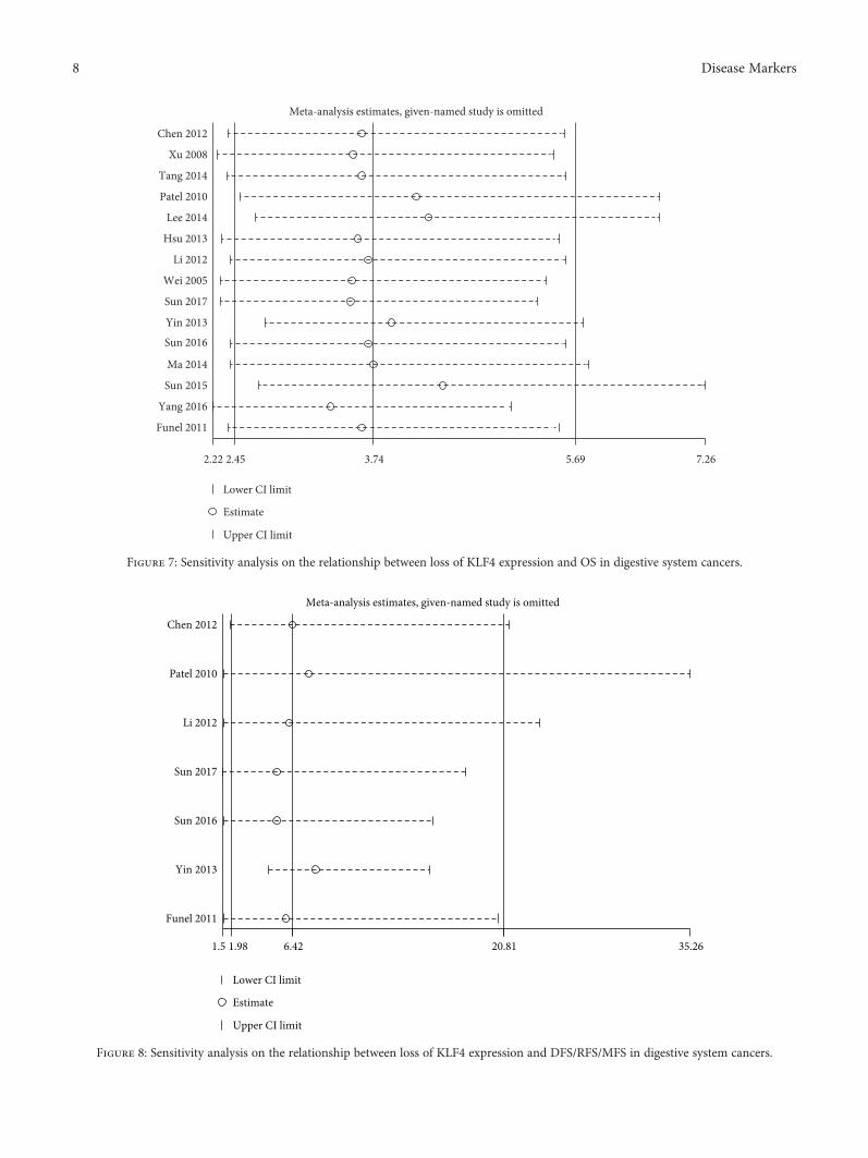
2.22 3.742.45 5.69 7.26
Chen 2012
Xu 2008
Tang 2014
Patel 2010
Lee 2014
Hsu 2013
Li 2012
Wei 2005
Sun 2017
Sun 2016
Yin 2013
Ma 2014
Sun 2015
Yang 2016
Funel 2011
Meta-analysis estimates, given-named study is omitted
Lower CI limit
Upper CI limit
Estimate
Figure 7: Sensitivity analysis on the relationship between loss of KLF4 expression and OS in digestive system cancers.
1.5 1.98 20.816.42 35.26
Chen 2012
Patel 2010
Li 2012
Sun 2017
Sun 2016
Yin 2013
Funel 2011
Meta-analysis estimates, given-named study is omitted
Lower CI limit
Upper CI limit
Estimate
Figure 8: Sensitivity analysis on the relationship between loss of KLF4 expression and DFS/RFS/MFS in digestive system cancers.
8 Disease Markers

hazard ratios demonstrated that loss of KLF4 expression wasassociated with a poorer OS (HR=1.61, 95% CI: 1.17–2.20,P = 0 003) and DFS/RFS/MFS (HR=1.99, 95% CI: 1.12–3.52, P = 0 001) as well as DSS (HR=1.73, 95% CI: 1.08–2.77, P = 0 022) in digestive system malignancies withoutregard to subgroup-confounding factors. In the subgroupanalysis, most results of which were consistent with the cor-responding overall result. But the existence of insignificanteven opposing results should also be noted. First, in the sub-group analysis on the basis of ethnicity, loss of KLF4 expres-sion yielded a better prognosis in Caucasian patients for DFS/RFS/MFS (HR=0.59, 95% CI: 0.36–0.94) which is likelycaused by the limited number of included studies (n = 2) orinherent differences of ethnicity. Second, as for differentmeasurement methods, inconsistent results were obtainedin patients detected by RT-PCR for OS and DFS/RFS/MFS(HR=0.93, 95% CI: 0.62–1.39 and HR=0.25, 95% CI:0.09–0.73, resp.). The relatively small amounts of studiesand the involvement of certain isoforms, in particular KLFα[44], may explain those discrepancies in some extent. Third,in the subgroup analysis according to the type of cancer, forESCC patients, loss of KLF4 expression showed a trend ofbetter prognosis though without statistical significance(HR=0.83, 95% CI: 0.57–1.23, P = 0 355) which suggestedthat, similar to head and neck squamous cell carcinoma(HNSCC) [45], KLF4 might be a malignant transformation-related gene in ESCC, which still needs further investigation.
Based on the evidence presented in our meta-analysis,loss of KLF4 expression could be a poorer prognostic bio-marker in most kinds of digestive system tumors exceptESCC. Whereas, this study has several limitations. First,because of a limited amount of included studies of eachtype of cancers, the results of some carcinomas were statis-tically insignificant and might be less powerful. Second, theliteratures were restricted to English-written papers, whichprobably introduced language bias. Third, the HRs of someliteratures, extrapolated based on Tierney’s method, wereless reliable than those directly provided in the originalarticles. Fourth, the cut-off values in the studies were notuniform, which might be a source of heterogeneity. Fifth,significant heterogeneity existed in those studies recruitedin this meta-analysis. Although we used a random-effectmodel and conducted subgroup analyses to explore thepotential source of heterogeneity, there were unacceptableheterogeneities in those subgroups. Many factors couldhave contributed the heterogeneity observed among thosestudies, such as different population characteristics, patho-logical grade, histology type, or study designs. Finally, somepublication bias was inevitable because positive results aremore easily accepted by journals than negative or nullresults. Given all the above limitations, our results shouldbe considered cautiously.
In conclusion, the present meta-analysis according topublished articles demonstrated that loss of KLF4 expressionwas associated with poorer survival in most kinds of digestivesystem cancer patients, such as gastric, hepatic, pancreatic,and colorectal cancers. Additionally, although not statisti-cally significant, we observed that loss of KLF4 expressionpredicted a trend of better survival in ESCC patients. At last,
our results should be interpreted carefully for the aforemen-tioned heterogeneity and limitations. To strengthen our find-ings, the prognostic value of KLF4 in digestive systemmalignancies should be further confirmed by large-scaleand standard investigations, in particular, in those patientswith ESCC.
Conflicts of Interest
The authors declare that they have no conflicts of interest.
References
[1] L. A. Torre, F. Bray, R. L. Siegel, J. Ferlay, J. Lortet-Tieulent,and A. Jemal, “Global cancer statistics, 2012,” CA: A CancerJournal for Clinicians, vol. 65, no. 2, pp. 87–108, 2015.
[2] R. L. Siegel, K. D. Miller, and A. Jemal, “Cancer statistics,2017,” CA: A Cancer Journal for Clinicians, vol. 67, no. 1,pp. 7–30, 2017.
[3] S. F. Yet, M. M. McA'Nulty, S. C. Folta et al., “Human EZF, aKrüppel-like zinc finger protein, is expressed in vascular endo-thelial cells and contains transcriptional activation and repres-sion domains,” The Journal of Biological Chemistry, vol. 273,no. 2, pp. 1026–1031, 1998.
[4] J. M. Shields, R. J. Christy, and V. W. Yang, “Identificationand characterization of a gene encoding a gut-enrichedKrüppel-like factor expressed during growth arrest,” TheJournal of Biological Chemistry, vol. 271, no. 33,pp. 20009–20017, 1996.
[5] B. B. McConnell and V. W. Yang, “Mammalian Krüppel-likefactors in health and diseases,” Physiological Reviews, vol. 90,no. 4, pp. 1337–1381, 2010.
[6] D. Wei, M. Kanai, S. Huang, and K. Xie, “Emerging role ofKLF4 in human gastrointestinal cancer,” Carcinogenesis,vol. 27, no. 1, pp. 23–31, 2006.
[7] X. Chen, E. M. Whitney, S. Y. Gao, and V. W. Yang,“Transcriptional profiling of Krüppel-like factor 4 revealsa function in cell cycle regulation and epithelial differenti-ation,” Journal of Molecular Biology, vol. 326, no. 3,pp. 665–677, 2003.
[8] N. Tiwari, N. Meyer-Schaller, P. Arnold et al., “Klf4 is a tran-scriptional regulator of genes critical for EMT, includingJnk1 (Mapk8),” PLoS One, vol. 8, no. 2, article e57329, 2003.
[9] N. Zhang, J. Zhang, L. Shuai et al., “Krüppel-like factor 4negatively regulates β-catenin expression and inhibits theproliferation, invasion and metastasis of gastric cancer,”International Journal of Oncology, vol. 40, no. 6, pp. 2038–2048, 2012.
[10] Z. Liu, H. Yang, W. Luo et al., “Loss of cytoplasmic KLF4expression is correlated with the progression and poor prog-nosis of nasopharyngeal carcinoma,” Histopathology, vol. 63,no. 3, pp. 362–370, 2013.
[11] Z. S. Lin, H. C. Chu, Y. C. Yen, B. C. Lewis, and Y. W. Chen,“Krüppel-like factor 4, a tumor suppressor in hepatocellularcarcinoma cells reverts epithelial mesenchymal transition bysuppressing slug expression,” PLoS One, vol. 7, no. 8, articlee43593, 2012.
[12] N. Tiwari, N. Meyer-Schaller, P. Arnold et al., “Klf4 is a tran-scriptional regulator of genes critical for EMT, includingJnk1 (Mapk8),” PLoS One, vol. 8, no. 2, article e57329, 2013.
9Disease Markers

[13] Q. Li, Z. Jia, L. Wang et al., “Disruption of Klf4 in villin-positive gastric progenitor cells promotes formation andprogression of tumors of the antrum in mice,” Gastroenterol-ogy, vol. 142, no. 3, pp. 531–542, 2012.
[14] J. P. Katz, N. Perreault, B. G. Goldstein et al., “Loss of Klf4 inmice causes altered proliferation and differentiation and pre-cancerous changes in the adult stomach,” Gastroenterology,vol. 128, no. 4, pp. 935–945, 2005.
[15] Y. Shimada, T. Okumura, S. Sekine et al., “Expression analysisof iPS cell-inductive genes in esophageal squamous cell carci-noma by tissue microarray,” Anticancer Research, vol. 32,no. 12, pp. 5507–5514, 2012.
[16] M. Q. Ma, H. D. Zhang, P. Tang, H. J. Jiang, and C. G. Chen,“Association of Krüppel-like factor 4 expression with theprognosis of esophageal squamous cell carcinoma patients,”International Journal of Clinical and Experimental Pathology,vol. 7, no. 10, pp. 6679–6685, 2014.
[17] D. Wei, W. Gong, M. Kanai et al., “Drastic down-regulation ofKrüppel-like factor 4 expression is critical in human gastriccancer development and progression,” Cancer Research,vol. 65, no. 7, pp. 2746–2754, 2005.
[18] L. S. Hsu, C. P. Chan, C. J. Chen et al., “Decreased Krüppel-likefactor 4 (KLF4) expression may correlate with poor survival ingastric adenocarcinoma,” Medical Oncology, vol. 30, no. 4,p. 632, 2013.
[19] M. X. Li, Q. Wang, B. Wang et al., “Association between gut-enriched Krüppel-like factor and prognosis of patients withgastric cancer,” Zhonghua wei chang wai ke za zhi= Chinesejournal of gastrointestinal surgery, vol. 15, no. 7, pp. 732–735,2012.
[20] N. Funel, M. Morelli, E. Giovannetti et al., “Loss of heterozy-gosity status of D9S105 marker is associated with downregula-tion of Krüppel-like factor 4 expression in pancreatic ductaladenocarcinoma and pancreatic intraepithelial lesions,”Pancreatology, vol. 11, no. 1, pp. 30–42, 2011.
[21] Z. Yang, D. Li, Z. Liu et al., “BIRC7 and KLF4 expression inbenign and malignant lesions of pancreas and their clinico-pathological significance,” Cancer Biomarkers, vol. 17, no. 4,pp. 437–444, 2016.
[22] H. T. Hsu, P. R. Wu, C. J. Chen et al., “High cytoplasmicexpression of Krüppel-like factor 4 is an independentprognostic factor of better survival in hepatocellular carci-noma,” International Journal of Molecular Sciences, vol. 15,no. 6, pp. 9894–9906, 2014.
[23] H. Sun, H. Tang, D. Xie et al., “Krüppel-like factor 4 blockshepatocellular carcinoma dedifferentiation and progressionthrough activation of hepatocyte nuclear factor-6,” ClinicalCancer Research, vol. 22, no. 2, pp. 502–512, 2016.
[24] H. Sun, Z. Peng, H. Tang et al., “Loss of KLF4 andconsequential downregulation of Smad7 exacerbate onco-genic TGF-β signaling in and promote progression ofhepatocellular carcinoma,” Oncogene, vol. 36, no. 21,pp. 2957–2968, 2017.
[25] H. Y. Chen, Y. M. Lin, H. C. Chung et al., “miR-103/107 pro-mote metastasis of colorectal cancer by targeting the metasta-sis suppressors DAPK and KLF4,” Cancer Research, vol. 72,no. 14, pp. 3631–3641, 2012.
[26] N. V. Patel, A. M. Ghaleb, M. O. Nandan, and V. W. Yang,“Expression of the tumor suppressor Krüppel-like factor 4as a prognostic predictor for colon cancer,” Cancer Epidemi-ology, Biomarkers & Prevention, vol. 19, no. 10, pp. 2631–2638, 2010.
[27] W. Tang, Y. Zhu, J. Gao et al., “MicroRNA-29a promotescolorectal cancer metastasis by regulating matrix metallopro-teinase 2 and E-cadherin via KLF4,” British Journal of Cancer,vol. 110, no. 2, pp. 450–458, 2014.
[28] L. L. Sun, J. Y. Wu, Z. Y. Wu et al., “A three-gene signature andclinical outcome in esophageal squamous cell carcinoma,”International Journal of Cancer, vol. 136, no. 6, pp. 569–577,2015.
[29] X. Yin, Y. W. Li, J. J. Jin et al., “The clinical and prognosticimplications of pluripotent stem cell gene expression inhepatocellular carcinoma,” Oncology Letters, vol. 5, no. 4,pp. 1155–1162, 2013.
[30] J. Xu, X. F. LüB, H. Gu, Y. Fang, Q. Huang, and M. Lai,“Dynamic down-regulation of Krüppel-like factor 4 incolorectal adenoma-carcinoma sequence,” Journal of CancerResearch and Clinical Oncology, vol. 134, no. 8, pp. 891–898, 2008.
[31] H. Y. Lee, J. B. Ahn, S. Y. Rha et al., “High KLF4 level innormal tissue predicts poor survival in colorectal cancerpatients,” World Journal of Surgical Oncology, vol. 12,no. 232, 2014.
[32] A. Stang, “Critical evaluation of the Newcastle-Ottawa scalefor the assessment of the quality of nonrandomized studiesin meta-analyses,” European Journal of Epidemiology, vol. 25,no. 9, pp. 603–605, 2010.
[33] F.Wang, J. Zhou, Y. Zhang et al., “The value of microRNA-155as a prognostic factor for survival in non-small cell lungcancer: a meta-analysis,” PLoS One, vol. 10, no. 8, articlee0136889, 2015.
[34] J. F. Tierney, L. A. Stewart, D. Ghersi, S. Burdett, and M. R.Sydes, “Practical methods for incorporating summary time-to-event data into meta-analysis,” Trials, vol. 8, no. 1, p. 16,2007.
[35] J. P. Higgins, S. G. Thompson, J. J. Deeks, and D. G. Altman,“Measuring inconsistency in meta-analyses,” British MedicalJournal, vol. 327, no. 7414, pp. 557–560, 2003.
[36] A. R. Black, J. D. Black, and J. Azizkhan-Clifford, “Sp1 andKrüppel-like factor family of transcription factors in cellgrowth regulation and cancer,” Journal of Cellular Physiology,vol. 188, no. 2, pp. 143–160, 2001.
[37] F. Zammarchi, M. Morelli, M. Menicagli et al., “KLF4 is anovel candidate tumor suppressor gene in pancreatic ductalcarcinoma,” The American Journal of Pathology, vol. 178,no. 1, pp. 361–372, 2011.
[38] J. L. Yori, D. D. Seachrist, E. Johnson et al., “Krüppel-likefactor 4 inhibits tumorigenic progression and metastasis in amouse model of breast cancer,” Neoplasia, vol. 13, no. 7,pp. 601–610, 2011.
[39] J. L. Yori, E. Johnson, G. Zhou, M. K. Jain, and R. A. Keri,“Krüppel-like factor 4 inhibits epithelial-to-mesenchymaltransition through regulation of E-cadherin gene expression,”The Journal of Biological Chemistry, vol. 285, no. 22,pp. 16854–16863, 2010.
[40] J. Zheng, Y. Liu, Y. Qiao, L. Zhang, and S. Lu, “miR-103 pro-motes proliferation and metastasis by targeting KLF4 in gastriccancer,” International Journal of Molecular Sciences, vol. 18,no. 5, 2017.
[41] H. Lv, Z. Zhang, Y. Wang, C. Li, W. Gong, and X. Wang,“MicroRNA-92a promotes colorectal cancer cell growth andmigration by inhibiting KLF4,” Oncology Research, vol. 23,no. 6, pp. 283–290, 2016.
10 Disease Markers

[42] A. Cercek, J. Wheler, P. E. Murray, S. Zhou, and L. Saltz,“Phase 1 study of APTO-253 HCl, an inducer of KLF4, inpatients with advanced or metastatic solid tumors,” Investiga-tional New Drugs, vol. 33, no. 5, pp. 1086–1092, 2015.
[43] B. D. Rowland and D. S. Peeper, “KLF4, p21 and context-dependent opposing forces in cancer,” Nature Reviews Cancer,vol. 6, no. 1, pp. 11–23, 2006.
[44] D. Wei, L. Wang, M. Kanai et al., “KLF4α up-regulation pro-motes cell cycle progression and reduces survival time ofpatients with pancreatic cancer,” Gastroenterology, vol. 139,no. 6, pp. 2135–2145, 2010.
[45] S. K. Tai, M. H. Yang, S. Y. Chang et al., “Persistent Krüppel-like factor 4 expression predicts progression and poor progno-sis of head and neck squamous cell carcinoma,”Cancer Science,vol. 102, no. 4, pp. 895–902, 2011.
11Disease Markers

Submit your manuscripts athttps://www.hindawi.com
Stem CellsInternational
Hindawi Publishing Corporationhttp://www.hindawi.com Volume 2014
Hindawi Publishing Corporationhttp://www.hindawi.com Volume 2014
MEDIATORSINFLAMMATION
of
Hindawi Publishing Corporationhttp://www.hindawi.com Volume 2014
Behavioural Neurology
EndocrinologyInternational Journal of
Hindawi Publishing Corporationhttp://www.hindawi.com Volume 2014
Hindawi Publishing Corporationhttp://www.hindawi.com Volume 2014
Disease Markers
Hindawi Publishing Corporationhttp://www.hindawi.com Volume 2014
BioMed Research International
OncologyJournal of
Hindawi Publishing Corporationhttp://www.hindawi.com Volume 2014
Hindawi Publishing Corporationhttp://www.hindawi.com Volume 2014
Oxidative Medicine and Cellular Longevity
Hindawi Publishing Corporationhttp://www.hindawi.com Volume 2014
PPAR Research
The Scientific World JournalHindawi Publishing Corporation http://www.hindawi.com Volume 2014
Immunology ResearchHindawi Publishing Corporationhttp://www.hindawi.com Volume 2014
Journal of
ObesityJournal of
Hindawi Publishing Corporationhttp://www.hindawi.com Volume 2014
Hindawi Publishing Corporationhttp://www.hindawi.com Volume 2014
Computational and Mathematical Methods in Medicine
OphthalmologyJournal of
Hindawi Publishing Corporationhttp://www.hindawi.com Volume 2014
Diabetes ResearchJournal of
Hindawi Publishing Corporationhttp://www.hindawi.com Volume 2014
Hindawi Publishing Corporationhttp://www.hindawi.com Volume 2014
Research and TreatmentAIDS
Hindawi Publishing Corporationhttp://www.hindawi.com Volume 2014
Gastroenterology Research and Practice
Hindawi Publishing Corporationhttp://www.hindawi.com Volume 2014
Parkinson’s Disease
Evidence-Based Complementary and Alternative Medicine
Volume 2014Hindawi Publishing Corporationhttp://www.hindawi.com