
The Possible Role of Cranio-Cervical Trauma and Abnormal ... · PDF fileCRANIO-CERVICAL TRAUMA...
33
Physiol. Chem. Phys. & Med. NMR (20 September 2011) 41: 1–17 The Possible Role of Cranio-Cervical Trauma and Abnormal CSF Hydrodynamics in the Genesis of Multiple Sclerosis Raymond V. Damadian and David Chu FONAR Corporation 110 Marcus Drive, Melville, NY 11747 E-mail: [email protected] Abstract: UPRIGHT ® Multi-Position™ MR scanning has uncovered a key set of new observa- tions regarding Multiple Sclerosis (MS), which observations are likely to provide a new under- standing of the origin of MS. The new findings may also lead to new forms of treatment for MS. The UPRIGHT ® MRI has demonstrated pronounced anatomic pathology of the cervical spine in five of the MS patients studied and definitive cervical pathology in the other three. The pathology was the result of prior head and neck trauma. All eight MS patients entered the study on a first come first serve basis without priority, and all but one were found to have a history of serious prior cer- vical trauma which resulted in significant cervical pathology. The cervical pathology was visualized by UPRIGHT ® MRI. Upright cerebrospinal fluid (CSF) cinematography and quantitative measure- ments of CSF velocity, CSF flow and CSF pressure gradients in the upright patient revealed that significant obstructions to CSF flow were present in all MS patients. The obstructions are believed to be responsible for CSF “leakages” of CSF from the ventricles into the surrounding brain parenchyma which “leakages” can be the source of the MS lesions in the brain that give rise to MS symptomatology. The CSF flow obstructions are believed to result in increases in intracranial pres- sure (ICP) that generate “leakages” of the CSF into the surrounding brain parenchyma. In all but one MS patient, anatomic pathologies were found to be more severe in the upright position than in the recumbent position. Similarly, CSF flow abnormalities were found to be more severe in the up- right position than in the recumbent position in all but one MS patient. Images of the MS patient anatomic pathologies and CSF flow abnormalities are provided with comparison images from nor- mal examinees in Figures 1–16. KEY WORDS: Multiple Sclerosis, Cranio-Cervical Trauma, CSF Hydrodynamics, CSF Leaks, Intracranial Pressure, CSF Peak Velocity, CSF Pressure Gradient, CSF Flow
Transcript of The Possible Role of Cranio-Cervical Trauma and Abnormal ... · PDF fileCRANIO-CERVICAL TRAUMA...

Physiol. Chem. Phys. & Med. NMR (20 September 2011) 41: 1–17
The Possible Role of Cranio-Cervical Traumaand Abnormal CSF Hydrodynamicsin the Genesis of Multiple Sclerosis
Raymond V. Damadian and David Chu
FONAR Corporation110 Marcus Drive, Melville, NY 11747
E-mail: [email protected]
Abstract: UPRIGHT® Multi-Position™ MR scanning has uncovered a key set of new observa-tions regarding Multiple Sclerosis (MS), which observations are likely to provide a new under-standing of the origin of MS. The new findings may also lead to new forms of treatment for MS.The UPRIGHT® MRI has demonstrated pronounced anatomic pathology of the cervical spine infive of the MS patients studied and definitive cervical pathology in the other three. The pathologywas the result of prior head and neck trauma. All eight MS patients entered the study on a first comefirst serve basis without priority, and all but one were found to have a history of serious prior cer-vical trauma which resulted in significant cervical pathology. The cervical pathology was visualizedby UPRIGHT® MRI. Upright cerebrospinal fluid (CSF) cinematography and quantitative measure-ments of CSF velocity, CSF flow and CSF pressure gradients in the upright patient revealed thatsignificant obstructions to CSF flow were present in all MS patients. The obstructions are believedto be responsible for CSF “leakages” of CSF from the ventricles into the surrounding brainparenchyma which “leakages” can be the source of the MS lesions in the brain that give rise to MSsymptomatology. The CSF flow obstructions are believed to result in increases in intracranial pres-sure (ICP) that generate “leakages” of the CSF into the surrounding brain parenchyma. In all butone MS patient, anatomic pathologies were found to be more severe in the upright position than inthe recumbent position. Similarly, CSF flow abnormalities were found to be more severe in the up-right position than in the recumbent position in all but one MS patient. Images of the MS patientanatomic pathologies and CSF flow abnormalities are provided with comparison images from nor-mal examinees in Figures 1–16.
KEY WORDS: Multiple Sclerosis, Cranio-Cervical Trauma, CSF Hydrodynamics, CSF Leaks,Intracranial Pressure, CSF Peak Velocity, CSF Pressure Gradient, CSF Flow
mg
Text Box
(Click here to view the videos of the CSF obstructions of the MS patients)

* flow in the absence of italics is generic, the presence of italics specifies cc/sec
THE ADVENT of MRI and its unique abilities to generate the tissue contrasts needed tosee detail in the body’s soft tissues was an important step forward in medical imaging. Aparticularly important example was the ability to visualize the plaque lesions of MultipleSclerosis (1). Traditionally the symptom-generating lesions in the brain and spinal cord ofMultiple Sclerosis (MS) patients are ascribed to tissue specific autoimmune interactions.These are believed to generate the parenchymal lesions of MS. More recently the adventof phase coded MR imaging has made it possible to visualize and quantify the dynamicflow* of the cerebrospinal fluid (CSF) within the cranial vault and spinal canal.
The present inquiry originated in the course of performing UPRIGHT® MR images ona patient (patient #1, Figure 1a) with an established diagnosis of MS. It was noted thatone of the MS brain lesions conspicuously appeared to be arising directly from the CSFwithin the lateral ventricle (Figure 1a arrow #1). An MS lesion, appearing to arise fromthe ventricular CSF, brought to our attention the unexplained tendency for MS lesions tobe peri-ventricular in their distribution (2) (Figure I, Figure II). Considered conjointly, anMS lesion appearing to arise directly from ventricular CSF (Figure 1a) and the tendencyof MS lesions to be peri-ventricular in their distribution (Figure I, Figure II) engenderedthe question whether abnormal CSF hydrodynamics (e.g., elevated intracranial pressure[ICP] or abnormal flow dynamics) was playing a role in the genesis of MS lesions. To ad-dress the question, eight patients with an established diagnosis of MS and seven normalexaminees were studied in the FONAR UPRIGHT® Multi-Position™ MRI.
Materials and Methods
The first eight MS patients who volunteered for the study were scanned. There was no se-lection among MS patients. They were all scanned in the order in which they volunteered.
MRI scans were performed on a 0.6 T UPRIGHT® scanner (FONAR Corporation,Melville, NY) with a quadrature head-neck combination coil. The patient bed can be ro-tated to any angle between the horizontal and vertical position in the space between thetwo poles of the upright magnet.
Regular clinical anatomical scans of the head and neck were acquired. Ciné phase con-trast scans of CSF flow were imaged using a phase contrast RF-spoiled gradient echo se-quence with TR = 19-22 ms, TE = 9-12 ms, slice thickness = 8 mm, flip angle = 20-25°,matrix = 256x128 zero filled to 256x256, and NEX = 2. Data acquisition was retrospec-tively gated using ECG or pulse oximeter covering the entire cardiac cycle.
Thirty-two (32) uniformly spaced time frames were obtained by linear interpolation inpost-processing.
To visualize the overall CSF flow pattern, a single slice of FOV = 26 cm was imagedin the midline sagittal plane. In order to quantify the CSF flow, an axial slice of FOV =16 cm at the mid C-2 level and perpendicular to the spinal canal was imaged (velocity en-coding along the slice-select direction, venc = 3-11 cm/s).
Quantification of CSF flow was accomplished by manually drawing the Region of In-terest (ROI) around the spinal canal and spinal cord in the axial mid C-2 phase contrast
2 DAMADIAN AND CHU

CRANIO-CERVICAL TRAUMA AND ABNORMAL CSF HYDRODYNAMICS IN MS 3
scan. Phase offset was corrected by requiring the spinal cord to have zero net phasechange over the whole cardiac cycle.
MR examinations of MS patients (Figures 1–8) and normal examinees (Figures 9-15)in the study were performed in both the upright and recumbent positions using theFONAR UPRIGHT® Multi-Position™ MRI. Examinees were deemed normal if they ex-hibited uninterrupted dorsal and ventral CSF flow on upright sagittal CSF flow images(e.g., Figure 15c). MS patients #7 and #8 could not be scanned recumbent. Lower limbparalysis prevented MS patient #7. Severe vertigo and emesis in the recumbent positionprevented MS patient #8. CSF cinematography (ciné) was obtained both in the sagittaland axial planes. Quantitative MR measurements of CSF flow (cc/sec) through the spinalcanal annulus obtained from axial MR images were calculated from the phase codedCSF flow image data. The flow data were obtained from axial images taken at the mid C-2level, unless specified otherwise. The CSF velocity (cm/sec) was measured as the aver-age annular proton velocity for each of the 32 imaging annuli acquired throughout thecardiac cycle. The peak CSF velocity was determined as the highest proton annular ve-locity measurement obtained for each phase (systole and diastole) of the cardiac cycle.The pressure gradient was derived from the measured CSF velocity data using theNavier-Stokes equation, with negligible contribution from the viscous term. There werethree notable findings.
Results
CSF Flow Imaging and Quantification in Multiple Sclerosis Patients
The first, and partially expected result stemming from the MR images of MS patient #1(Figure 1a) and the known peri-ventricular distribution of MS lesions (Figure I, II), wasthe finding of abnormal CSF flows in all eight MS patients (Figures 1–8, Table 2A, col.10). The abnormal CSF flows corresponded with the cranio-cervical structural abnormal-ities found on the patients’ MR images. The second finding was a history of severe cervi-cal trauma prior to the patient’s MS diagnosis in six of the eight MS patients and asignificant possibility of cervical trauma in a seventh patient (Table 1, MS patient #8). Thethird finding was the discovery that CSF inflow (cc/sec) and inflow velocity (cm/sec) inthe upright position is about half (53–56%) (Table 3) of what it was in the recumbent po-sition in both the MS patients and normal examinees. Alperin et al. have shown that theaverage (oscillatory) CSF volume (cc) in the upright position was less than half the os-cillatory volume (cc) in the supine position, but neither a reduction of CSF inflow veloc-ity (cm/sec) nor of CSF inflow (cc/sec) in the upright position have been previouslyreported (3).
The first important observation of this study of eight MS patients was that every MSpatient exhibited obstructions to their CSF flow when examined by phase coded CSFcinematography (ciné) in the upright position (Table 2A, col. 10 & 13). All MS patientsexhibited CSF flow abnormalities that were manifest on MR cinematography as inter-ruptions to flow or outright flow obstructions somewhere in the cervical spinal canal,depending on the location and extent of their cervical spine pathology (Table 2A, col.10, 11 & 13). Normal examinees did not display these flow obstructions (Table 2B, col.10 & 11).

1 2 3
Patient # History of Cranio-Cervical Trauma Nature of Trauma
Patient #1(MVA #1)
Motor vehicle accident 1978 — Rear-ended by pickup truck.Patient sustained whiplash. Wore neck brace for 2 monthsthereafter. Passenger seat occupant hospitalized.
Whiplash injury. Cervical collar worn 2months after accident.
Patient #2 (1992) While patient was standing on top of a 20-ft. extensionladder repairing his home rooftop, the extension linkdecoupled. Ladder extension section slid groundward withpatient on board. 16-oz. hammer fell 3 ft. and landed on top ofpatient’s head. Ladder extension section then further slid toground. Patient fell to ground hitting back of head. Patientsemi-conscious and dizzy after ground impact.
Hammer hit to top of patient’s head.Ladder fell backward when extensionlink decoupled. Patient landed hittingback of head. Semi-conscious anddizzy following impact.
Patient #3(MVA #2)
Major car accident 30 years ago. Rear ended by pickup truck.Car trunk collapsed to level of rear car window. Patientbelieves head whiplashed during accident. 2 years later secondcar accident, hit tree. Thrown forward on impact.
Neck and head trauma from rear-endingby pickup truck. Patient describedaccident as “whiplash”.
Patient #4 17 years old today – Patient age 6 years old was picked up byangry parent and shaken back and forth by shoulders. Head wascaused to shake back and forth and against a wall. 8 years later(2007 age 14), hit in face by teenager which caused his rightarm to go limp. Taken to Schneider’s Children’s Hospital,Great Neck L.I. (NSUH). Diagnosed as MS from MRI images.
Severe parental shaking age 6 (11 yearsprior to MS diagnosis). Exacerbatingincident immediately prior to MSdiagnosis (2007). Patient hit in face byteenager. Right eye bruised and rightarm went limp. MRI taken immediatelyfollowing injury showed multiple peri-ventricular MS lesions.
Patient #5 No accident trauma. Ballroom ballerina/tap/ modern dancesince age 3. 4–5 hours dancing/day from ages 12 to 17.
Possible dance head neck trauma.Intense dancing 1990 to 1995. MSsymptoms started in 2004.
Patient #6(MVA #3)
Motor vehicle accident 1998 at age 19. 11 years prior to MShit a post while driving in parking lot. Head went forward thenback (reverse whiplash). Could not move neck after accident.Fractured left clavicle. Wore neck collar more than 2 weeks.Unable to move left arm. Second accident playing baseball.Hit back of neck. Blurred vision (optic neuritis) followed plusnumbness in left shoulder and tingling.
Modified whiplash — head forward thenback. Neck injury 11 years prior and 6months prior. Second incident provokedsymptoms.
Patient #7(MVA #4)
Severe car accident 1995. “Black ice”. Car spun, hit divider,wrapped around tree. 2 months after accident developedspasms in each leg and impaired locomotion. Neck surgery2003. Deteriorated markedly following surgery. Lost controlof both legs. Currently wheelchair bound.
Seat belt came across left side of neck.Bruises to left neck.
Patient #8(MVA #5)
Age 2-3 involved in a severe motor vehicle accident thattotalled the car. Patient believes she was in the customarysitting position (no seat belt or infant seat).
Possible neck injury from motor vehicleaccident.
Avg. # years trauma preceded MS diagnosis
TABLE 1. Clinical History and Symptomatology of MS Patients

4 5 6
Years PhysicalTrauma Preceded
Diagnosis Description of MS Image Pathology Current Symptoms
8 Two peri-ventricular MS lesions penetrating corpus callosumfrom lateral ventricle into adjacent brain parenchyma. Lateralventricle swollen mid-line (Sag T2).
Loss of vision left eye — optic atrophyleft eye. Loss of muscle strength bothlegs. Wheelchair bound.
2 Occipital horn hydrocephalus plus peri-ventricular edema i.e.“interstitial edema”, MS lesions predominantly adjacent toenlarged occipital horns on both right and left.
Memory problems, sleep apnea, bladderand bowel incontinence, impotence,visual difficulties, fatigue.
21 Solitary MS lesion adjacent to left occipital horn and peri-ventricular interstitial edema anterior horns.
Transient episodes of kaleidoscopic vision,numbness left hand, frequent falling.Wears leg brace to diminish falling.Severe foot drop right foot. Severe righthand weakness. Loss of bladder control.Loss of fine motor control.
8 Multiple peri-ventricular MS lesions radiating off lateralventricle. Multiple peri-ventricular lesions. Also pronouncedinterstitual edema lesions both anterior and occipital horns.
Backaches, headaches, numbness in theface. Lack of energy for 17 year old.Sleeps a lot. Gets many canker sores inmouth.
9 One lesion posterior to left occipital horn. Interstitial edemaaround entire ventricle. Peri-ventricular lesions.
Right arm numbness, sternal numbness.Numbness from mid-sternum to rightshoulder extending to right fingertips.
11 Solitary lesion Flair Axial midway up left lateral border leftventricle, peri-ventricular interstitial edema. Possible secondMS lesion adjacent occipital horn.
Tingling numbness left shoulder.Blurred vision left eye. Visual blurringafter exercise.
1 Parenchymal MS appearing lesion superior to right occipitalhorn. Couple small peri-ventricular lesions. MS lesionsadjacent to left lateral ventricle midway in AP direction. Peri-ventricular interstitial edema.
Loss of motor control lower legs.Wheelchair bound. Fatigue.
27 Multiple peri-ventricular lesions, pronounced hydrocephalus ofthe occipital horns of the lateral ventricles with accompanyingperi-ventricular lesions and edema, prominent peri-ventricularinterstitial edema and hydrocephalus in the main body of thelateral ventricles anteriorly.
Optic neuritis, severe vertigo, nauseaand vomiting when supine, stumblingwhen walking.
11

1 2 3 4 5 6 7 8 9
Up. Rec. Up. Rec. Up. Rec. Upright RecumbentPeak Peak Peak Peak Press. Press. MRI Image MRI ImageSys. Sys. Dias. Dias. Grad. Grad. Analysis AnalysisCSF CSF CSF CSF Peak Peak toVel. Vel. Vel. Vel. Peak to Peak
(outflow) (outflow) (inflow) (inflow) mmHg/cm mmHg/cmPatient # cm/sec cm/sec cm/sec cm/sec
Patient #1 .67 1.52 .40 .745 .012 .024 1)C2 c.clock rot. 16°, 1)C2 c.clock rot. 5.7°(MVA #1) 2)C6/7 cd.c., c.sten. 2)no c.sten., no cd.c.
P<.05
Patient #2 2.58 1.39 1.047 1.033 .054 .031 1)r.lis. C5, 1)vent. can.obs.,2)cd.c., c.sten. C5/6 2)r.lis., c.sten. red.3)ligfl. cd.c. C6/7, 3)ligfl. obst. absent
ligfl. can. obst. C6/7P<.05 P<.05 P<.05 P<.05 P<.05 4)c.sten. C6/7
Patient #3 1.14 .336 .394 .702 .016 .011 1)r.lis. C4, C5, 1)r.lis. red.,(MVA #2) 2)cd.c., c.sten. C4/5, 2)no sp.cd. abut. C5/6
Patient #4 1.80 2.71 .464 1.36 .036 .063 1)sp.cd. post. 1)sp.cd. more ant.,2)C3/4 dors. can.obs., 2)dors. can. pat.,3)CTE 2mm 3)CTE 1mm4)sp.cd. abut. post.
P<.05 P<.05 P<.05 P<.05 P<.05 wall sp.can. C3/4
Patient #5 2.03 2.14 .731 1.55 .050 .046 1)dsc. hrn. at C3/4, 1)C2/3, C5/6 dsc. hrn.C4/5, C5/6 2)C3/4 hrn. absent
2)dsc. bulges at C2/3, 3)C4/5less dsc. bulgeC6/7 4)C6/7 dsc. bulge not
P<.05 P<.05 P<.05 P<.05 P<.05 P<.05 present
Patient #6 .79 .94 .32 .80 .014 .020 1)CTE 2mm 1)CTE 2mm(MVA #3)
Patient #7 .865 — .51 — .024 — 1)cd.c. C2/3 to C5/6 1)cd.c. red. C2 to C6(MVA #4). 2)r.lis C5 2)C5 r.lis. red.
(2003 conventionalrecumbent MRI)
Patient #8 .818 — .380 — .020 — 1)post. disp. sp.cd. at(MVA #5) C2/3, obst. dors.
CSFfl. at C2/32)markedly enlarged
occipital horns lat.ventcls.
3)peri-ventcl. ed.occipital horns
4)CTE – T2 axials5)rot. C3 c. wise
Mean Value 1.34 1.51 .531 1.03 .028 .033(MS patients) (8 pat.) (6 pat.) (8 pat.) (6 pat.) (8 pat.) (6 pat.)
TABLE 2A. Anatomic Images, CSF Flow Images and CSF Flow Quantification of MS Patients
see glossary
a-ant., anterior; abut., abutment; b-bel., below; c-c.clock, counter clockwise; c.sten., spinal canal stenosis; c.wise, clockwise; can., canal; can.obs., spinaldors., dorsal; dsc., disc; e-ed., edema; g-grad., gradient; h-hrn., herniation; i-inter., interruption; inta., intact; l-lat., lateral; ligfl., ligamentum flavum;red., reduced; rot., rotation; s-sp., spinal; sp.cd., spinal cord; sys., systolic; u-unobst., unobstructed; Up., upright; v-vel., velocity; vent., ventral;—— BOLDED numbers Col. 2-7 (P< .05)

10 11 12 13
UPRIGHT MRI Recumbent MRI MRI Image CSF CinéCiné Ciné Differences Differences
CSF Flow Analysis CSF Flow Analysis Up./Rec. Up./Rec.
1)dors. CSFfl. obst. at C2/3 1)dors. CSFfl. faint but inta., 1)C2 rotated 16° 1)vent. CSFfl. obst. Up. 12)vent. CSFfl. obst. C3 to C5/6, 2)vent. CSFfl. inta. to C6/7, Up., 5.7° Rec. but not Rec.3)vent. CSFfl. not obst. by C6/7 3)vent. CSFfl. obst. at C6/7
dsc. hrn. when Up. by disc. hrn.
1)dors. CSFfl. obst. top C2 to C3/4, 1)vent. CSFfl. obst. C3/4 & bel. 1)no infolding ligfl. Rec., 1)CSFfl. obst. Up., 22)dors. CSFfl. obst. C5/6, 2)less c.sten. Rec. 2)full circumspinal3)vent. CSFfl. obst. bel. C3/4 CSFfl. Rec.
1)dors. CSFfl. obst. C2/3 to C5/6, 1)vent. & dors. CSFfl. inta. 1)C4, C5 listhesis, much 1)CSFfl. obst. Up., 32)vent. CSFfl. obst. C2/3 & bel. less Rec. 2)CSFfl. unobst. Rec.
1)dors. CSFfl. obst. bel. C2/3, 1)dors. CSFfl. obst. bel. C2/3, 1)cord more ant. Rec. 1)same CSFfl. obst. 42)vent. CSFfl. inta. 2)vent. CSFfl. inta. Up. & Rec.
1)dors. CSFfl. obst. C2/3 to C5, 1)vent. CSFfl. int. less when 1)C5/6 hrn. less Rec. 1)vent. CSFfl. less inter. 52)vent. CSFfl. obst. at C2/3, C3/4, patient Rec. Rec.
C4/5, C5/6
1)vent. CSFfl. obst. C2/3 to C6 1)no CSFfl. int. when patient Rec. 1)no difference 1)vent. CSFfl. obst. 6C2/3 to C6 Up.
2)no CSFfl. inter. Rec.
1)dors. CSFfl. obst. C2/3 to C4/5, 1)dors. CSFfl. obst. C2/3 & bel. 1)less cd.c. C2 to C6, 1)CSFfl. obst. at C2/3 72)vent. CSFfl. obst. at C5 (2003 conventional recumbent 2)r.lis. red. Rec. but restored at C4/5 Up.
MRI) 2)not restored bel. C4/5Rec. (2003 conventionalrecumbent)
1)dors. CSFfl. obst. C2/3 8
see glossary
canal obstruction; cd.c., cord compression; cent., central; CSFfl., CSF flow; CTE, cerebellar tonsil ectopia; d-dias., diastolic; disp., displaced;m-MVA, motor vehicle accident; o-obst., obstructed, obstruction; p-pat., patent; post., posterior; press., pressure; r-r.lis., retrolisthesis; Rec., recumbent;ventcls., ventricles
1 2 3 4 5 6 7 8 9
Up. Rec. Up. Rec. Up. Rec. Upright RecumbentPeak Peak Peak Peak Press. Press. MRI Image MRI ImageSys. Sys. Dias. Dias. Grad. Grad. Analysis AnalysisCSF CSF CSF CSF Peak Peak toVel. Vel. Vel. Vel. Peak to Peak
(outflow) (outflow) (inflow) (inflow) mmHg/cm mmHg/cmPatient # cm/sec cm/sec cm/sec cm/sec
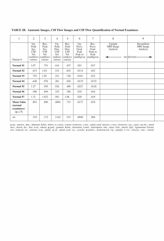
Normal #1 1.07 .751 .414 .457 .021 .017
Normal #2 .613 1.03 .332 .825 .0114 .023
Normal #3 .752 1.05 .321 .746 .0181 .023
Normal #4 .640 .976 .301 .626 .0135 .0135
Normal #5 1.27 .595 .526 .488 .0227 .0126
Normal #6 .586 .849 .325 .786 .018 .016
Normal #7 1.32 1.022 .583 1.08 .020 .019
Mean Value .893 .896 .4004 .715 .0177 .018(normalexaminees)
(n = 7)
+/- .319 .172 .1124 .215 .0040 .004
TABLE 2B. Anatomic Images, CSF Flow Images and CSF Flow Quantification of Normal Examinees
a-ant., anterior; abut., abutment; b-bel., below; c-c.clock, counter clockwise; c.sten., spinal canal stenosis; c.wise, clockwise; can., canal; can.obs., spinaldors., dorsal; dsc., disc; e-ed., edema; g-grad., gradient; h-hrn., herniation; i-inter., interruption; inta., intact; l-lat., lateral; ligfl., ligamentum flavum;red., reduced; rot., rotation; s-sp., spinal; sp.cd., spinal cord; sys., systolic; u-unobst., unobstructed; Up., upright; v-vel., velocity; vent., ventral;
see glossary

10 11 12 13
UPRIGHT MRI Recumbent MRI MRI Image CSF CinéCiné Ciné Differences Differences
CSF Flow Analysis CSF Flow Analysis Up./Rec. Up./Rec.
CSFfl. inta. vent. & dors. CSFfl. inta. vent. & dors.
CSFfl. inta. vent. & dors. CSFfl. inta. vent. & dors.
CSFfl. inta. vent. & dors. CSFfl. inta. vent. & dors.
CSFfl. inta. vent. & dors. CSFfl. inta. vent. & dors.
CSFfl. inta. vent. & dors. CSFfl. inta. vent. & dors.
CSFfl. inta. vent. & dors. CSFfl. inta. vent. & dors.
CSFfl. inta. vent. & dors. CSFfl. inta. vent. & dors.
canal obstruction; cd.c., cord compression; cent., central; CSFfl., CSF flow; CTE, cerebellar tonsil ectopia; d-dias., diastolic; disp., displaced;m-MVA, motor vehicle accident; o-obst., obstructed, obstruction; p-pat., patent; post., posterior; press., pressure; r-r.lis., retrolisthesis; Rec., recumbent;ventcls., ventricles
see glossary

Significant differences in MS patient CSF flow cinematography (ciné) in the sagittalmidplane were observed between the upright and recumbent positions, while no posi-tional differences in ciné flow were observed in normal examinees (Table 2B, col. 10, 11& 13). CSF flow differences between the two positions were found in the six MS patientsthat were scanned both upright and recumbent (Table 2A, col. 10, 11 & 13). Obstructionsof spinal CSF flow in both the dorsal and ventral spinal canals, when viewed sagittally,were found by cinematography in five of the eight MS patients when they were examinedin the upright position (Table 2A, patients #1, #2, #3, #5 and #7, col. 10). Patient #3exhibited both dorsal and ventral CSF flow obstruction only in the upright position (Table2A, col. 10). Dorsal and ventral CSF flows in patient #3 were unobstructed in the recum-bent position (Table 2A, col. 11).
Regarding the quantitation of CSF flow (cc/sec) abnormalities, determinations of peakCSF velocities (cm/sec) produced the most pronounced differences between the MSpatients and normal examinees (Table 2A, col. 2, 3, 4, & 5). While the peak velocity mea-surements in our study were measured differently than the peak velocities by Haughton etal. [Haughton et al. (4) determined the peak velocity to be the highest voxel velocity mea-sured in the scan as compared to the highest annular velocity measured in the scan in ourdetermination], both peak velocity methods found the peak velocity determination to bethe most sensitive measure for detecting CSF flow abnormalities.
Among the MS patients, three of the eight patients had significantly (P < .05) elevatedpeak CSF outflow (systolic) velocities (cm/sec) from the brain (2.58, 1.80, 2.03 cm/sec)in the upright position (Table 2A, col. 2, patients #2, #4 and #5) compared to the meanvalue for the normal examinees in the upright position (.893 ± .32 cm/sec, Table 2B,col. 2). Outflow velocities for all three of these MS patients were more than twice the up-right outflow peak velocities for the normal examinees. Four (patients #1, #2, #4, and #5)had significantly elevated peak CSF outflow velocities (cm/sec) (1.52, 1.39, 2.71 and 2.14cm/sec, Table 2A, col. 3) in the recumbent position, two of which (2.71 and 2.14 cm/sec)were more than twice the normal value (.896 ± .17 cm/sec, Table 2B, col. 3). A fifth MSpatient (patient #3) had a recumbent CSF outflow velocity of .336 cm/sec that was sig-nificantly reduced relative to normal (.896 ± .17 cm/sec).
In addition, two of the eight MS patients (Table 2A, col. 4, patients #2 and #5) exhib-ited significantly elevated peak inflow velocities in the upright position (Table 2A, col. 4,1.047, and .731 cm/sec) relative to the peak inflow velocities of normal examinees (.400cm/sec) in the upright position (Table 2B, col. 4). Importantly, therefore, five of the eightMS patients had at least one significantly abnormal peak CSF velocity measurement inthree of the parameters measured (upright outflow, recumbent outflow, and uprightinflow), and three of the MS patients exhibited elevated peak velocities in both the uprightand recumbent positions (patients #2, #4, and #5, Table 2A, col. 2 & 3).
Sharply Reduced CSF Inflow Velocity in the Upright Position
Additionally it was found that peak CSF inflow (1.023 cc/sec) and peak CSF inflow ve-locity (0.400 cm/sec) (Table 3) were sharply reduced in normal examinees in the uprightposition when compared to inflow and inflow velocity in the recumbent position. Bothpeak inflow (cc/sec) and peak inflow velocity (cm/sec) in the upright position were foundto be about half (53%–56% respectively), of what they were in the recumbent position(Table 3) in the normal examinees. Except for patient #2 and normal examinee #5, both
10 DAMADIAN AND CHU

MS patients and the normal examinees exhibited reduced inflow velocity in the uprightposition (Table 2A, col. 4 & 5; Table 2B, col. 4 & 5). In the case of patient #2, the antic-ipated velocity reduction arising from the upright position was offset by the peak veloc-ity acceleration arising from the patient’s pathology (Table 2A, col. 4 & 8).
This striking reduction of CSF inflow into the brain in the upright position in both MSpatients (Table 2A) and normals (Table 2B, Table 3) was unexpected, inasmuch as cere-bral blood flow in normal subjects is unaffected by position (5). The observed reductionin CSF inflow in the upright position apparently constitutes normal physiology. The un-expected observation that CSF flow is significantly reduced in the upright position (or sig-nificantly increased in the recumbent position) raises interesting questions regarding thephysiological significance of the increased CSF flow of recumbency.
The High Percentage of “Normal” Examinees That Did Not Qualify as Normal
Another unexpected finding was the high percentage of “normal” adults that did not qual-ify as normal. It was found that a large percentage of normal examinees (as high as 75%)did not qualify as normal with respect to their cervical spine anatomy, e.g., exhibiting lo-calized disc herniations (or significant bulges) at C5/6 or elsewhere, or localized inter-ruptions of CSF flow. Such examinees were entirely asymptomatic currently andhistorically, but were nonetheless unable to meet a standard for normal cervical spineanatomy. With the cervical spine being the most active segment of the spine, the finding,though unexpected, is not inconsistent with the cervical spine’s high degree of bio-mechanical activity.
A Possible Physiologic Role of Nocturnal Sleep Enabled By Enhanced CSF FlowInto the Brain in Recumbency
Since one of the physiological roles attributed to the CSF is the delivery of nutrients tothe brain and the removal of toxic metabolic waste, the increase in CSF flow facilitatedby recumbency engenders the consideration that the normal nocturnal sleep process mayin fact be playing an active role in facilitating the removal of metabolic waste from thebrain and delivering nutrients. Recumbent sleep may be enabling increased CSF inflowinto the brain for the physiologic purpose of delivering nutrients to the brain and cleansing
CRANIO-CERVICAL TRAUMA AND ABNORMAL CSF HYDRODYNAMICS IN MS 11
TABLE 3: Change of CSF Inflow With Position in Normal Examinees(see Tables 2A and 2B for velocities)
1 2 3 4Peak Peak Peak Peak
INFLOW INFLOW INFLOW INFLOW(cc/sec) (cc/sec) Velocity Velocity
UPRIGHT RECUMBENT (cm/sec) (cm/sec)UPRIGHT RECUMBENT
1.023 1.935 .400 .715% Difference Up/Rec % Difference Up/Rec
53% 56%

it of toxic metabolic by-products*. Similarly, it may account, in some measure, for thebenefits of recumbent sleep in the medical healing process.
Prior Histories of Significant and Severe Trauma
The most unexpected finding in this MR study of MS patients was the revelation thatwhen carefully questioned, six of the eight Multiple Sclerosis patients had a prior historyof severe trauma to the neck (Table 1, col. 2) with one patient (patient #2) having sus-tained both neck and head trauma. In addition, there is a significant likelihood that traumahad a role in the genesis of a seventh patient’s MS (Table 1, patient #8, col. 2). The findingis consistent with prior reports that trauma may have a causative role in the onset of MS(6,7,8). All seven patients had distinct cervical anatomic pathology on their current MRimages that corresponded with their trauma histories, thereby establishing that the histor-ical trauma events contributed directly to their permanent pathologies of the cervical spine(Table 2A, col. 8 & 9) and that their cervical trauma histories were not immaterial. Fourhad received neck injuries in motor vehicle accidents, three of which were whiplash in-juries, and the fourth a “reverse whiplash” (neck flexion preceding neck extension) injury(patient #7). A fifth, patient #8, was involved in a severe motor vehicle accident at age 2–3that “totalled” the car in which she was riding without a seat belt or infant seat.
Noteworthy was the fact that the trauma and particularly motor vehicle trauma,notwithstanding its severity, was never correlated by either the patients or their physicianswith the onset of their MS symptomatology. The symptoms of head and neck trauma,however, can be long lasting (e.g., 17 yrs.) (9). In all but two of the patients (patients #2and #7, Table 1, col. 4) the trauma preceded the onset of MS symptoms by more than 8years. When the mean value was calculated for all eight MS patients, the average numberof years patient trauma preceded the patient’s MS diagnosis, was 11 years. In addition,the abnormal CSF flow dynamics found in the MS patients of this study corresponded tothe MR cervical pathology that was visualized (Table 2A, col. 2–9).
In the UPRIGHT® MRI examination of the eight MS patients, four of the eight exhib-ited severe cervical anatomic pathology (patients #1, #2, #3 and #7, Table 2A, col. 8). Theremaining four patients had less severe but still serious cervical anatomic pathology(Table 2A, col. 8) and two (MS patients #2 [Figure 2] and #8 [Figure 8]) exhibited con-spicuous swelling of the body of the cerebral lateral ventricles or of the occipital horns ofthe lateral ventricles.
Anatomic Pathology and CSF Flow Obstruction More Severe With the PatientUpright
In all but one (patient #6) of the seven MS patients that were imaged in both the uprightand recumbent positions, the visualized anatomic pathology was more severe in theupright position than in the recumbent position (Table 2A, col. 8 & 9). Patient #1, for ex-ample, exhibited a 16° mal-rotation of C-2 on the patient’s upright axial image (Figure 1e)
12 DAMADIAN AND CHU
* The authors thank Charles Green, FONAR Corporation scientist and engineer, for his proposal that the in-creased CSF flow into the brain of recumbency might have the beneficial effect of enhancing CSF cleansing ofthe brain.

FIGURE 1a. Sagittal T2-weighted image of a Multiple Sclerosis patient (patient #1) showing two peri-ventricular MS plaques (arrows 1 & 2) perpen-dicular to the ventricular wall. Lesion 1 (arrow 1) exhibits an explicit connection between ventricular CSF and an MS plaque. Lesion 2 exhibits a sim-ilar connection to ventricular CSF but in a less striking manner. The images visualizing the CSF “leaks” of Figure 1a were obtained on March 11, 2010with the patient upright in the FONAR UPRIGHT® Multi-Position™ MRI.

FIGURE I. (Figure 61-2, Stark, D. B., Bradley, W. B. Jr., Eds. Magnetic Resonance Imaging, Ed.III Vol. III, 1999, S. K. Lakhanpal, K. R. Maravilla, “Multiple Sclerosis”, ch. 61, p. 1381, Mosby,Inc.) Axial fluid-attenuated inversion recovery (FLAIR) image at the level of the corona radiatashows multiple hyperintense MS plaques in the periventricular and subcortical white matter. Sev-eral plaques with the corona radiata demonstrate a characteristic appearance, with the long axis ofthe plaque oriented perpendicular to the axis of the lateral ventricles (parallel with the white mat-ter fibers within the corona radiata), known as Dawson’s fingers (arrows). Noteworthy is the au-thor’s description of the plaque axis being parallel to the white matter fibers within the corona, anatural pathway for “leaking” CSF. (Author’s additions to the original published legends of Fig-ure 61-2 appear in italics.)

FIGURE IIA and IIB. (Figure 61-3A and 61-3B, Stark, D. B., Bradley, W. B. Jr., Eds. MagneticResonance Imaging, Ed. III Vol. III, 1999, S. K. Lakhanpal, K. R. Maravilla, “Multiple Sclerosis”,ch. 61, p. 1381, Mosby, Inc.) Sagittal spin-density-weighted (A) and T2-weighted (B) images in apatient with MS show multiple periventricular plaques, again demonstrating the characteristic elon-gated appearance of the plaques oriented perpendicular to the ventricular wall (arrows). This ap-pearance is often best demonstrated on sagittal T2-weighted images such as these. Image B exhibitscontiguity between the peri-ventricular MS plaques (arrows) and ventricular CSF. The MS plaqueat the right most arrow when carefully examined exhibits a stem, similar to Figure 1a, connectingthe MS plaque to ventricular CSF. (Author’s additions to the original published legends of Figure61-3A and 61-3B appear in italics.)

FIGURE 1b–1h. The first MS patient (patient #1) exhibited peri-ventricular MS lesions (Figures 1g & 1h) that were adjacent to the occipital horns of the lateral ventricles as well as to the anteriorhorns. Additionally, MS patient #1 exhibited a non-uniform distribution of peri-ventricular interstitial edema (PVIE). Peri-ventricular interstitial edema suggestive of CSF leakage was present an-terolaterally in both right and left lateral ventricles in the axial image of Figure 1h (black arrows) and was most pronounced adjacent to the anterior horn of the right lateral ventricle (Figure 1g blackarrow). The patient also exhibited a sixteen degree (16°) counter-clockwise rotation of C2 in the upright position (Figure 1e white arrow) which reduced to a five degree (5°) rotation in the recum-bent position (Figure 1f). Additionally, disc herniations and disc protrusions were observed to be present in the upright position at all cervical levels from C2/3 to C6/7 (Figure 1b) with the mostprominent protrusions/herniations occurring at C4/5, C5/6, and C6/7 abutting the spinal cord and obstructing the ventral spinal canal. The disc herniation and cord abutment at C6/7 was the mostpronounced (Figure 1b white arrow). An impediment to dorsal CSF flow manifest as hypertrophy and infolding of the ligamentum flava at C2/3 and C3/4 dorsally (Figure 1b black arrow) was alsovisualized.
The visualized anatomic obstructions of the dorsal and ventral spinal canals resulted in corresponding dorsal and ventral interruptions of CSF flow in the spinal canal (Figure 1c). Axial CSF flowmeasured in the recumbent position at C4 was interrupted from 1 o’clock to 6 o’clock in the left lateral spinal canal (Figure 1d white arrow).
mg
Text Box
CLICK ON IMAGES WITH A BLUE BORDER TO VIEW MRI CINES (MOVIES)

FIGURE 2a–2f. MS patient #2 exhibited MS lesions adjacent to both occipital horns of the lateral ventricles (Figure 2c black arrows), MS lesions adjacent to both anterior horns (Figure 2c) and hy-drocephalus of the lateral ventricles (Figure 2c) and (Figure 2d white arrow). The presence of non-uniform peri-ventricular interstitial edema at the anterior horns was also evident (Figure 2c whitearrow). Anatomically, cerebellar tonsil ectopia (CTE) was seen abutting the brainstem (Figure 2b white arrows) and was manifest as incomplete and dorsally obstructed CSF flow in the posteriorforamen magnum secondary to cerebellar tonsil obstruction (Figure 2b white arrows).
Additionally, anatomic impedance and obstruction of CSF in the ventral spinal canal (Figure 2f opposite white arrow) was visualized at C5 and C4 secondary to a posterolisthesis of C5 and adisc herniation abutting and posteriorly displacing the spinal cord at C5/6. The anatomic CSF obstruction of the ventral spinal canal visualized in the patient’s upright sagittal image of the cervicalspine (Figure 2f opposite white arrow) was manifest as corresponding impairments of CSF flow ventrally and dorsally from C4 to C5 (Figure 2e black arrows).
mg
Text Box
CLICK ON IMAGES WITH A BLUE BORDER TO VIEW MRI CINES (MOVIES)

FIGURE 3a–3f. MS patient #3 exhibited an MS lesion adjacent to the occipital horn of the left lateral ventricle (Figure 3d black arrow) and enhanced peri-ventricular interstitial edema at the ante-rior horns (white arrow). Anatomical degradation of cervical vertebra C4 and C5 and obstructive disruption of the spinal canal at this level is visualized in the upright image of the cervical spine(Figure 3a). Dorsal and ventral CSF flow is likewise interrupted at C4 and C5 (Figure 3b long white arrows). Dorsal and ventral CSF flow is unobstructed anatomically at C2 (Figure 3a) in the up-right position and unobstructed both sagittally (Figure 3b short white arrows) and axially (Figure 3e) with respect to CSF flow in the upright position. In the recumbent position, however, CSF flowis obstructed dorsally at C2 (Figure 3f white arrow), in contrast to unobstructed dorsal CSF flow at C2 when the patient is upright (Figure 3e). CSF flow in the recumbent position (Figure 3c whitearrowhead), however, is also obstructed ventrally at the same C4 and C5 cervical levels that exhibit the anatomic disintegration visible in the upright MR images of the patient’s cervical spine(Figure 3a).
mg
Text Box
CLICK ON IMAGES WITH A BLUE BORDER TO VIEW MRI CINES (MOVIES)

FIGURE 4a–4g. MS patient #4 exhibited a pronounced aggregate of MS lesions in peri-ventricular distribution around the lateral ventricles (Figure 4d and 4c) increasing in frequency in the direc-tion of the occipital horns (Figure 4d black arrow). Irregular peri-ventricular interstitial edema is pronounced at the anterior horns (Figure 4c short white arrow). The density of MS lesions is mostpronounced adjacent to the occipital horns (Figure 4c) where, in addition, what appears to be a CSF “leakage” striation (Figure 4c) arising from the right occipital horn (white arrow) is conspicuousand suggestive of an increase in ventricular CSF pressure within the lateral ventricle. Patient #4 also exhibits a posterior displacement of the spinal cord within the spinal canal abutting the poste-rior wall of the spinal canal (Figure 4a) at the level of cervical disc C3/4 (white arrow). The anatomic obstruction of the dorsal spinal canal resulting from the posterior displacement of the spinalcord and its abutment of the posterior wall of the canal (Figure 4a), is accompanied by an obstruction of dorsal CSF flow in the spinal canal (Figure 4b black arrow). Axial MR images of the spinalcanal taken in both the upright position (Figure 4f) and in the recumbent position (Figure 4g) exhibit a corresponding absence of dorsal CSF flow in the upright position (Figure 4f) at the mid C-4level (Figure 4e and 4f) and also at the mid C-3 level in the recumbent position (Figure 4g).
mg
Text Box
CLICK ON IMAGES WITH A BLUE BORDER TO VIEW MRI CINES (MOVIES)

FIGURE 5a–5g. MS patient #5 exhibited peri-ventricular MS lesions (Figure 5e white arrows) on the upright sagittal FLAIR images of the brain. The upright axial FLAIR images (Figures 5g and5f) show MS lesions adjacent to the left occipital horn of the lateral ventricle (Figure 5g black arrow) and lesions attached to the lateral wall of the left ventricle (Figure 5f white arrow). Addition-ally, irregular peri-ventricular interstitial edema is present most pronounced in the right occipital horn (Figure 5g black arrow), with the additional suggestion of CSF “leakage” (Figure 5g small blackarrow) suggestive of an increase in intraventricular CSF pressure connecting the left occipital horn to the MS lesion. Anatomically, MS patient #5 exhibited cervical disc bulges indenting the thecalsac and anatomically interfering with CSF flow at C4/5, C5/6 and C6/7 (Figure 5a). Direct cervical disc abutment of the spinal cord is exhibited at C5/6. Correspondingly, CSF flow is interruptedventrally at C2/3, C3/4, C4/5, C5/6 and C6/7 in the upright sagittal images of CSF flow (Figure 5b). Additionally, significant compromise of the dorsal spinal canal at C2/3 (Figure 5a white arrow)that appears obstructive of CSF is manifest as an obstruction and absence of CSF flow dorsally from C2/3 to C6/7 (Figure 5b white arrow). The axial image of CSF flow obtained at mid C-3 (Fig-ure 5c) exhibits an absence of CSF flow dorsally but satisfactory ventral flow (Figure 5c white arrow) corresponding with the upright CSF flow imaging of the sagittal plane that exhibits disc inter-rupted ventral CSF flow but absent dorsal CSF flow (Figure 5b white arrow). The recumbent axial imaging of CSF flow (Figure 5d) exhibits increased annular flow compared to CSF flow in theupright position at C3 (Figure 5c) but the same annular distribution of CSF flow that shows intact ventral flow but absent dorsal flow.
mg
Text Box
CLICK ON IMAGES WITH A BLUE BORDER TO VIEW MRI CINES (MOVIES)

FIGURE 6a–6g. MS patient #6 exhibited an MS lesion proximate to the wall of the left lateral ventricle (Figure 6f & 6g white arrows). CSF flow in the dorsal spinal canal is unobstructed anatom-ically in the upright position (Figure 6a) and correspondingly unobstructed in the dynamic images of dorsal CSF flow (Figure 6b black arrow). Obstruction of ventral CSF flow (Figure 6b whitearrow) in the upright position corresponding to the cervical disc herniations (Figure 6a) that obstruct the ventral spinal canal and abut the spinal cord is evident in Figure 6b. The cervical disc her-niations at C3/4, C4/5 and C5/6 responsible for the obstruction are visualized in the upright T2 image of the cervical spine (Figure 6a black arrows) where they are seen indenting the thecal sac abut-ting the cord and anatomically obstructing the CSF ventrally (Figure 6a). The ventral CSF flow obstruction of MS patient #6 is visible only with the patient upright. When weight loading of theC-spine is removed with the patient in the recumbent position (Figure 6c), CSF flow is restored ventrally and both normal dorsal and ventral CSF flow are simultaneously visualized (Figure 6c blackarrows).
mg
Text Box
CLICK ON IMAGES WITH A BLUE BORDER TO VIEW MRI CINES (MOVIES)

FIGURE 7a–7f. MS patient #7 exhibited MS lesions adjacent to the left occipital horn (Figure 7f & 7e black arrows) and proximate to the right occipital horn (Figure 7d & 7e small black arrow).Additionally, striations suggestive of CSF “leakages” appear in the upright axial FLAIR images of patient #7 (Figure 7f, 7e and 7d white arrows). Also present is an irregular peri-ventricular inter-stitial edema suggestive of increased intracranial pressure that is exhibited as hyperintensities contiguous with the anterior horns of the lateral ventricle. The hyperintensity is most pronounced con-tiguous with the right anterior horn of the lateral ventricle (Figure 7e anterior white arrow). The peri-ventricular edema is also visible contiguous with the lateral walls of the left and right anteriorhorns of the lateral ventricles (Figure 7d anterior white arrows).
Anatomically severe compression of the spinal cord is visible in MS patient #7 from C2/3 to C5/6 obstructing the ventral spinal canal. The disc compressions of the cord (Figure 7a) are furthercompounded by an additional retrolisthesis of C5 when the patient is upright (Figure 7b) that compresses the cord further and displaces it posteriorly to a greater extent in the upright position (Fig-ure 7b white arrow) under the added weight load. Additionally, hypertrophies of the ligamentum flavum (Figure 7b intersecting white arrows) compress the spinal cord dorsally and obstruct the dor-sal canal (Figure 7a).
The dynamic upright imaging of CSF flow (Figure 7c) exhibits a corresponding obstruction of CSF flow dorsally (Figure 7c black arrow) from C2/3 to C4/5 and impairs CSF flow ventrally inthe same region.
mg
Text Box
CLICK ON IMAGES WITH A BLUE BORDER TO VIEW MRI CINES (MOVIES)

FIGURE 8a–8f. MS patient #8 exhibited a pronounced peri-ventricular distribution of MS lesions (Figure 8a). In addition, multiple cerebral pathologies suggestive of increased intracranial pres-sure (ICP) were seen. Most conspicuous was the hydrocephalus of the occipital horns of the lateral ventricles visualized in the Upright T2 axial images of the brain (Figure 8e white arrow) and inaxial Flair images of the brain (Figure 8f). Particularly prominent was the pronounced edema seen adjacent to the occipital horns of the lateral ventricles (Figure 8f black arrows) strongly sugges-tive of CSF “leakage”, possibly secondary to an increased ICP, into the surrounding brain parenchyma. Similarly, the conspicuous collar of interstitial edema surrounding the lateral ventricles in theupright Flair sagittal image of the brain (Figure 8c) and the conspicuous ventricular dilatation of the body of the lateral ventricles (Figure 8b white arrow) are further suggestive of an increased in-tracranial pressure (ICP) being the origin of the CSF “leakage” seen in Figures 8e and 8f.
mg
Text Box
CLICK ON IMAGES WITH A BLUE BORDER TO VIEW MRI CINES (MOVIES)

FIGURE 9a–9c. UPRIGHT® normal examinee #1 exhibits continuous ventral and dorsal sagittal CSF flow (Figure 9a & 9c black channels) as well as uninterrupted 360° annular circumspinal flow(black annulus) visualized in the axial image obtained at mid C-2 (Figure 9b).
FIGURE l0a–10c. UPRIGHT® normal examinee #2 shows full patency of the ventral and dorsal spinal canals (Figure 10a) manifest as uninterrupted ventral and dorsal sagittal CSF flow (Figure l0bblack channels) and as uninterrupted 360° annular circumspinal CSF flow in the axial image obtained at mid C-2 (Figure 10c black annulus).

FIGURE 11a–11c. UPRIGHT® normal examinee #3 exhibits patent ventral and dorsal spinal canals (Figure 11a) confirmed by intact ventral and dorsal CSF flow in upright sagittal CSF flow(Figure 11b black channels) and full 360° annular circumspinal CSF flow in the axial image obtained at mid C-2 (Figure 11c black annulus).
FIGURE 12a–12c. UPRIGHT® normal examinee #4 exhibits patent ventral and dorsal spinal canals (Figure 12a) with full UPRIGHT® ventral and dorsal CSF flow (Figure 12c black channels) andfull 360° annular circumspinal recumbent CSF flow in the axial image obtained at mid C-2 (Figure 12b black annulus).

FIGURE 13a–13c. UPRIGHT@ normal examinee #5 exhibits patent vemtral and dorsal spinal canals (Figure l3a) visualized as uninterrupted UPRIGHT@ CSF flow ventrally and dorsally in the sagit-tal CSF image (Figure 13b black channels) as well as in full 360° annular circumspinal CSF flow in the UPRIGHT@ axial image obtained at mid C-2 (Figure l3c black annulus).
FIGURE 14a–14c. UPRIGHT® normal examinee #6 exhibits patent ventral and dorsal spinal canals (Figure 14a) confirmed by uninterrupted ventral and dorsal CSF flow in the UPRIGHT® sagittalimage of CSF flow (Figure 14c black channels) and by full 360° annular circumspinal CSF flow in the axial recumbent image obtained at mid C-2 (Figure 14b black annulus).

FIGURE 15a–15c. UPRIGHT® normal examinee #7 exhibits patent ventral and dorsal CSF channels (Figure 15a) confirmed by full 360° annular circumspinal CSF flow in the UPRIGHT® axialimage obtained at mid C-2 (Figure 15b black annulus) and by the uninterrupted ventral and dorsal CSF flows exhibited in the UPRIGHT® sagittal CSF flow study (Figure 15c black channels).
mg
Text Box
CLICK ON IMAGES WITH A BLUE BORDER TO VIEW MRI CINES (MOVIES)

CSF Pixel* Velocities at mid C-2 in MS Patient #8 Before and After Successful Treatment.
The malalignment of C-1 found by the FONAR UPRIGHT® MRI images of the cervical spine of MS patient #8 in the upright position was successfully treated byDr. Scott Rosa, using the Atlas Orthogonal (AO) instrumentation. She is the first MS patient of this study of MS patients that has been treated thus far. The patient'ssymptoms, severe vertigo accompanied by vomiting when recumbent and stumbling from unequal leg length, ceased upon treatment. Figure 16a are the pixel* velocitymaps of CSF in the peri-spinal CSF annulus at mid C-2 in the upright symptomatic patient prior to treatment. The CSF void in the center is the spinal cord. Figure 16bare the pixel velocity maps of the upright asymptomatic patient immediately following treatment with the AO instrument. Pixel velocities were obtained from the axialCSF flow MR images obtained at C-2. Figure 16b exhibits an overall reduction in CSF velocity as well as a distinct reduction in the number of CSF flow jets (red),compared to the number of flow jets present in the symptomatic patient prior to treatment (Figure 16a). In addition, average CSF velocity (average peak height) was re-duced in the asymptomatic MS patient following treatment as compared to the symptomatic patient prior to treatment. CSF Flow was also more homogeneous (less peakheight variation) in the asymptomatic patient than in the symptomatic patient. The CSF pixel velocities of Figure 16 were computed and mapped by FONAR scientists-engineers Michael Boitano and Bob Wolf. The CSF flow measurements obtained immediately following successful AO treatment of the patient and the cessation of herMS symptoms also exhibited a 28.6% reduction of the patient's measured CSF pressure gradient. The patient is currently being maintained free of MS symptoms (ver-tigo and vomiting on recumbency) by weekly treatments with the AO instrument.
* 3D pixels (voxels) Dolar, M.T., Haughton, V.M., Iskandar, B.J. and Quigley, M. (2004) Am. J. Neuroradiol., 25:142 reported analogous reductionsin CSF pixel velocities in Chiari I patients after surgical decompression.
Pre-treatmentFigure 16a
Post-treatmentFigure 16b
Diastolic CSF FlowInflow
Diastolic CSF FlowInflow

that reduced to a 5.7° rotation (Figure 1e & 1f) when the patient was recumbent. Anotherexample is patient #2. In patient #2, the spinal canal stenosis in the upright position atC5/6 (Figure 2f opposite white arrows; Table 2A, col. 8), that was the result of disc her-niation, osteophyte compression and retrolisthesis of C-5 obstructing the anterior spinalcanal, was further compounded in the upright position by anterior infolding of the liga-mentum flavum obstructing the dorsal spinal canal at C5/6 and at C6/7. The canal steno-sis in patient #2 was substantially reduced in the recumbent position where theligamentum flavum infolding became non-existent and non-obstructive of the dorsalspinal canal when the patient was recumbent (Table 2A, col. 8 & 9). See Table 2A, col. 8& 9 for the remainder of the important differences in anatomic pathology in the uprightand recumbent positions. Similarly, obstructions of CSF flow were more pronounced inthe upright position than in the recumbent position (Table 2A, col. 10 & 11).
Upright and Recumbent MR Images of Multiple Sclerosis Patients
Upright (and recumbent) MR images of the MS patients and normal examinees are pre-sented in Figures 1–16.
The compared upright and recumbent MR imaging findings of the brain and cervicalspine of the Multiple Sclerosis patients are described in Figures 1–8. The MR images ofthe normal examinees are contained in Figures 9–15.
As described in Figures 1–8, ALL MS patients exhibited specific anatomic pathologiesof the cervical spine and corresponding obstructions of CSF flow. Four of the MS patients(MS patients #1, #2, #3 and #7) exhibited severe anatomic pathology, while the remain-ing four (MS patients #4, #5, #6 and #8) exhibited less striking cervical spine anatomicpathology that was nonetheless accompanied by significant obstructions to CSF flow (pa-tient #4, Figures 4b, 4f, 4g: patient #5, Figures 5b, 5c, 5d: patient #6, Figures 6b, 6e)which CSF flow obstructions could result in increases in ventricular intracranial pressure(ICP), CSF leakages and the genesis of Multiple Sclerosis lesions. Additionally, the hy-drocephalus of the occipital horns of the lateral ventricles (Figure 8e) and the ventriculardilatation of the body of the lateral ventricles (Figure 8b) are consistent with the likeli-hood of an increased ICP in patient #8.
The findings raise the possibility that interventions might be considered to restore nor-mal intracranial CSF flow dynamics and intracranial pressure (ICP) as well as surgicalprocedures to correct the causative anatomy if non-invasive procedures prove insufficient.
Discussion
Struck and Haughton have pointed out in their study of CSF flow obstruction in Chiari pa-tients that “the increased CSF flow velocities are associated with steeper pressure gradi-ents across the foramen magnum” (10).
Alperin et al. have further established that there is a linear correlation between the mea-sured CSF pressure gradient and the measured CSF Intracranial Pressure (ICP) when CSFdynamics are measured in vivo (11). As Alperin reported, “A twofold increase in the am-plitude of the oscillating pressure (ICP) yielded a twofold increase in the amplitude of thepressure gradient” (11, p. 881).
CRANIO-CERVICAL TRAUMA AND ABNORMAL CSF HYDRODYNAMICS IN MS 13

Accordingly, the elevated peak CSF velocities measured in the MS patients of thisstudy would indicate the existence of elevated intracranial pressures (ICP) in these MSpatients.
Additionally, three of the eight MS patients (Table 2A, col. 6 & 7, patients #2, #4 and#5) directly exhibited elevated peak-to-peak CSF pressure gradients by MRI.
Accordingly, the increases in the peak-to-peak pressure gradients of these MS patientsand the accompanying ICP increases can directly be the origin of the CSF “leaks” thatappear evident in MS patient images and evident in their peri-ventricular distribution(Figure 1a, Figures I, II). Consistent with the findings of Struck and Haughton (10), theMS patients of this study who exhibited elevated CSF peak inflow velocities in the up-right position (Table 2A, col. 4, patients #2 and #5, 1.047, .731) also exhibited elevatedpeak-to-peak pressure gradients when upright (Table 2A, col. 6, .054, .050 mmHg/cm) ascompared to the normal examinees (Table 2B, col. 6, .0177).
The existence of peri-ventricular interstitial edema, Figure 1–8, in the MR images ofthe brain of all eight of the MS patients of this study is further consistent with the prospectthat an increase in ICP is playing a role in generating MS “plaque” lesions.
The Possible Role of CSF “Leaks” in the Genesis of MS Lesions
The most important finding of this study is that cerebrospinal fluid “leaks” from the ven-tricles of the brain into surrounding brain parenchyma, possibly secondary to trauma in-duced blockages of CSF flow and resulting increases in ICP, may be playing an importantetiologic role in the genesis of Multiple Sclerosis. The existence of such possible CSF“leaks” contributing to MS plaque formation could not be known until MS plaques them-selves became readily visible on medical images. The advent of MRI made this a reality(1). Such CSF “leaks” could not have been seen prior to MRI, and a role for CSF “leak-age” in the genesis of MS could not have been known prior to the advent of MRI and priorto the availability of phase coded MR imaging. These combined technologies have nowmade CSF flows directly visible and quantifiable.
The first suggestion of this possibility arose from the T2 weighted sagittal brain imageof a patient with MS (Figure 1a, patient #1) displaying an explicit CSF connection be-tween ventricular CSF and one of the patient’s MS lesions (Figure 1a, arrow #1). Anotherlesion in the same image exhibits a similar direct connection to ventricular CSF but in aless striking manner (Figure 1a, arrow #2). In addition, the peri-ventricular distributionof MS lesions naturally gives rise to the question that if MS lesions are not correlated inany way to CSF hydrodynamics, why are they not randomly distributed throughout thewhite matter of the brain, instead of being clustered around the ventricles of the brain.Further consistent with the possibility that MS plaques originate as CSF “leaks” sec-ondary to trauma, is the existence of Dawson’s fingers (Figure I) where the “long axis ofthe (MS) plaque” is “parallel with the white matter fibers in the corona radiata”, i.e., notwithin the white matter fibers themselves but parallel to them. “Dawson’s fingers” mightwell be the “leak” pathways of cerebrospinal fluid originating in the ventricle and joiningthe body of the MS plaque within the brain parenchyma. Parallel to the white matterfibers would be the path of least resistance for “leaking” CSF to diffuse within the brainparenchyma, i.e., alongside the white matter fibers.
Protein is the principal ingredient, other than water, of the cerebrospinal fluid. CSF con-tains approximately 15 to 40 mg/dL of protein (12). CSF gel electrophoresis has estab-
14 DAMADIAN AND CHU

lished that there are “more than 300 polypeptides in CSF” (13). In addition, “nine anti-genic species have been demonstrated in CSF that are absent in serum” (14). The ques-tion naturally arises whether the “leakage” of these CSF antigenic proteins, like theantigenic tau proteins they are known to contain (15), could be the source of the antigensgenerating the autoimmune reactions known to be the origin of MS lesions.
If trauma induced “leakage” of CSF proteins into the surrounding brain parenchyma,and particularly “leakage” of antigenic proteins, is contributing to the formation of MSplaques, then the vascular expansion stenting of the Azygous and Internal Jugular Veinsrecommended by Zamboni et al. (16) could be monitored after installation by UP-RIGHT® phase coded MRI measurements of CSF flow. Upright phase coded imaging ofCSF flow would assure that installed expansion stents are achieving the corrections ofCSF flow dynamics and intracranial pressure (ICP) that are needed to terminate plaquegenerating CSF “leaks”.
It is possible that those patients who currently do not respond to the Zamboni vein ex-pansion stents or those who relapse are relapsing or not responding because the necessaryrestoration of normal CSF hydrodynamics and normal ICP has not been fully accom-plished by the initial venous stenting procedure or is not being maintained.
Schoser et al. have reported that an increase in ICP is associated with an increase inblood velocity in the straight sinus (17). One possible explanation, therefore, for the suc-cess of the Zamboni et al. expansion stent procedure (16) could be that the Zamboni ex-pansion stent is diminishing blood flow velocity in the straight sinus and BVR (Basil Veinof Rosenthal), thereby reducing ICP and diminishing plaque generating CSF “leaks”.
Alperin et al. increased the measured ICP in their experimental animal (baboon) by re-stricting “jugular venous outflow” by “applying pressure over the neck region”. The re-duced “jugular venous outflow” resulted in an increase in the animal’s measuredintracranial pressure (ICP), demonstrating that reduced “jugular venous outflow can resultin increased ICP”. The reduced jugular venous outflow observed by Zamboni to exist inMS patients further suggests the presence of an elevated intracranial pressure in MS pa-tients and that the successful response of MS patients to Zamboni et al.’s placement ofexpansion stents placed in the Internal Jugular Vein is the result of lowering MS patients’ICP closer to normal.
Noteworthy in this context is the recent report by McKee et al. who found abnormalforms of the immunoreactive tau proteins in lesions in the brain and spinal cords of pro-fessional athletes who had experienced repetitive head trauma (18). Since the tau proteinsare a normal component of CSF (12), the possibility arises that “leaks” of ventricular CSF(Figures 1a, I, II, 1g, 1h, 2c, 3d, 4c, 4d, 5f, 5g, 7d, 7e, and 7f), secondary to trauma, couldalso be the origin of the antigenic tau proteins found in these repetitive head traumapatients (18).
In addition, the tau proteins have been identified as a significant participant inAlzheimer’s disease. The possibility that they too are originating in the ventricular CSF,possibly secondary to increases in ICP, raises the prospect that Alzheimer’s may also be theresult of pathologic CSF hydrodynamics, which if corrected could halt the progress ofAlzheimer’s symptoms. Accordingly, while multiple authors (6,7,8) have fruitfully calledattention to the correlation between trauma and the onset of MS, perhaps the “missinglink” to date has been the inability to directly “see” the CSF “leaks” and CSF flow ob-structions that have now been made visible by phase coded MR imaging. This new power
CRANIO-CERVICAL TRAUMA AND ABNORMAL CSF HYDRODYNAMICS IN MS 15

to dynamically visualize CSF hydrodynamics and its abnormalities opens the prospect ofmedically restoring pathologic CSF flow dynamics to normal under MR image guidance,thereby eliminating pathogenetic CSF leakages and the symptomatologies to which theygive rise.
Myelogenesis is a normal physiologic process that repairs damaged myelin over time(19). If the myelin injuring process, i.e., “leaked” antigenic CSF proteins, could be ter-minated, there is the possibility that with the continuing injury from CSF “leakage” ter-minated, the demyelinated axons of MS lesions could be remyelinated by normalphysiologic myelogensis and the MS lesions repaired.
The findings further suggest that going forward, victims of Motor Vehicle Whiplash in-juries with persisting symptoms, e.g., headache, neck pain, should be scanned byUPRIGHT® MRI to assure that their CSF hydrodynamics and cervical anatomy (C1-C7)are normal. Should their CSF hydrodynamics prove abnormal, they should be monitoredby UPRIGHT® MRI to assure they are restoring to normal over time, or ultimately de-compressed by expansion stenting or cervical realignment if they are not.
Cervical Spine Trauma Pathology and Resulting CSF Flow Obstructions MayIncrease ICP and Produce Plaque Generating CSF “Leakage”
In conclusion, the results of our investigation suggest that Multiple Sclerosis may be bio-mechanical in origin wherein traumatic injuries to the cervical spine result in cervicalpathologies that impede the normal circulation of CSF to and from the brain. The result-ing obstruction of CSF outflow from the brain impairs the outflow of CSF from the lat-eral ventricles of the brain where 500 cc of cerebrospinal fluid is generated daily by thechoroid plexuses (20). The obstruction to CSF outflow would result in an increase in ven-tricular CSF pressure (ICP) which in turn could result in “leakage” of cerebrospinal fluidand its content of more than 300 polypeptides and at least six (6) antigenic proteins (e.g.,tau proteins) into surrounding brain parenchyma. The attachment of antigenic proteins tosurrounding brain nerve fibers would stimulate the antigen-antibody reactions that pro-duce the axon demyelinations characteristic of MS.
The authors wish to thank Scott Rosa, David Harshfield, Francis Smith and Jevan Damadian, et al.for initiating the use of the FONAR UPRIGHT® Multi-Position™ MRI technology for visualizingcerebellar tonsil ectopia (CTE) in automobile whiplash injuries (21). Their demonstration of thepower of the FONAR UPRIGHT® Multi-Position™ MRI technology for imaging brain injury pa-tients prompted us to enquire about the role of brain injury in Multiple Sclerosis when we observed,for the first time, the ventricular CSF connection to an MS lesion that was seen in patient #1.
References
1. Young, I.R., Hall, A.S., Pallis, C.A., Bydder, G.M., Legg, N. J., Steiner, R.E. (1981) Lancet,318:1063–1066.
2. Lakhanpal, S.K. and Maravilla, K.B. (1999) in Magnetic Resonance Imaging, Eds. Stark,David D., Bradley, Jr., William G., Edition III, p. 1381, Figure 61-2, 61-3, Mosby, Inc.
3. Alperin, N., Sivaramakrishnan, A., Hushek, S.G., (2005) Journal of Magnetic ResonanceImaging, 22:591–596.
16 DAMADIAN AND CHU

4. Haughton, V.M., Korosec, F.R., Medow, J.E., Dolar, M.T., and Iskandar, B.J. (2004) AJNR,Am. J. Neuroradiol., 24:169.
5. Ouchi, Y., Nobezawa, S., Yoshikawa, E., Futatsubashi, M., Kanno, T., Okada, H., Torizuka, T.,Nakayama, T. and Tanaka, K. (2001) J. Cerebral Blood Flow and Metabolism, 21, p. 1058(1058–1066).
6. Brain, W., Wilkinson, M. (1957) Brain 80:456–478.7. Poser, C.M. (2000) Arch. Neurol. 57:1074–1077.8. Martinelli, V. (2000) Neurol. Sci., 21:5849–5852.9. Bunketorp, L., Nordholm, L., Carlsson, J. (2002) Eur. Spine J. 11:227 (227–234).10. Struck, A.F and Haughton, V.M. (2009) Radiology 253, No. 1, p. 185.11. Alperin, N., Lee, S., Loth, F., Raskin, P., Lichtor, T. (2000) Radiology 217:877–885,
Figure 8.12. Felgenhauer, K. (1974) Klin. Wochenschr. 52(24):1158–1164.13. Merril, C.R., Goldman, D., Sedman, S.A., Ebert, M.H. (1981) Science 211:1437–1438.14. Laterre, C., Heremans, J., Carbonara, A. (1964) Clin. Chim. Acta. 10:197.
Bock E. (1973) A Manual of Quantitative Immunoelectrophoresis, Axelen, N.H., Kroll, J.,Weeke, B., Eds., Universitetsforlaget, Oslo, pp. 119–124.
15. Sjögren, M., Vanderstichele, H., Ågren, H., Zachrisson, O., Edsbagge, M., Wikkelsø, C.,Skoog, I., Wallin, A., Wahlund, L., Marcusson, J., Nägga, K., Andreasen, N., Davidsson, P.,Vanmechelen, E., Blennow, K. (2001) Clinical Chemistry 47: 1776–1781.
16. Zamboni, P., Galeotti, R., Menegatti, E., Malagoni, A., Gianesini, S., Bartolomei, I., Mascoli,F., Salvi, F. (2009) J. Vascular Surgery 50(6):1348–1358.
17. Schoser, B., Reimenschneider, N., Hansen C. (1999) J. Neurosurg. 91:744–749.18. McKee, A., Gavett, B., Stern, R., Nowinski, C., Cantu, R., Kowall, N., Perl, D., Hedley-
Whyte, E., Price, B., Sullivan, C., Morin, P., Lee, H., Kubilus, C., Daneshvar, D., Wulff, M.,Budson, A. (2010) J. Neuropathol. Exp. Neurol. 69(9):918–929.
19. Xin, M., Yue, T., Ma, Z., Wu, F., Gow, A. and Lu, Q. (2005) J. Neurosci. 25(6):1354–1365.20. Department of Neurological Surgery, Columbia University Medical Center
(http://www.columbianeurosurgery.org/conditions/adult-hydrocephalus).21. Freeman, M., Rosa, S., Harshfield, D., Smith, F., Bennett, R., Centeno, C., Kornel, E.,
Nystrom, A., Heffez, D., Kohles, S. (2010) Brain Injury, 24(7–8):988–994.
Received June 8, 2011;accepted June 22, 2011.
CRANIO-CERVICAL TRAUMA AND ABNORMAL CSF HYDRODYNAMICS IN MS 17


![Guidelines on the Management of Abnormal Cervical Cytology [2008]](https://static.fdocuments.in/doc/165x107/620748ab49d709492c2fdd95/guidelines-on-the-management-of-abnormal-cervical-cytology-2008.jpg)

