The Polyketide Synthase Gene pks4 of Trichoderma reesei Provides ... - Eukaryotic Cell · protein...
10
The Polyketide Synthase Gene pks4 of Trichoderma reesei Provides Pigmentation and Stress Resistance Lea Atanasova, a Benjamin P. Knox, b * Christian P. Kubicek, a,c Irina S. Druzhinina, a,c Scott E. Baker b * Microbiology Group, Research Area Biotechnology and Microbiology, Institute of Chemical Engineering, Vienna University of Technology, Vienna, Austria a ; Chemical and Biological Process Development Group, Pacific Northwest National Laboratory, Richland, Washington, USA b ; Austrian Center of Industrial Biotechnology, GmBH, c/o Institute of Chemical Engineering, Vienna University of Technology, Vienna, Austria c Species of the fungal genus Trichoderma (Hypocreales, Ascomycota) are well-known for their production of various secondary metabolites. Nonribosomal peptides and polyketides represent a major portion of these products. In a recent phylogenomic in- vestigation of Trichoderma polyketide synthase (PKS)-encoding genes, the pks4 from T. reesei was shown to be an orthologue of pigment-forming PKSs involved in synthesis of aurofusarin and bikaverin in Fusarium spp. In this study, we show that deletion of this gene in T. reesei results in loss of green conidial pigmentation and in pigmentation alteration of teleomorph structures. It also has an impact on conidial cell wall stability and the antagonistic abilities of T. reesei against other fungi, including forma- tion of inhibitory metabolites. In addition, deletion of pks4 significantly influences the expression of other PKS-encoding genes of T. reesei. To our knowledge, this is the first indication that a low-molecular-weight pigment-forming PKS is involved in de- fense, mechanical stability, and stress resistance in fungi. T he economically important genus Trichoderma (Hypocreales, Ascomycota, Dikarya) is well-known for its mycotrophic life- style and for the broad range of biotrophic interactions with plants and animals. Moreover, it contains several cosmopolitan species characterized by their outstanding environmental opportunism. These properties have given rise to the use of several species in agriculture as biopesticides and biofertilizers, while T. reesei is utilized for production of bioenergy-related enzymes (1). The molecular basis for the opportunistic success of Trichoderma is not yet well understood. While there is some evidence for a role of some secreted proteins (2, 3), less is known about a possible role(s) of secondary metabolites. In this respect, Trichoderma spp. are probably best known for production of peptaibols, which are nonribosomal peptides with antimicrobial and plant defense- stimulating activities (4). However, the role of polyketide syn- thases (PKSs) in Trichoderma ecophysiology is not well studied. Trichoderma spp. polyketides are produced by iterative PKSs, multifunctional enzymes consisting of several active sites capable of catalyzing the fusion of variable numbers of coenzyme A (CoA)-linked acyl monomers, such as acetyl-CoA and malonyl- CoA, into polymers known as polyketides. They can be further grouped into nonreducing (NR) and reducing (RD) PKSs accord- ing to their domain organization (5). Recently, Baker et al. (6) used a phylogenomic approach to study the PKS repertoire in T. reesei, T. atroviride, and T. virens, and their findings enabled the putative in silico prediction of some of the respective products. A total of 11 PKS-encoding genes were found in the T. reesei ge- nome, among which 2 occur only in T. reesei and 9 have ortho- logues in T. virens or/and T. atroviride PKSs (6). pks4 (Trire2: 82208, Triat2:79, and Trive2:77826 in T. reesei, T. atroviride, and T. virens, respectively), which encodes an enzyme of the nonre- ducing type (clade I), has been shown to have orthologues in other fungi, i.e., PKSs associated with synthesis of aurofusarin in Fusar- ium graminearum (7–9), bikaverin in F. fujikuroi (10, 11), and DHN melanin in Aspergillus spp. (12–19). It was therefore hy- pothesized that PKS4 would likewise be involved in the produc- tion of the characteristic green pigment of Trichoderma (6). Pigment-forming PKSs are known to have functions beyond providing the color of conidia. For example, DHN-melanin is involved in virulence in Aspergillus spp. (15, 16, 20). In this study, we used a reverse genetic approach to examine the functions of pks4 in the ecophysiology of T. reesei. We found that PKS4 is indeed responsible for the pigmentation of conidia and the non- melanized structures of fruiting bodies, but its loss of function also impacts the stability of the conidial wall and the antagonistic abil- ities of T. reesei against other fungi, including formation of inhib- itory metabolites. In addition, we demonstrate that deletion of pks4 significantly changes the expression of other PKS-encoding genes of T. reesei. To our knowledge, this is the first indication that low-molecular-weight pigments can be involved in defense, me- chanical stability, and stress resistance in fungi. MATERIALS AND METHODS Deletion of the pks4 gene in T. reesei. The pks4 gene was deleted by utilizing a double-joint PCR method as described by Yu et al. (21). Briefly, DNA fragments of 5=- and 3=-flanking regions of pks4 were fused with a hygromycin B (hyg) selection marker. Amplification of the 5=-flanking sequence was done using primers F1 and R3 (CAATGGCCGAATGTTC TAGC and GGAACAAGTTGAGCCAGAGC, respectively), the 3=-flank- ing region was amplified with primers F4 and R6 (GCAATACACGGTG Received 22 April 2013 Accepted 1 September 2013 Published ahead of print 13 September 2013 Address correspondence to Irina S. Druzhinina, [email protected]. * Present address: Benjamin P. Knox, Department of Medical Microbiology and Immunology, University of Wisconsin—Madison, Madison, Wisconsin, USA; Scott E. Baker, Environmental Molecular Sciences Laboratory, Pacific Northwest National Laboratory, Richland, Washington, USA. Supplemental material for this article may be found at http://dx.doi.org/10.1128 /EC.00103-13. Copyright © 2013, American Society for Microbiology. All Rights Reserved. doi:10.1128/EC.00103-13 The authors have paid a fee to allow immediate free access to this article. November 2013 Volume 12 Number 11 Eukaryotic Cell p. 1499 –1508 ec.asm.org 1499 on April 17, 2020 by guest http://ec.asm.org/ Downloaded from
Transcript of The Polyketide Synthase Gene pks4 of Trichoderma reesei Provides ... - Eukaryotic Cell · protein...

The Polyketide Synthase Gene pks4 of Trichoderma reesei ProvidesPigmentation and Stress Resistance
Lea Atanasova,a Benjamin P. Knox,b* Christian P. Kubicek,a,c Irina S. Druzhinina,a,c Scott E. Bakerb*
Microbiology Group, Research Area Biotechnology and Microbiology, Institute of Chemical Engineering, Vienna University of Technology, Vienna, Austriaa; Chemical andBiological Process Development Group, Pacific Northwest National Laboratory, Richland, Washington, USAb; Austrian Center of Industrial Biotechnology, GmBH, c/oInstitute of Chemical Engineering, Vienna University of Technology, Vienna, Austriac
Species of the fungal genus Trichoderma (Hypocreales, Ascomycota) are well-known for their production of various secondarymetabolites. Nonribosomal peptides and polyketides represent a major portion of these products. In a recent phylogenomic in-vestigation of Trichoderma polyketide synthase (PKS)-encoding genes, the pks4 from T. reesei was shown to be an orthologue ofpigment-forming PKSs involved in synthesis of aurofusarin and bikaverin in Fusarium spp. In this study, we show that deletionof this gene in T. reesei results in loss of green conidial pigmentation and in pigmentation alteration of teleomorph structures. Italso has an impact on conidial cell wall stability and the antagonistic abilities of T. reesei against other fungi, including forma-tion of inhibitory metabolites. In addition, deletion of pks4 significantly influences the expression of other PKS-encoding genesof T. reesei. To our knowledge, this is the first indication that a low-molecular-weight pigment-forming PKS is involved in de-fense, mechanical stability, and stress resistance in fungi.
The economically important genus Trichoderma (Hypocreales,Ascomycota, Dikarya) is well-known for its mycotrophic life-
style and for the broad range of biotrophic interactions with plantsand animals. Moreover, it contains several cosmopolitan speciescharacterized by their outstanding environmental opportunism.These properties have given rise to the use of several species inagriculture as biopesticides and biofertilizers, while T. reesei isutilized for production of bioenergy-related enzymes (1).
The molecular basis for the opportunistic success of Trichodermais not yet well understood. While there is some evidence for a roleof some secreted proteins (2, 3), less is known about a possiblerole(s) of secondary metabolites. In this respect, Trichoderma spp.are probably best known for production of peptaibols, which arenonribosomal peptides with antimicrobial and plant defense-stimulating activities (4). However, the role of polyketide syn-thases (PKSs) in Trichoderma ecophysiology is not well studied.Trichoderma spp. polyketides are produced by iterative PKSs,multifunctional enzymes consisting of several active sites capableof catalyzing the fusion of variable numbers of coenzyme A(CoA)-linked acyl monomers, such as acetyl-CoA and malonyl-CoA, into polymers known as polyketides. They can be furthergrouped into nonreducing (NR) and reducing (RD) PKSs accord-ing to their domain organization (5). Recently, Baker et al. (6)used a phylogenomic approach to study the PKS repertoire in T.reesei, T. atroviride, and T. virens, and their findings enabled theputative in silico prediction of some of the respective products. Atotal of 11 PKS-encoding genes were found in the T. reesei ge-nome, among which 2 occur only in T. reesei and 9 have ortho-logues in T. virens or/and T. atroviride PKSs (6). pks4 (Trire2:82208, Triat2:79, and Trive2:77826 in T. reesei, T. atroviride, andT. virens, respectively), which encodes an enzyme of the nonre-ducing type (clade I), has been shown to have orthologues in otherfungi, i.e., PKSs associated with synthesis of aurofusarin in Fusar-ium graminearum (7–9), bikaverin in F. fujikuroi (10, 11), andDHN melanin in Aspergillus spp. (12–19). It was therefore hy-pothesized that PKS4 would likewise be involved in the produc-tion of the characteristic green pigment of Trichoderma (6).
Pigment-forming PKSs are known to have functions beyondproviding the color of conidia. For example, DHN-melanin isinvolved in virulence in Aspergillus spp. (15, 16, 20). In this study,we used a reverse genetic approach to examine the functions ofpks4 in the ecophysiology of T. reesei. We found that PKS4 isindeed responsible for the pigmentation of conidia and the non-melanized structures of fruiting bodies, but its loss of function alsoimpacts the stability of the conidial wall and the antagonistic abil-ities of T. reesei against other fungi, including formation of inhib-itory metabolites. In addition, we demonstrate that deletion ofpks4 significantly changes the expression of other PKS-encodinggenes of T. reesei. To our knowledge, this is the first indication thatlow-molecular-weight pigments can be involved in defense, me-chanical stability, and stress resistance in fungi.
MATERIALS AND METHODSDeletion of the pks4 gene in T. reesei. The pks4 gene was deleted byutilizing a double-joint PCR method as described by Yu et al. (21). Briefly,DNA fragments of 5=- and 3=-flanking regions of pks4 were fused with ahygromycin B (hyg) selection marker. Amplification of the 5=-flankingsequence was done using primers F1 and R3 (CAATGGCCGAATGTTCTAGC and GGAACAAGTTGAGCCAGAGC, respectively), the 3=-flank-ing region was amplified with primers F4 and R6 (GCAATACACGGTG
Received 22 April 2013 Accepted 1 September 2013
Published ahead of print 13 September 2013
Address correspondence to Irina S. Druzhinina, [email protected].
* Present address: Benjamin P. Knox, Department of Medical Microbiology andImmunology, University of Wisconsin—Madison, Madison, Wisconsin, USA; ScottE. Baker, Environmental Molecular Sciences Laboratory, Pacific Northwest NationalLaboratory, Richland, Washington, USA.
Supplemental material for this article may be found at http://dx.doi.org/10.1128/EC.00103-13.
Copyright © 2013, American Society for Microbiology. All Rights Reserved.
doi:10.1128/EC.00103-13
The authors have paid a fee to allow immediate free access to this article.
November 2013 Volume 12 Number 11 Eukaryotic Cell p. 1499–1508 ec.asm.org 1499
on April 17, 2020 by guest
http://ec.asm.org/
Dow
nloaded from
AGAACGA and TGCGGAGGATCGAGACTATT, respectively), and thehygromycin B sequence was amplified with the primers hygF and hygR(GCTGGAGCTAGTGGAGGTCA and CGGTCGGCATCTACTCTATT,respectively). In a second PCR, the fragments were assembled into a singlelinear construct (21). The third PCR amplification of the final constructwas performed using nested primers F2 and R5 (AGGTACGCATGGAGACAACA and TACACACGCACTCACGCATA, respectively), leaving F1and R6 available for downstream knockout verification. Similar to previ-ously published methods (22), a protoplast polyethylene glycol-mediatedtransformation and selection scheme was utilized for introduction of thelinear transforming DNA construct and subsequent selection on hygro-mycin. Albino transformants were selected and examined for double-crossover-mediated pks4 replacement with the hygromycin cassette byPCR amplification using primer pairs F1/R6 and F2/R5.
Verification of the pks4 absence in the genome of T. reesei �pks4 wastested by a specific quantitative PCR (qPCR) with primers for pks4 (Table1) and the following amplification protocol: initial denaturation step for 3min at 95°C, followed by 40 cycles of denaturation for 15 s at 95°C, an-nealing for 20 s at 54°C, and extension at 72°C for 15 s.
Somatic incompatibility. The somatic compatibility of �pks4 mu-tants and the parental strain was tested in confrontation assays on 2.4%potato dextrose agar (PDA; BD Difco, Germany) at 28°C for 7 days indarkness. All three strains were cultivated together on multiple plates,which were then micro- and macroscopically screened for flat zones, bar-rage zones, and anastomoses, which indicate somatic incompatibility be-tween the opponents.
Morphological observations. The parental strain QM 6a and bothdeletion mutants were cultivated in darkness and a 12-h light cycle on2.4% PDA (BD Difco, Germany) at 28°C for 7 days. The spore density wasmeasured quantitatively per cm2 of the developed colony. For this pur-pose, three 6.2-cm2 agar fragments were cut from cultures pregrown at28°C for 7 days and were rinsed separately in 15 ml of water containing0.1% Tween 80 until visually all conidia were washed out. The opticaldensity at 590 nm (OD590) of the suspension was measured in a Biolog(Hayward, CA) turbidimeter calibrated to the Biolog standard for fila-mentous fungi. The final concentration value was calculated based on thecalibration curve inferred from serial dilutions of the standard suspen-sion. Furthermore, conidial size was assessed by measuring the length of40 conidia per each strain under 400� magnification with a light micro-scope.
Mycelial growth rate and carbon source utilization. Growth ratesand carbon utilization profiles of the strains were analyzed using a phe-notype microarray system with Biolog FF microplates for filamentousfungi (Biolog Inc., Hayward, CA) as described by Druzhinina et al. (23)and Atanasova et al. (24). Briefly, the strains were cultivated on 2.4% PDAfor 5 days in darkness, and conidial inocula were prepared by rolling asterile, wetted cotton swab over conidiating areas of the plates. Theconidia were then suspended in sterile Biolog FF inoculating fluid (0.25%phytagel, 0.03% Tween 40), gently mixed, and adjusted to a transmissionof 75% at 590 nm (by using a Biolog standard turbidimeter; 4 � 107 sporesin 10 ml of phytagel). A total of 90 �l of the conidial suspension wasdispensed into each of the wells of the Biolog FF microplates and incu-bated at 28°C in darkness and with a 12-h ligh-dark cycle. The OD750 (fordetection of mycelial growth) was measured after 18, 24, 42, 48, 66, 72, 90,and 96 h using a microplate reader. The growth rate of each strain wasassessed based on the averaged mycelial density, measured on all 95 car-bon sources after 0, 24, 48, and 72 h of incubation in darkness. Statisticalanalyses were performed using the Statistica software package (version6.1; StatSoft Inc., Tulsa, OK).
Response to illumination. All three strains were inoculated in theBiolog FF phenotype microarray plates containing 95 carbon sources andwater as described above, incubated in light (20-cm distance to a MasterTLD 15-W/840 lamp) or in darkness at 28°C for 5 days. Mycelial densitywas measured at 750 nm. The growth rates and carbon utilization patternswere compared, and the data were statistically analyzed.
UV sensitivity. The tolerance to UV irradiation of the pigment-defi-cient spores was tested at 254 nm by using four HNS 15-W OFR UVlamps. The conidia collected after 5 days of growth on PDA plates werefiltered through sterile glass wool to remove hyphal fragments. The vol-ume of the spore suspension that contained 4 � 107 (75% turbidity) wasdetermined using a Biolog turbidimeter at 590 nm. The suspensions werethen diluted several times to obtain ca. 20 spores per 100 �l, which werethen plated on four PDA plates per sample (QM 6a and both �pks4 mu-tants) and four plates for each sample control. Open plates were thenexposed to UV illumination for 5, 7, and 10 min, whereas the control wasprotected from UV light. Finally, the plates were incubated for 48 h at28°C, the single-spore colonies observed under 10� magnification werecounted, and the percentage of germinated spores was normalized to thespore numbers obtained from control plates. Statistical analyses were per-formed using the Statistica software package (version 6.1; StatSoft Inc.,Tulsa, OK).
Mechanical stability of T. reesei conidial cell walls. Spores of themutants and the parental strain collected from 5-day-old cultures on PDAplates were transferred to a carbon supporter and were coated with a 4-nmAu-Pd layer under vacuum conditions in a high-vacuum evaporation unit(Sputter Coater Quorum Q150T S; Quorum Technologies, Germany).Spores of QM 6a and both mutants were then separately studied, observ-ing 10 � 2 different fields for each strain under a FEI Quanta 200 fieldemission scanning electron microscope (FEGSEM) with 5 kV and highpressure under 25,000� magnification.
Mating tests. In vitro matings were carried out on PDA medium (BDDifco, Germany) at room temperature and cycling daylight in dual con-frontation assays of QM 6a or pks4 deletion mutants (MAT1-2) against T.reesei CBS 999.97 (MAT1-1). Plates were incubated for 10 to 20 days, untilstromata formed. Single ascospore progeny were recovered from matureperithecia, and the obtained F1 strains were then purified and tested forsomatic incompatibility as described above. Strains that expressed thesomatic incompatibility reaction were considered unique genets and thuswere used further in this study. The obtained F1 generations of theMAT1-1 and MAT1-2 pks4 deletion progeny were further crossed as de-scribed above.
PCR verification of MAT loci and pks4 inheritance. Mycelia wereharvested after 2 to 4 days of growth on PDA (Difco, Germany) at 28°C,and genomic DNA was isolated by using a DNeasy plant minikit (Qiagen,Germany) following the manufacturer’s protocol. The mating type of theprogeny strains was determined by PCR amplification of mat1-1-3 andmat1-2-1 as described by Druzhinina et al. (25). Furthermore, the prog-eny were screened for inheritance of the pks4 gene by using the PCRprimers PKS4-2fw (TCATTATACACGGACTTT) and PKS4-1rev (TATAAGCCTGACTGTAGT) under the following conditions: 1 min of an ini-tial denaturation step at 94°C, followed by 30 cycles of 1 min of denatur-ation at 94°C, 1 min of annealing at 50°C, and 90 s of extension at 72°C.Final extension was carried out at 72°C for 7 min. The results were verifiedby qPCR with the primer pair for the pks4 gene listed in Table 1 under theconditions described above. The cycle thresholds obtained for pks4 werecompared to that for the tef1 housekeeping gene.
Mycoparasitic potential. The mycoparasitic abilities of T. reesei QM6a and the two pks4 deletion mutants were assessed in dual confrontationtests against the plant pathogenic fungi Rhizoctonia solani, Sclerotinia scle-rotiorum, and Alternaria alternata (the “hosts”). Agar plugs with freshcultures of Trichoderma and the host fungus were each placed on oppositesides of a PDA plate, 1 cm from the edge. The cultures were incubated at28°C for 10 days under a 12-h light-dark cycle. The antagonistic potentialwas quantified as the percent reduction of the host’s growth, corrected forits growth when confronted with itself (the growth of antagonist againstitself was set as the zero inhibition rate).
Production of VOCs. The impact of pks4 gene on volatile organiccompounds (VOCs) production was tested in “sandwich” culture assays.T. reesei strains were first cultivated on a PDA plate for 2 days at 28°C, andthen they were sealed together with a plate containing a fresh plug of a host
Atanasova et al.
1500 ec.asm.org Eukaryotic Cell
on April 17, 2020 by guest
http://ec.asm.org/
Dow
nloaded from

TA
BLE
1T
richoderma
reeseirepertoireofP
KS-en
coding
genes,th
eirm
ostclosely
relatedorth
ologsfor
wh
ichprodu
ctsor
fun
ctions
have
beeniden
tified,an
dth
eprim
ersan
dan
nealin
gtem
peratures
used
forth
eexpression
analysis
Protein
IDa
Nam
eb
Ror
NR
and
clade(s)
No.of
amin
oacids
PK
SIden
tity(%
)E
-value
Accession
no.(s)
NC
BI
Blastp
identifi
cationR
eference(s)
orsou
rceForw
ard/reverseprim
ers(5=–3=)
Tm
(°C)
Tr82208
PK
S4N
Rclade
I2,146
Yellow
-greencon
idialpigm
ent
polyketidesyn
thase
470.0
AC
J13039alb1
fromA
spergillusfum
igatus28
CC
AG
CA
GA
CA
GA
TA
CA
AC
/A
AC
AG
TC
CA
GG
CT
CA
TT
A54
Tr81964
PK
S8N
Rclade
I,II1,863
Un
characterized
protein47
0.0JN
257714adaA
fromA
.nigeror
vrtAfrom
Penicillium
aethiopicumin
volvedin
anth
racenon
ean
dn
aphth
acenedion
eprodu
ction,respectively
29,30A
TA
AC
AC
TG
GC
GT
CA
CA
T/
CA
AT
CA
GC
AC
AG
CA
AT
CT
C54.7
Tr105804
PK
S3N
Rclade
III2,116
PK
Sin
volvedin
lipidm
etabolism54
0.0X
P_
003719468P
KS16
fromB
otryotiniafuckeliana
Botrytis
cinereaT
4G
enom
eC
onsortiu
mC
CG
TA
TC
TC
TG
CT
GT
AT
C/
GT
GA
AC
CA
TC
TT
GA
AG
GA
NA
c
Tr73621
Singlet,P
KS1S
NR
cladeIII
2,633U
nch
aracterizedprotein
370.0
EG
E04288
Ph
enolpth
iocerolsynth
esis(ppsB
)from
Trichophyton
equinum
Trichophyton
equinumC
BS
127.97G
enom
eC
onsortiu
m
AG
CA
TA
AG
CG
GA
AT
AC
AT
C/
AG
CC
TG
AG
AA
GA
GT
TG
AT
50.2
Tr65172
PK
S1R
Dclade
I,lovastatin
/citrin
indiketide
2,598U
nch
aracterizedlovastatin
/citrinin
-like
diketidesyn
thase
35/400.0/2E
-93B
AC
20566/AA
P32477
MlcB
fromP
.citrinumsyn
thesizes
diketideportion
oflovastatin/P
KS
foroch
ratoxinA
fromA
.ochraceus
31,32A
AC
AT
CA
AT
CT
CA
AC
AT
C/
AC
AC
AT
CG
GT
AT
AA
GT
AT
A57
Tr65891
PK
S22,374
370.0
ELQ
362436-M
ethylsalicylic
acidsyn
thase
fromM
agnaportheoryzae
33G
GA
CA
TA
TT
CA
AC
AG
GA
TT
CT
C/
GG
TG
GC
AA
CA
TC
TT
CA
AG
54
Tr60118
PK
S62,415
460.0
AA
R90259
Un
characterized
PK
Sfrom
Cochliobolus
heterostrophus5
TC
AA
GT
GG
TC
TC
CT
CT
AT
T/
AA
TG
TG
CT
GT
CT
CA
AT
CC
54.7
Tr106272
PK
S92,612
540.0
AA
R92209
Un
characterized
PK
S2from
Gibberella
moniliform
is5
CC
GT
AT
CT
CT
GC
TG
TA
TC
/A
TC
GT
CT
GT
GA
TG
AA
GT
G54.7
Tr73618
Singlet,P
KS2S
2,56741/40
0.0B
AC
20566/AA
D34559
MlcB
fromP
.citrinuman
dLovF
fromA
.terreussyn
thesize
diketideportion
oflovastatinan
dcitrin
in,respectively
31,34T
AC
CA
TT
AC
AC
AG
AC
TT
G/
AG
CA
AT
CA
CA
AC
AT
CA
TA
50.2
Tr59482
PK
S5R
Dclade
III,T
-toxin2,205
Un
characterized
T-toxin
-likesyn
thase
340.0
AB
B76806
PK
S1an
dP
KS2
ofC.
heterostrophusrequ
iredfor
synth
esisofT
-toxin
35T
CT
CA
TT
GA
TG
CG
TG
GT
A/
GC
TT
GG
AC
TC
TC
AT
TC
AT
AT
C57
Tr65116
PK
S7R
Dclade
IV,
fum
onisin
s2,434
Un
characterized
fum
onisin
-likesyn
thase
400.0
ELQ
64206M
ycocerosicacid
synth
asefrom
M.oryzae
33A
AG
AA
GA
TG
TC
CG
CA
AC
T/
AA
GC
AC
TC
AT
AC
AC
AA
CC
TN
A
aA
ccording
toth
eJG
Igen
ome
portal(http://gen
ome.jgi-psf.org/T
rire2/Trire2.h
ome.h
tml).
bB
asedon
the
PK
Sgrou
ping
ofBaker
etal.(6).
cNA
,not
available.
Polyketide Synthase 4 (pks4) from Trichoderma reesei
November 2013 Volume 12 Number 11 ec.asm.org 1501
on April 17, 2020 by guest
http://ec.asm.org/
Dow
nloaded from

fungus, so that the host fungus was facing the T. reesei culture from thetop. No hyphal contact was established between the two confronted fungi.The plates were cocultivated under the same conditions for 4 days. Thecolony radii of the host fungi were measured every 24 h.
Production of water-soluble compounds. Water-soluble com-pounds (WSCs) were assessed by growing Trichoderma strains on thePDA plates covered by cellophane, which was removed together withTrichoderma mycelium after 60 h. Agar plugs of the host fungi were thenput in the middle of the plates and were cultivated for a further 4 daysunder 12-h light-dark cycling at 28°C. The colony radii of the fungi weremeasured every 24 h. Additionally, the same experiment was performedwhen T. reesei was pregrown under the same conditions as describedabove under the influence of volatile compounds of the host fungus. Thelatter was plated on a fresh PDA plate and was sealed on top of the T. reeseiplate. The plate with the host fungus was then removed simultaneouslywith Trichoderma and the cellophane. The same host fungus was thenplated on this plate, and its growth was observed for 4 days.
Gene expression analysis. Confrontation assays for T. reesei QM 6aand both �pks4 strains against Rhizoctonia solani were carried out onpotato dextrose agar plates covered with cellophane at 28°C and with a12-h illumination cycle. To compensate for the effects caused by nutrientdepletion, self-confrontation experiments were included as controls.Peripheral hyphal zones from each confrontation stage were sampled andshock-frozen in liquid nitrogen. Mycelia of five replicate plates were har-vested and pooled together before the RNA extraction when (i) thehyphae were ca. 20 mm apart (before the contact) and (ii) at contact ofTrichoderma and host hyphae. The biomass was ground to a fine powderunder liquid nitrogen, and total RNA was isolated by using the RNeasyextraction kit (Qiagen, Germany). For cDNA synthesis, RNA (5 �g) thatwas DNase treated (DNase I, RNase-free; Fermentas, Germany) was re-verse transcribed by using the RevertAid first-strand cDNA kit (Fermen-tas, Germany) according to the manufacturer’s protocol with a combina-tion of the provided oligo(dT) and random hexamer primers.
All real-time PCR experiments were performed in an iCycler IQ (Bio-Rad, Germany). For the reaction mixture, IQ SYBR green supermix (Bio-Rad, Germany) was prepared for 25-�l assay mixtures with the standardMgCl2 concentration (3 mM) and final primer concentrations of 100 nMeach. The primers used are given in Table 1. The amplification protocolconsisted of an initial denaturation step (3 min at 95°C) followed by 40cycles of denaturation (15 s at 95°C), annealing (20 s at the temperaturegiven in Table 1 for each primer), and extension (72°C for 15 s). The tef1(elongation factor 1�-encoding) gene, whose expression remained con-stant under all conditions tested (�20 relative percent), was used as areference. Determination of the PCR efficiency was performed using trip-licate reaction mixtures from a dilution series of cDNA (0.1, 10�2, 10�3,and 10�2). Amplification efficiency was then calculated from the givenslopes in the IQ5 optical system software v2.0. The qPCR were performedwith the cDNA of 5 pooled biological replicates for each species and con-dition separately. Expression ratios were calculated by the Pfaffl testmodel implemented in the relative expression software tool (REST) (26).The expression of pks genes in the mutants under nonantagonistic condi-tions (confrontation to itself) was measured by using QM 6a as a control.The mathematical model used for the expression analysis was based oncorrection for exact PCR efficiencies and the mean crossing-point devia-tion between the sample group and the control group (26).
RESULTSGeneration of pks4 knockout strains of T. reesei QM 6a. To gen-erate pks4 knockout strains, a linear DNA construct was designedto replace the reading frame of pks4 with a hygromycin B (hyg)selection marker under the T. reesei pki1 promoter via homolo-gous recombination (21). Transformants with the expected albinophenotype were picked from plates, and the deletion of pks4 wasverified by PCR and by qPCR as described in Materials and Meth-
ods. Two verified deletants, �pks4-1 and �pks4-2, were selectedfor further studies.
Biolog phenotype microarray analyses verified that the pheno-types of both candidate strains were consistent and similar to theparental strain (see Fig. S1 in the supplemental material). Further-more, the genetic identity of the mutants and the influence of thepks4 knockout on the recognition of the QM 6a genotype weretested in a plate confrontation assay. Both mutants expressed so-matic incompatibility reactions (a flat zone) to the parental QM 6astrain but not to each other, and no anastomoses were observed incontact with QM 6a. These findings suggested that the two dele-tion strains are genetically identical. In contrast, the parentalstrain recognized the mutants as nonself and thus reacted antag-onistically, which eventually led to the partial overgrowth of themutants.
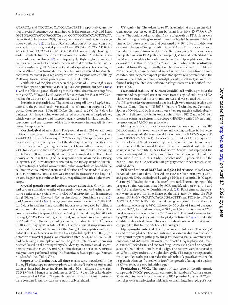
�pks4 mutants are devoid of conidial pigmentation. The de-fault hypothesis of this work was that PKS4 is involved in pigmentformation. This was confirmed: morphological examination onPDA plates revealed that both pks4 deletion mutants lost theirgreen conidial pigmentation (Fig. 1a), while formation of the yel-low pigment and its secretion into the medium, a characteristic ofT. reesei, remained unaffected. No green conidia were formed un-der any of the cultivation conditions in either light or darkness,and the color was not recovered by cultivation on any of 95 carbonsources of the Biolog phenotype microarrays, 5 of which areshown in Fig. 1b.
The �pks4 strains did not show any statistically significant dif-ference in the intensity of conidia production on PDA: after 7days, QM 6a, �pks4-1, and �pks4-2 produced on average 9.03 �106, 9.16 � 106, and 9.94 � 106 spores per cm2, respectively (anal-ysis of variance [ANOVA], P � 0.05) (Fig. 1c). Spore size re-mained unchanged (ANOVA, P � 0.05) (data not shown); how-ever, the conidia of the deletion mutants showed less mechanicalstability against reduced air pressure (Fig. 1d).
In order to test whether pks4 is solely responsible for conidialpigmentation, we crossed the �pks4 mutants (which, due to theirQM 6a background, possess MAT1-2 [27]) with the T. reeseiMAT1-1 strain CBS 999.97. In total, 34 pure single-spore strainswere isolated from mature ascospores. A total of 21, 10, and 3cultures contained purely white, green, or yellowish conidia, re-spectively. All strains from the first-generation progeny (F1) werescreened by PCR for mating types, and there was a nearly equaldistribution (15 MAT1-2 and 18 MAT1-1) that was independentof the phenotype (see Fig. S2 in the supplemental material). Spe-cific pks4 primers were designed (Table 1) and used to test the F1
progeny for the presence of the pks4 gene. We found that all thestrains with green conidia had indeed inherited the wild-typegene, whereas the yellow and albino conidia phenotype conidiadid not contain the pks4 gene (see Fig. S2). This proved that theloss of the conidial green pigmentation is directly caused by thepks4 deletion and that this gene is involved in its production.
Loss of green pigmentation caused reduced resistance to UV.The lack of green conidia pigmentation was also reflected in anincreased sensitivity to UV light: after 7 min of UV exposure (seeMaterials and Methods for details) sixty-four percent of greenspores survived, whereas only 8 to 20% of white conidia were ableto germinate (Table 2). Prolongation of exposure time (to 10 min)led to a ca. 92% reduction of germination for both �pks4 mutants(Table 2), while germination of QM 6a was decreased by only60%.
Atanasova et al.
1502 ec.asm.org Eukaryotic Cell
on April 17, 2020 by guest
http://ec.asm.org/
Dow
nloaded from
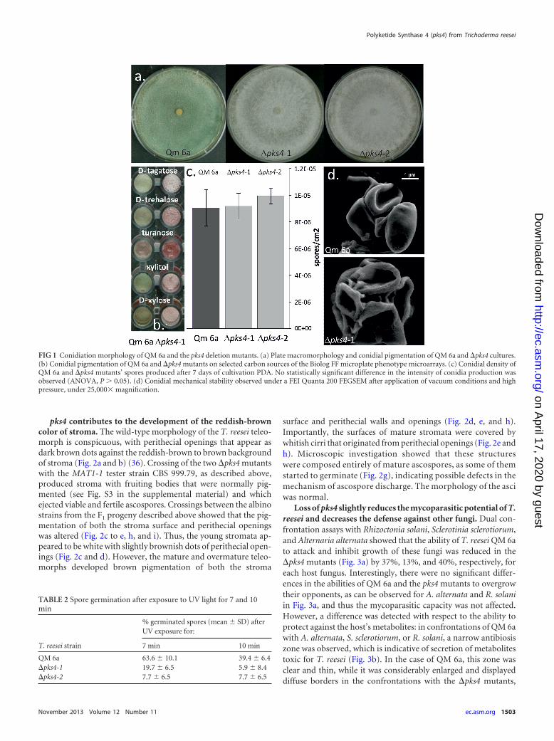
pks4 contributes to the development of the reddish-browncolor of stroma. The wild-type morphology of the T. reesei teleo-morph is conspicuous, with perithecial openings that appear asdark brown dots against the reddish-brown to brown backgroundof stroma (Fig. 2a and b) (36). Crossing of the two �pks4 mutantswith the MAT1-1 tester strain CBS 999.79, as described above,produced stroma with fruiting bodies that were normally pig-mented (see Fig. S3 in the supplemental material) and whichejected viable and fertile ascospores. Crossings between the albinostrains from the F1 progeny described above showed that the pig-mentation of both the stroma surface and perithecial openingswas altered (Fig. 2c to e, h, and i). Thus, the young stromata ap-peared to be white with slightly brownish dots of perithecial open-ings (Fig. 2c and d). However, the mature and overmature teleo-morphs developed brown pigmentation of both the stroma
surface and perithecial walls and openings (Fig. 2d, e, and h).Importantly, the surfaces of mature stromata were covered bywhitish cirri that originated from perithecial openings (Fig. 2e andh). Microscopic investigation showed that these structureswere composed entirely of mature ascospores, as some of themstarted to germinate (Fig. 2g), indicating possible defects in themechanism of ascospore discharge. The morphology of the asciwas normal.
Loss of pks4 slightly reduces the mycoparasitic potential of T.reesei and decreases the defense against other fungi. Dual con-frontation assays with Rhizoctonia solani, Sclerotinia sclerotiorum,and Alternaria alternata showed that the ability of T. reesei QM 6ato attack and inhibit growth of these fungi was reduced in the�pks4 mutants (Fig. 3a) by 37%, 13%, and 40%, respectively, foreach host fungus. Interestingly, there were no significant differ-ences in the abilities of QM 6a and the pks4 mutants to overgrowtheir opponents, as can be observed for A. alternata and R. solaniin Fig. 3a, and thus the mycoparasitic capacity was not affected.However, a difference was detected with respect to the ability toprotect against the host’s metabolites: in confrontations of QM 6awith A. alternata, S. sclerotiorum, or R. solani, a narrow antibiosiszone was observed, which is indicative of secretion of metabolitestoxic for T. reesei (Fig. 3b). In the case of QM 6a, this zone wasclear and thin, while it was considerably enlarged and displayeddiffuse borders in the confrontations with the �pks4 mutants,
FIG 1 Conidiation morphology of QM 6a and the pks4 deletion mutants. (a) Plate macromorphology and conidial pigmentation of QM 6a and �pks4 cultures.(b) Conidial pigmentation of QM 6a and �pks4 mutants on selected carbon sources of the Biolog FF microplate phenotype microarrays. (c) Conidial density ofQM 6a and �pks4 mutants’ spores produced after 7 days of cultivation PDA. No statistically significant difference in the intensity of conidia production wasobserved (ANOVA, P � 0.05). (d) Conidial mechanical stability observed under a FEI Quanta 200 FEGSEM after application of vacuum conditions and highpressure, under 25,000� magnification.
TABLE 2 Spore germination after exposure to UV light for 7 and 10min
T. reesei strain
% germinated spores (mean � SD) afterUV exposure for:
7 min 10 min
QM 6a 63.6 � 10.1 39.4 � 6.4�pks4-1 19.7 � 6.5 5.9 � 8.4�pks4-2 7.7 � 6.5 7.7 � 6.5
Polyketide Synthase 4 (pks4) from Trichoderma reesei
November 2013 Volume 12 Number 11 ec.asm.org 1503
on April 17, 2020 by guest
http://ec.asm.org/
Dow
nloaded from
suggesting that metabolites secreted by the other fungi had pene-trated the colonies of the deletion mutants.
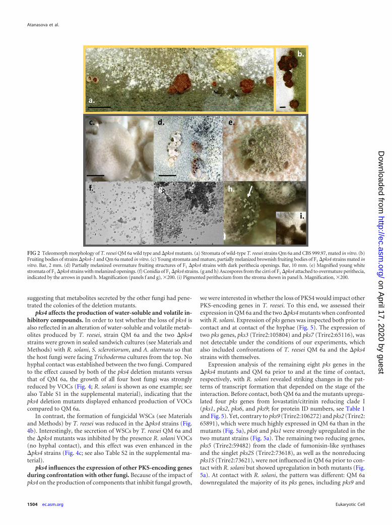
pks4 affects the production of water-soluble and volatile in-hibitory compounds. In order to test whether the loss of pks4 isalso reflected in an alteration of water-soluble and volatile metab-olites produced by T. reesei, strain QM 6a and the two �pks4strains were grown in sealed sandwich cultures (see Materials andMethods) with R. solani, S. sclerotiorum, and A. alternata so thatthe host fungi were facing Trichoderma cultures from the top. Nohyphal contact was established between the two fungi. Comparedto the effect caused by both of the pks4 deletion mutants versusthat of QM 6a, the growth of all four host fungi was stronglyreduced by VOCs (Fig. 4; R. solani is shown as one example; seealso Table S1 in the supplemental material), indicating that thepks4 deletion mutants displayed enhanced production of VOCscompared to QM 6a.
In contrast, the formation of fungicidal WSCs (see Materialsand Methods) by T. reesei was reduced in the �pks4 strains (Fig.4b). Interestingly, the secretion of WSCs by T. reesei QM 6a andthe �pks4 mutants was inhibited by the presence R. solani VOCs(no hyphal contact), and this effect was even enhanced in the�pks4 strains (Fig. 4c; see also Table S2 in the supplemental ma-terial).
pks4 influences the expression of other PKS-encoding genesduring confrontation with other fungi. Because of the impact ofpks4 on the production of components that inhibit fungal growth,
we were interested in whether the loss of PKS4 would impact otherPKS-encoding genes in T. reesei. To this end, we assessed theirexpression in QM 6a and the two �pks4 mutants when confrontedwith R. solani. Expression of pks genes was inspected both prior tocontact and at contact of the hyphae (Fig. 5). The expression oftwo pks genes, pks3 (Trire2:105804) and pks7 (Trire2:65116), wasnot detectable under the conditions of our experiments, whichalso included confrontations of T. reesei QM 6a and the �pks4strains with themselves.
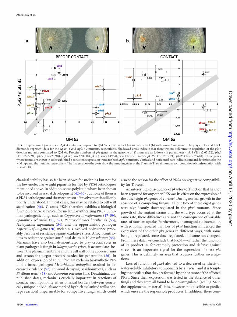
Expression analysis of the remaining eight pks genes in the�pks4 mutants and QM 6a prior to and at the time of contact,respectively, with R. solani revealed striking changes in the pat-terns of transcript formation that depended on the stage of theinteraction. Before contact, both QM 6a and the mutants upregu-lated four pks genes from lovastatin/citrinin reducing clade I(pks1, pks2, pks6, and pks9; for protein ID numbers, see Table 1and Fig. 5). Yet, contrary to pks9 (Trire2:106272) and pks2 (Trire2:65891), which were much highly expressed in QM 6a than in themutants (Fig. 5a), pks6 and pks1 were strongly upregulated in thetwo mutant strains (Fig. 5a). The remaining two reducing genes,pks5 (Trire2:59482) from the clade of fumonisin-like synthasesand the singlet pks2S (Trire2:73618), as well as the nonreducingpks1S (Trire2:73621), were not influenced in QM 6a prior to con-tact with R. solani but showed upregulation in both mutants (Fig.5a). At contact with R. solani, the pattern was different: QM 6adownregulated the majority of its pks genes, including pks9 and
FIG 2 Teleomorph morphology of T. reesei QM 6a wild type and �pks4 mutants. (a) Stromata of wild-type T. reesei strains Qm 6a and CBS 999.97, mated in vitro. (b)Fruiting bodies of strains �pks4-1 and Qm 6a mated in vitro. (c) Young stromata and mature, partially melanized brownish fruiting bodies of F1 �pks4 strains mated invitro. Bar, 2 mm. (d) Partially melanized overmature fruiting structures of F1 �pks4 strains with dark perithecia openings. Bar, 10 mm. (e) Magnified young whitestromata of F1 �pks4 strains with melanized openings. (f) Conidia of F1 �pks4 strains. (g and h) Ascospores from the cirri of F1 �pks4 attached to overmature perithecia,indicated by the arrows in panel h. Magnification (panels f and g), �200. (i) Pigmented perithecium from the stroma shown in panel h. Magnification, �200.
Atanasova et al.
1504 ec.asm.org Eukaryotic Cell
on April 17, 2020 by guest
http://ec.asm.org/
Dow
nloaded from

pks2, which were strongly upregulated before the contact with R.solani (Fig. 5). Interestingly, the two singlet pks genes (pks1S andpks2S) were differentially downregulated at the contact betweenthe pks4 mutants and R. solani (Fig. 5b), whereas they werestrongly upregulated in QM 6a at this stage.
Most of the eight pks genes were downregulated in the mutants,whereas two singlet genes, pks2S, a member of the lovastatin/cit-rinin clade, and pks1S, which belongs to nonreducing clade III,were upregulated (see Fig. S4 in the supplemental material).
DISCUSSION
In this study, we functionally characterized the role of thepolyketide synthase PKS4 in T. reesei. pks4 belongs to the nonre-ducing clade I of fungal pks-encoding genes, which includes genesassociated with pigment production, such as aurofusarin (7–9)and bikaverin (10, 11), but also DHN melanin (12–19). While theformer comprise substances of relatively low molecular weights,melanins—the dark to black pigments—are of high molecularmass that derive from oxidative polymerization of phenolic com-pounds (20, 37). Melanins are pigments that occur in all biologicalkingdoms and serve many functions, such as defense against en-vironmental stresses including UV light, oxidizing agents, andionizing radiation, and they also contribute to fungal pathogenesis(38).The chemical structures of the conidial pigments of T. reesei,and of Trichoderma spp. in general, have not been elucidated yet,
but due to their green and sometimes yellowish color they appearnot to be melanins. Yellow pigments from Aspergillus niger havebeen shown to be dimeric linear naphtho-y-pyrones (4, 7, 13).However, Benitez et al. (39) preliminarily characterized the conid-ial pigment of Trichoderma spp. (T. viride at that time) as a non-indolic melanin-like polyphenol. Consistent with these data,Csiktusnádi Kiss et al. (40) identified the main pigment fractionsof T. harzianum as oxidation polymers originating from mono-mer molecules containing polar substructures and double bondsin the alkyl chain. Here, we showed that the final dark browncomponent of the fruiting body and the stroma color, which likelyrepresents melanin, are independent of PKS4, but the colorizationis delayed in the early stages of fruiting body development. In theabsence of pks4, the young stroma of T. reesei are colorless (white)with slightly darkened openings of perithecia, indicating someretained pigmentation. Mature and overmature stromata, how-ever, showed some dark brown coloration on the surface and per-ithecia walls, indicating that melanin was still synthesized. Thequestion of whether the pigments synthesized by PKS4 are mela-nins is important, because our data showed that PKS4 is involvedin antagonism and defense against other fungi and in the mechan-ical stability of the conidium. A role in antagonism is also sup-ported by earlier findings indicating that pks4 is upregulated dur-ing antagonism and mycoparasitic contact of T. reesei with R.solani (41). A role in defense, stress resistance, virulence, and me-
FIG 3 Antagonistic potentials of QM 6a and the pks4-1 deletion mutant.Confrontations panels are shown only for �pks4-1, but both mutants revealedidentical mycoparasitic patterns. (a) Front sides of confrontation plates, withplant-pathogenic fungi always on the left side and Trichoderma QM 6a or themutant always on the right. (b) Back sides of confrontation plates, with plant-pathogenic fungi always on the left side and T. reesei QM 6a or the mutantstrain always on the right. Antibiosis zones for �pks4 strains are indicated bythe arrows. Aa, Ss, Bs, and Rs mark Alternaria alternata, Sclerotinia sclerotio-rum, Botrytis cinerea, and Rhizoctonia solani, respectively.
FIG 4 Effects of VOCs and WSCs from pks4 deletion mutants and QM 6a ongrowth of R. solani. (a) Reduction of R. solani growth by the VOCs secreted byT. reesei QM 6a and �pks4 mutants after 4 days of incubation. Both mutantsconsistently reduced the growth of R. solani. (b) R. solani growth on PDAmedium containing WSCs of T. reesei and the pks4 deletion mutants. (c)Growth of R. solani on medium with WSCs secreted by the pks4 mutants andQM 6a in the presence of VOCs from R. solani during growth of the T. reeseistrains.
Polyketide Synthase 4 (pks4) from Trichoderma reesei
November 2013 Volume 12 Number 11 ec.asm.org 1505
on April 17, 2020 by guest
http://ec.asm.org/
Dow
nloaded from
chanical stability has so far been shown for melanins but not forthe low-molecular-weight pigments formed by PKS4 orthologuesmentioned above. In addition, some polyketides have been shownto be involved in sexual development (42–46) but none of them isa PKS4 orthologue, and the mechanism of involvement is still onlypoorly understood. In most cases, this may be related to cell wallstabilization (46). T. reesei PKS4 therefore exhibits a biologicalfunction otherwise typical for melanin-synthesizing PKSs: in hu-man pathogenic fungi, such as Cryptococcus neoformans (47–59),Sporothrix schenckii (51, 52), Paracoccidioides brasiliensis (53),Histoplasma capsulatum (54), and the opportunistic pathogenAspergillus fumigatus (20), melanin is involved in virulence, prob-ably because of resistance against oxidative stress. Also, it contrib-utes to resistance against antifungal drugs in H. capsulatum (55).Melanins have also been demonstrated to play crucial roles inplant pathogenic fungi: in Magnaporthe grisea, it accumulates be-tween the plasma membrane and the cell wall of the appressoriumand creates the turgor pressure needed for penetration (56). Inaddition, expression of an A. alternata melanin biosynthetic PKSin the insect pathogen Metarhizium anisopliae resulted in in-creased virulence (57). In wood decaying Basidiomycota, such asPhellinus weirii (58) and Pleurotus ostreatus (I. S. Druzhinina, un-published data), melanin is crucially important in reactions ofsomatic incompatibility when physical borders between geneti-cally unique individuals are marked by thick melanized walls (bar-rage reaction) impermeable for competitive fungi, which could
also be the reason for the effect of PKS4 on vegetative compatibil-ity for T. reesei.
An interesting consequence of pks4 loss of function that has notbeen reported for any other PKS was its effect on the expression ofthe other eight pks genes of T. reesei. During normal growth in theabsence of a competing fungus, all but two of these eight geneswere significantly downregulated in the pks4 mutants. Sincegrowth of the mutant strains and the wild type occurred at thesame rate, these differences are not the consequence of variablerates of nutrient uptake. Furthermore, an antagonistic interactionwith R. solani revealed that loss of pks4 function influenced theexpression of the other pks genes in different ways, with somebeing upregulated, some downregulated, and some not changed.From these data, we conclude that PKS4 — or rather the functionof its product in, for example, protection and defense againststress—is an important signal for the expression of these pksgenes. This is definitely an area that requires further investiga-tions.
Loss of function of pks4 also led to a decreased synthesis ofwater-soluble inhibitory components by T. reesei, and it is tempt-ing to speculate that they are formed by one or more of the affectedPKSs. Since their expression was tested in the absence of otherfungi and they were all found to be downregulated (see Fig. S4 inthe supplemental material), it is, however, not possible to predictwhich ones are the responsible producers. In addition, these com-
FIG 5 Expression of pks genes in �pks4 mutants compared to QM 6a before contact (a) and at contact (b) with Rhizoctonia solani. The gray circles and blackdiamonds represent data for the �pks4-1 and �pks4-2 mutants, respectively. Shadowed areas indicate that there was no difference in regulation of the pks4deletion mutants compared to QM 6a. Protein numbers of pks genes in the genome of T. reesei are as follows (in parentheses): pks1 (Trire2:65172), pks2(Trire2:65891), pks5 (Trire2:59482), pks6 (Trire2:60118), pks8 (Trire2:81964), pks9 (Trire2:106272), pks1S (Trire2:73621), pks2S (Trire2:73618). Those geneswhose names are shown in color exhibited a consistent expression trend for both �pks4 mutants. Vertical and horizontal bars indicate standard deviations for thewild type and the mutants, respectively. The images above the plots show the sampling stage of the T. reesei (T) strains under each condition of confrontation withR. solani (R).
Atanasova et al.
1506 ec.asm.org Eukaryotic Cell
on April 17, 2020 by guest
http://ec.asm.org/
Dow
nloaded from
ponents could also be products of other enzymes, such as nonri-bosomal peptide synthases, of which T. reesei produces 10 (59).
Finally, it was interesting that deletion of pks4 increased theproduction of VOCs by T. reesei. The chemical natures of theVOCs from T. reesei have not been identified yet, but their diver-sity compared to those from other Trichoderma spp. showed thatthey are composed mainly of long aliphatic acids and that theyhave alcohols and esters (60, 61), which are usually products offatty acid catabolism (62). Biosynthesis of these compounds maybe favored by the lack of PKS activity, which results in an increasedaccess to the cellular pools of acetyl- and malonyl-CoA.
The functions of PKSs have so far mainly been investigatedwith respect to the role of their products in human or plant patho-genesis. Our data showed that PKS4 also influences several biolog-ical functions in T. reesei that are not only related to the interactionwith other organisms. Transcriptomic analyses of T. reesei haverecently shown that many pks genes are maximally expressed dur-ing rapid vegetative growth (63–65), which is not a pattern thatwould be expected for genes whose functions are traditionallyviewed as unrelated to growth (i.e., secondary metabolites). It willthus be worthwhile to perform a deeper investigation of the regu-lation and role of pks genes in fungal physiology.
ACKNOWLEDGMENTS
This study was supported by a grant of the Austrian Science Fund (FWF)to C.P.K. (P 21266). S.E.B. and B.P.K. were supported by funding from theU.S. DOE Biomass Program.
We thank Aldin Saracevic and Yuan Zhi-Lin for laboratory assistanceand Bernhard Pummer for the SEM images.
REFERENCES1. Druzhinina IS, Seidl-Seiboth V, Herrera-Estrella A, Horwitz BA, Ken-
erley CM, Monte E, Mukherjee PK, Zeilinger S, Grigoriev IV, KubicekCP. 2011. Trichoderma: the genomics of opportunistic success. Nat. Rev.Microbiol. 16:749 –759.
2. Druzhinina IS, Shelest E, Kubicek CP. 2012. Novel traits of Trichodermapredicted through the analysis of its secretome. FEMS Microbiol. Lett.337:1–9.
3. Druzhinina IS, Kubicek CP. 2013. Ecological genomics of Trichoderma.In Martin F (ed), Ecological genomics of fungi. Wiley-Blackwell, Oxford,United Kingdom.
4. Mukherjee PK, Horwitz BA, Kenerley CM. 2012. Secondary metabolismin Trichoderma: a genomic perspective. Microbiology 158:35– 45.
5. Kroken S, Glass NL, Taylor JW, Yoder OC, Turgeon BG. 2003. Phy-logenomic analysis of type I polyketide synthase genes in pathogenic andsaprobic ascomycetes. Proc. Natl. Acad. Sci. U. S. A. 100:15670 –15675.
6. Baker SE, Perrone G, Richardson NM, Gallo A, Kubicek CP. 2012.Phylogenomic analysis of polyketide synthase-encoding genes inTrichoderma. Microbiology 158:147–154.
7. Frandsen RJ, Schütt C, Lund BW, Staerk D, Nielsen J, Olsson S, GieseH. 2011. Two novel classes of enzymes are required for the biosynthesis ofaurofusarin in Fusarium graminearum. J. Biol. Chem. 286:10419 –10428.
8. Kim JE, Han KH, Jin J, Kim H, Kim JC, Yun SH, Lee YW. 2005. Putativepolyketide synthase and laccase genes for biosynthesis of aurofusarin inGibberella zeae. Appl. Environ. Microbiol. 71:1701–1708.
9. Malz S, Grell MN, Thrane C, Maier FJ, Rosager P, Felk A, Albertsen KS,Salomon S, Bohn L, Schafer W, Giese H. 2005. Identification of a genecluster responsible for the biosynthesis of aurofusarin in the Fusariumgraminearum species complex. Fungal Genet. Biol. 42:420 – 433.
10. Linnemannstöns P, Schulte J, del Mar Prado M, Proctor RH, Avalos J,Tudzynski B. 2002. The polyketide synthase gene pks4 from Gibberellafujikuroi encodes a key enzyme in the biosynthesis of the red pigmentbikaverin. Fungal Genet. Biol. 37:134 –148.
11. Wiemann P, Willmann A, Straeten M, Kleigrewe K, Beyer M, HumpfHU, Tudzynski B. 2009. Biosynthesis of the red pigment bikaverin inFusarium fujikuroi: genes, their function and regulation. Mol. Microbiol.72:931–946.
12. Baker SE. 2008. Aspergillus genomics and DHN-melanin conidial pig-mentation, p 73– 85. In Varga J, Samson RA (ed),Aspergillus in thegenomic era. Wageningen Academic Publishers, Wageningen, The Neth-erlands.
13. Chiang YM, Meyer KM, Praseuth M, Baker SE, Bruno KS, Wang CC.2011. Characterization of a polyketide synthase in Aspergillus niger whoseproduct is a precursor for both dihydroxynaphthalene (DHN) melaninand naphtho-c-pyrone. Fungal Genet. Biol. 48:430 – 437.
14. Jørgensen TR, Park J, Arentshorst M, van Welzen AM, Lamers G,Vankuyk PA, Damveld RA, van den Hondel CA, Nielsen KF, FrisvadJC, Ram AF. 2011. The molecular and genetic basis of conidial pigmen-tation in Aspergillus niger. Fungal Genet. Biol. 48:544 –553.
15. Langfelder K, Jahn B, Gehringer H, Schmidt A, Wanner G, BrakhageAA. 1998. Identification of a polyketide synthase gene (pksP) of Aspergillusfumigatus involved in conidial pigment biosynthesis and virulence. Med.Microbiol. Immunol. (Berl.) 187:79 – 89.
16. Tsai HF, Chang YC, Washburn RG, Wheeler MH, Kwon-Chung KJ.1998. The developmentally regulated alb1 gene of Aspergillus fumigatus: itsrole in modulation of conidial morphology and virulence. J. Bacteriol.180:3031–3038.
17. Tsai HF, Fujii I, Watanabe A, Wheeler MH, Chang YC, Yasuoka Y,Ebizuka Y, Kwon-Chung KJ. 2001. Pentaketide melanin biosynthesis inAspergillus fumigatus requires chain length shortening of a heptaketideprecursor. J. Biol. Chem. 276:29292–29298.
18. Watanabe A, Fujii I, Sankawa U, Mayorga ME, Timberlake WE, Ebi-zuka Y. 1999. Re-identification of Aspergillus nidulans wA gene to code fora polyketidesynthase of naphthopyrone. Tetrahedron Lett. 40:91–94.
19. Watanabe A, Fujii I, Tsai H, Chang YC, Kwon-Chung KJ, Ebizuka Y.2000. Aspergillus fumigatus alb1 encodes naphthopyrone synthase whenexpressed in Aspergillus oryzae. FEMS Microbiol. Lett. 192:39 – 44.
20. Heinekamp T, Thywißen A, Macheleidt J, Keller S, Valiante V, Bra-khage AA. 2012. Aspergillus fumigatus melanins: interference with thehost endocytosis pathway and impact on virulence. Front. Microbiol.3:440. doi:10.3389/fmicb.2012.00440.
21. Yu JH, Hamari Z, Han KH, Seo JA, Reyes-Domínguez Y, ScazzocchioC. 2004. Double-joint PCR: a PCR-based molecular tool for gene manip-ulations in filamentous fungi. Fungal Genet. Biol. 41:973–981.
22. Penttila M, Nevalainen H, Ratto M, Salminen E, Knowles J. 1987. Aversatile transformation system for the cellulolytic filamentous fungusTrichoderma reesei. Gene 61:155–164.
23. Druzhinina IS, Schmoll M, Seiboth B, Kubicek CP. 2006. Global carbonutilization profiles of wild-type, mutant, and transformant strains ofHypocrea jecorina. Appl. Environ. Microbiol. 72:2126 –2133.
24. Atanasova L, Druzhinina IS. 2010. Global nutrient profiling by pheno-type microarrays: a tool complementing genomic and proteomic studiesin conidial fungi. J. Zhejiang Univ. Sci. B 11:151–168.
25. Druzhinina IS, Komon-Zelazowska M, Atanasova L, Seidl V, KubicekCP. 2010. Evolution and ecophysiology of the industrial producer Hypo-crea jecorina (anamorph Trichoderma reesei) and a new sympatric aga-mospecies related to it. PLoS One 5(2):e9191. doi:10.1371/journal.pone.0009191.
26. Pfaffl MW, Horgan GW, Dempfle L. 2002. Relative expression softwaretool (REST) for group-wise comparison and statistical analysis of relativeexpression results in real-time PCR. Nucleic Acids Res. 30:e36. doi:10.1093/nar/30.9.36.
27. Seidl V, Seibel C, Kubicek CP, Schmoll M. 2009. Sexual development inthe industrial workhorse Trichoderma reesei. Proc. Natl. Acad. Sci. U. S. A.106:13909 –13914.
28. Pihet M, Vandeputte P, Tronchin G, Renier G, Saulnier P, GeorgeaultS, Mallet R, Chabasse D, Symoens F, Bouchara JP. 2009. Melanin is anessential component for the integrity of the cell wall of Aspergillus fumiga-tus conidia. BMC Microbiol. 9:177. doi:10.1186/1471-2180-9-177.
29. Li Y, Chooi YH, Sheng Y, Valentine JS, Tang Y. 2011. Comparativecharacterization of fungal anthracenone and naphthacenedione biosyn-thetic pathways reveals an �-hydroxylation-dependent Claisen-like cycli-zation catalyzed by a dimanganesethioesterase. J. Am. Chem. Soc. 133:15773–15785.
30. Chooi YH, Cacho R, Tang Y. 2010. Identification of the viridicatumtoxinand griseofulvin gene clusters from Penicillium aethiopicum. Chem. Biol.17:483– 494.
31. Abe Y, Suzuki T, Mizvno T, Ono C, Iwamoto K, Hosobuchi M,Yoshikawa H. 2002. Molecular cloning and characterization of an ML-
Polyketide Synthase 4 (pks4) from Trichoderma reesei
November 2013 Volume 12 Number 11 ec.asm.org 1507
on April 17, 2020 by guest
http://ec.asm.org/
Dow
nloaded from

236B (compactin) biosynthetic gene cluster in Penicillium citrinum. Mol.Genet. Genomics 267:636 – 646.
32. O’Callaghan J, Caddick MX, Dobson AD. 2003. A polyketide synthasegene required for ochratoxin A biosynthesis in Aspergillus ochraceus. Mi-crobiology 149:3485–3491.
33. Xue M, Yang J, Li Z, Hu S, Yao N, Dean RA, Zhao W, Shen M, ZhangH, Li C, Liu L, Cao L, Xu X, Xing Y, Hsiang T, Zhang Z, Xu JR, PengYL. 2012. Comparative analysis of the genomes of two field isolates of therice blast fungus Magnaporthe oryzae. PLoS Genet. 8(8):e1002869. doi:10.1371/journal.pgen.1002869.
34. Hendrickson L, Davis CR, Roach C, Nguyen DK, Aldrich T, McAda PC,Reeves CD. 1999. Lovastatin biosynthesis in Aspergillus terreus: character-ization of blocked mutants, enzyme activities and a multifunctionalpolyketide synthase gene. Chem. Biol. 6:429 – 439.
35. Baker SE. 2006. Aspergillus niger genomics: past, present and into thefuture. Med. Mycol. 1:S17–S21.
36. Samuels GJ, Chaverri P, Farr DF, McCray EB. 2010, last update.Trichoderma online. Systematic Mycology and Microbiology Laboratory,ARS, USDA. http://nt.ars-grin.gov/taxadescriptions/keys/FrameListAllTaxa.cfm?gen�Trichoderma.
37. Riley PA. 1997. Melanin. Int. J. Biochem. Cell Biol. 29:1235–1239.38. Eisenman HC, Casadevall A. 2012. Synthesis and assembly of fungal
melanin. Appl. Microbiol. Biotechnol. 93:931–940.39. Benítez T, Villa TG, García Acha I. 1976. Some chemical and structural
features of the conidial wall of Trichoderma viride. Can. J. Microbiol. 22:318 –321.
40. Csiktusnádi Kiss G, Forgács E, Cserháti T, Vizcaino JA. 2000. Colourpigments of Trichoderma harzianum. Preliminary investigations withthin-layer chromatography-Fourier transform infrared spectroscopy andhigh-performance liquid chromatography with diode array and massspectrometric detection. J. Chromatogr. A 896:61– 68.
41. Atanasova L, Crom S, Gruber S, Coulpier F, Seidl-Seiboth V, KubicekCP, Druzhinina IS. 2013. Comparative transcriptomics reveals differentstrategies of Trichoderma mycoparasitism. BMC Genomics 14:121. doi:10.1186/1471-2164-14-121.
42. Wolf JC, Mirocha CJ. 1973. Regulation of sexual reproduction in Gib-berella zeae (Fusarium roseum ‘Graminearum’) by F-2 (zearalenone). Can.J. Microbiol. 19:725–734.
43. Zimmerman WC, Blanchette RA, Burnes TA, Farrell RL. 1995. Melaninand perithecial development in Ophiostoma piliferum. Mycologia 87:857–863.
44. Graziani S, Vasnier C, Daboussi MJ. 2004. Novel polyketide synthasefrom Nectria haematococca. Appl. Environ. Microbiol. 70:2984 –2988.
45. Engh I, Nowrousian M, Kück U. 2007. Regulation of melanin biosyn-thesis via the dihydroxynaphthalene pathway is dependent on sexual de-velopment in the ascomycete Sordaria macrospora. FEMS Microbiol. Lett.275:62–70.
46. Nowrousian M. 2009. A novel polyketide biosynthesis gene cluster isinvolved in fruiting body morphogenesis in the filamentous fungi Sordariamacrospora and Neurospora crassa. Curr. Genet. 55:185–198.
47. Nosanchuk JD, Rosas AL, Lee SC, Casadevall A. 2000. Melanisation ofCryptococcus neoformans in human brain tissue. Lancet 355:2049 –2050.
48. Rosas AL, Nosanchuk JD, Feldmesser M, Cox GM, McDade HC,Casadevall A. 2000. Synthesis of polymerized melanin by Cryptococcusneoformans in infected rodents. Infect. Immun. 68:2845–2853.
49. Rosas AL, Nosanchuk JD, Gómez BL, Edens WA, Henson JM, Casade-vall A. 2000. Isolation and serological analyses of fungal melanins. J.Immunol. Methods 244:69 – 80.
50. Casadevall A, Rosas AL, Nosanchuk JD. 2000. Melanin and virulence inCryptococcus neoformans. Curr. Opin. Microbiol. 3:354 –358.
51. Romero-Martinez R, Wheeler M, Guerrero-Plata A, Rico G, Torres-Guerrero H. 2000. Biosynthesis and functions of melanin in Sporothrixschenckii. Infect. Immun. 68:3696 –3703.
52. Morris-Jones R, Youngchim S, Gómez BL, Aisen P, Hay RJ, NosanchukJD, Casadevall A, Hamilton AJ. 2003. Synthesis of melanin-like pigmentsby Sporothrix schenckii in vitro and during mammalian infection. Infect.Immun. 71:4026 – 4033.
53. Gómez BL, Nosanchuk JD, Díez S, Youngchim S, Aisen P, Cano LE,Restrepo A, Casadevall A, Hamilton AJ. 2001. Detection of melanin-likepigments in the dimorphic fungal pathogen Paracoccidioides brasiliensis invitro and during infection. Infect. Immun. 69:5760 –5767.
54. Nosanchuk JD, Gómez BL, Youngchim S, Díez S, Aisen P, Zancopé-Oliveira RM, Restrepo A, Casadevall A, Hamilton AJ. 2002. Histoplasmacapsulatum synthesizes melanin-like pigments in vitro and during mam-malian infection. Infect. Immun. 70:5124 –5131.
55. Hamilton AJ, Holdom MD. 1999. Antioxidant systems in the pathogenicfungi of man and their role in virulence. Med. Mycol. 37:375–389.
56. Howard RJ, Valent B. 1996. Breaking and entering: host penetration bythe fungal rice blast pathogen Magnaporthe grisea. Annu. Rev. Microbiol.50:491–512.
57. Tseng MN, Chung PC, Tzean SS. 2011. Enhancing the stress toleranceand virulence of an entomopathogen by metabolic engineering of dihy-droxynaphthalene melanin biosynthesis genes. Appl. Environ. Microbiol.77:4508 – 4519.
58. Li CY. 1981. Phenoloxidase and peroxidase activities in zone lines ofPhellinus weirii. Mycologia 73:811– 821.
59. Kubicek CP, Herrera-Estrella A, Seidl-Seiboth V, Martinez DA, Dru-zhinina IS, Thon M, Zeilinger S, Casas-Flores S, Horwitz BA, Mukher-jee PK, Mukherjee M, Kredics L, Alcaraz LD, Aerts A, Antal Z, Atana-sova L, Cervantes-Badillo MG, Challacombe J, Chertkov O, et al. 2011.Comparative genome sequence analysis underscores mycoparasitism asthe ancestral life style of Trichoderma. Genome Biol. 12:R40.
60. Nemcovic M, Jakubíková L, Víden I, Farkas V. 2008. Induction ofconidiation by endogenous volatile compounds in Trichoderma spp.FEMS Microbiol. Lett. 284:231–236.
61. Siddiquee S, Cheong BE, Taslima K, Kausar H, Hasan MM. 2012.Separation and identification of volatile compounds from liquid culturesof Trichoderma harzianum by GC-MS using three different capillary col-umns. J. Chromatogr. Sci. 50:358 –367.
62. Savage TJ, Hristova MK, Croteau R. 1996. Evidence for an elongation/reduction/C1-elimination pathway in the biosynthesis of n-heptane inxylem of Jeffrey pine. Plant Physiol. 111:1263–1269.
63. Metz B, Seidl-Seiboth V, Haarmann T, Kopchinskiy A, Lorenz P,Seiboth B, Kubicek CP. 2011. Expression of biomass-degrading enzymesis a major event during conidium development in Trichoderma reesei.Eukaryot. Cell 10:1527–1735.
64. Portnoy T, Margeot A, Le Crom S, Linke R, Atanasova L, Fekete E,Sándor E, Karaffa L, Druzhinina IS, Seiboth B, Kubicek CP. 2011. TheCRE1 carbon catabolite repressor of the fungus Trichoderma reesei: a mas-ter regulator of carbon assimilation. BMC Genomics 12:269. doi:10.1186/1471-2164-12-269.
65. Karimi-Aghcheh R, Bok JW, Phatale PA, Smith KM, Baker SE, LichiusA, Omann M, Zeilinger S, Seiboth B, Rhee C, Keller NP, Freitag M,Kubicek CP. 2013. Functional analyses of Trichoderma reesei LAE1 revealconserved and contrasting roles of this regulator. G3 3:369 –378.
Atanasova et al.
1508 ec.asm.org Eukaryotic Cell
on April 17, 2020 by guest
http://ec.asm.org/
Dow
nloaded from