The placenta, Membranes , and umbilical · PDF fileWhen, after birth , the placenta is viewed...
50
The placenta, Membranes , The placenta, Membranes , and umbilical cord and umbilical cord and umbilical cord and umbilical cord
Transcript of The placenta, Membranes , and umbilical · PDF fileWhen, after birth , the placenta is viewed...

The placenta, Membranes ,The placenta, Membranes ,and umbilical cordand umbilical cord
The placenta, Membranes ,The placenta, Membranes ,and umbilical cordand umbilical cord

Development of the placenta is a highly regulatedDevelopment of the placenta is a highly regulatedprocess that is essential for normal fetal growthprocess that is essential for normal fetal growth
and development, and for maintenance of aand development, and for maintenance of ahealthy pregnancy. The placenta fulfills severalhealthy pregnancy. The placenta fulfills several
critical roles as it preventing rejection of the fetalcritical roles as it preventing rejection of the fetalallograft; transporting and metabolizing nutrients,allograft; transporting and metabolizing nutrients,
and providing peptide and steroid hormones.and providing peptide and steroid hormones.

•Developmental Anatomy And Structure

Development of the placenta and fetus is aDevelopment of the placenta and fetus is acontinuous process that begins at the time ofcontinuous process that begins at the time of
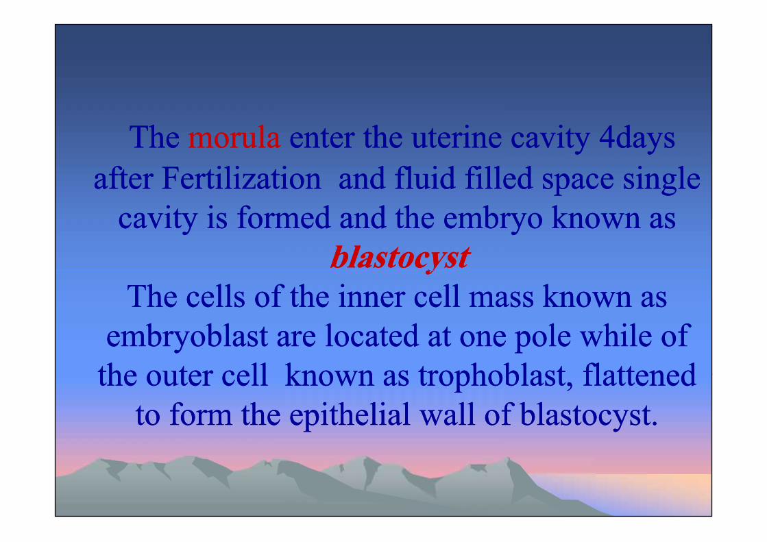
fertilization. Thefertilization. The morulamorula enter the uterine cavityenter the uterine cavity44days afterdays after Fertilization and fluid filled spaceFertilization and fluid filled space
single cavity is formed and the embryo known assingle cavity is formed and the embryo known asblastocystblastocyst
The cells of the inner cell mass known asThe cells of the inner cell mass known asembryoblast are located at one pole while of theembryoblast are located at one pole while of the
outer cell known as trophoblast, flattened toouter cell known as trophoblast, flattened toform the epithelial wall of blastocyst.form the epithelial wall of blastocyst.
Development of the placenta and fetus is aDevelopment of the placenta and fetus is acontinuous process that begins at the time ofcontinuous process that begins at the time of
fertilization. Thefertilization. The morulamorula enter the uterine cavityenter the uterine cavity44days afterdays after Fertilization and fluid filled spaceFertilization and fluid filled space
single cavity is formed and the embryo known assingle cavity is formed and the embryo known asblastocystblastocyst
The cells of the inner cell mass known asThe cells of the inner cell mass known asembryoblast are located at one pole while of theembryoblast are located at one pole while of the
outer cell known as trophoblast, flattened toouter cell known as trophoblast, flattened toform the epithelial wall of blastocyst.form the epithelial wall of blastocyst.
TheThe morulamorula enter the uterine cavityenter the uterine cavity 44daysdaysafterafter Fertilization and fluid filled space singleFertilization and fluid filled space single
cavity is formed and the embryo known ascavity is formed and the embryo known asblastocystblastocyst
The cells of the inner cell mass known asThe cells of the inner cell mass known asembryoblast are located at one pole while ofembryoblast are located at one pole while of
the outer cell known as trophoblast, flattenedthe outer cell known as trophoblast, flattenedto form the epithelial wall of blastocyst.to form the epithelial wall of blastocyst.

INTRODUCTIONINTRODUCTIONThe blastocyst is bathed in uterine
secretions that provide the embryo withoxygen and metabolic substrates. However,this soon becomes inadequate for furtherdevelopment, and the embryo must thenimplant in the uterine wall.
The blastocyst is bathed in uterinesecretions that provide the embryo withoxygen and metabolic substrates. However,this soon becomes inadequate for furtherdevelopment, and the embryo must thenimplant in the uterine wall.

At the beginning of the 2nd week , theblastocyst is partially embedded in theendometrial stroma .The trophoblast differentiates into :1- An inner ,actively proliferating layer ,thecytotrophoblast.2-An outer layer,the syncytiotrophoblast
At the beginning of the 2nd week , theblastocyst is partially embedded in theendometrial stroma .The trophoblast differentiates into :1- An inner ,actively proliferating layer ,thecytotrophoblast.2-An outer layer,the syncytiotrophoblast

By dayBy day 99 lacuna develop in syncytiotrophoblast.lacuna develop in syncytiotrophoblast.Sebsequently,maternal sinusoid are eroded bySebsequently,maternal sinusoid are eroded bythe syncytiothe syncytio-- trophoblast maternal blood enterstrophoblast maternal blood entersthe lacunar network, and by the end of thethe lacunar network, and by the end of the 22ndnd
week, a primitive uteroplacental circulationweek, a primitive uteroplacental circulationbegins.begins.

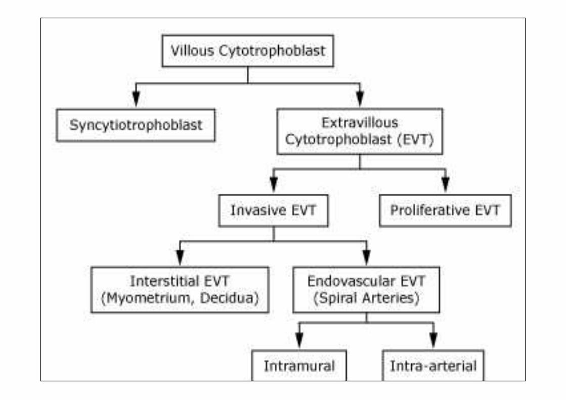
The progenitor villous trophoblast cell isThe progenitor villous trophoblast cell isthe stem cell of the placenta. It proliferatesthe stem cell of the placenta. It proliferatesthroughout gestation, differentiating alongthroughout gestation, differentiating alongtwo pathways totwo pathways toinvasive extravillous trophoblast (EVT)invasive extravillous trophoblast (EVT)and the syncytiotrophoblast ,as show inand the syncytiotrophoblast ,as show infollowing figure.following figure.
••The progenitor villous trophoblast cellThe progenitor villous trophoblast cellis the stem cell of the placenta. Itis the stem cell of the placenta. Itproliferates throughout gestation,proliferates throughout gestation,differentiating along two pathways todifferentiating along two pathways toform nonproliferative invasiveform nonproliferative invasiveextravillous trophoblast (EVT) or theextravillous trophoblast (EVT) or thesyncytiotrophoblast (show figuresyncytiotrophoblast (show figure 11).).
بسـم ا الرمحن الرحيـمبسـم ا الرمحن الرحيـم﴾﴾يرفـع اللــه الذين آمنـوا منكــم والذين أوتوا العلــم درجات واللـه مبا تعملون خبرييرفـع اللــه الذين آمنـوا منكــم والذين أوتوا العلــم درجات واللـه مبا تعملون خبري﴿﴿
صـدق ا العظيـمصـدق ا العظيـم))٢٨٢٨اادلـةاادلـة((

The extra villous trophoblast ( EVT )is responsible for invasion, thereby anchoring theplacenta to the decidua and myometrium.
The syncytiotrophoblastis a specialized epithelium covering the villoustree and has several functions, such as transport ofgases, nutrients, and waste products and synthesisof peptide and steroid hormones that regulateplacental, fetal, and maternal systems.

Alterations in villous trophoblastdifferentiation are seen in variouspathophysiological situations and mayunderlie several pregnancy disorders.
Alterations in villous trophoblastdifferentiation are seen in variouspathophysiological situations and mayunderlie several pregnancy disorders.
Endovascular EVTsEndovascular EVTs are associated withare associated withspiral arteries, either within the vessel wallspiral arteries, either within the vessel wall(intramural) or replacing endothelium(intramural) or replacing endothelium(intra(intra--arterial). EVTs transform the narrowarterial). EVTs transform the narrowspiral arteries to wide uteroplacentalspiral arteries to wide uteroplacentalarteries, which distribute blood into a lowarteries, which distribute blood into a lowresistance vascular network .resistance vascular network .
Endovascular EVTsEndovascular EVTs are associated withare associated withspiral arteries, either within the vessel wallspiral arteries, either within the vessel wall(intramural) or replacing endothelium(intramural) or replacing endothelium(intra(intra--arterial). EVTs transform the narrowarterial). EVTs transform the narrowspiral arteries to wide uteroplacentalspiral arteries to wide uteroplacentalarteries, which distribute blood into a lowarteries, which distribute blood into a lowresistance vascular network .resistance vascular network .

Defects in EVT invasion occur withDefects in EVT invasion occur withpreeclampsia and intrauterine growthpreeclampsia and intrauterine growthrestriction (IUGR),restriction (IUGR), where some spiralwhere some spiralarteries are not invaded at all and some arearteries are not invaded at all and some aresuperficially invaded, leading to lack of thesuperficially invaded, leading to lack of thenormal physiological adaptation of spiralnormal physiological adaptation of spiralarteries to pregnancy, reduced blood flowarteries to pregnancy, reduced blood flowinto the intervillous space, and relativeinto the intervillous space, and relativehypoxia/ischemiahypoxia/ischemia..
Defects in EVT invasion occur withDefects in EVT invasion occur withpreeclampsia and intrauterine growthpreeclampsia and intrauterine growthrestriction (IUGR),restriction (IUGR), where some spiralwhere some spiralarteries are not invaded at all and some arearteries are not invaded at all and some aresuperficially invaded, leading to lack of thesuperficially invaded, leading to lack of thenormal physiological adaptation of spiralnormal physiological adaptation of spiralarteries to pregnancy, reduced blood flowarteries to pregnancy, reduced blood flowinto the intervillous space, and relativeinto the intervillous space, and relativehypoxia/ischemiahypoxia/ischemia..

VASCULATURE
The proportion of the placenta occupied byblood vessels increases throughout gestationto facilitate nutrient transport. The twoumbilical arteries and the vein divide intonetworks of secondary vessels, and thenfurther divide into tertiary vessels beforepenetrating the chorionic plate and enteringthe main stem villi.
The proportion of the placenta occupied byblood vessels increases throughout gestationto facilitate nutrient transport. The twoumbilical arteries and the vein divide intonetworks of secondary vessels, and thenfurther divide into tertiary vessels beforepenetrating the chorionic plate and enteringthe main stem villi.

These then divide two to five timesto form rami chorii (intermediatevilli) and further divide to formramuli chorii, some of whichterminate in the terminal villi, thefunctional units of exchange asshown in following figure.
These then divide two to five timesto form rami chorii (intermediatevilli) and further divide to formramuli chorii, some of whichterminate in the terminal villi, thefunctional units of exchange asshown in following figure.


The terminal villi each contain upThe terminal villi each contain uptoto 1010 capillaries which formcapillaries which formcapillary loops and occasionalcapillary loops and occasionalsinusoids, perhaps to reducesinusoids, perhaps to reduceresistance and slow blood flow inresistance and slow blood flow inorder to maximize time for gas andorder to maximize time for gas andnutrient exchange.nutrient exchange.
The terminal villi each contain upThe terminal villi each contain uptoto 1010 capillaries which formcapillaries which formcapillary loops and occasionalcapillary loops and occasionalsinusoids, perhaps to reducesinusoids, perhaps to reduceresistance and slow blood flow inresistance and slow blood flow inorder to maximize time for gas andorder to maximize time for gas andnutrient exchange.nutrient exchange.

Structure of placentaStructure of placenta

By the beginning of theBy the beginning of the 44th month , the placentath month , the placentahashas 22 components:components:11-- A fetal portion:A fetal portion: formed by chorion frondosumformed by chorion frondosum( the villli on embryonic pole , continue to grow( the villli on embryonic pole , continue to growand expand ,thus giving rise to chorionand expand ,thus giving rise to chorionfrondosum;bushy chorion).frondosum;bushy chorion).
22--A maternal portionA maternal portion ::formed by decidua basalis.formed by decidua basalis.
On the fetal side ,the placenta is borderedOn the fetal side ,the placenta is borderedby the chorionic plate; on its maternalby the chorionic plate; on its maternalside,it is bordered by the decidua basalis,ofside,it is bordered by the decidua basalis,ofwhich the decidual plate is intimatlywhich the decidual plate is intimatlyincorporated into the placenta.incorporated into the placenta.Between the chorionic and decidual platesBetween the chorionic and decidual platesare the intervillous spaces that are filledare the intervillous spaces that are filledwith maternal blood.with maternal blood.
On the fetal side ,the placenta is borderedOn the fetal side ,the placenta is borderedby the chorionic plate; on its maternalby the chorionic plate; on its maternalside,it is bordered by the decidua basalis,ofside,it is bordered by the decidua basalis,ofwhich the decidual plate is intimatlywhich the decidual plate is intimatlyincorporated into the placenta.incorporated into the placenta.Between the chorionic and decidual platesBetween the chorionic and decidual platesare the intervillous spaces that are filledare the intervillous spaces that are filledwith maternal blood.with maternal blood.

During theDuring the 44thth andand 55thth months ,the deciduamonths ,the deciduaforms anumber of septa,the decidualforms anumber of septa,the decidualsepta,which project into the intervillous spacesepta,which project into the intervillous spacebut do not reach the chorionic plate. As abut do not reach the chorionic plate. As aresult of this septum formation,the placenta isresult of this septum formation,the placenta isdivided into a number of compartmentsdivided into a number of compartmentswhich are calledwhich are called cotyledonscotyledons.Since the.Since thedecidual septa do not reach the chorionicdecidual septa do not reach the chorionicplate ,contact between the intervillous spacesplate ,contact between the intervillous spacesin the various cotyledons is maintainedin the various cotyledons is maintained..
During theDuring the 44thth andand 55thth months ,the deciduamonths ,the deciduaforms anumber of septa,the decidualforms anumber of septa,the decidualsepta,which project into the intervillous spacesepta,which project into the intervillous spacebut do not reach the chorionic plate. As abut do not reach the chorionic plate. As aresult of this septum formation,the placenta isresult of this septum formation,the placenta isdivided into a number of compartmentsdivided into a number of compartmentswhich are calledwhich are called cotyledonscotyledons.Since the.Since thedecidual septa do not reach the chorionicdecidual septa do not reach the chorionicplate ,contact between the intervillous spacesplate ,contact between the intervillous spacesin the various cotyledons is maintainedin the various cotyledons is maintained..

As a result of fetus growth and uterusAs a result of fetus growth and uterusexpantion ,the placenta also enlarged.Itsexpantion ,the placenta also enlarged.Itsincrease in surface area roughly parallelsincrease in surface area roughly parallelsthat of the expanding uterus. And throughthat of the expanding uterus. And throughpregnancy it covers approximatelypregnancy it covers approximately 1515--3030 ٪٪of the internal surface of the uterus.of the internal surface of the uterus.
As a result of fetus growth and uterusAs a result of fetus growth and uterusexpantion ,the placenta also enlarged.Itsexpantion ,the placenta also enlarged.Itsincrease in surface area roughly parallelsincrease in surface area roughly parallelsthat of the expanding uterus. And throughthat of the expanding uterus. And throughpregnancy it covers approximatelypregnancy it covers approximately 1515--3030 ٪٪of the internal surface of the uterus.of the internal surface of the uterus.

Full term placenta:Full term placenta:At full term ,the placenta has a discoid shape ,At full term ,the placenta has a discoid shape ,
adiameter ofadiameter of 1515--2525 cmcm, is approximately, is approximately 33 cmcmthick, and has a weight of aboutthick, and has a weight of about 500500--600600 gmgm..At birth , it is torn from the uterine wall and ,At birth , it is torn from the uterine wall and ,approximatelyapproximately 3030 minutesminutes after birth of the child , isafter birth of the child , isexpelled from the uterine cavity.expelled from the uterine cavity.When, after birth , the placenta is viewed from theWhen, after birth , the placenta is viewed from thematernal side ,maternal side ,1515 --2020 cotyledonscotyledons covered by a thincovered by a thinlayer of decidua basalis are clearly recognizable.layer of decidua basalis are clearly recognizable.

The fetal surface of the placenta is covered entirelyThe fetal surface of the placenta is covered entirelyby the chorionic plate. A number of large arteriesby the chorionic plate. A number of large arteriesand veinsand veins (the chorionic vessels )(the chorionic vessels )convergeconvergetoward the umbilical cord .toward the umbilical cord .The chorion in turn covered by the amnion.The chorion in turn covered by the amnion.
Attachment of the umbilical cord is usuallyAttachment of the umbilical cord is usuallyeccentric and occasionally even marginal .eccentric and occasionally even marginal .Rarely however , does it insert into the chorionicRarely however , does it insert into the chorionicmembranes outside the placenta (velamentousmembranes outside the placenta (velamentousinsertion ).insertion ).


Circulation of placentaCirculation of placentaCotyledon receive their blood throughCotyledon receive their blood through 8080--100100
spiral arteries that pierce the decidual plate andspiral arteries that pierce the decidual plate andenter the intervillous space at more or lessenter the intervillous space at more or lessregullar intervals .regullar intervals .The lumen of spiral artery is narrow ,resultingThe lumen of spiral artery is narrow ,resulting
in an increased blood pressure when enter thein an increased blood pressure when enter theintervillous space.intervillous space.This pressure forces the blood deep into theThis pressure forces the blood deep into the
intervillous spaces and bathes numerous smallintervillous spaces and bathes numerous smallvilli of villous tree in oxygenated blood.villi of villous tree in oxygenated blood.
Cotyledon receive their blood throughCotyledon receive their blood through 8080--100100spiral arteries that pierce the decidual plate andspiral arteries that pierce the decidual plate andenter the intervillous space at more or lessenter the intervillous space at more or lessregullar intervals .regullar intervals .The lumen of spiral artery is narrow ,resultingThe lumen of spiral artery is narrow ,resulting
in an increased blood pressure when enter thein an increased blood pressure when enter theintervillous space.intervillous space.This pressure forces the blood deep into theThis pressure forces the blood deep into the
intervillous spaces and bathes numerous smallintervillous spaces and bathes numerous smallvilli of villous tree in oxygenated blood.villi of villous tree in oxygenated blood.

As the pressure decreases blood flows backAs the pressure decreases blood flows backfrom chorionic plate toward the decidua.,thenfrom chorionic plate toward the decidua.,thento maternal circulation throuh endometrialto maternal circulation throuh endometrialveins.veins.Since the maternal blood in the intervillousSince the maternal blood in the intervillous
space is separated from fetal blood by aspace is separated from fetal blood by achorionic derivative, the human placenta ischorionic derivative, the human placenta isconsider to be ofconsider to be of hemochorialhemochorial type.type.

Function of placentaFunction of placenta

11-- Prevention of the fetal allograft rejection.Prevention of the fetal allograft rejection.
22--Metabolic functions:Metabolic functions:--In addition toIn addition to gas and nutrient exchange, thegas and nutrient exchange, theplacenta is capable of synthesizing glycogen andplacenta is capable of synthesizing glycogen andcholesterol, which are energy sources to thecholesterol, which are energy sources to thedeveloping fetus. Additionally, cholesterol is andeveloping fetus. Additionally, cholesterol is animportant precursor for hormone production byimportant precursor for hormone production bythe fetothe feto--placental unit, also has a rule in proteinplacental unit, also has a rule in proteinmetabolism and lactate removal.metabolism and lactate removal.

33--Anticoagulant activity :Anticoagulant activity :Thrombosis in the placental vasculature canThrombosis in the placental vasculature canresult in pregnancy loss . To prevent stasisresult in pregnancy loss . To prevent stasisand coagulation of blood in the lowand coagulation of blood in the lowvelocity intervillous space, the trophoblastvelocity intervillous space, the trophoblastactively secretes substances (nitric oxideactively secretes substances (nitric oxideand carbon monoxide) that prevent plateletand carbon monoxide) that prevent plateletand leukocyte adhesion and aggregation toand leukocyte adhesion and aggregation tothe trophoblast surface . The trophoblastthe trophoblast surface . The trophoblastsurface also has anticoagulant activity.surface also has anticoagulant activity.
33--Anticoagulant activity :Anticoagulant activity :Thrombosis in the placental vasculature canThrombosis in the placental vasculature canresult in pregnancy loss . To prevent stasisresult in pregnancy loss . To prevent stasisand coagulation of blood in the lowand coagulation of blood in the lowvelocity intervillous space, the trophoblastvelocity intervillous space, the trophoblastactively secretes substances (nitric oxideactively secretes substances (nitric oxideand carbon monoxide) that prevent plateletand carbon monoxide) that prevent plateletand leukocyte adhesion and aggregation toand leukocyte adhesion and aggregation tothe trophoblast surface . The trophoblastthe trophoblast surface . The trophoblastsurface also has anticoagulant activity.surface also has anticoagulant activity.
44--Endocrine function:Endocrine function:--The placenta is not innervated, hence anyThe placenta is not innervated, hence anycommunication between it, the mother, and thecommunication between it, the mother, and thefetus must involve humoral agents. Thefetus must involve humoral agents. Thesignaling molecules secreted by the placenta cansignaling molecules secreted by the placenta canact locally through paracrine and autocrineact locally through paracrine and autocrineregulation. The placenta also acts as anregulation. The placenta also acts as animportant endocrine organ and is responsible forimportant endocrine organ and is responsible forthe release of hormones into both the fetal andthe release of hormones into both the fetal andmaternal circulation. The hormones produced bymaternal circulation. The hormones produced bythe placenta can be split into two categories:the placenta can be split into two categories:
44--Endocrine function:Endocrine function:--The placenta is not innervated, hence anyThe placenta is not innervated, hence anycommunication between it, the mother, and thecommunication between it, the mother, and thefetus must involve humoral agents. Thefetus must involve humoral agents. Thesignaling molecules secreted by the placenta cansignaling molecules secreted by the placenta canact locally through paracrine and autocrineact locally through paracrine and autocrineregulation. The placenta also acts as anregulation. The placenta also acts as animportant endocrine organ and is responsible forimportant endocrine organ and is responsible forthe release of hormones into both the fetal andthe release of hormones into both the fetal andmaternal circulation. The hormones produced bymaternal circulation. The hormones produced bythe placenta can be split into two categories:the placenta can be split into two categories:

AA-- Peptide hormonesPeptide hormones :: human chorionichuman chorionicgonadotrophin (hCG), human placentalgonadotrophin (hCG), human placentallactogen (hPL), cytokines, growth hormonelactogen (hPL), cytokines, growth hormone(GH), insulin(GH), insulin--like growth factors (IGF's),like growth factors (IGF's),corticotropin releasing hormone (CRH),corticotropin releasing hormone (CRH),placental growth factor (PIGF)placental growth factor (PIGF)
BB--Steroid hormonesSteroid hormones :: estrogens,estrogens,progesterone and glucocorticoidsprogesterone and glucocorticoids
5-Imprenting genes:-Imprinting refers to the differential
expression of genetic material dependingon whether it was inherited from the maleor female parent.
5-Imprenting genes:-Imprinting refers to the differential
expression of genetic material dependingon whether it was inherited from the maleor female parent.

PLACENTAL TRANSFERPLACENTAL TRANSFER

The syncytiotrophoblast layer of the placenta isThe syncytiotrophoblast layer of the placenta isthe main site of exchange for nutrients andthe main site of exchange for nutrients andgases between the maternal blood stream andgases between the maternal blood stream andthe fetus. The efficient transfer of nutrients andthe fetus. The efficient transfer of nutrients andsolutes across the placenta is essential forsolutes across the placenta is essential fornormal fetal growth and development.normal fetal growth and development.
The syncytiotrophoblast layer of the placenta isThe syncytiotrophoblast layer of the placenta isthe main site of exchange for nutrients andthe main site of exchange for nutrients andgases between the maternal blood stream andgases between the maternal blood stream andthe fetus. The efficient transfer of nutrients andthe fetus. The efficient transfer of nutrients andsolutes across the placenta is essential forsolutes across the placenta is essential fornormal fetal growth and development.normal fetal growth and development.

Mechanisms :Mechanisms :-- There are several mechanisms byThere are several mechanisms bywhich transfer occurs:which transfer occurs:
11-- Solvent drag :Solvent drag : Solvent drag is the movementSolvent drag is the movement(bulk flow) of water in which solutes and(bulk flow) of water in which solutes andnutrients are dissolved. Bulk flow has beennutrients are dissolved. Bulk flow has beendemonstrated in the perfused human placentaldemonstrated in the perfused human placentalcotyledon in response to hydrostatic pressurecotyledon in response to hydrostatic pressurechanges .changes .

22--Simple diffusion:Simple diffusion: Simple diffusion is theSimple diffusion is thepassive transfer of solutes driven bypassive transfer of solutes driven byconcentration and electrical gradients. Allconcentration and electrical gradients. Allsolutes are transferred by diffusion, but thesolutes are transferred by diffusion, but therelative contribution is dependent onrelative contribution is dependent onmolecular properties. As an example,molecular properties. As an example,lipophilic molecules, such as respiratorylipophilic molecules, such as respiratorygases, are readily exchanged by simplegases, are readily exchanged by simplediffusiondiffusion
22--Simple diffusion:Simple diffusion: Simple diffusion is theSimple diffusion is thepassive transfer of solutes driven bypassive transfer of solutes driven byconcentration and electrical gradients. Allconcentration and electrical gradients. Allsolutes are transferred by diffusion, but thesolutes are transferred by diffusion, but therelative contribution is dependent onrelative contribution is dependent onmolecular properties. As an example,molecular properties. As an example,lipophilic molecules, such as respiratorylipophilic molecules, such as respiratorygases, are readily exchanged by simplegases, are readily exchanged by simplediffusiondiffusion

33--Transcellular transfer:Transcellular transfer: There are three types:There are three types:
** Channels .Channels .
** Facilitated diffusion .Facilitated diffusion .
** Carrier mediated active transportCarrier mediated active transport
Facilitated diffusion
** Channels .Channels .
** Facilitated diffusion .Facilitated diffusion .
** Carrier mediated active transportCarrier mediated active transport
Facilitated diffusion
44-- Endocytosis and exocytosis:Endocytosis and exocytosis: DuringDuringendocytosis, material is engulfed in a sample ofendocytosis, material is engulfed in a sample ofextracellular fluid following invagination of the cellextracellular fluid following invagination of the cellsurface to form a fluid filled vesicle. Exocytosis is thesurface to form a fluid filled vesicle. Exocytosis is thereverse of this process, where vesicles fuse with thereverse of this process, where vesicles fuse with thecell membrane to release their contents. This processcell membrane to release their contents. This processcan be receptor mediated, that is, it is triggered by acan be receptor mediated, that is, it is triggered by aspecific interaction between the solute and a receptorspecific interaction between the solute and a receptoron the cell membrane.on the cell membrane.
44-- Endocytosis and exocytosis:Endocytosis and exocytosis: DuringDuringendocytosis, material is engulfed in a sample ofendocytosis, material is engulfed in a sample ofextracellular fluid following invagination of the cellextracellular fluid following invagination of the cellsurface to form a fluid filled vesicle. Exocytosis is thesurface to form a fluid filled vesicle. Exocytosis is thereverse of this process, where vesicles fuse with thereverse of this process, where vesicles fuse with thecell membrane to release their contents. This processcell membrane to release their contents. This processcan be receptor mediated, that is, it is triggered by acan be receptor mediated, that is, it is triggered by aspecific interaction between the solute and a receptorspecific interaction between the solute and a receptoron the cell membrane.on the cell membrane.

Transfer of specific substancesTransfer of specific substances

--Respiratory gas exchange :Respiratory gas exchange :-- Both oxygen andBoth oxygen andcarbon dioxide are lipophilic molecules which willcarbon dioxide are lipophilic molecules which willcross the placenta by simple diffusion.cross the placenta by simple diffusion.Glucose transport :Glucose transport :-- ByBy facilitated diffusionfacilitated diffusionAmino acid transport :Amino acid transport :--By active transport.By active transport.Fatty acid transport :Fatty acid transport :--Fats are transport byFats are transport bysimple difusion.simple difusion.
--Respiratory gas exchange :Respiratory gas exchange :-- Both oxygen andBoth oxygen andcarbon dioxide are lipophilic molecules which willcarbon dioxide are lipophilic molecules which willcross the placenta by simple diffusion.cross the placenta by simple diffusion.Glucose transport :Glucose transport :-- ByBy facilitated diffusionfacilitated diffusionAmino acid transport :Amino acid transport :--By active transport.By active transport.Fatty acid transport :Fatty acid transport :--Fats are transport byFats are transport bysimple difusion.simple difusion.

Immunoglobulin G transfer :Immunoglobulin G transfer :-- Maternal antibodies areMaternal antibodies arereadily transported across the placenta to conferreadily transported across the placenta to conferimmunity to the fetus. From early in the secondimmunity to the fetus. From early in the secondtrimester the concentration of Immunoglobulin Gtrimester the concentration of Immunoglobulin G(IgG) in fetal blood increases , with most antibodies(IgG) in fetal blood increases , with most antibodiesbeing acquired in the third trimester. IgG is transportedbeing acquired in the third trimester. IgG is transportedacross the syncytiotrophoblast via the Fc receptorsacross the syncytiotrophoblast via the Fc receptors(pinocytosis).(pinocytosis).
Immunoglobulin G transfer :Immunoglobulin G transfer :-- Maternal antibodies areMaternal antibodies arereadily transported across the placenta to conferreadily transported across the placenta to conferimmunity to the fetus. From early in the secondimmunity to the fetus. From early in the secondtrimester the concentration of Immunoglobulin Gtrimester the concentration of Immunoglobulin G(IgG) in fetal blood increases , with most antibodies(IgG) in fetal blood increases , with most antibodiesbeing acquired in the third trimester. IgG is transportedbeing acquired in the third trimester. IgG is transportedacross the syncytiotrophoblast via the Fc receptorsacross the syncytiotrophoblast via the Fc receptors(pinocytosis).(pinocytosis).

Drugs :Drugs :-- Most drugs cross the placenta by simpleMost drugs cross the placenta by simplediffusion.diffusion.
Factors that affect transfer include molecularFactors that affect transfer include molecularweight, degree of ionization, lipid solubility, proteinweight, degree of ionization, lipid solubility, proteinbinding, and fetal and placental blood flow.binding, and fetal and placental blood flow.Nonionized, nonprotein bound, lipid soluble drugsNonionized, nonprotein bound, lipid soluble drugswith molecular weight belowwith molecular weight below 600600 Daltons freelyDaltons freelycross the placentacross the placenta

MembranesMembranes
MembranesMembranes
The placental membranes are composed ofThe placental membranes are composed oftwo layers; the layer nearest the fetustwo layers; the layer nearest the fetus(facing the amniotic cavity) is the(facing the amniotic cavity) is theamnionamnion, the layer immediately beyond it, the layer immediately beyond itis theis the chorion.chorion.The amnion is a membranous tissue with anThe amnion is a membranous tissue with anepithelial layer made up of short cuboidalepithelial layer made up of short cuboidalcells and a mesenchymal layer consistingcells and a mesenchymal layer consistingof minimally cellular fibrous tissue.of minimally cellular fibrous tissue.
The placental membranes are composed ofThe placental membranes are composed oftwo layers; the layer nearest the fetustwo layers; the layer nearest the fetus(facing the amniotic cavity) is the(facing the amniotic cavity) is theamnionamnion, the layer immediately beyond it, the layer immediately beyond itis theis the chorion.chorion.The amnion is a membranous tissue with anThe amnion is a membranous tissue with anepithelial layer made up of short cuboidalepithelial layer made up of short cuboidalcells and a mesenchymal layer consistingcells and a mesenchymal layer consistingof minimally cellular fibrous tissue.of minimally cellular fibrous tissue.

TheThe chorionchorion has an upper layer of minimallyhas an upper layer of minimallycellular fibrous tissue and, more importantly,cellular fibrous tissue and, more importantly,an epithelial cellular layer composed ofan epithelial cellular layer composed oftrophoblast cells adjacent to the decidua. Thetrophoblast cells adjacent to the decidua. Thetrophoblast cells of the chorion are derivedtrophoblast cells of the chorion are derivedfrom the trophoblast cells which form thefrom the trophoblast cells which form theentire epithelial lining of the placenta. Theyentire epithelial lining of the placenta. Theyare the cells that communicate with theare the cells that communicate with thematernal blood, anchor the placenta, andmaternal blood, anchor the placenta, andprovide the hormones that support theprovide the hormones that support thepregnancy.pregnancy.
TheThe chorionchorion has an upper layer of minimallyhas an upper layer of minimallycellular fibrous tissue and, more importantly,cellular fibrous tissue and, more importantly,an epithelial cellular layer composed ofan epithelial cellular layer composed oftrophoblast cells adjacent to the decidua. Thetrophoblast cells adjacent to the decidua. Thetrophoblast cells of the chorion are derivedtrophoblast cells of the chorion are derivedfrom the trophoblast cells which form thefrom the trophoblast cells which form theentire epithelial lining of the placenta. Theyentire epithelial lining of the placenta. Theyare the cells that communicate with theare the cells that communicate with thematernal blood, anchor the placenta, andmaternal blood, anchor the placenta, andprovide the hormones that support theprovide the hormones that support thepregnancy.pregnancy.
Umbilical cordUmbilical cordThe umbilical cord connects the body of theThe umbilical cord connects the body of thefetus with the placenta. It is normallyfetus with the placenta. It is normallycomposed of two umbilical arteries and ancomposed of two umbilical arteries and anumbilical vein supported by loose gelatinousumbilical vein supported by loose gelatinoustissue calledtissue called Wharton's JellyWharton's Jelly. The cord. The cordincreases in length as the fetus grows and itincreases in length as the fetus grows and ithas a characteristic twist or coil .has a characteristic twist or coil .The average length isThe average length is 5555 cmcm, with a wide range, with a wide rangetypically considered normal (ie.typically considered normal (ie. 3535 toto 8080 cm)cm)
The umbilical cord connects the body of theThe umbilical cord connects the body of thefetus with the placenta. It is normallyfetus with the placenta. It is normallycomposed of two umbilical arteries and ancomposed of two umbilical arteries and anumbilical vein supported by loose gelatinousumbilical vein supported by loose gelatinoustissue calledtissue called Wharton's JellyWharton's Jelly. The cord. The cordincreases in length as the fetus grows and itincreases in length as the fetus grows and ithas a characteristic twist or coil .has a characteristic twist or coil .The average length isThe average length is 5555 cmcm, with a wide range, with a wide rangetypically considered normal (ie.typically considered normal (ie. 3535 toto 8080 cm)cm)


![PowerPoint Presentation · PDF fileattachment Placenta Uterus Placenta previa (complete) Placenta Cervix Umbilical Cord 4th week: 2mm long baby amnion forms [cushion] cord connects](https://static.fdocuments.in/doc/165x107/5a9f279f7f8b9a8e178c6556/powerpoint-presentation-placenta-uterus-placenta-previa-complete-placenta-cervix.jpg)