The phytoestrogen genistein inhibits EGFR/PI3K/NF -kB ...
36
The phytoestrogen genistein inhibits EGFR/PI3K/NF-kB activation and induces apoptosis in human endometrial hyperplasial cells Journal: RSC Advances Manuscript ID: RA-ART-04-2015-006167.R1 Article Type: Paper Date Submitted by the Author: 28-May-2015 Complete List of Authors: Shukla, Vinay; CSIR-Central Drug Research Institute, Division of Endocrinology Chandra, Vishal; CSIR-Central Drug Research Institute, Division of Endocrinology Sankhwar, Pushplata; King George's Medical University, Department of Obstetrics and Gynecology Popli, Pooja; CSIR-Central Drug Research Institute, Division of Endocrinology Kaushal, Jyoti; CSIR-Central Drug Research Institute, Division of Endocrinology Sirohi, Vijay; CSIR-Central Drug Research Institute, Division of Endocrinology Dwivedi, Anila; CSIR-Central Drug Research Institute, Division of Endocrinology RSC Advances
Transcript of The phytoestrogen genistein inhibits EGFR/PI3K/NF -kB ...
The phytoestrogen genistein inhibits EGFR/PI3K/NF-kB
activation and induces apoptosis in human endometrial
hyperplasial cells
Journal: RSC Advances
Manuscript ID: RA-ART-04-2015-006167.R1
Article Type: Paper
Date Submitted by the Author: 28-May-2015
Complete List of Authors: Shukla, Vinay; CSIR-Central Drug Research Institute, Division of Endocrinology Chandra, Vishal; CSIR-Central Drug Research Institute, Division of
Endocrinology Sankhwar, Pushplata; King George's Medical University, Department of Obstetrics and Gynecology Popli, Pooja; CSIR-Central Drug Research Institute, Division of Endocrinology Kaushal, Jyoti; CSIR-Central Drug Research Institute, Division of Endocrinology Sirohi, Vijay; CSIR-Central Drug Research Institute, Division of Endocrinology Dwivedi, Anila; CSIR-Central Drug Research Institute, Division of Endocrinology
RSC Advances
The phytoestrogen genistein inhibits EGFR/PI3K/NF-kB activation and induces
apoptosis in human endometrial hyperplasial cells
Vinay Shukla1, Vishal Chandra
1, Pushplata Sankhwar
2, Pooja Popli
1, Jyoti Bala Kaushal
1,
Vijay Kumar Sirohi1, Anila Dwivedi
1*
1Division of Endocrinology, CSIR- Central Drug Research Institute, Lucknow-226031, U.P.,
India
2Department of Obstetrics and Gynecology, King George’s Medical University, Lucknow-
226001, U.P., India
Short title: Genistein inhibits EGFR/PI3K/NF-kB activation
Correspondence: A. Dwivedi, Division of Endocrinology, CSIR-Central Drug Research
Institute, Lucknow-226031, Uttar Pradesh, India.
Tel: +91 0522 277 2486; Fax: +91 0522 2623405/2623938
E-mail: [email protected]
Page 1 of 33 RSC Advances
Abstract
Endometrial hyperplasia is an estrogen-dependent disease and is the most frequent precursor
of endometrial cancer, diagnosed in pre- and peri-menopausal women. Aside from estrogenic
induction, peculiar activation of the epidermal growth factor receptor (EGFR) signal is well
known to coordinate with endometrial hyperplasia and its related carcinoma and could be an
important factor in aetiology of endometrial hyperplasia. Genistein is an abundant isoflavone
in soy, and play an important role in therapy of various diseases, however, the mechanism of
action of genistein, towards endometrial hyperplasia is largely unknown. The current study
was undertaken to explore the effect of genistein on cellular growth and EGFR -mediated
signalling pathway in endometrial hyperplasia. Results demonstrated that genistein
significantly suppressed the growth of human endometrial hyperplasial cells through EGFR
inhibition and its downstream effectors PI3K/Akt and NF-κB. Genistein induced apoptosis in
human endometrial hyperplasial cells through intrinsic pathway. Genistein also decreased
NF-κB nuclear accumulation which regulates cellular proliferation and p53-dependent
apoptosis. In conclusion, genistein inhibits cell proliferation through discontinued EGFR
signalling, and induces apoptosis in primary endometrial hyperplasial cells via inhibiting the
cell survival pathway PI3K/Akt and NF-κB.
Key words: genistein; EGFR; endometrial hyperplasia; apoptosis
1. Introduction
Genistein has been identified as the predominant isoflavone in soybean enriched foods1. In
numerous epidemiological studies, this phytoestrogen has great potential as an inexpensive,
Page 2 of 33RSC Advances
bioavailable chemotherapeutic agent with high antioxidant capacity as well as effective in the
treatment of different types of cancer and diseases, like breast cancer2, prostate cancer
3,
ovarian cancer4, colon cancer
5, endometrial cancer
6,7 and endometriosis
8. Several
investigations in various in vitro and in vivo models have demonstrated that genistein
modulates estrogen sensitive parameters in selective estrogen receptor modulator (SERM)
like manner and it may be referred as a phyto-SERM9. Thereby, genistein acts as a
pleiotropic substance, which influences multiple actions that are involved in cell
proliferation, cell invasion, anti-angiogenesis and apoptotic cell death via multiple
intracellular signaling pathways10-12
. Genistein inhibits EGFR- associated tyrosine kinase13,14
and EGF-induced tyrosine phosphorylation, degradation of EGFR in HepG2 cells15
.
Genistein is capable of exhibiting NF-κB dependent- and NF-κB independent- apoptotic
control via reactive oxygen species (ROS) generation depending on genetic cell types12
, and
inhibits NF-κB activity through the MEK5/ERK5 pathway in breast cancer cells; it also
suppresses cell proliferation and induces apoptosis16
.
Endometrial hyperplasia is a relatively common pelvic gynaecological condition that affects
women of all age groups, with the majority of cases representing with abnormal uterine
bleeding17
. Endometrial hyperplasia is a condition of excessive and abnormal proliferation of
the endometrial glands of the endometrium, or inner lining of the uterus through estrogen
hormones18
. It is a significant risk factor for the development or even co-existence of
endometrial cancer with prolonged unopposed estrogenic stimulation and may also occur
because of chronic disorder such as obesity, diabetes, polycystic ovarian syndrome and
delayed childbearing19
. The endometrial hyperplasia condition is commonly managed with
progestin therapy with surveillance or hysterectomy20,21
. Some women desire retention of
fertility, in which case, standard surgical treatment, comprising hysterectomy, bilateral
salpingo-oophorectomy and lymph node dissection is unacceptable, so careful monitoring
Page 3 of 33 RSC Advances

and treatment of women with this disorder is essential and there is a greater need for fertility-
sparing treatments22,23
.
There are some reports on the inhibitory effect of genistein on endometrial hyperplasia, in
randomized double-blind, placebo and progesterone-controlled clinical trials, and in
premenopausal women24-26
. However, the mechanism of action of genistein, towards
endometrial hyperplasia is largely unknown. The current study was undertaken to explore the
effect of the genistein on cellular growth and EGFR-mediated signalling pathway in
endometrial hyperplasia. Our results demonstrated that genistein significantly suppresses the
growth of human endometrial hyperplasial cells through inhibition of EGFR and its
downstream effectors PI3K/Akt and NF-κB.
2. Materials and methods
2.1. Chemicals and antibodies
Genistein, [(3-(4,5-dimethylthiazol-2-yl)-2,5-diphenyltetrazolium bromide)] (MTT) ,
collagenase, DNase, propidium iodide (PI), Annexin V-FITC (fluorescein isothiocyanate)-
labeled apoptosis detection kit were purchased from Sigma-Aldrich, USA. All culture media
and other reagents were also purchased from Sigma-Aldrich, USA. Anti-cytokeratin-7, -ERα,
-ERβ, -PR, -PCNA, -c-Fos, -c-Jun, -β-catenin, -IGF-1, -p-Bad (ser 112), -Bad, -Bim, -
PUMAα, -NOXA, -p-CREB (ser 133), -CREB, -p53, -p-Mdm2 (ser 166), -Mdm2, -pPI3K
(tyr 485), -PI3K, p-Akt (ser 473), -Akt, p-NF-κB p65 (ser 536), -NF-κB p65, -β-actin
antibodies, peroxidase- and fluorescein isothiocyanate (FITC)-conjugated secondary
antibodies were procured from Santa Cruz (Dallas, TX, USA). Antibodies for cleaved
caspase-3 and -9, cleaved PARP, Bax, EGFR, p-EGFR (tyr 1173), Bcl-2, and BclXL, were
Page 4 of 33RSC Advances

purchased from Cell Signalling Technology, Life Sciences (Boston, MA, USA). Fig.1 shows
the chemical structure of genistein.
2.2. Endometrial tissue collection and primary culture of endometrial cells
Endometrial hyperplasia samples were collected in the operating room of the Department of
Obstetrics and Gynecology, King George’s Medical University, Lucknow, Uttar Pradesh,
India. Endometrial hyperplasia samples (five different cases of atypical hyperplasia) were
collected from patients with abnormal uterine bleeding (age: 25–40 years). Normal
endometrial samples (three different cases) were collected from the patients undergoing
hysterectomy for uterine prolapse reasons. A specific informed consent was obtained from
each patient and the study was approved by the local Human Ethics Committee.
Histopathological testing were carried out by expert gynecologists and pathologists from
Department of Obstetrics and Gynecology and Department of Pathology, King George’s
Medical University, such as gland-to-stroma ratio, gland’s shape and size,
nuclear/cytoplasmic ratio, cytologic atypia, hyperchromatosis by simple staining and
expression of estrogen receptor, progesterone receptor and proliferation marker (Ki67) by
immunohistochemistry, who approved the category of samples as hyperplasia to be of
atypical type and after the testing and evaluation, tissue samples were taken for further
studies.
Briefly, tissue were collected in MEM, minced in 1 mm pieces and incubated with 1 mg/ml
collagenase and DNase (2 mg/ml) in MEM for 2 h at 37ºC with regular mixing. Digested
tissue was mechanically dissociated through a 1 ml tip and resuspended in 2 ml of fresh
MEM. The cells were separated from tissue clumps and debris by filtration through an 18-
mesh sterile gauze and centrifugation, washed twice with MEM containing 10% fetal bovine
Page 5 of 33 RSC Advances

serum, 1 mM sodium pyruvate, 2 mM L-glutamine, and 2% of antibiotic-antimycotic solution
(Sigma–Aldrich, USA) and then transferred into tissue culture flasks (75 cm2, Corning,
USA). Cells were incubated at 37ºC with saturating humidity and 5% CO2. Prior to
experiments, cells were cultured in phenol red-free MEM supplemented with 10% charcoal
stripped fetal bovine serum and 1% antibiotic-antimycotic solution27,28
. The human
endometrial hyperplasial cells were characterized by examining the expression of
cytokeratin-7 (an epithelial marker) by immunocytochemistry. (Supplementary Fig.1 )
2.3. Cell viability assay
Cell viability was determined by MTT assay. Cells were seeded (3 × 10³
cells/well) into 96-
well plate and treated with genistein (25, 50, 100, 150, and 200 µM ) for 48 h. At the end of
incubation, MTT (0.5 mg/ml) (Sigma) was added and incubated for 2 h at 37ºC. After 2 h of
incubation, supernatants were removed and 100 µl of DMSO was added. The formazan
crystals formed inside the viable cells were solubilized in DMSO and the OD was read with
Microquant (Biotech, USA) at 540 nm. The IC50 values for each compound were determined
by Compusyn software. The experiments were performed three times with five replicates in
each.
2.4. ELISA for EGFR activation
Levels of phosphorylated and total EGFR were quantified by using ELISA kit (Invitrogen).
In brief, primary endometrial hyperplasial cells were treated as indicated in Fig. 3 B. At the
end of incubation, cell lysate was prepared by lysing the cells in buffer containing 10 mM
Tris pH 7.4, 100 mM NaCl, 1 mM EDTA, 1 mM EGTA, 1 mM NaF, 20 mM Na4P2O7, 2 mM
Page 6 of 33RSC Advances
Na3VO4, 1% Triton X-100, 10% glycerol, 0.1% SDS and 0.5% deoxycholate supplemented
with protease and phosphatase inhibitors. EGFR activation was measured by following the
manufacturer’s instructions. OD was taken with Microquant ELISA reader (Biotech, USA) at
450 nm. The experiments were repeated three times with three replicates in each.
2.5. Co-immunoprecipitation assay
Interaction between EGF ligand and EGRF proteins was studied by co-immunoprecipitation
of the complex followed by immunoblotting. Briefly, 2 µg anti-EGFR antibody was added to
500 µg of cell lysate and samples were incubated for overnight at 4ºC. In negative control,
cell lysate was incubated with related non-immune serum instead of anti EGFR. Then 100 µl
of Protein A-Sepharose beads (Sigma-Aldrich, USA) suspension was added and samples
were incubated for 1 h at 4ºC with constant rocking. Immunoprecipitated complexes were
collected by centrifugation at 3000×g for 2 min at 4ºC and then washed three times with
RIPA buffer (Sigma-Aldrich, USA), then resuspended in Laemmli sample buffer to a final
concentration and heated for 5 min at 95ºC. The supernatants were collected by
centrifugation at 12,000×g for 30 s at room temperature. Equal amounts of
immunoprecipitated proteins were separated by 8% SDS-PAGE and transferred on PVDF
membrane (Millipore). The proteins were probed with anti-EGF, followed by the related
secondary peroxidase conjugated antibody. Antibody binding was detected by using
enhanced chemiluminescence detection system (GE Healthcare). Bands were detected by Gel
Doc imaging system (Bio-Rad) and analyzed by densitometry using Quantity One Software
(v. 4.5.1).
Page 7 of 33 RSC Advances

2.6. Annexin-V/propidium iodide labeling and flow cytometry assay for apoptosis
Cells (2 × 10⁵ cells/ml) were cultured in 6-well plates and treated with genistein (50 µM and
100 µM,) for 48 h. Adherent and non-adherent cells were probed with FITC-conjugated
Annexin-V and PI for 15 min. The staining profiles were determined with FACScan and
Cell-Quest software. The experiments were performed three times.
2.7. Measurement of Mitochondrial Membrane Potential (MMP)
In brief, human endometrial hyperplasial cells were treated with genistein (50 and 100µM)
for 48 h and harvested by trypsinization. Cells were incubated with 2 mL of medium
containing JC-1 dye (1 µg/mL) for 15 min at 37 °C. Stained human endometrial hyperplasial
cells were washed with PBS and subjected to flow cytometry analyses as per standard
protocol using FL1, and FL2 channel. The experiments were performed three times.
2.8. Immunofluorescence imaging by florescence microscopy and confocal microscopy
Cells were grown on coverslips in 12-well plate and treated with vehicle, 50 and 100 µM of
genistein in primary endometrial hyperplasial cells and normal endometrial cells for 24h.
Cells were then fixed in methanol and acetone in 1:1 ratio at 4ºC and permeabilized with
0.1% Triton X-100. Cells were washed with PBS and blocked with 1% BSA and incubated
with p-EGFR, EGFR, and NF-κB antibody for overnight followed by 1h incubation with
fluorescence-tagged secondary anti-rabbit antibody, then counterstained with DAPI for 5
min. Images were captured at 40X with the NIS-Elements F 3.0 camera and 63X using Carl
Zeiss LSM 510 META microscope and analysed using LSM-Image-Examiner Software to
Page 8 of 33RSC Advances
detect fluorescence and DAPI emissions. In negative controls, cells not exposed to primary
antibodies.
2.9. Western blot analysis
Endometrial primary culture cells were treated with vehicle or 50 and 100 µM concentrations
of genistein for 48 h. After each treatment, cells were lysed in lysis buffer (Sigma-Aldrich,
USA) supplemented with a protease inhibitor cocktail (PIC). Equal amounts of protein were
separated by gel electrophoresis and transferred to Immuno-Blot PVDF membrane
(Millipore). The membrane was blocked for 1 hr in 5% skimmed milk and incubated with
primary antibody overnight at 4ºC. The membranes were then incubated with secondary
antibody for 1 h. Antibody binding was detected by using enhanced chemiluminescence
detection system (GE Healthcare). After developing, the membrane was stripped and re-
probed with β-actin, Bad, CREB, Mdm2, PI3K, Akt, and NF-κB p65 antibodies.
Quantitation of band intensity was performed by densitometry using Quantity-One software
(v.4.5.1). Each experiment was performed three times.
2.10. Real-time polymerase chain reaction
Total RNA from genistein treated and untreated endometrial hyperplasial cells was extracted
using the Tri-reagent by following the manufacturer’s instructions. cDNA was synthesized
from 5 µg of total RNA using first strand cDNA synthesis kit. The quantification of the
selected genes by real time-PCR was performed with a LightCycler (Roche). The nucleotide
sequences of the primers used have been given in Supplementary Table 1. Expressions of the
investigated genes were compared to the steady expression of GAPDH. The PCR system was
Page 9 of 33 RSC Advances
programmed according to the manufacturer’s instructions. The experiments were repeated
three times.
2.11. Statistical analysis
Results are expressed as Mean ± S.E. for at least three separate determinations for each
experiment. Statistical significance was determined by ANOVA and Newmann Keul’s test. P
values less than 0.05 were considered significant.
3. Results
3.1. Genistein inhibits endometrial hyperplasia cell viability
The effect of genistein on cell viability was examined by MTT assay. Genistein reduced the
viability of human endometrial hyperplasial cells in a dose-dependent manner with IC50 of
~75 µM (P< 0.001), This showed that genistein has an anti-proliferative effect on primary
endometrial hyperplasia cells without affecting the normal endometrial cells (Figure 2 A, B
and D).
3.2. Effect of genistein on expression of proliferation markers
For analysis of proliferation marker proteins and mRNA expression, human endometrial
hyperplasial cells and normal endometrial cells were treated with 50 and 100 µM genistein.
A significant reduction in ERα, PR, PCNA, β-catenin, IGF-1, c-fos, and c-jun protein was
Page 10 of 33RSC Advances

observed in dose-dependent manner, whereas significant induction was observed in the
expression of ERβ in human endometrial hyperplasial cells. The densitometric analysis
showed that in human endometrial hyperplasial cells, genistein caused reduction by ~ 50 %
in ERα (p <0.001) , ~ 52 % in PR (p <0.01), ~ 48 % in PCNA (p<0.001), ~ 50 % in β-
catenin (p <0.001), IGF-1 (p <0.001), c-fos (p <0.001), c-jun (p <0 .001), whereas ERβ
expression was induced by ~ 45% (p <0.001) at 100 µM concentration (Fig.2 C). Also, the
dose- dependent effects were observed on expression of proliferative markers
(Supplementary Fig. 2). No significant changes was found in the levels of ERα, PR, PCNA,
IGF-1 protein expression in human normal endometrial cells treated with genistein
(Supplementary Fig.3).
Besides this, we also studied the effect of genistein on mRNA expression of proliferative
markers and found a reduction of ~ 0.7 fold in ERα, ~ 0.6 fold in PR and PCNA, ~ 0.5 fold
in β-catenin expression at 100 µM concentration (Fig.2 E). These results show that the
reduction in protein levels under the influence of genistein, was due to the inhibition at their
respective mRNA expression levels and not due to protein degradation.
3.3. Genistein inhibits EGFR activation, EGF-EGFR interaction and antagonises EGF-
induced EGFR activation
To identify the molecular mechanism responsible for genistein toxicity, we sought to
investigate the diverse biological responses triggered in primary endometrial hyperplasial
cells by the genistein. Since the genistein was found to inhibit proliferation of endometrial
hyperplasial cells, we went onto see if the genistein also prevents binding of EGF to EGFR .
Co-immunoprecipitation studies indicated that genistein like EGFR inhibitor significantly
Page 11 of 33 RSC Advances

reduced the formation of EGF-EGFR complex (p<0.01) and in presence of EGF (p<0.001)
(Fig. 3 A).
Furthermore, we analyzed the effect of genistein on p-EGFR (tyr 1173) and total EGFR
activation using ELISA, and immunocytochemistry (20X) (Fig. 3 B, and C ). At 50 and 100
µM concentration, the genistein significantly inhibited phosphorylation of EGFR and more
significant at 100 µM (p < 0.001). No significant change in the level of total EGFR was
observed in the human endometrial hyperplasial cells on treatment with indicated
concentrations of the genistein.
3.4. Genistein down regulates the expression of NF-κB p65 and its nuclear localization
in endometrial hyperplasial cells
Genistein was found to reduce the nuclear localization of NF-κB in endometrial hyperplasial
cells without affecting normal endometrial cells (Fig. 6 A and B). It also reduced the
expression of NF-κB in endometrial hyperplasial cells (Fig. 6 A). Phosphorylation status of
NF-κB (ser 536) was significantly suppressed by genistein in endometrial hyperplasial cells.
The densitometric analysis showed that genistein reduced the expression of p-NF-κB by 50%
(p <0.001), at a concentration of 100 µM of genistein.
3.5. Genistein induces p53-dependent apoptosis in human endometrial hyperplasial
cells via intrinsic pathway
In order to check if the loss in cell viability on treatment with the genistein is due to induction
of apoptosis, we analyzed Annexin V/PI stained cells by flow cytometry. Genistein increased
the percentage of apoptotic cells at 100 µM and apoptotic cell fraction was approximately ~
Page 12 of 33RSC Advances

35 % higher in comparison to control (Fig. 4 A). We also checked protein expression of pro-
apoptotic markers Bax, Bad, Bim and anti-apoptotic markers BCL-2, BCLXL expression in
the human endometrial hyperplasia cells treated with 50 and 100 µM genistein. The possible
mechanism of genistein induced apoptosis was also examined in endometrial hyperplasial
cells. It has been reported that anti-apoptotic BCL-2 family proteins are involved in caspase
dependent apoptosis. Results showed that genistein noticeably induced Bim expression. We
found that genistein increased the p53 (Fig. 7 A) and active (cleaved) caspase-9 expression
in a concentration-dependent manner. The subsequent activation of Mdm2, caspase-3 and
PARP were also measured. A significant reduction of anti-apoptotic protein was observed
dose-dependently whereas significant induction was observed in the expression of pro-
apoptotic protein in human endometrial hyperplasial cells. About ~ 50 % reduction was
observed in BCL-2, BCLXL, (p <0.001) expression whereas ~ 45 % upregulation in
phosphorylated CREB and ~ 85 % in Bax (p <0.001), ~ 90 % NOXA ,~70% in PUMAα
(p <0.001), ~ 90 % in phosphorylated Bad , ~ 75 % in Bim (p <0.001) , ~ 70 % in cleaved
caspase-9 , 90 % in cleaved caspase-3 (p <0.001), ~ 80 % in cleaved PARP (p <0.001)
protein expression at 100 µM genistein treatment was detected in human endometrial
hyperplasial cells (Fig. 5 A, and B). Besides this, genistein also reduced the expression of
Mdm2 ~ 45 % (p <0.001) and induced the expression of p53 ~ 60 % (p <0.001) at 100 µM
concentration (Fig.7 A). Dose-dependent effects of genistein were also observed on
expression of apoptotic markers in human endometrial hyperplasial cells and significant
changes were found (Supplementary Fig. 2). However, no significant change was found in
expression levels of Bax , BCL-2 proteins in human normal endometrial cells
(Supplementary Fig. 3).
Further, we also studied the effect of genistein on the mRNA expression of apoptotic genes
by quantitative real time PCR and found, ~ 1.4 and ~0.8 fold induction in Bax and caspase-3
Page 13 of 33 RSC Advances
expression respectively whereas the BCL-2 expression was found to be reduced by ~ 0.6 fold
at 100 µM concentration (Fig.5 C). These results clearly indicated that the reduction in
protein expression levels of apoptotic markers was due to reduced mRNA expression caused
by genistein.
Next, the mitochondrial membrane potential (MMP) of hyperplasial cells was analyzed and
results showed ~80 % drop in MMP, in the presence of 100 µM genistein (P>0.001). These
results show that the apoptotic signaling pathway activated by genistein is likely to be
mediated via the mitochondrial pathway or intrinsic pathway (Fig.4 B).
3.6. Genistein interferes with the PI3K/Akt survival pathway in endometrial
hyperplasial cells
We also studied the effect of genistein on the PI3K/Akt pathway, an important cell survival
and proliferation pathway in endometrial hyperplasia development. Phosphorylation status of
PI3K (tyr 485) and Akt (ser 473) was significantly downregulated by genistein. The
densitometric analysis of immunoblots showed that the genistein decreased the intracellular
levels of phosphorylated PI3K by ~ 50 % (p <0.001), which in turn decreased the activation
of Akt by ~ 55% (p <0.001), at a concentration of 100 µM of genistein (Fig. 7 B).
4. Discussion
Genistein is reported to bind to ERα and ERβ, though with higher affinity to ERβ 29
, and
behaves either as an estrogen agonist or antagonist to one or both ERs depending on genistein
dosage and timing of exposure30
. Endometrial hyperplasia represents a non-physiological,
precancerous non-invasive and abnormal proliferation of the endometrium31
. In this study, we
have established a human primary atypical endometrial hyperplasial cell culture for studying
Page 14 of 33RSC Advances

effect of genistein on the human tissue. It was interesting to observe that the genistein
significantly decreased the growth of human endometrial hyperplasial cells but not of normal
human endometrial cells. While exploring the mechanistic action of genistein, we found that
the genistein exerts anti-proliferative activity via reducing EGF binding to EGFR and
causing the inhibition EGFR activation as observed in primary human endometrial
hyperplasial cells. EGFR and its family members are the major contributors of a complex
signaling cascade that modulates proliferation, survival, differentiation, apoptosis, adhesion
and signaling of cancer cells. Due to their extraordinary role in the progression of cancer,
EGFR and its family members have emerged as attractive agent for cancer therapy32,33
. The
phosphorylated tyrosine 1173 of EGFR can function as a docking site for PI3K/Akt signaling
pathway34
. This is the first study to report the EGFR mediated-growth inhibitory action of
genistein on endometrial hyperplasial cells.
The BCL-2 family, has fundamental role in the regulation of apoptosis. This family includes
pro-apoptotic proteins (Bax, Bad, Bak, PUMAα, Bim and Bid) and anti-apoptotic proteins
(BCLXL, MCL-1 and BCL-2)35
. It has been demonstrated that proteins of the BCL-2 family
are critical death regulators of mitochondrial integrity and its dependent apoptosis. Some of
them are governed by PI3K/Akt signaling pathways through translational and post-
translational modifications. Bad binds to BCL-2 or BCLXL and inhibits their anti-apoptotic
potential. We have found that the genistein induced the expression of pro-apoptotic markers
at mRNA and protein level, in endometrial hyperplasial cells. Also genistein induced the
activation and cleavage of caspase-9, caspase-3 and caused a significant drop in
mitochondrial transmembrane potential (MMP) indicating involvement of intrinsic pathway,
in genistein-induced apoptosis in endometrial hyperplasial cells. Genistein, in particular, is
recognized as an inhibitor of tyrosine-specific protein kinase36
, and induces apoptosis in
human colon cancer cells through inhibiting NF-κB pathway5. EGFR blockade alone may not
Page 15 of 33 RSC Advances
be sufficient for the control of growth of human endometrial hyperplasial cells because of the
independent activation of Akt and NF-κB. The transcription factor NF-kB is a regulator of
genes encoding cytokines and cytokine receptors molecules that drive immune and
inflammatory responses. NF-kB is involved in both proliferation and apoptosis via anti-
apoptotic genes BCL-2, BCLXL and XIAP, growth inducible ErbB2 and cell cycle regulating
gene cyclin D137-40
. Because NF-kB has a central role in the regulation of survival and
apoptotic pathways, we measured the effect of genistein on expression of the NF-kB in
endometrial hyperplasial cells. Genistein inhibited the activation of PI3K and Akt which led
to inhibition of activation of downstream effector, NF-kB. Further, it was interesting to note
that genistein significantly increased the expression of ERβ protein besides increasing
apoptotic signals which are known to be functionally involved in the rhythmic proliferation
and differentiation of human endometrium41
. Also, genistein suppressed p-Akt and induced
the activation of p53. The transcription factor p53 is known to activate genes involved in
apoptosis, DNA repair, growth arrest, and angiogenesis42
. Altogether, these data suggest that
genistein significantly down regulates the EGFR and PI3K/Akt/NF-κB signalling which
might be considered as one of the mechanisms responsible for specific apoptotic and anti-
proliferative activity of genistein in human endometrial hyperplasial cells.
5. Conclusion
To conclude, genistein inhibits cell proliferation through discontinued EGFR signalling, and
induces apoptosis in primary endometrial hyperplasial cells via inhibiting the cell survival
pathway PI3K/Akt and NF-κB. Thus, our findings have helped elucidate the mechanisms by
which genistein may contribute to the prevention of endometrial hyperplasia.
Page 16 of 33RSC Advances

Acknowledgments
We thank Mr AL Vishwakarma, Dr K Mitra, and Dr Kavita Singh, SAIF-facility, CSIR-
CDRI for help in flow cytometric analysis and confocal microscopy. Financial support was
provided by Council of Scientific and Industrial Research (CSIR) and Indian Council of
Medical Research (ICMR), New Delhi. This is CDRI communication number X2/2015-AD.
References
1 G.G.C. Kuhnle, C.D. Aquila, S.A. Runswick, S.A. Bingham, Variability of phytoestrogen
content in foods from different sources, Food Chemistry, 2009, 113, 4, 1184-1187.
2 J. Chen, Y. Duan, X. Zhang, Y. Ye, B. Ge, and J. Chen, Genistein induces apoptosis by the
inactivation of the IGF-1R/p-Akt signaling pathway in MCF-7 human breast cancer cells,
Food Funct., 2015.
3 N.P. Aditya, M. Shim, Y. Hanjoo, Y.J. Lee, S. Ko, Antiangiogenic effect of combined
treatment with curcumin and genistein on human prostate cancer cell line, Journal of
Functional Food., 2014, 8, 204–213.
4 K-A Hwang, N.-H. Kang, B.-R. Yi, H.-R. Lee, M.-A. Park and K.-C. Choi, Genistein, a
soy phytoestrogen, prevents the growth of BG-1 ovarian cancer cells induced by 17β
estradiol or bisphenol A via the inhibition of cell cycle progression, International Journal of
Oncology., 2013, 42, 733-740.
5 Y. Luo, S.X. Wang, Z.-Q. Zhou, Z. Wang, Y.G. Zhang, Y. Zhang, et al., Apoptotic effect
of genistein on human colon cancer cells via inhibiting the nuclear factor-kappa B (NF-κB)
pathway, Tumor Biology, 2014, 35, 11, 11483-11488.
Page 17 of 33 RSC Advances

6 B. Parajuli, S.J. Shin, S.H. Kwon, S.D. Cha, H.G. Lee, I. Bae, et al., The Synergistic
Apoptotic Interaction of Indole-3-Carbinol and Genistein with TRAIL on Endometrial
Cancer Cells, J Korean Med Sci., 2013, 28, 527-533.
7 S. Whirledge, L.T. Senbanjo, and J.A. Cidlowski, Genistein Disrupts Glucocorticoid
Receptor Signaling in Human Uterine Endometrial Ishikawa Cells, Environmental
Health.,2015, Perspectives volume 123, number 1.
8 S. Sutrisno, D. Mastryagung, R. Khairiah, F. Tridiyawati, N. Artina, D.Y.N. Hidayati et al.
The effects of genistein on estrogen receptor expression, cell proliferation, and apoptosis in
endometriosis cell culture, J Exp Integr Med., 2014, 4, 3, 201-206.
9 P. Diel, S. Olff, S. Schmidt, H. Michna, Molecular identification of potential selective
estrogen receptor modulator (SERM) like properties of phytoestrogens in the human breast
cancer cell line MCF-7, Planta Med., 2001, 67, 6, 510-4.
10 S. Banerjee, Y. Li, Z. Wang, F.H. Sarkar, Multi-targeted therapy of cancer by genistein,
Cancer Letters, 2008, 269, 226-242.
11 Y. Qian, T. Guan, M. Huang, L. Cao, Y. Li, H. Cheng et al., Neuroprotection by the soy
isoflavone, genistein, via inhibition of mitochondria dependent apoptosis pathways and
reactive oxygen induced-NF-κB activation in a cerebral ischemia mouse model, Neurochem
Int., 2012, 60, 759-67.
12 Y.K. Lee, O.J. Park, Soybean isoflavone genistein regulates apoptosis through NF-κB
dependent and independent pathways, Exp Toxicol Pathol., 2013, 65, 1-6.
13 B.F. El-Rayes, S. Ali, I.F. Ali, P.A. Philip, J. Abbruzzese and F.H. Sarkar, Potentiation of
the Effect of Erlotinib by Genistein in Pancreatic Cancer: The Role of Akt and Nuclear
Factor-KB, Cancer Res., 2006, 66, 21, 10553-9.
Page 18 of 33RSC Advances

14 A. Gruca, Z. Krawczyk, W. Szeja, G. Grynkiewicz, and A. Rusin, Synthetic Genistein
Glycosides Inhibiting EGFR Phosphorylation Enhance the Effect of Radiation in HCT 116
Colon Cancer Cells, Molecules, 2014, 19, 18558-18573.
15 E.B. Yang, D.F. Wang, P. Mack, L.Y.Cheng, Genistein, a tyrosine kinase inhibitor,
reduces EGF-induced EGF receptor internalization and degradation in human hepatoma
HepG2 cells, Biochem Biophys Res Commun., 1996, 224, 309–317.
16 Z. Li, J. Li, B. Mo, C. Hu, H. Liu, H. Qi, et al., Genistein induces cell apoptosis in MDA-
MB-231 breast cancer cells via the mitogenactivated protein kinase pathway, Toxicol In
Vitro., 2008, 22, 1749-53.
17 R.J. Kurman, P.F. Kaminski, H.J. Norris, The behavior of endometrial hyperplasia. A
long-term study of "untreated" hyperplasia in 170 patients, Cancer., 1985, 56, 403–412.
18 A.G. Guinn, I.N. Mashin, D.A. Zahkarov, Proliferation, mitosis orientation and
morphogenetic changes in the uterus of mice following chronic treatment with both estrogen
and glucocorticoid hormones, J Endocrinol., 2001, 169, 23-31.
19 L.A. Brinton, M.L. Berman, R. Mortel, L.B. Twiggs, R.J. Barrett, G.D. Wilbanks, et
al., Reproductive, menstrual, and medical risk factors for endometrial cancer:results from a
case control study, Am J Obstet Gynecol., 1992, 167, 1317–25.
20 D.E. Marsden, N.F. Hacker, Optimal management of endometrial hyperplasia, Best Pract
Res Clin Obstet Gynaecol., 2001, 15, 393–405.
21 T.J.Clark, D. Neelakantan, J.K. Gupta, The management of endometrial hyperplasia: an
evaluation of current practice, Eur J Obstet Gynecol Reprod Biol., 2006, 125, 259–264.
Page 19 of 33 RSC Advances
22 T.J. Mathews, B.E. Hamilton, Delayed childbearing: more women are having their
first child later in life, NCHS data brief, 2009, number 21. Hyattsville, MD: National
Center for Health Statistics.
23 B. Sherry, H.M. Blanck, D.A. Galuska, L. Pan, W.H. Dietz, L. Balluz, 2010, MMWR:
vital signs: statespecific obesity prevalence among adults— United States, 2009. Atlanta,
GA: Centers for Disease Control and Prevention.
24 Z. Lian, K. Niwa, K. Tagami, M. Hashimoto, J. Gao, Y. Yokoyama et al., Preventive
effects of isoflavones, genistein and daidzein, on estradiol-17 beta-related endometrial
carcinogenesis in mice, Jpn J Cancer Res., 2001, 92, 7, 726-734.
25 A. Bitto, R. Granese, O. Triolo, D. Villari, D. Maisano, D. Giordano, et al., Genistein
aglycone: A new therapeutic approach to reduce endometrial Hyperplasia, Phytomedicine,
2010, 17, 844–850.
26 R. Granese, A. Bitto, F. Polito, O. Triolo, D. Giordano, A. Santamaria, et al., Genistein
reduces angiogenesis and apoptosis in women with endometrial hyperplasia, Botanics:
Targets and Therapy, 2015, 5, 27–32.
27 S. Genc, E. Attar, F. Gurdol, S. Kendigelen, A. Bilir, H. Serdaroglu, The effect of COX-
2 inhibitor, nimesulide, on angiogenetic factors in primary endometrial carcinoma cell
culture, Clin Exp Med., 2007, 7, 6–10.
28 V. Chandra, I. Fatima , M. Manohar, P. Popli,V.K. Sirohi, M.K. Hussain, et al.,
Inhibitory effect of 2- (piperidinoethoxyphenyl)-3- (4-hydroxyphenyl)-2H-benzo(b)pyran (K-
1) on human primary endometrial hyperplasia cells mediated via combined suppression of
Wnt/b-catenin signaling and PI3K/Akt survival pathway, Cell Death and Disease, 2014, 5,
e1380.
Page 20 of 33RSC Advances
29 J. An, C. Tzagarakis-Foster, T.C. Scharschmidt, N. Lomri, and D.C. Leitman, Estrogen
receptor beta-selective transcriptional activity and recruitment of coregulators by
phytoestrogens, J. Biol. Chem., 2001, 276, 17808–17814.
30 W.N. Ratna, Inhibition of estrogenic stimulation of gene expression by genistein, Life
Sci., 2002, 71, 865–877.
31 L.C.Horn, A. Meinel, R. Handzel, J. Einenkel, Histopathology of endometrial
hyperplasia and endometrial carcinoma: an update, Ann Diagn Pathol., 2007, 11, 297–311.
32 A. Wells, EGF receptor, Int J Biochem Cell Biol.,1999, 31, 637–43.
33 J.R. Grandis, J.C. Sok, Signaling through the epidermal growth factor receptor during the
development of malignancy, Pharmacol Ther., 2004, 102, 1,37–46.
34 J. Pourazar, A. Blomberg, F.J. Kelly, D.E. Davies, S.J. Wilson, S.T. Holgate et al., Diesel
exhaust increases EGFR and phosphorylated C-terminal Tyr 1173 in the bronchial
epithelium, Part Fibre Toxicol., 2008, 5, 8.
35 T. Engel, D.C. Henshall, Apoptosis, Bcl-2 family proteins and caspases: the ABCs of
seizure-damage and epileptogenesis? Int J Physiol Pathophysiol Pharmacol, 2009, 1, 97.
36 T. Akiyama, J. Ishida, S. Nakagawa, H. Ogawara, S.Watanabe, N. Itoh, et al., Genistein,
a specific inhibitor of tyrosine-specific protein kinases, J. Biol. Chem., 1987, 262, 5592.
37 M. Barkett, and T.D. Gilmore, Control of apoptosis by Rel/NF-kappaB transcription
factors, Oncogene, 1999, 18, 6910–6924.
38 K.M. Ryan, M.K. Ernst, N.R. Rice & K.H. Vousden, Role of NF-kappaB in p53-
mediated programmed cell death, Nature, 2000, 404, 892-897.
Page 21 of 33 RSC Advances
39 M. Karin, and A. Lin, NF-κB at the crossroads of life and death, Nat. Immunol., 2002, 3,
221–227.
40 X. Dolcet, D. Llobet, J. Pallares, X. Matias-Guiu, NF-kB in development and progression
of human cancer, Virchows Arch., 2005, 446, 5, 475–482.
41 T Shiozawa, SF Li, K Nakayama, T Nikaido, S Fujii: Relationship between the
expression of cyclins/cyclin-dependent kinases and sex-steroid receptors/Ki67 in normal
human endometrial glands and stroma during the menstrual cycle. Mol Hum Reprod, 1996,
2:745-752.
42 B. Vogelstein, D. Lane, and A.J. Levine, Surfing the p53 network, Nature, 2000, 408,
307-310.
Figure legends
Fig. 1: Chemical structure of genistein.
Fig. 2: Cellular growth pattern of primary human (A) endometrial hyperplasial cells and (B)
normal primary human endometrial cells. Cells were treated with various concentrations of
compound genistein (25, 50, 100, 150 and 200 µM) for 48 h. Cell viability was measured by
MTT assay. The percentage of viable cells was calculated as the ratio of treated cells to the
control cells. (C) Effect of genistein on expression of proliferation markers in human
primary endometrial hyperplasial cells by western blotting. Cells were treated with vehicle,
50 and 100 µM of genistein for 48 h. β-Actin was used as internal control to correct loading
error. Densitometric quantitation of protein expression levels is shown as % changes. Data
are expressed as mean of five different experiments on human endometrial hyperplasial
samples; mean±S.E.. p values are a- p<0.001, b-P<0.01, c-P<0.05 and d-P>0.05 versus
Page 22 of 33RSC Advances
control. (D) Genistein inhibits endometrial hyperplasial cell proliferation morphological
changes in primary human endometrial hyperplasial cells treated with vehicle, genistein 50
and 100 µM, in MEM containing 10% charcoal stripped FBS. (E) mRNA expression of
proliferative genes in human hyperplasial cells was analysed by real time PCR. Results are
expressed as mean ± SEM, n = 3. p values are a-p < 0.001, b-p < 0.01, c-p < 0.05 and d-p >
0.05 vs. control.
Fig. 3: Genistein suppresses EGFR signalling in endometrial hyperplasial cells. (A) Effect on
EGF-EGFR complex formation as determined by co-immunoprecipitation in human
endometrial hyperplasial cells. Cells were incubated as shown in the Fig. for 48 h. Cell
lysates were immunoprecipitated with anti-EGFR antibody and subsequently immunoblotted
with anti-EGF antibody. NC, is the negative control. Lower panel shows the densitometric
analysis of bands. (B) Genistein inhibited the activation of EGFR. Quantified degree of p-
EGFR relative to total EGFR expression as determined by ELISA, in primary endometrial
hyperplasial cells. (C) Florescence microscopy to demonstrate the effect of genistein on
expression pattern of p-EGFR and EGFR by immunocytochemistery. Human endometrial
hyperplasial cells were treated with vehicle or indicated concentration of genistein for 24 h.
Cells were fixed, permeabilized, incubated with p-EGFR and EGFR antibody for overnight,
and incubated with FITC-conjugated anti-rabbit antibody for 1 h. The preparations were
washed and counterstained with DAPI and cell images were grasped using a Nikon
fluorescence microscope at 20×. Results are expressed as mean ±S.E., n=3. p values are a-
p<0.001, b-p<0.01, c-p<0.05 and d-p>0.05 vs. control.
Fig. 4: Genistein induces apoptosis in human endometrial hyperplasial cells. (A) Analysis of
apoptosis in human endometrial hyperplasial cells treated with genistein by flow cytometric
analysis of annexin-V/PI-stained cells after 48-h culture. Groups are control (vehical), 50 and
100 µM of genistein. (AV+/PI
- -intact cells; AV
-/PI
+ -nonviable/necrotic cells; AV
+/PI
- and
Page 23 of 33 RSC Advances

AV+/PI
+ -apoptotic cells. (B). Mitochondrial membrane potential (MMP) were assessed by
JC-1 staining using flow cytometry analysis. Representative images of flow cytometry of
vehicle and 50 and 100 µM of genistein treated cells are shown in the upper panel and the
percentage of cell with mean ±S.E. is shown in the lower panel, n=3. p values are a-p<0.001,
b<0.01, c-p<0.05 and d-p>0.05 vs. control.
Fig. 5: Effect of genistein on apoptotic markers. (A and B) Human endometrial hyperplasial
cells were treated with vehicle or genistein at various concentrations (50 and 100 µM) for 48
h. 25 µg of whole cell lysate protein in each lane was probed for the expression proteins. β-
actin was used as a control to correct for loading. Representative blots are shown (left panels)
and densitometric quantitation of protein expression levels are shown as fold changes (right
panels).Results are expressed as mean ±S.E., n=3. p values are a-p<0.001, b<0.01, c-p<0.05
and d-p>0.05 vs. control. (C) mRNA expression of apoptotic genes in human hyperplasial
cells was analysed by Real time PCR. Results are expressed as mean ± SEM, n = 3. p
values are a-p < 0.001, b-p < 0.01, c-p < 0.05 and d-p > 0.05 vs. control.
Fig. 6: Confocal microscopy to demonstrate the effect of genistein on localization pattern of
NF-kB in human (A) endometrial hyperplasial cells and (B) normal endometrial cells. Cells
were treated with vehicle, 50 and 100 µM of genistein for 24 h. Cells were fixed,
permeabilized, incubated with NF-kB antibody overnight, and subsequently incubated with
FITC-conjugated anti-rabbit antibody for 2 h. The preparations were washed and
counterstained with DAPI (Sigma-Aldrich). Representative micrographs demonstrating the
distribution of NF-kB are shown. Cell images were grasped using a confocal fluorescence
microscope (63X zoom).
Fig. 7: (A) Western blot analysis to see the expression of p53 and p-Mdm2 and Mdm2
proteins in human endometrial hyperplasial cells. Cells were treated with the indicated
Page 24 of 33RSC Advances
concentrations of genistein for 48h, and 25 µg whole cell lysate in each lane was probed for
the expression of different proteins using specific antibodies. β-actin was used as a control to
correct for loading. Densitometric quantitation of protein expression levels are shown as fold
changes in the right panel. Results are expressed as mean± SE, n = 3. p values are a-p<0.001,
b<0.01, c-p<0.05 and d-p>0.05 vs. control. (B) Phosphorylation status of PI3K, Akt and Nf-
κB expression as determined by western blot analysis. Human endometrial hyperplasial cells
were treated with vehicle, 50 and 100 µM of genistein for 48 h. Twenty-five micrograms of
whole cell lysate protein in each lane was probed for the expression of p-PI3K(tyr485), PI3K,
p-Akt (ser473), Akt, p-Nf-κB and Nf-κB using specific antibodies. β-actin was used as a
control to correct for loading. Densitometric quantitation of protein expression levels are
shown as fold changes in right panel. Data are expressed as mean of three different
experiments; mean ± S.E., p values are a-p<0.001, b<0.01, c-p<0.05 and d-p>0.05 vs.
control.
Fig. 8: Schematic representation of the action of genistein on inhibition of EGFR signalling
in endometrial hyperplasia cells. The genistein inhibits the EGF-EGFR interaction and leads
to subsequent induction of apoptosis, inhibition of cell proliferation and cell survival.
Page 25 of 33 RSC Advances

Fig. 1: Chemical structure of genistein.
Page 26 of 33RSC Advances

Fig. 2: Cellular growth pattern of primary human (A) endometrial hyperplasial cells and (B) normal primary human endometrial cells respectively. Cells were treated with various concentrations of compound genistein (25, 50, 100, 150 and 200 µM) for 48 h. Cell viability was measured by MTT assay. The percentage of viable
cells was calculated as the ratio of treated cells to the control cells. (C) Effect of genistein on expression of proliferation markers in human primary endometrial hyperplasial cells by western blotting. Cells were
treated with vehicle, 50 and 100 µM of genistein for 48 h. β-Actin was used as internal control to correct loading error. Densitometric quantitation of protein expression levels is shown as % changes. Data are
expressed as mean of five different experiments on human endometrial hyperplasial samples; mean±S.E.. p values are a- p<0.001, b-P<0.01, c-P<0.05 and d-P>0.05 versus control. (D) Genistein inhibits endometrial hyperplasial cell proliferation morphological changes in primary human endometrial
hyperplasial cells treated with vehicle, genistein 50 and 100 µM, in MEM containing 10% charcoal stripped FBS. (E) mRNA expression of proliferative genes in human hyperplasial cells was analysed by real time
PCR. Results are expressed as mean ± SEM, n = 3. p values are a-p < 0.001, b-p < 0.01, c-p < 0.05 and
Page 27 of 33 RSC Advances

d-p > 0.05 vs. control. 26x39mm (600 x 600 DPI)
Page 28 of 33RSC Advances

Fig. 3: Genistein suppresses EGFR signalling in endometrial hyperplasial cells. (A) Effect on EGF-EGFR complex formation as determined by co-immunoprecipitation in human endometrial hyperplasial cells. Cells were incubated as shown in the fig. for 48 h. Cell lysates were immunoprecipitated with anti-EGFR antibody
and subsequently immunoblotted with anti-EGF antibody. NC, is the negative control. Lower panel shows the densitometric analysis of bands. (B) Genistein inhibited the activation of EGFR. Quantified degree of p-EGFR relative to total EGFR expression as determined by ELISA, in primary endometrial hyperplasial cells. (C) Florescence microscopy to demonstrate the effect of genistein on expression pattern of p-EGFR and EGFR by immunocytochemistery. Human endometrial hyperplasial cells were treated with vehicle or indicated concentration of genistein for 24 h. Cells were fixed, permeabilized, incubated with p-EGFR and EGFR
antibody for overnight, and incubated with FITC-conjugated anti-rabbit antibody for 1 h. The preparations were washed and counterstained with DAPI and cell images were grasped using a Nikon fluorescence microscope at 20×. Results are expressed as mean ±S.E., n=3. p values are a-p<0.001, b-p<0.01, c-
p<0.05 and d-p>0.05 vs. control.
Page 29 of 33 RSC Advances

24x30mm (600 x 600 DPI)
Page 30 of 33RSC Advances

Fig. 4: Genistein induces apoptosis in human endometrial hyperplasial cells. (A) Analysis of apoptosis in human endometrial hyperplasial cells treated with genistein by flow cytometric analysis of annexin-V/PI-stained cells after 48-h culture. Groups are control (vehical), 50 and 100 µM of genistein. (AV+/PI- -intact
cells; AV-/PI+ -nonviable/necrotic cells; AV+/PI- and AV+/PI+ -apoptotic cells. (B). Mitochondrial membrane potential (MMP) were assessed by JC-1 staining using flow cytometry analysis. Representative images of flow cytometry of vehicle and 50 and 100 µM of genistein treated cells are shown in the upper
panel and the percentage of cell with mean ±S.E. is shown in the lower panel, n=3. p values are a-p<0.001, b<0.01, c-p<0.05 and d-p>0.05 vs. control.
16x12mm (600 x 600 DPI)
Page 31 of 33 RSC Advances

Fig. 5: Effect of genistein on apoptotic markers. (A and B) Human endometrial hyperplasial cells were treated with vehicle or genistein at various concentrations (50 and 100 µM) for 48 h. 25 µg of whole cell
lysate protein in each lane was probed for the expression proteins. β-actin was used as a control to correct
for loading. Representative blots are shown (left panels) and densitometric quantitation of protein expression levels are shown as fold changes (right panels).Results are expressed as mean ±S.E., n=3. p values are a-p<0.001, b<0.01, c-p<0.05 and d-p>0.05 vs. control. (C) mRNA expression of apoptotic
genes in human hyperplasial cells was analysed by Real time PCR. Results are expressed as mean ± SEM, n = 3. p values are a-p < 0.001, b-p < 0.01, c-p < 0.05 and d-p > 0.05 vs. control.
22x31mm (600 x 600 DPI)
Page 32 of 33RSC Advances
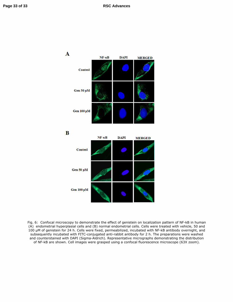
Fig. 6: Confocal microscopy to demonstrate the effect of genistein on localization pattern of NF-kB in human (A) endometrial hyperplasial cells and (B) normal endometrial cells. Cells were treated with vehicle, 50 and 100 µM of genistein for 24 h. Cells were fixed, permeabilized, incubated with NF-kB antibody overnight, and
subsequently incubated with FITC-conjugated anti-rabbit antibody for 2 h. The preparations were washed and counterstained with DAPI (Sigma-Aldrich). Representative micrographs demonstrating the distribution
of NF-kB are shown. Cell images were grasped using a confocal fluorescence microscope (63X zoom).
Page 33 of 33 RSC Advances

Fig. 7: (A) Western blot analysis to see the expression of p53 and p-Mdm2 and Mdm2 proteins in human endometrial hyperplasial cells. Cells were treated with the indicated concentrations of genistein for 48h, and
25 µg whole cell lysate in each lane was probed for the expression of different proteins using specific antibodies. β-actin was used as a control to correct for loading. Densitometric quantitation of protein
expression levels are shown as fold changes in the right panel. Results are expressed as mean± SE, n = 3. p values are a-p<0.001, b<0.01, c-p<0.05 and d-p>0.05 vs. control. (B) Phosphorylation status of PI3K, Akt
and Nf-κB expression as determined by western blot analysis. Human endometrial hyperplasial cells were treated with vehicle, 50 and 100 µM of genistein for 48 h. Twenty-five micrograms of whole cell lysate
protein in each lane was probed for the expression of p-PI3K(tyr485), PI3K, p-Akt (ser473), Akt, p-Nf-κB and Nf-κB using specific antibodies. β-actin was used as a control to correct for loading. Densitometric
quantitation of protein expression levels are shown as fold changes in right panel. Data are expressed as mean of three different experiments; mean ± S.E., p values are a-p<0.001, b<0.01, c-p<0.05 and d-
p>0.05 vs. control.
Page 34 of 33RSC Advances

Fig. 8: Schematic representation of the action of genistein on inhibition of EGFR signalling in endometrial hyperplasia cells. The genistein inhibits the EGF-EGFR interaction and leads to subsequent induction of
apoptosis, inhibition of cell proliferation and cell survival.
Page 35 of 33 RSC Advances