The Phycodnaviridae: The Story of How Tiny Giants Rule the World
43
University of Nebraska - Lincoln DigitalCommons@University of Nebraska - Lincoln Papers in Plant Pathology Plant Pathology Department 1-1-2009 e Phycodnaviridae: e Story of How Tiny Giants Rule the World W. H. Wilson Bigelow Laboratory for Ocean Sciences, [email protected] James L. Van Een University of Nebraska - Lincoln, [email protected] M. J. Allen Plymouth Marine Laboratory Follow this and additional works at: hp://digitalcommons.unl.edu/plantpathpapers Part of the Plant Pathology Commons is Article is brought to you for free and open access by the Plant Pathology Department at DigitalCommons@University of Nebraska - Lincoln. It has been accepted for inclusion in Papers in Plant Pathology by an authorized administrator of DigitalCommons@University of Nebraska - Lincoln. Wilson, W. H.; Van Een, James L.; and Allen, M. J., "e Phycodnaviridae: e Story of How Tiny Giants Rule the World" (2009). Papers in Plant Pathology. Paper 208. hp://digitalcommons.unl.edu/plantpathpapers/208
Transcript of The Phycodnaviridae: The Story of How Tiny Giants Rule the World
University of Nebraska - LincolnDigitalCommons@University of Nebraska - Lincoln
Papers in Plant Pathology Plant Pathology Department
1-1-2009
The Phycodnaviridae: The Story of How Tiny GiantsRule the WorldW. H. WilsonBigelow Laboratory for Ocean Sciences, [email protected]
James L. Van EttenUniversity of Nebraska - Lincoln, [email protected]
M. J. AllenPlymouth Marine Laboratory
Follow this and additional works at: http://digitalcommons.unl.edu/plantpathpapersPart of the Plant Pathology Commons
This Article is brought to you for free and open access by the Plant Pathology Department at DigitalCommons@University of Nebraska - Lincoln. Ithas been accepted for inclusion in Papers in Plant Pathology by an authorized administrator of DigitalCommons@University of Nebraska - Lincoln.
Wilson, W. H.; Van Etten, James L.; and Allen, M. J., "The Phycodnaviridae: The Story of How Tiny Giants Rule the World" (2009).Papers in Plant Pathology. Paper 208.http://digitalcommons.unl.edu/plantpathpapers/208
�
Published in James L. Van Etten (ed.) Lesser Known Large dsDNA Viruses. Current Topics in Microbiology and Immunology 328 (2009), pp. �–42. Copyright © Springer-Verlag Berlin Heidelberg 2009. Used by permission.
The Phycodnaviridae: The Story of How Tiny Giants Rule the WorldW. H. Wilson, J. L.Van Etten, M. J. Allen
Corresponding author — W. H. Wilson, Bigelow Laboratory for Ocean Sciences, �80 McKown Point, P.O. Box 475, West Boothbay Harbor, ME 04575-0475, USA; e-mail [email protected]
Contents
Introduction . . . . . . . . . . . . . . . . . . . . . . . . . . . . . . . . . . . . . . . . . . . .2Hosts . . . . . . . . . . . . . . . . . . . . . . . . . . . . . . . . . . . . . . . . . . . . . . . .5
Chlorella and Chloroviruses. . . . . . . . . . . . . . . . . . . . . . . . . . . . . . . . . .5Emiliania huxleyi and Coccolithoviruses . . . . . . . . . . . . . . . . . . . . . . . . . . .7Prasinophytes and Prasinoviruses . . . . . . . . . . . . . . . . . . . . . . . . . . . . . .7Prymnesiophytes and Prynmesioviruses. . . . . . . . . . . . . . . . . . . . . . . . . . .8Phaeophyceae and Phaeoviruses . . . . . . . . . . . . . . . . . . . . . . . . . . . . . . .9Raphidophytes and Raphidoviruses . . . . . . . . . . . . . . . . . . . . . . . . . . . . ��
Virion Morphology/Structure . . . . . . . . . . . . . . . . . . . . . . . . . . . . . . . . . ��Propagation Strategies. . . . . . . . . . . . . . . . . . . . . . . . . . . . . . . . . . . . . . �5Genome Structure and Properties . . . . . . . . . . . . . . . . . . . . . . . . . . . . . . . �8Coding Potential . . . . . . . . . . . . . . . . . . . . . . . . . . . . . . . . . . . . . . . . . 20
tRNAs, Introns, and Inteins . . . . . . . . . . . . . . . . . . . . . . . . . . . . . . . . . 20Characterized Gene Products . . . . . . . . . . . . . . . . . . . . . . . . . . . . . . . . 2�Unique Genes . . . . . . . . . . . . . . . . . . . . . . . . . . . . . . . . . . . . . . . . . 22
Core Genes and Phycodnaviridae Evolution . . . . . . . . . . . . . . . . . . . . . . . . . . 25Inter-genus Variation . . . . . . . . . . . . . . . . . . . . . . . . . . . . . . . . . . . . . 25Intra-genus Variation. . . . . . . . . . . . . . . . . . . . . . . . . . . . . . . . . . . . . 26
Summary . . . . . . . . . . . . . . . . . . . . . . . . . . . . . . . . . . . . . . . . . . . . . 3�References. . . . . . . . . . . . . . . . . . . . . . . . . . . . . . . . . . . . . . . . . . . . . 32
Abstract The family Phycodnaviridae encompasses a diverse and rapidly expanding col-lection of large icosahedral, dsDNA viruses that infect algae. These lytic and ly-sogenic viruses have genomes ranging from �60 to 560 kb. The family consists of six genera based initially on host range and supported by sequence compari-sons. The family is monophyletic with branches for each genus, but the phycod-naviruses have evolutionary roots that connect them with several other families of large DNA viruses, referred to as the nucleocytoplasmic large DNA viruses (NCLDV).The phycodnaviruses have diverse genome structures, some with
Wilson, Van EttEn, & allEn in Lesser Known Large dsdna Viruses (2009)2
large regions of noncoding sequence and others with regions of ssDNA. The genomes of members in three genera in the Phycodnaviridae have been se-quenced. The genome analyses have revealed more than �000 unique genes, with only �4 homologous genes in common among the three genera of phy-codnaviruses sequenced to date. Thus, their gene diversity far exceeds the number of so-called core genes. Not much is known about the replication of these viruses, but the consequences of these infections on phytoplankton have global affects, including influencing geochemical cycling and weather patterns.
Introduction
The illuminated region is only a small part of the 3.7-km mean depth of the ocean, yet it houses several of the great engines of planetary control (Tett 1990). Absorption of heat energy by the ocean and light energy by tiny float-ing marine plants, known as phytoplankton, are the engines that help reg-ulate many aspects of the global environment. Phytoplankton (microalgae) form the base of the marine food web and their photosynthetic activities pro-vide an important carbon sink that influences the global carbon cycle and even climate (Charlson et al. �987). Conservative estimates suggest there is somewhere between �00,000 to several million species of algae and that only approximately 40,000 have been identified. The Phycodnaviridae consists of a family of viruses that infect these globally important players. A literal trans-lation of Phycodnaviridae is, DNA viruses that infect algae. Given the num-ber of potential hosts, it is incredible so few phycodnaviruses have been iso-lated to date. With only approximately 150 formal identifications (Wilson et al. 2005b) and at least �00 others mentioned in the literature, it is certain that most phycodnaviruses, containing an almost infinite reservoir of genetic di-versity, remain to be discovered.
The Phycodnaviridae comprise a genetically diverse (Dunigan et al. 2006), yet morphologically similar, family of large icosahedral viruses that infect marine or freshwater eukaryotic algae with dsDNA genomes ranging from �60 to 560 kb (Van Etten et al. 2002). Members of the Phycodnaviridae are cur-rently grouped into six genera (named after the hosts they infect): Chlorovi-rus, Coccolithovirus, Prasinovirus, Prymnesiovirus, Phaeovirus, and Raphido-virus (Wilson et al. 2005b). Nomenclature anomalies have already crept into the system with the formation of the genus Coccolithovirus, a group of vi-ruses that infect Emiliania huxleyi (an alga species in the class Prymnesiophy-ceae), which logically should have been classified within the genus Prymne-siovirus. However, phylogenetic analysis of the DNA polymerase gene from these viruses indicated they belong to a new genus (Schroeder et al. 2002). It is likely the Phycodnaviridae will eventually be split into several subfami-lies and numerous genera as more isolates are characterized and the current nomenclature will probably become obsolete. Complete genome sequences have been obtained from representatives of the Chlorovirus, Coccolithovirus and Phaeovirus genera (Dunigan et al. 2006) and evolutionary analysis of their
PhycodnaViridae : HoW tiny Giants RulE tHE WoRld 3
genomes places them within a major, monophyletic assemblage of large eu-karyotic dsDNA viruses termed the Nucleo-Cytoplasmic Large DNA Viruses (NCLDVs) (Iyer et al. 200�, 2006; Allen et al. 2006c; Raoult et al. 2004). There are five families in the NCLDV clade that include Poxviridae, Iridoviridae, As-farviridae, Phycodnaviridae and Mimiviridae. Probably, the virus family Asco-viridae should also be included in this grouping because these viruses have clearly evolved from the Iridoviruses (see the chapter by V.G. Chinchar et al., this volume). The grouping of the NCLDVs is significant for two reasons: (i) as the name suggests, it implies a likely propagation mechanism for mem-bers of the Phycodnaviridae where replication is probably initiated in the nu-cleus and is completed in the cytoplasm; (ii) the NCLDVs are proposed to have an ancient evolutionary lineage (e.g., Villarreal and DeFilippis 2000;lyer et al. 2006; Raoult et al. 2004). Phylogeny of the NCLDVs constructed by cla-distic analysis indicates that the major families may have diverged prior to the divergence of the major eukaryotic lineages 2-3 billion years ago (Iyer et al. 2006;Raoult et al. 2004). The finding that there are only 14 genes in com-mon, from a pool of approximately �000 genes, between three genomes from different genera of the Phycodnaviridae (Allen et al. 2006c) supports the idea that the Chlorovirus, Coccolithovirus and Phaeovirus genera diverged a long time ago. As more Phycodnaviridae genomes are sequenced, there is likely to be an explosion of exciting gene discoveries with novel functions.
Reports of virus-like particles (VLPs) in at least 44 taxa of eukaryotic al-gae have appeared since the early �970s (Van Etten et al. �99�). These in-clude incidental observations of VLPs in electron micrographs, for example the first description of VLPs in E. huxleyi was reported in �974 (Manton and Leadbeater �974). Although these investigators did not publish an electron micrograph showing VLPs in E. huxleyi, they mentioned that VLPs in Chrys-ochromulina mantoniae (plates 65 and 66, Manton and Leadbeater �974) resem-bled those of VLPs commonly found in moribund or dead Coccolithus huxleyi cells (now referred to as E. huxleyi). Many images containing VLPs are prob-ably labeled miscellaneous and filed into obscurity in laboratories around the world (e.g.. Figure �). These old images could be a valuable resource to help identify new viruses and susceptible host strains. It was not until �979 that a phycodnavirus was even isolated; the virus infected the marine uni-cellular alga Micromonas pusilla (Mayer and Taylor �979). However, this re-port was largely ignored until the early �990s (Cottrell and Suttle �99�) when high concentrations of viruses in aquatic environments were being described (Bergh et al. 1989). Perhaps most significantly, in the early 1980s a group of viruses were characterized that infect freshwater unicellular, eukaryotic, ex-symbiotic chlorella- like green algae, called chloroviruses (Meints et al. �98�; Van Etten et al. �98�, �982, �983a). These reports were followed in the early 1990s by research into marine filamentous brown algal viruses (Muller et al. �990; Henry and Meints �992; Muller and Stache �992). Thereafter, use of ge-netic markers such as virus-encoded DNA polymerases, a core gene present in all NCLDVs (Iyer et al. 200�, 2006; Allen et al. 2006c), revealed that phy-coviruses are a diverse and ubiquitous component of aquatic environments (Chen and Suttle 1995; Chen et al. 1996; Short and Suttle 2002). The field of
Wilson, Van EttEn, & allEn in Lesser Known Large dsdna Viruses (2009)4
phycodnavirology is now well established and expanding rapidly; indeed there have been four international meetings dedicated to the subject (Bergen, Norway �998; Galway, Ireland 2000; Hiroshima, Japan 2002; and Amster-dam, Netherlands 2005). Previous algal virus reviews have focused primar-ily on the chloroviruses (Van Etten and Meints �999; Van Etten et al. 2002, 2003; Kang et al. 2005; Dunigan et al. 2006; Yamada et al. 2006) because of the extensive research on these viruses. The ecological aspects of marine phyto-plankton viruses were reviewed recently by Brussaard (2004). The current chapter will provide a broad overview of the Phycodnaviridae, attempting to describe the novelty and incredible genetic diversity of this ancient group of viruses. Starting with a brief introduction to the algal hosts these viruses in-fect, the review will then cover virus propagation strategies, genome struc-ture, an analysis of known and novel genes, and finish with a discussion on core genes and their implication in Phycodnaviridae evolution.
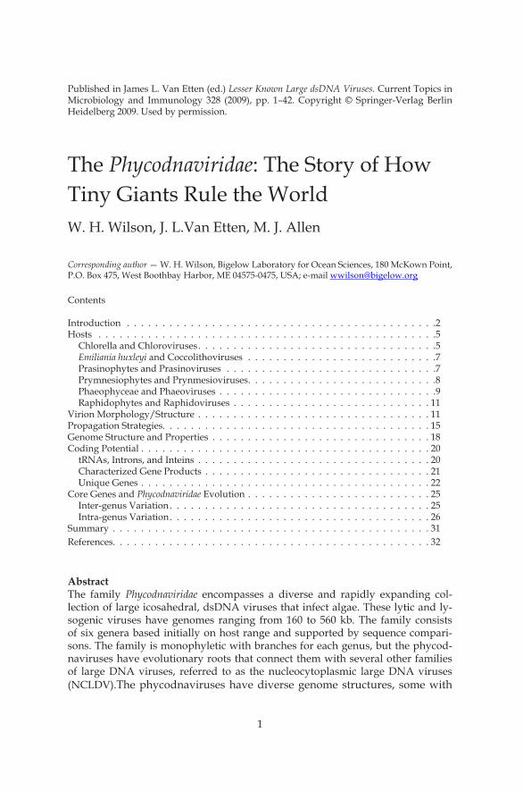
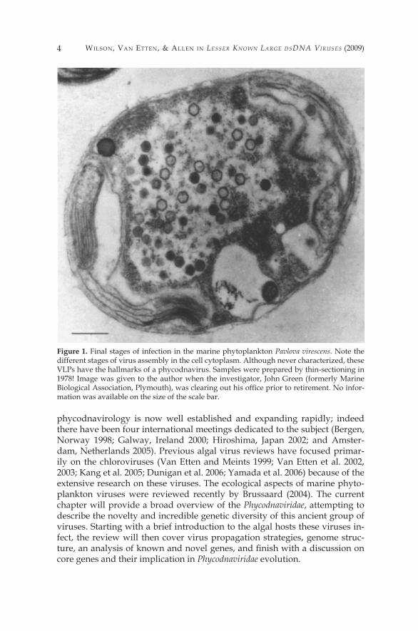
Figure 1. Final stages of infection in the marine phytoplankton Pavlova virescens. Note the different stages of virus assembly in the cell cytoplasm. Although never characterized, these VLPs have the hallmarks of a phycodnavirus. Samples were prepared by thin-sectioning in �978! Image was given to the author when the investigator, John Green (formerly Marine Biological Association, Plymouth), was clearing out his office prior to retirement. No infor-mation was available on the size of the scale bar.
PhycodnaViridae : HoW tiny Giants RulE tHE WoRld 5
Hosts
Eukaryotic algae are a group of oxygen-evolving, photosynthetic organ-isms that include seaweeds (macroalgae) and a large diverse group of mi-croorganisms generically referred to as microalgae. Photosynthetic prokary-otes such as pico-cyanobacteria (e.g. Synechococcus or Prochlorococcus) are not included in this group, however, they are infected by bacteriophage-like vi-ruses (Suttle 2000a, 2000b; Clokie and Mann 2006). Eukaryotic algae range in size from the smallest known eukaryote, Ostreococcus, at approximately � [µm in diameter (Derelle et al. 2006; Palenik et al. 2007), through numerous chain-forming and colonial species that are visible to the naked eye to the large kelp (seaweed) forests in coastal regions. Algae consist of at least five distinct evolutionary lineages (plants, cercozoa, alveolates, heterokonts and discicristates) (Baldauf 2003) and they are ubiquitous in marine, freshwater and terrestrial habitats. The number of algal species (mostly microalgae) has been estimated to be as high as several million; hence their overall diversity is probably enormous. It is likely that viruses infect all of these species; fur-thermore, not all of these viruses will be assigned to the Phycodnaviridae fam-ily. Indeed, other types of viruses that infect algae are being discovered and characterized (e.g., ssRNA, dsRNA, and ssDNA containing viruses, Tai et al. 2003; Brussaard et al. 2004a; Nagasaki et al. 2004). Thus, algal virology is in its infancy.
Chlorella and Chloroviruses
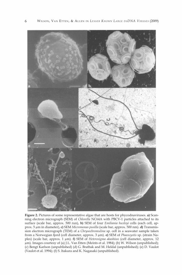
The genus Chlorella consists of small, unicellular, non-motile, asexual green algae with a global distribution (Figure 2A) (e.g., Shihra and Krauss �965). They have a simple developmental cycle and reproduce by mitotic di-vision. Vegetative cells increase in size and, depending on the species and en-vironmental conditions, divide into two, four, eight or more progeny, which are released by rupture or enzymatic digestion of the parental walls. Chlo-rella species are usually free living, although many species have symbiotic relationships with organisms from different classes in the animal kingdom including Rhizopoda, Ciliata, Hydrozoa and Turbellaria (Reisser �992). To date, the only described chloroviruses infect symbiotic chlorella, often referred to as zoochlorellae, such as those associated with the protozoan Paramecium bur-saria, the coelenterate Hydrozoa viridis and the heliozoon Ancanthocystis turfa-cea (Kawakami and Kawakami �978; Meints et al. �98�; Van Etten et al. �982; Bubeck and Pfitzner 2005). Fortunately, many of these chlorella strains can be grown in the laboratory, independent of their symbiotic partner, including P. bursaria-associated chlorella isolates NC64A and Pbi. The chloroviruses can be produced in large quantities and assayed by plaque formation. Chlorovi-ruses are ubiquitous in nature and have been isolated from freshwater col-lected throughout the world (Yamata et al. 2006). Typically, the virus liters in native waters are �-�00 plaque-forming units (PFU) per ml, but liters as
Wilson, Van EttEn, & allEn in Lesser Known Large dsdna Viruses (2009)6
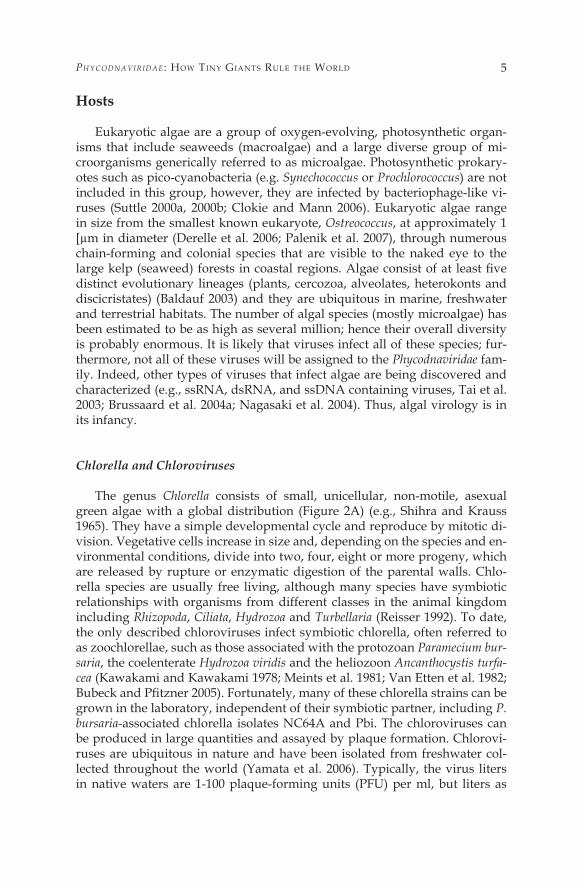
Figure 2. Pictures of some representative algae that are hosts for phycodnaviruses. a) Scan-ning electron micrograph (SEM) of Chlorella NC64A with PBCV-� particles attached to its surface (scale bar, approx. 500 run), b) SEM of four Emiliania huxleyi cells (each cell, ap-prox. 5 µm in diameter), c) SEM Micromonas pusilla (scale bar, approx. 500 nm). d) Transmis-sion electron micrograph (TEM) of a Chrysochromulina sp. cell in a seawater sample taken from a Norwegian fjord (cell diameter, approx. 3 µm). e) SEM of Phaeocystis sp. (strain Na-ples) (scale bar, approx. � µm). f) SEM of Heterosigma akashiwo (cell diameter, approx. �2 µm). Images courtesy of (a) J.L. Van Etten (Meints et al. �984); (b) W. Wilson (unpublished); (c) Bengt Karlson (unpublished) (d) G. Bratbak and M. Heldal (unpublished); (e) D. Vaulot (Vaulot et al. �994); (f) S. Itakura and K. Nagasaki (unpublished).
PhycodnaViridae : HoW tiny Giants RulE tHE WoRld 7
high as 100,000 PFU/ml of native water have been obtained. Titers fluctuate with the seasons, with the highest liters occurring in the spring. Chlorovirus PBCV-�, which infects Chlorella NC64A, is currently the best-studied phycod-navirus (Van Etten 2003; Yamada et al. 2006).
Emiliania huxleyi and Coccolithoviruses
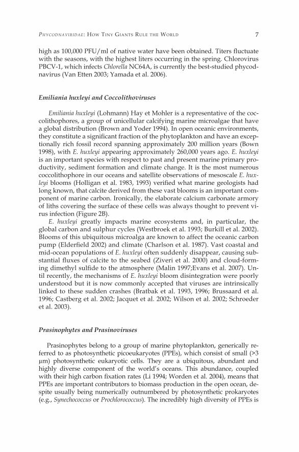
Emiliania huxleyi (Lohmann) Hay et Mohler is a representative of the coc-colithophores, a group of unicellular calcifying marine microalgae that have a global distribution (Brown and Yoder �994). In open oceanic environments, they constitute a significant fraction of the phytoplankton and have an excep-tionally rich fossil record spanning approximately 200 million years (Bown �998), with E. huxleyi appearing approximately 260,000 years ago. E. huxleyi is an important species with respect to past and present marine primary pro-ductivity, sediment formation and climate change. It is the most numerous coccolithophore in our oceans and satellite observations of mesoscale E. hux-leyi blooms (Holligan et al. 1983, 1993) verified what marine geologists had long known, that calcite derived from these vast blooms is an important com-ponent of marine carbon. Ironically, the elaborate calcium carbonate armory of liths covering the surface of these cells was always thought to prevent vi-rus infection (Figure 2B).
E. huxleyi greatly impacts marine ecosystems and, in particular, the global carbon and sulphur cycles (Westbroek et al. �993; Burkill et al. 2002). Blooms of this ubiquitous microalga are known to affect the oceanic carbon pump (Elderfield 2002) and climate (Charlson et al. 1987). Vast coastal and mid-ocean populations of E. huxleyi often suddenly disappear, causing sub-stantial fluxes of calcite to the seabed (Ziveri et al. 2000) and cloud-form-ing dimethyl sulfide to the atmosphere (Malin 1997;Evans et al. 2007). Un-til recently, the mechanisms of E. huxleyi bloom disintegration were poorly understood but it is now commonly accepted that viruses are intrinsically linked to these sudden crashes (Bratbak et al. �993, �996; Brussaard et al. �996; Castberg et al. 2002; Jacquet et al. 2002; Wilson et al. 2002; Schroeder et al. 2003).
Prasinophytes and Prasinoviruses
Prasinophytes belong to a group of marine phytoplankton, generically re-ferred to as photosynthetic picoeukaryotes (PPEs), which consist of small (>3 µm) photosynthetic eukaryotic cells. They are a ubiquitous, abundant and highly diverse component of the world’s oceans. This abundance, coupled with their high carbon fixation rates (Li 1994; Worden et al. 2004), means that PPEs are important contributors to biomass production in the open ocean, de-spite usually being numerically outnumbered by photosynthetic prokaryotes (e.g., Synechococcus or Prochlorococcus). The incredibly high diversity of PPEs is
Wilson, Van EttEn, & allEn in Lesser Known Large dsdna Viruses (2009)8
only now being appreciated (Diez et al. 200�; Moon-van der Staay et al. 200�; Zeidner et al. 2003; Fuller et al. 2006; Worden 2006). Among the many classes included in the PPEs, members of the class Prasinophyceae are ubiquitous in clone libraries collected from oceanic samples and they often dominate the PPE community (Not et al. 2004; Worden 2006). These include members in the gen-era Ostreococcus, the smallest know eukaryote (Chrétiennot-Dinet et al. �995), Bathycoccus (Eikrem and Throndsen �990), and Micromonas (Figure 2C), a major component in many oceanic and coastal regions (Not et al. 2005).
Despite the obvious ubiquity and abundance of these algae, relatively lit-tle research has been conducted on prasinoviruses and only a few viruses have been described. This is perhaps ironic, given that the first characterized virus assigned to the Phycodnaviridae was a prasinovirus that infects Micromonas pu-silla (Mayer and Taylor �979). M pusilla-specific viruses are the only prasinovi-ruses that have been described. These viruses are easy to isolate, simply add-ing a small volume of filtered coastal seawater to an exponentially growing culture of M pusilla usually results in virus-induced lysis. Research has focused largely on their ecological role and their genetic diversity (Cottrell and Sut-tle 1991,1995b; Sahlsten 1998; Sahlsten and Karlson 1998; Zingone et al. 1999, 2006). M pusilla viruses can lyse up to 25% of the daily Micromonas population (Evans et al. 2003); however, the high growth rates of the hosts, coupled with the high diversity of both host (Worden 2006) and virus (Chen et al. �996), al-lows them to propagate in a stable co-existence (Cottrell and Suttle �995a) as compared to the bloom-bust scenario observed with Coccolithovirus infection of E. huxleyi blooms (Bratbak et al. �993; Jacquet et al. 2002; Wilson et al. 2002).
Except for a report of a transient Ostreococcus bloom off the eastern coast of the United States, whose rapid decline was partially attributed to viruses (O’Kelly et al. 2003), no other reports exist of viruses infecting prasinophytes. However, the authors are aware of an unpublished investigation on the isola-tion of prasinoviruses that infect the smallest known eukaryotic cell, Ostreo-coccus tauri. These dsDNA viruses are readily isolated by plaque assay and are morphologically similar to the M pusilla viruses (K. Weynberg, N. Grims-ley, H. Moreau, personal communication). Of the six Phycodnaviridae genera, the prasinoviruses and viruses of PPEs are the least represented in the litera-ture despite their global and ecological importance.
Prymnesiophytes and Prymnesioviruses
Algae assigned to the class Prymnesiophyceae, from the division Hap-tophyta (and generally referred to as haptophytes), occur predominantly in marine environments. These algae have a global distribution and they are of-ten associated with large-scale microalgal blooms. The principle characteris-tic of the haptophytes is the haptonema, a filiform organelle or Hagella whose length ranges from barely detectable (Green and Pienaar �977) to many times that of the mother cell (Figure 2D) (Parke et al. �959). The presence of two-layered microfibrillar scales on the cell surface is also a characteristic and it is traditionally considered an important taxonomic character at the species
PhycodnaViridae : HoW tiny Giants RulE tHE WoRld 9
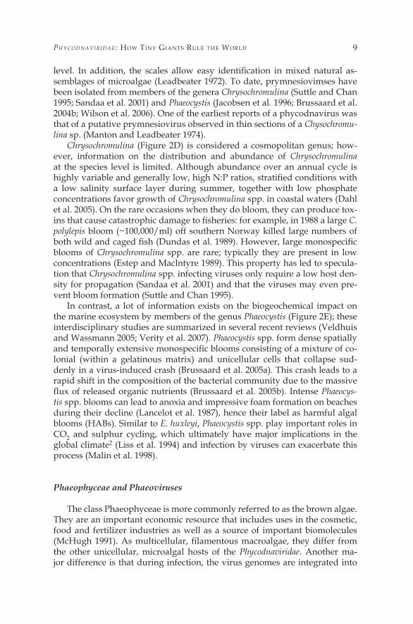
level. In addition, the scales allow easy identification in mixed natural as-semblages of microalgae (Leadbeater �972). To date, prymnesiovimses have been isolated from members of the genera Chrysochromulina (Suttle and Chan �995; Sandaa et al. 200�) and Phaeocystis (Jacobsen et al. �996; Brussaard et al. 2004b; Wilson et al. 2006). One of the earliest reports of a phycodnavirus was that of a putative prymnesiovirus observed in thin sections of a Chysochromu-lina sp. (Manton and Leadbeater �974).
Chrysochromulina (Figure 2D) is considered a cosmopolitan genus; how-ever, information on the distribution and abundance of Chrysochromulina at the species level is limited. Although abundance over an annual cycle is highly variable and generally low, high N:P ratios, stratified conditions with a low salinity surface layer during summer, together with low phosphate concentrations favor growth of Chrysochromulina spp. in coastal waters (Dahl et al. 2005). On the rare occasions when they do bloom, they can produce tox-ins that cause catastrophic damage to fisheries: for example, in 1988 a large C. polylepis bloom (~�00,000/ml) off southern Norway killed large numbers of both wild and caged fish (Dundas et al. 1989). However, large monospecific blooms of Chrysochromulina spp. are rare; typically they are present in low concentrations (Estep and Maclntyre �989). This property has led to specula-tion that Chrysochromulina spp. infecting viruses only require a low host den-sity for propagation (Sandaa et al. 200�) and that the viruses may even pre-vent bloom formation (Suttle and Chan �995).
In contrast, a lot of information exists on the biogeochemical impact on the marine ecosystem by members of the genus Phaeocystis (Figure 2E); these interdisciplinary studies are summarized in several recent reviews (Veldhuis and Wassmann 2005; Verity et al. 2007). Phaeocystis spp. form dense spatially and temporally extensive monospecific blooms consisting of a mixture of co-lonial (within a gelatinous matrix) and unicellular cells that collapse sud-denly in a virus-induced crash (Brussaard et al. 2005a). This crash leads to a rapid shift in the composition of the bacterial community due to the massive flux of released organic nutrients (Brussaard et al. 2005b). Intense Phaeocys-tis spp. blooms can lead to anoxia and impressive foam formation on beaches during their decline (Lancelot et al. �987), hence their label as harmful algal blooms (HABs). Similar to E. huxleyi, Phaeocystis spp. play important roles in CO2 and sulphur cycling, which ultimately have major implications in the global climate2 (Liss et al. �994) and infection by viruses can exacerbate this process (Malin et al. �998).
Phaeophyceae and Phaeoviruses
The class Phaeophyceae is more commonly referred to as the brown algae. They are an important economic resource that includes uses in the cosmetic, food and fertilizer industries as well as a source of important biomolecules (McHugh 1991). As multicellular, filamentous macroalgae, they differ from the other unicellular, microalgal hosts of the Phycodnaviridae. Another ma-jor difference is that during infection, the virus genomes are integrated into
Wilson, Van EttEn, & allEn in Lesser Known Large dsdna Viruses (2009)�0
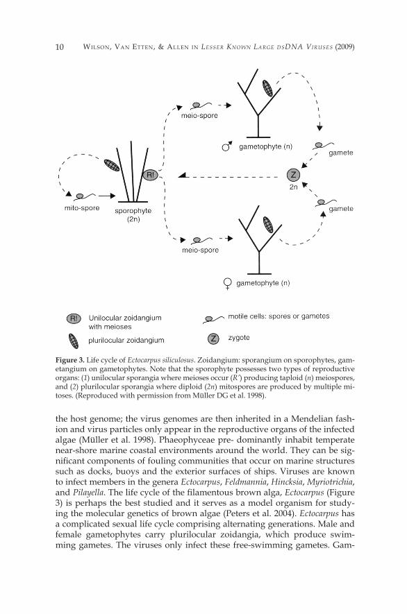
the host genome; the virus genomes are then inherited in a Mendelian fash-ion and virus particles only appear in the reproductive organs of the infected algae (Müller et al. �998). Phaeophyceae pre- dominantly inhabit temperate near-shore marine coastal environments around the world. They can be sig-nificant components of fouling communities that occur on marine structures such as docks, buoys and the exterior surfaces of ships. Viruses are known to infect members in the genera Ectocarpus, Feldmannia, Hincksia, Myriotrichia, and Pilayella. The life cycle of the filamentous brown alga, Ectocarpus (Figure 3) is perhaps the best studied and it serves as a model organism for study-ing the molecular genetics of brown algae (Peters et al. 2004). Ectocarpus has a complicated sexual life cycle comprising alternating generations. Male and female gametophytes carry plurilocular zoidangia, which produce swim-ming gametes. The viruses only infect these free-swimming gametes. Gam-
Figure 3. Life cycle of Ectocarpus siliculosus. Zoidangium: sporangium on sporophytes, gam-etangium on gametophytes. Note that the sporophyte possesses two types of reproductive organs: (1) unilocular sporangia where meioses occur (R’) producing taploid (n) meiospores, and (2) plurilocular sporangia where diploid (2n) mitospores are produced by multiple mi-toses. (Reproduced with permission from Müller DG et al. �998).

PhycodnaViridae : HoW tiny Giants RulE tHE WoRld ��
ete fusion results in zygotes that develop into diploid sporophytes. The spo-rophytes produce unilocular sporangia where meiosis leads to the formation of haploid spores. These spores go on to produce the next gametophyte gen-eration. The sporophytes can also develop plurilocular sporangia by mitotic cell division. The diploid spores can therefore repeat the diploid sporophyte phase (Müller et al. �998). Virus particles are only formed in gametangia or sporangia (zoidangia) cells of the host (Müller �996). Since viruses displace the normal reproductive organs, the algae are rendered sterile; however, this does not affect their vegetative growth (Müller et al. �990).
Raphidophytes and Raphidoviruses
Microalgae in the class Raphidophyceae are important bloom-forming species found in coastal and subartic regions of the world’s oceans, although freshwater species also exist (Figueroa and Rengefors 2006). Members are often associated with toxic red tides (Khan et al. 1997) and subsequent fish kills, particularly in the aquaculture industry (Hiroishi et al. 2005). Classical identification of the Raphidophytes relies on morphological differences, but it is extremely difficult to distinguish some of the species due to a pleomor-phology that changes shapes and sizes in various environmental conditions (Khan et al. �995). Consequently, efforts to develop rapid and sensitive poly-merase chain reaction (PCR)-based diagnostic techniques are ongoing (Kai et al. 2006). To date, the only viruses reported in this genus infect the red tide forming species Heterosigma akashiwo (Figure 2F); the viruses are referred to as HaV (Heterosigma akashiwo virus) (Nagasaki and Yamaguchi �997, �998a; Nagasaki et al. �999b). H. akashiwo is a single species belonging to the genus Heterosigma (Throndsen �996) and, although not usually associated with hu-man illness, blooms of H. akashiwo have caused massive fish kills, typically of caged fish such as salmon and yellowtail (Honjo 1993; Khan et al. 1997). The susceptibility of H. akashiwo to HaV differs among clonal strains in the labo-ratory (Nagasaki and Yamaguchi �998b; Nagasaki et al. �999a; Tarutani et al. 2006). Marine field surveys and cross-reactivity tests between H. akashiwo host strains and HaV clones suggest that this strain-dependent infection plays an important role in determining clonal composition and effectively maintain-ing intraspecies diversity in natural H. akashiwo populations (Tarutani et al. 2000;Tomaru et al. 2004). This diversity probably contributes to the success of H. akashiwo as a ubiquitous and problematic bloom former in coastal regions.
Virion Morphology/Structure
Despite the wide host range displayed by the Phycodnaviridae, all members have similar structural morphology; this is consistent (independent of genomic information) with a common ancestry. Few have been studied in great detail, but the overlying pattern suggests a general conservation of structure. The life-
Wilson, Van EttEn, & allEn in Lesser Known Large dsdna Viruses (2009)�2
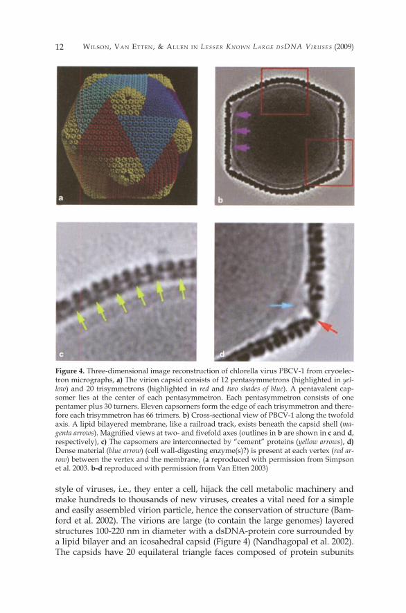
style of viruses, i.e., they enter a cell, hijack the cell metabolic machinery and make hundreds to thousands of new viruses, creates a vital need for a simple and easily assembled virion particle, hence the conservation of structure (Bam-ford et al. 2002). The virions are large (to contain the large genomes) layered structures �00-220 nm in diameter with a dsDNA-protein core surrounded by a lipid bilayer and an icosahedral capsid (Figure 4) (Nandhagopal et al. 2002). The capsids have 20 equilateral triangle faces composed of protein subunits
Figure 4. Three-dimensional image reconstruction of chlorella virus PBCV-� from cryoelec-tron micrographs, a) The virion capsid consists of �2 pentasymmetrons (highlighted in yel-low) and 20 trisymmetrons (highlighted in red and two shades of blue). A pentavalent cap-somer lies at the center of each pentasymmetron. Each pentasymmetron consists of one pentamer plus 30 turners. Eleven capsorners form the edge of each trisymmetron and there-fore each trisymmetron has 66 trimers. b) Cross-sectional view of PBCV-� along the twofold axis. A lipid bilayered membrane, like a railroad track, exists beneath the capsid shell (ma-genta arrows). Magnified views at two- and fivefold axes (outlines in b are shown in c and d, respectively), c) The capsomers are interconnected by “cement” proteins (yellow arrows), d) Dense material (blue arrow) (cell wall-digesting enzyme(s)?) is present at each vertex (red ar-row) between the vertex and the membrane, (a reproduced with permission from Simpson et al. 2003. b-d reproduced with permission from Van Etten 2003)
PhycodnaViridae : HoW tiny Giants RulE tHE WoRld �3
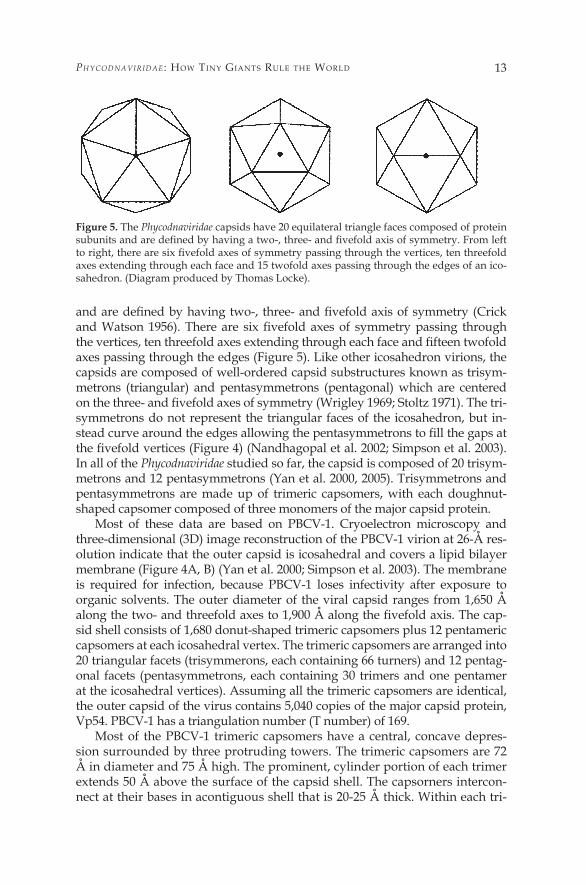
and are defined by having two-, three- and fivefold axis of symmetry (Crick and Watson 1956). There are six fivefold axes of symmetry passing through the vertices, ten threefold axes extending through each face and fifteen twofold axes passing through the edges (Figure 5). Like other icosahedron virions, the capsids are composed of well-ordered capsid substructures known as trisym-metrons (triangular) and pentasymmetrons (pentagonal) which are centered on the three- and fivefold axes of symmetry (Wrigley 1969; Stoltz 1971). The tri- symmetrons do not represent the triangular faces of the icosahedron, but in-stead curve around the edges allowing the pentasymmetrons to fill the gaps at the fivefold vertices (Figure 4) (Nandhagopal et al. 2002; Simpson et al. 2003). In all of the Phycodnaviridae studied so far, the capsid is composed of 20 trisym-metrons and �2 pentasymmetrons (Yan et al. 2000, 2005). Trisymmetrons and pentasymmetrons are made up of trimeric capsomers, with each doughnut-shaped capsomer composed of three monomers of the major capsid protein.
Most of these data are based on PBCV-�. Cryoelectron microscopy and three-dimensional (3D) image reconstruction of the PBCV-� virion at 26-Å res-olution indicate that the outer capsid is icosahedral and covers a lipid bilayer membrane (Figure 4A, B) (Yan et al. 2000; Simpson et al. 2003). The membrane is required for infection, because PBCV-� loses infectivity after exposure to organic solvents. The outer diameter of the viral capsid ranges from �,650 Å along the two- and threefold axes to 1,900 Å along the fivefold axis. The cap-sid shell consists of �,680 donut-shaped trimeric capsomers plus �2 pentameric capsomers at each icosahedral vertex. The trimeric capsomers are arranged into 20 triangular facets (trisymmerons, each containing 66 turners) and �2 pentag-onal facets (pentasymmetrons, each containing 30 trimers and one pentamer at the icosahedral vertices). Assuming all the trimeric capsomers are identical, the outer capsid of the virus contains 5,040 copies of the major capsid protein, Vp54. PBCV-� has a triangulation number (T number) of �69.
Most of the PBCV-� trimeric capsomers have a central, concave depres-sion surrounded by three protruding towers. The trimeric capsomers are 72 Å in diameter and 75 Å high. The prominent, cylinder portion of each trimer extends 50 Å above the surface of the capsid shell. The capsorners intercon-nect at their bases in acontiguous shell that is 20-25 Å thick. Within each tri-
Figure 5. The Phycodnaviridae capsids have 20 equilateral triangle faces composed of protein subunits and are defined by having a two-, three- and fivefold axis of symmetry. From left to right, there are six fivefold axes of symmetry passing through the vertices, ten threefold axes extending through each face and �5 twofold axes passing through the edges of an ico-sahedron. (Diagram produced by Thomas Locke).
Wilson, Van EttEn, & allEn in Lesser Known Large dsdna Viruses (2009)�4
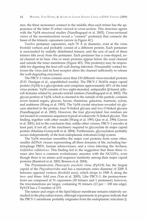
mer, the three monomers connect in the middle; thus each trimer has the ap-pearance of the letter H when viewed in cross-section. This structure agrees with the Vp54 structural studies (Nandhagopal et al. 2002). Cross-sectional views of the reconstructions reveal a “cement” protein(s) that connects the bases of the trimeric capsomers (arrow in Figure 4C).
Twelve pentamer capsomers, each 70 Å in diameter, exist at the virus fivefold vertices and probably consist of a different protein. Each pentamer is surrounded by radially distributed trimers, and the axis of each of these trimers tilts away from the pentamer. Each pentamer has a cone-shaped, ax-ial channel at its base. One or more proteins appear below the axial channel and outside the inner membrane (Figure 4D). This protein(s) may be respon-sible for digesting the host cell wall during infection. Presumably contact be-tween the virus and its host receptor alters the channel sufficiently to release the wall-degrading enzyme(s).
The PBCV-� virion contains more than ��0 different virus-encoded proteins (D.D. Dunigan et al., unpublished results). The PBCV-� 54-kDa major capsid protein (Vp54) is a glycoprotein and comprises approximately 40% of the total virus protein. Vp54 consists of two eight-stranded, antiparallel (β-barrel, jelly-roll domains related by pseudo-sixfold rotation (Nandhagopal et al. 2002). The glycan portion of Vp54, which is oriented to the outside of the particle, contains seven neutral sugars, glucose, fucose, rhamnose, galactose, mannose, xylose, and arabinose (Wang et al. �993). The Vp54 crystal structure revealed six gly-cans attached to the protein, four N-linked glycans and two O-linked glycans (Nandhagopal et al. 2002). However, the four glycosylated Asn residues are not located in consensus sequences typical of eukaryotic N-linked glycans. This finding, together with other results (Wang et al. 1993; Que et al. 1994; Graves et al. 200�), led to the conclusion that, unlike other viruses, PBCV-� encodes at least part, if not all, of the machinery required to glycosylate its major capsid protein (Markine-Goriaynoff et al. 2004). Furthermore, glycosylation probably occurs independently of the host endoplasmic reticulum-Golgi system.
The Vp54 structure resembles the major coat proteins from some other, smaller dsDNA viruses representing all three domains of life, including bac-teriophage PRD�, human adenoviruses, and a virus infecting the Archaea, Sulfolobus solfataricus. This finding led to the suggestion that these three vi-ruses also have a common evolutionary ancestor with the NCLDVs, even though there is no amino acid sequence similarity among their major capsid proteins (Bamford et al. 2002; Benson et al. 2004).
The Prymnesiovirus Phaeocystis pouchetii virus (PpVOl) has the largest capsid of the Phycodnaviridae and has a maximum outer diameter of 2200 Å between opposed vertices (fivefold axes), which drops to 1900 Å along the two- and three- fold axes (Yan et al. 2005). Like PBCV-�, the pentasymme-trons are composed of 3� capsomers (30 trimers and � pentamer); however, the trisymmetrons are larger, containing 9� trimers (�3 per ~ �00 nm edge). PpVOl has a T number of 2�9.
The nature and origin of the lipid bilayer membrane remains relatively un-studied in the phycodnaviruses, although experiments in progress indicate that the PBCV-� membrane probably originates from the endoplasmic reticulum (J.
PhycodnaViridae : HoW tiny Giants RulE tHE WoRld �5
Heuser et al., unpublished results). A lipid profile of virus EhV-86 is very sim-ilar to that of its host E. huxleyi, suggesting the direct requisition of host mem-brane lipids during virion assembly (T. Dunn, personal communication).
Over the last decade we have been increasingly aware that the Phycod-naviridae is a highly diverse family whose members share very little else in common other than the nature of their genomic material and their morphol-ogy. The following sections discuss this diversity with regards to lifestyle, genomic structure, coding and noncoding potential and infection strategy.
Propagation Strategies
Members of the Phycodnaviridae have a variety of lifestyles. The replica-tion strategies of three of these viruses will be briefly described in this sec-tion, PBCV-� representing the chloroviruses, EsV-� representing the phaeovi-ruses and EhV-86 representing the coccolithoviruses.
PBCV-1 initiates infection by attaching rapidly, specifically and irreversibly to the chlorella cell wall (Meints et al. �984, �988), probably at a unique virus vertex (Onimatsu et al. 2006); attachment is immediately followed by degrada-tion of the host wall at the point of contact by a virus-packaged enzyme(s) (Fig-ure 6). The chloroviruses encode several proteins involved in polysaccharide degradation that may be involved in degrading the cell wall (see Sect. 6.3.�). Following host cell wall degradation, the viral internal membrane presumably fuses with the host membrane, resulting in entry of the viral DNA and virion-associated proteins into the cell, leaving an empty virus capsid attached to the cell wall. This process triggers a rapid depolarization of the host membrane (probably triggered by a virus encoded potassium channel located in the virus internal membrane) (Frohns et al. 2006) and the rapid release of potassium ions from the cell (Neupartl et al. 2008); this depolarization may function to prevent infection by a second virus (Mehmel et al. 2003;Frohns et al. 2006). The rapid loss of potassium ions and associated water fluxes from the host also lowers its turgor pressure and this lowering is postulated to aid ejection of DNA from the virions into the host (Neuparti et al. 2008).
Circumstantial evidence indicates that the viral DNA and probably DNA-associated proteins quickly move to the nucleus where early transcription is detected within 5-�0 min p.i. (Schuster et al. �986). Unlike EhV-86 early gene expression (Allen et al. 2006a), the PBCV-� early expressed genes are scat-tered throughout the virus genome. Shortly after infection, host chromosomal DNA begins to be degraded (Agarkova et al. 2006), presumably to aid in in-hibiting host transcription and to provide the recycling of nucleotides for viral DNA replication. In this immediate-early phase of infection, the host is repro-grammed to transcribe viral RNAs. Very little is known as to how this occurs, but chromatin remodeling may be involved. PBCV-� encodes a ��9 amino acid SET domain containing protein (referred to as vSET) that di-methylates Lys27 in histone 3 (Manzur et al. 2003). vSET is packaged in the PBCV-� virion and accumulating evidence indicates that vSET is involved in repression of host transcription following PBCV-� infection (S. Mujtaba et al., unpublished data).
Wilson, Van EttEn, & allEn in Lesser Known Large dsdna Viruses (2009)�6
Viral DNA replication begins 60-90 min after infection (Van Etten et al. �984) and is followed by transcription of late genes (Schuster et al. �986). Ul-trastructural studies of PBCV-�-infected chlorella suggest that the nuclear membrane remains intact, at least during the early stages of virus replication (Meints et al. �986). However, a functional host nucleus is not required for virus replication since PBCV-� can replicate, albeit poorly and with a small burst size, in UV-irradiated cells (Van Etten et al. �986). Approximately 2-3 h postinfection (p.i.), assembly of virus capsids begins in localized regions in the cytoplasm, called virus assembly centers, which become prominent at 3-4 h p.i. (Meints et al. 1986). By 5- 6 h p.i., the cytoplasm becomes filled with
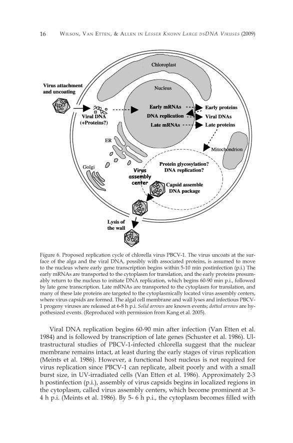
Figure 6. Proposed replication cycle of chlorella virus PBCV-�. The virus uncoats at the sur-face of the alga and the viral DNA, possibly with associated proteins, is assumed to move to the nucleus where early gene transcription begins within 5-�0 min postinfection (p.i.) The early mRNAs are transported to the cytoplasm for translation, and the early proteins presum-ably return to the nucleus to initiate DNA replication, which begins 60-90 min p.i., followed by late gene transcription. Late mRNAs are transported to the cytoplasm for translation, and many of these late proteins are targeted to the cytoplasmically located virus assembly centers, where virus capsids are formed. The algal cell membrane and wall lyses and infectious PBCV-� progeny viruses are released at 6-8 h p.i. Solid arrows are known events; dotted arrows are hy-pothesized events. (Reproduced with permission from Kang et al. 2005).
PhycodnaViridae : HoW tiny Giants RulE tHE WoRld �7
infectious progeny virus particles and by 6-8 h p.i., localized lysis of the host cell releases progeny (Van Etten et al. �983b). Approximately �,000 particles are released per cell, of which approximately 30% are infectious.
A different lifestyle occurs in the phaeoviruses: in contrast to the lytic life-style of the chloroviruses, the phaeoviruses are lysogenic (Müller et al. �998; Van Etten et al. 2002). Viruses EsV-� and EfasV-� only infect free-swimming, wall-less gametes of their filamentous hosts Ectocarpus siliculousus and Ec-tocarpus fasciculatus, respectively. Following virion attachment, the viral ge-nomic DNA and associated protein material is released into the cytoplasm and then immediately moves to the nucleus within 5 min p.i. (Maier et al. 2002). The genomic material is then integrated into the host genome (pre-sumably using a virally encoded integrase like the one in virus EsV-�; R.H. Meints et al., unpublished data). The viral genome is transmitted mitotically to all the cells in the developing host thallus (Braütigam et al. �995; Delar-oque et al. �999; Müller et al. �99�). The virus genome remains latent in the vegetative filamentous cells until the formation of algal reproductive cells, ei-ther sporangia or gametangia, in the mature host. This differentiation triggers virus DNA replication in the sporangia or gametangia, followed by break-down of the nuclear membrane, virion assembly and release of infectious vi-rions from the cells (Wolf et al. �998, 2000; Müller et al. �998).
The coccolithoviruses have a different propagation strategy than either the lytic PBCV-� or the latent EsV-� systems, which may be similar to the life-style of the ancestral phycodnavirus (Allen et al. 2006c). The host alga E. hux-leyi is covered with a calcium carbonate shell (Figure 2B) that would appear to create a physical barrier to virus adsorption. However, despite this barrier, virus adsorption is rapid and appears to be intrinsically linked to the cell cy-cle of the host (W.H. Wilson, unpublished data). Whereas both PBCV-� and EsV-� are completely dependent on the host transcriptional machinery, the coccolithoviruses are unique among the phycodnaviruses in that they have several RNA polymerase encoding genes (Wilson et al. 2005a). These genes suggest a viral replication strategy that could be either entirely or partially independent of the host nucleus. Viral transcription begins immediately after infection, but it is limited to a distinct and localized �00-kb region in the virus genome; this region contains a unique promoter element (Allen et al. 2006a, 2006d). The only genes transcribed during the Isth after infection have this el-ement. None of these genes match anything in the databases and are referred to as ORFans (Fischer and Eisenberg �999). Intriguingly, proteomic analysis of EhV-86 virions did not detect any transcriptional machinery packaged in mature virions, therefore a host nuclear RNA polymerase(s) is presumably re-sponsible for this early transcription (M.J. Allen, unpublished data). Between � and 2 h p.i., a second transcriptional phase begins with gene expression occurring from the remainder of the genome. Since viral RNA polymerase components are expressed in this second phase, viral replication may no lon-ger be nuclear dependent and transcription may move to the cytoplasm. Un-like the other phycodnavirus systems, where nascent virions accumulate in the cytoplasm prior to release by cell lysis, the coccolithovirus virions are re-leased gradually in a controlled fashion. One possibility is that the virus uti-
Wilson, Van EttEn, & allEn in Lesser Known Large dsdna Viruses (2009)�8
lizes the ready-made lith export pathway to maintain this controlled and or-derly release. It has also been suggested that the coccolithoviruses use the virally encoded sphingolipid metabolism pathway to manipulate the sphin-golipids in the host cell wall or membrane, possibly to prevent programmed cell death and to prolong the length of infection (Han et al. 2006).
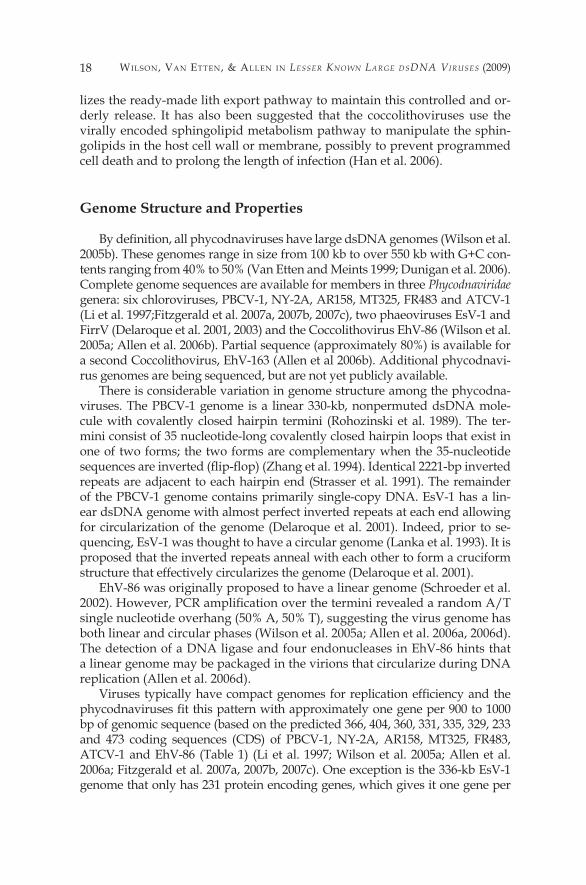
Genome Structure and Properties
By definition, all phycodnaviruses have large dsDNA genomes (Wilson et al. 2005b). These genomes range in size from �00 kb to over 550 kb with G+C con-tents ranging from 40% to 50% (Van Etten and Meints �999; Dunigan et al. 2006). Complete genome sequences are available for members in three Phycodnaviridae genera: six chloroviruses, PBCV-�, NY-2A, AR�58, MT325, FR483 and ATCV-� (Li et al. �997;Fitzgerald et al. 2007a, 2007b, 2007c), two phaeoviruses EsV-� and FirrV (Delaroque et al. 200�, 2003) and the Coccolithovirus EhV-86 (Wilson et al. 2005a; Allen et al. 2006b). Partial sequence (approximately 80%) is available for a second Coccolithovirus, EhV-�63 (Allen et al 2006b). Additional phycodnavi-rus genomes are being sequenced, but are not yet publicly available.
There is considerable variation in genome structure among the phycodna-viruses. The PBCV-� genome is a linear 330-kb, nonpermuted dsDNA mole-cule with covalently closed hairpin termini (Rohozinski et al. �989). The ter-mini consist of 35 nucleotide-long covalently closed hairpin loops that exist in one of two forms; the two forms are complementary when the 35-nucleotide sequences are inverted (flip-flop) (Zhang et al. 1994). Identical 2221-bp inverted repeats are adjacent to each hairpin end (Strasser et al. �99�). The remainder of the PBCV-� genome contains primarily single-copy DNA. EsV-� has a lin-ear dsDNA genome with almost perfect inverted repeats at each end allowing for circularization of the genome (Delaroque et al. 200�). Indeed, prior to se-quencing, EsV-� was thought to have a circular genome (Lanka et al. �993). It is proposed that the inverted repeats anneal with each other to form a cruciform structure that effectively circularizes the genome (Delaroque et al. 200�).
EhV-86 was originally proposed to have a linear genome (Schroeder et al. 2002). However, PCR amplification over the termini revealed a random A/T single nucleotide overhang (50% A, 50% T), suggesting the virus genome has both linear and circular phases (Wilson et al. 2005a; Allen et al. 2006a, 2006d). The detection of a DNA ligase and four endonucleases in EhV-86 hints that a linear genome may be packaged in the virions that circularize during DNA replication (Allen et al. 2006d).
Viruses typically have compact genomes for replication efficiency and the phycodnaviruses fit this pattern with approximately one gene per 900 to 1000 bp of genomic sequence (based on the predicted 366, 404, 360, 33�, 335, 329, 233 and 473 coding sequences (CDS) of PBCV-�, NY-2A, AR�58, MT325, FR483, ATCV-� and EhV-86 (Table �) (Li et al. �997; Wilson et al. 2005a; Allen et al. 2006a; Fitzgerald et al. 2007a, 2007b, 2007c). One exception is the 336-kb EsV-� genome that only has 23� protein encoding genes, which gives it one gene per
PhycodnaViridae : HoW tiny Giants RulE tHE WoRld �9
roughly�450 bp. The 366 PBCV-� protein-encoding genes are evenly distributed on both strands and, with one exception, intergenic space is minimal. In fact, 275 ORFs are separated by fewer than �00 nucleoddes. The exception is a �788-nucleotide sequence near the middle of the genome. This DNA region, which contains many stop codons in all reading frames, encodes �� tRNA genes.
However, some repetitive DNA occurs in the PBCV-�, EsV-� and EhV-86 genomes. Both EsV-� and PBCV-� contain approximately 2-kb inverted re-peats adjacent to the terminal ends (Strasser et al. �99�; Delaroque et al. 200�). In addition to the terminal repeats, tandem repeats are located throughout the EsV-� genome and comprise approximately �2% of the total genome size (Delaroque et al. 200�). A similar proportion of the Feldmannia sp. virus (FsV) genome also consists of repetitive DNA (Lee et al. �995). Intriguingly, EhV-86 has three repeat families (none of which are located at the ends of the ge-nome); one family is postulated to act as an origin of replication (adding cre-dence to the circular mode of replication model), another family is postulated to contain immediate early promoter elements and the last family has a large repetitive proline rich domain that may bind calcium (Allen et al. 2006d). The repetitive regions in these Phycodnaviridae genomes, while hindering se-quencing projects, may play a role in recombination between viruses that al-lows genetic information to be exchanged with themselves and with their hosts (Allen et al. 2006b, 2006c).
Limited sequence information is currently available for the phycodnavi-rus hosts, although this situation is changing. The Chlorella NC64A and E. huxleyi genomes are currently being sequenced by the Joint Genome Insti-tute and drafts of the two genomes will soon be available. There is evidence
Table 1 Genome data of sequenced phycodnaviruses
Genome %G+CGenus Virus Host size (bp) content ORFs tRNAs Nts/gene
Chlorovirus PBCV-� Chlorella 330,743a 40.0 366 �� 904 NC64A NY-2A Chlorella 368,683a 40.7 404 7 9�3 NC64A AR�58 Chlorella 344,690a 40.8 360 6 957 NC64A MT325 Chlorella Pbi 3�4,335a 45.3 33� �0 950 FR483 Chlorella Pbi 32�,240a 44.6 335 9 959 ATCV-� Chlorella SAG 288,047a 49.4 329 �� 875 3.83Phaeovirus EsV-� Ectocarpus 335,593 5�.7 23� 0 �453 siliculousus FirrV-� Feldmannia �9�,667b �56 0 �229 irregularisCoccolithovirus EhV-86 Emiliania 407,339 40.2 472 5 863 huxleyi
a Does not include the nts at the hairpin ends b Is not one continuous contig
Wilson, Van EttEn, & allEn in Lesser Known Large dsdna Viruses (2009)20
that both E. huxleyi and EhV-86share a putative phosphate permease gene, the large degree of difference between the genes suggesting this transfer was not recent; however, the best and only match of the virus was from the host (Wilson et al. 2005a). Host E. huxleyi also appears to contain numerous G+C-rich repetitive elements (B. Read, personal communication) similar to the re-petitive elements in EhV-86 (Allen et al. 2006d); therefore the virus may have acquired these elements from its host.
The origin of the chlorella virus genes is a mystery. Microarrays that con-tain all 366 PBCV-� genes are available and only two of the PBCV-� genes hy-bridize with host chlorella DNA (G. Yanai-Balser et al., unpublished data). Furthermore, the G+C content of PBCV-� and Chlorella NC64A differ signif-icantly, 40% versus 67% (Van Etten et al. �985). Therefore, it seems unlikely that PBCV-� recently acquired its genes from this host.
Some of the phycodnavirus genomes have methylated bases. For exam-ple, genomes from 37 chlorella viruses contain 5-methylcytosine (5mC) in amounts varying from 0.�2% to 47.5% of the total cytosine. In addition, 24 of the 37 viral DNAs contain N6-methyladenine (6 mA) in amounts varying from �.5% to 37% of the total adenine (Van Etten et al. �99�). The methyl-ated bases occur in specific DNA sequences; thus it is not surprising that the chlorella viruses encode multiple 5mC and 6 mA DNA methyltransferases. However, it was a surprise to discover that approximately 25% of the virus-encoded DNA methyltransferases have a companion DNA site-specific (re-striction) endonuclease, some which have unique specificities (e.g., Xia et al. �986, �987; Nelson et al. �998; Van Etten et al. �99�). Some of the virus en-coded site-specific endonucleases are unusual in that they only cleave one strand of dsDNA, for example virus NYs-1 nickase cleaves /CC sites (Xia et al. 1988; Chan et al. 2004) and NY-2A nickase cleaves R/AG sites (Zhang et al. 1998; Chan et al. 2006). Thus, the virus-infected chlorellae are the first non-prokaryotic source of DNA site-specific endonucleases.
The phaeovirus EsV-� genome also has low levels of methylated bases (�% and 3% of the total cytosines and adenines, respectively, are methylated) (Lanka et al. �993; Delaroque et al. 200�). However, the EsV-� DNA methyl-transferases do not have companion site-specific endonucleases.
Coding Potential
tRNAs f IntronSf and Interns
In general, CDSs tend to be evenly distributed on both strands with min-imal intergenic spaces. Exceptions to this rule include a �788-nucleotide se-quence in the middle of the PBCV-� genome that contains a polycistronic gene encoding �� tRNA genes (co-transcribed as a large precursor and then processed to mature tRNAs) (Dunigan et al. 2006). All the sequenced chloro-viruses have a similar pattern, although the nature of the tRNAs and their ge-nomic locations vary among the viruses (ATCV-�,�� tRNAs, two duplicated;

PhycodnaViridae : HoW tiny Giants RulE tHE WoRld 2�
MT325, ten tRNAs, one duplicated; FR483, nine tRNAs, one duplicated; NY-2A, seven tRNAs, one duplicated; AR�58, six tRNAs, one duplicated) (Fitzgerald et al. 2007a, 2007b, 2007c). The coccolithovirus, EhV-86, encodes five tRNAs, four of which are clustered together (Wilson etal. 2005a). A sec-ond EhV strain (EhV-99Bl) contains a sixth tRNA and also encodes a putative isoleucyi tRNA synthetase to complement the Ilu-tRNA (A. Lanzen, personal communication). In contrast, the phaeovirus EsV-� does not have any tRNA encoding genes (Delaroque et al. 200�).
Codon usage in viral proteins is often correlated with the abundance and nature of virus-encoded tRNAs (Kropinski and Sibbald �999). However, it is interesting that there is such a large variation in the number and type of tRNAs contained within these phycodnavirus genomes. Not only does the number of tRNAs in closely related viruses (often infecting the same host) differ, but the nature of the tRNAs also vary. For example, only three tRNAs are conserved among the six chloroviruses that have been sequenced (Fitzgerald et al. 2007c).
In addition to a �3-nt intron in a PBCV-� Tyr-tRNA gene, PBCV-� con-tains two other types of introns: a self-splicing intron in a transcription factor TFIIS-like gene and a spliceosomal-processed intron in its DNA polymerase gene. Chloroviruses MT325 and FR483 lack both of these introns (Fitzgerald et al. 2007a). A spliceosomal-processed intron was also identified in a pyrim-idine dimer-specific glycosylase gene in 15 chloroviruses including NY-2A and AR�58 (Sun et al. 2000; Fitzgerald et al. 2007b). An intron is also present at an identical location in the ATCV-1 pyrimidine dimer-specific glycosylase gene, but this intron has little sequence identity to those from viruses NY-2A and AR�58 (Fitzgerald et al. 2007c). Like PBCV, NY-2A also contains an in-tron in its DNA polymerase and TFIIS-like genes; in addition, NY-2A also contains another self-splicing intron in a gene of unknown function. The coc-colithovirus EhV-86 contains two self-splicing introns; one in the DNA poly-merase gene and one in a nucleic acid independent nucleoside triphospha-tase gene (Wilson et al. 2005a).
In addition to introns, some phycodnavirus proteins contain inteins. In-teins are the protein equivalent of introns: following translation they are auto-catalytically spliced to create a mature protein product. Recently, two inteins were identified in the NY-2A genome (the first to be identified in the chloro-viruses): one 337 amino acid intein is in a ribonucleotide reductase large sub-unit protein and the other is in a putative helicase protein (384 amino acids) (Fitzgerald et al. 2007b). However, inteins may be more common in a class of phycodnaviruses that infect the raphido-phytes (Nagasaki et al. 2005). The raphidoviruses contain a 232 amino acid intein in their DNA polymerase pro-tein, which is distantly related to an intein in the Mimivirus. This intein was present in all ten raphidoviruses examined.
Characterized Gene Products
Many PBCV-�-encoded enzymes are either the smallest or among the smallest proteins of their family. The small sizes and the finding that many
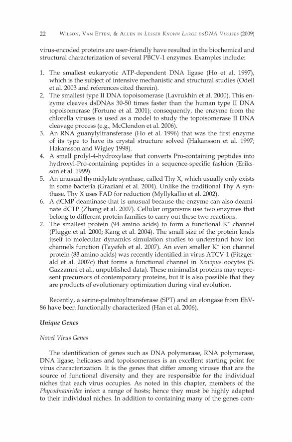
Wilson, Van EttEn, & allEn in Lesser Known Large dsdna Viruses (2009)22
virus-encoded proteins are user-friendly have resulted in the biochemical and structural characterization of several PBCV-� enzymes. Examples include:
�. The smallest eukaryotic ATP-dependent DNA ligase (Ho et al. �997), which is the subject of intensive mechanistic and structural studies (Odell et al. 2003 and references cited therein).
2. The smallest type II DNA topoisomerase (Lavrukhin et al. 2000). This en-zyme cleaves dsDNAs 30-50 times faster than the human type II DNA topoisomerase (Fortune et al. 200�); consequently, the enzyme from the chlorella viruses is used as a model to study the topoisomerase II DNA cleavage process (e.g., McClendon et al. 2006).
3. An RNA guanylyltransferase (Ho et al. 1996) that was the first enzyme of its type to have its crystal structure solved (Hakansson et al. �997; Hakansson and Wigley �998).
4. A small prolyl-4-hydroxylase that converts Pro-containing peptides into hydroxyl-Pro-containing peptides in a sequence-specific fashion (Eriks-son et al. �999).
5. An unusual thymidylate synthase, called Thy X, which usually only exists in some bacteria (Graziani et al. 2004). Unlike the traditional Thy A syn-thase. Thy X uses FAD for reduction (Myllykallio et al. 2002).
6. A dCMP deaminase that is unusual because the enzyme can also deami-nate dCTP (Zhang et al. 2007). Cellular organisms use two enzymes that belong to different protein families to carry out these two reactions.
7. The smallest protein (94 amino acids) to form a functional K+ channel (Plugge et al. 2000; Kang et al. 2004). The small size of the protein lends itself to molecular dynamics simulation studies to understand how ion channels function (Tayefeh et al. 2007). An even smaller K+ ion channel protein (83 amino acids) was recently identified in virus ATCV-1 (Fitzger-ald et al. 2007c) that forms a functional channel in Xenopus oocytes (S. Gazzamni et al., unpublished data). These minimalist proteins may repre-sent precursors of contemporary proteins, but it is also possible that they are products of evolutionary optimization during viral evolution.
Recently, a serine-palmitoyltransferase (SPT) and an elongase from EhV-86 have been functionally characterized (Han et al. 2006).
Unique Genes
Novel Virus Genes
The identification of genes such as DNA polymerase, RNA polymerase, DNA ligase, helicases and topoisomerases is an excellent starting point for virus characterization. It is the genes that differ among viruses that are the source of functional diversity and they are responsible for the individual niches that each virus occupies. As noted in this chapter, members of the Phycodnaviridae infect a range of hosts; hence they must be highly adapted to their individual niches. In addition to containing many of the genes com-
PhycodnaViridae : HoW tiny Giants RulE tHE WoRld 23
monly found in other DNA viruses, sequencing phycodnavirus genomes have revealed genes not previously found in viruses. These gene products may provide clues as to their niche adaptation.
An example of this is the putative sphingolipid biosynthesis pathway present in EhV-86 (Wilson et al. 2005a). Sphingolipid synthesis leads to the formation of ceramide, which can affect apoptosis; thus this is an intrigu-ing choice of a pathway for a virus to manipulate. Sphingolipids are en-riched in Upid rafts and may play a crucial role during viral release (Han et al. 2006). EhV-86 contains a cluster of genes encoding sphingolipid metabolic enzymes similar to those in other organisms, from yeast to mammals (Wil-son et al. 2005a). A particularly surprising example is the viral gene, ehv050, which encodes both subunits of the eukaryotic serine palmitoyltransferases (SPT) (Han et al. 2006). SPT is the enzyme required for the first and rate-lim-iting step in the pathway and catalyses the condensation of serine with pal-mitoyl-CoA (Hanada and Nishijima 2003). The novel fusion protein encoded by EhV-86 is not only active, it has a preference for myristoyl-CoA (Han et al. 2006). The relevance of this is unclear, but it is likely to be fundamental to the infection strategy of the virus.
The chloroviruses are also unusual because they often encode enzymes involved in sugar metabolism. Three PBCV-� encoded enzymes glutamine:fructose-6-phosphate amindotransferase, UDP-glucose dehydrogenase (UDP- GlcDH), and hyaluronan synthase (HAS) are involved in the synthesis of hyaluronan (hyaluronic acid), a linear polysaccharide composed of alter-nating β-1,4-glucuronic acid and β-1,3-N-acetylglucosamine residues (DeAn-gelis et al. �997; Landstein et al. �998). All three genes are transcribed early in PBCV-� infection and hyaluronan accumulates on the external surface of the infected chlorella cells (Graves et al. �999).
However, not all chloroviruses have a has gene. Surprisingly, many chlo-roviruses that lack a has gene have a gene encoding a functional chitin syn-thase (CHS). Furthermore, cells infected with these viruses produce chitin fibers on their external surface (Kawasaki et al. 2002). Chitin, an insoluble lin-ear homopolymer of P-l,2-linked N-acetyl-glucosamine residues, is a com-mon component of insect exoskeletons, shells of crustaceans, and fungal cell walls (Muzzarelli et al. �986). Some chloroviruses contain both has and chs genes and form both hyaluronan and chitin on the surface of their infected cells (Kawasaki et al. 2002; Yamada and Kawasaki 2005). Finally, a few chlo-roviruses appear to lack both genes because no extracellular polysaccharides are formed on the host surface of cells infected with these viruses (Graves et al. �999). The fact that many chloroviruses encode enzymes involved in these energy-demanding extracellular polysaccharide biosynthetic pathways sug-gests that the polysaccharides are important in the virus life cycles. However, at present this function is unknown.
Two PBCV-�-encoded enzymes, GDP-D-mannose dehydratase (GMD) and fucose synthase, comprise a highly conserved, three-step pathway that converts GDP-D- mannose to GDP-L-fucose (Tonetti et al. 2003). However, the PBCV-� GMD not only has the predicted dehydratase activity, it has a re-ductase activity that produces GDP-D-rhamnose. Both fucose and rhamnose
Wilson, Van EttEn, & allEn in Lesser Known Large dsdna Viruses (2009)24
are present in the glycans attached to PBCV-� major capsid protein. Interest-ingly, another chlorella virus, ATCV-�, encodes a GMD that only has the de-hydratase activity (Fruscione et al. 2008).
The chlorella viruses degrade the host cell wall during infection and also during virus release. Therefore, it is not surprising that the viruses encode several enzymes that may be involved in these processes (see Yamata et al. 2006 for details). For example, PBCV-� encodes two chitinases, a chitosanase, a β-l-3 glucanase and an enzyme that cleaves polymers of either β-or -l,4-linked glucuronic acids (Sugimoto et al. 2004). Enzymes packaged in the vi-rion and involved in virus infection are typically expressed late in the infec-tion cycle and one of the chitinases and the chitosanase meet this requirement and are packaged in the PBCV-� virion. However, the composition of the host chlorella cell walls and the enzymatic digestion of the walls during infection are still under active investigation.
PBCV-� encodes four enzymes involved in polyamine biosynthesis: omithine decarboxylase (ODC), homospermidine synthase, agmatine imi-nohydrolase, and N-carbamoylputrescine amidohydrolase (Baumann et al. 2007; Kaiser et al. �999;Morehead et al. 2002). ODC catalyzes the decarbox-ylation of omithine to putrescine, which is the first and the rate-limiting en-zymatic step in the polyamine biosynthetic pathway. The PBCV-�-encoded ODC is the smallest characterized ODC (Morehead et al. 2002). The PBCV-� enzyme is also interesting because it decarboxylates arginine better than omithine (Shah et al. 2004). These four enzymes form a complete polyamine biosynthetic pathway that allows the formation of homospermidine through the precursors arginine, agmatine, N-car-bamoylputrescine and putrescine (Baumann et al. 2007). However, the relevance of this pathway to virus rep-lication is unclear. Although polyamines are present in large amounts in the capsids of some viruses, that is not true for PBCV-� (Kaiser et al. �999).
As more phycodnavirus sequences become available, we predict that the number of unique and interesting gene pathways found in these viruses will increase. Each of the viruses sequenced to date appears to have its own quirks; this pattern is likely to be repeated as more diverse phycodnaviruses are studied. For example, EsV-� contains at least six putative hybrid histi-dine kinases (two-component systems which form part of a stimulus-respon-sive transduction pathway) that are wide-spread in archaea and bacteria, but have not previously been found in viruses (Delaroque et al. 2000, 200�). The relevance of these genes to EsV-� infection is unknown, but is likely to be fundamental to successful infection.
Database ORFan Genes (Unknown Genes)
The wealth of genetic potential contained within the phycodnaviruses is per-haps best represented not by the unique proteins encoded by the viruses, but by the genes encoding proteins that do not have any matches in GenBank. For ex-ample, 86% of the EhV-86 gene products lack GeneBank homologs (Wilson et al. 2005a). These genes/CDSs/ORFs are commonly referred to as database OR-Fans (Fischer and Eisenberg 1999). Indeed, this is exemplified by a 100-kb re-

PhycodnaViridae : HoW tiny Giants RulE tHE WoRld 25
gion in EhV-86, which contains approximately �50 genes, none of which have a known function (Allen et al. 2006c). Half of these genes are associated with a unique promoter element that drives their expression, but only during the ear-liest stages of infection (Allen et al. 2006a, 2006d). Thus these genes encode pro-teins that undoubtedly play a crucial and integral role during virus infection.
Core Genes and Phycodnaviridae Evolution
Inter-genus Variation
There is accumulating evidence that the large dsDNA viruses referred to as nuclear- cytoplasmic large DNA viruses (NCLDV) (Phycodnaviridae, Pox-viridae, Mimiviridae, Asfariviridae, and Iridoviridae) shared a common ancestor (Iyer et al. 200�, 2006; Raoult et al. 2004). Genome-wide bioinformatic sur-veys have suggested an ancestral virus genome composed of at least 50 genes that have subsequently been retained or discarded in a distinctive evolution-ary traceable pattern in these highly divergent virus families (Iyer et al. 200�). Nine genes are present in all NCLDV members (known as group I genes), and 22 additional genes are present in at least three of the five families (group II and III genes). Lineage-specific gene loss and gain within NCLDV families obviously has led to the highly diverse properties of present day viruses.
High evolutionary rates, horizontal gene transfers and nonorthologous gene displacements make accurate phylogenetic resolution difficult over great periods of time, especially when using single-gene phylogenetic trees (Filee et al. 2003). Consequently, studies on whole genomes are becoming more popular when constructing evolutionary relationships between virus families (lyer et al. 200�; Allen et al. 2006c). In a comparison of PBCV-�, EsV-� and EhV-86, only �4 genes are common in all three viruses, indicating a large amount of inter-genus genomic variation between Phycodnaviridae genera (Allen et al. 2006c). A phylogenetic tree was constructed by concatenating six of the common gene products; the results indicate that the phycodnaviruses form a distinct clade from the other large DNA viruses, yet the tree fails to provide adequate resolution of the genera within the Phycodnaviridae (Figure 7). However, the presence of genes encoding six RNA polymerase subunits in the EhV-86 genome (many of which are found in the other NCLDV virus families) clearly separates EhV-86 from PBCV-1 and EsV-1. This finding sug-gests that the ancestral Phycodnaviridae lineage diverged with one branch giv-ing rise to EhV-86 and the second branch giving rise to the PBCV-� and EsV-� lineages. The change in lifestyle represented by this loss of RNA polymerase function (i.e., from nuclear independence to nuclear dependent transcription) probably contributes to the high diversity among present day genera in the Phycodnaviridae. Since ancestral NCLDVs contained RNA polymerase func-tion, it is likely that of all the phycodnaviruses sequenced so far, EhV-86 rep-resents the virus with the lifestyle most similar to the ancestral virus (Allen et al. 2006c). As more phycodnavirus genomic sequences become available, we may be able to elucidate the complex history of phycodnavirus evolution.
Wilson, Van EttEn, & allEn in Lesser Known Large dsdna Viruses (2009)26
Intra-genus Variation
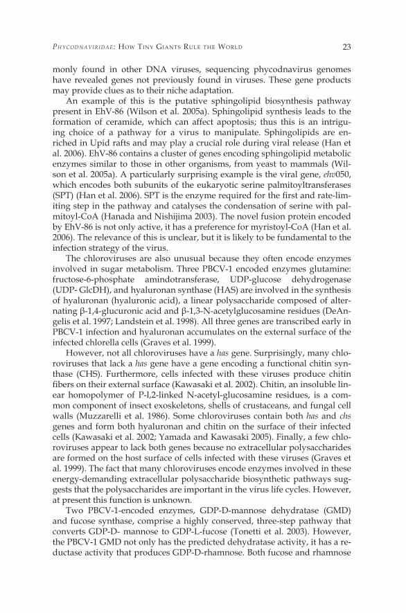
With little sequence and genomic data available from the majority of the phycodnaviruses identified so far, it is difficult to assess intra-genus varia-tion. Currently, sequencing efforts have focused on members in just three families: the chloroviruses, phaeoviruses and coccolithoviruses. Therefore, we are just beginning to appreciate the genetic diversity that exists among these genera. Initial results indicate that the huge degree of inter-genus varia-tion can, at least, be matched by a large intra-genus variation.
Figure 7. Phylogenetic inference tree based on a distance matrix algorithm between concate-nated conserved domains from A�8-like helicase, D6R-like helicase, A32-like ATPase, DNA polymerase, thio-oxidoreductase from members of the NCLDV group (Neighbor, in PHY-LIP version 3.6b). Numbers at nodes indicate bootstrap values retrieved from �00 replicates for both the neighbor-joining and parsimony analyses. The bar depicts one base substitu-tion per ten amino acids. (Diagram adapted from Allen et al. 2006d). Viruses included are African swine fever virus (ASFV), Amsacta moorei entomopoxvirus (AMEV), Melanoplus san-guinipes entomopoxvirus (MSEV), bovine papular stomatitis virus (BPSV), fowlpox virus (FWPV), sheeppox virus (SPPX), swinepox virus (SWPX), vaccinia virus (VACV), Mollus-cum contagiosum virus (MOCV), myxoma virus (MYXV), Yaba monkey tumor virus (YMTV), Paramecium bursaria chlorella virus � (PBCV-1), Ectocarpus siliculosus virus � (ESV-1), Emili-ania huxleyi virus 86 (EhV-86), frog virus 3 (FV3), invertebrate iridescent virus 6 (IIV6), Re-gina ranavirus (RRV), lymphocystis disease virus � (LCDV) and mimivirus.
PhycodnaViridae : HoW tiny Giants RulE tHE WoRld 27
Chloroviruses
The six sequenced chlorella viruses provide the best opportunity to study intra-genus variation within the Phycodnaviridae. The chlorella vi-ruses are grouped by the host they infect: viruses PBCV-�, NY-2A and AR�58 infect Chlorella NC64A (Fitzgerald et al. 2007b), MT325 and FR483 infect Chlorella Pbi (Fitzgerald et al. 2007a), and ATCV-� infects Chlorella SAG 3.83 (Fitzgerald et al. 2007c). Approximately 80% of the genes are found in all six chloroviruses, suggesting that the majority of the genes are essential for successful viral infection of chlorella. As expected, viruses infecting the same host are most similar; the average protein amino acid identity between PBCV-� and NY-2A or AR�58 homologs is 73%, with 87% identity between NY-2A and AR�58 homologs. PBCV-� and MT325 or FR483 homologs have 50% amino acid identity; 86% identity exists be-tween MT325 and FR483. PBCV-� and ATCV-� have 49% amino acid iden-tity between homologs.
The amino acid differences between homologous proteins can be ex-ploited to aid in understanding how certain proteins function. For example, electrophysiological experiments have been conducted with the 94 amino acid PBCV-� encoded potassium ion channel protein (called Kcv) in Xen-opus oocytes. Kcv-like proteins were cloned and sequenced from 40 addi-tional chlorella viruses; �6 amino acid substitutions occurred in 94 of the amino acids, producing six new Kcv-like proteins that formed functional potassium selective channels in Xenopus oocytes. However, the biophysi-cal properties of some of the new Kcv channels differed from PBCV-� Kcv. The amino acid changes, together with the different electrophysiological properties, observed in the six Kcv-like channels were used to guide site-directed amino acid substitutions, either singularly or in combination, to identify key residues that conferred specific properties to Kcv (Kang et al. 2004; Gazzarrini et al. 2004).
Each of the six sequenced chloroviruses contains genes that encode unique proteins. For example, ATCV-� is unique among these viruses in con-taining genes encoding dTDP-D-glucose 4,6 dehydratase, ribonucleotide-tri-phosphate reductase, and mucin-desulphatating sulphatase; MT325 encodes a functional aquaglyceroporin (Gazzarrini et al. 2006), FR483 an alkyi sul-phatase and a functional potassiumion transporter (M. Kang et al., unpub-lished data); NY-2A an ubiquitin; AR�58 a calcium transporting ATPase; and a functional Cu/Zn superoxide dismutase (M. Kang et al., unpublished data) is unique to PBCV-�.
Using the gene order of PBCV-� as the model, there is a high degree of gene colinearity between the three viruses, PBCV-�, NY-2A and AR�58 that infect Chlorella NC64A. Unlike the co-linearity between the three NC64A vi-ruses, PBCV-� has only slight co-linearity with the two viruses, MT325 and FR483 that infect Chlorella Pbi and virus ATCV-� that infects Chlorella SAG 3.83 (L.A. Fitzgerald et al., unpublished data).
Wilson, Van EttEn, & allEn in Lesser Known Large dsdna Viruses (2009)28
Not all PBCV-� genes are required for virus replication in the laboratory. For example, four spontaneously derived PBCV-� mutants were isolated that contain 27- to 37-kb deletions at the left end of the 330-kb genome (Landstein et al. �995). Two of these mutants have deletions beginning at nucleotide co-ordinates 4.9 or �6 kb and ending at 42 kb. In total, these two deleted regions, which probably resulted from recombination, encode 28 putative proteins. The other two mutants, which probably arose from nonhomologous recombina-tion, lack the entire left terminal 37-kb of the PBCV-� genome, including the 2.2-kb terminal inverted-repeat region. The deleted left terminus was replaced by the transposition of an inverted 7.7- or �8.5-kb copy from the right end of the PBCV-� genome. These regions encode 26 single-copy ORFs, of which 23 are common to those deleted in the first two mutant viruses. Taken together, approximately 40 kb of single-copy DNA encoding 3� ORFs at the left end of the genome, or �2% of the PBCV-� genome, is unnecessary for PBCV-� replica-tion in the laboratory. However, replication of the PBCV-� deletion mutants is attenuated; i.e., their burst sizes are about half of wild-type virus.
The deletion mutants also indicate that the size of the inverted terminal re-peats in PBCV-1 can vary. This conclusion is consistent with the finding that the size and sequence of the inverted repeat region is not conserved among chlorella viruses (Strasser et al. �99�; Yamada and Higashiyama �993). This lack of conservation is somewhat surprising because one predicts that the DNA ter-mini might be essential for either virus DNA replication and/or DNA packag-ing. These results also indicate that the virus DNA packaging machinery tol-erates significant differences in genome size; for example the largest deletion in PBCV-� creates a genome of approximately 302 kb, whereas chlorella virus NY-2A has a genome of roughly 370 kb. Similar large deletions were reported in the left terminus of the chlorella virus CVK� genome (Songsri et al. �995).
Large insertions also occur in the chlorella viruses. Comparison of PBCV-� and CVK2 genomes revealed that an approximately �5 kb region in the PBCV-� left terminal region is absent in the CVK2 genome. However, CVK2 contains a 22.2-kb insert in this region that contains five gene copies of a ho-molog of PBCV-� glyco-protein Vp260; this 22.2-kb sequence is absent in the PBCV-� genome (Chuchird et al. 2002; Nishida et al. �999).
The sizes and locations of the deletions and transpositions in the chlorella viruses resemble poxviruses (Turner and Moyer �990) and ASFV (Blasco et al. �989) deletion mutants. Like PBCV-�, poxviruses and ASFV genomes have inverted terminal repeats and covalently closed hairpin ends. Models to ex-plain the generation of deletions and deletion/transpositions in the poxvirus genomes (Shchelkunov and Totmenin �995; Turner and Moyer �990) may be relevant to the chlorella viruses.
Several other observations reflect the diversity of chlorella virus genomes.
�. The NC64A virus NY-2A genome is roughly 40 kb larger than the PBCV-� 330- kb genome and contains approximately 30 more genes (Fitzgerald et al. 2007b).
2. Southern hybridization and DNA sequence analyses indicate that not all PBCV- � genes exist in all of the NC64A virus isolates. Also, an extra ORF
PhycodnaViridae : HoW tiny Giants RulE tHE WoRld 29
is often inserted between co-linear genes. The insertion of extra genetic el-ements, referred to as morons (for more DNA), between adjacent genes also occurs in related lambda phages (Hendrix et al. 2000).
3. Sequence analyses of a gene (pdg) encoding a UV-specific DNA repair en-zyme from 42 NC64A viruses revealed that �5 of them contain a 98-nucle-otide spliceosomal-processed intron that is �00% conserved; four other vi-ruses contain an identically positioned 8�-nucleotide intron that is nearly �00% conserved (Sun et al. 2000). In contrast, the nucleotides in the pdg coding regions (exons) from the intron-containing viruses are 84%-�00% identical. The �00% identity of the 98-nucleotide intron sequence in �5 vi-ruses and the near �00% identity of an 8�-nucleotide intron sequence in another four viruses imply that either the intron was acquired recently or that there is strong selective pressure to maintain the DNA sequence of the intron once it is in the pdg gene. However, the abilities of intron-containing and intron-lacking viruses to repair UV-damaged DNA in the dark were indistinguishable (Sun et al. 2000). These findings contradict the dogma that intron sequences are more variable than exon sequences.
4. Yamada and his colleagues (Nishida et al. �998; Yamada et al. �994) re-ported that 8% of the NC64A viruses isolated in Japan contain a self-splic-ing group I intron. This intron is inserted in the gene encoding either a tran-scriptional elongation factor TFIIS (~60% of the viruses) or an unidentified ORF encoding a �4.2-kDa polypeptide (~40% of the viruses); however, in a few viruses the intron is in the major capsid protein gene. Yamada et al. (�994) suggested that the self- splicing intron might function as a mobile ge-netic element; e.g., transferring genes between algae and protists.
To summarize, considerable variation occurs in the chlorovirus genomes and the total number of genes in the chlorovirus gene pool exceeds that of a single isolate. The different sizes of the chlorovirus genomes as well as the large deletions and insertions, suggest that dynamic and frequent rearrange-ments of virus genomes occur in natural environments. The fact that the left end of the chlorella virus genome is tolerant to deletions/insertions/rear-rangements suggests that a recombinational hotspot in this region allows vi-ruses to exchange genes among themselves and possibly with their host(s).
Coccolithoviruses
In addition to the fully sequenced English Channel Coccolithovirus, EhV-86, there is good sequence coverage (>80%) of a second Norwegian virus. Eh V-�63 (Allen et al. 2006b). Both strains are capable of infecting many, but not all, of the same E. huxleyi strains and they have nearly identical sized ge-nomes (Allen et al. 2007). The sequence of a third strain, another Norwegian strain closely related to EhV- �63, is also near completion. Sequencing the coccolithoviruses is very difficult because of the highly repetitive nature of the genomes. Direct comparison of EhV- 86 and Eh V-�63 genomic sequences reveal that of the 202 CDSs for which there is full sequence in both genomes, only 20 are completely identical at the nucleotide level and an additional �7
Wilson, Van EttEn, & allEn in Lesser Known Large dsdna Viruses (2009)30
at the protein level (Allen et al. 2006b). Although these37 conserved CDSs are distributed throughout the genome, some of them appear to be clustered to-gether in four regions. Many of the remaining fully sequenced EhV- �63 CDSs have 95%-99% identity with their EhV-86 counterparts. One CDS, ehvl42, is highly variable (86.9% at the nucleotide level, 79.�% at the amino acid level), the function of this gene product is unknown, but it may be a KELCH-like protein. KELCH-like proteins in the poxviruses contribute to virulence, host range and reproduction; therefore this EhV-86 protein may contribute to the variation in host range displayed by these viruses (Tulman et al. 2002; Pires de Miranda et al. 2003; Kochneva et al. 2005). Since the EhV-�63 genome se-quence is unfinished, it is not possible to draw any firm conclusions as to which genes are absent from EhV-�63, but present in EhV-86. However, a ge-nomic rearrangement occurs in Eh V-�63. A putative EhV-86 phosphate per-mease (ehvll7) has been partially deleted and replaced with a putative endo-nuclease (ehvll7A) (Allen et al. 2006b). The significance of this replacement is unknown, but it may represent an overall like-for-like replacement because both gene products have the potential to increase the phosphate available to the virus, though by very different mechanisms. One mechanism is by in-creasing phosphate uptake and the other by degrading host nucleic acid.
A genome-wide microarray-based survey, used to assess the genomic variation without sequencing, has revealed a large amount of variation in ge-nomic content among the coccolithoviruses (Allen and Wilson 2006; Allen et al. 2007). In a screen of a dozen virus isolates, 7� out of 425 CDSs tested were either absent or highly variable in at least one isolate. All core NCLDV genes identified in EhV-86 are present in all isolates tested to date. The advantage of this genomic snapshot analysis can be seen when considering the most phylogenetically and geographically distinct isolates, EhV-�63 and EhV-V2. EhV-�63 and EhV-V2 have at least 27 and 28 missing or highly variable CDSs, respectively, in comparison with EhV-86 (26 common to both strains). The largest variation occurred between isolates obtained from the same geo-graphic location, but separated by time of sampling (Schroeder et al. 2002), suggesting that the coccolithoviruses are excellent examples of the classic evolutionary rule of thumb that variation within populations is often greater than variation between populations. Despite the large degree of variation in genome content, the overall genome size is similar between coccolithovi-ruses, suggesting that physical genome size, in addition to genome content, may be crucial to the virus (Allen et al. 2007). Whether this is due to genomic packaging, replication or some other unknown process is not known.
Phaeoviruses
A comparison ofEsV-� (23� genes, 335 kbp) and FiirV-� (�56 genes, 23� kbp) genomes revealed that 93 FiirV-� genes are related to EsV-genes (Dela-roque et al. 2003). The most highly conserved genes (>50% amino acid iden-tity) predictably include the core NCLDV genes. The majority of the virus-encoded proteins have 3�%-50% amino acid identity. However, the gene order is remarkably different between the two viral genomes, suggesting a
PhycodnaViridae : HoW tiny Giants RulE tHE WoRld 3�
high rate of recombination and divergence since their last common ancestor. Whereas the chloroviruses and coccolithoviruses (discussed in Sects. 7.2.� and 7.2.2) only infect one algal species, phaeoviruses often infect two distinct, but taxonomically similar, filamentous brown algae, Ectocarpus and Feldma-nnia (Delaroque et al. 2003). Despite the closely related host organisms and similar life styles, their genomes are remarkably different. Partial genomic se-quence is also available for another Feldmannia virus, FsV, and the gene or-der is similar to that ofFiirV-�. Of particular interest are large sections of the EsVgenome that have no FiirV-� homologs; presumably these sections of the genome have either been lost in the FiirV-� lineage or acquired by the EsV-� lineage (Delaroque et al. 200�, 2003). It is possible that the genes obtained in this region are essential for successful infection of Ectocarpus hosts, but are not needed for Feldmannia infection. However, these viruses have almost in-distinguishable lysogenic replication cycles and the extra genomic regions in EsV-� may be predominantly redundant (Delaroque et al. 2003).
Summary
The Phycodnaviridae is an ancient, genetically diverse, yet morphologi-cally similar family of large icosahedral viruses that infect eukaryotic algae; they have dsDNA genomes ranging from �80 to 560 kb. Currently, the Phy-codnaviridae consist of six genera named after the hosts they infect (Chlorovi-rus, Coccolithovirus, Prasinovirus, Prymnesiovirus, Phaeovirus and Raphidovirus). Their hosts, eukaryotic algae, are an incredibly diverse group of organisms that represent at least five distinct evolutionary lineages. They are ubiquitous in marine, freshwater and terrestrial habitats, and the number of algal species may be as high as several million. Their role in global carbon and nutrient cy-cling make O2-evolving eukaryotic algae arguably the most important micro-organisms for maintaining the status quo of life on the planet. It is likely that viruses infect all eukaryotic algae and it is reasonable to assume that many of these viruses will be assigned to the Phycodnaviridae. Therefore, it is perhaps surprising that only a few hundred (at most) phycodnaviruses have been isolated, less than a dozen have been sequenced and only one (PBCV-�) has been studied in any depth. Thus it is obvious that a huge gap exists in our knowledge and understanding of the Phycodnaviridae.
The Phycodnaviridae clearly group into a single family. Their common evo-lutionary origin is reflected not only in their appearance (despite their range in sizes, these viruses are structurally and morphologically similar) but also in their genomic content. As with the other NCLDV viruses (Poxviridae, Iri-doviridae, Mimiviridae, and Asfarviridae), their common evolutionary origin is revealed by the so-called core genes, which are responsible for essential vi-ral functions such as DNA replication, RNA transcription and virion assem-bly; these genes have been conserved since their ancient last common ances-tor. Yet the enormous divergence that has occurred since that ancestral virus graced the world has created a truly fascinating group of viruses that will en-thrall researchers for many years to come. We have hardly scratched the sur-
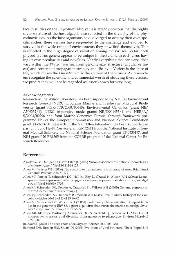
Wilson, Van EttEn, & allEn in Lesser Known Large dsdna Viruses (2009)32
face in studies on the Phycodnaviridae, yet it is already obvious that the highly diverse nature of the host algae is also reflected in the diversity of the phy-codnaviruses. As the host organisms have diverged to occupy their own spe-cific niches, these viruses have responded to the challenge and evolved to survive in the wide range of environments they now find themselves. This is reflected in the huge degree of variation among the viruses. So far, each phycodnavirus genera appear to be unique in lifestyle, with each virus hav-ing its own peculiarities and novelties. Nearly everything that can vary, does vary within the Phycodnaviridae, from genome size, structure (circular or lin-ear) and content, to propagation strategy and life style. Variety is the spice of life, which makes the Phycodnaviridae the spiciest of the viruses. As research-ers recognize the scientific and commercial worth of studying these viruses, we predict they will not be regarded as lesser known for very long.
Acknowledgments Research in the Wilson laboratory has been supported by Natural Environment Research Council (NERC) programs Marine and Freshwater Microbial Biodi-versity (grant NER/T/S/2000/00640); Environmental Genomics (grant NE/A509332/�); NERC responsive mode grants NE/D00�455/� and NER/A/S/2003/00296 and from Marine Genomics Europe, through framework pro-gramme FP6 of the European Commission and National Science Foundation grant EF-0723730. Research in the Van Etten laboratory has been supported in part by Public Health Service grant GM3244� from the National Institute of Gen-eral Medical Sciences, the National Science Foundation grant EF-0333�97, and NIH grant P20-RR�565 from the COBRE program of the National Center for Re-search Resources.
ReferencesAgarkova IV, Dunigan DD, Van Etten JL (2006) Virion-associated restriction endonucleases
of chloroviruses. J Virol 80:8��4-8�23 Allen MJ, Wilson WH (2006) The coccolithovirus microarray: an array of uses. Brief Funct
Genomic Proteomic 5:273-279 Allen MJ, Forster T, Schroeder DC, Hall M, Roy D, Ghazal P, Wilson WH (2006a) Locus-
specific gene expression pattern suggests a unique propagation strategy for a giant algal virus. J Virol 80:7699-7705
Allen MJ, Schroeder DC, Donkin A, Crawfurd KJ, Wilson WH (2006b) Genome comparison of two Coccolithoviruses. Virology J 3:�5
Allen MJ, Schroeder DC, Holden MTG, Wilson WH (2006c) Evolutionary history of the Coc-colithoviridae. Mol Biol Evol 23:86-92
Allen MJ, Schroeder DC, Wilson WH (2006d) Preliminary characterization of repeat fami-lies in the genome of EhV-86, a giant algal virus that infects the marine microalga Emili-ania huxleyi. Arch Virology �5�:525-535
Allen MJ, Martinez-Martinez J, Schroeder DC, Somerfield PJ, Wilson WH (2007) Use of microarrays to assess viral diversity: from genotype to phenotype. Environ Microbiol 9:97�-982
Baldauf SL (2003) The deep roots of eukaryotes. Science 300:�703-�706 Bamford DH, Bumett RM, Stuart DI (2002) Evolution of viral structure. Theor Popul Biol
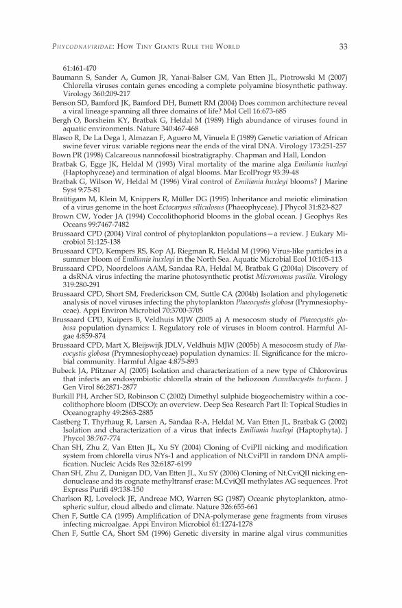
PhycodnaViridae : HoW tiny Giants RulE tHE WoRld 33
6�:46�-470 Baumann S, Sander A, Gumon JR, Yanai-Balser GM, Van Etten JL, Piotrowski M (2007)
Chlorella viruses contain genes encoding a complete polyamine biosynthetic pathway. Virology 360:209-2�7
Benson SD, Bamford JK, Bamford DH, Bumett RM (2004) Does common architecture reveal a viral lineage spanning all three domains of life? Mol Cell �6:673-685
Bergh O, Borsheim KY, Bratbak G, Heldal M (�989) High abundance of viruses found in aquatic environments. Nature 340:467-468
Blasco R, De La Dega I, Almazan F, Aguero M, Vinuela E (�989) Genetic variation of African swine fever virus: variable regions near the ends of the viral DNA. Virology �73:25�-257
Bown PR (�998) Calcareous nannofossil biostratigraphy. Chapman and Hall, London Bratbak G, Egge JK, Heldal M (�993) Viral mortality of the marine alga Emiliania huxleyi
(Haptophyceae) and termination of algal blooms. Mar EcolProgr 93:39-48 Bratbak G, Wilson W, Heldal M (�996) Viral control of Emiliania huxleyi blooms? J Marine
Syst 9:75-8� Braütigam M, Klein M, Knippers R, Müller DG (�995) Inheritance and meiotic elimination
of a virus genome in the host Ectocarpus siliculosus (Phaeophyceae). J Phycol 3�:823-827 Brown CW, Yoder JA (�994) Coccolithophorid blooms in the global ocean. J Geophys Res
Oceans 99:7467-7482 Brussaard CPD (2004) Viral control of phytoplankton populations—a review. J Eukary Mi-
crobiol 5�:�25-�38Brussaard CPD, Kempers RS, Kop AJ, Riegman R, Heldal M (�996) Virus-like particles in a
summer bloom of Emiliania huxleyi in the North Sea. Aquatic Microbial Ecol �0:�05-��3 Brussaard CPD, Noordeloos AAM, Sandaa RA, Heldal M, Bratbak G (2004a) Discovery of
a dsRNA virus infecting the marine photosynthetic protist Micromonas pusilla. Virology 3�9:280-29�
Brussaard CPD, Short SM, Frederickson CM, Suttle CA (2004b) Isolation and phylogenetic analysis of novel viruses infecting the phytoplankton Phaeocystis globosa (Prymnesiophy-ceae). Appi Environ Microbiol 70:3700-3705
Brussaard CPD, Kuipers B, Veldhuis MJW (2005 a) A mesocosm study of Phaeocystis glo-bosa population dynamics: I. Regulatory role of viruses in bloom control. Harmful Al-gae 4:859-874
Brussaard CPD, Mart X, Bleijswijk JDLV, Veldhuis MJW (2005b) A mesocosm study of Pha-eocystis globosa (Prymnesiophyceae) population dynamics: II. Significance for the micro-bial community. Harmful Algae 4:875-893
Bubeck JA, Pfitzner AJ (2005) Isolation and characterization of a new type of Chlorovirus that infects an endosymbiotic chlorella strain of the heliozoon Acanthocystis turfacea. J Gen Virol 86:287�-2877
Burkill PH, Archer SD, Robinson C (2002) Dimethyl sulphide biogeochemistry within a coc-colithophore bloom (DISCO): an overview. Deep Sea Research Part II: Topical Studies in Oceanography 49:2863-2885
Castberg T, Thyrhaug R, Larsen A, Sandaa R-A, Heldal M, Van Etten JL, Bratbak G (2002) Isolation and characterization of a virus that infects Emiliania huxleyi (Haptophyta). J Phycol 38:767-774
Chan SH, Zhu Z, Van Etten JL, Xu SY (2004) Cloning of CviPII nicking and modification system from chlorella virus NYs-� and application of Nt.CviPII in random DNA ampli-fication. Nucleic Acids Res 32:6187-6199
Chan SH, Zhu Z, Dunigan DD, Van Etten JL, Xu SY (2006) Cloning of Nt.CviQII nicking en-donuclease and its cognate methyltransf erase: M.CviQII methylates AG sequences. Prot Express Purifi 49:138-150
Charlson RJ, Lovelock JE, Andreae MO, Warren SG (�987) Oceanic phytoplankton, atmo-spheric sulfur, cloud albedo and climate. Nature 326:655-66�
Chen F, Suttle CA (1995) Amplification of DNA-polymerase gene fragments from viruses infecting microalgae. Appi Environ Microbiol 6�:�274-�278
Chen F, Suttle CA, Short SM (�996) Genetic diversity in marine algal virus communities
Wilson, Van EttEn, & allEn in Lesser Known Large dsdna Viruses (2009)34
as revealed by sequence analysis of DNA polymerase genes. Appi Environ Microbiol 62:2869-2874
Chrétiennot-Dinet MJ, Courties C, Vaquer A, Neveux J, Claustre H, Lautier J, Machado MC (�995) A new marine picoeucaryote: Ostreococcus tauri gen. et sp. nov. (Chlorophyta, Pra-sinophyceae). Phycologia 34:285-292
Chuchird N, Nishida K, Kawasaki T, Fujie M, Usami S, Yamada T (2002) A variable region on the Chlorovirus CVK2 genome contains five copies of the gene for Vp260, a viral gly-coprotein. Virology 295:289-298
Clokie MRJ, Mann NH (2006) Marine cyanophages and light. Environ Microbiol 8:2074-2082
Cottrell MT, Suttle CA (�99�) Wide-spread occurrence and clonal variation in viruses which cause lysis of a cosmopolitan, eukaryotic marine phytoplankter, Micromonas pusilla. Mar Ecol Progr78:l-9
Cottrell MT, Suttle CA (�995a) Dynamics of a lytic virus infecting the photosynthetic marine icoflagellate Micromonas pusilla. Limnol Oceanography 40:730-739
Cottrell MT, Suttle CA (�995b) Genetic diversity of algal viruses which lyse the photo-synthetic picoflagellate Micromonas pusilla (Prasinophyceae). Appi Environ Microbiol 6�:3088-309�
Crick FH, Watson JD (�956) Structure of small viruses. Nature �77:473-75 Dahl E, Bagoien E, Edvardsen B, Stenseth NC (2005) The dynamics of Chrysochromulina spe-
cies in the Skagerrak in relation to environmental conditions. J Sea Res 54:�5-24 DeAngelis PL, Jing W, Graves MV, Burbank DE, Van Etten JL (�997) Hyaluronan synthase
of chlorella virus PBCV-�. Science 278:�800-�803 Delaroque N, Maier I, Knippers R, Muller DG (�999) Persistent virus integration into the ge-
nome of its algal host, Ectocarpus siliculosus (Phaeophyceae). J Gen Virol 80:�367-�370 Delaroque N, Boland W, Muller DG, Knippers R (2003) Comparisons of two large phaeovi-
ral genomes and evolutionary implications. J Mol Evol 57:6�3-622 Delaroque N, Muller DG, Bothe G, Pohl T, Knippers R, Boland W (200�) The complete DNA
sequence of the Ectocarpus siliculosus virus EsV-� genome. Virology 287:��2-�32 Derelle E, Ferraz C, Rombauts S, Rouze P, Worden AZ, Robbens S, Partensky F, Degroeve S,
Echeynie S, Cooke R, Saeys Y, Wuyts J, Jabbari K, Bowler C, Panaud 0, Piegu B, Ball SG, Ral JP, Bouget FY, Piganeau G, De Baets B, Picard A, Delseny M, Demaille J, Van de Peer Y, Moreau H (2006) Genome analysis of the smallest free-living eukaryote Ostreococcus tauri unveils many unique features. Proc Nat Acad Sci USA �03:��647-��652
Diez B, Pedros-Alio C, Massana R (200�) Study of genetic diversity of eukaryotic picoplank-ton in different oceanic regions by small-subunit rRNA gene cloning and sequencing. Appi Environ Microbiol 67:2932-294�
Dundas I, Johannessen OM, Berge G, Heimdal B (�989) Toxic algal bloom in Scandinavian waters, May-June �988. Oceanography 2:9-�4
Dunigan DD, Fitzgerald LA, Van Etten JL (2006) Phycodnaviruses: a peek at genetic diver-sity. Virus Res ��7:��9-�32
Eikrem W, Throndsen J (�990) The ultrastructure of Bathycoccus gen-nov and Bathycoccus prasinos sp-nov, a nonmotile picoplanktonic alga (Chlorophyta, Prasinophyceae) from the Mediterranean and Atlantic. Phycologia 29:344-350
Elderfield H (2002) Climate change: carbonate mysteries. Science 296:1618-1621 Eriksson M, Myllyharju J, Tu H, Hellman M, Kivirikko KI (�999) Evidence for 4-hydoxypro-
line in viral proteins: characterization of a viral prolyl 4 hydroxylase and its peptide sub-strates. J Biol Chem 274:22�3�-22�34
Estep KW, Maclntyre F (�989) Taxonomy, life cycle, distribution and dasmotrophy of Chrys-ochromulina: a theory accounting for scales, haptonema, muciferous bodies and toxicity. Mar Ecol Progr 57:��-2�
Evans C, Archer SD, Jacquet S, Wilson WH (2003) Direct estimates of the contribution of vi-ral lysis and microzooplankton grazing to the decline of a Micromonas spp. population. Aquatic Microbial Ecol 30:207-2�9
Evans C, Kadner SV, Darroch LJ, Wilson WH, Liss PS, Malin G (2007) The relative signif-
PhycodnaViridae : HoW tiny Giants RulE tHE WoRld 35
icance of viral lysis and microzooplankton grazing as pathways of dimethylsulfonio-propionate (DMSP) cleavage: an Emiliania huxleyi culture study. Limnol Oceanogr 52:�036-�045
Figueroa RI, Rengefors K (2006) Life cycle and sexuality of the freshwater raphidophyte Go-nyostomum semen (Raphidophyceae). J Phycol 42:859-87�
Filee J, Forterre P, Laurent J (2003) The role played by viruses in the evolution of their hosts: a view based on informational protein phylogenies. Res Microbiol �54: 237-243
Fischer D, Eisenberg D (�999) Finding families for genomic ORFans. Bioinformatics �5:759-762
Fitzgerald LA, Graves MV, Li X, Feldblyum T, Hartigan J, Van Etten JL (2007a) Sequence and annotation of the 3�4-kb MT325 and the 32�-kb FR483 viruses that infect Chlorella Pbi. Virology 358:459-47�
Fitzgerald LA, Graves MV, Li X, Feldblyum T, Nierman WC, Van Etten JL (2007b) Sequence and annotation of the 369-kb NY-2A and the 345-kb AR�58 viruses that infect Chlorella NC64A. Virology 358:472-484
Fitzgerald LA, Graves MV, Li X, Hartigan J, Pfitzner AJ, Hoffart E, Van Etten JL (2007c) Se-quence and annotation of the 288-kb ATCV-� virus that infects an endosymbiotic chlo-rella strain of the heliozoon Acanthocystis turfacea. Virology 362:350-36�
Fortune JM, Lavrukhin 0V, Gumon JR, Van Etten JL, Lloyd RS, OsheroffN (200�) Topoisom-erase II from chlorella virus PBCV-� has an exceptionally high DNA cleavage activity. J Biol Chem 276:2440�-24408
Frohns F, Kasmann A, Kramer D, Schafer B, Mehmel M, Kang M, Van Etten JL, Gazzarrini S, Moroni A, Thiel G (2006) Potassium ion channels of chlorella viruses cause rapid de-polarization of host cells during infection. J Virol 80:2437-2444
Fruscione F, Sturia L, Duncan, G, Van Etten JL, Valbuzzi, P, De Flora A, Di Zanni E, Tonetti M (2008) Differential role of NADP+ and NADPH in the activity and structure of GDP-D-mannose 4,6-dehydratase from two chlorella viruses. J. Biol Chem 283:�84-�93
Fuller NJ, Campbell C, Allen DJ, Pitt FD, Zwirglmaieri K, Le Gall F, Vaulot D, Scanlan DJ (2006) Analysis of photosynthetic picoeukaryote diversity at open ocean sites in the Arabian Sea using a PCR biased towards marine algal plastids. Aquatic Microbial Ecol 43:79-93
Gazzarrini S, Kang M, Van Etten JL, Tayefeh S, Kast SM, DiFrancesco D, Thiel G, Moroni A (2004) Long-distance interactions within the potassium channel pore are revealed by molecular diversity of viral proteins. J Biol Chem 279:28443-28449
Gazzarrini S, Kang M, Epimashko S, Van Etten JL, Dainty J, Theil G, Moroni A (2006) Chlo-rella virus MT325 encodes water and potassium channels that interact synergistically. Proc Nati Acad Sci U S A �03:5355-5360
Graves MV, Burbank DE, Roth R, Heuser J, DeAngelis PL, Van Etten JL (�999) Hyaluronan synthesis in virus PBCV-� infected chlorella-like green algae. Virology 257:�5-23
Graves MV, Bemadt CT, Cemy R, Van Etten JL (200�) Molecular and genetic evidence for a virus-encoded glycosyltransferase involved in protein glycosylation. Virology 285:332-345
Graziani S, Xia Y, Gumon JR, Van Etten JL, Leduc D, Skouloubris S, Myllykallio, Hiebl U (2004) Functional analysis of FAD-dependent thymidylate synthase ThyX from Parame-cium bursaria chlorella virus-�. J Biol Chem 279:54340-54347
Green JC, Pienaar RN (�977) The taxonomy of the order Isochrysidales (Prymnesiophyceae) with special reference to the genera Isochrysis Parke, Dicrateria Parke and Imantonia Reynolds. J Mar Biol Assoc UK 57:7-�7
Hakansson K, Wigley DB (�998) Structure of a complex between a cap analogue and mRNA guanylyl transferase demonstrates the structural chemistry of RNA capping. Proc Nati Acad Sci USA 95:�505-�5�0
Hakansson K, Doherty AJ, Shuman S, Wigley DB (1997) X-ray crystallography reveals a large conformational change during guanyl transfer by mRNA capping enzymes. Cell 89:543-553
Han G, Gable K, Yan L, Allen MJ, Wilson WH, Moitra P, Harmon JM, Dunn TM (2006) Ex-pression of a novel marine viral single-chain serine palmitoyltransferase and construc-
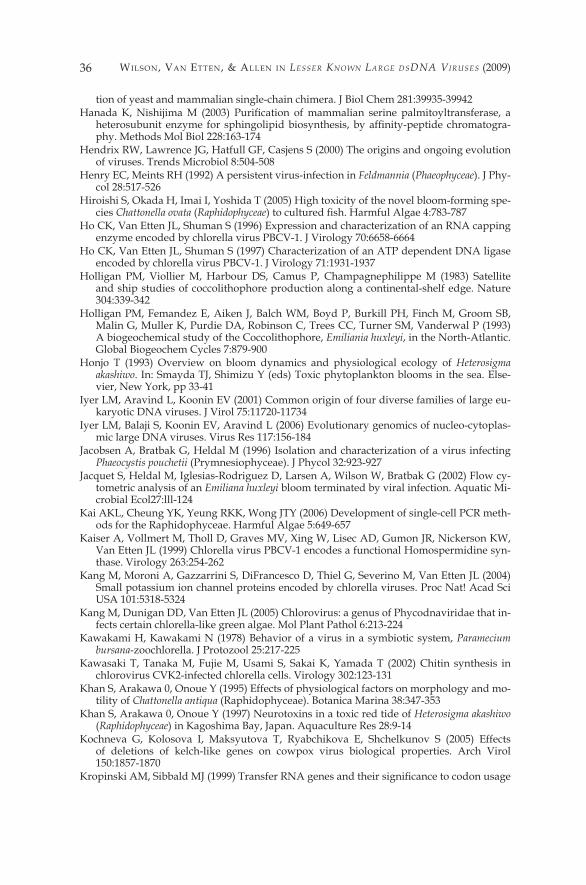
Wilson, Van EttEn, & allEn in Lesser Known Large dsdna Viruses (2009)36
tion of yeast and mammalian single-chain chimera. J Biol Chem 28�:39935-39942 Hanada K, Nishijima M (2003) Purification of mammalian serine palmitoyltransferase, a
heterosubunit enzyme for sphingolipid biosynthesis, by affinity-peptide chromatogra-phy. Methods Mol Biol 228:�63-�74
Hendrix RW, Lawrence JG, Hatfull GF, Casjens S (2000) The origins and ongoing evolution of viruses. Trends Microbiol 8:504-508
Henry EC, Meints RH (�992) A persistent virus-infection in Feldmannia (Phaeophyceae). J Phy-col 28:5�7-526
Hiroishi S, Okada H, Imai I, Yoshida T (2005) High toxicity of the novel bloom-forming spe-cies Chattonella ovata (Raphidophyceae) to cultured fish. Harmful Algae 4:783-787
Ho CK, Van Etten JL, Shuman S (�996) Expression and characterization of an RNA capping enzyme encoded by chlorella virus PBCV-�. J Virology 70:6658-6664
Ho CK, Van Etten JL, Shuman S (�997) Characterization of an ATP dependent DNA ligase encoded by chlorella virus PBCV-�. J Virology 7�:�93�-�937
Holligan PM, Viollier M, Harbour DS, Camus P, Champagnephilippe M (�983) Satellite and ship studies of coccolithophore production along a continental-shelf edge. Nature 304:339-342
Holligan PM, Femandez E, Aiken J, Balch WM, Boyd P, Burkill PH, Finch M, Groom SB, Malin G, Muller K, Purdie DA, Robinson C, Trees CC, Turner SM, Vanderwal P (�993) A biogeochemical study of the Coccolithophore, Emiliania huxleyi, in the North-Atlantic. Global Biogeochem Cycles 7:879-900
Honjo T (�993) Overview on bloom dynamics and physiological ecology of Heterosigma akashiwo. In: Smayda TJ, Shimizu Y (eds) Toxic phytoplankton blooms in the sea. Else-vier, New York, pp 33-4�
Iyer LM, Aravind L, Koonin EV (200�) Common origin of four diverse families of large eu-karyotic DNA viruses. J Virol 75:��720-��734
Iyer LM, Balaji S, Koonin EV, Aravind L (2006) Evolutionary genomics of nucleo-cytoplas-mic large DNA viruses. Virus Res ��7:�56-�84
Jacobsen A, Bratbak G, Heldal M (�996) Isolation and characterization of a virus infecting Phaeocystis pouchetii (Prymnesiophyceae). J Phycol 32:923-927
Jacquet S, Heldal M, Iglesias-Rodriguez D, Larsen A, Wilson W, Bratbak G (2002) Flow cy-tometric analysis of an Emiliana huxleyi bloom terminated by viral infection. Aquatic Mi-crobial Ecol27:lll-�24
Kai AKL, Cheung YK, Yeung RKK, Wong JTY (2006) Development of single-cell PCR meth-ods for the Raphidophyceae. Harmful Algae 5:649-657
Kaiser A, Vollmert M, Tholl D, Graves MV, Xing W, Lisec AD, Gumon JR, Nickerson KW, Van Etten JL (�999) Chlorella virus PBCV-� encodes a functional Homospermidine syn-thase. Virology 263:254-262
Kang M, Moroni A, Gazzarrini S, DiFrancesco D, Thiel G, Severino M, Van Etten JL (2004) Small potassium ion channel proteins encoded by chlorella viruses. Proc Nat! Acad Sci USA �0�:53�8-5324
Kang M, Dunigan DD, Van Etten JL (2005) Chlorovirus: a genus of Phycodnaviridae that in-fects certain chlorella-like green algae. Mol Plant Pathol 6:2�3-224
Kawakami H, Kawakami N (�978) Behavior of a virus in a symbiotic system, Paramecium bursana-zoochlorella. J Protozool 25:2�7-225
Kawasaki T, Tanaka M, Fujie M, Usami S, Sakai K, Yamada T (2002) Chitin synthesis in chlorovirus CVK2-infected chlorella cells. Virology 302:�23-�3�
Khan S, Arakawa 0, Onoue Y (�995) Effects of physiological factors on morphology and mo-tility of Chattonella antiqua (Raphidophyceae). Botanica Marina 38:347-353
Khan S, Arakawa 0, Onoue Y (�997) Neurotoxins in a toxic red tide of Heterosigma akashiwo (Raphidophyceae) in Kagoshima Bay, Japan. Aquaculture Res 28:9-�4
Kochneva G, Kolosova I, Maksyutova T, Ryabchikova E, Shchelkunov S (2005) Effects of deletions of kelch-like genes on cowpox virus biological properties. Arch Virol �50:�857-�870
Kropinski AM, Sibbald MJ (1999) Transfer RNA genes and their significance to codon usage
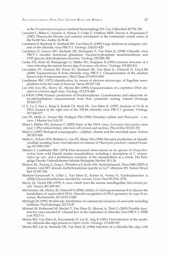
PhycodnaViridae : HoW tiny Giants RulE tHE WoRld 37
in the Pseudomonas aeruginosa lamboid bacteriophage D3. Can J Microbiol 45:79�-796 Lancelot C, Billen G, Soumia A, Weisse T, Colijn F, Veldhuis MJW, Davies A, Wassmann P
(�987) Phaeocystis blooms and nutrient enrichment in the continental coastal zones of the North Sea. Ambio �6:38-46
Landstein D, Burbank D, Nietfeldt JW, Van Etten JL (�995) Large deletions in antigenic vari-ants of the chlorella virus PBCV-�. Virology 2�4:4�3-420
Landstein D, Graves MV, Burbank DE, DeAngelis P, Van Etten JL (�998) Chlorella virus PBCV-� encodes functional glutamine: fructose-6-phosphate amidotransferase and UDP-glucose dehydrogenase enzymes. Virology 250:388-396
Lanka STJ, Klein M, Ramsperger U, Müller DG, Knippers R (�993) Genome structure of a virus infecting the marine brown alga Ectocarpus siliculosus. Virology �93:802-8��
Lavrukhin 0V, Fortune JM, Wood TG, Burbank DE, Van Etten JL, Osheroff N, Lloyd RS (2000) Topoisomerase II from chlorella virus PBCV-�. Characterization of the smallest known type II topoisomerase. J Biol Chem 275:69�5-692�
Leadbeater BSC (1972) Identification, by means of electron microscopy, of flagellate nano-plankton from the coast of Norway. Sarsia 49:�07-�24
Lee AM, Ivey RG, Henry EC, Meints RH (�995) Characterization of a repetitive DNA ele-ment in a brown algal virus. Virology 2�2:474-480
Li WKW (�994) Primary production of Prochlorophytes, Cyanobacteria, and eukaryotic ul-tra-phytoplankton—measurements from flow cytometric sorting. Limnol Oceanogr 39:�69-�75
Li Y, Lu Z, Sun L, Ropp S, Kutish GF, Rock DL, Van Etten JL (1997) Analysis of 74 kb of DNA located at the right end of the 330-kb chlorella virus PBCV-� genome. Virology 237:360-377
Liss PS, Malin G, Turner SM, Holligan PM (1994) Dimethyl sulfide and Phaeocystis - a re-view. J Marine Sys 5:4�-53
Maier I, Müller DG, Katsaros C (2002) Entry of the DNA virus, Ectocarpus fasciculatus virus type � (Phycodnaviridae), into host cell cytosol and nucleus. Phycol Res 50:227-23�
Malin G (�997) Biological oceanography—sulphur, climate and the microbial maze. Nature 387:857-859
Malin G, Wilson WH, Bratbak G, Liss PS, Mann NH (�998) Elevated production of dimeth-ylsulfide resulting from viral infection of cultures of Phaeocystis pouchetii. Limnol Ocean-ogr 43:�389-�393
Manton I, Leadbeater BSC (�974) Fine structural observations on six species of Chrysochro-mulina from wild Danish marine nanoplankton, including a description of C. campan-ulifera sp. nov. and a preliminary summary of the nanoplankton as a whole. Det Kon-gelige Danske Videnskabemes Selskab Biologiske Skrifter 20:�-26
Manzur KL, Farooq A, Zeng L, Plotnikova 0, Koch AW, Sachchidanand, Zhou MM (2003) A dimeric viral SET domain methyltransferase specific to Lys27 ofhistone H3. Nature Struct Biol �0:�87-�96
Markine-Goriaynoff N, Gillet L, Van Etten JL, Korres H, Verma N, Vanderplasschen A (2004) Glycosyltransferases encoded by viruses. J Gen Virol 85:274�-2754
Mayer JA, Taylor FJR (1979) A virus which lyses the marine nanoflagellate Micromonas pu-silla. Nature 28�:299-30�
McClendon AK, Dickey JS, Osheroff N (2006) Ability of viral topoisomerase II to discern the handedness of supercoiled DNA: bimodal recognition of DNA geometry by type II en-zymes. Biochemistry 45:��674-��680
McHugh DJ (�99�) Worldwide distribution of commercial resources of seaweeds including Geldium. Hydrobiologia 22�:�9-29
Mehmel M, Rothermel M, Meckel T, Van Etten JL, Moroni A, Thiel G (2003) Possible func-tion for virus encoded K+ channel Kcv in the replication of chlorella virus PBCV-�. FEBS Lett 552:7-��
Meints RH, Van Etten JL, Kuczmarski D, Lee K, Ang B (�98�) Viral-infection of the symbi-otic chlorella-like alga present in Hydra viridis. Virology ��3:698-703
Meints RH, Lee K, Burbank DE, Van Etten JL (�984) Infection of a chlorella-like alga with
Wilson, Van EttEn, & allEn in Lesser Known Large dsdna Viruses (2009)38
the virus, PBCV-�: Ultrastructural studies. Virology �38:34�-346 Meints RH, Lee K, Van Etten JL (�986) Assembly site of the virus PBCV-� in a chlorella-like
green alga: ultrastructural studies. Virology �54:240-245 Meints RH, Burbank DE, Van Etten JL, Lamport DTA (�988) Properties of the Chlorella re-
ceptor for the virus PBCV-�. Virology �64:�5-2�Moon-van der Staay SY, De Wachter R, Vaulot D (200�) Oceanic �8S rDNA sequences from
picoplankton reveal unsuspected eukaryotic diversity. Nature 409:607-6�0 Morehead TA, Gumon JR, Adams B, Nickerson KW, Fitzgerald LA, Van Etten JL (2002)
Omithine decarboxylase encoded by chlorella virus PBCV-�. Virology 30�:�65-�75 Müller DG (�99�) Mendelian segregation of a virus genome during host meiosis in the ma-
rine brown alga Ectocarpus siliculosus. J Plant Physiol �37:739-743 Müller DG (�996) Host-virus interactions in marine brown algae. Hydrobiologia 327:2�-28 Müller DG, Kawai H, Stache B, Lanka S (�990) A virus-infection in the marine brown alga
Ectocarpus siliculosus (Phaeophyceae). Bot Acta �03:72-82 Müller DG, Stache B (1992) Worldwide occurrence of virus-infections in filamentous marine
brown-algae. Helgolander Meeresunters 46:�-8 Müller DG, Kapp M, Knippers R (�998) Viruses in marine brown algae. In: Advances in vi-
rus research, Vol. 50. Academic, San Diego, pp 49-67 Muzzarelli R, Jeuniaux C, Gooday GW (eds) (�986) Chitin in nature and technology. Ple-
num Press, New York Myllykallio H, Lipowski G, Leduc D, Filee J, Forterre P, Liebl U (2002) An alternative flavin-
dependent mechanism for thymidylate synthesis. Science 297:�05-�07Nagasaki K, Yamaguchi M (�997) Isolation of a virus infectious to the harmful bloom caus-
ing microalga Heterosigma akashiwo (Raphidophyceae). Aquatic Microbial Ecol �3:�35-�40 Nagasaki K, Yamaguchi M (�998a) Effect of temperature on the algicidal activity and the
stability of HaV (Heterosigma akashiwo virus). Aquatic Microbial Ecol �5:2��-2�6 Nagasaki K, Yamaguchi M (1998b) Intra-species host specificity of HaV (Heterosigma
akashiwo virus) clones. Aquatic Microbial Ecol �4:�09-��2 Nagasaki K, Tarutani K, Yamaguchi M (�999a) Cluster analysis on algicidal activity of HaV
clones and virus sensitivity of Heterosigma akashiwo (Raphidophyceae). J Plankton Res 2�:22�9-2226
Nagasaki K, Tarutani K, Yamaguchi M (�999b) Growth characteristics of Heterosigma akashiwo virus and its possible use as a microbiological agent for red tide control. Appi Environ Microbiol 65:898-902
Nagasaki K, Tomaru Y, Katanozaka N, Shirai Y, Nishida K, Itakura S, Yamaguchi M (2004) Isolation and characterization of a novel single-stranded RNA virus infecting the bloom-forming diatom Rhizosolenia setigera. Appi Environ Microbiol 70:704-7��
Nagasaki K, Shirai Y, Tomaru Y, Nishida K, Pietrokovski S (2005) Algal viruses with dis-tinct intraspecies host specificities include identical intein elements. Appi Environ Mi-crobiol 7�:3599-3607
Nandhagopal N, Simpson AA, Gumon JR, Yan X, Baker TS, Graves MV, Van Etten JL, Ross-mann MG (2002) The structure and evolution of the major capsid protein of a large, lipid-containing DNA virus. Proc Nail Acad Sci U S A 99:�4758-�4763
Nelson M, Burbank DE, Van Etten JL (�998) Chlorella viruses encode multiple DNA meth-yltransferases. Biol Chem 379:423-428
Neupartl M, Meyer C, Woll I, Frohns F, Kang M, Van Etten JL, Kramer D, Hertel B, Moroni A, Thiel G (2008) Chlorella viruses evoke a rapid release of �C from host cells during early phase of infection. Virology 372:340-348
Nishida K, Suzuki S, Kimura Y, Nomura N, Fujie M, Yamada T (�998) Group I introns found in chlorella viruses: Biological implications. Virology 242:3�9-326
Nishida K, Kimura Y, Kawasaki T, Fujie M, Yamada T (�999) Genetic variation of chlorella viruses: variable regions localized on the CVK2 genomic DNA. Virology 255:376-384
Not F, Latasa M, Marie D, Cariou T, Vaulot D, Simon N (2004) A single species, Micromonas pusilla (Prasinophyceae), dominates the eukaryotic picoplankton in the western English channel. Appi Environ Microbiol 70:4064-4072
PhycodnaViridae : HoW tiny Giants RulE tHE WoRld 39
Not F, Massana R, Latasa M, Marie D, Colson C, Eikrem W, Pedros-Alio C, Vaulot D, Si-mon N (2005) Late summer community composition and abundance of photosynthetic picoeukaryotes in Norwegian and Barents Seas. Limnol Oceanogr 50:�677-�686
Odell M, Malinina L, Sriskanda V, Teplova M, Shuman S (2003) Analysis of the DNA join-ing repertoire of chlorella virus DNA ligase and a new crystal structure of the ligase-ad-enylate intermediate. Nucleic Acids Res 3�:5090-5�00
O’Kelly CJ, Sieracki ME, Thier EC, Hobson �C (2003) A transient bloom of Ostreococcus (Chlorophyta, Prasinophyceae) in the West Neck Bay, Long Island, New York. J Phy-col 39:850-854
Onimatsu H, Suganuma K, Uenoyama S, Yamada T (2006) C-terminal repetitive motifs in Vpl30 present at the unique vertex of the chlorovirus capsid are essential for binding to the host chlorella cell wall. Virology 353:432-442
Palenik B, Grimwood J, Aerts A, Rouze P, Salamov A, Putnam N, Dupont C, Jorgensen R, Derelle E, Rombauts S, Zhou K, Otillar R, Merchant SS, Podell S, Gaasterland T, Nap-oli C, Gendler K, Manuell A, Tai V, Vallon 0, Piganeau G, Jancek S, Heijde M, Jabbari K, Bowler C, Lohr M, Robbens S, Wemer G, Dubchak I, Pazour GJ, Ren Q, Paulsen I, Del-wiche C, Schmutz J, Rokhsar D, Van de Peer Y, Moreau H, Grigoriev IV (2007) The tiny eukaryote Ostreococcus provides genomic insights into the paradox of plankton specia-tion. Proc Nat! Acad Sci USA �04:7705-77�0
Parke M, Manton I, Clarke B (1959) Studies on marine flagellates. V. Morphology and mi-croanatomy of Cyrysochromulina strobilus sp. nov. J Marine Biol Assoc UK 38:�69-�88
Peters AF, Marie D, Scomet D, Kloareg B, Cock JM (2004) Proposal of Ectocarpus siliculosus (Ectocarpales, Phaeophyceae) as a model organism for brown algal genetics and genom-ics. J Phycol 40:�079-�088
Pires de Miranda M, Reading PC, Tscharke DC, Murphy BJ, Smith GL (2003) The vaccinia virus kelch-like protein C2L affects calcium-independent adhesion to the extracellular matrix and inflammation in a murine intradermal model. J Gen Virol 84:2459-2471
Plugge B, Gazzamni S, Nelson M, Cerana R, Van Etten JL, Derst C, DiFrancesco D, Moroni A, Thiel G (2000) A potassium channel protein encoded by chlorella virus PBCV-�. Sci-ence 287:�64�-�644
Que Q, Li Y, Wang IN, Lane LC, Chaney WG, Van Etten JL (1994) Protein glycosylation and myristylation in chlorella virus PBCV-� and it antigenic variants. Virology 203:320-327
Raoult D, Audio S, Robert C, Abergel C, Renesto P, Ogata H, La Scola B, Suzan M, Claverie JM (2004) The �.2 megabase genome sequence of mimivirus. Science 306:�344-�350
Reisser W (ed) (�992) Algae and symbioses. Biopress, Bristol, UK Rodriguez-Trelles F, Tarrio R, Ayala FJ (2006) Origins and evolution of spliceosomal in-
trons. Annu Rev Genet 40:47-76Rohozinski J, Girton LE, Van Etten JL (�989) Chlorella viruses contain linear nonpermuted
double strand DNA genomes with covalently closed hairpin ends. Virology �68:363-369Sahlsten E (�998) Seasonal abundance in Skagerrak-Kattegat coastal waters and host spec-
ificity of viruses infecting the marine photosynthetic flagellate Micromonas pusilla. Aquatic Microbial Ecol �6:�03-�08
Sahlsten E, Karlson B (�998) Vertical distribution of virus-like particles (VLP) and viruses infecting Micromonas pusilla during late summer in the southeastern Skagerrak, North Atlantic. J Plankton Res 20:2207-22�2
Sandaa RA, Heldal M, Castberg T, Thyrhaug R, Bratbak G (200�) Isolation and characteriza-tion of two viruses with large genome sizes infecting Chrysochromulina ericina (Prymne-siophyceae) and Pyramimonas orientalis (Prasinophyceae). Virology 290:272-280
Schroeder DC, Oke J, Malin G, Wilson WH (2002) Coccolithovirus (Phycodnaviridae): char-acterisation of a new large dsDNA algal virus that infects Emiliania huxleyi. Arch Virol-ogy �47:�685-�698
Schroeder DC, Oke J, Hall M, Malin G, Wilson WH (2003) Virus succession observed during an Emiliania huxleyi bloom. Appi Environ Microbiol 69:2484-2490
Schuster AM, Girton L, Burbank DE, Van Etten JL (�986) Infection of a chlorella-like alga with the virus PBCV-�: transcriptional studies. Virology �48:�8�-�89
Shah R, Coleman CS, Mir K, Baldwin J, Van Etten JL, Grishin NV, Pegg AE, Stanley BA,
Wilson, Van EttEn, & allEn in Lesser Known Large dsdna Viruses (2009)40
Phillips MA (2004) Paramecium bursaria chlorella virus-� encodes an unusual arginine decarboxylase that is a close homolog of eukaryotic ornithine decarboxylases. J Biol Chem 279:35760-35767
Shchelkunov SN, Totmenin AV (�995) Two types of deletions in orthopoxvirus genomes. Vims Genes 9:23�-245
Shihra I, Krauss RW (�965) Chlorella physiology and taxonomy of forty-one isolates. Col-lege Park, MD, University of Maryland Press
Short SM, Suttle CA (2002) Sequence analysis of marine virus communities reveals that groups of related algal viruses are widely distributed in nature. Appi Environ Micro-biol 68:�290-�296
Simpson AA, Nandhagopal N, Van Etten JL, Rossmann MG (2003) Structural analyses of Phycodnaviridae and Iridoviridae. Acta Crystallogr D Biol Crystallogr 59:2053-2059
Songsri P, Hamazaki T, Ishikawa Y, Yamada T (�995) Large deletions in the genome of chlo-rella virus CVK�. Virology 2�4:405-4�2
Stoltz DB (�97�) The structure of icosahedral cytoplasmic deoxyriboviruses. J Ultrastruct Res 37:2�9-239
Strasser P, Zhang YP, Rohozinski J, Van Etten JL (1991) The termini of the chlorella virus PBCV-� genome are identical 2.2-kbp inverted repeats. Virology �80:763-769
Sugimoto I, Onimatsu H, Fujie M, Usami S, Yamada T (2004) vAL-�, a novel polysaccharide lyase encoded by Chlorovirus CVK2. FEBS Lett 559:5�-56
Sun L, Li Y, McCullough AK, Wood TG, Lloyd RS, Adams B, Gumon JR, Van Etten JL (2000) Intron conservation in a UV-specific DNA repair gene encoded by chlorella viruses. J Mol Evol 50:82-92
Suttle CA (2000a) Cyanophages and their role in the ecology of cyanobacteria. In: Whitton BA, Potts M (eds) The ecology of cyanobacteria: their diversity in time and space. Klu-wer, Boston, pp 563-589
Suttle CA (2000b) Ecological, evolutionary and geochemical consequences of viral infection of cyanobacteria and eukaryotic algae. In: Hurst CJ (ed) Viral ecology. Academic, pp 247-296
Suttle CA, Chan AM (�995) Viruses Infecting the marine Prymnesiophyte Chrysochromu-lina spp- isolation, preliminary characterization and natural-abundance. Mar Ecolo Progr ��8:275-282
Tai V, Lawrence JE, Lang AS, Chan AM, Culley AI, Suttle CA (2003) Characterization of HaRNAV, a single-stranded RNA virus causing lysis of Heterosigma akashiwo (Raphido-phyceae). J Phycol 39:343-352
Tarutani K, Nagasaki K, Yamaguchi M (2000) Viral impacts on total abundance and clonal composition of the harmful bloom-forming phytoplankton Heterosigma akashiwo. Appi Environ Microbiol 66:49�6-4920
Tarutani K, Nagasaki K, Yamaguchi M (2006) Virus adsorption process determines vi-rus susceptibility in Heterosigma akashiwo (Raphidophyceae). Aquat Microbial Ecol 42:209-2�3
Tayefeh S, Kloss T, Thiel G, Hertel B, Moroni A, Kast SM (2007) Molecular dynamics simula-tion of the cytosolic mouth in Kcv-type potassium channels. Biochemistry 46:4826-4839
Tett P (1990) The photic zone. In: Herring PJ, Campbell AK, Whitfield M, Maddock L (eds) Light and life in the sea. Cambridge University Press, Cambridge, pp 59-87
Throndsen J (�996) Note on the taxonomy of Heterosigma akashiwo (Raphidophyceae). Phy-cologia 35:367
Tomaru Y, Tarutani K, Yamaguchi M, Nagasaki K (2004) Quantitative and qualitative im-pacts of viral infection on a Heterosigma akashiwo (Raphidophyceae) bloom in Hiroshima Bay, Japan. Aquatic Microbial Ecol 34:227-238
Tonetti M, Zanardi D, Gumon JR, Fmscione F, Armirotti A, Damonte G, Sturia L, De Flora A, Van Etten JL (2003) Paramecium bursaria chlorella virus � encodes two enzymes in-volved in the biosynthesis of GDP-L-fucose and GDP-D-rhamnose. J Biol Chem 278:2�559-2�565
Tulman ER, Afonso CL, Lu Z, Zsak L, Sur JH, Sandybaev NT, Kerembekova UZ, Zaitsev VL, Kutish GF, Rock DL (2002) The genomes of sheeppox and goatpox vimses. J Virol

PhycodnaViridae : HoW tiny Giants RulE tHE WoRld 4�
76:6054-606�Turner PC, Moyer RW (�990) The molecular pathogenesis of poxviruses. Curr Top Micro-
biol Immunol �63:�25-�5�Van Etten JL (2003) Unusual life style of giant chlorella viruses. Annu Rev Genet 37:�53-�95 Van Etten JL, Meints RH (�999) Giant viruses infecting algae. Annu Rev Microbiol
53:447-494 Van Etten JL, Meints RH, Burbank DE, Kuczmarski D, Cuppels DA, Lane LC (�98�) Isola-
tion and characterization of a virus from the intracellular green-alga symbiotic with Hy-dra viridis. Virology ��3:704-7��
Van Etten JL, Meints RH, Kuczmarski D, Burbank DE, Lee K (�982) Viruses of symbiotic chlorella-like algae isolated from Paramecium bursaria and Hydra viridis. Proc Nail Acad Sci US A 79:3867-387�
Van Etten JL, Burbank DE, Kuczmarski D, Meints RH (�983a) Virus-infection of culturable chlorella-like algae and development of a plaque-assay. Science 2�9:994-996
Van Etten JL, Burbank DE, Xia Y, Meints RH (1983b) Growth cycle of a virus, PBCV-1, that infects chlorella-like algae. Virology �26:��7-�25
Van Etten JL, Burbank DE, Joshi J, Meints RH (�984) DNA synthesis in a chlorella-like alga following infection with the virus PBCV-�. Virology �34:443-449
Van Etten JL, Schuster AM, Girton L, Burbank DE, Swinton D, Hattman S (�985) DNA methylation of viruses infecting a eukaryotic chlorella-like alga. Nucleic Acids Res �3:347�-3478
Van Etten JL, Burbank DE, Meints RH (�986) Replication of the algal virus PBCV-� in UV-ir-radiated chlorella. Intervirology 26:��5-�20
Van Etten JL, Lane LC, Meints RH (�99�) Viruses and virus-like particles of eukaryotic al-gae. Microbiol Rev 55:586-620
Van Etten JL, Graves MV, Muller DG, Boland W, Delaroque N (2002) Phycodnaviridae—large DNA algal viruses. Arch Virol �47:�479-�5�6
Veldhuis MJW, Wassmann P (2005) Bloom dynamics and biological control of a high bio-mass HAB species in European coastal waters: a Phaeocystis case study. Harmful Al-gae 4:805-809
Verity P, Brussaard C, Nejstgaard J, van Leeuwe M, Lancelot C, Medlin L (2007) Current understanding of Phaeocystis ecology and biogeochemistry, and perspectives for future research. Biogeochemistry 83:3��-330
Villarreal LP, DeFilippis VR (2000) A hypothesis for DNA viruses as the origin of eukary-otic replication proteins. J Virol 74:7079-7084
Wang IN, Li Y, Que Q, Bhattacharya M, Lane LC, Chaney WG, Van Etten JL (1993) Evidence for virus-encoded glycosylation specificity. Proc Nati Acad Sci U S A 90:3840-3844
Westbroek P, Brown CW, Vanbleijswijk J, Brownlee C, Brummer GJ, Conte M, Egge J, Femandez E, Jordan R, Knappertsbusch M, Stefels J, Veldhuis M, Vanderwal P, Young J (�993) A model system approach to biological climate forcing—the example of Emiliania huxleyi. Glob Planet Change 8:27-46
Wilson WH, Tarran GA, Schroeder D, Cox M, Oke J, Malin G (2002) Isolation of viruses re-sponsible for the demise of an Emiliania huxleyi bloom in the English Channel. J Marine Biol Assoc UK 82:369-377
Wilson WH, Schroeder DC, Allen MJ, Holden MT, Parkhill J, Barrell BG, Churcher C, Ham-lin N, Mungall K, Norbertczak H, Quail MA, Price C, Rabbinowitsch E, Walker D, Crai-gon M, Roy D, Ghazal P (2005a) Complete genome sequence and lytic phase transcrip-tion profile of a Coccolithovirus. Science 309:1090-1092
Wilson WH, Van Etten JL, Schroeder DS, Nagasaki K, Brussaard C, Delaroque N, Bratbak C, Suttle C (2005b) Family: Phycodnaviridae. In: Fauquet CM, Mayo MA, Maniloff J, Dus-selberger U, Ball LA (eds) Virus taxonomy, VIIIth ICTV report. Elsevier/Academic, Lon-don, pp �63-�75
Wolf S, Maier I, Katsaros C, Müller DG (�998) Virus assembly in Hincksia Hincksiae (Eco-carpales, Phaeophyceae). An electron and fluorescence microscopic study. Protoplasma 203:�53-�67
Wilson, Van EttEn, & allEn in Lesser Known Large dsdna Viruses (2009)42
Wilson WH, Schroeder DC, Ho J, Canty M (2006) Phylogenetic analysis ofPgV-�02P, a new virus from the English Channel that infects Phaeocystis globosa. J Marine Biol Assoc UK 86:485-490
Wolf S, Müller DG, Maier I (2000) Assembly of a large icosahedral DNA virus, MclaV-�, in the marine alga Myriotrichia clavaeformis (Dictyosiphonales, Phaeophyceae). Eur J Phy-col 35:�63-�7�
Worden AZ (2006) Picoeukaryote diversity in coastal waters of the Pacific Ocean. Aquatic Microbial Ecol 43:�65-�75
Worden AZ, Nolan JK, Palenik B (2004) Assessing the dynamics and ecology of marine picophytoplankton: the importance of the eukaryotic component. Limnol Oceanogr 49:�68-�79
Wrigley NG (�969) An electron microscope study of the structure of Serocesthis iridescent virus. J Gen Virol 5:�23-�34
Xia Y, Burbank DE, Uher L, Rabussay D, Van Etten JL (1986) Restriction endonuclease ac-tivity induced by PBCV-� virus infection of a chlorella-like green alga. Mol Cell Biol 6:�430-�439
Xia Y, Burbank DE, Uher L, Rabussay D, Van Etten JL (1987) IL-3A virus infection of chlo-rella-like green alga induces a DNA restriction endonuclease with novel sequence speci-ficity. Nucleic Acids Res 15:6075-6090
Xia Y, Morgan R, Schildkraut I, Van Etten JL (1988) A site-specific single strand endonucle-ase activity induced by NYs-� virus infection of a chlorella like green alga. Nucleic Ac-ids Res �6:9477-9487
Yamada T, Higashiyama T (�993) Characterization of the terminal inverted repeats and their neighboring tandem repeats in the chlorella CVK� virus genome. Mol Gen Genet 24�:554-563
Yamada T, Kawasaki T (2005) Microbial synthesis of hyaluronan and chitin: New ap-proaches. J Biosci Bioeng 99:52�-528
Yamada T, Tamura K, Aimi T, Songsri P (�994) Self-splicing group I introns in eukaryotic viruses. Nucleic Acids Res 22:2532-2537
Yamada T, Onimatsu H, Van Etten JL (2006) Chlorella viruses. In: Advances in virus re-search. Academic, pp 293-336
Yan X, Olson NH, Van Etten JL, Bergoin M, Rossmann MG, Baker TS (2000) Structure and assembly of large lipid-containing dsDNA viruses. Nat Struct Biol 7:�0�-�03
Yan X, Chipman PR, Castberg T, Bratbak G, Baker TS (2005) The marine algal virus PpVOl has an icosahedral capsid with T=2�9 quasisymmetry. J Virol 79:9236-9243
Zeidner G, Preston CM, Delong EF, Massana R, Post AF, Scanlan DJ, Beja 0 (2003) Molecu-lar diversity among marine picophytoplankton as revealed by psbA analyses. Environ Microbiol 5:2�2-2�6
Zhang Y, Nelson M, Nietfeldt J, Xia Y, Burbank DE, Ropp S, Van Etten JL (1998) Chlorella virus NY-2A encodes at least twelve DNA endonuclease/methyltransferase genes. Vi-rology 240:366-375
Zhang Y, Maley F, Maley GF, Duncan G, Dunigan DD, Van Etten JL (2007) chloroviruses encode a bifunctional dCMP-dCTP deaminase that produces two key intermediates in dTTP formation. J Virol 8�:7662-767�
Zhang YP, Strasser P, Grabherr R, Van Etten JL (1994) Hairpin loop structure at the termini of the chlorella virus PBCV-� genome. Virology 202:�079-�082
Zingone A, Samo D, Forlani G (1999) Seasonal dynamics in the abundance of Micromonas pusilla (Prasinophyceae) and its viruses in the Gulf of Naples (Mediterranean Sea). J Plankton Res 2�:2�43-2�59
Zingone A, Natale F, Biffali E, Borra M, Forlani G, Samo D (2006) Diversity in morphology, infectivity, molecular characteristics and induced host resistance between two vimses infecting Micromonas pusilla. Aquatic Microbial Ecol 45:�-�4
Ziveri P, Broerse ATC, van Hinte JE, Westbroek P, Honjo S (2000) The fate of coccoliths at 48 degrees N2� degrees W, northeastern Atlantic. Deep-Sea Research. Part Il.Topical Studies in Oceanography 47:�853-�875