The peroxisomal receptor Pex19p forms a helical...
10
The peroxisomal receptor Pex19p forms a helical mPTS recognition domain Nicole Schueller 1,3 , Simon J Holton 1,3 , Krisztian Fodor 1 , Morlin Milewski 1 , Petr Konarev 1 , Will A Stanley 1,4 , Janina Wolf 2 , Ralf Erdmann 2 , Wolfgang Schliebs 2 , Young-Hwa Song 1 and Matthias Wilmanns 1, * 1 EMBL c/o DESY, Notkestrasse 85, Hamburg, Germany and 2 Department of Systems Biology, Faculty of Medicine, Institute for Physiological Chemistry, Ruhr University of Bochum, Bochum, Germany The protein Pex19p functions as a receptor and chaperone of peroxisomal membrane proteins (PMPs). The crystal structure of the folded C-terminal part of the receptor reveals a globular domain that displays a bundle of three long helices in an antiparallel arrangement. Complementary functional experiments, using a range of truncated Pex19p constructs, show that the structured a-helical domain binds PMP-targeting signal (mPTS) seq- uences with about 10 lM affinity. Removal of a conserved N-terminal helical segment from the mPTS recognition domain impairs the ability for mPTS binding, indicating that it forms part of the mPTS-binding site. Pex19p var- iants with mutations in the same sequence segment abolish correct cargo import. Our data indicate a divided N-terminal and C-terminal structural arrangement in Pex19p, which is reminiscent of a similar division in the Pex5p receptor, to allow separation of cargo-targeting signal recognition and additional functions. The EMBO Journal (2010) 29, 2491–2500. doi:10.1038/ emboj.2010.115; Published online 8 June 2010 Subject Categories: membranes & transport; structural biology Keywords: biogenesis; peroxisome; receptor; structural biology; translocation Introduction Peroxisomes are single-membrane organelles with distinct translocation pathways for peroxisomal matrix proteins and peroxisomal membrane proteins (PMPs). Most matrix proteins include a short peroxisomal-targeting signal (PTS) motif. Two different motifs, categorized as type 1 (PTS1) and type 2 (PTS2), are recognized by the specific receptors, Pex5p and Pex7p, respectively. Structures of the PTS1 receptor Pex5p in the absence and presence of a cargo have unravelled how the conformation of the receptor adapts to recognize peroxisomal matrix proteins through their C-terminal PTS1 motif (Gatto et al, 2000; Stanley et al, 2006). In contrast, several PMPs have one or more membrane protein-targeting signal (mPTS) motifs with a cluster of basic residues in a predicted a-helical conformation (Fransen et al, 2001; Jones et al, 2001; Rottensteiner et al, 2004). This binding site is generally flanked by one or two transmembrane segments. Correct topogenesis of most of these targets, categorized as class-I mPTS, within the peroxisomal membrane, however, requires two peroxins: Pex3p and Pex19p. These findings have led to diverse arguments that consider Pex19p as a PMP receptor, an assembly factor, a PMP chaperone or a factor combining more than a single function (Eckert and Erdmann, 2003; Jones et al, 2004; Halbach et al, 2006). Recent data have reconciled earlier hypotheses by indicating complementary tasks for Pex3p and Pex19p in PMP topogenesis (Pinto et al, 2006). In this model, Pexp19p recognizes newly translated PMPs in the cytosol, thus functioning as a soluble PMP receptor. In a second step, the PMP cargo is directed to the peroxisomal membrane by the binding of Pex19p to mem- brane-bound Pex3p, assigning Pex3p the function of a Pex19p co-receptor (Heiland and Erdmann, 2005; Matsuzono et al, 2006; Matsuzaki and Fujiki, 2008). The first step, in which Pex19p shields the hydrophobic PMP Pex19p-binding site, may also be considered as a chaperone-like activity. Furthermore, there is evidence that Pex19p binds to the docking and assembly complex Pex13p/Pex14p, indicating a functional role in the formation of a multi-component importomer complex (Fransen et al, 2004; Neufeld et al, 2009). In vivo, Pex19p has been found to be associated with the peroxisomal membrane (Gotte et al, 1998; Matsuzono et al, 1999) and with the ER (Hoepfner et al, 2005) showing its dynamic nature and involvement in shuttling processes. The importance of correct peroxisome function is highlighted by the existence of fatal human genetic peroxisomal biogen- esis disorders. For example, a frame-shift mutation to the Pex19p coding region (Met255) is one of the underlying causes of Zellweger syndrome (Matsuzono et al, 1999). Pex19p is a soluble 299-residue protein with a conserved C-terminal farnesylation site (Kammerer et al, 1997; Matsuzono et al, 1999). However, little is known about its structural organization. On the basis of an analysis of splice variants and available binding data, a three-domain organi- zation of Pex19p was proposed (Mayerhofer et al, 2002). Limited proteolysis and biophysical data indicated that Pex19p consists of a folded C-terminal part, preceded by a flexible N-terminal sequence (Shibata et al, 2004). Mutagenesis data have mapped the primary PMP-binding site to the C-terminal Pex19p domain (Fransen et al, 2005). Other in vitro data indicate that only full-length Pex19p is capable of PMP binding, thus indicating a contribution from the N-terminal part of Pex19p in PMP recognition Received: 4 July 2009; accepted: 6 May 2010; published online: 8 June 2010 *Corresponding author. EMBL-Hamburg, Notkestrasse 85, Hamburg 22603, Germany. Tel.: þ 49 40 89902 126, Fax: þ 49 40 89902 149; E-mail: [email protected] 3 These authors contributed equally to this work 4 Present address: ARC Centre of Excellence in Plant Energy Biology, The University of Western Australia, 35 Stirling Highway, Crawley 6009, Western Australia, Australia The EMBO Journal (2010) 29, 2491–2500 | & 2010 European Molecular Biology Organization | All Rights Reserved 0261-4189/10 www.embojournal.org & 2010 European Molecular Biology Organization The EMBO Journal VOL 29 | NO 15 | 2010 EMBO THE EMBO JOURNAL THE EMBO JOURNAL 2491
Transcript of The peroxisomal receptor Pex19p forms a helical...
The peroxisomal receptor Pex19p forms a helicalmPTS recognition domain
Nicole Schueller1,3, Simon J Holton1,3,Krisztian Fodor1, Morlin Milewski1,Petr Konarev1, Will A Stanley1,4,Janina Wolf2, Ralf Erdmann2,Wolfgang Schliebs2, Young-Hwa Song1
and Matthias Wilmanns1,*1EMBL c/o DESY, Notkestrasse 85, Hamburg, Germany and2Department of Systems Biology, Faculty of Medicine, Institute forPhysiological Chemistry, Ruhr University of Bochum, Bochum,Germany
The protein Pex19p functions as a receptor and chaperone
of peroxisomal membrane proteins (PMPs). The crystal
structure of the folded C-terminal part of the receptor
reveals a globular domain that displays a bundle of
three long helices in an antiparallel arrangement.
Complementary functional experiments, using a range of
truncated Pex19p constructs, show that the structured
a-helical domain binds PMP-targeting signal (mPTS) seq-
uences with about 10 lM affinity. Removal of a conserved
N-terminal helical segment from the mPTS recognition
domain impairs the ability for mPTS binding, indicating
that it forms part of the mPTS-binding site. Pex19p var-
iants with mutations in the same sequence segment
abolish correct cargo import. Our data indicate a divided
N-terminal and C-terminal structural arrangement in
Pex19p, which is reminiscent of a similar division in the
Pex5p receptor, to allow separation of cargo-targeting
signal recognition and additional functions.
The EMBO Journal (2010) 29, 2491–2500. doi:10.1038/
emboj.2010.115; Published online 8 June 2010
Subject Categories: membranes & transport; structural
biology
Keywords: biogenesis; peroxisome; receptor; structural
biology; translocation
Introduction
Peroxisomes are single-membrane organelles with distinct
translocation pathways for peroxisomal matrix proteins and
peroxisomal membrane proteins (PMPs). Most matrix
proteins include a short peroxisomal-targeting signal (PTS)
motif. Two different motifs, categorized as type 1 (PTS1) and
type 2 (PTS2), are recognized by the specific receptors, Pex5p
and Pex7p, respectively. Structures of the PTS1 receptor
Pex5p in the absence and presence of a cargo have unravelled
how the conformation of the receptor adapts to recognize
peroxisomal matrix proteins through their C-terminal PTS1
motif (Gatto et al, 2000; Stanley et al, 2006). In contrast,
several PMPs have one or more membrane protein-targeting
signal (mPTS) motifs with a cluster of basic residues in a
predicted a-helical conformation (Fransen et al, 2001; Jones
et al, 2001; Rottensteiner et al, 2004). This binding site
is generally flanked by one or two transmembrane segments.
Correct topogenesis of most of these targets, categorized as
class-I mPTS, within the peroxisomal membrane, however,
requires two peroxins: Pex3p and Pex19p.
These findings have led to diverse arguments that consider
Pex19p as a PMP receptor, an assembly factor, a PMP chaperone
or a factor combining more than a single function (Eckert
and Erdmann, 2003; Jones et al, 2004; Halbach et al, 2006).
Recent data have reconciled earlier hypotheses by indicating
complementary tasks for Pex3p and Pex19p in PMP topogenesis
(Pinto et al, 2006). In this model, Pexp19p recognizes newly
translated PMPs in the cytosol, thus functioning as a soluble
PMP receptor. In a second step, the PMP cargo is directed to
the peroxisomal membrane by the binding of Pex19p to mem-
brane-bound Pex3p, assigning Pex3p the function of a Pex19p
co-receptor (Heiland and Erdmann, 2005; Matsuzono et al,
2006; Matsuzaki and Fujiki, 2008). The first step, in which
Pex19p shields the hydrophobic PMP Pex19p-binding site, may
also be considered as a chaperone-like activity.
Furthermore, there is evidence that Pex19p binds to the
docking and assembly complex Pex13p/Pex14p, indicating a
functional role in the formation of a multi-component
importomer complex (Fransen et al, 2004; Neufeld et al,
2009). In vivo, Pex19p has been found to be associated with
the peroxisomal membrane (Gotte et al, 1998; Matsuzono
et al, 1999) and with the ER (Hoepfner et al, 2005) showing
its dynamic nature and involvement in shuttling processes.
The importance of correct peroxisome function is highlighted
by the existence of fatal human genetic peroxisomal biogen-
esis disorders. For example, a frame-shift mutation to the
Pex19p coding region (Met255) is one of the underlying
causes of Zellweger syndrome (Matsuzono et al, 1999).
Pex19p is a soluble 299-residue protein with a conserved
C-terminal farnesylation site (Kammerer et al, 1997;
Matsuzono et al, 1999). However, little is known about its
structural organization. On the basis of an analysis of splice
variants and available binding data, a three-domain organi-
zation of Pex19p was proposed (Mayerhofer et al, 2002).
Limited proteolysis and biophysical data indicated that
Pex19p consists of a folded C-terminal part, preceded by
a flexible N-terminal sequence (Shibata et al, 2004).
Mutagenesis data have mapped the primary PMP-binding
site to the C-terminal Pex19p domain (Fransen et al, 2005).
Other in vitro data indicate that only full-length Pex19p is
capable of PMP binding, thus indicating a contribution
from the N-terminal part of Pex19p in PMP recognitionReceived: 4 July 2009; accepted: 6 May 2010; published online:8 June 2010
*Corresponding author. EMBL-Hamburg, Notkestrasse 85, Hamburg22603, Germany. Tel.: þ 49 40 89902 126, Fax: þ 49 40 89902 149;E-mail: [email protected] authors contributed equally to this work4Present address: ARC Centre of Excellence in Plant Energy Biology,The University of Western Australia, 35 Stirling Highway, Crawley 6009,Western Australia, Australia
The EMBO Journal (2010) 29, 2491–2500 | & 2010 European Molecular Biology Organization | All Rights Reserved 0261-4189/10
www.embojournal.org
&2010 European Molecular Biology Organization The EMBO Journal VOL 29 | NO 15 | 2010
EMBO
THE
EMBOJOURNAL
THE
EMBOJOURNAL
2491
(Shibata et al, 2004). In contrast, the N-terminal part of
Pex19p is sufficient to bind both Pex3p, which establishes
the Pex3p/Pex19p PMP receptor complex, and the peroxiso-
mal assembly factor Pex14p (Muntau et al, 2003; Fransen
et al, 2004; Jones et al, 2004; Shibata et al, 2004; Neufeld
et al, 2009). The importance of the very C-terminus of Pex19p
varies for different PMP interactions. A splice variant of
human Pex19p, lacking residues 273–299, is able to bind
Pex3p and the PMPs ALDP, ALDRP and PMP70 (Mayerhofer
et al, 2002). To date, there are conflicting reports about the
biological impact of Pex19p farnesylation (Vastiau et al,
2006). Removing the C-terminal CAAX (A¼ aliphatic,
X¼ any amino acid) motif, and thus the farnesylation site,
from Pex19p, affects its ability to bind several PMPs (Fransen
et al, 2002; Rucktaschel et al, 2009). However, in some
investigations, the absence of farnesylation of Pex19p in
Saccharomyces cerevisiae has no effect on the yeast cell
viability (Fransen et al, 2001; Vastiau et al, 2006).
To unravel the molecular basis of the unusual Pex19p
function as both a PMP-docking factor and receptor, we
have investigated the natively folded part of Pex19p that
comprises the C-terminal segment of the protein sequence.
The high-resolution crystal structure of the C-terminal Pex19p
domain reveals an a-helical bundle with an overall arrange-
ment that is without precedence in other available protein
structures. Quantitative-peptide-binding experiments and
complementary functional data show that this domain is a
functional mPTS-binding module in Pex19p.
Results
Selection of Pex19p constructs for functional/structural
characterization
As a prerequisite for structural analysis of the human Pex19p
PMP receptor, we searched for suitable fragments of the
protein by combining earlier published data with our own
findings. In an earlier investigation, residue 156 was identi-
fied as a main proteolytic cleavage site, allowing separation
of the N- and C-terminal parts of the protein (Shibata et al,
2004). By using a modified proteolysis protocol, we were able
to reveal two additional prominent cleavage sites at residue
positions 281 and 283 of the Pex19p sequence (Figure 1). On
the basis of these data, we selected either residue 283 or the
native C-terminus as the protein fragment boundary.
For further functional and structural studies, we used
Pex19p constructs with three different N-termini (residues
1, 161, 182). The rational for the choice of residue 161 as the
N-terminus was to allow separated structural/functional
analysis of the C-terminal part of Pex19p. The full-length
version of protein was used as reference for comparison. The
third construct with the N-terminus at residue 182 emerged
from the structure of the Pex19p C-terminal domain
(see below). All three constructs were expressed with two
different C-termini (residues 283, 299), generating a total of
six different Pex19p fragments for functional characterization
(Figure 1A). Folding of all Pex19p constructs investigated was
verified by circular dichroism (CD) (Supplementary Figure
S1). Whereas the CD spectra of the two Pex19p constructs
that include the complete N-terminal part (1–283, 1–299)
indicate a significant unfolded protein content, in agreement
with earlier biophysical data (Shibata et al, 2004), the
spectra of all remaining wild-type constructs were basically
indistinguishable and indicate that the C-terminal part of
Pex19p is mostly a-helical.
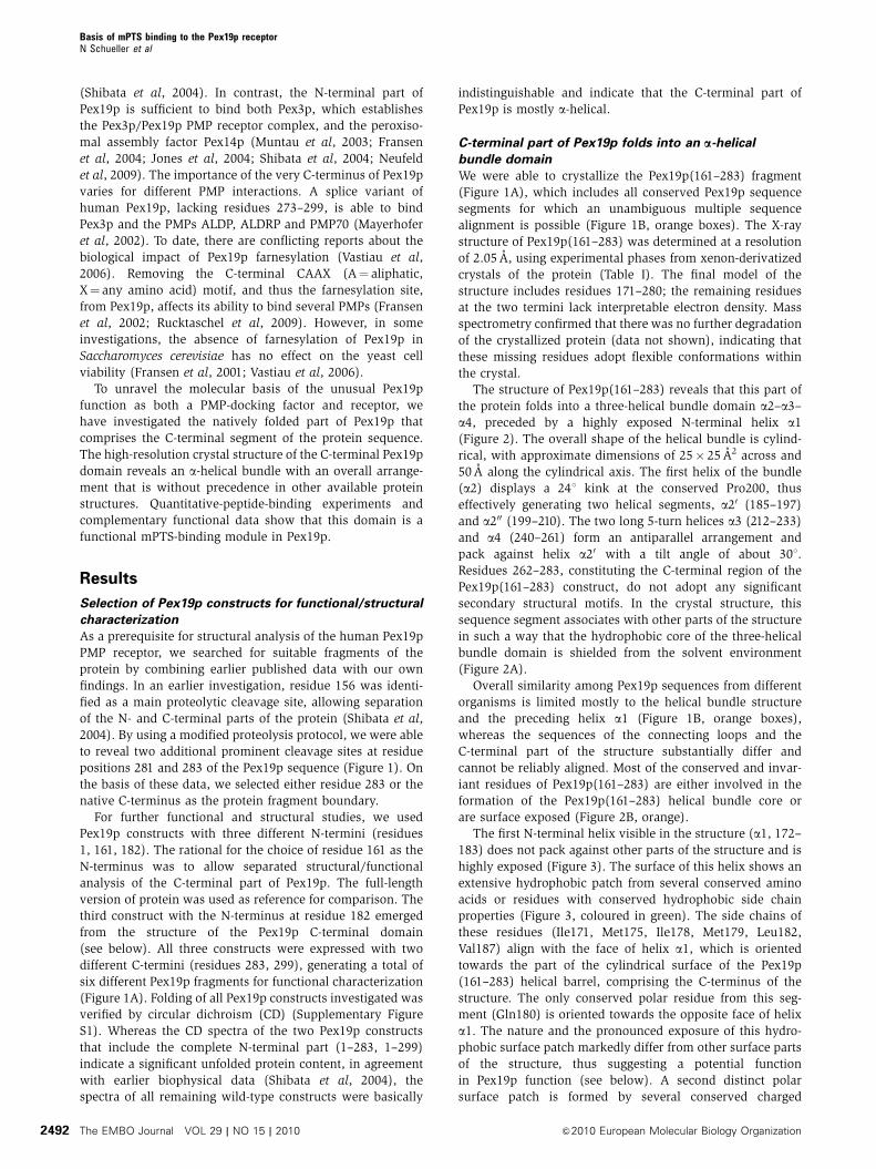
C-terminal part of Pex19p folds into an a-helical
bundle domain
We were able to crystallize the Pex19p(161–283) fragment
(Figure 1A), which includes all conserved Pex19p sequence
segments for which an unambiguous multiple sequence
alignment is possible (Figure 1B, orange boxes). The X-ray
structure of Pex19p(161–283) was determined at a resolution
of 2.05 A, using experimental phases from xenon-derivatized
crystals of the protein (Table I). The final model of the
structure includes residues 171–280; the remaining residues
at the two termini lack interpretable electron density. Mass
spectrometry confirmed that there was no further degradation
of the crystallized protein (data not shown), indicating that
these missing residues adopt flexible conformations within
the crystal.
The structure of Pex19p(161–283) reveals that this part of
the protein folds into a three-helical bundle domain a2–a3–
a4, preceded by a highly exposed N-terminal helix a1
(Figure 2). The overall shape of the helical bundle is cylind-
rical, with approximate dimensions of 25� 25 A2 across and
50 A along the cylindrical axis. The first helix of the bundle
(a2) displays a 241 kink at the conserved Pro200, thus
effectively generating two helical segments, a20 (185–197)
and a200 (199–210). The two long 5-turn helices a3 (212–233)
and a4 (240–261) form an antiparallel arrangement and
pack against helix a20 with a tilt angle of about 301.
Residues 262–283, constituting the C-terminal region of the
Pex19p(161–283) construct, do not adopt any significant
secondary structural motifs. In the crystal structure, this
sequence segment associates with other parts of the structure
in such a way that the hydrophobic core of the three-helical
bundle domain is shielded from the solvent environment
(Figure 2A).
Overall similarity among Pex19p sequences from different
organisms is limited mostly to the helical bundle structure
and the preceding helix a1 (Figure 1B, orange boxes),
whereas the sequences of the connecting loops and the
C-terminal part of the structure substantially differ and
cannot be reliably aligned. Most of the conserved and invar-
iant residues of Pex19p(161–283) are either involved in the
formation of the Pex19p(161–283) helical bundle core or
are surface exposed (Figure 2B, orange).
The first N-terminal helix visible in the structure (a1, 172–
183) does not pack against other parts of the structure and is
highly exposed (Figure 3). The surface of this helix shows an
extensive hydrophobic patch from several conserved amino
acids or residues with conserved hydrophobic side chain
properties (Figure 3, coloured in green). The side chains of
these residues (Ile171, Met175, Ile178, Met179, Leu182,
Val187) align with the face of helix a1, which is oriented
towards the part of the cylindrical surface of the Pex19p
(161–283) helical barrel, comprising the C-terminus of the
structure. The only conserved polar residue from this seg-
ment (Gln180) is oriented towards the opposite face of helix
a1. The nature and the pronounced exposure of this hydro-
phobic surface patch markedly differ from other surface parts
of the structure, thus suggesting a potential function
in Pex19p function (see below). A second distinct polar
surface patch is formed by several conserved charged
Basis of mPTS binding to the Pex19p receptorN Schueller et al
The EMBO Journal VOL 29 | NO 15 | 2010 &2010 European Molecular Biology Organization2492

residues, most of which are located on helix 2a0 (Figure 3,
coloured in magenta). One of the residues in this surface
patch, Lys193, is invariant (Figures 1 and 2B).
Interestingly, for all four protomers in the asymmetric unit,
helix a1 forms an identical antiparallel coiled-coil interaction
with the equivalent helix from either a crystallographic or
non-crystallographic symmetry-related molecule (Supple-
mentary Figure S2). However, none of our solution data
support oligomerization of Pex19p(161–283) or any other
Pex19p constructs studied in this contribution (data not
shown), suggesting that the observed assembly is transient.
Further structural features of Pex19p, including the observa-
tion of a large internal cavity, are described in the
Supplementary data.
C-terminal helical bundle constitutes the Pex19p
mPTS-binding site
In the absence of structural information for Pex19p in com-
plex with PMP target proteins, we became interested in
whether the C-terminal domain of Pex19p, as visualized in
the X-ray structure, constitutes a functional mPTS-binding
element. We, therefore, tested three different Pex19p frag-
ments (1–299, 161–283, 161–299), using an earlier estab-
lished methodology for PMP-peptide scans (Rottensteiner
et al, 2004; Halbach et al, 2005). The analysis included
seven mPTS-peptide motifs from six PMPs, which were
scanned in seven two-residue steps each, using earlier pub-
lished mPTS motifs (Halbach et al, 2005) (Figure 4A–D). The
remaining four spots of each membrane were used for control
experiments. All three Pex19p constructs showed a signifi-
cant ability for specific binding of most of the selected mPTS
peptides. The analysis revealed the strongest binding pattern
for peptides from Pex11p, Pex13p, Pex16p, ALDP and the
second mPTS motif of Pex26p.
On the basis of these observations and peptide solubility
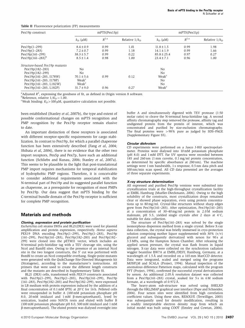
tests, we selected two mPTS peptides, Pex13p and Pex11p, for
quantitative binding affinity measurements using fluores-
cence polarization (FP) (Table II; Supplementary Figure S3).
The C-terminal Pex19p(161–283) fragment used for X-ray
structural analysis binds the two peptides with equilibrium
Figure 1 Pex19p sequence/structure relationships. (A) Pex19p fragments used for investigation; the sequence segment, whose 3D structure wedetermined in this study, is highlighted in yellow. The C-termini and N-termini of each protein fragment are listed. (B) Multiple sequencealignment of the C-terminal part of Pex19p, showing sequence/structure relationships. A representative set of Pex19p sequences is shown,including H. sapiens (UNIPROT code P40855), M. musculus (Q8VCI5), C. griseus (Q60415), R. rattus (Q9QYU1), C. elegans (P34453),H. polymorpha (Q96WN7), P. pastoris (Q9Y8C5), S. cerevisiae (Q07418), Y. lipolytica (Q96W74) and A. thaliana (Q9SRQ3). The sequences arequite divergent, thus allowing reliable multiple alignments for restricted sequence segments only (orange boxes). Invariant, highly conserved(maximum one deviating amino acid) and conserved (maximum three deviating amino acids) residue positions are indicated by ‘*’, ‘:’ and ‘.’symbols, respectively. The sequence numbers refer to the H. sapiens Pex19p sequence. The Pex19p sequence segment for which the 3Dstructure has been determined is indicated by a line above the alignment. Cylinders indicate the positions of a-helices. The mPTS-binding helixa1 is coloured in green. Those residues that are involved in the formation of the central Pex19p cavity (cf. Supplementary Figure S4) areindicated by ‘D’ symbols. Cys296, which presents the C-terminal farnesylation site, is shown in yellow characters and black background.Surface exposed polar amino acids with conserved residue properties are shown on magenta background (cf. Figure 3). Surface exposedhydrophobic amino acids with conserved residue properties are shown on green background (cf. Figure 3). The latter are clustered at theN-terminus of the Pex19p structure, covering helix a1. Sequences were aligned using CLUSTALW (Pearson, 1994), followed by manualadjustments. A full-colour version of this figure is available at The EMBO Journal Online.
Basis of mPTS binding to the Pex19p receptorN Schueller et al
&2010 European Molecular Biology Organization The EMBO Journal VOL 29 | NO 15 | 2010 2493
dissociation constants of 8.5 and 23.4 mM, respectively.
Interestingly, when adding the C-terminal tail (284–299)
that comprises the Pex19p farnesylation site, the binding
affinity decreases by about five-fold, suggesting a potentially
inhibiting effect of the non-farnesylated tail. For comparison,
Pex19p versions that include the N-terminal part of the
sequence (1–160) show about the same or slightly increased
binding affinity as Pex19p(161–283), regardless the presence
or absence of the C-terminal tail. The data thus indicate that
the C-terminal part of Pex19p is sufficient to recognize the
mPTS motif, however, potentially complemented by a pre-
sently still uncharacterized, additional function of the
N-terminal part of Pex19p in enhancing the mPTS-binding
affinity.
We were further interested in identifying the molecular
features in the C-terminal helical bundle domain of
Pex19p(161–283) that are responsible for mPTS-motif recog-
nition. The high amount of sequence conservation of residues
from helix a1 and the presence of an extensive hydrophobic
patch generated by the structure of this helix (Figures 1B
and 3, coloured in green) suggested that this region of Pex19p
could be responsible for mPTS binding. To assess this hypo-
thesis, we expressed and purified two additional versions of
Pex19p that lack the corresponding sequence segment (182–
283, 182–299). Comparative analysis of the CD spectra of
these Pex19p fragments show that their fold content is
virtually indistinguishable from other C-terminal Pex19p
constructs (Supplementary Figure S1). Initial pepblot analysis
revealed that the truncated Pex19p(182–283) construct basi-
cally has lost the ability to bind to any of the mPTS peptides
used in this qualitative investigation (Figure 4E). Quantitative
FP experiments confirmed that for both Pex19p fragments
lacking the sequence segment that includes helix a1,
Pex19p(182–283) and Pex19p(182–299) have entirely lost
the ability to bind to both mPTS peptides (Table II), showing
that the presence of helix a1 is essential for mPTS
recognition.
In an attempt to further locate residue-specific mPTS-
binding sites, we mutated two conserved surface residues
from helix a1, Ile178 and Leu182 (Figures 1B and 3), either
into a tryptophan or proline, within the Pex19p(161–283)
construct that was used for 3D structure determination.
The rational of these mutations was to test for potential steric
Table I Crystallographic statistics
Native Xenon derivatized
Data collectionSpace group P212121 P212121
Cell dimensions (A) a¼ 67.1 a¼ 67.6b¼ 91.0 b¼ 91.3c¼ 122.3 c¼ 127.7
Wavelength (A) 0.98 1.50Overall resolution range (A) 73–2.05 55–2.87Highest resolution range (A) 2.16–2.05 3.10–2.87Number of unique reflections 47 467 17 855Multiplicity 6.2 20.3Mean I/s(I) 7.6 (2.0) 6.2 (2.7)Completeness (%) 99.6 (100) 99.6 (97.1)Anomalous completeness (%) — 99.1 (96.0)Ra
sym 6.2 (45.2) 7.2 (26.2)Mosaicity (deg) 0.38 0.46
PhasingFigure of merit (acentric/centric) — 0.70/0.62
(0.34/0.29)Number of sites — 4
RefinementProtein atoms 3717Other atoms 192
Rbconv/Rc
free 21.5/25.0RMS deviations
Bond lengths (A) 0.015Bond angles (deg) 1.574
aRsym ¼P
h
Pj Ih;j � Ihh i��
��P
h
Pj Ih;j, where Ih,j is the intensity of
the jth observation of unique reflection h.bRconv ¼
Ph jFoh
j � jFchj
��
��=P
h jFohj, where Foh
and Fchare the
observed and calculated structure factor amplitudes for reflection h.cRfree is equivalent to Rconv, but is calculated using a 5% disjoint setof reflections excluded from the maximum likelihood refinementstages.
Figure 2 Crystal structure of the mPTS-binding domain of Pex19p(161–283). (A) Ribbon representation of the Pex19p(161–283) structure.The secondary structural elements and the termini of the visible part of the Pex19p(161–283) sequence are labelled. Those parts of the structurefor which a reliable multiple sequence alignment exists (orange boxes, cf. Figure 1) are shown as orange ribbons. (B) Side chains of invariantand highly conserved residues (for definition see Figure 1 caption) of the Pex19p(161–283) structure are shown in orange and yellow,respectively, in stick presentation and are labelled. Oxygen and nitrogen atoms are shown in red and blue, respectively. The illustration revealsthat the hydrophobic core of the Pex19p(161–283) helical bundle is most conserved. A full-colour version of this figure is available at TheEMBO Journal Online.
Basis of mPTS binding to the Pex19p receptorN Schueller et al
The EMBO Journal VOL 29 | NO 15 | 2010 &2010 European Molecular Biology Organization2494

clashes and for structural interference of the formation of
helix a1. As expected, the Pex19p(161–283, I178P) and
Pex19p(161–283, L182P) variants show a slight but signifi-
cant loss of secondary structural content, most likely because
of disruption of helix a1 because of the insertion of a proline
residue, whereas the spectra of the two equivalent tryptophan
variants were basically indistinguishable with wild-type
Pex19p(161–283) (Supplementary Figure S1, panels C and D).
All Pex19p mutants led to a complete or almost complete
loss of binding of the Pex11p mPTS peptide, whereas the
observed effects on Pex13p binding were not as strong
(Table II). Collectively, the data indicate that the binding
modes of the two mPTS motifs are probably not identical,
which may reflect the lack of specific, directed interactions
within a Pex19p-mPTS interface that seems to be dominated
by hydrophobic residues from the Pex19p helix a1 (Figure 3)
and the two mPTS motifs.
We also tested the importance of helix a1 in Pex19p for
mPTS binding in vivo by monitoring Pex19p variants for their
capacity to functionally complement a peroxisome biogenesis
defect in PEX19-deficient fibroblasts. Functional complemen-
tation restores correct topogenesis of peroxisomal matrix and
membrane proteins and formation of peroxisomes. Thus,
only those cells in which functional Pex19p enables the
correct assembly of peroxin complexes at the peroxisomal
membrane exhibit a punctuate pattern for the PTS1-cargo
EGFP-SCP, by co-localizing it with the endogenous PMP
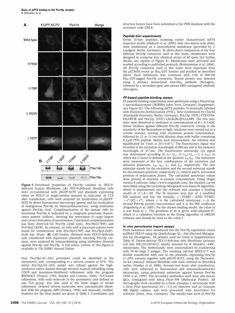
Pex14p (Figure 5). For this assay, we used the same single-
residue Pex19p variants that were investigated for in vitro
binding of mPTS peptides, however, in context of the full-
length Pex19p sequence (Figure 3). In contrast to the trypto-
phan variants of Pex19p (I178W, L182W), which showed
only mild defects, the proline mutants (I178P, L182P) could
not rescue the import defect of the Pex19p-deficient cells for
EGFP-SCP2 (Figure 5A). After careful inspection of three
independent transfection experiments, we could not detect
a single cell with a punctuate Pex14p and GFP-SCP2 pattern,
although immunolabelling with antibodies directed against
Pex19p revealed a transfection rate of up to 50% (data not
shown). Western blotting of lysates of the transfected cells
revealed similar steady-state levels for all Pex19p variants
(Figure 5B). In conclusion, our in vitro mPTS-binding data,
which characterized both a Pex19p construct lacking the
N-terminal helix (182–283) and four additional single-residue
mutants, together with complementary in vivo functional
readout data, which are believed to be sensitive to the
complete pool of PMPs needed for peroxisome biogenesis,
both identify the same N-terminal region of the C-terminal
Pex19p helical bundle domain to be critically involved in
mPTS recognition. To what extent the mPTS-binding site is
conserved for different PMPs and whether other Pex19p
sequence regions are additionally involved, still remain to
be determined.
Discussion
Structure of Pex19p(161–283) defines an autonomous
mPTS-binding domain
The structural and functional investigation of the C-terminal
Pex19p mPTS-binding domain allows us to review earlier
findings of the function of Pex19p as a PMP receptor/chaper-
one (Jones et al, 2004). FP analysis with two mPTS peptides
from Pex11p and Pex13p shows that the autonomously folded
C-terminal Pex19p(161–283) fragment is sufficient to recog-
nize the mPTS motifs from at least two PMP targets (Table II;
Supplementary Figure S3), thus allowing its assignment
as the Pex19p mPTS-binding domain. These findings are in
agreement with a recent study in which penta-peptides were
systematically inserted to assess the ability of Pex19p to bind
to class-I PMP targets (Fransen et al, 2005). All insertion sites
that were shown to abolish PMP binding are within the
sequence boundaries of the Pex19p(161–283) crystal struc-
ture (Supplementary Figure S4). In addition, the loss-of-
function insertions are generally located within the a-helices
of the structure, whereas most of the insertions with no
observed effect are found within the connecting loops.
Figure 3 Pex19p(161–283) surface patches with conserved residue properties. (A) Worm-type ribbon representation of the Pex19p(161–283)structure; (B) semi-transparent surface and ribbon representation of the Pex19p(161–283) structure in two different orientations, in which theright panel is rotated around a vertical axis in the paper plane by 1801 with respect to the left panel. The side chain contributions of polarsurface residues with conserved amino-acid properties are shown in magenta. The side chain contributions of hydrophobic residues withconserved amino-acid properties are shown in green. All coloured residues are labelled. In the worm-type presentation of the Pex19p(161–283)structure, oxygen, nitrogen and sulphur atoms are shown in atom-specific colours: red, blue, yellow, respectively. A full-colour version of thisfigure is available at The EMBO Journal Online.
Basis of mPTS binding to the Pex19p receptorN Schueller et al
&2010 European Molecular Biology Organization The EMBO Journal VOL 29 | NO 15 | 2010 2495

Thus, the data indicate that the structural integrity of the
helical bundle found in the Pex19p structure is critical to
recognize PMPs (Fransen et al, 2005).
A recent study unravelled that, while several Pex19p
fragments truncated at residue 261 retain peroxisome-restor-
ing activity, slightly shorter Pex19p constructs, truncated at
residue 255, have lost this functional ability (Matsuzono et al,
2006). In the structure of the Pex19p mPTS-binding domain,
residue 261 coincides with the C-terminus of helix a4
(Figure 2). As the preceding residues (255–260) are involved
in the packing of the antiparallel arrangement of the two long
helices a3 and a4, the Pex19p truncation data can be inter-
preted as an indication that the structural integrity of the
C-terminal Pex19p domain is critical for its function as an
mPTS-binding epitope. The same region is also highly sensi-
tive to structural perturbations (Supplementary Figure S4).
As the first helix a1 points away from the remaining core
of the helical bundle in the structure of Pex19p(161–283) and
is highly conserved (Figures 1 and 3), we investigated its
contribution to mPTS binding separately. A Pex19p(182–283)
variant without this helix almost completely abolished the
ability to bind several mPTS motifs (Table II), indicating that
the corresponding sequence segment in Pex19p is critical for
mPTS recognition. These data were corroborated by in vivo
assays in Pex19-deficient fibroblasts, which confirmed the
importance of the helical structure of this sequence segment
for maintaining a functional peroxisomal translocation
system (Figure 5).
Potential functions of Pex19p sequence segments
flanking the C-terminal helical bundle domain
Interestingly, we found a moderate effect in enhancing the
overall binding affinity in the presence of the N-terminal part
of Pex19p(1–160) (Table II). These data support earlier
investigations, which indicated that most of the N-terminal
part of Pex19p, which is associated with Pex3p binding and
membrane targeting, is required for PMP recognition as well
(Shibata et al, 2004; Matsuzono et al, 2006). These differ-
ences could reflect additional functional requirements of
Pex19p for PMP recognition, stabilization and import beyond
mPTS recognition. These considerations are supported by
recent findings that PMP-cargo loading of Pex19p leads to
increased binding of the N-terminal part of Pex19p to Pex3p,
which is membrane associated and has been characterized as
a PMP-docking factor (Pinto et al, 2006). Interestingly, there
is also increasing evidence for non-mPTS-binding sites in
several PMP targets (Vizeacoumar et al, 2006), thus raising
questions about the exclusiveness of mPTS-binding sites for
PMP recognition by Pex19p.
Emerging molecular mechanisms in peroxisomal
import receptors
To date, three peroxins have been identified and character-
ized to function as import receptors: Pex5p, Pex7p and
Pex19p. In contrast to the PMP receptor/chaperone Pex19p,
Pex5p and Pex7p are receptors for peroxisomal matrix
proteins with specific recognition signal sequences (Stanley
et al, 2007a). Interestingly, the PTS/cargo-binding modules of
all three receptors assemble with co-receptor components
either by intramolecular interactions with additional domains
or by hetero-assemblies with other peroxins (Schliebs and
Kunau, 2006), which together allow targeting of the respec-
tive receptor/cargo complexes to the peroxisomal membrane.
Notably, Pex5p and Pex19p share the same sequence
division, comprising a folded C-terminal PTS-binding domain
and a less folded N-terminal segment that is involved
in membrane docking of the respective receptor/cargo
complexes. Moreover, the two peroxisomal receptors display
similar a-helical bundle arrangements. However, whereas
considerable conformational changes in the seven-fold
repeated TPR array in Pex5p on cargo recognition have
Figure 4 In vitro binding of mPTS-peptide motifs by Pex19p.Peptide blot data showing the binding profile of several Pex19pfragments to mPTS peptides. (A) Layout of the mPTS motifs(Halbach et al, 2005), consisting of 27 residues that were scannedover with 15-mer peptides in two-residue steps, indicated by Pex2p(residues 141–167, FV0IG0LL0KL0GG0LI0NFLIFLQRGKFATLT, spots1–7), Pex11p-b (residues 180–206, GG0GL0PQ0LA0LK0LR0LQVLLLARVLRGHPP, spots 8–14), Pex13p (residues 171–197, LK0IH0FT0KV0
FS0AF0ALVRTIRYLYRRLQR, spots 15–21), Pex16p (residues 109–135,RW0LV0IA0LI0QL0AK0AVLRMLLLLWFKAGL, spots 22–28), ALDP(residues 62–88, AA0KA0GM0NR0VF0LQ0RLLWLLRLLFPRVLC, spots29–35), Pex26p motif-1 (residues 247–273, FF0SL0PF0KK0SL0LA0ALILCLLVVRFDPAS, spots 36–42), Pex26p motif-2 (residues 270–296,DP0AS0PS0SL0HF0LY0KLAQLFRWIRKAAFS, spots 43–49). In addition,the sequence segment covering helix a1 of Pex19p (residues164–190, EG0DG0EG0NI0LP0IM0QSIMQNLLSKDVLYP, spots 50–56),the C-terminal PTS1 sequence of mSCP2 (residues 129–143,MKLQNLQLQPGNAKL, spot 57, negative control), a poly-histidinesequence (HHHHHH, spots 58–59, positive control) and an emptyspot (60, negative control) were analysed. Spot numbers areindicated to the left and to the right. Experimental data are shownin (B) Pex19p(1–299), (C) Pex19p(161–299), (D) Pex19p(161–283)and (E) Pex19p(182–283).
Basis of mPTS binding to the Pex19p receptorN Schueller et al
The EMBO Journal VOL 29 | NO 15 | 2010 &2010 European Molecular Biology Organization2496
been established (Stanley et al, 2007b), the type and extent of
possible conformational changes on mPTS recognition and
PMP recognition by the Pex19p receptor remain elusive
to date.
An important distinction of these receptors is associated
with different receptor-specific requirements for cargo stabi-
lization. In contrast to Pex19p, for which a parallel chaperone
function has been extensively described (Fang et al, 2004;
Shibata et al, 2004), there is no evidence that the other two
import receptors, Pex5p and Pex7p, have such an additional
function (Schliebs and Kunau, 2006; Stanley et al, 2007a).
This seems to be plausible in the light that post-translational
PMP import requires mechanisms for temporal stabilization
of hydrophobic PMP regions. Therefore, it is conceivable
to consider additional requirements associated with the
N-terminal part of Pex19p and its suggested parallel function
as chaperone, as a prerequisite for recognition of most PMPs
by Pex19p. Our data suggest that mPTS binding by the
C-terminal bundle domain of the Pex19p receptor is sufficient
for complete PMP recognition.
Materials and methods
Cloning, expression and protein purificationEscherichia coli strains DH5a and BL21(DE3) were used for plasmidamplification and protein expression, respectively. Homo sapiensPEX19 DNA encoding Pex19p(1–299), Pex19p(1–283), Pex19p(161–299), Pex19p(161–283), Pex19p(182–283) and Pex19p(182–299) were cloned into the pETM11 vector, which includes anN-terminal poly-histidine tag with a TEV cleavage site, using theNcoI and BamHI sites. Owing to the presence of an internal NcoIsite in the two longest constructs, the 50 cut was performed withBsmBI to create an NcoI compatible overhang. Single-point mutantswere generated with the QuikChange Site-Directed Mutagenesis kit(Stratagene), according to the manufacturer’s instructions. Allprimers that were used to create the different length constructsand the mutants are described in Supplementary Table SI.
BL21 (DE3) cells, transformed with PEX19 constructs associatedwith Pex19p(1–299), Pex19p(1–283), Pex19p(161–299), Pex19p(161–283), Pex19p(182–283) and Pex19p(182–299), were expressedin LB medium with protein expression induced by the addition of afinal concentration of 0.1 mM IPTG at 201C for 16 h. Pelleted cellswere resuspended in buffer A (100 mM potassium phosphate pH8.0, 20 mM imidazol and 1 mM b-mercaptoethanol), lysed bysonication, loaded onto NiNTA resin and eluted with buffer B(100 mM potassium phosphate pH 8.0, 500 mM imidazol and 1 mMb-mercaptoethanol). The eluted protein was dialysed overnight into
buffer A and simultaneously digested with TEV protease (1:50molar ratio) to cleave the N-terminal hexa-histidine tag. A secondaffinity chromatography step removed the protease, affinity tag andundigested protein from the protein of interest, which wasconcentrated and purified by size-exclusion chromatography.The final proteins were 498% pure as judged by SDS–PAGE(Supplementary Figure S5).
Circular dichroismCD experiments were performed on a Jasco J-810 spectropolari-meter. Proteins were dialysed into 10 mM potassium phosphate(pH 8.0) and 1 mM DTT. Far UV spectra were recorded between185 and 260 nm (1 mm cuvette, 0.1 mg/ml protein concentration,as determined by specific absorbance at 280 nm). The machinesettings were 1 nm bandwidth, 1 s response, 0.5 nm data pitch and100 nm/min scan speed. All CD data presented are the averagesof three separate experiments.
X-ray structure determinationAll expressed and purified Pex19p versions were submitted intocrystallization trials at the high-throughput crystallization facilityat EMBL Hamburg (Mueller-Dieckmann, 2006). Owing to the highsolubility of the constructs, most crystallization drops remainedclear or showed phase separation, even using protein concentra-tions up to 80 mg/ml. Crystal-like structures without sharp edgesgrew from Pex19p(161–283). After optimization, Pex19p(161–283)at a concentration of 30–40 mg/ml protein in 2.0 M sodiummalonate, pH 5.5, yielded single crystals after 2 days at 41C,suitable for data collection.
The structure of Pex19p(161–283) was solved by the singleisomorphous dispersion method from a single crystal. Before X-raydata collection, the crystal was briefly immersed in cryo-protectionsolution comprising mother liquor supplemented with 30% (v/v)glycerol and subsequently derivatized with xenon for 90 s at3.5 MPa, using the Hampton Xenon Chamber. After releasing theapplied xenon pressure, the crystal was flash frozen in liquidnitrogen. X-ray data were collected on the synchrotron radiationwiggler beamline BW7A at EMBL/DESY, Hamburg, Germany, at awavelength of 1.5 A and recorded on a 165 mm MarCCD detector.Data were integrated, scaled and merged using the programsMOSFLM and SCALA (Project, 1994). Manual inspection of theanomalous difference Patterson maps, calculated with the programFFT (Project, 1994), confirmed the successful crystal derivatizationby xenon. An additional 2.05 A resolution dataset was collectedfrom a Pex19p(161–283) crystal, soaked for 5 s in 6 M sodiumformate, at a wavelength of 0.972 A.
The heavy-atom sub-structure was solved using SHELXDthrough the HKL2MAP graphical user interface (Pape and Schneider,2004). Four xenon sites were identified from high correlationcoefficient values. Using these sites, RESOLVE (Terwilliger, 2003)was subsequently used for density modification, resulting ina readily interpretable electron density map from which aninitial model was built using COOT (Emsley and Cowtan, 2004).
Table II Fluorescence polarization (FP) measurements
Pex19p construct mPTS(Pex13p) mPTS(Pex11p)
kD (mM) R2 a Relative 1/kD kD (mM) R2 a Relative 1/kD
Pex19p(1–299) 8.4±0.9 0.99 1.01 11.8±1.5 0.99 1.98Pex19p(1–283) 7.2±0.7 0.99 1.18 14.1±1.9 0.99 1.66Pex19p(161–299) 38.3±5.7 0.99 0.22 85.8±25.4 0.97 0.27Pex19p(161–283)b 8.5±1.4 0.98 1.00 23.4±7.1 0.96 1.00
Structure-based Pex19p mutantsPex19p(182–283) No NoPex19p(182–299) No NoPex19p(161–283, I178W) 70.1±5.6 0.99 0.12 Weakc
Pex19p(161–283, I178P) Weakc NoPex19p(161–283, L182W) Weakc NoPex19p(161–283, L182P) 31.7±9.0 0.96 0.27 Weakc
aAdjusted R2, expressing the goodness of fit, as defined in Origin version 8 software.bReference, relative 1/KD¼ 1.00.cWeak binding: KD4100mM, quantitative calculation not possible.
Basis of mPTS binding to the Pex19p receptorN Schueller et al
&2010 European Molecular Biology Organization The EMBO Journal VOL 29 | NO 15 | 2010 2497
Four Pex19p(161–283) protomers could be identified in theasymmetric unit, corresponding to a solvent content of 65%. Theinitial Pex19p(161–283) model was refined against the high-resolution native dataset through iterative manual rebuilding usingCOOT and maximum-likelihood refinement with the programREFMAC5 (Project, 1994; Emsley and Cowtan, 2004). TLS-basedrefinement, with each molecule in the asymmetric unit defined asone TLS group, was also used in the latter stages of modelrefinement. Ordered solvent molecules were automatically identi-fied with the program ARP (Project, 1994) and manually verified.Statistics for the final model are given in Table I. Coordinates and
structure factors have been submitted to the PDB database with theaccession code 2WL8.
Peptide blot experimentsSeven 15-mer peptides, scanning earlier characterized mPTSsequence motifs (Halbach et al, 2005) with two-amino-acid shifts,were synthesized on a nitrocellulose membrane (provided by CLandgraf, Berlin, Germany). To allow direct comparison of the fourdifferent Pex19p constructs used in this study, membranes weredesigned to comprise four identical arrays of 60 spots (for furtherdetails, see caption of Figure 4). Membranes were activated andwashed according to published protocols (Rottensteiner et al, 2004).All Pex19p constructs used in this study were expressed fromthe pETM30 vector as His6-GST fusions and purified as describedabove. Each membrane was overlayed with 3 ml of 800 nMHis6-GST-tagged Pex19p constructs. Bound protein was detectedusing a primary monoclonal Anti-His6 antibody (Novagen),followed by a secondary goat anti-mouse HRP-conjugated antibody(Novagen).
FP-based peptide-binding assaysFP peptide-binding experiments were performed using a FluoroLog-3 spectrofluorometer (HORIBA Jobin Yvon, Germany) (Supplemen-tary Figure S2). The following mPTS peptides, N-terminally labelledwith fluorescein isothiocyanate (FITC), were synthesized by Charite(Humboldt University, Berlin, Germany): Pex13p, (FITC)-FTKVFSA-FALVRTIR and Pex11p, (FITC)-LALKLRLQVLLLARV. The two pep-tides were dissolved in methanol at concentrations of 0.1–0.5 mM,before titration against different Pex19p constructs. Owing to thesensitivity of the fluorophore to light, titrations were carried out in areverse manner, starting with maximum protein concentration,followed by 12 or 13 two-fold dilution steps with buffer containing0.2mM FITC peptide. Before each measurement, the solution wasequilibrated for 5 min at 20±0.011C. The fluorescence signal wasrecorded at the excitation wavelength of 488 nm and at the emissionwavelength of 517 nm. The fluorescence anisotropy (A) signalwas determined according to A¼ (Ivv�G � Ivh)/(IvvþG � 2 � Ivh), inwhich the G-factor is defined as the quotient Ivh/Ihh. The intensitieswere measured at the four combinations of the excitation andemission polarizers, Ivh, Ihh, Ivv and Ivh, respectively. The firstsubscript stands for the excitation and the second subscript standsfor the emission polarizer, respectively (v, vertical and h, horizontalposition of polarization plane). The calculated anisotropy valueswere plotted as function of protein concentration. Using Originversion 8 software (http://www.originlab.com), the titration curveswere fitted using the Levenberg–Marquardt non-linear fit algorithm,which is implemented into the software and assumes a bindingmodel of AþB¼AB. The fit function was that of a modifiedHill analysis and has the formula: y¼ STARTþ (END�START)� xn/(Kd
nþ xn), where y is the calculated anisotropy, x is thetitrated Pex19p protein concentration and n is the Hill coefficient(Pogenberg et al, 2005). For the chosen binding model, the value ofn was fixed to 1. The goodness of fit is given with adjusted R2,which is a validation function in the fitting algorithm of ORIGINsoftware and should be close to the value 1.
In vivo peroxisome import assaysPoint mutations were introduced into the Pex19p expression vectorpcDNA3-PEX19 using the Quickchange XL—Site-Directed Mutagen-esis kit (Stratagene). All primers used are listed in SupplementaryTable SI. Patient-derived PEX19-deficient skin fibroblasts (primarycell line RW/mf/0854872, kindly donated by R Wanders, AMC,Amsterdam, The Netherlands) were immortalized by transfectingwith SV-40 large T antigen. The resulting cell-line DPEX19 T wasdouble transfected with one of the plasmids expressing Pex19p(1–299) variants together with pEGFP-SCP2, using the Nucleofec-tor-kit (Amaxa). Human fibroblast cells were cultured as describedearlier (Stanley et al, 2006). Seventy-two hours after transfection,cells were subjected to fluorescence and immunofluorescencemicroscopy, using polyclonal antiserum against human Pex14p(Will et al, 1999). The secondary antibodies used were anti-mouseIgGs, conjugated with Alexa Fluor-594 (Molecular Probes). Allmicrographs were recorded on a Zeiss Axioplan 2 microscope witha Zeiss Plan-Apochromat 63� /1.4 oil objective and an AxiocamMR digital camera, and were processed with AxioVision 4.6software (Zeiss, Jena, Germany). The steady-state level of Pex19p
Figure 5 Functional properties of Pex19p variants in PEX19-deficient human fibroblasts. (A) PEX19-deficient fibroblast cellswere co-transfected with pEGFP-SCP2 and plasmids expressingeither wt PEX19 or single-residue mutants as indicated. At 72 hafter transfection, cells were analysed for localization of pEGFP-SCP2 by direct fluorescence microscopy (green) and for localizationof endogenous Pex14p by immunofluorescence, using antibodiesagainst Pex14p (red). Complementation of the mutant cells byfunctional Pex19p is indicated by a congruent punctuate fluores-cence pattern (yellow), showing the restoration of cargo importand correct formation of peroxisomes. Functional complementationhas been observed for Pex19p (wild type), Pex19p(I178W) andPex19p(L182W). In contrast, no cells with a punctuate pattern werefound for transfections with Pex19p(I178P) and Pex19p(L182P).Scale bar, 10mm. (B) Cell lysates, obtained from PEX19-deficientcells transfected with expression plasmids encoding Pex19p var-iants, were analysed by immunoblotting using antibodies directedagainst Pex14p and Pex19p. A full-colour version of this figure isavailable at The EMBO Journal Online.
Basis of mPTS binding to the Pex19p receptorN Schueller et al
The EMBO Journal VOL 29 | NO 15 | 2010 &2010 European Molecular Biology Organization2498
expression was assessed by immunoblot analyses, using mono-clonal mouse antibodies against HsPex19p (BD Biosciences).
Supplementary dataSupplementary data are available at The EMBO Journal Online(http://www.embojournal.org).
Acknowledgements
We thank Janica Meine for laboratory support and Marc Fransen(Katholieke Universiteit Leuven, Belgium) for fruitful discussionsduring the early phase of the project. Ron Wanders (AMCAmsterdam, The Netherlands) has kindly provided PEX19-deficientskin fibroblasts. Manfred Roessle is thanked for technical assistanceto obtain SAXS data. Vivian Pogenberg is thanked for critical
assessment and advice on fluorescence-polarization experiments.Chris Williams (EMBL) is thanked for critical comments on theexperimental approach and the paper. The project has been sup-ported by 3D-REPERTOIRE (EC, LSHG-CT-2005512028) to MW.
Author contributions: NS and SJH designed and carried out mostof the experiments and contributed to writing of the paper. KF,MM, WAS, YHS, JW and WS carried out specific experimentsand wrote the respective experiment sections in the paper. REsupported the work by JW and WS. MW coordinated the projectand wrote the paper.
Conflict of interest
The authors declare that they have no conflict of interest.
References
Eckert JH, Erdmann R (2003) Peroxisome biogenesis. Rev PhysiolBiochem Pharmacol 147: 75–121
Emsley P, Cowtan K (2004) Coot: model-building tools for molecu-lar graphics. Acta Crystallogr D Biol Crystallogr 60 (Part 12 Part 1):2126–2132
Fang Y, Morrell JC, Jones JM, Gould SJ (2004) PEX3 functions as aPEX19 docking factor in the import of class I peroxisomalmembrane proteins. J Cell Biol 164: 863–875
Fransen M, Brees C, Ghys K, Amery L, Mannaerts GP, Ladant D, VanVeldhoven PP (2002) Analysis of mammalian peroxin interactionsusing a non-transcription-based bacterial two-hybrid assay.Mol Cell Proteomics 1: 243–252
Fransen M, Vastiau I, Brees C, Brys V, Mannaerts GP, Van VeldhovenPP (2004) Potential role for Pex19p in assembly of PTS-receptordocking complexes. J Biol Chem 279: 12615–12624
Fransen M, Vastiau I, Brees C, Brys V, Mannaerts GP, Van VeldhovenPP (2005) Analysis of human Pex19p’s domain structureby pentapeptide scanning mutagenesis. J Mol Biol 346:1275–1286
Fransen M, Wylin T, Brees C, Mannaerts GP, Van Veldhoven PP(2001) Human pex19p binds peroxisomal integral membraneproteins at regions distinct from their sorting sequences. MolCell Biol 21: 4413–4424
Gatto Jr GJ, Geisbrecht BV, Gould SJ, Berg JM (2000) Peroxisomaltargeting signal-1 recognition by the TPR domains of humanPEX5. Nat Struct Biol 7: 1091–1095
Gotte K, Girzalsky W, Linkert M, Baumgart E, Kammerer S,Kunau WH, Erdmann R (1998) Pex19p, a farnesylatedprotein essential for peroxisome biogenesis. Mol Cell Biol 18:616–628
Halbach A, Landgraf C, Lorenzen S, Rosenkranz K, Volkmer-EngertR, Erdmann R, Rottensteiner H (2006) Targeting of the tail-anchored peroxisomal membrane proteins PEX26 and PEX15occurs through C-terminal PEX19-binding sites. J Cell Sci 119(Part 12): 2508–2517
Halbach A, Lorenzen S, Landgraf C, Volkmer-Engert R, Erdmann R,Rottensteiner H (2005) Function of the PEX19-binding site ofhuman adrenoleukodystrophy protein as targeting motif in manand yeast. PMP targeting is evolutionarily conserved. J Biol Chem280: 21176–21182
Heiland I, Erdmann R (2005) Biogenesis of peroxisomes.Topogenesis of the peroxisomal membrane and matrix proteins.FEBS J 272: 2362–2372
Hoepfner D, Schildknegt D, Braakman I, Philippsen P, Tabak HF(2005) Contribution of the endoplasmic reticulum to peroxisomeformation. Cell 122: 85–95
Jones JM, Morrell JC, Gould SJ (2001) Multiple distinct targetingsignals in integral peroxisomal membrane proteins. J Cell Biol153: 1141–1150
Jones JM, Morrell JC, Gould SJ (2004) PEX19 is a predominantlycytosolic chaperone and import receptor for class 1 peroxisomalmembrane proteins. J Cell Biol 164: 57–67
Kammerer S, Arnold N, Gutensohn W, Mewes HW, Kunau WH,Hofler G, Roscher AA, Braun A (1997) Genomic organization andmolecular characterization of a gene encoding HsPXF, a humanperoxisomal farnesylated protein. Genomics 45: 200–210
Matsuzaki T, Fujiki Y (2008) The peroxisomal membrane proteinimport receptor Pex3p is directly transported to peroxisomes by anovel Pex19p- and Pex16p-dependent pathway. J Cell Biol 183:1275–1286
Matsuzono Y, Kinoshita N, Tamura S, Shimozawa N, Hamasaki M,Ghaedi K, Wanders RJ, Suzuki Y, Kondo N, Fujiki Y (1999)Human PEX19: cDNA cloning by functional complementation,mutation analysis in a patient with Zellweger syndrome, andpotential role in peroxisomal membrane assembly. Proc Natl AcadSci USA 96: 2116–2121
Matsuzono Y, Matsuzaki T, Fujiki Y (2006) Functional domainmapping of peroxin Pex19p: interaction with Pex3p isessential for function and translocation. J Cell Sci 119 (Part 17):3539–3550
Mayerhofer PU, Kattenfeld T, Roscher AA, Muntau AC (2002)Two splice variants of human PEX19 exhibit distinct functionsin peroxisomal assembly. Biochem Biophys Res Commun 291:1180–1186
Mueller-Dieckmann J (2006) The open-access high-throughputcrystallization facility at EMBL Hamburg. Acta Crystallogr DBiol Crystallogr 62 (Part 12): 1446–1452
Muntau AC, Roscher AA, Kunau WH, Dodt G (2003) Interaction ofPEX3 and PEX19 visualized by fluorescence resonance energytransfer (FRET). Adv Exp Med Biol 544: 221–224
Neufeld C, Filipp FV, Simon B, Neuhaus A, Schuller N, David C,Kooshapur H, Madl T, Erdmann R, Schliebs W, Wilmanns M,Sattler M (2009) Structural basis for competitive interactions ofPex14 with the import receptors Pex5 and Pex19. EMBO J 28:745–754
Pape T, Schneider TR (2004) HKL2MAP: a graphical user interfacefor phasing with SHELX programs. J Appl Cryst 37: 843–844
Pearson WR (1994) Using the FASTA program to search protein andDNA sequence databases. Methods Mol Biol 24: 307–331
Pinto MP, Grou CP, Alencastre IS, Oliveira ME, Sa-Miranda C,Fransen M, Azevedo JE (2006) The import competence of aperoxisomal membrane protein is determined by Pex19p beforethe docking step. J Biol Chem 281: 34492–34502
Pogenberg V, Guichou JF, Vivat-Hannah V, Kammerer S, Perez E,Germain P, de Lera AR, Gronemeyer H, Royer CA, Bourguet W(2005) Characterization of the interaction between retinoic acidreceptor/retinoid X receptor (RAR/RXR) heterodimers and trans-criptional coactivators through structural and fluorescenceanisotropy studies. J Biol Chem 280: 1625–1633
Project CC (1994) The CCP4 suite: programs for protein crystal-lography. Acta Cryst D 50: 760–763
Rottensteiner H, Kramer A, Lorenzen S, Stein K, Landgraf C,Volkmer-Engert R, Erdmann R (2004) Peroxisomal membraneproteins contain common Pex19p-binding sites that are anintegral part of their targeting signals. Mol Biol Cell 15: 3406–3417
Rucktaschel R, Thoms S, Sidorovitch V, Halbach A, Pechlivanis M,Volkmer-Engert R, Alexandrov K, Kuhlmann J, Rottensteiner H,Erdmann R (2009) Farnesylation of Pex19p is required forits structural integrity and function in peroxisome biogenesis.J Biol Chem 284: 20885–20896
Schliebs W, Kunau WH (2006) PTS2 co-receptors: diverse proteinswith common features. Biochim Biophys Acta 1763: 1605–1612
Basis of mPTS binding to the Pex19p receptorN Schueller et al
&2010 European Molecular Biology Organization The EMBO Journal VOL 29 | NO 15 | 2010 2499

Shibata H, Kashiwayama Y, Imanaka T, Kato H (2004) Domainarchitecture and activity of human Pex19p, a chaperone-likeprotein for intracellular trafficking of peroxisomal membraneproteins. J Biol Chem 279: 38486–38494
Stanley WA, Filipp FV, Kursula P, Schuller N, Erdmann R, SchliebsW, Sattler M, Wilmanns M (2006) Recognition of a functionalperoxisome type 1 target by the dynamic import receptor pex5p.Mol Cell 24: 653–663
Stanley WA, Fodor K, Marti-Renom MA, Schliebs W, Wilmanns M(2007a) Protein translocation into peroxisomes by ring-shapedimport receptors. FEBS Lett 581: 4795–4802
Stanley WA, Pursiainen NV, Garman EF, Juffer AH, Wilmanns M,Kursula P (2007b) A previously unobserved conformation forthe human Pex5p receptor suggests roles for intrinsicflexibility and rigid domain motions in ligand binding.BMC Struct Biol 7: 24
Terwilliger TC (2003) Automated main-chain model building bytemplate matching and iterative fragment extension. ActaCrystallogr D Biol Crystallogr 59 (Part 1): 38–44
Vastiau IM, Anthonio EA, Brams M, Brees C, Young SG, Van deVelde S, Wanders RJ, Mannaerts GP, Baes M, Van Veldhoven PP,Fransen M (2006) Farnesylation of Pex19p is not essential forperoxisome biogenesis in yeast and mammalian cells. Cell MolLife Sci 63: 1686–1699
Vizeacoumar FJ, Vreden WN, Aitchison JD, Rachubinski RA (2006)Pex19p binds Pex30p and Pex32p at regions required for theirperoxisomal localization but separate from their peroxisomaltargeting signals. J Biol Chem 281: 14805–14812
Will GK, Soukupova M, Hong X, Erdmann KS, Kiel JA, Dodt G,Kunau WH, Erdmann R (1999) Identification and characterizationof the human orthologue of yeast Pex14p. Mol Cell Biol 19:2265–2277
Basis of mPTS binding to the Pex19p receptorN Schueller et al
The EMBO Journal VOL 29 | NO 15 | 2010 &2010 European Molecular Biology Organization2500