
The Pattern, Risk Factors and Clinico-Aetiological ...mis.zu.edu.eg/ajied/Ajied_System_Files/Tinea...
12
Original article Akinboro et al., Afro-Egypt J Infect Endem Dis 2011; 1 (2):53-64 www.mis.zu.edu.eg/ajied/home.aspx 53 The Pattern, Risk Factors and Clinico-Aetiological Correlate of Tinea Capitis Among the Children in a Tropical Community Setting of Osogbo, South-Western Nigeria Adeolu O. Akinboro 1 , Olayinka A. Olasode 2 , Olaniyi Onayemi 3 1 Dermatology Unit, Department of Internal Medicine, LAUTECH Teaching Hospital, and College of Health Sciences, Ladoke Akintola University of Technology,Ogbomoso,Oyo State,Nigeria 2 Department of Dermatology and Venereology, Obafemi Awolowo University Teaching Hospital Complex,and College of Health Sciences,Obafemi Awolowo University, Ile - Ife, Osun State, Nigeria 3 Department of Dermatology and Venereology, Obafemi Awolowo University Teaching Hospital Complex,and College of Health Sciences,Obafemi Awolowo University, Ile - Ife, Osun State, Nigeria Corresponding author: Adeolu O. Akinboro email: [email protected] mobile: +234 8136 873 2240 Received :1 / 12 /2011 Accepted after revision: 15 /12 /2011 Keywords : Tinea .capitis, Clinical types, dermatophytes, risk factors Background and study aim: Tinea. capitis is an important infective dermatologic disease of worldwide distribution among children. Its frequency is increasing especially in the developing world, and has become an endemic disease in some places. To determine the prevalence, the risk factors, pattern and clinico - aetiological correlate of Tinea capitis among the children in Ilie community. Patients and Methods: A total of 370 children aged 5 – 16 years; 185 with Tinea capitis as subjects and 185 relatively healthy children as controls. Multistage sampling method was employed, and house to house survey for Tinea capitis was conducted. The diagnosis of Tinea capitis was made and scrapings were obtained for microscopy and culture. Results: The mean age was 7.31 + 2.52 years for the subjects and 7.40 + 2.43 years for the controls. The mean age of onset of T. capitis was 5.2 + 2.039 years. The prevalence of T. capitis in Ilie Community was 43.5%. Contact with animals, soil and individuals with T. capitis were the prevalent risk factors for T. capitis. Large family size did contribute but not significantly to the spread of Tinea. Also, history of atopy did correlate but not significantly with disease chronicity. A total of 120 isolate representing 7 different dermatophytes including; T. metangrophytes (67.5%) as the leading organism were isolated. There was no case of mixed infection. Other isolates include T. tonsuran (13.3%), T. rubrum (10.8%), M. aodounii (2.5%), M. gypseum (2.5%), T. violaceum (1.7%), T. soudanence (1.7%). Trichophyton metangrophytes was the most frequent organism causing the predominant non – inflammatory type of T. capitis (Gray patch and Black dot type) though not exclusively among children age group 5-8 years and 9-12 years, followed by T. tonsurans. A bold step must be taken to effectively reduce contact with the sources of infection. Conclusion: Tinea capitis remains an endemic disease reaching variable epidemic proportion in some populations as seen in Ilie among the children. The non – inflammatory type (GPTC, BDTC) were the prevalent clinical types of T. capitis in Ilie. INTRODUCTION Ringworm which can also be simply referred to as Tinea, is an infectious fungal disease of the skin and its integuments.[1] It is one of the superficial infections of worldwide distribution but of extreme prevalence in the humid regions of the world. [1] Tinea capitis (T. capitis) is a public health problem in Nigeria though not a reportable disease. Like other infectious diseases, it has its heavy toll on children between ages of 4 and 14 years because of frequent body contact that occur among children. [2,3] Gugnani and Njoku-obi [3]had earlier attributed the public health problems caused by dermatophytosis in Nigeria to the warm humid climate, crowded living , poor
-
Upload
truongphuc -
Category
Documents
-
view
216 -
download
0
Transcript of The Pattern, Risk Factors and Clinico-Aetiological ...mis.zu.edu.eg/ajied/Ajied_System_Files/Tinea...
Original article
Akinboro et al., Afro-Egypt J Infect Endem Dis 2011; 1 (2):53-64
www.mis.zu.edu.eg/ajied/home.aspx
53
The Pattern, Risk Factors and Clinico-Aetiological Correlate
of Tinea Capitis Among the Children in a Tropical
Community Setting of Osogbo, South-Western Nigeria
Adeolu O. Akinboro1, Olayinka A. Olasode
2, Olaniyi Onayemi
3
1Dermatology Unit, Department of Internal Medicine, LAUTECH Teaching Hospital, and College of Health Sciences, Ladoke Akintola University of Technology,Ogbomoso,Oyo State,Nigeria
2Department of Dermatology and Venereology, Obafemi Awolowo University Teaching Hospital Complex,and College of Health Sciences,Obafemi Awolowo University, Ile - Ife, Osun State, Nigeria
3Department of Dermatology and Venereology, Obafemi Awolowo University Teaching Hospital Complex,and College of Health Sciences,Obafemi Awolowo University, Ile - Ife, Osun State, Nigeria
Corresponding author: Adeolu O. Akinboro email: [email protected] mobile:
+234 8136 873
2240
Received :1 / 12 /2011
Accepted after revision: 15 /12 /2011 Keywords : Tinea .capitis, Clinical
types, dermatophytes, risk factors
Background and study aim: Tinea.
capitis is an important infective
dermatologic disease of worldwide
distribution among children. Its frequency
is increasing especially in the developing
world, and has become an endemic
disease in some places. To determine the
prevalence, the risk factors, pattern and
clinico - aetiological correlate of Tinea
capitis among the children in Ilie
community. Patients and Methods: A total of 370
children aged 5 – 16 years; 185 with
Tinea capitis as subjects and 185
relatively healthy children as controls.
Multistage sampling method was
employed, and house to house survey for
Tinea capitis was conducted. The
diagnosis of Tinea capitis was made and
scrapings were obtained for microscopy
and culture.
Results: The mean age was 7.31 + 2.52 years for the subjects and 7.40 + 2.43
years for the controls. The mean age of
onset of T. capitis was 5.2 + 2.039 years.
The prevalence of T. capitis in Ilie
Community was 43.5%. Contact with
animals, soil and individuals with T.
capitis were the prevalent risk factors for
T. capitis. Large family size did
contribute but not significantly to the
spread of Tinea. Also, history of atopy
did correlate but not significantly with
disease chronicity. A total of 120 isolate
representing 7 different dermatophytes
including; T. metangrophytes (67.5%) as
the leading organism were isolated. There
was no case of mixed infection. Other
isolates include T. tonsuran (13.3%), T.
rubrum (10.8%), M. aodounii (2.5%), M. gypseum (2.5%), T. violaceum (1.7%), T.
soudanence (1.7%). Trichophyton
metangrophytes was the most frequent
organism causing the predominant non –
inflammatory type of T. capitis (Gray
patch and Black dot type) though not
exclusively among children age group 5-8
years and 9-12 years, followed by T.
tonsurans. A bold step must be taken to
effectively reduce contact with the
sources of infection. Conclusion: Tinea capitis remains an
endemic disease reaching variable
epidemic proportion in some populations
as seen in Ilie among the children. The
non – inflammatory type (GPTC, BDTC)
were the prevalent clinical types of T.
capitis in Ilie.
INTRODUCTION Ringworm which can also be simply
referred to as Tinea, is an infectious
fungal disease of the skin and its integuments.[1] It is one of the
superficial infections of worldwide
distribution but of extreme prevalence
in the humid regions of the world. [1] Tinea capitis (T. capitis) is a public
health problem in Nigeria though not
a reportable disease. Like other
infectious diseases, it has its heavy toll on children between ages of 4 and
14 years because of frequent body
contact that occur among children.
[2,3] Gugnani and Njoku-obi [3]had earlier attributed the public health
problems caused by dermatophytosis
in Nigeria to the warm humid climate, crowded living , poor
Original article
Akinboro et al., Afro-Egypt J Infect Endem Dis 2011; 1 (2):53-64
www.mis.zu.edu.eg/ajied/home.aspx
54
sanitary conditions of the
majority of the populace, and same has been enhancing the spread of the disease. In the past
decades, the disease remained endemic in
Nigeria, largely because of lack of information
on its prevalence and the absence of necessary control measures in place [4]. The ringworm of
the head is a public place ill: in the schools and
other public places where children are found and have continue to be an issue of concern among
parents and teachers that cares.
The epidemiology of T. capitis varies from place to place [5]. These variations range from
changing prevalence, shifting agents, to seasonal
escalations. Fathi et al attributed this variation to
people's habits, lifestyle, and standards of hygiene, climate conditions and levels of
education. [5]
Previous works on T. capitis in our environment placed emphasis on prevalence and implicated
aetiological agents, clinical types were not
properly defined. Therefore, findings from a community based study of T. capitis was aimed
at bridging existing gaps, proffer solution to
lingering wildfire and continuous threat of T.
capitis among children and may serve as an incentive to a more comprehensive approach to
the management of T. capitis and generate data
for further studies.
PATIENTS AND METHODS
This study was conducted in Ilie community, a
village in Olorunda Local government headquarters in Osogbo, the capital of Osun
State, Nigeria. Ilie is located in the tropical rain
forest belt of South-Western part of Nigeria and is about 500 kilometers from Abuja the capital
city of Nigeria. Geographically Ilie lies
approximately on latitude 400N of equator and
longitude 7.340E of Greenwich meridian.
Ilie is an organized community outreach centre
of Ladoke Akintola University Teaching
Hospital, where medical students undergo their community medicine posting. Vegetation in Ilie
is a mixture of Savannah and semi-tropical
forest. There are two distinct seasons; the wet
and the dry seasons. The former occurs between April and October, while the later takes place
from November to March.
Largely the community is an agrarian and fishing community, other occupations of the people
include trading, cloth dying and wood carving.
The community is an example of typical African
setting and settlement; the dwellers have large
and extended family .Children and adults are living together in compounds with household
numbers varying from two to five per compound.
Houses are built mostly with mud. Only very few
houses were cemented and were built years past and presently were dilapidated and in a state of
disrepute. Also, domestic animals and pets are
kept in very close association with humans.
The people are mainly Yorubas, but other ethnic
groups including Ibos, Hausas, and some
minority group that work as manual labourers, serving the need of the community.
From population projection of 2006 national
census, about 5500 people are currently living in
Ilie. Population growth rate was put at 2.9% per annum. The estimated under 15 years (44% of
Nigeria population) population was projected at
2,420 by year 2009.
The social amenities in the community include
post office, police post, electricity, pipe borne
water and a community health centre. Community dermatology services are grossly
lacking. Only the parents that filled informed
consent form had their children recruited into the
study. A systematic community based epidemiological study of Tinea capitis infection
has not been conducted in the area.
Study design: Cross-sectional study.
STUDY POPULATION: Three hundred and
seventy children were recruited. One hundred
and eighty five children with T. capitis and 185
sex matched children without any chronic or severe disease as control. The subjects and
control group were recruited between August and
December 2010.
Selection criteria for patients and controls: Children with symptoms or signs suggestive of
T. capitis, resident of Ilie, whose parents filled the consent form and aged 5 to 16years with
voluntary intention to participate in the study,
were recruited. The inclusion criteria for contols
were the same except that, they were without T. capitis.
Exclusion criteria for patients and controls: All non - Ilie’s residents and those whose parent’s refused to sign the informed consent .
Clinical Survey for Tinea capitis: The purpose
and benefits of the study were explained to the parents and their children in the local Yoruba
language. Survey for T. capitis was done in the
evening when parents and children had returned
back to their homes from school and farm.
Original article
Akinboro et al., Afro-Egypt J Infect Endem Dis 2011; 1 (2):53-64
www.mis.zu.edu.eg/ajied/home.aspx
55
Multistage sampling method, comprising of
successive random sampling was employed. Sixteen communities were randomly selected
from the thirty two communities in Ilie village.
From the randomly selected community,
compounds were randomly recruited into the study. Then from the selected compounds houses
were recruited. Compound with two houses had
one house randomly selected; two out of three, three out of four, four out of five houses, and five
out of six houses were also selected randomly for
inclusion into the study.
Children from selected houses had the survey
questionnaire self administered by the
investigators. The questionnaire focused on
socio-demographic characteristics like age, sex, and child’s education level, parent’s occupation,
number of children in the family and average
monthly income, race and religion. Clinical history such as age at onset of T. capitis, duration
of T. capitis, history of contact with animal, soil,
individuals with T. capitis, and place of barbing were recorded. All the children in the selected
houses had all areas of their scalp thoroughly
examined for clinical types of T. capitis and
lymphadenopathy. Diagnosis of clinical types was also made as seen among the children
include; Scaly annular patch, inflammatory black
dot pattern, Inflammatory T. capitis (kerion), Inflammatory T. capitis (Favus), and
inflammatory pustular type.
Sample Collection and laboratory processing:
The affected area of the head was cleaned with alcohol, hairs and scales were collected into dry,
clean envelope for mycological examination
using the technique described by Fathi et al. [3]. The hair scrapping was transported from the field
in a dry clean envelope. Identification of all
specimens taken from the scalp was done by direct microscopy with 10% KOH, The scrapings
and the pieces of hair were plated out
immediately as soon as investigator arrived from
the field on daily basis separately on culture media. Slide culture technique was also used.
The dermatophyte specific Potatoe agar was
used. Each of the culture plates were incubated at 27°C for 4 weeks and then macro and micro
morphological studies of cultured colonies was
done for the presence of dermatophytes.
STATISTICAL ANALYSIS:
Data was analyzed using Statistical Programme
for Service Solution 16.0 (SPSS Chicago Inc.,
IL, and U.S.A.).The socio-demographic variables of the patients were summarized using the
Student’s t-test for numeric variables and Chi
square tests for categorical variables. Clinical variables such as age at onset, duration of T.
capitis, family and atopy history, close contact
with soil, animals, and individual with T. capitis.
Other clinical variables like alopecia, scaling, pruritus, and lymphadenopathy were also
summarized using the Student’s t-test for
numeric variables and Chi square tests for categorical variable as applicable. The pattern of
animal contact was represented with bar chart .
RESULTS
The mean age was 7.31 + 2.52 years for the children with T. capitis and 7.40 + 2.43 years for
the apparently normal children recruited into the
study as controls. There was no significant
difference between the age of both group statistically (t test = 0.74 df = 278, p = 0.67) .
The range and mode were 5-16 and 5 years
respectively for both T. capitis group and control. (Table 1)
A total of 425 children were randomly recruited
and examined before the sample size was completed. Overall prevalence of T. capitis in
this population was 43.53%. Prevalence was
highest among the age group 5- 8 years ,139
(57.43%) followed by age group 9- 12 years, 35 (42.12%) and least among age group 13-16
years, 89 (20.94%). Tinea Capitis was prevalent
among the boys than the girls. The overall prevalence among the boys was 131 (30.82%),
and among the girls were 54 (12.70%). The
highest prevalence was recorded among boys 5 –
8 years, 91(37.6%), the least was among girls 9-12 years, 6 (6.38%). T capitis was not recorded
among girls 13 - 16 years( Table 1).
Table (1) also shows the distribution of children per families’ of T. capitis and the control groups.
Most of the subjects (T. capitis group) came
from a larger family with many children than the control. The mean number of children per
family for the subjects and controls were, 4.81 +
2.012 and 4.63 + 2.210 respectively. The most
frequent number of children encountered in the study per family was 5 for the subjects, and was
4 for the controls. The number of children per
family for the subjects ranged from 1 – 15, for the controls it ranged from 1- 12. However, this
difference was not statistically significant, (t =
0.787, df = 368, p = 0.155).
T. capitis was rare before the age of one year,
only 4 (0.9%) of the subjects had the infection
before one year. Most of the subjects had T.
Original article
Akinboro et al., Afro-Egypt J Infect Endem Dis 2011; 1 (2):53-64
www.mis.zu.edu.eg/ajied/home.aspx
56
capitis by the age of 6 year. The mean age of
onset of T. capitis was 5.2 + 2.039 years and the most frequent age of onset was 5 years. The
duration of T capitis among children was
0.6+1.031with a range of 0.1 – 13years. ,
99(49.1%) children have had T. capitis for about one year. Also, 52 children (28.1%) had T.capitis
for at least 2 years, while 14 (7.6%), 9 (4.9%), 11
(5.9%) and 4 (2.2%) had T. capitis for at least 3, 4, 5 and 6 years respectively. Only 4 (2.2%) of T.
capitis population had the infection recurrently
for more than 8years.
There was no statistically significant difference
between children with T. capitis and the control
group in terms of educational attainment (Table
2). Majority of the pupils, T. capitis and control group were in the primary school, 105 (56.8%)
vs 94 (50.8%) respectively. This was closely
followed by children in pre-school age, 68 (36.8%) vs 74 (40.0%) respectively for both T.
capitis and control group. Only a few of the
children randomly recruited were in the secondary school at the time of survey, 11
(5.9%) vs 17 (9.2%) for T. capitis group and
control group respectively( X 2
=75.474, df= 2, p
= 0.000 ) Whereas, the entire pupils in the control group were enrolled in one form of
formal education or the other, one child, (0.5%)
of the Tinea group had no form of education. Statistically, there was no significant difference
in the educational attainment of both groups. (X
2 = 3.147 df = 3, p = 0.344)( Table 2).
The two groups however differ significantly in terms of occupation and religion. While majority
of the parents of children with T. capitis group
were predominantly farmers and fisher men, the control group were largely civil servants and
men that were self employed in trades such as
road side mechanics, drivers, patent medicine store operators and the likes. Statistically this
difference was found to be significant. (X2 =
99.219, df= 2, p = 0.000).
From Table (2) , children with T. capitis were likely to have farmers and fishermen as parent,
while the parents of the control population were
likely to be civil servants and self employed individuals.
The Average income of the families of the
subject and controls were evaluated. Most families in the T capitis group, 140 (76.1%) earn
less than N5 000; 00($33.00) per month which is
much below the Nigerian minimum wage, while
income among the parents of the children in the control group spread across the entire income
bracket. The average family income per month
for the subjects was N8 260 ($52.00) + 11 749($74.36), and controls was N16
880:00($106.83) + 14 513:00($91.85). The range
of income per month for the subjects and control
group was N1, 000 – N 50 000 ($6.3.00- 316.00) and N3 000 - N 60 000 ($18.98 - 379.74)
respectively. The difference in the family
incomes was found to be statistically significant. (t test = -6.269, df = 368, p = 0.000) (Table 2).
Assessment was made for the possible risk
factors for Tinea among the children. Majority of the children with T. capitis had significant
animal contact, 166 (89.7%) than the children in
the control group, 105 (56.8%). The existed
difference was statistically significant. (X2 =
51.316 df =1, p = 0.000). Meanwhile, figure( 1)
shows the pattern of animal contact among the
children with T. capitis and controls. The predominant animal contacted was goat among
both group; 125/185 and 74/185 respectively.
Frequent contact with goat and other animals being kept for commercial purposes was highest
among children with T. capitis than control
group (Goat and sheep: 26/185 and 2/185; sheep
alone 5/185 and 2/185 respectively).
Contact with pets like dog and cat were frequent
among controls than the children with T. capitis
(14/185 and 8/185 respectively ).
Contact with soil was also more common among
children with T. capitis than control 156 (84.3%)
vs 14 (7.6%) respectively. The difference was
statistically signifacnt, (X 2
= 2.194, df =1, p = 0.000). Also frequent among children with T.
capitis than the control was the positive history
of previous contact with individual that had ringworm, 146 (78.9%) vs 38 (20.5%)
respectively. The observation was also found to
be significant, (X 2
= 1.261, df =1, p = 0.000). Household contact was significantly higher than
contact in the classroom. (X2 = 72.339, df =1, p =
0.000). One hundred and thirty five (73.0%) of
the children with T. capitis use various available village barbers while 30 (16.2%), either weaved
or barb their head at home. The difference found
between self barbing and public barbing was statistically significant, (X
2 = 8.096, df =1, p =
0.017). (Table 3)
History suggestive of various atopic diathesis was also commoner among children with T.
capitis than controls, 54 (29.45%) vs 4 (2.2%),
statistically the difference was also significant.
(X2 = 51.116, df =1, p = 0.000). (Table 3)
Rhinitis was the prevalent atopy suggestive

Original article
Akinboro et al., Afro-Egypt J Infect Endem Dis 2011; 1 (2):53-64
www.mis.zu.edu.eg/ajied/home.aspx
57
symptom 45/54(83.3%), this was followed by
Vernal conjunctivitis 4/54 (7.45%), Atopic dermatitis 3 (5.5%), and Asthma 2 (3.7%)(Table
3).
Family history of T. capitis in the parents of
children with T. capitis infection and the control was present in 45/185 (24.3%) and 4/185 (2.2%)
of the control. Statistically, the difference
between the two groups was significant (X2 =
39.543, df =1, p = 0.000). However, there was an
non-significant positive correlation between
history of atopy and the duration of T. capitis in this study. (r = 0.024, p > 0.05) (Table 3)
The study examined the pattern of symptoms
found among the recruited children, scalp scaling
was the predominant symptom found among 179/185 (96.8%) more than as found in the
controls, 12/185 (6.3%). The difference was
found to be statistically significant (X2 =
346.754, df =1, p = 0.000) (Table 3) .Scalp
pruritus followed by scaling as the second most
frequently encountered symptom in this study among children with T. capitis than among the
controls and the existed difference in this pattern
of presentation was found to be statistically
significant, 161/185 (87.0%) vs 27/185 (14.6%), (X
2 = 3.018, df =1, p = 0.000)(Table 3).
However, hair loss (alopecia) was present both as
symptom and examination finding significantly among children with T .capitis than the controls,
139/185 (75.1%) vs 0 (0.0) (X2 = 222.641, df =1,
p = 0.000). Alopecia was predominantly patchy
and non - scarring 134 (96.4%) than patchy scarring form which was present in 5 (3.6%)
(Table 3 ).
Adenopathy was also significantly present among children with T. capitis than control ,47
/185 (25.4%) vs 5/185 (2.7%), (X2 = 41.978, df
=1, p = 0.000). Fourty four (23.78%) children had lymphadenopathy at the posterior cervical
area and 3/47 (6.38%) had it in the post auricular
area( Table 3).
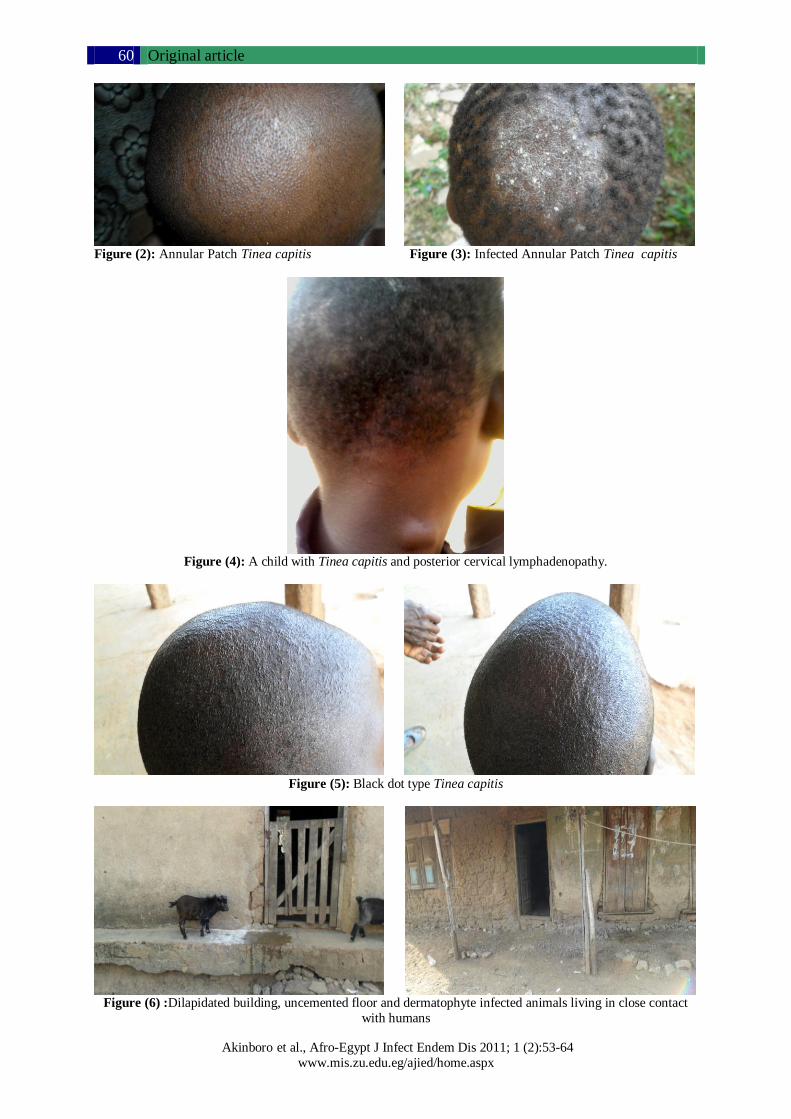
The various clinical types of T.capitis were examined for; the non-inflammatory form of T.
capitis was prevalent: “Gray patch” Tinea capitis
(GPTC) 86/185 (46.5%), “Black dot” Tinea capitis (BDTC) 78(42.2), Seborrheic dermatitis
type Tinea capitis (SDTC) 18 (9.7%), and the
pustular inflammatory type 3 (2.2%). No case of Kerion or Favus was recorded (Fig 2,3,4&5)
Attempt was made to correlate clinical type of T.
capitis with species of organism responsible. T.
metangrophyte was responsible for non- inflammatory T. capitis: BDTC and GPTC and
SDTC .There was no clear cut correlation
between dermatophytes and clinical types (r = 0.025, p = 0.567) .
The scaling and hairs from children’s scalp were
obtained for microscopy and culture. Scrapping was only possible and obtainable from 179
children. One hundred and sixty two samples
were positive for fungal element microscopically
while 17 samples showed no growth. Culture confirmed growth of dermatophytes only in 120
samples. Isolated dermatophytes represented 2
genera; Trichophyton and Microsporon, and 8 different dermatophytes including Trichophyton
metangrophytes as the leading organism isolated.
There was no case of mixed infection. Other
isolates of this study include T. tonsuran, T.rubrum, T.violaceum, T. soudanence,
Microsporon auodounii Microsporum gypseum.
Trichophyton metangrophytes was the most frequent organism causing infection in children,
followed by T. tonsuran as shown in Table( 4).

Original article
Akinboro et al., Afro-Egypt J Infect Endem Dis 2011; 1 (2):53-64
www.mis.zu.edu.eg/ajied/home.aspx
58
Table (1): General Characteristics of Children with Tinea capitis and the Controls
Variables T. capitis group Control
Age of participants(years)
Mean + SD 7.31+ 2.52 7.40+ 2.43
Mode 5 5
Range 5-16 5-16
Number of children per family
Mean + SD 4.81 + 2.012 4.63 + 2.210
Mode 5 4
Range 1 – 15 1 – 12
Mean age at onset of T. capitis(years)
Mean + SD 5.2 + 2.039
Mode 5 Range 1 month – 11years
Duration of Tinea capitis(years) Mean + SD 0.6 +1.031
Mode 1
Range 1month -13year
Age and Sex Prevalence of T. capitis
Age in years Number examined Number infected(prevalence) Total(prevalence)
Male Female Total (%)
5 – 8 242 91(37.60) 48 (19.83) 139 (57.43)
9 – 12 94 29(30.85) 6 (6.38) 35 (37.23)
13 – 16 89 11(12.35) 0 (0.0) 11 (12.35)
Total 425 131(30.82) 54 (12.70) 185 (43.53)
Table (2): Educational attainment, occupation and religion, ethnicity, and average family income per month
distribution of patients and controls
Educational attainment* Tinea capitis group (N, %) Control group (N, %)
None 1 (0.5) 0 (0.0)
Pre-school 68 (36.8) 74 (40.0)
Primary 105 (56.8) 94 (50.8) Secondary 11 (5.9) 17 (9.2)
Occupation**
Predominantly farming 130 (70.3) 7(3.8)
Predominantly fishing 14 (7.6) 0 (0.0)
Other forms of self employment 19 (10.3) 104 (56.2)
Unemployed 0 (0.0) 0 (0.0)
Civil servants 22 (11.9) 74 (40.0)
Religion***
Christianity 28 (15.1) 102 (55.1)
Islam 157 (84.9) 77 (41.6)
Traditional 0 (0) 6 (3.2)
Ethnicity****
Yoruba 183(98.9) 176 (95.1)
Hausa 0 (0.0) 6 (3.2)
Igbo 0 (0.0) 3 (1.6)
Others 2 (1.1) 0 (0.0)
Average income per month(Naira)***** 0 – 5000 140 (76.1) 56 (30.3)
5,001 - 10,000 17 (9.2) 31 (16.8)
10,001 - 20,000 13 (7.1) 52 (28.1)
20,001 - 50,000 14 (7.6) 43(23.2)
> 50,000 0(0.0) 3 (1.6)
Mean + SD 8,260 + 11,749 16,880 + 14,513
Range 1,000.00 – 50,000 3,000 – 60,000
*X 2 =3.14*X 2 =3.147, df = 3 p = 0.344 (N = 185), **X 2 =3.147, df= 3, p = 0.369 (N = 185),
***X 2 =75.474, df= 2, p = 0.000 (N = 185), **** X 2 = 5.136 df =2, p = 0.048,
***** t- test = -6.269, df = 368, p = 0.000

Original article
Akinboro et al., Afro-Egypt J Infect Endem Dis 2011; 1 (2):53-64
www.mis.zu.edu.eg/ajied/home.aspx
59
Table (3): Comparism of some risk factors, clinical history and examination findings: children with Tinea.
capitis and controls.
Clinical variables Tinea capitis group N( %) Control group (N %) X2
Yes No Yes No
Animal contact 166 (89.7) 19 (10.3) 105 (56.8) 80 (43.2) 51.316*
Soil contact 156 (84.3) 29 (15.7) 14 (7.6) 171 (92.4) 2.194*
Human contact 146 (78.9) 39 (21.1) 38 (20.5) 147 (79.5) 1.261*
Atopy History 54 (29.8) 131 (68.5) 4 (2.2) 181 (97.3) 51.116*
Family history 45 (24.3) 140 (75.7) 4 (2.2) 181 (97.8) 39.543*
Clinical History
Scalp pruritus 161 (87.0) 24 (13.0) 27 (14.6) 158 (85.4) 1.942*
Scalp scaling 179 (96.8) 6 (3.2) 12 (6.3) 173 (96.6) 3.018*
Adenopathy 47 (25.4) 138 (74.6) 5 (2.7) 180 (97.3) 41.978* Alopecia 139 (75.1) 46 (24.9) 0 (0.0) 185 (100.0) 2.226*
Type of Atopy
Rhinitis 45(83.3) 2(3.7)
3(5.5)
4(7.4)
2(3.7) 0(0.0)
0.(0.0)
0(0.0)
Asthma 2.331
Dermatitis
Conjuctivity
Contacts
Family member 135(73.0) 14(7.6) 72.339*
Class member 14(16.7) 27(14.6)
Barbing saloon 155 (83.3) 30 (16.2) 8.096*
Home Barbing 134 (16.7) 49 (26.5)
Weaving 0 (0) 2 (0.1)
*df =1, p < 0.001 N = number
Table (4): Distribution of dermatophytes isolated according to sex among the children in Ilie Community
Species Isolates according to sex
Total (%) Male Female
Trichophyton mentagrophytes 81(67.5) 67 14
Trichophyton tonsuran 16(13.3) 10 6
Trichophyton rubrum 13(10.8) 10 3
Microsporum audouinii 3 (2.5) 2 1
Microsporium gypseum 3 (2.5) 2 1
Trichophyton violaceum 2(1.7) 2 0
Trichophyton soudanense 2 (1.7) 2 0
120 (100) 95 (79.17) 25 (20.83)
Figure (1): shows the pattern of animal contact: patients and controls
X2 = 51.316, df= 1, p < 0.001
Original article
Akinboro et al., Afro-Egypt J Infect Endem Dis 2011; 1 (2):53-64
www.mis.zu.edu.eg/ajied/home.aspx
60
Figure (2): Annular Patch Tinea capitis Figure (3): Infected Annular Patch Tinea capitis
Figure (4): A child with Tinea capitis and posterior cervical lymphadenopathy.
Figure (5): Black dot type Tinea capitis
Figure (6) :Dilapidated building, uncemented floor and dermatophyte infected animals living in close contact
with humans

Original article
Akinboro et al., Afro-Egypt J Infect Endem Dis 2011; 1 (2):53-64
www.mis.zu.edu.eg/ajied/home.aspx
61
DISCUSSION
The overall prevalence of T.capitis in this study
was 43.5%. This prevalence rates is widely in
excess of previously recorded prevalence of 4% to 30% described among school children in the
Western and Southern part of Africa. [6, 7]
Soyinka [8] recorded a higher prevalence of 55% in a population based study and they
thought there was an epidemic of T. capitis
among school pupils. Tinea Capitis has been
known to reach epidemic proportions among school children. The observed prevalence in this
study is higher than 14.05% recorded by Ajao
and Akintunde in Ile- Ife [9] and similarly prevalence was highest among boys (30.82%)
compared to girls (12.7 %).
In the past decades, several studies have
concluded that T. capitis is an important dermatologic condition widely distributed
throughout the world, more importantly among
children. Its frequency is increasing, and aetiological agents vary from one geographic
location to another . Several other Nigerian [3, 4,
8, 10, 11, 12, 13] and international studies [14, 15] had documented differences in occupation,
parent income as a significant risk factors for T.
capitis as shown in this study. Parents of
children with T. capitis in this study were more likely to be predominantly farmers and
fishermen, while those of controls were
predominantly office workers or self employed in other forms of trade. This is in line with
epidemiological reasoning that, low income
earning or poverty, malnutrition and general poor
social conditions experienced by subsistent farmers, coupled with unlimited exposure to
potential sources of infections such as
contaminated soil and animals highly predisposed children in Ilie to T.capitis. The
strong link between animals and dermatophyte
had been previously documented by Abdulkadir [16], Ameh and Okolo [17] in the Northern states
of Nigeria.
The study also found that children in Ilie
community live in close contact with animals (predominantly goat, sheep and dogs) which
were being kept for business , family food supply
and hunting purposes. Abdulkadir [16] had earlier confirmed the enzootic ringworm of
horses, dogs, and livestock as common source of
sporadic infection among owners or their care takers which might include the owner’s children.
Animal type ringworm was also viewed by
Macura as an occupational hazard for farmers
and pet keepers [18] .Study from Sokoto state of
Nigeria where livestock and pet domestication
was found as a common practice in households, had suggested domestic animals as important
reservoir of tinea. Direct or indirect contact with
fungus contaminated objects of livestock rearing
like dung, fencing, halters, rope, harness and grooming brushes were found as extremely
important in the natural dissemination of the
disease.[17]
Other authors in the region had observed that
Tinea transmission was encouraged by poor
living unsanitary condition with overcrowding [3]. Soil in the homes of livestock keepers and
playground has been viewed by several authors
as containing fungal element that dropped from
the body of infected animals or primarily a geophillic agent [19]. The high frequency of T.
capitis among children had therefore been linked
to intense close contact among children especially at play grounds and at home. This
study showed that contact were more intense at
home amongst family members and neighbors than in the classroom, this is understandable
because pupils spend few hours in the school,
and most hours are spent at home and
neighborhood play grounds.
In addition, most children with T. capitis came
from large families (> 4 children), this seems not
significant enough as a sole factor that could risk the children and sustain the infection in the
community. This observation is similar to the
former findings of Ajao et al. in Ile – Ife. [9]
Like other previous studies [3,4,8,10,11,12,13] boys were predominantly infected than girls,
with a ratio of 2.4: 1 which was greater than 1.8:
1 recorded by Kalla et al.[20] in India but lower
than a high ratio 5: 1 recorded by Gugnani and
Njoku-Obi in South-eastern Nigeria.[3] The
mean age of infection with dermatophyte was 7.31+2.52 years, which is closer to the
observation of other workers [8,21]. The most
frequent age group affected was 5 – 8 years with
most likely age of infection being 5 years. Gugnani and Njoku-Obi recorded age group 3- 7
years in their study [3]. Ayanbimpe et al [21]
also documented the highest rate of infection in the age bracket 10–14 years; this was closely
followed by similar age grouped 5–8 years which
was observed in this study. Tinea capitis was not seen among girls older than 13 year. Soyinka and
others had adduced this high prevalence of T.
capitis among the boys to continuous and
sustained exposure to infective agents and close body contact at the play ground, and the fact that
most boys visit the same set of barbers that

Original article
Akinboro et al., Afro-Egypt J Infect Endem Dis 2011; 1 (2):53-64
www.mis.zu.edu.eg/ajied/home.aspx
62
harbor infective agents on their barbing
equipment [8,12,21] .Furthermore, most of the girls older than 13 years may prefer to weave
their hair rather than visiting barber’s shop, they
practice better hair and general hygiene and may
carry their own weaving equipment and thus reduce contact with infective agents on the hair
dresser’s hand [9]. Encouragement of personal
hygiene has therefore become important in the prevention and control of this endemic disease.
Many of the children infected with T.capitis barb
in public saloon in this study. Barbing saloon in Ilie are untidy and the practice of equipment
sterilization was foreign to the operators of the
four barbing saloon serving the village and the
environment. David et al. [12] also demonstrated evidence of fungal element in barbing equipment
in a recent study in Mubi, Adamawa state of
Nigeria and suggested sensitization of public health workers and saloon customers on the need
for sterilization of barbing equipment in all
saloon. Ayanbimpe et al.[21] also attributed high incidence of T. capitis to continuous contact in
barber’s shop. With continuous and intense
transmission of T. capitis, unless a bold step is
taken to control the infection at the barber’s shop, complete eradication of the condition
might be a mirage.
This study demonstrated a non-significant positive correlation between duration of T.
capitis and history of atopy. The commonest
documented atopy condition was rhinitis. Hay
and Shennan [22] and other workers [23] had previously reported association between the
presence of atopy and chronic dermatophytosis,
asthma and hay fever was the commonest atopic disease reported in their study. Other
associations of chronic dermatophytosis include
immediate- type hypersensitivity and elevated IgE levels which were not examined in this
study.
In respect to the spectrum of isolated species of
dermatophyte, it has been established that organism varies from one geographical location
to another, even within a country, state, or local
government area the patterns of isolates have varied overtime. In this study, the isolated
dermatophytes were zoophylic, anthropophilic or
geophilic. The organisms belonged to two general Trichophyton and Microsporon, and
seven species which include T. metagrophyte,
T.tonsurans, T.rubrum , M.audouinii,
M.gypseum, T.violaceum, T.soudanens were isolated. The high prevalence of T. metagrophyte
(67.5%) in this study was similar to a recent
finding by Nweke [13] in Anambra among
normadic herdsmen living in camps. Jain et al. in Rajasthan district of India also found T.
metagrophyte as the second leading agent in their
study[24]. Jha et al. in eastern Nepal also found
similar pattern [25]. This finding is not surprising because 166 (89.7%) and 156 (84.3%) of the
children recruited into the study had history of
unlimited contact with animals mainly goats and dogs. These animals, mainly live stocks have
been kept for commercial purposes and almost
all the households have their share in the livestock raring. The animals live in close
contact with humans, direct contact is possible
because animals are not been kept in special pen,
and the children also participate in the raring of the animals. Animals have been variously
implicated in Tinea transmission. This profile of
isolate in this study was similar to findings of Ajao and Akintunde [9] but Microsporum
aoduoinii was the leading agent in their study.
The finding of T. tonsurans,T. rubrum,T. soudanense,T. violaceous, M gypseum is not
strange in this environment. They have been
isolated in various studies in Nigeria in the past
as aetiological agent of T. capitis. [3, 4, 8, 10, 11, 12, 13]
It is worthy of note also, to comment on the high
burden 42/179 (23.5%) of other non-dermatophyte fungi infection of the children’s
scalp. Isolated in the study include: Penicillium
spp, Blastomyces dermatidis, Candida albican,
and Gliocladium spp. This is also not unusual because the children live in close contact with
soil both at home and in the school. Oyeka and
Ugwu had also noted that this non – dermatophyte spores are uquibitous and may
transiently colonized human skin [26]. The
finding of this organism should be taken serious because isolates of non – dermatophyte was not
found mixed with other dermatophyte organism,
which will portend their isolation as been
contaminants. More so, dermatophyte specific culture media (Potatoe agar) was used.
In this current study, the non- inflammatory T.
capitis (GPTC; 46.5%, BDTC; 42.2%, and SDTC; 9.7%), were more common than the
inflammatory type (Pustular; 2.2%). Previous
findings also corroborated the fact that, gray patch T. capitis (GPTC) or black dot (BDTC)
type are likely to be the leading clinical type in
any epidemiologic survey [20, 27]. The
incidence of inflammatory type (2.27%) is lower when compared with the findings of other
authors [27,28] . Mixed morphology of T.capitis
Original article
Akinboro et al., Afro-Egypt J Infect Endem Dis 2011; 1 (2):53-64
www.mis.zu.edu.eg/ajied/home.aspx
63
was rarely reported in literature [28]. There was
no case of mixed morphology in this study. Several other studies confirmed the non
inflammatory (GPTC or BDTC) to be the leading
clinical types, and in some studies both almost
occurred at the same proportion or frequency [20, 25, 27, 29]. The inflammatory types of T.
capitis are uncommon in the present study.
Nnoruka et al. [30] in Enugu state of Nigeria found a low incidence of kerion (9 ,3.1%) in
their study, while Grover et al. [27] in India
found a higher incidence of inflammatory T. capitis at 32%.
According to Gugnani and Najoku-Obi [3] the
clinical appearance of T.capitis is most variable,
and depends on the type of hair invasion, the level of host resistance and the degree of
inflammatory host response. The ectothrix agent
(T. metagrophyte) was prevalent in this study as a cause of the predominant non – inflammatory
type, though not mutually exclusive for these
clinical types alone in the study. The isolates of this study were mainly Trichophyton species that
characteristically invaded the hair while
producing large-spored ectothrix in chains as
seen in T. mentagrophytes or the endothrix type as documented for T. tonsurans, T. soudanense,
T. violaceum, T. yaoundei, T. gourvilii and rarely
same may be demonstrated by T. rubrum. However, previous studies have shown that
clinical presentation is not correctly indicative of
the type of fungus or vice versa, as it also
depends on other unknown factors [25,29], but Grover et al. in their study found endothrix
agents to be responsible for BDTC and ectothrix
agent to be responsible for GPTC, but the finding was not mutually exclusive also in their study.
[27]
CONCLUSIONS
Tinea capitis remains an endemic disease
reaching variable epidemic proportion in some populations as seen in Ilie among the children.
The non – inflammatory type (GPTC, BDTC)
were the prevalent clinical types of T. capitis in
Ilie. Close animal and soil contact, poor sanitary condition at home, extreme of poverty were the
most potent risk factors for contracting and
sustaining the epidemic in this population. The cheapest means of prevention and controlling
this infection depends on education of parent and
sibling on reducing or preventing undue contact with the source of contagion.
RECOMMENDATIONS
The following are recommended:
1) Health education about infectious disease
such T. capitis: including modes of transmission to the parents and children, and
the importance of personal hygiene.
2) There is need for the establishment of community dermatological services to cater
for the teeming needs of the children.
3) Since livestock keeping is a lifestyle of the
Ilie people, I therefore suggest the need for construction of animal’s pen by each
household to reduce in- house contact with
animals. This can be shouldered by the local health authority.
4) There is a need for effective community
veterinary services for prompt treatment of
infected animals.
5) Study of dermatophyte infections among
animals in Ilie and its correlation with human
dermatophytes is suggested as a future study.
Funding: This work was not funded by any
agency.
Conflicts of interest: There are no conflicts of
interest in the course of this research work.
Ethical approval : Ethical clearance was obtained from the ethical committee of Ladoke
Akintola University Teaching hospital, Osogbo.
Informed consents were obtained from parents of
the children.
REFERENCES
1. Scott DW: Superficial Mycoses in Pederson D (Edn) Large animal Dermatology.publish by W.B
Saunders company Harcort Brace jovanich Inc.,
1988:172 – 175.
2. Macura A B: Dermatophyte infection. int. J of
Dermatology 1993;32:313.
3. Gugnani HC, Njoku-Obi ANU. Tinea capitis in
school children in East Nigeria. Mykosen 1986; 29: 132-144.
4. Anosike JC, Keke IR, Uwaezuoke JC, Anozie, J
C, Obiukwu C, Nwoke B E B et al. Prevalence
and distribution of ringworm infection in primary
school children in parts of Eastern, Nigeria. J. of Appl. Sc. and Environ. Manag 2005; 9 (3): 21-25.
5. Fathi HI, Al-Samarai AG. Prevalence of tinea
capitis among school children in Iraq. Eastern
Mediterranean Health Journal 2000;06 (1):128 –
137.
6. Morar N, Dlova NC, Gupta AK, Aboobaker
J. Tinea capitis in Kwa-Zulu Natal, South Africa.
Paediatr Dermatol 2004; 21: 444 – 447.

Original article
Akinboro et al., Afro-Egypt J Infect Endem Dis 2011; 1 (2):53-64
www.mis.zu.edu.eg/ajied/home.aspx
64
7. Shrank, AB. In A. B. Shrank and R. R. Harman.
The incidence of skin diseases in a Nigerian
teaching hospital dermatological clinic. Brit. J.
Dermatol 1966; 78:239.
8. Soyinka F. Epidemiologic study of dermatophyte
infections in Nigeria (Clinical Survey and Laboratory Investigations). Mycopathologia 1978; 63(2): 99-103.
9. Ajao AO, Akintunde CA. Studies on the
prevalence of tinea capitis in Ile-Ife.
Mycopathologia 2005;89: 43 – 48
10. Ogunbiyi A O, Owoaje E, Ndahi A. Prevalence
of skin disorders in school children in Ibadan,
Nigeria. Pediatric Dermatology 2005; 22: 6-10:
493- 498. 11. Enemuor C, Amedu AS. Prevalence of
superficial mycoses in primary schoolchildren in
Anyigba, Kogi State, Nigeria. African Journal of
Microbiology Research 2009; 3 (2): 62-65.
12. David DL, Edward A, Zaruwa MZ, Addass PA.
Barbing Saloon Associated Fungal Disease
Infection in Mubi, Adamawa State-Nigeria.
World J. Med. Sci.2010; 5 (1): 17- 21.
13. Nweke EI. Dermatophytosis among children of
Fulani/Hausa herdsmen living in southeastern
Nigeria. RevIberoam Micol.2010; 27(4):191– 194
14. Male O. The significance of mycology in medicine. In: Hawksworth DL (ed.), Frontiers in
Mycology. Wallingford: CAB International,
1990: 131– 136.
15. Havlickova B, Czaika VA, Friedrich M.
Epidemiological trends in skin mycoses
worldwide. Mycoses 2004: 51 (4), 2–15.
16. Abdulkadir I. Dermatophytosis In: Infectious
Diseases of livestock in Nigeria- An Outline. Pub
Ahmadu Bello University Press Limited,
Zaria,1989; 274- 275.
17. Ameh I.G , R.U Okolo. Dermatophytosis among children: Domestic animal as a predisposing
factor in Sokoto, Nigeria. Pakistan Journal of
Biological science 2004, 7 (7):1109 - 1112.
18. Macura A.B. Dermatophyte infection. Int J
Dermatol 1993; 32(5):313-323
19. Chineme, C.N, Adekeye J.O, Bida S.A.
Trichophyton verrucossum in Young goat. Bull
Hlth Prod Afr 1981 ; 29(1):75-78
20. Kalla G, Begra B, Solanki A, Goyal A, Batra
A. Clinicomycological study of tinea capitis in
desert district of Rajasthan, Ind J Dermatol Venereol Leprol 1995;6 : 342- 343.
21. Ayanbimpe GM, Taghir H, Diya A, Wapwera S.
Tinea capitis among primary school children in
some parts of central Nigeria. Mycoses 2008; 51,
336 – 340.
22. Hay RJ, Shennan G. Chronic dermatophyte
infections. II. Antibody and cell-mediate immune
responses. Br J Dermatol 1982; 106: 191– 195.
23. Jones HE, Reinhardt JH, Rinaldi MG. Acquired
immunity to dermatophytosis. Arch Dermatol
1974; 109: 840 – 848.
24. Jain N, Sharma M, Saxena VN. Clinico-mycological profile of dermatophytosis in Jaipur,
Rajasthan. Indian J Dermatol Venereol Leprol
2008; 74 (3): 274- 275.
25. Jha BN, Garg VK , Agrawa S , Khanal B, Arun
Agwalla A. Tinea capitis in eastern .Nepal Int J
Dermatol 2006; 45 (2): 100 – 102. 26. Oyeka CA, Ugwu IO. Fungal flora of human toe
webs. Mycoses 2002; 45:488 - 491.
27. Grover C, Arora P, Manchanda V. Tinea capitis
in the pediatric population: A study from North
India. Indian J Dermatol Venereol Leprol 2010;
76 (5): 527- 532.
28. Jahangir M, Hussain I, Khurshid K, Haroon TS.
A clinico-etiologic correlation in Tinea capitis.
Int J Dermatol 1999; 38: 275- 278.
29. Singal A, Rawat S, Bhattacharya S, Mohanty S,
Baruah MC . Clinico- mycological profile of
tinea capitis in North India and response to griseofulvin. J Dermatol 2001; 28: 22- 26.
30. Nnoruka EN, Ndu AN, Ohanu ME, Uzodimma
BA, Udoh IP. Patchy hair loss in school children:
Tinea capitis re-assessed. Journal of Pediatric
Infectious Disease 2007; (3):147- 151.


![SCIENCE CHINA Life Sciences - Springer · tions, such as tinea capitis, tinea corporis, tinea inguinalis, tinea manus, tinea unguium and tinea pedis [1–3]. Unlike](https://static.fdocuments.in/doc/165x107/5d1b54ac88c993283c8ce38a/science-china-life-sciences-springer-tions-such-as-tinea-capitis-tinea-corporis.jpg)


