The parasite Schistocephalus solidus secretes proteins ...
20
Berger et al. Parasites Vectors (2021) 14:436 https://doi.org/10.1186/s13071-021-04933-w RESEARCH The parasite Schistocephalus solidus secretes proteins with putative host manipulation functions Chloé Suzanne Berger 1,2,3 , Jérôme Laroche 2 , Halim Maaroufi 2 , Hélène Martin 1,2,4 , Kyung‑Mee Moon 5 , Christian R. Landry 1,2,4,6,7 , Leonard J. Foster 5 and Nadia Aubin‑Horth 1,2,3* Abstract Background: Manipulative parasites are thought to liberate molecules in their external environment, acting as manipulation factors with biological functions implicated in their host’s physiological and behavioural alterations. These manipulation factors are part of a complex mixture called the secretome. While the secretomes of various parasites have been described, there is very little data for a putative manipulative parasite. It is necessary to study the molecular interaction between a manipulative parasite and its host to better understand how such alterations evolve. Methods: Here, we used proteomics to characterize the secretome of a model cestode with a complex life cycle based on trophic transmission. We studied Schistocephalus solidus during the life stage in which behavioural changes take place in its obligatory intermediate fish host, the threespine stickleback (Gasterosteus aculeatus). We produced a novel genome sequence and assembly of S. solidus to improve protein coding gene prediction and annotation for this parasite. We then described the whole worm’s proteome and its secretome during fish host infection using LC–MS/MS. Results: A total of 2290 proteins were detected in the proteome of S. solidus, and 30 additional proteins were detected specifically in the secretome. We found that the secretome contains proteases, proteins with neural and immune functions, as well as proteins involved in cell communication. We detected receptor‑type tyrosine‑protein phosphatases, which were reported in other parasitic systems to be manipulation factors. We also detected 12 S. solidus‑specific proteins in the secretome that may play important roles in host–parasite interactions. Conclusions: Our results suggest that S. solidus liberates molecules with putative host manipulation functions in the host and that many of them are species‑specific. Keywords: Schistocephalus solidus, Secretome, Proteomics, Manipulation factor, Parasite, Behaviour © The Author(s) 2021. Open Access This article is licensed under a Creative Commons Attribution 4.0 International License, which permits use, sharing, adaptation, distribution and reproduction in any medium or format, as long as you give appropriate credit to the original author(s) and the source, provide a link to the Creative Commons licence, and indicate if changes were made. The images or other third party material in this article are included in the article’s Creative Commons licence, unless indicated otherwise in a credit line to the material. If material is not included in the article’s Creative Commons licence and your intended use is not permitted by statutory regulation or exceeds the permitted use, you will need to obtain permission directly from the copyright holder. To view a copy of this licence, visit http://creativecommons.org/licenses/by/4.0/. The Creative Commons Public Domain Dedication waiver (http://creativeco mmons.org/publicdomain/zero/1.0/) applies to the data made available in this article, unless otherwise stated in a credit line to the data. Background Parasites have major impacts on their hosts, including on their morphology [1], physiology [2], and behaviour [3, 4]. To induce these complex changes in their hosts, it has been proposed that parasites produce, store, and release manipulation factors that interfere with the host physiological and central nervous systems [5–7]. ese manipulation factors are thought to be part of a complex mixture of molecules called the secretome, which is a key element of parasite–host interactions [6]. e secretome of a parasite includes lipids [8], nucleic acids [9], and proteins [10], which are sometimes protected inside extracellular vesicles [11]. Using molecular and bioinfor- matics approaches, the proteomic fraction of secretomes Open Access Parasites & Vectors *Correspondence: Nadia.Aubin‑[email protected] 1 Département de Biologie, Université Laval, Quebec, QC, Canada Full list of author information is available at the end of the article
Transcript of The parasite Schistocephalus solidus secretes proteins ...

Berger et al. Parasites Vectors (2021) 14:436 https://doi.org/10.1186/s13071-021-04933-w
RESEARCH
The parasite Schistocephalus solidus secretes proteins with putative host manipulation functionsChloé Suzanne Berger1,2,3, Jérôme Laroche2, Halim Maaroufi2, Hélène Martin1,2,4, Kyung‑Mee Moon5, Christian R. Landry1,2,4,6,7, Leonard J. Foster5 and Nadia Aubin‑Horth1,2,3*
Abstract
Background: Manipulative parasites are thought to liberate molecules in their external environment, acting as manipulation factors with biological functions implicated in their host’s physiological and behavioural alterations. These manipulation factors are part of a complex mixture called the secretome. While the secretomes of various parasites have been described, there is very little data for a putative manipulative parasite. It is necessary to study the molecular interaction between a manipulative parasite and its host to better understand how such alterations evolve.
Methods: Here, we used proteomics to characterize the secretome of a model cestode with a complex life cycle based on trophic transmission. We studied Schistocephalus solidus during the life stage in which behavioural changes take place in its obligatory intermediate fish host, the threespine stickleback (Gasterosteus aculeatus). We produced a novel genome sequence and assembly of S. solidus to improve protein coding gene prediction and annotation for this parasite. We then described the whole worm’s proteome and its secretome during fish host infection using LC–MS/MS.
Results: A total of 2290 proteins were detected in the proteome of S. solidus, and 30 additional proteins were detected specifically in the secretome. We found that the secretome contains proteases, proteins with neural and immune functions, as well as proteins involved in cell communication. We detected receptor‑type tyrosine‑protein phosphatases, which were reported in other parasitic systems to be manipulation factors. We also detected 12 S. solidus‑specific proteins in the secretome that may play important roles in host–parasite interactions.
Conclusions: Our results suggest that S. solidus liberates molecules with putative host manipulation functions in the host and that many of them are species‑specific.
Keywords: Schistocephalus solidus, Secretome, Proteomics, Manipulation factor, Parasite, Behaviour
© The Author(s) 2021. Open Access This article is licensed under a Creative Commons Attribution 4.0 International License, which permits use, sharing, adaptation, distribution and reproduction in any medium or format, as long as you give appropriate credit to the original author(s) and the source, provide a link to the Creative Commons licence, and indicate if changes were made. The images or other third party material in this article are included in the article’s Creative Commons licence, unless indicated otherwise in a credit line to the material. If material is not included in the article’s Creative Commons licence and your intended use is not permitted by statutory regulation or exceeds the permitted use, you will need to obtain permission directly from the copyright holder. To view a copy of this licence, visit http:// creat iveco mmons. org/ licen ses/ by/4. 0/. The Creative Commons Public Domain Dedication waiver (http:// creat iveco mmons. org/ publi cdoma in/ zero/1. 0/) applies to the data made available in this article, unless otherwise stated in a credit line to the data.
BackgroundParasites have major impacts on their hosts, including on their morphology [1], physiology [2], and behaviour [3, 4]. To induce these complex changes in their hosts, it has been proposed that parasites produce, store, and
release manipulation factors that interfere with the host physiological and central nervous systems [5–7]. These manipulation factors are thought to be part of a complex mixture of molecules called the secretome, which is a key element of parasite–host interactions [6]. The secretome of a parasite includes lipids [8], nucleic acids [9], and proteins [10], which are sometimes protected inside extracellular vesicles [11]. Using molecular and bioinfor-matics approaches, the proteomic fraction of secretomes
Open Access
Parasites & Vectors
*Correspondence: Nadia.Aubin‑[email protected] Département de Biologie, Université Laval, Quebec, QC, CanadaFull list of author information is available at the end of the article
Page 2 of 20Berger et al. Parasites Vectors (2021) 14:436
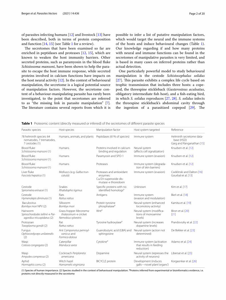
of parasites infecting humans [12] and livestock [13] have been described, both in terms of protein composition and function [14, 15] (see Table 1 for a review).
The secretomes that have been examined so far are enriched in peptidases and proteases [12, 15], which are known to weaken the host immunity barriers. Other secreted proteins, such as paramyosin in the blood fluke Schistosoma mansoni, have been shown to help the para-site to escape the host immune response, while secreted proteins involved in calcium functions have impacts on the host neural activity [12]. In the context of behavioural manipulation, the secretome is a logical potential source of manipulation factors. However, the secretome con-tent of a behaviour-manipulating parasite has rarely been investigated, to the point that secretomes are referred to as “the missing link in parasite manipulation” [7]. The literature contains several reports from which it is
possible to infer a list of putative manipulation factors, which would target the neural and the immune systems of the hosts and induce behavioural changes (Table 1). Our knowledge regarding if and how many proteins with neural and immune functions can be found in the secretomes of manipulative parasites is very limited, and is based in many cases on inferred proteins rather than actual detection.
One particularly powerful model to study behavioural manipulation is the cestode Schistocephalus solidus [27]. This parasite exhibits a complex life cycle based on trophic transmission that includes three hosts: a cope-pod, the threespine stickleback (Gasterosteus aculeatus, obligatory intermediate fish host), and a fish-eating bird, in which S. solidus reproduces [27, 28]. S. solidus infects the threespine stickleback’s abdominal cavity through the ingestion of a parasitized copepod [29]. The
Table 1 Proteomic content (directly measured or inferred) of the secretomes of different parasite species
(1) Species of human importance. (2) Species studied in the context of behavioural manipulation. aProteins inferred from experimental or bioinformatics evidence, i.e. proteins not directly measured in the secretome
Parasitic species Host species Manipulation factor Host system targeted Reference
78 helminth species: 64 nematodes, 7 trematodes, 7 cestodes (1)
Humans, animals, and plants Peptidases (61% of species) Immune system Helminth secretome data‑base (HSD)
Garg and Ranganathan [15]
Blood flukeSchistosoma mansoni (1)
Humans Proteins involved in calcium binding and regulation
Neural system(affects cell signalization)
Knudsen et al. [12]
Blood flukeSchistosoma mansoni (1)
Humans Paramyosin and SPO‑1 Immune system (evasion) Knudsen et al. [12]
Blood flukeSchistosoma mansoni (1)
Humans Proteases Immune system (degrada‑tion of skin barriers)
Knudsen et al. [12]
Liver flukeFasciola hepatica (1)
Molluscs (e.g. Galba trun-catula)
Proteases and antioxidant enzymes:
Cu/Zn‑superoxide dis‑mutase + thioredoxin
Immune system (evasion) Cwiklinski and Dalton [16]Gourbal et al. [13]
CestodeSpirometra erinacei (1)
SnakesRhabdophis tigrinus
Specific proteins with no identified homologsa
Unknown Kim et al. [17]
CestodeHymenolepis diminuta (1)
RatsRattus rattus
Antigens Immune system(evasion and modulation)
Bień et al. [18]
BaculovirusBombyx mori NPV (2)
SilkwormBombyx mori
Protein tyrosine phosphatasea
Neural system (enhanced locomotory activity)
Kamita et al. [19]
HairwormSpinochordodes tellinii + Par-
agordius tricuspidatus (2)
Grass‑hopper Meconema thalassinum + cricket
Nemobius sylvestris
Wnta Neural system (modifica‑tions of monoamine levels)
Biron et al. [20][21]
ProtozoanToxoplasma gondii (2)
RatRattus rattus
Tyrosine hydroxylasea Neural system (increases dopamine levels)
Prandovszky et al. [22]
FungusOphiocordyceps unilateralis
(2)
Ant Camponotus pennsyl-vanicus and
Formica dolosa
Guanobutyric acid (GBA) and sphingosine
Neural system (action not determined)
De Bekker et al. [23]
WaspCotesia congregata (2)
CaterpillarManduca sexta
Cytokinea Immune system (activation that results in feeding reduction)
Adamo et al. [24]
WaspAmpulex compressa (2)
Cockroach Periplaneta americana
Dopamine Neural system (represses the activity of neurons)
Libersat et al. [25]
AphidHormaphis cornu (2)
Witch hazelHamamelis virginiana
BICYCLE protein Development (induces galls—novel plant ‘organs’)
Korgaonkar et al. [26]

Page 3 of 20Berger et al. Parasites Vectors (2021) 14:436
consequences of the infection by S. solidus on the three-spine stickleback’s morphology [30], physiology [31], immune system [32], and behaviour [33] are well docu-mented. For example, sticklebacks infected by S. solidus show drastic behavioural changes that result in a loss of the anti-predator response [27]: infected fish are more exploratory [34], less anxious [35], and bolder in the pres-ence of a predator [36] than non-infected fish.
Most of these behavioural alterations seen in infected fish appear after several weeks, when the worm has grown to reach the infective stage within its intermedi-ate host (i.e. larger than 50 mg) [37]. The infective stage coincides with the time at which S. solidus is ready to reproduce in the bird [27, 38], which also generally cor-responds to the activation of the immune response in the host. In the first phase of infection, the adaptive immune response is generally not activated in the fish. It is only when the worm reaches the infective stage that an ineffective upregulation of the respiratory burst activ-ity occurs [32]. Nevertheless, activation of the immune response through the production of reactive oxygen spe-cies (ROS) by granulocytes has been shown to occur early during infection, depending on the genotype of the stick-leback population [39]. Several studies have suggested that the manipulation of the stickleback’s behaviour increases the worm’s transmission rate to its final avian host [27]. Yet, the adaptive value for S. solidus of such behavioural changes has never been demonstrated [40], and it is possible that these behavioural modifications in the fish may solely result from a side effect of infection [7], such as the effect of the parasite mass burden or of the activation of the host immune response [35, 41]. To demonstrate that behavioural changes in the host are the result of direct parasitic manipulation, the first step is to determine whether the parasite can liberate molecules in its external environment, and if yes, to study their func-tions in relation with the host’s phenotype perturbations.
This host–parasite system was used for the first experi-mental demonstration of a complex life cycle of a trophi-cally transmitted parasite in 1790 (as reviewed in [27]). A rich body of work on this model system during the past 50 years has shown, using enzymatic assays, that the activity of proteases [42] and transferases [43] is required for S. solidus survival and growth. Furthermore, a par-tial genome of the worm is available [44], and extensive transcriptome data has been produced [45, 46]. Quan-tification of the transcriptome dynamics across the life stages has uncovered that when the worm reaches the infective stage in its fish host, genes involved in neural pathways and sensory perception have higher expres-sion levels compared to the earlier stages in the same host, which are characterized by upregulation of growth-related pathways [46]. Furthermore, vesicles are present
inside the S. solidus tegument, as shown through scan-ning and transmission electron microscopy [47]. Moreo-ver, S. solidus excretes (through passive mechanisms) or secretes (through active mechanisms) molecules, including (uncharacterized) proteins, and these secre-tions are sufficient to affect its fish host behaviour [48]. Finally, a well-annotated genome of the threespine stick-leback host is also available [49], which is important to adequately differentiate proteins coming from the host and the parasite. The Schistocephalus–stickleback sys-tem is thus appropriate to test the presence of manipula-tion factors. However, the nature of the protein content of the S. solidus secretome has never been explored [7, 48]. Analysing the proteomic fraction of the secretome of S. solidus, and its potential enrichment in manipulation factors involved in neuronal and immune functions, will help us to understand if the behavioural changes of the host could be induced by parasitic manipulation through the secretome.
Here, we characterized the worm’s whole-body pro-teome and the protein fraction of the secretome of S. solidus using liquid chromatography-tandem mass spectrometry (LC–MS/MS) [50]. We focused on indi-viduals in the infective stage of development so that the secretome may include manipulation factors that could be associated with the fish host’s behavioural changes. In helminth parasites, a portion of proteins from the pro-teome are passively released in the external environment as metabolic waste products, contributing to an impor-tant fraction of their secretome content [51]. Therefore, the secretome is generally a subset of the proteome in terms of protein content [52]. We thus expected that the secretome of S. solidus would also mainly be a subset of its proteome. However, in the context of behavioural manipulation, we hypothesized that proteins could also be actively liberated by the parasite in its external envi-ronment [51], so that they would be enriched in the secretome compared to the proteome. Based on what has been described in previous parasitic systems (references reviewed in Table 1), if S. solidus manipulates stickleback behaviour with its secretions as we hypothesize, then its secretome would include proteases as well as proteins with neural and immune functions. Because the worm is not in direct contact with the vertebrate host brain [27], we also expected to detect proteins involved in cell com-munication, cell–cell signalling, or transport functions. These proteins would mediate the communication of the worm with its host’s brain to induce potential neural and immune changes, and ultimately behavioural alterations.
As the genome of S. solidus is used during LC–MS/MS as a reference database, a more thorough annotation of the genome than the one publicly available [44] could allow us to detect more proteins, including potential

Page 4 of 20Berger et al. Parasites Vectors (2021) 14:436
manipulation factors. We therefore used a multipronged approach combining genomics with proteomics (described in Fig. 1). We first sequenced the genome of S. solidus using a combination of long and short reads to combine longer contigs and improved annotation. Then, we investigated the global proteomic composition of the proteome and secretome of S. solidus using LC–MS/MS.
MethodsGenome sequencing of Schistocephalus solidusWorm collectionWe caught threespine sticklebacks from Lac Témisc-ouata (Québec, 47°40′33″N 68°50′15″W), where fish are known to be infected by S. solidus [35], using minnow traps in July 2015. Fish were brought to the Laboratoire Aquatique de Recherche en Sciences Environnementales et Médicales at Université Laval (Québec, Canada) and were maintained under a 12 h:12 h light/dark cycle and a water temperature of 15 °C. Fish were fed daily with a mixture of blood worms and Artemia. After 10 months, one fish (out of 40) exhibited the morphological changes typically induced by S. solidus [30] and was consequently sacrificed to collect the worm. The fish was euthanized with an overdose of MS-222 (75 mg/l mg/kg) and dis-sected to confirm the infection by S. solidus. The worm was immediately put in ethanol 90% and stored at 4 °C until genome sequencing. All the other fish were later used during a behavioural experiment [35].
DNA extraction and sequencingGenomic DNA was extracted using the DNeasy Blood and Tissue kit (Qiagen Inc., Valencia, CA, USA) from ~ 20 mg of tissues. After lysis, we added 4 µl of RNase A (10 mg/ml). Elution was done twice in 100 µl of elution buffer. To reach the desired concentration for the Nanopore library preparation and Illumina sequenc-ing, we concentrated DNA with a SpeedVac concentrator (Thermo Fisher, Waltham, MA, USA) for 1 h.
Illumina HiSeq X sequencing The DNA libraries for Illu-mina sequencing were prepared using a shotgun PCR-free library preparation (Lucigen) Illumina Library at the McGill University and Genome Quebec Innovation Cen-tre (Montréal, Canada). Sequencing was performed at the same centre on an Illumina HiSeq X instrument, using paired-end reads (PE150). A total of 157,475,128 reads were obtained. The estimated genome size is 540 MB [44].
Oxford Nanopore Technologies (MinION) sequenc-ing Library preparation for Oxford Nanopore sequenc-ing was done with a PCR-free ligation sequencing kit SQK-LSK108 (ONT, Oxford, UK). Briefly, approximately 2.5 µg of high-molecular-weight DNA was repaired using
the NEBNext FFPE Repair Mix (NEB, Ipswich, MA, USA) for 15 min at 20 °C before purification with AMPure XP beads. The repaired DNA was end-prepped with the NEBNext Ultra II End Repair/dA-Tailing Module (NEB, Ipswich, MA, USA) for 30 min at 20 °C and 30 min at 65 °C according to the manufacturer’s instructions and purified with AMPure XP beads. Adapter mix (ONT, Oxford, UK) and Blunt/TA Ligase Master Mix (NEB, Ipswich, MA, USA) were added to the purified end-prepped DNA and incubated for 15 min at room temperature. The library was then purified with AMPure XP beads and ABB wash buffer and recovered in elution buffer (ONT, Oxford, UK). Approximately 333 ng of the purified library was loaded onto a primed R9.4 SpotOn flow cell (FLO-MIN106) along with RBF (ONT, Oxford, UK) and library loading beads (ONT, Oxford, UK). Sequencing was performed with a MinION Mk1B sequencer running for 48 h, and MinKNOW software (provided by ONT, Oxford, UK) was used to control the sequencing process. Four librar-ies were sequenced using this protocol. Base calling was performed with albacore (read_fast5_basecaller.py, ONT Albacore Sequencing Pipeline Software v2.3.1) on all fast5 files. A total of 4,636,932 read sequences resulted from the base calling step for a total of 14,535,522,365 nucleotides.
Genome assembly and annotationAll fastq ONT files were pooled in one file prior to assembly. ONT sequences were assembled using Flye v2.4 [53, 54]. The final assembly produced 17,882 scaf-folds, the largest scaffold being 919,337 nt, with an N50 of 121,189 nt. The mean coverage across scaffolds was 20×. A first phase of correction (polishing) was carried out with nanopolish (v0.10.2) (https:// github. com/ jts/ nanop olish). A total of 3,473,881 changes were applied. A second correction phase with one Illumina HiSeq X paired-end sequence library (20× coverage) was also done with Pilon (v1.22) [55]. A total of 4,889,938 changes helped to improve the quality of the assembly sequences. Scaffolds that had an average coverage of less than 10× coverage and those that were shorter than 500 bp were removed because they were of limited use prior to genome annotation. These represented less than 1% of the data, and all contigs that contained hits with the tran-scripts from S. solidus [46] remained after this selection. This left a total of 15,357 scaffolds and 625,207,408 nucle-otides. This Whole Genome Shotgun project has been deposited at DDBJ/ENA/GenBank under the accession WEDG00000000.1 and the assembly ID ASM1759139v1.
Completeness of the genome assembly We used a data-set of 24,765 transcripts from S. solidus that we previously published [46] and mapped them on the de novo assem-bly using GMAP (v2019-03–15) [56] as implemented in

Page 5 of 20Berger et al. Parasites Vectors (2021) 14:436
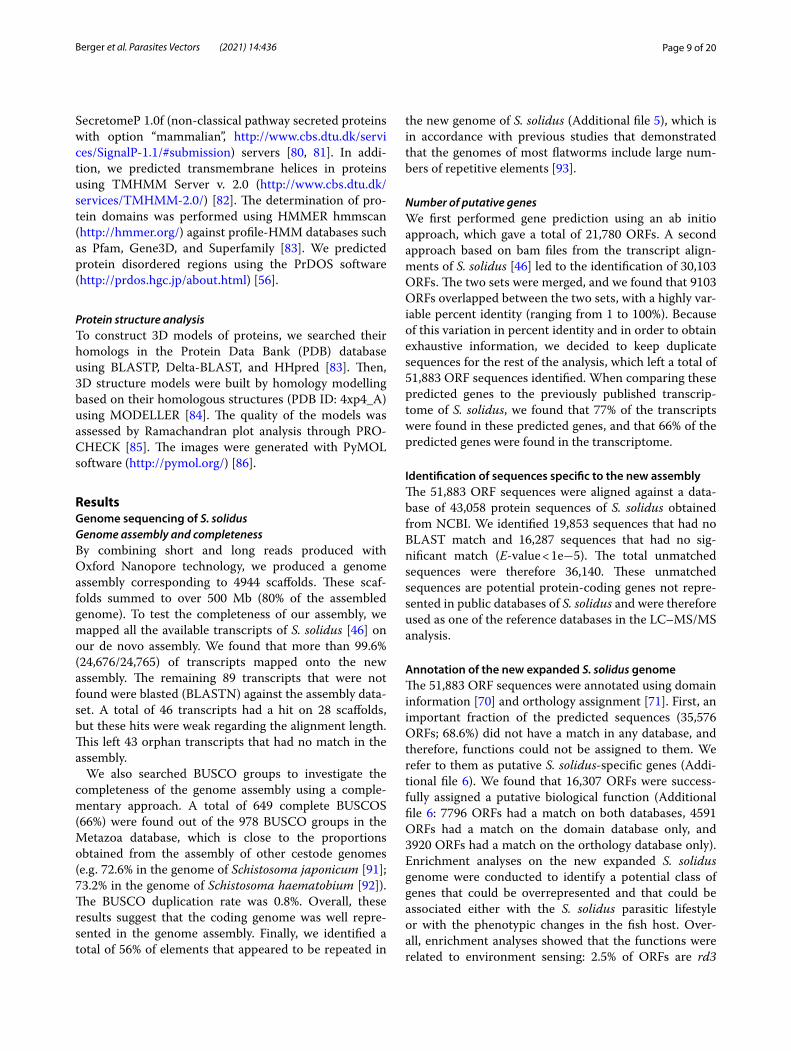
A multi-pronged approach to study the secretome of Schistocephalus solidus
In PBSfor 2 hours
Secretomestored at -20°C
Worm tissuesstored at -80°c
X 4 X 4 DNA extraction
Sequencing with Illumina and Oxford nanopore
Assembly and annotation
NEW GENOME
Worm stored at 4°C
in ethanol 90%
In-gel digestion
Protein precipitation Protein extraction
Protein prediction using as databases: larger proteome of S. solidus from UniProt
+ NEW GENOME of S. solidus
LC-MS/MS analysis
Proteins detected in the secretome but not in tissues
MANIPULATION FACTORS
S. solidus infected fishhave behavioural
perturbations
X 1
Fig. 1 The multipronged approach designed to detect potential manipulation factors in the secretome of Schistocephalus solidus. On the left, the proteomic approach based on LC–MS/MS aims at describing the global proteomic composition of the proteome and the secretome of S. solidus. On the right, the genomic approach aims at producing a novel genome sequence and assembly of S. solidus to improve protein coding gene prediction and annotation for this parasite. The genome, whose quality is improved, can be used as a new reference database to infer proteins in the proteome and the secretome of S. solidus. Combined, these approaches allow us to characterize the secretome of S. solidus at the infective stage, including the uncovering of proteins detected only in that fraction and not in its proteome, thus representing potential manipulation factors

Page 6 of 20Berger et al. Parasites Vectors (2021) 14:436
the pipeline GAWN v0.3.2 (https:// github. com/ enorm andeau/ gawn). An assembly quality analysis was per-formed using the Benchmarking Universal Single-Copy Orthologs (BUSCO), and BUSCO groups were searched in the Metazoa database [57].
Protein coding gene prediction To find out the propor-tion of repeated regions in the genome and to obtain a masked assembly prior to running BRAKER2 to predict protein coding genes, we built a RepeatModeler (1.0.8) database [58] based on the new genome sequence and ran RepeatMasker (4.0.6) based on that database [59]. We used BRAKER2 [60–65] for protein coding gene pre-diction using two approaches. The first approach was ab initio as it was not based on external data to find open reading frames (ORFs) but only on genes predicted by GeneMark-ES that are selected for training Augustus [60, 61]. The second approach used the alignment (bam files) of the transcripts from S. solidus [46] on the genome. The two sets of ORFs obtained with the two approaches were merged, and duplicate sequences were removed, which allowed us to obtain a final number of predicted ORFs. We used a BLAST analysis to quantify the percentage of the S. solidus transcriptome we previously published [46] that was found in the genes predicted using Augustus and vice versa.
Identification of sequences specific to the new assem-bly The predicted ORFs were locally aligned using BLAST+ [66, 67] against a database of 43,058 protein sequences from S. solidus obtained from release 230 of the NCBI (March 2019, https:// www. ncbi. nlm. nih. gov). From the predicted ORFs, we selected those that had no BLAST match or that had no significant match based on the fact that the length of the alignment was less that 80% of the length of the query or that had less than 90% of identi-cal nucleotides over the length of the query [68]. These unmatched sequences that were specific to the assembly were used as one of the reference databases during LC–MS/MS analysis (see “Protein identification”).
Genome annotation A functional annotation of the pre-dicted ORFs that were obtained with BRAKER2 [60–65] was performed using HMMER (version 3.3) [69] against the PFAM domain database (release 32) [70] and using orthology assignment with eggNOG-mapper (Evolution-ary Genealogy of Genes: Non-supervised Orthologous Groups) (version 2.0.1) [71].
Mass spectrometry characterization of the worm proteome and secretomeWorm and secretome collection for mass spectrometry analysisSampling of experimental individuals Whole worms were collected from wild-caught fish acclimatized to lab-oratory conditions. Juvenile sticklebacks came from Lac Témiscouata (Québec, 47°40′33″N 68°50′15″W), the same lake that was used to collect a worm for genome sequenc-ing. Fish were caught using a seine in August 2016. They were brought to the Laboratoire Aquatique de Recherche en Sciences Environnementales et Médicales at Université Laval where they were raised for 1 year in 80-l tanks under a light/dark photoperiod of 12 h:12 h and a temperature of 15 °C reflecting their natural environment conditions (Québec, Canada). Fish were fed daily with brine shrimps.
Collection of proteome and secretome samples In sum-mer of 2017, 51 fish were individually isolated in 2-l tanks. The day following isolation, fish were injected with 100 µl of phosphate-buffered saline (PBS; pH 7.4, Life Tech-nologies) in their abdominal cavity in order to sample their fluids to detect infection by S. solidus following the method described in [72]. This protocol was repeated the next day. If fish were detected as infected, they were euthanized with an overdose of MS-222 (75 mg/l mg/kg) and dissected to confirm the infection by S. solidus and to collect the worm and its secretome. Fish sex, size, and mass, and S. solidus mass and number in each fish were noted. We found that five fish were infected, each har-bouring a worm whose weight was above 50 mg (worm 1 = 485.3 mg; worm 2 = 504.1 mg; worm 3 = 286.5 mg; worm 4 = 544.5 mg; worm 5 = 220.9 mg). All the worms used to collect proteome and secretome samples were far above the mass threshold of 50 mg, which is commonly used to define the parasitic infective stage that coincides with the appearance of the behavioural changes in the fish host [27, 37, 38]. Furthermore, behavioural changes in fish infected by worms of similar masses were previously reported in the Témiscouata population [48]. Therefore, we considered these worms and secretome samples to be representative of the manipulative stage of S. solidus. We only selected fish that were infected by a single worm to prevent potential effects of multiple infections on the pro-teomic content of the secretome.
The worm secretome was collected according to a pro-tocol adapted from [73]. Each worm was rinsed with 1 ml of PBS (pH 7.4, Life Technologies) to remove fish fluids and then immediately put in a 2-ml tube of PBS. The tube was covered with aluminium foil to protect the worm from light and placed in a water recipient at the same temperature as the fish tanks (15 °C) for 2 h. The worm was removed from the tube, and a tablet of complete

Page 7 of 20Berger et al. Parasites Vectors (2021) 14:436
Mini Protease Inhibitor (Sigma) was added to the tube to protect the proteins from the protease activity. The worm tissues were then snap-frozen in liquid nitrogen and stored dry at −80 °C for proteomics analysis. The liquid in which the worm was incubated (PBS + poten-tial secretome collected) was stored at −20 °C. Further experiments were therefore conducted with five worms and their respective secretome.
Preparation of worm tissues and secretome for in‑gel digestionWorms were individually washed with 2 ml of PBS (pH 7.4, Life Technologies) to remove potential remain-ing contaminants from the fish. They were then cut into three equal pieces that were each put into a tube contain-ing 700 µl of lysis buffer (4% (w/v) sodium dodecyl sul-fate (SDS) in 100 mM Tris, pH 8–10, mM dithiothreitol [DTT]). Six sterile ceramic beads (size 2.8 mm) were added to each lysis tube, and the samples were homoge-nized for 20 s at 6000 rpm using a Precellys homogenizer (Bertin Technologies). Homogenization was repeated three times. Samples were put on ice for 1 min between each run. Samples were then spun at 10,000×g at 4 °C for 10 min. For each individual worm, the three homogen-ates obtained were pooled together and redistributed into equal volumes into two tubes. Samples were heated at 95 °C for 10 min, then spun for 10 min at room tempera-ture. The supernatant of each sample was collected into a new tube, and protein concentration was calculated using the extinction coefficient measured with a NanoDrop 1000 spectrophotometer (A280 nm; Thermo Scientific). The concentration obtained for each worm lysate was respectively: 24.14 mg/ml; 22.03 mg/ml; 15.66 mg/ml; 28.53 mg/ml; and 11.37 mg/ml. Worm lysates were kept at −20 °C before performing in-gel digestion.
For the secretome, the protein concentration of each of the liquids in which the worms were incubated (“secretome” samples) was measured using a Pierce Coomassie (Bradford) Protein Assay Kit (Fisher Sci-entific). Bovine serum albumin (BSA) at 2 mg/ml was used as standard (seven-point standard curve ranging from 0 to 25 µg/ml). The concentrations obtained were: 13.11 µg/ml; 15.09 µg/ml; 9.83 µg/ml; 16.59 µg/ml; and 7.46 µg/ml. For each secretome sample, 10 µg of pro-teins were precipitated with trichloroacetic acid (TCA) [74]. Precipitated samples were directly used for in gel-digestion.
In‑gel digestionFor each sample, 50 µg of proteins from worm lysate and 10 µg of proteins from secretome were resolved on a 12% SDS–polyacrylamide gel electrophoresis (SDS-PAGE) with a BenchMark protein ladder (Invitrogen) and a
negative control (SDS-PAGE loading buffer and water). Migration was performed during 60 min at 175 V for the worm lysates, and during 30 min at 175 V for secretomes. Coomassie blue G250 (PMID: 15,174,055) was used for staining overnight (Additional file 1). Each migration lane was cut into five fractions for worm lysates and three fractions for secretomes. In-gel digestion was performed on these fractions according to a previously developed protocol [75]. Briefly, after cutting the gel into slices, pro-teins were reduced using 10 mM dithiothreitol (DTT) for 45 min at 56 °C, then alkylated with 55 mM iodoaceta-mide (IAA) for 30 min at room temperature in the dark. Digestion was performed overnight at 37 °C with trypsin (Promega V5113; 0.1–1 µg of trypsin depending on the gel staining intensity). The next day, peptides were extracted using an organic solvent (100 µl acetonitrile) and a step-wise protocol (40% acetonitrile then 100% ace-tonitrile), and dried (< 50 μl). Following in-gel digestion, a STAGE-TIP protocol using C18 extraction disks (3M Empore) was performed to desalt the samples [76, 77]. Samples were acidified with trifluoroacetic acid (TFA) (pH < 2.5) before being passed through the tip. At the end of the STAGE-TIP protocol, samples were dried com-pletely using a Vacufuge Plus concentrator (Eppendorf ) and stored at −20 °C until LC–MS/MS analysis.
LC–MS/MS analysisThe peptides were analysed using a quadrupole time-of-flight mass spectrometer (Impact II; Bruker Daltonics) coupled to an Easy-nLC 1000 high-performance liquid chromatography (HPLC) system (Thermo Fisher). More information about the LC–MS/MS analysis can be found in the supplementary information (Additional file 2).
Protein identificationWe searched the detected mass spectra against the S. soli-dus genome using MaxQuant (version 1.6.1.0) [78]. Two searches were independently performed: the first search used the larger proteome of S. solidus (43,058 entries, downloaded June 21, 2018, and updated May 6, 2019) from the Universal Protein Resource release 2018–05 and 2019–03 (UniProt https:// www. unipr ot. org/) as a refer-ence database, which includes proteins predicted from the partial genome of S. solidus that was then currently available [44], as well as proteins predicted from the de novo transcriptome [45, 46]. For the second search, we used the unmatched sequences that we reported to be specific to our genome assembly (36,140 sequences, see Results) as a reference database. The search included common contaminants and variable modifications of methionine oxidation, and N-acetylation of the proteins. The data was filtered for matches passing a 1% false dis-covery rate set by MaxQuant [78]. We included the larger
Page 8 of 20Berger et al. Parasites Vectors (2021) 14:436
proteome of the threespine stickleback host (29,032 entries, downloaded June 21, 2018) from the Universal Protein Resource release 2018–05 (UniProt https:// www. unipr ot. org/) in the search in order to include proteins originating from the fish host during the MaxQuant data search. Subsequent analyses were performed with all the proteins inferred using the larger proteome of S. solidus and the unmatched sequences of the new genome as ref-erence databases.
Data analysis of the proteomeData analysis was performed with Python custom scripts (version 3.6.4) using Jupyter notebooks (version 5.4.0). A template of the custom script is available in the sup-plementary information (Additional file 3). Proteins detected in each worm sample were retrieved using MaxQuant. We filtered out of the dataset protein IDs that were solely attributed to the threespine stickleback (Additional file 4), protein IDs with REV coding (reverse hits for false discovery rate filtering), and protein IDs with CON coding (contaminant hits that were added into the search). In some cases, several protein IDs were found by MaxQuant for a specific protein because of high sequence similarities between them. Research on NCBI (https:// www. ncbi. nlm. nih. gov/) and UniProt (https:// www. unipr ot. org/) databases demonstrated that the pro-tein IDs detected for one protein were probably isoforms with identical functions. These multiple protein IDs were kept during annotation to obtain exhaustive functional information, but only the first ID of each protein was kept for enrichment analyses to avoid an overrepresenta-tion of specific processes or functions (see below). This final dataset was used to describe the global composition of the proteome of S. solidus.
We performed two distinct enrichment analyses for the proteins detected in at least one worm sample and for the proteins detected in all worm samples. For proteins with several protein IDs, we kept only the first ID in order to prevent enrichment bias during analysis. Enrichment analysis was performed using the tool FunRich (version 3.1.3) [79]. We constructed a custom reference database using the protein IDs and the Gene Ontology (GO) anno-tation (biological process, cellular component, molecular function) of all the proteins described in the larger pro-teome of S. solidus available on UniProt (https:// www. unipr ot. org/ 43058 entries). P-values for enrichment were obtained with a hypergeometric test corrected with the Bonferroni method.
Data analysis of the secretomeData analysis was performed with Python using the same approach as for the proteome. We separated proteins into two categories: proteins that were shared between
the proteome and the secretome samples, and those that were found only in a secretome sample. For the pro-teins that were shared between the proteome and the secretome samples, we performed two distinct enrich-ment analyses: one for the proteins detected in at least one secretome sample and one for the proteins detected in all secretome samples. The enrichment analysis was performed using the FunRich tool (version 3.1.3) [79] using the same approach as for the proteome.
We investigated the annotation of proteins found only in the secretome and not the proteome using a three-step approach. During the first step, the protein ID obtained with the MaxQuant analysis was searched in UniProt to retrieve its information if available (protein name and sequence, corresponding coding gene, protein function, localization, and/or structural information). During the second step, we inferred the function of the protein based on sequence homologies using the Basic Local Alignment Search Tool (BLAST) on NCBI (https:// blast. ncbi. nlm. nih. gov/ Blast. cgi). We used BLASTP (protein–protein) using the non-redundant protein sequences (nr) database (August 2018) and TBLASTN (protein-translated nucle-otide) using the nucleotide collection (nr/nt) database (August 2018). During the last step, when no informa-tion was found with the previous steps or to confirm the information previously found, we used the Pfam data-base (version 32.0) [70] to infer the function of the pro-tein based on domain organization homologies. In some cases, several protein IDs were found by MaxQuant for a specific protein because of high sequence similarities between these IDs. We found by applying the previous approach that in all of these cases, the putatively redun-dant protein IDs detected for one protein were indeed isoforms with identical functions. These multiple protein IDs were nevertheless kept during annotation (see below) to obtain exhaustive functional information.
Annotation of proteinsIn order to obtain an exhaustive annotation of proteins, we conducted complementary approaches based on sequence and structure analyses (as described below) [56, 80–90] for the proteins detected in the proteome and/or enriched in the secretome for which few or no annota-tion was available. These analyses were also performed for the proteins that were detected using the unmatched sequences specific to our genome assembly as a refer-ence database, for which limited or no annotation was available.
Protein sequence analysisWe predicted secreted proteins using the SignalP 5 .0 (classical secretory proteins with option “eukary-ota”, http:// www. cbs. dtu. dk/ servi ces/ Signa lP/) and
Page 9 of 20Berger et al. Parasites Vectors (2021) 14:436
SecretomeP 1.0f (non-classical pathway secreted proteins with option “mammalian”, http:// www. cbs. dtu. dk/ servi ces/ Signa lP-1. 1/# submi ssion) servers [80, 81]. In addi-tion, we predicted transmembrane helices in proteins using TMHMM Server v. 2.0 (http:// www. cbs. dtu. dk/ servi ces/ TMHMM-2. 0/) [82]. The determination of pro-tein domains was performed using HMMER hmmscan (http:// hmmer. org/) against profile-HMM databases such as Pfam, Gene3D, and Superfamily [83]. We predicted protein disordered regions using the PrDOS software (http:// prdos. hgc. jp/ about. html) [56].
Protein structure analysisTo construct 3D models of proteins, we searched their homologs in the Protein Data Bank (PDB) database using BLASTP, Delta-BLAST, and HHpred [83]. Then, 3D structure models were built by homology modelling based on their homologous structures (PDB ID: 4xp4_A) using MODELLER [84]. The quality of the models was assessed by Ramachandran plot analysis through PRO-CHECK [85]. The images were generated with PyMOL software (http:// pymol. org/) [86].
ResultsGenome sequencing of S. solidusGenome assembly and completenessBy combining short and long reads produced with Oxford Nanopore technology, we produced a genome assembly corresponding to 4944 scaffolds. These scaf-folds summed to over 500 Mb (80% of the assembled genome). To test the completeness of our assembly, we mapped all the available transcripts of S. solidus [46] on our de novo assembly. We found that more than 99.6% (24,676/24,765) of transcripts mapped onto the new assembly. The remaining 89 transcripts that were not found were blasted (BLASTN) against the assembly data-set. A total of 46 transcripts had a hit on 28 scaffolds, but these hits were weak regarding the alignment length. This left 43 orphan transcripts that had no match in the assembly.
We also searched BUSCO groups to investigate the completeness of the genome assembly using a comple-mentary approach. A total of 649 complete BUSCOS (66%) were found out of the 978 BUSCO groups in the Metazoa database, which is close to the proportions obtained from the assembly of other cestode genomes (e.g. 72.6% in the genome of Schistosoma japonicum [91]; 73.2% in the genome of Schistosoma haematobium [92]). The BUSCO duplication rate was 0.8%. Overall, these results suggest that the coding genome was well repre-sented in the genome assembly. Finally, we identified a total of 56% of elements that appeared to be repeated in
the new genome of S. solidus (Additional file 5), which is in accordance with previous studies that demonstrated that the genomes of most flatworms include large num-bers of repetitive elements [93].
Number of putative genesWe first performed gene prediction using an ab initio approach, which gave a total of 21,780 ORFs. A second approach based on bam files from the transcript align-ments of S. solidus [46] led to the identification of 30,103 ORFs. The two sets were merged, and we found that 9103 ORFs overlapped between the two sets, with a highly var-iable percent identity (ranging from 1 to 100%). Because of this variation in percent identity and in order to obtain exhaustive information, we decided to keep duplicate sequences for the rest of the analysis, which left a total of 51,883 ORF sequences identified. When comparing these predicted genes to the previously published transcrip-tome of S. solidus, we found that 77% of the transcripts were found in these predicted genes, and that 66% of the predicted genes were found in the transcriptome.
Identification of sequences specific to the new assemblyThe 51,883 ORF sequences were aligned against a data-base of 43,058 protein sequences of S. solidus obtained from NCBI. We identified 19,853 sequences that had no BLAST match and 16,287 sequences that had no sig-nificant match (E-value < 1e−5). The total unmatched sequences were therefore 36,140. These unmatched sequences are potential protein-coding genes not repre-sented in public databases of S. solidus and were therefore used as one of the reference databases in the LC–MS/MS analysis.
Annotation of the new expanded S. solidus genomeThe 51,883 ORF sequences were annotated using domain information [70] and orthology assignment [71]. First, an important fraction of the predicted sequences (35,576 ORFs; 68.6%) did not have a match in any database, and therefore, functions could not be assigned to them. We refer to them as putative S. solidus-specific genes (Addi-tional file 6). We found that 16,307 ORFs were success-fully assigned a putative biological function (Additional file 6: 7796 ORFs had a match on both databases, 4591 ORFs had a match on the domain database only, and 3920 ORFs had a match on the orthology database only). Enrichment analyses on the new expanded S. solidus genome were conducted to identify a potential class of genes that could be overrepresented and that could be associated either with the S. solidus parasitic lifestyle or with the phenotypic changes in the fish host. Over-all, enrichment analyses showed that the functions were related to environment sensing: 2.5% of ORFs are rd3

Page 10 of 20Berger et al. Parasites Vectors (2021) 14:436
genes known to be expressed in photoreceptor cells [94]; cell division, growth, and development: 1.7% of ORFs encode for proteins involved in the movement of micro-tubules, and 1.2% of ORFs are atrophin-coding genes involved in development [95]; as well as cell physiology: 1% of ORFs encode for PARP proteins involved in various cell physiological processes [96].
Characterization of the proteomeUsing mass spectrometry (LC–MS/MS), we detected 2290 proteins in samples of whole S. solidus tissues, among which 1467 proteins were detected in all worm samples (Additional file 7). Among these 2290 proteins, 246 proteins were only detected using the new genome of S. solidus as a reference database during the LC–MS/MS analysis, with 113 proteins detected in all worms. The new genome sequence and annotation therefore provide a significant additional resource for S. solidus functional genomics. Most of the 2290 proteins were functionally annotated, with the exception of the 246 proteins previ-ously mentioned, for which functions were further inves-tigated using a three-step approach based on sequence, structure, and phylogenetic analyses (see below). Using this approach, we found that the 2290 proteins of S. soli-dus included 40 proteins encoded by 27 genes that did not have any sequence or domain similarities with other known species, and for which functions could not be described (i.e. 1.7% of putative S. solidus-specific proteins in the proteome, compared to 68.6% of putative S. soli-dus-specific genes in the new genome) (Additional file 8).
Enrichment analyses in terms of biological process, cellular component, and molecular function (GO terms) were performed for all the proteins detected in at least one of the worm samples, and also for the proteins
detected in all worm samples. For proteins with sev-eral protein IDs, we kept only the first ID in order to prevent enrichment bias. We found that 14.9% of pro-teins detected in all worm samples were involved in protein metabolism processes, namely “translation” [GO:0006412] and “protein folding” [GO:0006457] (p < 0.001). These are typically highly expressed proteins, which explains their overrepresentation here [93]. Also, we found that 10.2% of proteins detected in all worm samples referred to mechanisms important in the func-tioning and the regulation of the cell cycle: “microtubule-based process” [GO:0007017], with the microtubules having major roles in cell division and growth, and “tri-carboxylic acid cycle” [GO:0006099], the latter referring to abundant metabolic enzymes that are involved in res-piration by producing ATP (p < 0.001) (Fig. 2a). We also found that 14.4% of proteins detected in all worm sam-ples were proteins with GTP binding [GO:0005525] and GTPase activity [GO:0003924] (p < 0.001) (Fig. 2b). Con-sistent with all the functions described above, we found that 23.2% of proteins detected in all worm samples were localized in the cytoplasm (p < 0.001).
The prediction of the functions of the 246 proteins that were detected using the new genome as a refer-ence database was further investigated with sequence and structure analyses (Additional file 7). Their func-tions were in accordance with the previous enrichment results, as we found for example proteins involved in microtubules (protein ID: g20896.t1) or with ATPase activity (protein ID: g10622.t1). Additionally, we found that six proteins were peptidases or proteases with con-served active sites, suggesting that they are functional (Table 2) and that a protein contained an amidase domain. This protein "g7530.t1″ is a fatty acid amide
a
microtubule-based process
[GO:0007017]
tricarboxylic acid cycle
[GO:0006099]
transla�on[GO:0006412]
protein folding[GO:0006457]
gluconeogenesis[GO:0006094]
glycoly�c process[GO:0006096]
b GTPase ac�vity[GO:0003924]
GTP binding[GO:0005525]
8,0%29,1%
4,3%3,9%
2,2%
10,6%
2,6%
8,7%
1,5%
4,9%
6,5%
7,9%
10,7%
12,3%
Fig. 2 The proteome and secretome of S. solidus include proteins involved in protein metabolism/cell growth/energy intake. Results that were significant (p < 0.001) for the enrichment analyses performed in terms of biological process (a) and molecular function (b). In each case, analysis was performed for the proteins detected in all worm (outer chart) and secretome (inner chart) samples

Page 11 of 20Berger et al. Parasites Vectors (2021) 14:436
hydrolase like (SsFAAH-like). FAAH enzymes degrade signalling lipids of the endocannabinoid class [97].
Additionally, 141 proteins were assigned both to the worm and to the fish host during LC–MS/MS analy-sis (Additional file 9). As it was not possible to directly determine if the 141 proteins were produced by the worm or the fish, we searched if these proteins could be core proteins, i.e. proteins conserved in all eukary-otes and whose functions are well characterized [98]. Using BLASTP and Pfam tools, we found that the 141 proteins had sequence similarities with 248 previously reported eukaryotic core proteins [98] (mean Expect-value 2.45e−11) and that they were composed of con-served domains. We therefore concluded that these proteins were expressed both in the worm and in the fish because of their fundamental functions in the cell, but that their peptide similarities did not allow us to determine their origin. Enrichment analysis per-formed on these 141 proteins confirmed that they were involved in cell physiology and in energy production as found for the whole proteome.
Characterization of the secretomeA total of 1568 proteins were detected in the secretome samples (Additional file 7). The numbers ranged between 781 and 1183 proteins depending on the sample, with 459 proteins detected in all secretome samples. As expected, the secretome of S. solidus was mostly a subset of the worm proteome, both in terms of protein number and functions, with a few exceptions described below. The whole protein content of the secretome of a given worm represented up to 59% of the whole protein content of the proteome of that same worm. In total, 1538 unique proteins were shared between at least one secretome
sample and one proteome sample, among which 385 proteins were detected in all secretomes and proteomes studied, and 74 proteins were detected in all secretomes and in at least one proteome. All the proteins that were shared between secretome and proteome samples had a functional annotation, except for 36 proteins that were previously detected in the proteome as S. solidus-specific and that were also secreted (2.3% of putative S. solidus-specific proteins in the secretome, compared to 68.6% of putative S. solidus-specific genes in the new genome and 1.7% of putative S. solidus-specific proteins in the pro-teome) (Additional file 8).
We found that the secretome was composed of pro-teins enriched for biological processes, cellular compo-nents, and molecular functions that were similar to the proteome. For the proteins detected in all secretomes, almost half of the proteins participated in the regulation of cell division and energy production. These proteins were enriched in functions such as the “microtubule-based process” [GO:0007017] involved in the regulation of cell division, and “glycolytic process” [GO:0006096] and “gluconeogenesis” [GO:0006094], these two pro-cesses being an important source of energy production (p < 0.001 hypergeometric test corrected with Bonferroni method) (Fig. 2a). Proteins were also enriched in domains with GTP binding [GO:0005525] and GTPase activity [GO:0003924] (Fig. 2b), as previously reported in the proteome (p < 0.001). Furthermore, 37.9% of the proteins detected in all the secretome samples were predicted to be localized in the cytoplasm (p < 0.001), similar to pro-teins in the proteome. Notably, we found that a signifi-cant number of proteins (6.1% of the proteins detected in all secretomes p < 0.001) were reported to be specifically localized into the extracellular space, an enrichment that was specific to the secretome.
Proteins unique to the secretomeWe found eight proteins that were detected in all secretome samples, but in none of the proteomes (Table 3). Among them, three proteins had fibronectin type-III domains. The first (protein ID: A0A0X3PH69) was a “Neogenin”, which is a protein involved in neural development with two fibronectin type-III domains. The second (protein ID: A0A0X3Q1B7; A0A0X3PKA1; A0A0X3Q8R6) was a receptor-type tyrosine-protein phosphatase eta that included three fibronectin type-III domains. The third (protein ID: A0A0V0JBL5) included two fibronectin type-III domains, but no additional information on the function of this protein was avail-able. Furthermore, we detected an uncharacterized pro-tein with a predicted molecular function corresponding to “neurotransmitter: sodium symporter activity (NSS)”,
Table 2 Peptidases detected in the proteome of S. solidus using its new expanded genome
The name, the identification number from the new genome assembly (ID), the length of the signal peptide (in numbers of amino acids), and the name of the conserved active sites are indicated for each peptidase
Peptidase name ID Signal peptide (amino acid)
Active site
Cathepsin B g20295.t1 1 to 26 Q131, C137, H306, N326
Aminopeptidase M17
g13261.t1 1 to 30 K280, R355
Aminopeptidase M17
g19803.t1 1 to 17 K284, R359
Aminopeptidase M17
g13265.t1 1 to 19 K317, R392
Peptidase family M49 g12315.t1 No E438, E439, H443, E495
Peptidase M16B g29053.t1 No E71, E141, L267, R368

Page 12 of 20Berger et al. Parasites Vectors (2021) 14:436
which is involved in transmembrane transport (pro-tein ID: A0A183S8K9; A0A0X3PDV9; A0A0X3PNC8; A0A0V0J682; A0A183T7R5), and a protein potentially acting at the cell membrane as a phospholipid scramblase (PLSCR) (protein ID: A0A0X3P711; A0A183SGM7). Lastly, three other uncharacterized proteins did not have information on function using the UniProt data-base, BLAST tools, and protein domain identification. It seems that these three proteins are specific to S. solidus, with the last protein (protein ID: A0A0X3Q756) having a secretory signal peptide.
Twenty-two proteins were detected in at least one of the five secretome samples, but in none of the proteomes (Table 4). Three proteins were composed of fibronectin type-III domains. The first (protein ID: A0A0X3NX35) detected in four out of five secretomes was described as a receptor-type tyrosine-protein phosphatase H. The second (protein ID: A0A183TLI3; A0A0X3PWG6; A0A0X3PW04; A0A0V0J316; A0A0X3PN23; A0A0X-3PJY8), which was detected in three secretomes, was either a tenascin (an extracellular matrix glycoprotein) or a receptor-type tyrosine-protein phosphatase F. The last protein of this type (protein ID: A0A0V0JA38), also detected in three secretomes, was, according to the BLAST result, a collagen-like protein, which is an impor-tant component of cuticle.
Two proteins appeared to be involved in immunity processes. The first (protein ID: A0A183TPG4;A0A0X3P3D7;A0A0X3PTB8), which was detected in three secretomes, was uncharacterized and had a cystatin domain which may be involved in immunomodula-tory functions. The second (protein ID: A0A0V0JBV1; A0A0V0J795; A0A0X3NVP3; A0A0X3P0M4), which was detected in the secretome of the largest worm only (worm 4), was according to the BLAST results an antigen
similar to the diagnostic antigen gp50 commonly used to detect parasitic diseases.
Two proteins were associated with transport func-tions. The first (protein ID: A0A183TIR8; A0A0X3PCX3; A0A183TT84; A0A0X3PT59) was detected in four out of five secretomes and was a sodium/glucose cotransporter involved in glucose homeostasis. The second (protein ID: A0A0X3NT74), which was only detected in the second largest worm (worm 2), was an intraflagellar transport protein required for ciliogenesis.
One protein (protein ID: A0A183TDP7), detected in four secretomes, had a knottin fold similar to a domain found in the Schistosoma parasitic trematodes, but for which the function is not characterized. It is a cysteine-rich protein and interestingly, BLASTP search against the UniProt database revealed that it is only found in Platy-helminthes, and amino acid multiple sequence alignment showed that it presents a new motif, C-x[6]-C-x(7)-CC-x(4)-C-x(9)-C-x[2]-C-x[6]-C-x(5)-CC-x(3)-C-x(4)-C. This type of protein is resistant to proteases.
Nine proteins had functions which could not be clearly determined by BLAST in UniProt and non-redundant protein sequences (GenBank) databases. Five other proteins were only detected using the new genome of S. solidus as a database reference. We fur-ther investigated the functions of these last 14 proteins with sequence, structure, and phylogenetic analysis: nine proteins were specific to S. solidus. Among them, two proteins (protein ID: A0A0V0J2U1; protein ID: A0A0X3PIM2) had secretion signal peptides, and one protein (protein ID: A0A0X3PXG6) had a transmem-brane helix (TMH) domain found in transmembrane proteins. Furthermore, one protein was identified as a peptidase M28B (glutamate carboxypeptidase 2)
Table 3 Proteins that are excreted/secreted by S. solidus, detected in five secretomes and not in proteomes
Protein IDs were taken from UniProt. When several protein IDs were assigned to one protein, these protein IDs corresponded to isoforms with identical functions. For each protein, functional annotation was first retrieved from searches with UniProt and BLAST tools. Complementary analyses based on sequence, structure, and phylogeny were used to obtain information
UniProt ID Information from UniProt and BLAST tools Information from sequence, structure, and phylogeny
A0A0X3PH69 Neogenin Fibronectin type‑III domains
A0A0X3Q1B7;A0A0X3PKA1;A0A0X3Q8R6 Receptor‑type tyrosine‑protein phosphatase eta Fibronectin type‑III domains
A0A0V0JBL5 Unknown Fibronectin type‑II domains
A0A183S8K9;A0A0X3PDV9;A0A0X3PNC8;A0A0V0J682;A0A183T7R5
Neurotransmitter: sodium symporter (NSS) Unknown
A0A0X3P711;A0A183SGM7 Phospholipid scramblase Unknown
A0A0V0J8W2 Unknown Gene specific to S. solidus (orphan)
A0A0X3P740 Unknown Gene specific to S. solidus (orphan)
A0A0X3Q756 Unknown Gene specific to S. solidus (orphan)Signal peptide (secreted)

Page 13 of 20Berger et al. Parasites Vectors (2021) 14:436
(protein ID: g1854.t1; g12541.t1), and one protein had fibronectin type-III domains (protein ID: g17644.t1).
DiscussionUnderstanding the molecular mechanisms behind behav-ioural changes in a vertebrate host infected by a non-cer-ebral parasite is a fascinating challenge. If these changes are the result of parasite manipulation of its host, one possible mechanism hinges on molecules secreted by the parasite (i.e. manipulation factors) that impact the host’s physiological, immunological, and central nervous sys-tems, and ultimately the behaviour of the host. Here, we
describe for the first time the protein component of the secretome of a parasite commonly referred as manipu-lative, using mass spectrometry (LC–MS/MS), with the objective to identify potential manipulation factors that could explain host behavioural changes. As expected, we found that the proteins that are excreted/secreted by S. solidus are mostly a subset of the proteins being expressed in the whole worm and that both included proteases. We also found that 30 secretome proteins were not detected in the proteome and were involved in neural, immune, and cell communication functions, therefore having the potential to interfere with the host’s
Table 4 Proteins that are excreted/secreted by S. solidus, detected in at least one secretome and not in proteomes
Protein IDs were taken from UniProt or from the new genome assembly. When several protein IDs were assigned to one protein, these protein IDs corresponded to isoforms with identical functions. For each protein, functional annotation was first retrieved from searches with UniProt and BLAST tools. Complementary analyses based on sequence, structure, and phylogeny were used to obtain information
Protein ID No. of secretomes for which the protein was detected
Information from UniProt and BLAST tools Information from sequence, structure, and phylogeny
A0A0X3NX35 4/5 Receptor‑type tyrosine‑protein phos‑phatase H
Fibronectin type‑III domains
A0A183TLI3;A0A0X3PWG6;A0A0X3PW04;A0A0V0J316;A0A0X3PN23;A0A0X3PJY8
3/5 ‑Tenascin?‑Receptor‑type tyrosine‑protein phos‑
phatase F?
Fibronectin type‑III domains
A0A0V0JA38 3/5 Collagen‑like protein Fibronectin type‑III domains
A0A183TPG4;A0A0X3P3D7;A0A0X3PTB8 3/5 Unknown Cystatin domain
A0A0V0JBV1;A0A0V0J795;A0A0X3NVP3;A0A0X3P0M4
1/5 (worm 4) Antigen Intrinsically disordered
A0A183TIR8;A0A0X3PCX3;A0A183TT84;A0A0X3PT59
4/5 Sodium/glucose cotransporter Unknown
A0A0X3NT74 1/5 (worm 2) Intraflagellar transport protein 81 homolog
Unknown
A0A183TDP7 4/5 Protein with a knottin fold Cysteine‑rich proteinSpecific to Platyhelminthes
A0A0V0J2U1 4/5 Unknown Gene specific to S. solidus (orphan)Signal peptide (secreted)
A0A0X3NRK5 4/5 Unknown Gene specific to S. solidus (orphan)
A0A0X3PMK5;A0A0X3PDL5;A0A0X3PEZ1 4/5 Unknown Gene specific to S. solidus (orphan)
A0A0X3PRL9 4/5 Unknown Gene specific to S. solidus (orphan)
A0A0X3NVW4 3/5 Unknown Gene specific to S. solidus (orphan)
A0A0X3PXG6 3/5 Unknown Gene specific to S. solidus (orphan)TMH (membrane)
A0A0X3PIM2 2/5 Unknown Gene specific to S. solidus (orphan)—Signal peptide (secreted)
A0A0X3PNW2 2/5 Unknown Gene specific to S. solidus (orphan)
A0A183TE24;A0A0X3PRC0 1/5(worm 4)
Unknown Gene specific to S. solidus (orphan)
g11241.t1 3/5 Unknown Intrinsically disordered
g1854.t1;g12541.t1 2/5 Unknown Peptidase M28B(glutamate carboxypeptidase 2)
g17644.t1 4/5 Unknown Fibronectin type‑III domains
g2.t1 3/5 Unknown Unknown
g6226.t1 1/5 Unknown Unknown

Page 14 of 20Berger et al. Parasites Vectors (2021) 14:436
physiological systems and behaviour. Finally, we high-lighted that the secretome of S. solidus included S. soli-dus-specific proteins that could play important roles in the tight interaction of the parasite with its fish host. All together, these proteins represent promising candidates to explain physiological and behavioural changes in the stickleback host.
The secretome of S. solidus is mostly a subset of its proteome with similar functionsThe global protein composition of the proteomes and the secretomes of S. solidus highlights functions that are cru-cial for its parasitic lifestyle and for its interactions with the external environment. As it is generally reported [52], we found that the proteins that are excreted/secreted by S. solidus (i.e. the secretome) are mostly a subset of the proteins being expressed in the whole worm (i.e. the pro-teome), both in terms of protein number and function. We found that both the proteome and the secretome of S. solidus at the infective stage (i.e. when behavioural changes appear in the fish host) were enriched in pro-teins involved in cell division, which was also found in the functional analysis of the genome we sequenced, as well as energy production. Concerning cell growth, our results may seem counterintuitive because a previous transcriptome analysis revealed that the non-infective stage of S. solidus shows higher expression levels of genes involved in growth and cell regulatory functions com-pared to the infective stage [46]. However, we did not analyse the proteome and the secretome of worms at the non-infective stage, such that we can only quantify that these processes are enriched at the infective stage but not their relative importance compared to other life stages of the parasite. We therefore only speculate that both the non-infective and the infective stages of S. solidus could rely on biological processes involving cell growth, but production levels would be much higher at the non-infective stage, considering the fact that growth occurs predominantly in the first 12 weeks after installa-tion in the fish host [99]. Concerning energy production, the results are also surprising, as the worm was empiri-cally described to use its glycogen reserve (its primary source of energy) mainly when it reaches its final avian host [100]. However, transcriptomic analyses demon-strated that glycogen metabolism and energy production in S. solidus are complex processes [46]. Specifically, six steps of the glycolysis cycle are highly expressed at the infective stage [46]. Furthermore, it is important to keep in mind that for tissue and secretome samples collection, worms were placed in a solution of PBS (pH 7.4) in the dark for 2 h. While this method is commonly used to collect parasitic secretomes [73], we cannot dismiss the possibility that the environmental change (from the body
cavity of the stickleback to the PBS tube) encountered by S. solidus at this step could have influenced the levels of proteins detected in the proteomes and secretomes. One can speculate that this environmental stress could have triggered cell growth and energy intake in the worm (for example, to counteract other metabolic effects), explain-ing the discrepancies between the proteomics data we obtained and the previous transcriptomic work [46]. To confirm our results, future work could replace the PBS solution by a medium that would better mimic the fish abdominal cavity, for example, by a medium already used in vitro for breeding [32]. The global analysis of the pro-teome and of the secretome of S. solidus therefore con-firms the importance of energy use for the worm, even before it reaches the bird.
We detected both in the proteome and the secretome of S. solidus proteins with GTP binding and GTPase activity. GTP-binding proteins also called G-proteins are known to regulate a variety of biological processes such as mediating signals by hormones and light, gene expression, cytoskeletal and microtubule organization, or vesicle trafficking [101–104]. In parasites, GTPases have been demonstrated to have important roles in the secretion of virulence factors [103, 104]. For instance, in Toxoplasma gondii, which is a parasite known to induce behavioural changes in rodents [105], Rab GTPases reg-ulate the secretion of proteins essential to invade host cells, and the modification of their expression results in aberrant transport of proteins [106].
The use of the new genome of S. solidus as a reference database during mass spectrometry analysis allowed us to detect six peptidases or proteases in the proteome of S. solidus, and one peptidase (peptidase M28B gluta-mate carboxypeptidase 2) detected only in the secretome fraction, with proteases and peptidases being typi-cally described as virulence factors for many parasites (Table 1). In S. solidus, proteases and peptidases may have important roles in weakening the fish immune response at the infective stage when the worm is ready to pass into its avian host to reproduce [32]. Injecting these proteases and peptidases, alone or in combination, into isolated head kidney samples from non-infected fish as in Scharsack et al. [32] would be necessary to confirm their potential role in disrupting the host immunity. Further-more, this shows that the new genome presented here is a valuable tool to identify proteins that are critical for the parasitic lifestyle. We also found an SsFAAH-like enzyme that could potentially degrade signalling lipids of the endocannabinoid class. Endocannabinoids have previously been reported as an important player in host-parasite interactions by promoting the activation of the immune response in the host [97, 107]. This enzyme was not detected in the secretome of S. solidus.

Page 15 of 20Berger et al. Parasites Vectors (2021) 14:436
The secretome of S. solidus also contains proteins not detected in the proteomeNeuronal and immune functionsTranscriptomic analysis demonstrated that when S. solidus reaches the infective stage, genes involved in neural pathways and sensory perception are expressed by the worm at higher levels [46]. Thus, we expected the secretome at the infective stage of S. solidus to be enriched with proteins involved in such neural func-tions (as described in other parasitic systems, reviewed in Table 1). Furthermore, behavioural changes in the stickleback infected by S. solidus generally appear in con-comitance with the activation of the immune response of the fish [32]. Therefore, we expected the secretome of S. solidus at the infective stage to include proteins involved in immunity (as reviewed in Table 1). We found that three proteins were playing potential roles in neural and immune functions. The first protein, which was detected in four secretomes but in none of the proteomes, had a knottin fold called UPF0506 composed of cysteines and generally found in Schistosoma parasites. In Schistosoma, the function of the proteins with such knottin fold is not defined. However, peptides with knottin domains (i.e. knottins) were described in venoms from various ani-mals. For venomous animals, knottins are neurotoxins having high specificity towards receptors in the nervous system of their prey or aggressor [108]. Furthermore, it is a cysteine-rich protein, and in parasites, cysteine-rich proteins play a role in invasion [109] and modula-tion [110] of the immune system. Therefore, this protein could be a promising manipulation factor if it could act as a neurotoxin in the brain of infected sticklebacks. The second protein, which was detected in three secretomes but in none of the proteomes, had a cystatin domain which may be involved in immunomodulatory func-tions. It was demonstrated in parasitic nematodes that cystastins are important secreted molecules that help parasites to evade immunity of the host [111]. Nema-tode cystatins inhibit host proteases involved in antigen processing and presentation, leading to a reduction of the host immune response [112]. This secreted protein could thereby explain in part why the immune response is only activated late in infected sticklebacks, which needs to be further studied. The third protein appeared to be an antigen similar to the diagnostic antigen gp50. This diagnostic antigen is used to detect parasitic infec-tion, for example, by Taenia solium [113]. However, the antigen was detected only in the secretome from the big-gest worm of our study and not in its proteome. Sam-pling over a longer time frame could alleviate this type of discrepancy between samples. One protein, which was detected in all secretomes but in none of the proteomes, had a “neurotransmitter: sodium symporter activity
(NSS)” annotation. In humans, it is a membrane pro-tein involved in the termination of synaptic transmission and the recycling of neurotransmitters at the membrane [114]. How this membrane protein could be secreted and act as a manipulation factor in the secretome is unclear. Therefore, we are cautious in interpreting the role of this protein as a manipulation factor, as it could be solely a waste product from the membrane of the parasite.
Cell communicationSchistocephalus solidus is located in the abdominal cav-ity of the threespine stickleback [27]. As the worm is not in direct contact with its host brain, we expected to detect proteins involved in cell communication or cell–cell signalling. During annotation of the new genome, 2.5% of genes were related to environmental sensing functions. Furthermore, we found that seven proteins detected only in the secretome fraction were charac-terized by fibronectin type-III (FNIII) domains. FNIII domains are widely found in animal proteins and are involved in cell–cell interactions [115]. The first protein with fibronectin type-III domains found in all secretomes but in none of the proteomes was identified as a neo-genin. In addition to playing roles in cell–cell adhesion, neogenins are involved in neural development in humans [116]. Three additional proteins that we identified with fibronectin type-III domains were described as receptor-type tyrosine-protein phosphatases (type eta, detected in all secretomes but in none of the proteomes; type H, detected in four secretomes but in none of the pro-teomes; type F, detected in three secretomes but in none of the proteomes). Receptor-type tyrosine-protein phos-phatases are involved in cell–cell communication [117]. It was previously shown that a baculovirus secretes a pro-tein tyrosine phosphatase, which acts on the neural sys-tem of its host, the silkworm Bombyx mori, and enhances its locomotory activity, so that it ultimately increases the virus dispersal [19] (Table 1). The set of phosphatases identified here are located in the membrane, such that they are predicted to have specific biological functions that differ from those of the protein tyrosine phosphatase found in the baculovirus. However, their abundant repre-sentation in the secretome is intriguing, and the impor-tance of the process of phosphorylation represents a future avenue of study in the context of behavioural changes in the fish host, such as increased exploration [34]. For the next two proteins identified with fibronec-tin type-III domains, the first protein was found in all secretomes but in none of the proteomes and had no clear function (protein ID: A0A0V0JBL5), as for the sec-ond protein that was detected in four secretomes but in none of the proteomes (protein ID: g17644.t1). The last protein appeared to be a collagen-like protein found in

Page 16 of 20Berger et al. Parasites Vectors (2021) 14:436
three secretomes. In Caenorhabditis elegans, the cuti-cle is an extracellular matrix made of small collagen-like proteins [118]. Thus, the role of this protein in cell com-munication is uncertain, and it may rather be a waste product excreted passively from the cuticle of S. solidus. In summary, fibronectin type-III proteins secreted by S. solidus appear to be good candidates for manipulation factors because of their roles in cell–cell signalling, but also in potential neural functions.
Additionally, we expected to detect proteins with trans-port functions in the secretome of S. solidus to mediate the communication between the worm localized in the host abdominal cavity and the host brain. We detected three proteins involved in transport or related functions at the membrane, including a protein that was detected in all secretomes but in none of the proteomes, a phos-pholipid scramblase. It is a transmembrane protein that is known in humans to bind to the 5’-promoter region of the inositol 1,4,5-triphosphate receptor type 1 gene (IP3R1) so that it enhances expression of the receptor [119]. Since these proteins detected here could be simply waste products coming from the membranes of the para-site, we thus have to be careful when speculating about the roles of these proteins, and functional analyses will be required.
The secretome includes S. solidus‑specific proteins that could play important roles in parasite–host interactionsDuring annotation of the new genome, we found that 68.6% of the predicted genes did not have any sequences or domain similarities with other known species that would enable accurate annotation, therefore represent-ing putative S. solidus-specific genes. Part of these genes are effectively translated, as 1.7% of the proteome and 2.3% of the secretome included putative proteins spe-cific to the worm. Specifically, we detected a total of 12 proteins only in the secretome fraction for which func-tions could not be clearly determined, but that were specific to S. solidus. Three proteins were detected to be secreted signal peptides (protein ID: A0A0X3Q756; protein ID: A0A0V0J2U1; protein ID: A0A0X3PIM2). We found that the proteins that are specifically enriched in the secretome are more likely to have secreted sig-nal peptides (10% of the proteins detected only in the secretome fraction had secreted signal peptides, while 0.17% of the proteins detected in the proteome had such signals). One protein had a transmembrane helix (TMH) domain found in transmembrane proteins (protein ID: A0A0X3PXG6). Proteins with TMH domains are used to diagnose parasitic infection, as they are highly specific to the parasite of interest [120, 121]. Interestingly, analyses conducted with the transcriptome of S. solidus previously
suggested that 19% of all the protein-coding genes could be S. solidus-specific [45]. In this study, producing a novel genome sequence and assembly of S. solidus led us to increase this estimation by three times. S. solidus-specific secreted proteins represent promising candidates to explain physiological and behavioural changes in the stickleback host. The threespine stickleback is an obliga-tory host of S. solidus [122]. Because of the potential coevolution between the worm and the fish [27], S. soli-dus may have developed highly specific molecular mech-anisms targeting the threespine stickleback physiological machinery to insure it can establish and grow in this fish only, until it is ready to pass into its final avian host. The S. solidus-specific proteins found here are therefore likely to be part of this unique set of molecules crucial for the survival of the worm, and we can assign them this eco-logical annotation [123]. It will be very interesting in the future to produce recombinant S. solidus-specific pro-teins and test their effects on the phenotype of the three-spine stickleback by functional analysis. These proteins could also serve to obtain structural information by X-ray crystallography or NMR spectroscopy, which could help us to discover their function. Finally, studies of how these proteins evolve between Schistocephalus populations and between species of Schistocephalus infecting different fish species [124] will shed additional light on the speci-ficity and potential function of these proteins.
ConclusionsThe secretome of S. solidus appears to be an important component of the molecular interaction between the parasite and its threespine stickleback host. In accord-ance with our predictions, we detected in the secretome of S. solidus at the infective stage (i.e. when behavioural changes appear in the stickleback) proteases, proteins involved in neural and immune functions, as well as proteins involved in cell communication. We also high-lighted in the secretome the presence of S. solidus-spe-cific proteins. In the future, comparative studies could be conducted to validate that the proteins detected in the secretome of S. solidus act as manipulation factors in the behaviour of its fish host: at the organism level, the anal-ysis of the secretome of the worm at the non-infective stage will confirm if the putative manipulative proteins reported in this study are effectively detected solely at the infective stage (or at highest levels) when the behavioural changes appear in the host. It would also be of special interest to study the secretome of S. solidus at the stage when it is able to reproduce, i.e. in the bird host. At the species level, comparing the secretome of S. solidus with the secretome of a closely related species Schistocephalus pungitii will be of great interest, as S. pungitii does not have known effects on the behaviour of the nine-spine

Page 17 of 20Berger et al. Parasites Vectors (2021) 14:436
stickleback, its specific intermediate host species [125]. Finally, functional studies testing effects of presence in non-infected sticklebacks of interesting proteins identi-fied in the secretome, such as receptor-type tyrosine-protein phosphatases or phospholipid scramblase, would allow us to better understand the contribution of these proteins in the behavioural changes. To conclude, we hope that the genomic and proteomic resources we pro-vide will help other researchers to investigate general questions on host–parasite interactions in nature.
Supplementary InformationThe online version contains supplementary material available at https:// doi. org/ 10. 1186/ s13071‑ 021‑ 04933‑w.
Additional file 1. Visualization on SDS‑PAGE gels of the protein content of the proteome and of the secretome for 5 S. solidus worms. (a) Proteome (i.e. tissues) extracts from 5 distinct worms. Note that for worm 1 and worm 2, the sample was loaded on gel either in duplicate or triplicate but only one lane was used for LC–MS/MS. (b) Secretome extracts from 5 distinct worms. On each gel, a BenchMark protein ladder (Invitrogen) and a negative control (SDS‑PAGE loading buffer and water) were also loaded.
Additional file 2. Methods information for LC/MS analysis.
Additional file 3. Template custom script for analysis of the proteome and secretome LC–MS/MS data using Python.
Additional file 4. List of the proteins that were solely attributed to the threespine stickleback host, and that were consequently discarded from analysis. All the protein IDs that were specific to the threespine stickleback Gasterosteus aculeatus are reported. For a proteome or a secretome sample, a value above 0 indicates protein detection. The Gene Ontology (GO) annotation (biological process, cellular component and molecular function) of each protein ID is reported.
Additional file 5. List of the repeated elements that were identified in the new genome of S. solidus. For each class of repeated elements the follow‑ing are indicated: the number of elements, their total length (bp) as well as their proportion (%) in the new genome.
Additional file 6. Annotation results of 16,307 ORFs predicted from the new expanded Schistocephalus solidus genome. Functional annotation was performed using PFAM (domain database) and eggNOG (orthology database). For each ORF match in each database: the accession number, the E‑value and the functional description of the match are indicated. The other ORFs (68.6%) predicted from the new genome did not match with these two databases, and are considered S. solidus‑specific.
Additional file 7. List of the proteins detected by LC–MS/MS in the proteome and the secretome of S. solidus. All the protein IDs that were detected in each of the proteome samples and/or secretome samples are reported. For a sample, a value above 0 indicates protein detection. The Gene Ontology (GO) annotation (biological process, cellular component and molecular function) of each protein ID is reported.
Additional file 8. List of the S. solidus‑specific proteins detected in the proteome and/or in the secretome of S. solidus. For each protein, the ID is reported from UniProt. If the protein is “not secreted”, then it was only detected in the proteome and not in the secretome. The Gene Ontology (GO) annotation (biological process, cellular component and molecular function) of each protein ID is reported.
Additional file 9. List of the 141 proteins that were assigned both to S. solidus and to the fish host during LC–MS/MS analysis. Each protein is assigned several protein IDs from the worm and the fish. The Gene Ontology (GO) annotation (biological process, cellular component and molecular function) of each protein ID is reported.
AcknowledgementsWe thank Karina Nielsen, Alexandra Sébastien, and Tom Clark from the Foster Lab for their advice during the preparation of the samples for mass spectrom‑etry analysis. The authors thank the personnel of the Laboratoire Aquatique de Recherche en Sciences Environnementales et Médicales at Université Laval for their help with fish rearing. We thank Eric Normandeau from Ressources Aquatiques Québec (RAQ) for his help to identify S. solidus‑specific proteins.
Authors’ contributionsCSB designed the study with input from NA‑H, CRL, LJF, and K‑MM. CSB performed the experiments to prepare the worms and their secretomes for mass spectrometry. K‑MM performed the experiments with the mass spec‑trophotometer and the MaxQuant software. HM prepared the worm samples for genome sequencing and performed the MinION sequencing. JL analysed the genome and prepared the files used for searches with the MaxQuant software. JL and H Maaroufi assembled and annotated the new genome and proteins. CSB analysed the data with input from NA‑H, CRL, and LJF. CSB and NA‑H wrote the manuscript with input from all authors. All authors read and approved the final manuscript.
FundingFunding for the project was provided by the Fonds de Recherche du Québec – Nature & Technologies (FRQNT), Projet en Équipe grant to N.A.H. and C.R.L.; a fellowship (international internship program) from FRQNT obtained through the Ressources Aquatiques Québec (RAQ) strategic cluster and a Student/PDF Research Grant award from the Canadian Society of Zoologists (CSZ) to C.S.B. Mass spectrometry infrastructure used here was supported by the Canada Foundation for Innovation, the BC Knowledge Development fund, and the BC Proteomics Network. Work in L.J.F.’s group was supported by Genome Canada and Genome British Columbia via the Pan‑Canadian Proteomics Initiative (264PRO).). C.R.L. holds the Canada Research Chair in Cellular Systems and Synthetic Biology.
Availability of data and materialsThe datasets supporting the conclusions of this article are included within the article and its additional files. Furthermore, the dataset of the new genome assembly is available from the NCBI BioProject with accession PRJNA576252. The mass spectrometry proteomics data have been deposited to the Pro‑teomeXchange Consortium via the PRIDE partner repository with the dataset identifier PXD016844.
Declarations
Ethics approval and consent to participateThis study was approved by the Comité de Protection des Animaux de l’Université Laval (CPAUL 2014069‑3).
Consent for publicationNot applicable.
Competing interestsThe authors declare that they have no competing interests.
Author details1 Département de Biologie, Université Laval, Quebec, QC, Canada. 2 Institut de Biologie Intégrative Et Des Systèmes (IBIS), Université Laval, Quebec, QC, Canada. 3 Ressources Aquatiques Québec (RAQ), Institut Des Sciences de La Mer de Rimouski, Quebec, Canada. 4 Département de Biochimie, Microbiologie Et Bioinformatique, Université Laval, Quebec, QC, Canada. 5 Department of Bio‑chemistry & Molecular Biology, Michael Smith Laboratories, University of Brit‑ish Columbia, Vancouver V6T 1Z4, Canada. 6 PROTEO, Le Réseau Québécois de Recherche Sur La Fonction, la structure et l’ingénierie des protéines, Université Laval, Quebec, Canada. 7 Centre de Recherche en Données Massives (CRDM), Université Laval, Quebec, Canada.
Received: 2 February 2021 Accepted: 6 August 2021

Page 18 of 20Berger et al. Parasites Vectors (2021) 14:436
References 1. Krist AC. Effect of the digenean parasite Proterometra macrostoma on
host morphology in the freshwater snail Elimia livescens. J Parasitol. 2000;86(2):262–7.
2. Jones D, Jones G, Rudnicka M, Click A, Reck‑Malleczewen V, Iwaya M. Pseudoparasitism of host Trichoplusia ni by Chelonus spp. as a new model system for parasite regulation of host physiology. J Insect Physiol. 1986;32(1):315–28.
3. Watson DW, Mullens BA, Petersen JJ. Behavioral fever response of Musca domestica (Diptera: Muscidae) to infection by Entomophthora muscae (Zygomycetes: Entomophthorales). J Invertebr Pathol. 1993;61(1):10–6.
4. Lafferty KD, Shaw JC. Comparing mechanisms of host manipulation across host and parasite taxa. J Exp Biol. 2013;216(1):56–66.
5. Adamo SA. Parasites: evolution’s neurobiologists. J Exp Biol. 2013;216(1):3–10.
6. Biron DG, Loxdale HD. Host‑parasite molecular cross‑talk dur‑ing the manipulative process of a host by its parasite. J Exp Biol. 2013;216(1):148–60.
7. Herbison R, Lagrue C, Poulin R. The missing link in parasite manipula‑tion of host behaviour. Parasit Vectors. 2018;11(1):222–7.
8. Coppens I, Vielemeyer O. Insights into unique physiological features of neutral lipids in Apicomplexa: from storage to potential mediation in parasite metabolic activities. Int J Parasitol. 2005;35(1):597–615.
9. Fromm B, Ovchinnikov V, Høye E, Bernal D, Hackenberg M, Marcilla A. On the presence and immunoregulatory functions of extracellular microRNAs in the trematode Fasciola hepatica. Parasite Immunol. 2017;39(1):e12399.
10. Grimwood J, Smith JE. Toxoplasma gondii: the role of parasite surface and secreted proteins in host cell invasion. Int J Parasitol. 1996;26(1):169–73.
11. Coakley G, Maizels RM, Buck AH. Exosomes and other extracellular vesicles: The new communicators in parasite infections. Trends Parasitol. 2015;31(1):477–89.
12. Knudsen GM, Medzihradszky KF, Lim K‑C, Hansell E, McKerrow JH. Proteomic analysis of Schistosoma mansoni cercarial secretions. Mol Cell Proteomics. 2005;4(12):1862–75.
13. Gourbal BEF, Guillou F, Mitta G, Sibille P, Thèron A, Pointier J‑P, et al. Excretory–secretory products of larval Fasciola hepatica investigated using a two‑dimensional proteomic approach. Mol Biochem Parasitol. 2008;161(1):63–6.
14. Ditgen D, Anandarajah EM, Meissner KA, Brattig N, Wrenger C, Liebau E. Harnessing the helminth secretome for therapeutic immunomodula‑tors. Biomed Res Int. 2014;15(1):964350–63.
15. Garg G, Ranganathan S. Helminth secretome database (HSD): a collec‑tion of helminth excretory/secretory proteins predicted from expressed sequence tags (ESTs). BMC Genomics. 2012;13(13):1471–2164.
16. Cwiklinski K, Dalton JP. Advances in Fasciola hepatica research using “omics” technologies. Int J Parasitol. 2018;48(1):321–31.
17. Kim D‑W, Yoo WG, Lee M‑R, Yang H‑W, Kim Y‑J, Cho S‑H, et al. Transcrip‑tome sequencing and analysis of the zoonotic parasite Spirometra erinacei spargana (plerocercoids). Parasit Vectors. 2014;7(1):368–87.
18. Bień J, Sałamatin R, Sulima A, Savijoki K, Conn DB, Näreaho A, et al. Mass spectrometry analysis of the excretory‑secretory (E‑S) products of the model cestode Hymenolepis diminuta reveals their immunogenic prop‑erties and the presence of new E‑S proteins in cestodes. Acta Parasitol. 2016;61(1):429–42.
19. Kamita SG, Nagasaka K, Chua JW, Shimada T, Mita K, Kobayashi M, et al. A baculovirus‑encoded protein tyrosine phosphatase gene induces enhanced locomotory activity in a lepidopteran host. Proc Natl Acad Sci. 2005;102(1):2584–9.
20. Biron DG, Marché L, Ponton F, Loxdale HD, Galéotti N, Renault L, et al. Behavioural manipulation in a grasshopper harbouring hairworm: a proteomics approach. Proc R Soc B. 2005;272(1):2117–26.
21. Biron DG, Ponton F, Marché L, Galeotti N, Renault L, Demey‑Thomas E, et al. “Suicide” of crickets harbouring hairworms: a proteomics investiga‑tion. Insect Mol Biol. 2006;15(6):731–42.
22. Prandovszky E, Gaskell E, Martin H, Dubey JP, Webster JP, McConkey GA. The neurotropic parasite Toxoplasma gondii increases dopamine metabolism. PLoS ONE. 2011;6(9):e23866.
23. De Bekker C, Quevillon LE, Smith PB, Fleming KR, Ghosh D, Patterson AD, et al. Species‑specific ant brain manipulation by a specialized fungal parasite. BMC Evol Biol. 2014;14(1):166–78.
24. Adamo SA, Kovalko I, Turnbull KF, Easy RH, Miles CI. The parasitic wasp Cotesia congregata uses multiple mechanisms to control host (Manduca sexta) behaviour. J Exp Biol. 2016;219(23):3750–8.
25. Libersat F. Wasp uses venom cocktail to manipulate the behavior of its cockroach prey. J Comp Physiol A. 2003;189(7):497–508.
26. Korgaonkar A, Han C, Lemire AL, Siwanowicz I, Bennouna D, Kopec R, et al. A novel family of secreted proteins linked to plant gall develop‑ment. BioRxiv. 2020. https:// doi. org/ 10. 1101/ 2020. 10. 28. 359562.
27. Barber I, Scharsack JP. The three‑spined stickleback‑Schistocephalus solidus system: an experimental model for investigating host‑parasite interactions in fish. Parasitology. 2010;137(3):411–24.
28. Clarke AS. Studies on the life cycle of the pseudophyllidean cestode Schistocephalus solidus. Proc Zool Soc London. 2010;124(1):257–302.
29. Hammerschmidt K, Kurtz J. Schistocephalus solidus: establishment of tapeworms in sticklebacks–fast food or fast lane? Exp Parasitol. 2007;116(2):142–9.
30. Dingemanse NJ, Oosterhof C, Van Der Plas F, Barber I. Variation in stick‑leback head morphology associated with parasite infection. Biol J Lin Soc. 2009;96(1):759–68.
31. Heins DC, Baker JA, Toups MA, Birden EL. Evolutionary significance of fecundity reduction in threespine stickleback infected by the diphyllobothriidean cestode Schistocephalus solidus. Biol J Lin Soc. 2010;100(1):835–46.
32. Scharsack JP, Koch K, Hammerschmidt K. Who is in control of the stick‑leback immune system: interactions between Schistocephalus solidus and its specific vertebrate host. Proc R Soc B. 2007;274(1629):3151–8.
33. Barber I, Huntingford FA. The effect of Schistocephalus solidus (Cestoda: pseudophyllidea) on the foraging and shoaling behav‑iour of three‑spined sticklebacks Gasterosteus aculeatus. Behaviour. 1995;132(1):1223–40.
34. Barber I. The effect of hunger and cestode parasitism on the shoaling decisions of small freshwater fish. J Fish Biol. 1995;47(1):524–36.
35. Grécias L, Valentin J, Aubin‑Horth N. Testing the parasite mass burden effect on alteration of host behaviour in the ‑stickleback system. J Exp Biol. 2018;221(1):174748.
36. Giles N. Behavioural effects of the parasite Schistocephalus solidus (Ces‑toda) on an intermediate host, the three‑spined stickleback Gasteros-teus aculeatus L. Animal Behaviour. 1983;131(1):1192–4.
37. Tierney JF, Crompton DWT. Infectivity of plerocercoids of Schistocepha-lus solidus (Cestoda: Ligulidae) and fecundity of the adults in an experi‑mental definitive host Gallus gallus. J Parasitol. 1992;78(1):1049–54.
38. Barber I, Svensson PA, Walker P. Behavioural responses to simu‑lated avian predation in female three spined sticklebacks: The effect of experimental Schistocephalus solidus infections. Behaviour. 2004;141(1):1425–40.
39. Weber JN, Steinel NC, Shim KC, Bolnick DI. Recent evolution of extreme cestode growth suppression by a vertebrate host. Proc Natl Acad Sci. 2017;114(25):6575–80.
40. Poulin R. Parasite manipulation of host behavior‑Chapter 5: An update and frequently asked questions. Adv Study Behav. 2010; 41(1):151–86.
41. Grécias L. Étude des causes proximales des changements de comporte‑ment de l’épinoche à trois épines (Gasterosteus aculeatus) par son parasite Schistocephalus solidus. Thesis Université Laval 2017; https:// corpus. ulaval. ca/ jspui/ handle/ 20. 500. 11794/ 27886
42. Polzer M, Conradt U. Identification and partial characterization of the proteases from different developmental stages of Schistocephalus solidus (Cestoda: Pseudophyllidae). Int J Parasitol. 1994;24(7):967–73.
43. Brophy PM, Papadopoulos A, Touraki M, Coles B, Körting W, Barrett J. Purification of cytosolic glutathione transferases from Schistocephalus solidus (plerocercoid): interaction with anthelmintics and products of lipid peroxidation. Mol Biochem Parasitolol. 1989;36(2):187–96.
44. International Helminth Genomes Consortium. Comparative genomics of the major parasitic worms. Nat Genet. 2019;51(1):163–74.
45. Hébert FO, Grambauer S, Barber I, Landry CR, Aubin‑Horth N. Transcrip‑tome sequences spanning key developmental states as a resource for the study of the cestode Schistocephalus solidus, a threespine stickle‑back parasite. GigaScience. 2016;5(1):1–19.

Page 19 of 20Berger et al. Parasites Vectors (2021) 14:436
46. Hébert FO, Grambauer S, Barber I, Landry CR, Aubin‑Horth N. Major host transitions are modulated through transcriptome‑wide reprogramming events in Schistocephalus solidus, a threespine stickleback parasite. Molecular Ecololgy. 2017;26(4):1118–30.
47. Yoneva A, Scholz T, Kuchta R. Comparative morphology of surface ultrastructure of diphyllobothriidean tapeworms (Cestoda: Diphyllobo‑thriidea). Invertebr Biol. 2018;137(1):38–48.
48. Berger CS, Aubin‑Horth N. The secretome of a parasite alters its host’s behaviour but does not recapitulate the behavioural response to infec‑tion. Proc R Soc B 287, 20200412.
49. Peichel CL, Sullivan ST, Liachko I, White MA. Improvement of the threespine stickleback genome using a Hi‑C‑based proximity‑guided assembly. J Hered. 2017;108(6):693–700.
50. Barrett J, Jefferies JR, Brophy PM. Parasite proteomics. Parasitol Today. 2000;16(9):400–3.
51. Harnett W. Secretory products of helminth parasites as immunomodu‑lators. Mol Biochem Parasitol. 2014;195(1):130–6.
52. Greenbaum D, Luscombe NM, Jansen R, Qian J, Gerstein M. Interrelating different types of genomic data, from proteome to secretome: ’oming in on function. Genome Res. 2001;11(9):1463–8.
53. Kolmogorov M, Yuan J, Lin Y, Pevzner PA. Assembly of long, error‑prone reads using repeat graphs. Nat Biotechnol. 2019;37(5):540–6.
54. Lin Y, Yuan J, Kolmogorov M, Shen MW, Pevzner PA. Assembly of long error‑prone reads using de Bruijn graphs. Proc Natl Acad Sci. 2016;113(52):E8396–405.
55. Walker BJ, Abeel T, Shea T, Priest M, Abouelliel A, Sakthikumar S, et al. Pilon: an integrated tool for comprehensive microbial variant detection and genome assembly improvement. PLoS ONE. 2014;9(11):e112963.
56. Ishida T, Kinoshita K. PrDOS: prediction of disordered protein regions from amino acid sequence. Nucleic Acids Res. 2007;35(1):W460–4.
57. Simão FA, Waterhouse RM, Ioannidis P, Kriventseva EV, Zdobnov EM. BUSCO: assessing genome assembly and annotation completeness with single‑copy orthologs. Bioinformatics. 2015;31(1):3210–2.
58. Smit A, Hubley R. RepeatModeler Open‑1.0. Institute of Systems Biol‑ogy, Seattle USA. <http:// www. repea tmask er. org>.
59. Hancock JM. RepeatMasker. Dictionary of Bioinformatics and Computa‑tional Biology. 2004.
60. Hoff KJ, Lomsadze A, Borodovsky M, Stanke M. Whole‑Genome annota‑tion with BRAKER. Methods Mol Biol. 2019;1962(1):65–95.
61. Hoff KJ, Lange S, Lomsadze A, Borodovsky M, Stanke M. BRAKER1: Unsupervised RNA‑Seq‑Based genome annotation with GeneMark‑ET and AUGUSTUS. Bioinformatics. 2016;32(5):767–9.
62. Stanke M, Diekhans M, Baertsch R, Haussler D. Using native and synteni‑cally mapped cDNA alignments to improve de novo gene finding. Bioinformatics. 2008;24(5):637–44.
63. Stanke M, Schöffmann O, Morgenstern B, Waack S. Gene prediction in eukaryotes with a generalized hidden Markov model that uses hints from external sources. BMC Bioinform. 2006;7(1):62–73.
64. Li H, Handsaker B, Wysoker A, Fennell T, Ruan J, Homer N, et al. The Sequence Alignment/Map format and SAMtools. Bioinformatics. 2009;25(16):2078–9.
65. Barnett DW, Garrison EK, Quinlan AR, Stromberg MP, Marth GT. BamTools: a C API and toolkit for analyzing and managing BAM files. Bioinformatics. 2011;27(1):1691–2.
66. Altschul SF, Gish W, Miller W, Myers EW, Lipman DJ. Basic local align‑ment search tool. J Mol Biol. 1990;215(3):403–10.
67. Camacho C, Coulouris G, Avagyan V, Ma N, Papadopoulos J, Bealer K, et al. BLAST+: architecture and applications. BMC Bioinform. 2009;10(1):421–30.
68. Anderson I, Brass A. Searching DNA databases for similarities to DNA sequences: when is a match significant? Bioinformatics. 1998;14(4):349–56.
69. Eddy SR. Accelerated Profile HMM Searches. PLoS Comput Biol. 2011;7(10):e1002195.
70. Finn RD, Coggill P, Eberhardt RY, Eddy SR, Mistry J, Mitchell AL, et al. The Pfam protein families database: towards a more sustainable future. Nucleic Acids Res. 2016;44(1):279–85.
71. Huerta‑Cepas J, Forslund K, Coelho LP, Szklarczyk D, Jensen LJ, von Mer‑ing C, et al. Fast Genome‑Wide functional annotation through orthol‑ogy assignment by eggNOG‑Mapper. Mol Biol Evol. 2017;34(8):2115–22.
72. Berger CS, Aubin‑Horth N. An eDNA‑qPCR assay to detect the presence of the parasite Schistocephalus solidus inside its threespine stickleback host. J Exp Biol. 2018;221(1):jeb178137.
73. Cao X, Fu Z, Zhang M, Han Y, Han Q, Lu K, et al. Excretory/secretory pro‑teome of 14‑day schistosomula, Schistosoma japonicum. J Proteomics. 2016;130(1):221–30.
74. Link AJ, LaBaer J. Trichloroacetic acid (TCA) precipitation of proteins. Cold Spring Harbor Protocol. 2011;2011(8):993–4.
75. Shevchenko A, Wilm M, Vorm O, Mann M. Mass spectrometric sequencing of proteins silver‑stained polyacrylamide gels. Anal Chem. 1996;68(5):850–8.
76. Rappsilber J, Ishihama Y, Mann M. Stop and go extraction tips for matrix‑assisted laser desorption/ionization, nanoelectrospray, and LC/MS sample pretreatment in proteomics. Anal Chem. 2003;75(3):663–70.
77. Rappsilber J, Mann M, Ishihama Y. Protocol for micro‑purification, enrichment, pre‑fractionation and storage of peptides for proteomics using StageTips. Nat Protoc. 2007;2(1):1896–906.
78. Cox J, Mann M. MaxQuant enables high peptide identification rates, individualized p.p.b.‑range mass accuracies and proteome‑wide protein quantification. Nat Biotechnol. 2008;26(1):1367–72.
79. Pathan M, Keerthikumar S, Ang C‑S, Gangoda L, Quek CYJ, Wil‑liamson NA, et al. FunRich: An open access standalone functional enrichment and interaction network analysis tool. Proteomics. 2015;15(15):2597–601.
80. Almagro Armenteros JJ, Tsirigos KD, Sønderby CK, Petersen TN, Winther O, Brunak S, et al. SignalP 5.0 improves signal peptide predictions using deep neural networks. Nat Biotechnol. 2019;37(4):420–3.
81. Bendtsen JD, Jensen LJ, Blom N, Von Heijne G, Brunak S. Feature‑based prediction of non‑classical and leaderless protein secretion. Protein Eng Des Sel. 2004;17(4):349–56.
82. Krogh A, Larsson B, von Heijne G, Sonnhammer EL. Predicting trans‑membrane protein topology with a hidden Markov model: application to complete genomes. J Mol Biol. 2001;305(3):567–80.
83. Potter SC, Luciani A, Eddy SR, Park Y, Lopez R, Finn RD. HMMER web server: 2018 update. Nucleic Acids Res. 2018;46(1):200–4.
84. Eswar N, Webb B, Marti‑Renom MA, Madhusudhan MS, Eramian D, Shen M‑Y, et al. Comparative protein structure modeling using MODELLER. Curr Protoc Protein Sci. 2007;2(1):1–30.
85. Laskowski RA, Rullmannn JA, MacArthur MW, Kaptein R, Thornton JM. AQUA and PROCHECK‑NMR: programs for checking the quality of protein structures solved by NMR. J Biomol NMR. 1996;8(4):477–86.
86. DeLano WL, Others. Pymol: An open‑source molecular graphics tool. CCP4 Newsletter on protein crystallography. 2002;40(1):82–92.
87. Sievers F, Higgins DG. Clustal Omega for making accurate alignments of many protein sequences. Protein Sci. 2018;27(1):135–45.
88. Guindon S, Dufayard J‑F, Lefort V, Anisimova M, Hordijk W, Gascuel O. New algorithms and methods to estimate maximum‑likelihood phylogenies: assessing the performance of PhyML 3.0. Syst Biol. 2010;59(3):307–21.
89. Gouet P, Courcelle E, Stuart DI, Métoz F. ESPript: analysis of multiple sequence alignments in PostScript. Bioinformatics. 1999;15(4):305–8.
90. Rambaut A. FigTree 1.4. 2 software. Institute of Evolutionary Biology, University of Edinburgh. 2014.
91. Luo F, Yin M, Mo X, Sun C, Wu Q, Zhu B, et al. An improved genome assembly of the fluke Schistosoma japonicum. PLOS Negl Trop Dis. 2019;13(8):e0007612.
92. Stroehlein AJ, Korhonen PK, Chong TM, Lim YL, Chan KG, Webster B, et al. High‑quality Schistosoma haematobium genome achieved by sin‑gle‑molecule and long‑range sequencing. Gigascience. 2019;8(9):1–24.
93. Maule AG, Marks NJ. Parasitic flatworms: Molecular biology, biochemis‑try, immunology and physiology. Chapter 1. CABI Publishing. 2006. 448 p.
94. Azadi S, Molday LL, Molday RS. RD3, the protein associated with Leber congenital amaurosis type 12, is required for guanylate cyclase traffick‑ing in photoreceptor cells. Proc Natl Acad Sci. 2010;107(49):21158–63.
95. Shen Y, Peterson AS. Atrophins’ emerging roles in development and neurodegenerative disease. Cell Mol Life Sci. 2009;66(1):437–46.
96. Vyas S, Chesarone‑Cataldo M, Todorova T, Huang Y‑H, Chang P. A systematic analysis of the PARP protein family identifies new functions critical for cell physiology. Nat Commun. 2013;4(1):2240–53.

Page 20 of 20Berger et al. Parasites Vectors (2021) 14:436
• fast, convenient online submission
•
thorough peer review by experienced researchers in your field
• rapid publication on acceptance
• support for research data, including large and complex data types
•
gold Open Access which fosters wider collaboration and increased citations
maximum visibility for your research: over 100M website views per year •
At BMC, research is always in progress.
Learn more biomedcentral.com/submissions
Ready to submit your researchReady to submit your research ? Choose BMC and benefit from: ? Choose BMC and benefit from:
97. Batugedara HM, Argueta D, Jang JC, Lu D, Macchietto M, Kaur J, et al. Host‑ and helminth‑derived endocannabinoids that have effects on host immunity are generated during infection. Infect Immun. 2018;86(1):1–26.
98. Parra G, Bradnam K, Korf I. CEGMA: a pipeline to accurately annotate core genes in eukaryotic genomes. Bioinformatics. 2007;23(9):1061–7.
99. Barber I, Svensson PA. Effects of experimental Schistocephalus solidus infections on growth, morphology and sexual development of female three‑spined sticklebacks Gasterosteus aculeatus. Parasitology. 2003;126(4):359–67.
100. Hopkins CA. Studies on cestode metabolism. I. Glycogen metabolism in Schistocephalus solidus in vivo. J Parasitol. 1950;36(4):384–90.
101. Bourne HR, Sanders DA, McCormick F. The GTPase superfamily: a con‑served switch for diverse cell functions. Nature. 1990;348(6297):125–32.
102. Takai Y, Sasaki T, Matozaki T. Small GTP‑binding proteins. Physiol Rev. 2001;81(1):153–208.
103. Mitra BN, Saito‑Nakano Y, Nakada‑Tsukui K, Sato D, Nozaki T. Rab11B small GTPase regulates secretion of cysteine proteases in the enteric protozoan parasite Entamoeba histolytica. Cell Microbiol. 2007;9(9):2112–25.
104. Ward GE, Tilney LG, Langsley G. Rab GTPases and the unusual secretory pathway of plasmodium. Parasitol Today. 1997;13(2):57–62.
105. Vyas A, Kim S‑K, Giacomini N, Boothroyd JC, Sapolsky RM. Behavioral changes induced by Toxoplasma infection of rodents are highly specific to aversion of cat odors. Proc Natl Acad Sci. 2007;104(15):6442–7.
106. Kremer K, Kamin D, Rittweger E, Wilkes J, Flammer H, Mahler S, et al. An overexpression screen of Toxoplasma gondii Rab‑GTPases reveals distinct transport routes to the micronemes. PLoS Pathogens. 2013;9(3):e1003213.
107. Struck TH, Wey‑Fabrizius AR, Golombek A, Hering L, Weigert A, Bleidorn C, et al. Platyzoan paraphyly based on phylogenomic data supports a noncoelomate ancestry of spiralia. Mol Biol Evol. 2014;31(7):1833–49.
108. Sachkova MY, Slavokhotova AA, Grishin EV, Vassilevski AA. Structure of the yellow sac spider Cheiracanthium punctorium genes provides clues to evolution of insecticidal two‑domain knottin toxins. Insect Mol Biol. 2014;23(4):527–38.
109. Michon P, Stevens JR, Kaneko O, Adams JH. Evolutionary relationships of conserved cysteine‑rich motifs in adhesive molecules of malaria parasites. Mol Biol Evol. 2002;19(7):1128–42.
110. Anandarajah EM, Ditgen D, Hansmann J, Erttmann KD, Liebau E, Brattig NW. SPARC (secreted protein acidic and rich in cysteine) of the intestinal nematode Strongyloides ratti is involved in mucosa‑associated parasite‑host interaction. Mol Biochem Parasitol. 2016;207(1):75–83.
111. Robinson MW, Massie DH, Connolly B. Secretion and processing of a novel multi‑domain cystatin‑like protein by intracellular stages of Trichinella spiralis. Mol Biochem Parasitol. 2007;151(1):9–17.
112. Hartmann S, Lucius R. Modulation of host immune responses by nema‑tode cystatins. Int J Parasitol. 2003;33(11):1291–302.
113. Hancock K, Pattabhi S, Greene RM, Yushak ML, Williams F, Khan A, et al. Characterization and cloning of GP50, a Taenia solium antigen diagnos‑tic for cysticercosis. Mol Biochem Parasitol. 2004;133(1):115–24.
114. Shi L, Quick M, Zhao Y, Weinstein H, Javitch JA. The Mechanism of a neurotransmitter:sodium symporter—Inward release of Na and substrate is triggered by substrate in a second binding site. Mol Cell. 2008;30(1):667–77.
115. Koide A, Bailey CW, Huang X, Koide S. The fibronectin type III domain as a scaffold for novel binding proteins. J Mol Biol. 1998;284(4):1141–51.
116. De Vries M, Cooper HM. Emerging roles for neogenin and its ligands in CNS development. J Neurochem. 2008;106(4):1483–92.
117. Tonks NK. Protein tyrosine phosphatases: from genes, to function, to disease. Nat Rev Mol Cell Biol. 2006;7(11):833–46.
118. Johnstone IL. The cuticle of the nematode Caenorhabditis elegans: a complex collagen structure. BioEssays. 1994;16(3):171–8.
119. Zhou Q, Ben‑Efraim I, Bigcas J‑L, Junqueira D, Wiedmer T, Sims PJ. Phospholipid scramblase 1 binds to the promoter region of the inositol 1,4,5‑triphosphate receptor type 1 gene to enhance its expression. J Biol Chem. 2005;280(41):35062–8.
120. Ohya K, Takahara Y, Kuroda E, Koyasu S, Hagiwara S, Sakamoto M, et al. Chlamydophila felis CF0218 is a novel TMH family protein with poten‑tial as a diagnostic antigen for diagnosis of C. felis infection. Clin Vacc Immunol. 2008;15(10):1606–15.
121. Wu H, Wang C, Jiang C, Xie Y, Liu L, Song Y, et al. Localization and char‑acterization of two putative TMH family proteins in Chlamydia psittaci. Microbiol Res. 2016;183(1):19–25.
122. Orr TSC, Hopkins CA, Charles GH. Host specificity and rejection of Schis-tocephalus solidus. Parasitology. 1969;59(1):683–90.
123. Pavey SA, Bernatchez L, Aubin‑Horth N, Landry CR. What is needed for next‑generation ecological and evolutionary genomics? Trends Ecol Evol. 2012;27(12):673–8.
124. Nishimura N, Heins DC, Andersen RO, Barber I, Cresko WA. Distinct lineages of Schistocephalus parasites in threespine and ninespine stickleback hosts revealed by DNA sequence analysis. PLoS ONE. 2011;6(7):e22505.
125. Heins DC, Ulinski B, Johnson J, Baker JA. Effect of the cestode macropar‑asite Schistocephalus pungitii on the reproductive success of ninespine stickleback Pungitius pungitius. Can J Zool. 2004;82(1):1731–7.
Publisher’s NoteSpringer Nature remains neutral with regard to jurisdictional claims in pub‑lished maps and institutional affiliations.