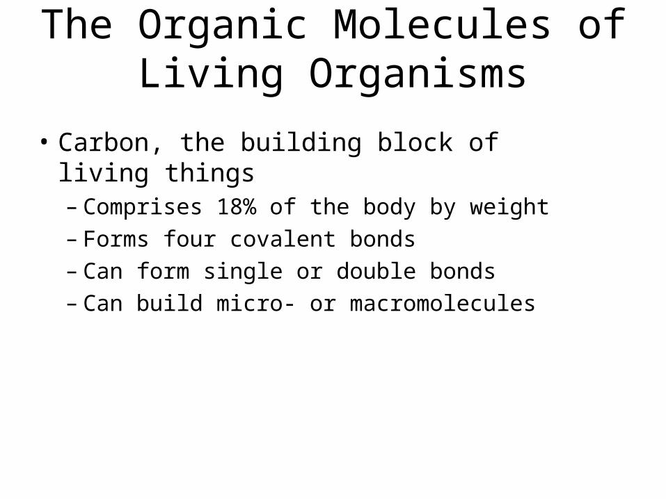
The Organic Molecules of Living Organisms Carbon, the building block of living things – Comprises...
53
-
date post
19-Dec-2015 -
Category
Documents
-
view
220 -
download
0
Transcript of The Organic Molecules of Living Organisms Carbon, the building block of living things – Comprises...

The Organic Molecules of Living Organisms
• Carbon, the building block of living things– Comprises 18% of the body by weight– Forms four covalent bonds– Can form single or double bonds– Can build micro- or macromolecules
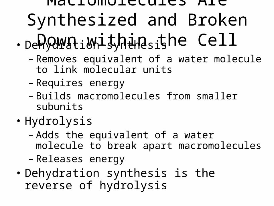
Macromolecules Are Synthesized and Broken Down within the Cell
• Dehydration synthesis– Removes equivalent of a water molecule to link
molecular units– Requires energy– Builds macromolecules from smaller subunits
• Hydrolysis– Adds the equivalent of a water molecule to break
apart macromolecules– Releases energy
• Dehydration synthesis is the reverse of hydrolysis
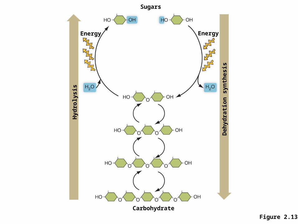
Figure 2.13
Sugars
Hyd
roly
sis
Deh
yd
rati
on
syn
thes
is
Carbohydrate
Energy Energy
Monomers and Polymers
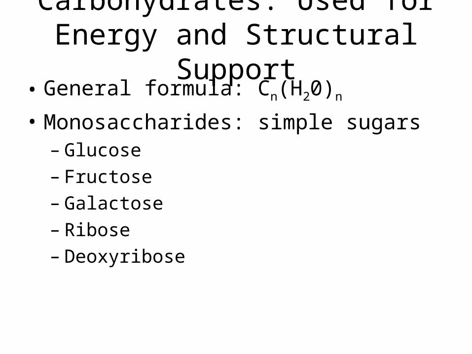
Carbohydrates: Used for Energy and Structural Support
• General formula: Cn(H20)n
• Monosaccharides: simple sugars– Glucose– Fructose– Galactose– Ribose– Deoxyribose
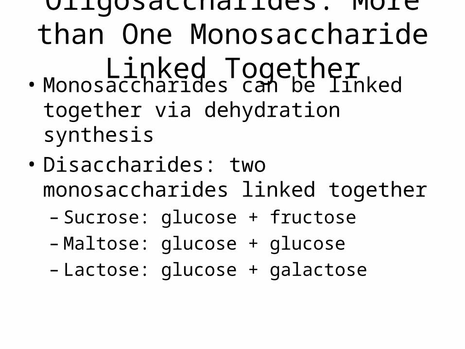
Oligosaccharides: More than One Monosaccharide Linked Together
• Monosaccharides can be linked together via dehydration synthesis
• Disaccharides: two monosaccharides linked together– Sucrose: glucose + fructose– Maltose: glucose + glucose– Lactose: glucose + galactose
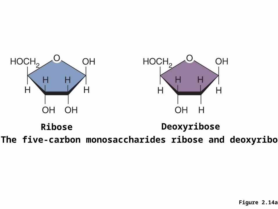
Figure 2.14a
a) The five-carbon monosaccharides ribose and deoxyribose.
Ribose Deoxyribose
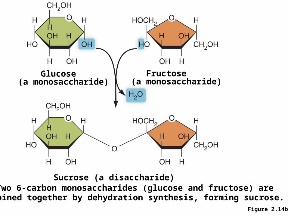
Figure 2.14b
Glucose (a monosaccharide)
Fructose (a monosaccharide)
Sucrose (a disaccharide)b) Two 6-carbon monosaccharides (glucose and fructose) are
joined together by dehydration synthesis, forming sucrose.
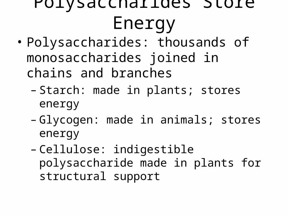
Polysaccharides Store Energy
• Polysaccharides: thousands of monosaccharides joined in chains and branches– Starch: made in plants; stores energy– Glycogen: made in animals; stores energy– Cellulose: indigestible polysaccharide made in
plants for structural support
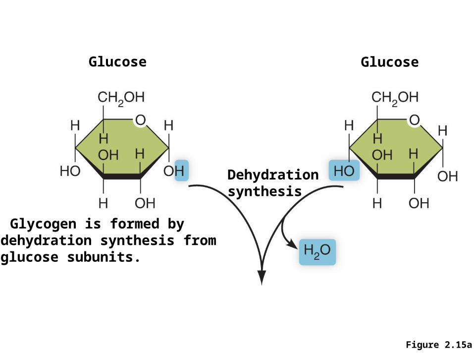
Figure 2.15a
Glucose Glucose
Dehydrationsynthesis
a) Glycogen is formed bydehydration synthesis fromglucose subunits.
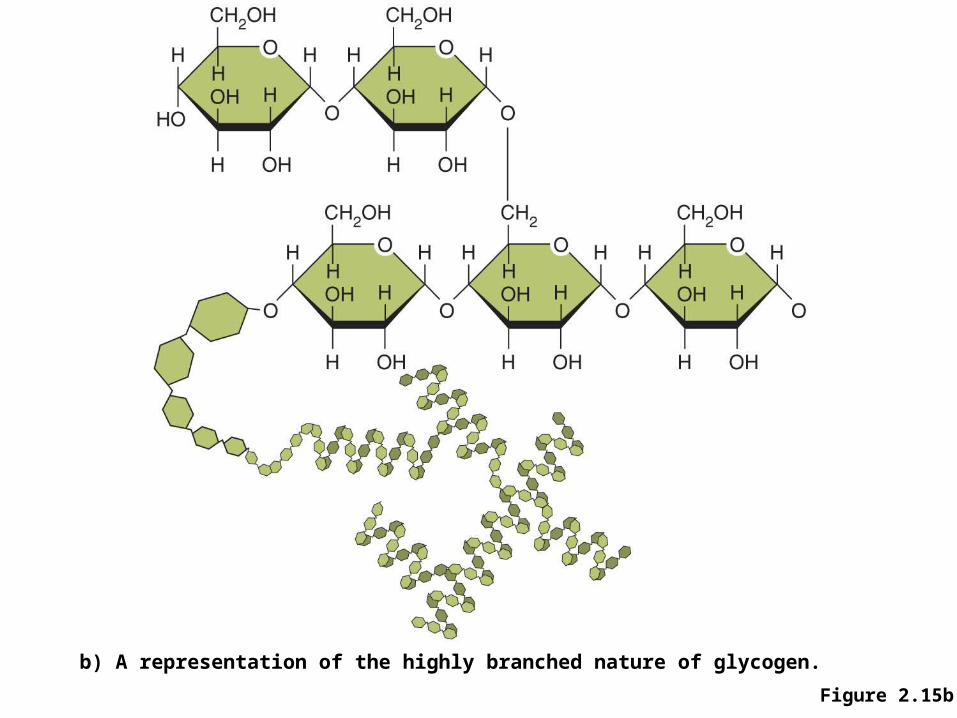
Figure 2.15b
b) A representation of the highly branched nature of glycogen.
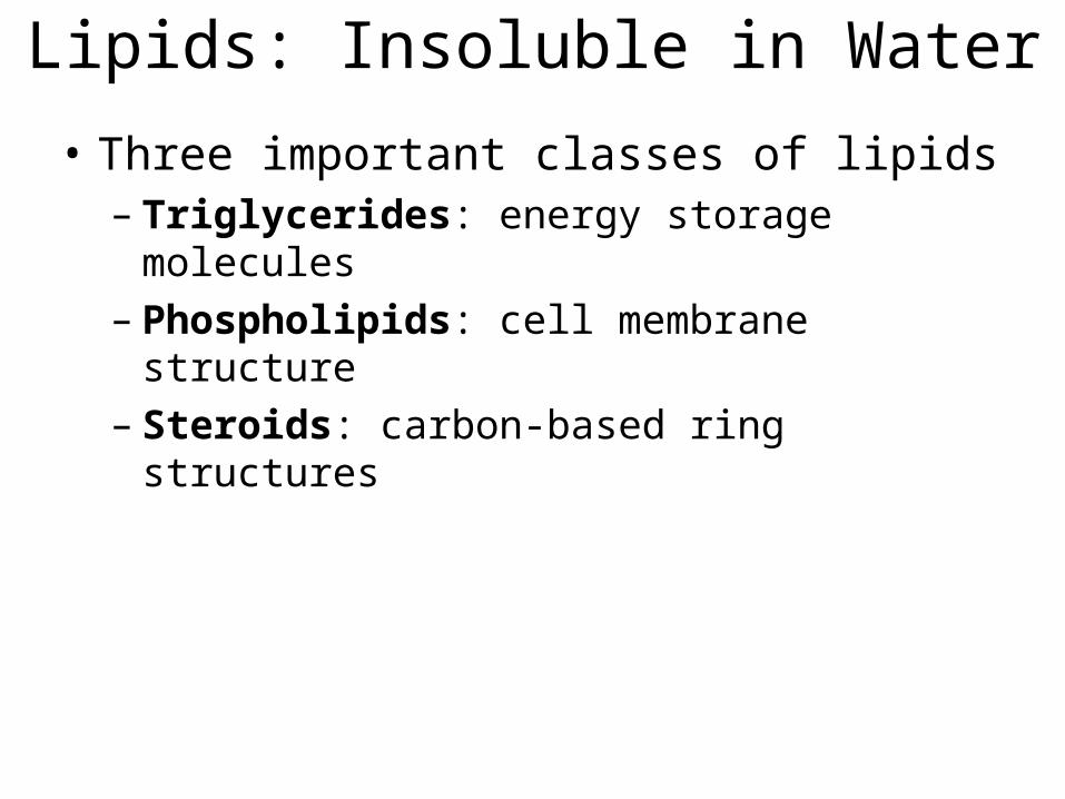
Lipids: Insoluble in Water
• Three important classes of lipids– Triglycerides: energy storage molecules– Phospholipids: cell membrane structure– Steroids: carbon-based ring structures
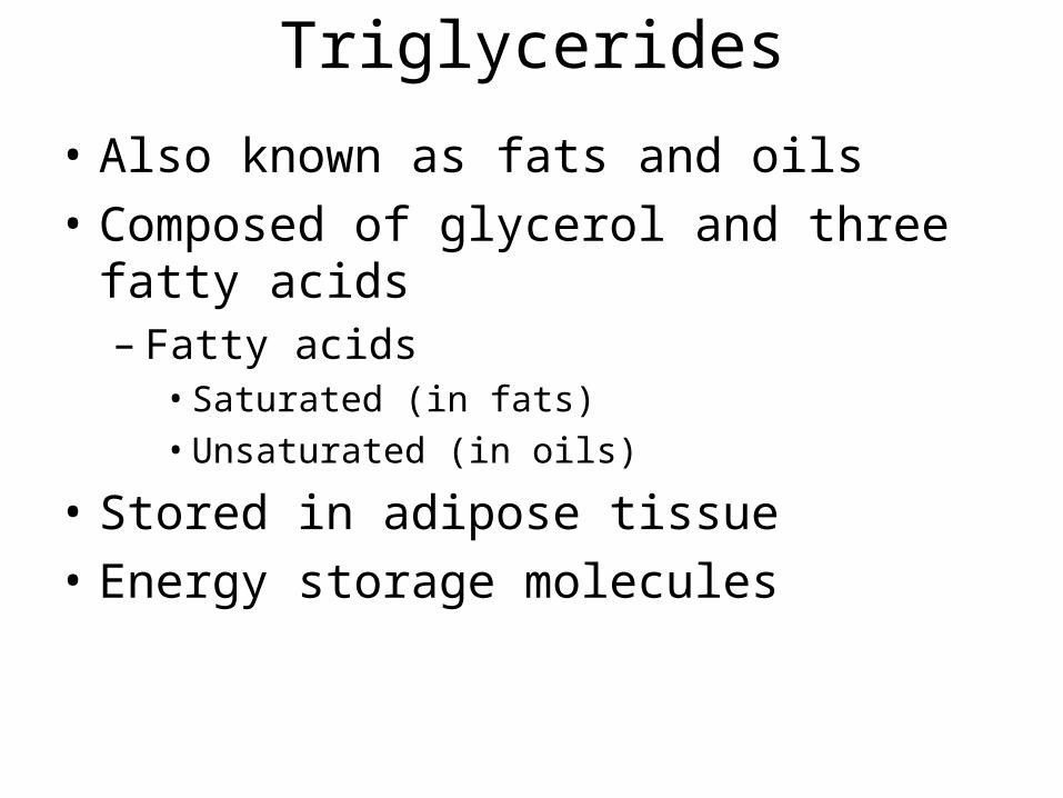
Triglycerides
• Also known as fats and oils• Composed of glycerol and three fatty acids
– Fatty acids• Saturated (in fats)• Unsaturated (in oils)
• Stored in adipose tissue• Energy storage molecules
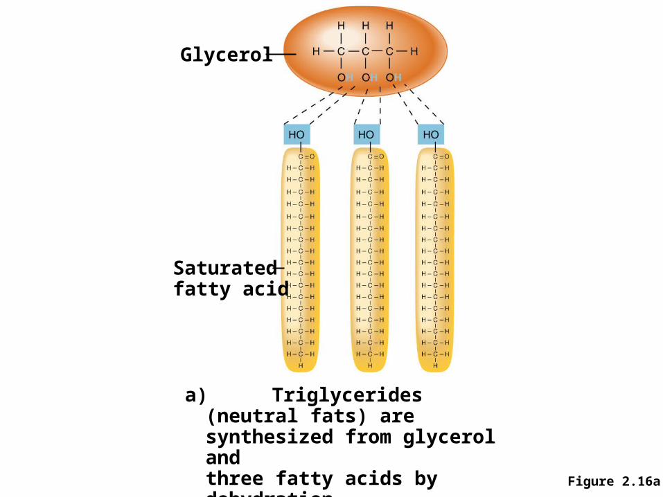
Figure 2.16a
Glycerol
Saturatedfatty acid
a) Triglycerides (neutral fats) aresynthesized from glycerol andthree fatty acids by dehydrationsynthesis.
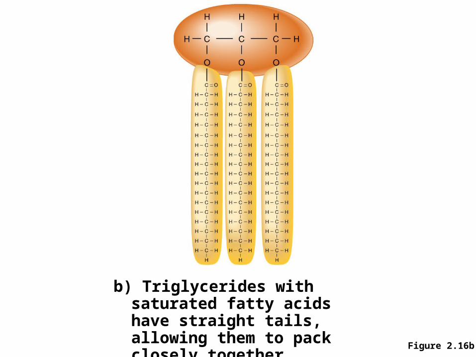
Figure 2.16b
b) Triglycerides with saturated fatty acids have straight tails, allowing them to pack closely together.
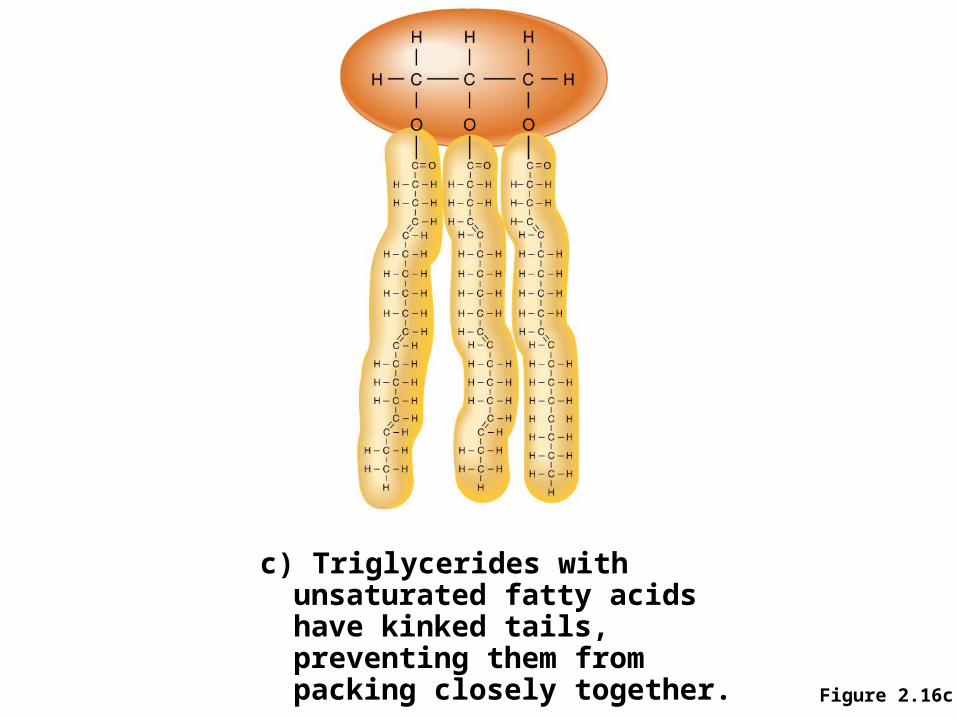
Figure 2.16c
c) Triglycerides with unsaturated fatty acids have kinked tails, preventing them from packing closely together.
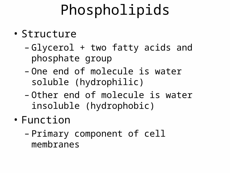
Phospholipids
• Structure– Glycerol + two fatty acids and phosphate group– One end of molecule is water soluble (hydrophilic)– Other end of molecule is water insoluble
(hydrophobic)
• Function– Primary component of cell membranes
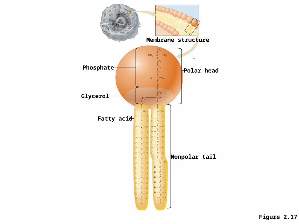
Figure 2.17
Phosphate
Glycerol
Fatty acid
Membrane structure
Polar head
Nonpolar tail
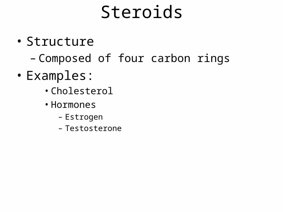
Steroids
• Structure– Composed of four carbon rings
• Examples:• Cholesterol• Hormones
– Estrogen– Testosterone
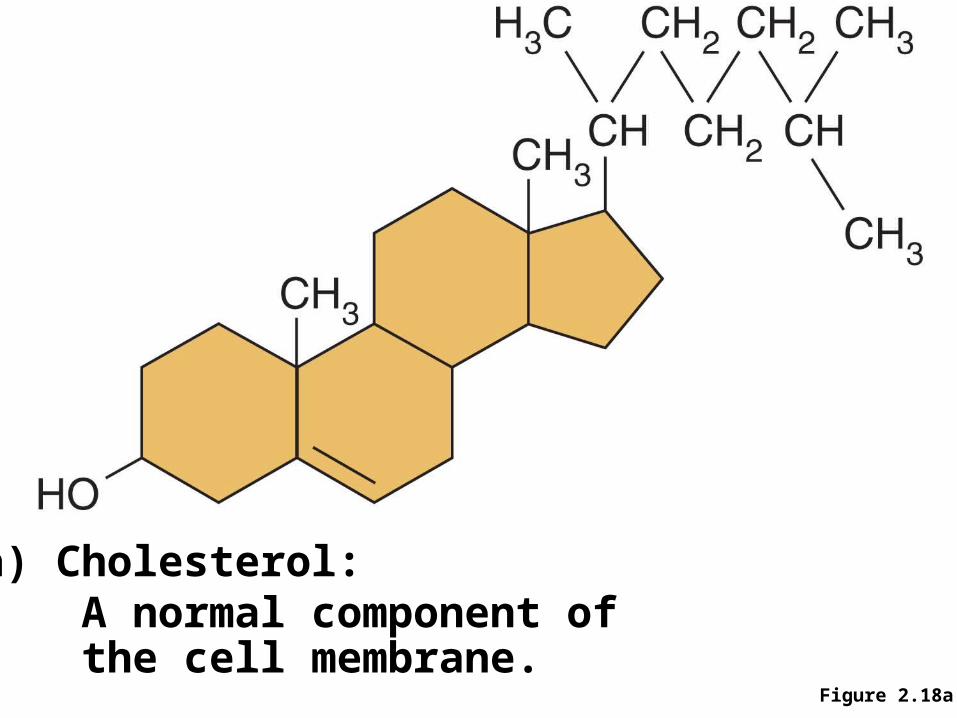
Figure 2.18a
a) Cholesterol: A normal component of the cell membrane.
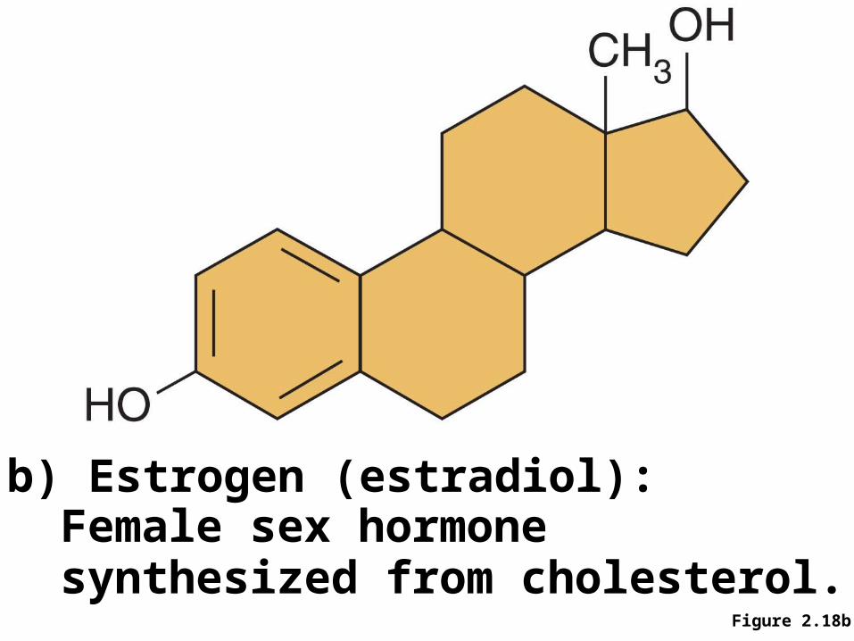
b) Estrogen (estradiol):Female sex hormonesynthesized from cholesterol.
Figure 2.18b
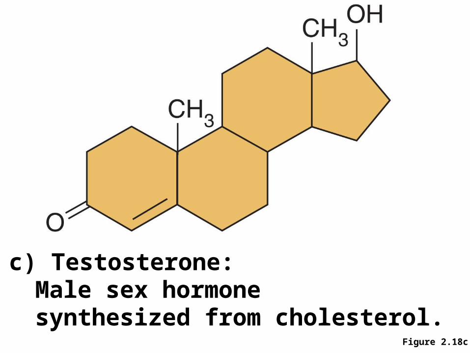
c) Testosterone:Male sex hormonesynthesized from cholesterol.
Figure 2.18c
Lipid Structure and Function
Proteins
• Long chains (polymers) of subunits called amino acids
• Amino acids– 20 different types– Amino end, carboxyl end, R group
• Amino acids are joined by peptide bonds, which are produced by dehydration synthesis reactions
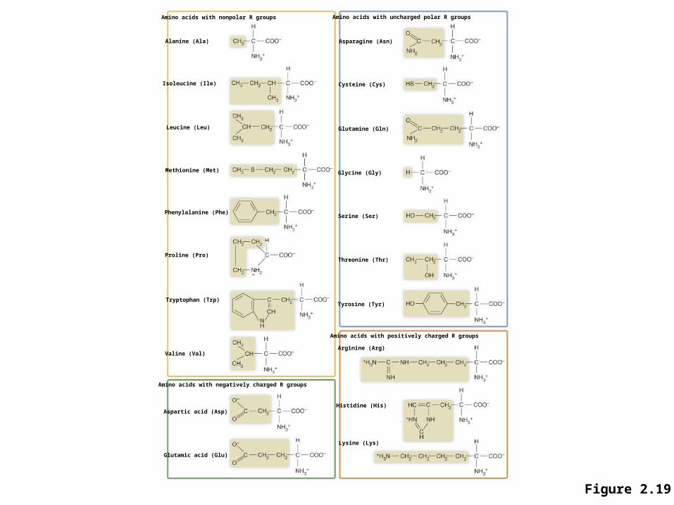
Figure 2.19
Amino acids with nonpolar R groups
Alanine (Ala)
Isoleucine (Ile)
Leucine (Leu)
Methionine (Met)
Phenylalanine (Phe)
Proline (Pro)
Tryptophan (Trp)
Valine (Val)
Amino acids with negatively charged R groups
Aspartic acid (Asp)
Glutamic acid (Glu)
Amino acids with uncharged polar R groups
Asparagine (Asn)
Cysteine (Cys)
Glutamine (Gln)
Glycine (Gly)
Serine (Ser)
Threonine (Thr)
Tyrosine (Tyr)
Amino acids with positively charged R groups
Arginine (Arg)
Histidine (His)
Lysine (Lys)
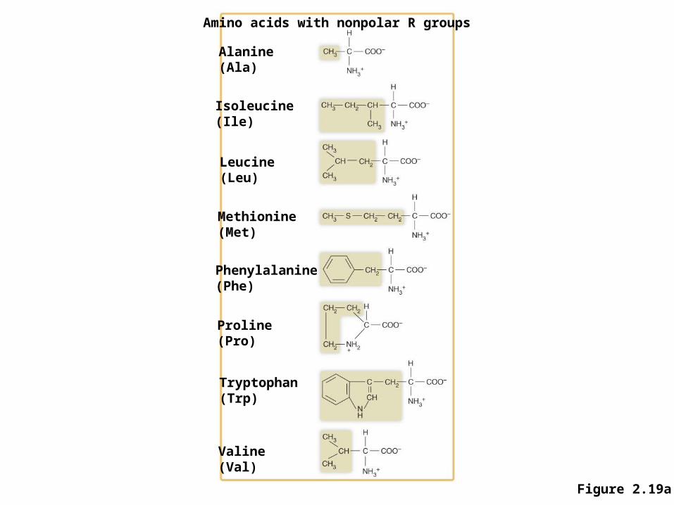
Figure 2.19a
Amino acids with nonpolar R groups
Alanine(Ala)
Isoleucine(Ile)
Leucine(Leu)
Methionine(Met)
Phenylalanine(Phe)
Proline(Pro)
Tryptophan(Trp)
Valine(Val)
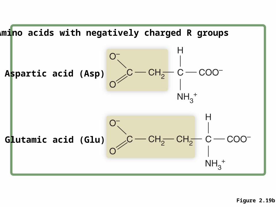
Figure 2.19b
Amino acids with negatively charged R groups
Aspartic acid (Asp)
Glutamic acid (Glu)
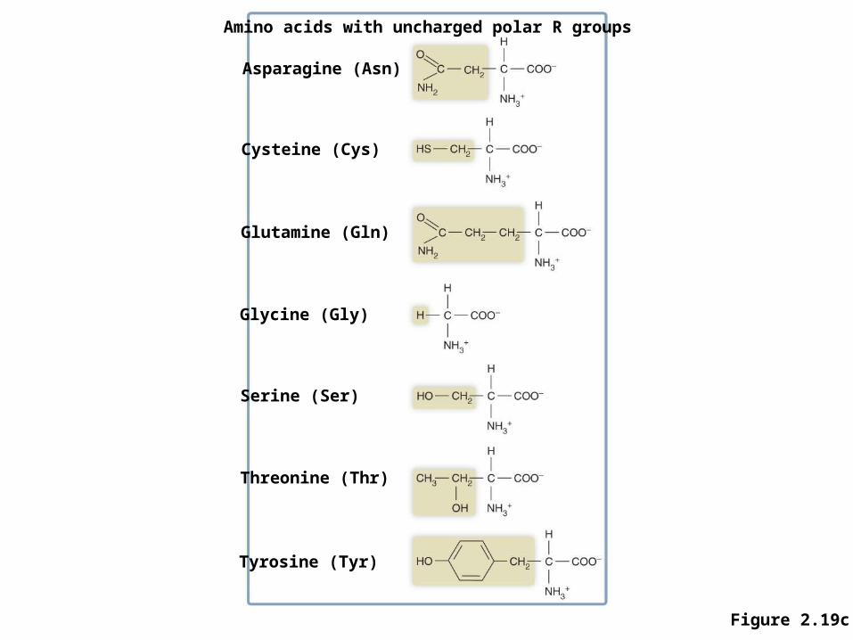
Figure 2.19c
Amino acids with uncharged polar R groups
Asparagine (Asn)
Cysteine (Cys)
Glutamine (Gln)
Glycine (Gly)
Serine (Ser)
Threonine (Thr)
Tyrosine (Tyr)
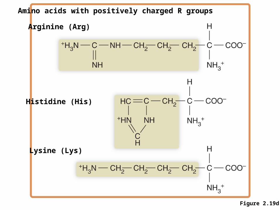
Figure 2.19d
Amino acids with positively charged R groups
Arginine (Arg)
Histidine (His)
Lysine (Lys)
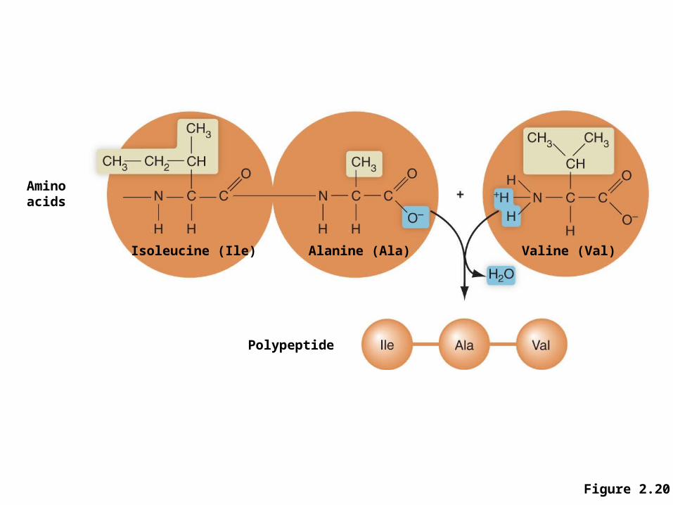
Figure 2.20
Aminoacids
Isoleucine (Ile) Alanine (Ala) Valine (Val)
Polypeptide
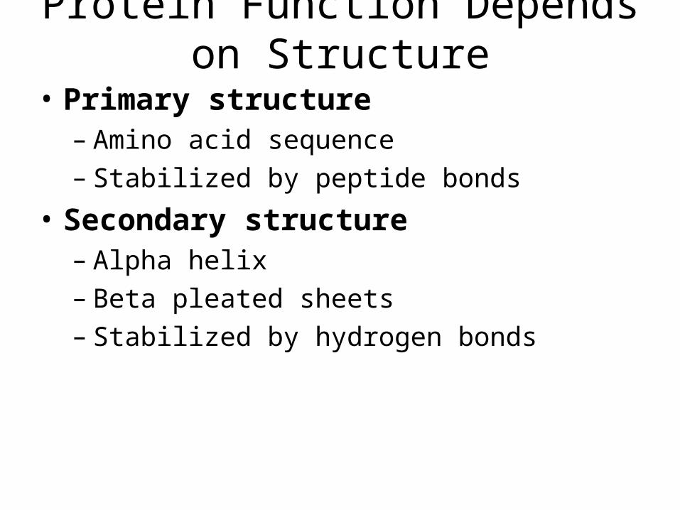
Protein Function Depends on Structure
• Primary structure– Amino acid sequence– Stabilized by peptide bonds
• Secondary structure– Alpha helix– Beta pleated sheets– Stabilized by hydrogen bonds
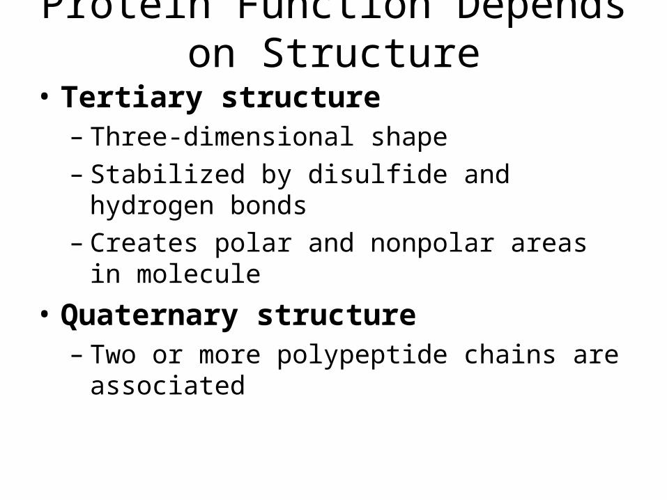
Protein Function Depends on Structure
• Tertiary structure– Three-dimensional shape – Stabilized by disulfide and hydrogen bonds– Creates polar and nonpolar areas in molecule
• Quaternary structure– Two or more polypeptide chains are associated
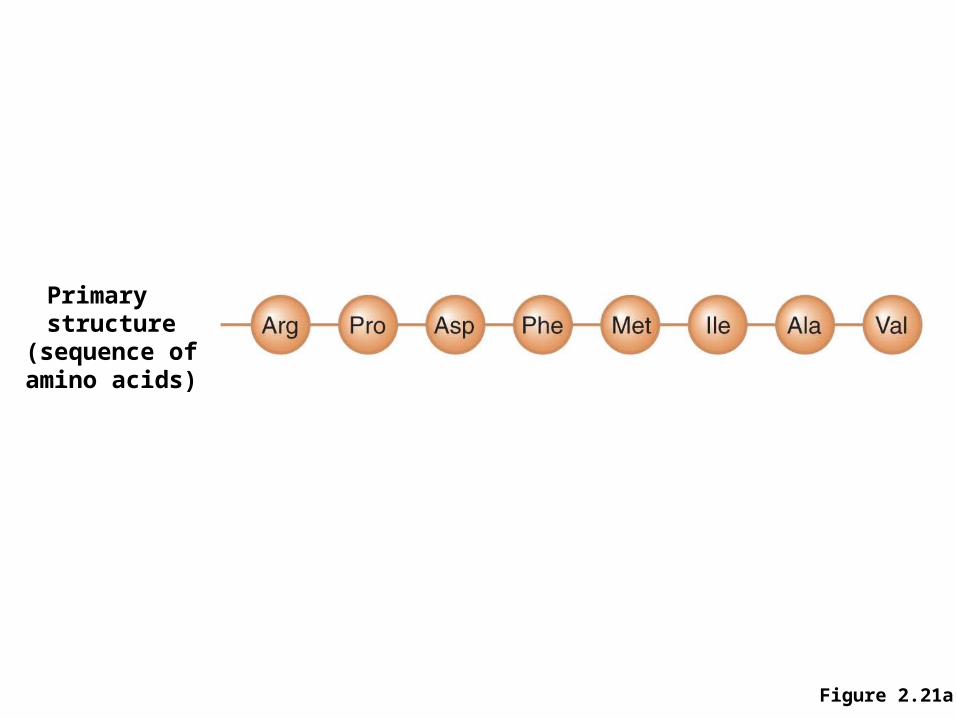
Figure 2.21a
Primary structure
(sequence ofamino acids)
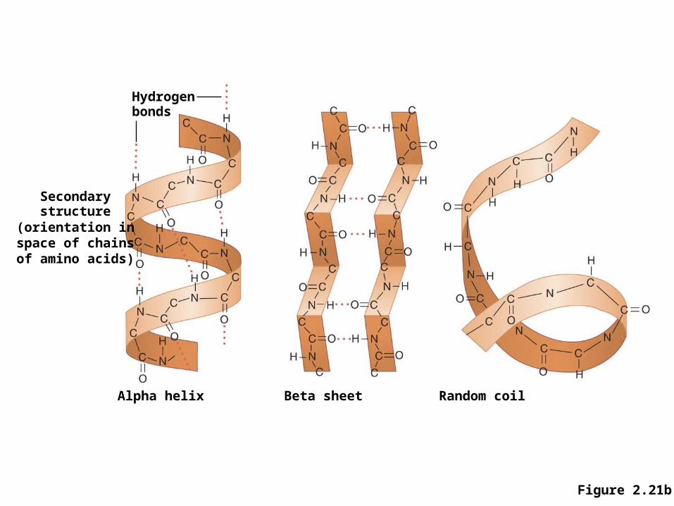
Figure 2.21b
Hydrogenbonds
Secondarystructure
(orientation inspace of chainsof amino acids)
Alpha helix Beta sheet Random coil
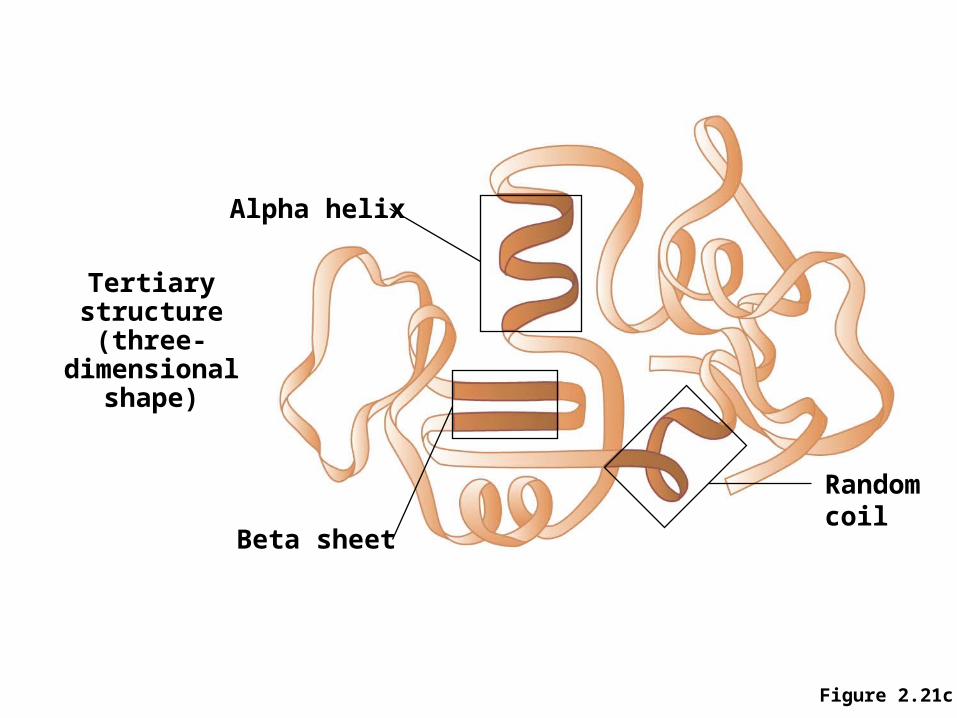
Figure 2.21c
Alpha helix
Beta sheet
Randomcoil
Tertiarystructure
(three-dimensionalshape)
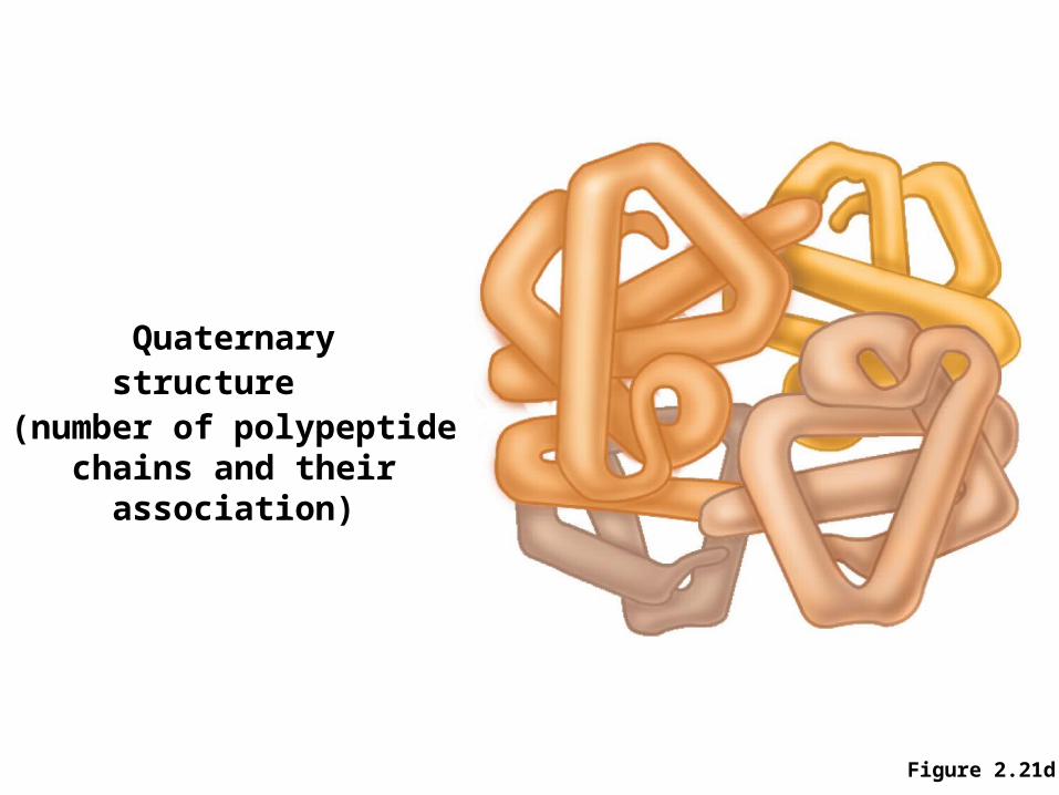
Figure 2.21d
Quaternarystructure
(number of polypeptidechains and their
association)
Protein Function Depends on Structure
• Denaturation– Permanent disruption of protein structure
• Can be damaged by temperature or changes in pH
– Leads to loss of biological function
Protein Structure
Enzymes Facilitate Biochemical Reactions
• Enzymes– Are proteins– Function as biological catalysts
• Speed up specific chemical reactions• Are not altered or consumed by the reaction
– Without enzymes, many biochemical reactions would not proceed quickly enough to sustain life
– Each enzyme is specific for a specific chemical reaction
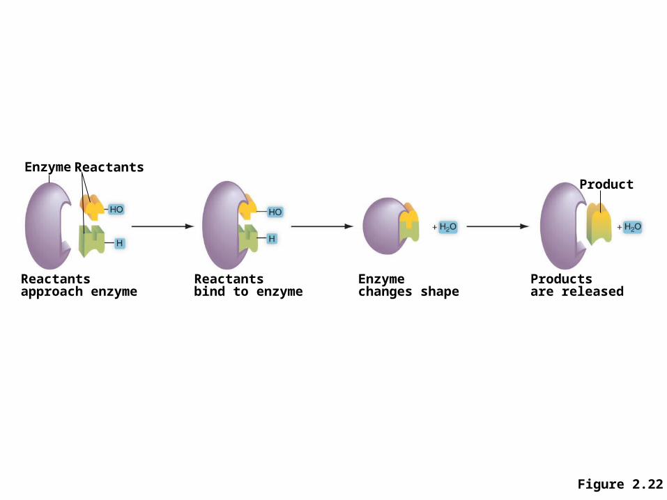
Figure 2.22
Enzyme Reactants
Reactantsapproach enzyme
Reactantsbind to enzyme
Enzymechanges shape
Productsare released
Product
Enzymes Facilitate Biochemical Reactions
• The functional shape of an enzyme is dependent on– Temperature– pH – Ion concentration– Presence of inhibitors
Nucleic Acids Store Genetic Information
• Two types– DNA: deoxyribonucleic acid– RNA: ribonucleic acid
• Functions– Store genetic information– Provide information used in making proteins
• Nucleic acids are long chains containing subunits known as nucleotides
Nucleic Acids Store Genetic Information
• Nucleotides: building blocks of nucleic acids• Each nucleotide contains
– 5 carbon sugar • DNA nucleotides: deoxyribose• RNA nucleotides: ribose
– Nitrogenous base– Phosphate group
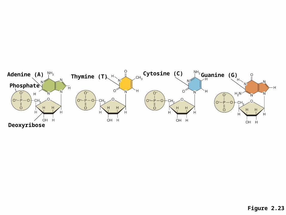
Figure 2.23
Adenine (A)
Phosphate
Deoxyribose
Thymine (T) Cytosine (C) Guanine (G)
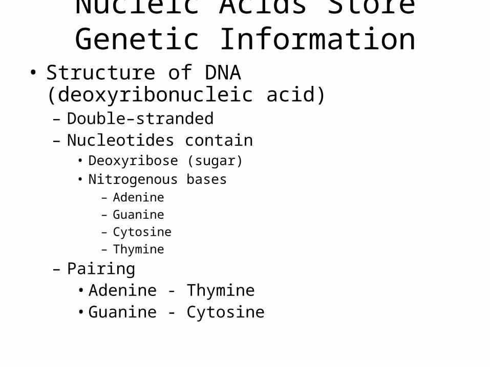
Nucleic Acids Store Genetic Information
• Structure of DNA (deoxyribonucleic acid)– Double–stranded– Nucleotides contain
• Deoxyribose (sugar)• Nitrogenous bases
– Adenine – Guanine– Cytosine– Thymine
– Pairing• Adenine - Thymine• Guanine - Cytosine
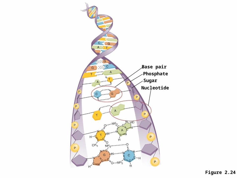
Figure 2.24
Base pair
Phosphate
Sugar
Nucleotide
Nucleic Acids Store Genetic Information
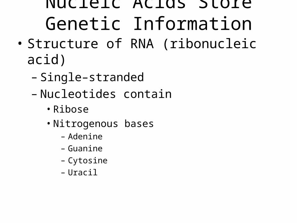
• Structure of RNA (ribonucleic acid)– Single–stranded– Nucleotides contain
• Ribose• Nitrogenous bases
– Adenine– Guanine– Cytosine– Uracil
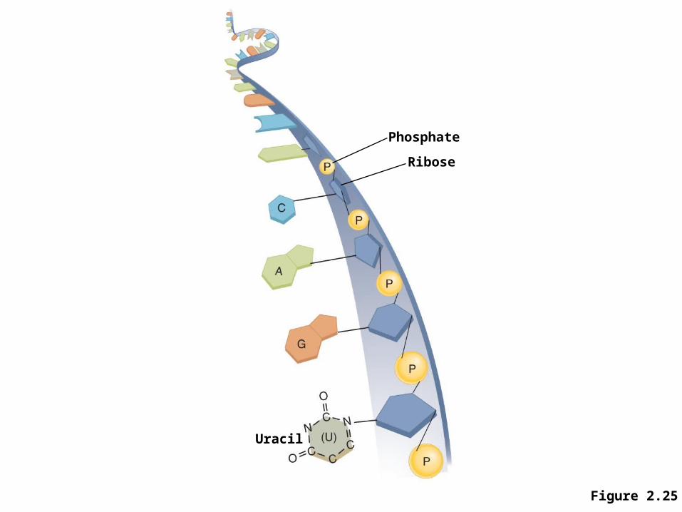
Figure 2.25
Phosphate
Ribose
Uracil
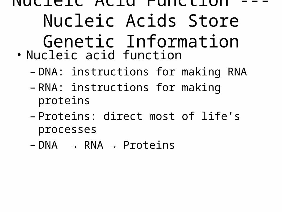
Nucleic Acid Function --- Nucleic Acids Store Genetic Information
• Nucleic acid function– DNA: instructions for making RNA– RNA: instructions for making proteins– Proteins: direct most of life’s processes– DNA → RNA → Proteins
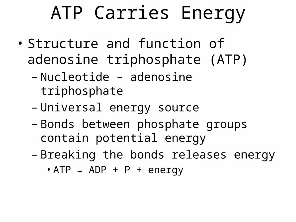
ATP Carries Energy
• Structure and function of adenosine triphosphate (ATP)– Nucleotide – adenosine triphosphate– Universal energy source– Bonds between phosphate groups contain
potential energy– Breaking the bonds releases energy
• ATP → ADP + P + energy
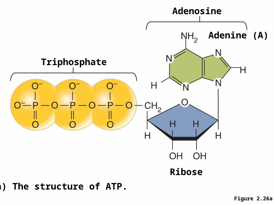
Figure 2.26a
Triphosphate
Adenosine
Ribose
Adenine (A)
a) The structure of ATP.
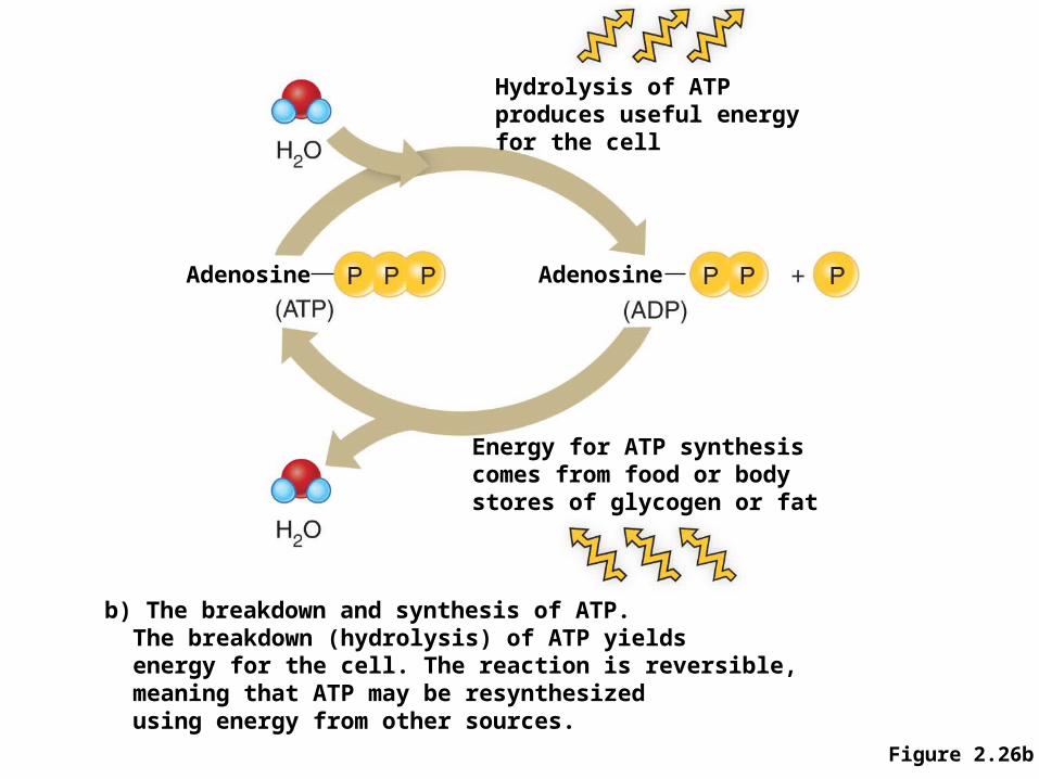
Figure 2.26b
Hydrolysis of ATPproduces useful energyfor the cell
AdenosineAdenosine
Energy for ATP synthesiscomes from food or bodystores of glycogen or fat
b) The breakdown and synthesis of ATP.The breakdown (hydrolysis) of ATP yieldsenergy for the cell. The reaction is reversible,meaning that ATP may be resynthesizedusing energy from other sources.