The orbit
21
The ORBIT ADZLEEN BINTI MOHMOOD
-
Upload
dzlnmhmd -
Category
Health & Medicine
-
view
57 -
download
4
Transcript of The orbit

The ORBITADZLEEN BINTI MOHMOOD

OUTLINESORBITAL VOLUME
BONY ORBIT
ORBTAL LMARGIN
ORBITAL ROOF, MEDIAL, FLOOR, LATERAL WALL
ORBITAL FORAMINA, DUCTS, CANALS AND FISSURES
PARANASAL SINUSES
EXTRAOCULAR MUSCLES
OPTIC NERVE

Orbital volumes
• Volume 30cc• Entrance 35mm
height 45mm width• Depth 40-45mm in
adults • Race & sex differ
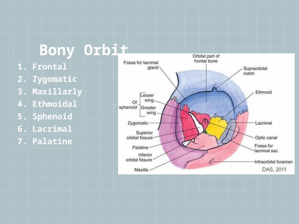
Bony Orbit1. Frontal
2. Zygomatic
3. Maxillarly
4. Ethmoidal
5. Sphenoid
6. Lacrimal
7. Palatine

Orbital margin


Orbital roof
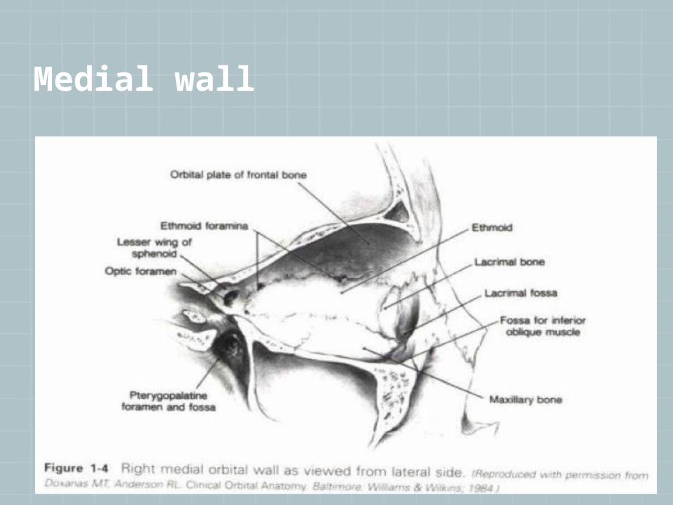
Medial wall

Orbital floor
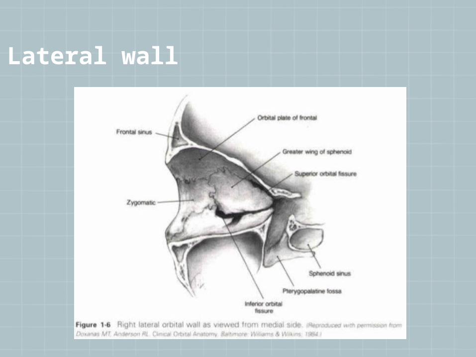
Lateral wall

Whitnall’s tubercle

Periorbital Sinuses
The eyes lie within two bony orbits, located on either side of the root of the nose.
They border the nasal cavity anteriorly and the ethmoidal air cells and the sphenoid sinus posteriorly.
The lateral walls border the middle cranial, temporal, and pterygopalatine fossae.
Superior to the orbit are the anterior cranial fossa and the frontal and supraorbital sinus.
The maxillary sinus and the palatine air cells are located inferiorly.
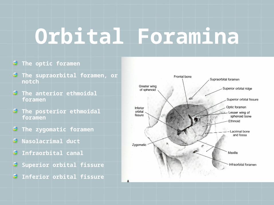
Orbital ForaminaThe optic foramen
The supraorbital foramen, or notch
The anterior ethmoidal foramen
The posterior ethmoidal foramen
The zygomatic foramen
Nasolacrimal duct
Infraorbital canal
Superior orbital fissure
Inferior orbital fissure

Extraocular Muscles
The four recti and two oblique muscles
All are supplied by CN III except superior oblique (CN IV ) and lateral rectus (CN VI)

Optic nerveThe optic nerve consists of more than 1 million axons that originate in the ganglion cell layer of the retina and extend toward the occipital cortex
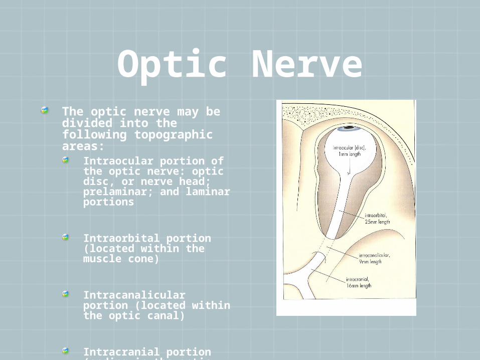
Optic NerveThe optic nerve may be divided into the following topographic areas:
Intraocular portion of the optic nerve: optic disc, or nerve head; prelaminar; and laminar portions
Intraorbital portion (located within the muscle cone)
Intracanalicular portion (located within the optic canal)
Intracranial portion (ending in the optic chiasm)
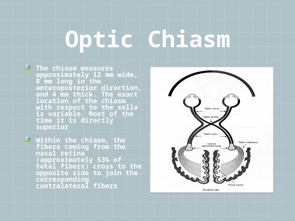
Optic ChiasmThe chiasm measures approximately 12 mm wide, 8 mm long in the anteroposterior direction, and 4 mm thick. The exact location of the chiasm with respect to the sella is variable. Most of the time it is directly superior
Within the chiasm, the fibers coming from the nasal retina (approximately 53% of total fibers) cross to the opposite side to join the corresponding contralateral fibers
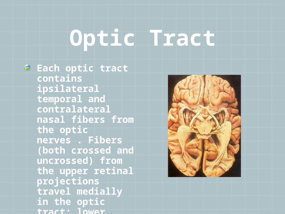
Optic TractEach optic tract contains ipsilateral temporal and contralateral nasal fibers from the optic nerves . Fibers (both crossed and uncrossed) from the upper retinal projections travel medially in the optic tract; lower projections move laterally.

Lateral geniculate body
The lateral geniculate body, or nucleus, is the synaptic zone for the higher visual projections
It has six alternating layers of gray and white matter. Layers 1, 4, and 6 of the lateral geniculate body contain axons from the contralateral optic nerve. Layers 2, 3, and 5 arise from the ipsilateral optic nerve
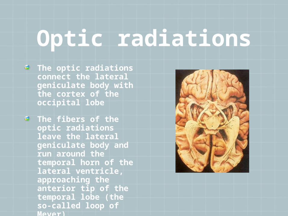
Optic radiationsThe optic radiations connect the lateral geniculate body with the cortex of the occipital lobe
The fibers of the optic radiations leave the lateral geniculate body and run around the temporal horn of the lateral ventricle, approaching the anterior tip of the temporal lobe (the so-called loop of Meyer)
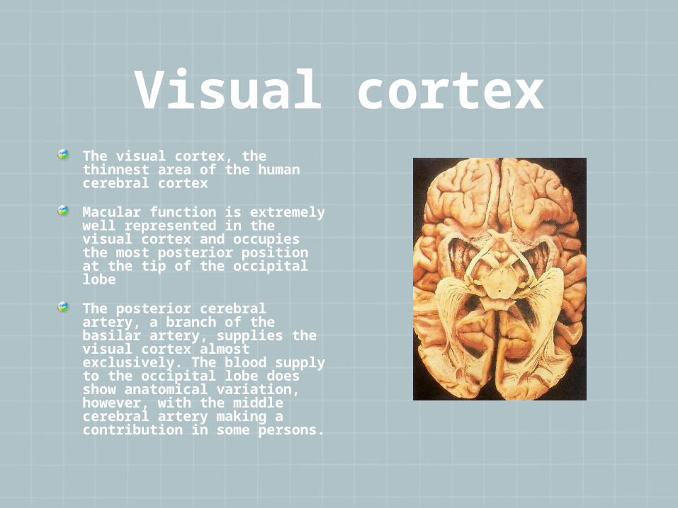
Visual cortexThe visual cortex, the thinnest area of the human cerebral cortex
Macular function is extremely well represented in the visual cortex and occupies the most posterior position at the tip of the occipital lobe
The posterior cerebral artery, a branch of the basilar artery, supplies the visual cortex almost exclusively. The blood supply to the occipital lobe does show anatomical variation, however, with the middle cerebral artery making a contribution in some persons.