Canadian Transportation Agency · Canadian Transportation Agency

Restorative Dentistry Review / Revue de la Dentisterie restauratrice
Complete Removable Prosthodontics /Prosthodontie complète amovible
Dental CAD/CAM Review / Revue de la CAO/FAO dentaire
CJRDP JCDRPCanadian Journal of Restorative Dentistry & Prosthodontics /The official publication of the Canadian Academy of Restorative Dentistry and Prosthodontics
Journal canadien de dentisterie restauratrice et de prosthodontiePublication officielle de l’Académie canadienne de
dentisterie restauratrice et de prosthodontie
www.cardp.ca Volume 8, No. 3 • Fall/automne 2015
CARDP Annual Meeting
Congrès annuel de l’ACDRP
Toronto 2015


Canadian Journal of Restorative Dentistry & Prosthodontics/Journal canadien de dentisterie restauratrice et de prosthodontie — Vol. 8, No. 3 — Fall/automne 2015 3
Canadian Journal of Restorative Dentistry & ProsthodonticsThe official publication of the Canadian Academy of Restorative Dentistry and Prosthodontics
Journal canadien de dentisterie restauratrice et de prosthodontiePublication officielle de l’Académie canadienne de dentisterie restauratrice et de prosthodontie
JCDRPCJRDP
EDITOR-IN-CHIEF/RÉDACTEUR EN CHEF ASSOCIATE EDITORS/RÉDACTEURS ASSOCIÉS
Publisher:Ettore Palmeri, MBA, AGDM, B.Ed., BAPalmeri Publishing Inc.Toronto, [email protected]
Office Administrators:Lobat Lali – [email protected] Ellis – [email protected] Palmeri, B.SC – [email protected]
Sales/Marketing:Mark Behar Bannelier – [email protected] Palmeri – [email protected]
Production Manager:Samira Sedigh, Design Dip. –[email protected]
Design & Layout: Tim Faller – [email protected] Sophie Faller
Internet Marketing Director: Ambianz Inc., Rashid Qadri
Canadian Office: 35-145 Royal Crest Court, Markham, ON L3R 9Z4Tel: 905-489-1970, Fax: 905-489-1971Email: [email protected]: www.palmeripublishing.com
Articles published express the viewpoints of the author(s) and do notnecessarily reflect the views and opinions of the Editorial Board.
All rights reserved. The contents of this publication may not be reproducedeither in part or in full without written consent of the copyright owner.
Printed in Canada Canadian Publications Mail Product Sale Agreement 40020046
CJRDP Editorial Board/Le comité de rédaction JCDRP
SECTION EDITORS/RÉDACTEURS DE SECTIONS
Occlusion and Temporo-Mandibular Dysfunctions /Occlusion et dysfonctionstemporo-mandibulairesDr. Kim ParlettBracebridge, [email protected]
Occlusion and Temporo-Mandibular Dysfunctions /Occlusion et dysfonctionstemporo-mandibulairesDr. Ian TesterSt. Catharines, [email protected]
Implant Dentistry /Dentisterie implantaireDr. Ron ZokolVancouver, British [email protected]
Implant Dentistry /Dentisterie implantaireDr. Yvan FortinQuébec City, Québec [email protected]
Practice Management /Gestion de pratiqueDental Materials / Matériaux dentaires Dr. Izchak BarzilayToronto, [email protected]
Practice Management /Gestion de pratiqueDr. Allan CoopersmithWestmount, [email protected]
Esthetic Dentistry /Dentisterie esthétiqueDr. Paresh ShahWinnipeg, Manitoba [email protected]
Restorative Dentistry /Dentisterie restauratriceDr. Peter Walford British [email protected]
Dental Technology /Technologie dentaireMr. Duane Baluke [email protected]
Dental Technology /Technologie dentaireMs. Mirjana Salkovich, [email protected]
Academic Liaison / Liason académiqueDr. Peter TaylorOakville, [email protected]
Dr. Hubert GaucherQuébec City, Qué[email protected]
Dr. Emmanuel J. RajczakHamilton, [email protected]
Dr. Maureen AndreaChester, Nova [email protected]
Dr. Dennis NimchukVancouver, British [email protected]

Canadian Journal of Restorative Dentistry & Prosthodontics/Journal canadien de dentisterie restauratrice et de prosthodontie — Vol. 8, No. 3 — Fall/automne 20154
CJRDP JCDRPwww.cardp.ca
Volume 8, No. 3
Fall/automne 2015
INDICATES PEER REVIEWED/INDIQUE REVUE DES PAIRS
In this issue/Dans cette édition
6 Editor’s Message / Message du Rédacteur
56 Academy News / Nouvelles de l’Académie
58 2015 Annual Scientific Meeting Program / Programme scientifique du Congrès annuel 2015
64 Call for Papers / Demande de communications
FEATURES/ARTICLES
Restorative Dentistry / Dentisterie restauratrice
10 Iatrogenically Induced Pulpitis as a Consequence of Operative Dentistry – Preventive and Remedial Considerations / Pulpite induite latrogéniquement, comme une conséquence de la dentisterie opératoire – considérations préventives et correctivesDr. Dennis P.A. Nimchuk, DDS, FRCD(C), (Certified Prosthodontist)
Complete Removable Prosthodontics / Prosthodontie complète amovible
28 Occlusal vertical dimension for complete removable dental prostheses / Dimension verticale de l’occlusion pour prothèses dentaires amovibles complètesDr. Samer Mesmar, DMD
Dr. Caroline Tram Nguyen, DMD, MS, FACP, FRCD
and Dr. Chris C. L. Wyatt, BSc, DMD, MSc, Dip Prosc
Dental CAD/CAM — CAO/FAO dentaire
42 CAD/CAM Advances in Prosthodontics / Les progrès de la CAO/FAO en Prosthodontie Dr. Begüm Akkayan, DDS, PhD
and Dr. Hubert Gaucher, DDS, MScD
10
28Comments/CommentairesAccess the Digital version at www.cardp.ca, Journal Section.Voir la version numérique à www.cardp.ca, Section Journal.

Visit us at Booth #3

Canadian Journal of Restorative Dentistry & Prosthodontics/Journal canadien de dentisterie restauratrice et de prosthodontie — Vol. 8, No. 3 — Fall/automne 20156
Revisiting our Journal’s Inaugural Message
Editor’s Message / Message du Rédacteur
Dr. Hubert Gaucher
Some background: I wish to highlight the fact thatthe Canadian Academy of Restorative Dentistry andProsthodontics (CARDP) is commemorating the 50thanniversary this year of one of its founding organizations:the Canadian Academy of Restorative Dentistry (CARD).Today, CARDP continues to promote and disseminateexcellence in the practice of Restorative Dentistry andProsthodontics across the nation. It is a ContinuingEducation driven provider and its Annual ScientificMeetings have earned a well deserved distinction dueto the impressive numbers of its high caliber speakers,state of the art meeting facilities, excellent socialprograms and leading Industry sponsors, all of whichencourage affinities among the membership from sea to sea.
In an endeavor to further its commitment to the dentalcommunity, CARDP launched its own Official Publication:CJRDP/JCDRP back in 2008. I invite you to revisitexcerpts of its Inaugural Message below, that kicked offa tradition of excellence and collaboration.
MESSAGE FROM THE EDITOR-IN-CHIEF
Keeping One’s Focus While Multitaskingand Sharing the Vision This, the inaugural issue of the Canadian Journalof Restorative Dentistry and Prosthodontics (CJRDP),reflects some of the diversities, and challengesthat we as practitioners must learn to cope withon a daily basis. Members of our Academy, as well as concerned clinicians at large, seek a comprehensive and integrated source ofinformation that can contribute to their professionaland personal needs. By its very nature, dentistrychallenges us to provide quality care within aframework of multiple inputs and the ever-presentcompetitive edge. Multitasking therefore becomespart and parcel of our clinical skills and anyresource that can assist in making our work easier and more fulfilling should be a welcomeaddition.
…So understanding the needs of our patientsis fundamental to our practice. CJRDP will keep its“eye on the ball” for you, as we explore information,procedures, technologies, and insights that canempower us.
It is therefore befitting that we underline theimportant contribution in this article “RelationshipBased Dentistry, The 21st Century Formula forSuccess.” Mr. Peter Barry speaks of the day-to-day“people skills” that are essential for our clinicalsuccess… – Moving to another article, “Aplasia of the Lacrimal and Salivary Glands: CasePresentation,” Dr. Renald Perusse presents a rarecase report detailing the genetic origins of the(ALSG) syndrome… – Also, Implant dentistry is wellrepresented in this issue with an article dealingwith the “Restoration of Posterior Implants:

Canadian Journal of Restorative Dentistry & Prosthodontics/Journal canadien de dentisterie restauratrice et de prosthodontie — Vol. 8, No. 3 — Fall/automne 2015 7
Editor’s Message / Message du Rédacteur
Simple Techniques for the Restorative Dentists and Dental Technicians.” In this case report, Dr. Doug Lobb summarizes the integration ofimplant supported restorations to tooth supportedceramic restorations.
By underlining our diversity, we inspirenewcomers to join us in our inclusive, qualityoriented outlook. In this issue, you will also read about our Academy’s mission, historicalbackground, activities and organizational structure.
Come share your vision – Your editorial teamawaits your input and extends a heartfelt invitationto all. What are your most reliable sources ofinformation? Where do you learn new procedures?How do you react to new technologies? What aresome of your insights on various dental topics?How do YOU conjugate multitasking and focus? We welcome “Letters to the Editor” – Moreover,we offer a “Members News” section as anaccessible means of informing and updating yourcolleagues. Feel free to use these features asoften as you like. CJRDP’s viability is the concernof each member…
Dr. Hubert Gaucher
We’ve covered a lot of ground since that first 2008 Issue!
CJRDP/JCDRP Some Facts:
n This Journal remains the only nationally peerreviewed publication exclusively dedicated toRestorative Dentistry and Prosthodontics.
n Scholars and dental practioners alike havecontributed original scientific manuscripts.
n We have received author contributions nationallyfrom 8 of the 10 provinces and internationally, 8 countries from 5 continents.
n The paper version of the quarterly CJRDP/JCDRPis circulated to over 13,000 dentists nationwide.
n The current digital version of the Journal and itsarchives are available at www.cardp.ca
n All regions of Canada are represented on theJournal’s Editorial Board.
n The Journal’s current Publisher, Palmeri PublishingInc. (Toronto) is the leading dental Publisher inCanada.
But we have been losing our momentum even thoughCARDP has a history of volunteerism and, I know for afact, abounds with talented members.
How to bolster our Journal: Palmeri Publishing Inc. needs to list our Journal withPubMed/Medline promptly, so that academicians, whosearticles are required to be published in such databases,will seek us out.
Along the same lines, a closer collaboration withAcademia endorsing our Journal’s mission, will causethose dental faculties to enjoy representation on theJournal’s Editorial board as well as the specificCARDP/CJRDP Awards for students and Facultymembers. To this end, an Academic Liaison has beenassigned to actively promote such a partnership.
Then there is the Association of Prosthodontists ofCanada (APC) and its provincial counterparts, numerousdental Academies and Associations, Study Clubs,Industry/Dental Laboratory leaders, as well as individualbenefactors who are all invited to become corporatesupporters and listed endorsers of CARDP’s OfficialPublication, and to voice their respective proposals forthe advancement of dental reconstructive research andservices nationwide.
And finally, you, our members and readers, far fromthe least of our assets, are expected to invest a minimaldegree of energy and skill in procuring your Journal witharticles, any length, either referred or authored byyourself, based on your own experiences (review, opinionpiece, open letter, case report, clinical tips, scientificmanuscript, and so on). You need not be a skilledwordsmith as we have an app for that. ☺ One shortpaper per annum would suffice.
It’s up to you.Steadfastly yours,
Dr. Hubert [email protected]
Comments/Commentaires

Canadian Journal of Restorative Dentistry & Prosthodontics/Journal canadien de dentisterie restauratrice et de prosthodontie — Vol. 8, No. 3 — Fall/automne 20158
Revoyons le message de notre journalinaugural
Editor’s Message / Message du Rédacteur
Dr. Hubert Gaucher
Contexte: Nous soulignons cette année le 50ièmeanniversaire de l’une des deux académies fondatricesde l’ACDRP, à savoir, l’Académie canadienne dedentisterie restauratrice (ACDR). Aujourd’hui l’ACDRPpoursuit toujours la promotion et la diffusion del’excellence dans la pratique de la dentisterierestauratrice et de la prosthodontie à travers tout lepays. Ses cours d’éducation continue et congrès annuelslui ont mérité une distinction enviable en raison de sesconférenciers de haut niveau, ses installations de fine pointe, ses excellents programmes sociaux etl’apport de ses commanditaires, tous regroupés pourfavoriser des affinités parmi nos membres d’un océan àl’autre.
En 2008, par souci d’engagement envers lacommunauté dentaire, l’ACDRP lançait sa proprepublication officielle: le JCDRP/CJRDP. Je vous invite à revoir certains extraits de son message inaugural ci-bas, qui ont donné le coup d’envoi à une tradition decollaboration et de qualité.
MESSAGE DU RÉDACTEUR EN CHEF
Polyvalence et partage de la vision: ne vous laissez pas dérouterCe numéro inaugural du Journal canadien dedentisterie restauratrice et de prosthodontieprésente la diversité ainsi que les défis que lesdentistes doivent relever tous les jours. Lesmembres de notre Académie, ainsi que tout autreclinicien, recherchent une source d’informationdétaillée et intégrée pouvant répondre à leursbesoins professionnels et personnels. De par sa nature, la médecine dentaire nous oblige àprodiguer des soins de qualité tout en tenantcompte de multiples critères et de la concurrenceomniprésente. La polyvalence devient donc unepartie essentielle de nos compétences cliniques,et toute ressource apte à nous aider à faciliter etvaloriser notre travail est appréciable.
Saisir les besoins de nos patients constitue lefondement de notre pratique. Le Journal canadiende dentisterie restauratrice et de prosthodontiedemeurera donc vigilant pour vous, en explorantl’information, les procédures, technologies etconnaissances dentaires pouvant enrichir l’exercicede notre profession.
Il est indiqué de souligner la contributionimportante de M. Peter Barry dans son article:“Relationship Based Dentistry, the 21st CenturyFormula for Success” (La dentisterie fondée surles rapports humains, la formule gagnante du21ième siècle) qui discute de nos capacitéscompatissantes en situation clinique. Dans unautre article, du Dr Ronald Pérusse cette fois-ci:“Aplasia of the Lacrimal and Salivary Glands: Case
Canadian Journal of Restorative Dentistry & Prosthodontics/Journal canadien de dentisterie restauratrice et de prosthodontie — Vol. 8, No. 3 — Fall/automne 2015 9
Editor’s Message / Message du Rédacteur
presentation” (Aplasie des glandes lacrymales etsalivaires: rapport de cas), l’origine génétique dece syndrome rare est détaillée. Aussi, la dentisterieimplantaire est représentée dans ce numéro avec “Restoration of Posterior Implants: SimpleTechniques for the Restorative Dentists and Dental Technicians” (Restauration des implantspostérieurs: techniques simples pour dentistes en restauration et techniciens dentaires) danslequel le Dr. Doug Lobb résume l’intégration desrestaurations implanto-portées aux restaurationsen céramique sur dentition naturelle.
Vous trouverez aussi dans ce Journal,l’historique, la structure et les activités de notreAcadémie. En mettant en évidence la diversité denos intérêts, nous attirerons de nouveaux membresqui s’associent à notre vision inclusive.
Venez partager cette vision avec votre équipede rédaction. Quelles sont vos sourcesd’information les plus fiables? D’où proviennentvos nouvelles techniques? Que pensez-vous desdernières technologies? Comment conjuguez-vousla polyvalence dans votre pratique?
Exprimez-vous dans vos Lettres au Rédacteur.Par ailleurs, la section accessible «Nouvelles desmembres» renseigne et tient vos collègues à joursur les faits qui vous intéressent. Servez-vous deces fonctions à volonté. La survie du JCDRPconcerne chacun de nos membres…
Dr Hubert GaucherRédacteur en chef
Nous avons fait beaucoup de chemin depuis cettepremière parution!
JCDRP/CJRDP Quelques données:n Ce Journal demeure la seule publication nationale
revue par des pairs qui se dédie exclusivement àla dentisterie restauratrice et la prosthodontie.
n Académiciens et praticiens également ontcontribué des manuscrits scientifiques originaux.
n Le Journal a reçu des articles d’auteurs provenantde 8 des 10 provinces au pays, et de 8 pays sur 5 continents.
n La version papier de notre périodique trimestrielest distribuée à plus de 13 000 dentistes auCanada.
n La version numérique actuelle ainsi que lesarchives du Journal se trouvent en ligne àwww.cardp.ca
n Toutes les régions du Canada sont représentéessur le Comité de rédaction.
n La Maison d’édition de notre Journal, PalmeriPublishing Inc. (Toronto) est chef de file en matièrede publications dentaires au Canada.
Or nous perdons notre vitesse de croisière malgrénos antécédents de bénévolat et, j’en sais quelquechose, nos membres fort talentueux.
Comment appuyer notre Journal:Palmeri Publishing Inc. se doit de lister notre Journaldans PubMed/Medline dans les meilleurs délais, afinque les académiciens, dont les articles sont tenus d’être publiés dans de telles banques de données, nouscôtoient.
Dans la même veine, une collaboration étroite avecle milieu universitaire qui appuie la mission de notreJournal, offrira en échange aux facultés dentaires, dejouir d’une représentation sur le comité de rédaction, enplus des prix d’excellence aux étudiants et enseignants.À cette fin, une personne de notre Académie est assignéeen liaison avec les facultés dans le but de promouvoirdes partenariats.
Puis il y a l’Association des prosthodontistes duCanada (APC) et ses contre-parties provinciales, lesnombreuses académies et associations dentaires, lesclubs d’étude, les représentants de l’industrie dentaireet les laboratoires, ainsi que les bienfaiteurs et mécènes,qui peuvent tous être invités à devenir membrescorporatifs listés de l’ACDRP, à soutenir notre Journalofficiel et à exprimer leurs propositions respectives pour l’avancement de la recherche et des services endentisterie de reconstruction à l’échelle nationale.
Et en toute fin, mais non les moindres, vous, membreset lecteurs, avez la tâche d’investir un effort minimalpour votre Journal en fournissant au moins un articlepar année, si court soit-il, signé ou encore référé parvous. Il peut s’agir d’une revue, d’une opinion, d’unelettre, d’un rapport de cas, de trucs du métier, de manuscrit scientifique, ainsi de suite. Ne vouspréoccupez pas de la prose; nous avons une applicationpour ça! ☺
Il n’en tient qu’à vous.Votre dévoué.
Hubert GaucherRédacteur en [email protected]
Comments/Commentaires
Canadian Journal of Restorative Dentistry & Prosthodontics/Journal canadien de dentisterie restauratrice et de prosthodontie — Vol. 8, No. 3 — Fall/automne 201510
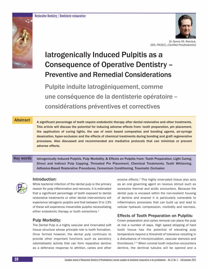
Dr. Dennis P.A. Nimchuk, DDS, FRCD(C), (Certified Prosthodontist)
Restorative Dentistry / Dentisterie restauratrice
Iatrogenically Induced Pulpitis as aConsequence of Operative Dentistry –Preventive and Remedial Considerations
Pulpite induite latrogéniquement, comme
une conséquence de la dentisterie opératoire –
considérations préventives et correctives
A significant percentage of teeth require endodontic therapy after dental restorative and other treatments.This article will discuss the potential for inducing adverse effects from: tooth preparation, pin placement,the application of curing lights, the use of resin based composites and bonding agents, air-syringedessication, hyper-occlusion and the effects of chemical treatments during bonding and graft regenerativeprocesses. Also discussed and recommended are mediative protocols that can minimize or preventadverse effects.
Abstract
Introduction:While bacterial infection of the dental pulp is the primaryreason for pulp inflammation and necrosis, it is estimatedthat a significant percentage of teeth exposed to dentalrestorative treatments or other dental interventions willexperience iatrogenic pulpitis and that between 9 to 13%of these will experience irreversible pulpitis necessitatingeither endodontic therapy or tooth extractions.1
Pulp Morbidity:The Dental Pulp is a highly vascular and innervated softtissue structure whose principle role is tooth formation.Once formed however, the dental pulp continues toprovide other important functions such as secretoryodontoblastic activity that can form reparative dentineas a defensive response to attrition, caries and other
erosive effects.2 This highly innervated tissue also actsas an oral governing agent on noxious stimuli such asexcessive thermal and acidic encounters. Because thedental pulp is encased within the non-resilient housingof dentine and enamel it is particularly vulnerable toinflammatory processes that can build up and lead tocellular hydraulic compression, morbidity and necrosis.
Effects of Tooth Preparation on Pulpitis: Crown preparation and caries removal can place the pulpat risk a number of ways. High speed stripping of hardtooth tissue has the potential of elevating pulptemperature beyond a threshold of tolerance resulting ina disturbance of microcirculation, vascular stenosis andthrombosis.3, 4 When coronal tooth reduction encountersdentine, the dentinal tubules will be opened and a
Iatrogenically Induced Pulpitis, Pulp Morbidity, & Effects on Pulpitis from: Tooth Preparation, Light Curing,Direct and Indirect Pulp Capping, Threaded Pin Placement, Chemical Treatments, Tooth Whitening,Adhesive-Based Restorative Procedures, Cementum Conditioning, Traumatic Occlusion
Key words

Canadian Journal of Restorative Dentistry & Prosthodontics/Journal canadien de dentisterie restauratrice et de prosthodontie — Vol. 8, No. 3 — Fall/automne 2015 11
Un nombre important de dents demande une thérapie endodontique après la restauration dentaire, et d’autres
traitements. Cet article parlera des possibilités pour induire les effets contraires, comme : préparation de la
dent, placement de tenon, pratique de polymérisation par la lumière, utilisation de composites à base de résine
comme agents de collage, dessiccation par air comprimé, sur-occlusion, et les effets de traitements chimiques
pendant les procédés de collage et de greffe régénératrice. Également on parlera, et on recommandera, des
protocoles médiateurs, qui peuvent minimiser ou prévenir ces effets adverses.
Résumé
Restorative Dentistry / Dentisterie restauratrice
communication with the pulp is created. The deeper thedentine is cut away the more permeable it will becomeand the more vulnerable the pulp will also become tothe ingress of microbial, chemical and physical irritants.5
Exposure of the pulp to the oral bacterial flora puts it atgreatest risk as bacterial contamination can cause severeinflammatory changes, resulting in micro-abcesses andprogressive pulpal necrosis.6
While the pulp has considerable resilience to recoverfrom irritation, cumulative repetitive injuries from caries,trauma from multiple restorative repair interventions orother traumas such as from occlusion or orthodonticsmay reduce the threshold of recovery due to the creationof pulpal micro-scarring to the vascular and nervecomponents of the pulp. An additional insult such as acrown preparation on a prior injured and predisposedtooth may be enough to cascade the pulp intodegeneration with ensuing irreversible pulpitis.7
Because the removal of tooth material by high and low-speed produces heat from the friction between the burand tooth substrate, it is prudent to use high volume water
and air spray and high volume air suction. (Figure 1)A critical threshold temperature of 41–42°C is irreversiblyharmful to pulpal tissue. A temperature rise to thismagnitude should not be attainable when normalhandpiece cooling water temperature of 30–34°C iseffectively utilized.8 According to Attrill et al., themaximum intrapulpal rise in temperature recorded forteeth prepared without water spray was 24.7°C, but only3.9°C in teeth prepared with water spray.9 (Figure 2)
According to Zach and Cohen doing in vivo studies, anintra-pulpal temperature increase of 5.5°C for 10 secondscaused histological changes in the pulp tissues,approximately 15% of which became irreversible pulpitis.When intra-pulpal temperature increases were sustainedat 11.1°C for 10 seconds, there was approximately 60–70%irreversible pulpitis.10 When using water spray in an in vitrostudy measuring intra-pulpal temperature increase with a0.5 mm residual dentin thickness, Firoozmand foundtemperature increases of 1.8°C, 1.4°C, and 0.7°C, with alow-speed handpiece, high-speed handpiece, and laser,respectively.11 These values are well below the physiologic
Figure 1: 4 Ports of Spray Figure 2: Spray from 4 Ports
Pulpite iatrogéniquement induite, morbidité pulpaire, & les effets pulpaires provenant: de la préparationdes dents, de la lumière polymérisante, coiffage pulpaire directe et indirecte, tenons vissés, traitementschimiques, blanchiment des dents, procédures adhésives des matériaux, conditionnement du cément,occlusion traumatique
Mots-clés

Canadian Journal of Restorative Dentistry & Prosthodontics/Journal canadien de dentisterie restauratrice et de prosthodontie — Vol. 8, No. 3 — Fall/automne 201512
5.5°C threshold described by Zach and Cohen so it is unlikely that
thermal pulpal damage will occur unless the water spray is turned
off or unless the water spray is diminished by close proximity
suctioning or by water flow obstruction.
According to Baldissara, Catapano and Scotti who evaluated
clinical and histological criteria, the main cause of postoperative
inflammation or necrosis of the pulp is injury to the dentine.12
It is apparent that the closer the cutting gets to the pulp the
greater will be the heat effect induced.
Diamond burs generate more heat than carbide unless the
carbide is worn. The difference of carbide vs. diamond selection
however, appears not to be significant, if water is used.13 However,
the practice that is most likely to induce pulp damage from
frictional heat arises when a worn carbide bur is used without
water spray to remove last vestiges of caries and hard affected
dentine, close to the pulp. This frequently used practice of shutting
off water when close to the pulp can generate a significant amount
of thermal injury, particularly if sustained for more than 10
seconds.9 A more suitable and safer method of removing last
vestiges of caries is by using a sharp spoon excavator.
The removal of deep caries itself has the potential for allowing
microrganisms to be breached into the pulp chamber, creating
a non-resolving pulpits.14 Proliferation of bacteria in the
superficial aspect of pulp tissue disorganizes the odontoblast
layer and kills cells either by direct toxic action or by excessive
acute inflammatory response. In some instances the pulp can
withstand the microbial insult and odontoblasts will respond by
deposition of new tubular dentin and deposition of mineralized
intra-tubular plugs.15
A systematic review of MEDLINE indicates that utilizing
a stepwise evacuation of dental caries that is very close to
the pulp, along with a treatment lining is effective for
pulp preservation by reducing bacteria and by promoting
remineralization of the carious dentine.16 The stepwise protocol,
at the first visit, recommends not removing the last remnants
of decay but recommends placing a layer of CaOH or other
similar material prior to making a provisional restoration.
Approximately nine months later, the site is re-entered and the
residual caries is removed, allowing time for a reparative dentine
bridge to be established.
Effects Of Light Curing on Pulpitis: Light-curing of resin-based products also generates heat and
has the capacity to increase the intra-pulp temperature high
enough to induce damaging effects to a vital pulp.17,18,19
Tosun et al. evaluated the temperature rise in the pulp
underneath caries-affected primary tooth dentine during adhesive
polymerization and found it could exceed 5.5°C, (the Zack and
Cohen threshold).20 Carious dentine is normally removed during
caries excavation, however, affected dentine is not usually
removed, so a significant portion of a cavity wall contains caries-
affected dentine.21 This practice is significant because carious
dentine or caries-affected dentine has a higher thermal
conductivity when exposed to curing light energy and will
generate greater heat compared to sound unaffected dentine
when at close proximity to the pulp.22 This is also the case with
dental adhesives which exhibit higher temperature rises during
polymerization than do the Resin-Based Composite materials
(RBC’s) which are placed over the adhesives.20
The process of light photo-activation of composite restorative
materials is multi-factorial and there are several reasons which
can contribute to cause a temperature rise inside the pulp
chamber and which are sufficient enough to cause pulpal
damage.23,24 Polymerization of light-cured composites generates
heat because of the energy absorbed during the irradiation
process of the light. This, in combination with the exothermic
reaction of addition polymerization that RBC’s undergo creates a
stepwise elevation of temperature.25 Heat generation can be
variable, depending on the intensity of the light source and on
the moderating thickness of bulk placements of composite. The
thicker the composite placed the less energy will reach the pulp.26
Another variable regarding temperature rise is the composition
of the RBC itself and which is dependent on the energy density
of the material used. For example Z100 with energy density of
28 J/cm2 promoted higher temperature increases than Z250
with energy density of 14J/cm2 when a light source of 700 W
was used for the same period of time.27 The manufacturer, (3M
ESPE), recommends a cure time of 40 seconds for Z100 and
20 seconds for Z250.
Although the light source largely determines the temperature
rise during curing, the heating effect also depends on the type
of curing unit, the quality of the light filter, the output intensity,
and the irradiation time.28 In a study of different types of curing
lights comparing LED, Quartz Halogen and Plasma Arc and
exposing for the same time intervals, it is the most powerful
light, the PAC unit, which registered the greatest amount of heat
output.29 The use of high-intensity PAC lights can cause increased
heat generation in the dental materials being cured, potentially
leading to pulpal damage. This is notably less so when using
LED or QTH lights.29,30 The practice of doubling up light-curing
units to speed up polymerization doubles up the heat and
should absolutely be avoided. (Figures 3, 4, 5, 6)
Adequate polymerization is a very important factor in
maximizing physical the properties, clinical performance and
Restorative Dentistry / Dentisterie restauratrice


Canadian Journal of Restorative Dentistry & Prosthodontics/Journal canadien de dentisterie restauratrice et de prosthodontie — Vol. 8, No. 3 — Fall/automne 201514
the biocompatibility of resin based filling materials as well as
resin based luting agents (RLA’s). Contemporary luting agents
that are used for the cementation of etchable, all ceramic
restorations for the most part, are resin-based and are either
light-cured or dual-cured. Problems associated with inadequate
polymerization include: inferior physical properties, solubility
in the oral environment and increased micro-leakage. These
factors have the potential to develop recurrent decay and
pulpal irritation. As well, when RLA’s are not fully polymerized,
leachable components such as Bis-GMA, UDMA, TEGDMA,
camphorquinone and HEMA may penetrate through dentin
tubules and exert potential pulpal injury and inhibit pulp tissue
repair, a cytotoxic effect.31,32 Chen et al. have shown that the
resin based components of dentine bonding agents also pose
a cytotoxic effect, exerting potentially harmful effects to the
pulp.33
The degree of conversion (DC), when using light-cured, filled,
composite materials or when using light-cured or dual-cured
luting agents can be highly variable giving values from 55% to
75% depending on the chemical composition of the material
and on the light source used.34,35,36 If the polymerizing light
is impeded by restoration material opacity, or by access, or by
bulk, then a dual-cure composite or dual-cure luting cement may
be a more appropriate consideration.37
Figure 3: High Intensity Curing Lights Figure 4: Heat Damage From Curing Lights — (a) Prior to Md. ¾ All-Ceramic Crowns.
Figure 5: Heat Damage From Curing Lights — (b) Radiograph, prior.
Figure 6: Heat Damage From Curing Lights — (c) RCT’s.
Restorative Dentistry / Dentisterie restauratrice
Canadian Journal of Restorative Dentistry & Prosthodontics/Journal canadien de dentisterie restauratrice et de prosthodontie — Vol. 8, No. 3 — Fall/automne 2015 15
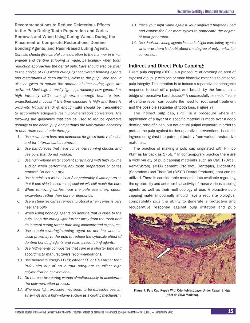
Recommendations to Reduce Deleterious Effects to the Pulp During Tooth Preparation and CariesRemoval, and When Using Curing Wands During thePlacement of Composite Restorations, DentineBonding Agents, and Resin-Based Luting Agents.Dentists should give careful consideration to the manner in which
enamel and dentine stripping is made, particularly when tooth
reduction approaches the dental pulp. Care should also be given
to the choice of LCU when curing light-activated bonding agents
and restorations in deep cavities, close to the pulp. Care should
also be given to reduce the amount of time curing lights are
activated. Most high intensity lights, particularly new generation,
high intensity LED’s can generate enough heat to burn
anaesthetized mucosa if the time exposure is high and there is
proximity. Notwithstanding, enough light should be transmitted
to accomplish adequate resin polymerization conversion. The
following are guidelines that can be used to reduce operative
damage to the dental pulp and perhaps the unfortunate necessity
to undertake endodontic therapy.
1. Use new, sharp burs and diamonds for gross tooth reduction
and for internal caries removal.
2. Use handpieces that have concentric running chucks and
use burs that do not wobble.
3. Use high-volume water coolant spray along with high volume
suction when performing any tooth preparation or caries
removal. Do not cut dry!
4. Use handpieces with at least 3 or preferably 4 water ports so
that if one side is obstructed, coolant will still reach the burr.
5. When removing caries near the pulp use sharp spoon
excavators rather than burs or diamonds.
6. Use a stepwise caries removal protocol when caries is very
near the pulp.
7. When using bonding agents on dentine that is close to the
pulp, keep the curing light further away from the tooth and
do interval curing rather than long concentrated exposures.
8. Use a pulp-covering/capping agent on dentine when in
close proximity to the pulp to reduce the cytotoxic effect of
dentine bonding agents and resin based luting agents.
9. Use high-energy composites that cure in a shorter time and
according to manufacturers recommendations.
10. Use moderate energy LCU’s; either LED or QTH rather than
PAC units but of an output adequate to effect high
polymerization conversions.
11. Do not use two curing wands simultaneously to accelerate
the polymerization process.
12. Whenever light exposure may seem to be excessive use, an
air syringe and a high-volume suction as a cooling mechanism.
13. Place your light wand against your ungloved fingernail bed
and expose for 2 or more cycles to appreciate the degree
of heat generated.
14. Use dual-cure luting agents instead of light-cure luting agents
whenever there is doubt about the degree of polymerization
conversion.
Indirect and Direct Pulp Capping:Direct pulp capping (DPC), is a procedure of covering an area of
exposed vital pulp with one or more bioactive materials to preserve
pulp integrity. The intention is to induce a reparative dentinogenic
response to seal off a pulpal wall breach by the formation a
bridge of reparative hard tissue.38 A successfully sealed-off zone
of dentine repair can obviate the need for root canal treatment
and the possible sequelae of tooth loss. (Figure 7)
The indirect pulp cap, (IPC), is a procedure where an
application of a layer of a specific material is made over a deep
dentine zone of close, but not actual pulpal exposure in order to
protect the pulp against further operative interventions, bacterial
ingress or against the potential toxicity from various restorative
materials.
The practice of making a pulp cap originated with Philipp
Pfaff as far back as 1756.39 In contemporary practice there are
a wide variety of pulp capping materials such as CaOH (Dycal,
Kerr-Sybron), (MTA) cement (ProRoot, Dentsply), Biodentine
(Septodont) and TheraCal (BISCO Dental Products), that can be
utilized. There is considerable research data available regarding
the cytotoxicity and antimicrobial activity of these various capping
agents as well as their methodology of use. A bioactive pulp
capping material optimally should have a requisite biological
compatibility plus the ability to generate a protective and
recuperative response against pulp irritation and pulp
Figure 7: Pulp Cap Repair With Odontoblast Layer Under Repair Bridge (after da Silva Modena).
Restorative Dentistry / Dentisterie restauratrice
Canadian Journal of Restorative Dentistry & Prosthodontics/Journal canadien de dentisterie restauratrice et de prosthodontie — Vol. 8, No. 3 — Fall/automne 201516
degeneration.40,41 CaOH has been used for decades with
successful but imperfect or unpredictable results.42 Currently,
there are several new materials that show evidence of
stimulating the dentine to develop the bridging of defects by
apatite formation and which do so more effectively than CaOH.
These materials utilize calcium silicates or calcium aluminates
and have been shown to be less cytotoxic and have better
antibacterial properties than CaOH. These newer materials are
considered to be more bioactive.41,45
Dycal has shown an ability to stimulate the pulp to lay down
reparative dentine. This however, may not be due to the bio-
inductive capacity of the material; rather it is more likely due to
the result of a defense mechanism by the pulp induced by the
inherent, irritant nature of calcium hydroxide.43 The toxicity of
Dycal, a high alkalinity, self-setting (2.5-3.5 min) radiopaque
calcium hydroxide-based material has been employed in direct
and indirect pulp-capping procedures and its shortcomings have
been well established.44,45 Dycal, while widely used in dental
practice for a very long time as the protective layer of choice,
has two additional disadvantages besides its relative toxicity:
poor adhesion to dentine and a significant solubility factor that
over time creates dissolution. These two factors are responsible
for allowing avenues of progressive micro-leakage to the pulp
as well as the creation of voids or defects (“tunnel defects”) in
the reparative dentine under the capping layer.46
MTA, Mineral trioxide aggregate, has a composition similar
to that of Portland Cement. Both are composed of calcium
phosphate, calcium, and silicon oxide. MTA is a powder that
contains trioxides and hydrophilic particles that allows the
material to set in the presence of moisture. It is applied as a
paste or putty depending on how much sterile water it is mixed
with. MTA, additionally, contains bismuth oxide, which provides
some radiopacity. It has an enhanced interaction with dentine
pulp tissue compared to CaOH showing faster and more
complete dentine bridge formation47,48 as well as significantly
less cytotoxicity.45 While MTA has been shown to be a reliable
and biocompatible pulp capping and dentin sealing material,
its formulation and chemical curing, plus its very long clinical
setting time, make it an inefficient material to use routinely for
restoration lining or as a pulp capping material.49
Biodentine is a powder-liquid material having chemistry similar
to MTA. The powder contains tricalcium silicate, dicalcium
silicate, calcium carbonate, and zirconium oxide; the liquid
contains water, calcium chloride (accelerator), and modified
polycarboxylate. Biodentine, like other silicate based materials
rates low in cytotoxicity.45 It does sets somewhat faster than
other calcium-silicate cements like MTA, but still takes beyond
ten minutes of set time when used as a pulp cap before a
restoration can be placed over it. This, in addition to the capsule
based tituration preparation makes for clinical inconvenience
compared to other materials.
TheraCal is a new category of resin-modified calcium silicates
based on a light-cured, resin-based, single-paste system.
TheraCal consists of CaO, calcium silicate particles (type III
Portland cement), Sr glass, fumed silica, barium sulphate, barium
zirconate and resin, containing Bis-GMA, and polydimethacrylate.
It is a highly radiopaque lining and capping material designed to
release calcium to promote the formation of reparative dentine.50
The biologic availability of calcium ions supplied from a pulp
capping or lining material is integral to stimulating pulp cells to
form new mineralized hard tissue. This is the basis of the
success of CaOH as well as the more recently introduced silicate
based products.51,52,53 TheraCal shows the highest Ca availability
of all tested materials.54 TheraCal also has distinctive application
advantages over other materials in that it is supplied directly
from a paste container and then light polymerized to form an
almost instantaneous hard, secure and low-solubility barrier
over which a restorative material can be immediately placed.
The objective of vital pulp therapy is to maintain the pulp
vitality and its function. Several factors influence the success
of direct pulp capping such as carious contamination, the age,
vitality and regenerative power of the pulp, the materials and
technique used to perform the procedure and the ability to
control pulp bleeding in a physiologic way. If pulp bleeding cannot
be controlled, it is difficult or impossible to create a wound seal
that would allow intimate contact of the capping material to the
pulp tissue. If a blood clot is left on the surface without direct
contact of a bioactive capping material, a chronic inflammatory
response impairing the healing process will ensue.55,56 Control
of pulp bleeding is usually attempted by placing a cotton pellet
soaked in a solution onto the bleed site. A variety of solutions
have been used, including saline, sodium hypochlorite
(concentrations ranging from 0.12% to 5.25%), hydrogen
peroxide, ferric sulfate and chlorhexidine. Saline or calcium
hydroxide solutions are the most benign to the pulp in performed
cytotoxicity tests.57 Saline exhibits the mildest pulp response
but is only moderately hemostatic. Sodium hypochlorite is
effectively hemostatic plus has the benefit of being antibacterial,
but it is more cytotoxic and will induce a pulpal inflammatory
response that fortunately in most cases is transient.
Chlorhexidine is antibacterial but is not very effective at
hemorrhage control. Ferric Sulfate is highly hemostatic but is
seriously cytotoxic, creating post-operative pulpal pain and
is not recommended.58 Ankaferd Blood Stopper (ABS) is a new
Restorative Dentistry / Dentisterie restauratrice

Imagine how much easier your day would be if you could breeze through posterior restorations. Productivity would go up and costly chair time would go down. Enjoy speed and simplicity with our Posterior Restorative Procedure. It combines Filtek™ Bulk Fill Posterior Restorative with three other innovative, time-saving products from 3M ESPE Dental. See how it can help keep you on schedule … because we know your time outside of work matters.
3M Science. Applied to Life., 3M, ESPE, Elipar, Filtek, Scotchbond and Sof-Lex are trademarks of 3M or 3M Deutschland GmbH. Used under license in Canada. © 3M 2015. All rights reserved. 1508-03096 E
3M™ ™™
™ ™ ™
+ + +
By using our four innovative technologies together, you can move through posterior restoratives with speed and simplicity - and keep your day on schedule.
Because we know your time outside of work matters.

Canadian Journal of Restorative Dentistry & Prosthodontics/Journal canadien de dentisterie restauratrice et de prosthodontie — Vol. 8, No. 3 — Fall/automne 201518
hemostatic agent derived from herbal extract that has been
shown to be as effective as ferric sulfate. It has been proposed
for bleeding control of exposed pulps but there is very little
research on it and its cytotoxicity on the pulp is not well
established.59
Recommendations on Vital Pulp Lining and Pulp Capping:Dentists should recognize that the dental pulp has good
recuperative powers even when it becomes exposed, but they
should also be aware that a carious exposure of the pulp
significantly reduces the probability of pulp survival and that
when bacterial contamination occurs, root canal therapy, should
be considered. In the case of an operative created exposure with
no contamination, pulp capping can have a high incidence of
success, particularly if best protocols and materials are utilized.
1. If the pulp bleeds, the bleeding needs to be arrested so
that the capping material can attach or be secured in a
stable way around the perimeter of the exposure. As a first
pass, use a cotton pellet moistened with saline solution
and very gently compress for a few minutes. If bleeding
continues repeat the process using NaOH which is more
cytotoxic, but not overtly so, if judiciously used.
2. When ready for capping, apply a thin lining of TheraCal
which is confirmed to be bioactive, sets immediately on
light activation, and has enough stability to receive a
hydrophilic self-etching dentine adhesive followed by a
restoration directly over it. Blot off moisture but do not
dessicate the area before applying the TheraCal layer.
3. Do not use high heat generating wands to cure the layer and
for the same reason do not exceed the minimal time needed
for polymerization. A gentle supplement of air syringe during
curing can help keep the heat from the light at minimal.
4. As a protective liner use the same protocol as in No. 2 & 3,
assuming there is no pulp bleed.
Effects of Threaded Pin Placement:Threaded and cemented pins or dentinal micro-screws are
commonly used in restorative dentistry to enhance the retention
and stability of restorative dental materials. According to Segovic
et al. (2002), the insertion of self threaded pins causes micro-
cracks in the dentine slightly in excess of 50% of cases. However,
there is very little information published on the effects that pin
placements have on the dental pulp or on the structural integrity
of the tooth or on the significance of the micro-cracks induced.
On the other hand, it is reasonable to presume that pin
placement into the pulp will create an inflammatory response
due to the mechanical insult and it is probable that unless the
pin channel is otherwise sealed it may act as a path of ingress
for bacterial leakage. If the penetration into the pulp is superficial
and the wound in not bacterially infective, it is reasonable to
assume that the pulp may respond by creating an isolating,
reparative zone of dentine. It is also reasonable to expect that
if there is pin perforation into the periodontium there will be a
localized inflammatory response that may create a chronic
periodontal condition.
Recommendations on Pin Placements: The use of retention pins to stabilize coronal restorations in vital
teeth poses some risks, particularly when they are placed in
narrow diameter teeth such as maxillary laterals and mandibular
incisors. Careful consideration should be given to the, type of pin
placed, the diameter of the pin, the placement location, the
number of pins and the risk versus benefit of utilizing pins.
1. Use sharp drills on ultra-low rpm’s to create the pin channel
so as not to induce heat or dentine crazing.
2. Choose to locate the drill holes at the 4 line angles of the
tooth where pin placement will be most remote from the
pulp. An exception is the mesial-buccal line angle of the
maxillary first molar where there is a pronounced pulp horn.
3. Self-shearing, straight pins are imprecise for bottoming out
or not. Pins with a coronal depth-limiting flange are more
accurate and may be less likely to create dentine stress.
4. If a pin must be bent, use a pin-bending tool to minimize
the torque that will be transferred to the tooth structure.
5. Consider the use of cementable pins that are passive,
compared to self-tapping pins. Use a cement that is
minimally cytotoxic such as conventional glass ionomer
compared to resin. A cemented pin will also create a pin
channel seal to resist possible bacterial micro-leakage.
6. Follow the contour of the root when aligning the drill to
minimize the risk of external perforation. (Figure 8)
Effects of Chemical Treatments to Enamel,Dentine and Cementum: The application of chemicals onto and into teeth is an integral
part of what clinical dentists do in everyday practice. Tooth
whitening is one of the most commonly used examples of
chemical application in dentistry. It utilizes hydrogen peroxide or
carbamide peroxide in the dental setting, but these agents are
also sold over the counter (OTC) for home use. In operative
dentistry various other substances are also utilized as conditioning
agents to prepare enamel, dentine and cementum for remedial
treatment enhancement. Agents such as phosphoric acid and
Restorative Dentistry / Dentisterie restauratrice

Canadian Journal of Restorative Dentistry & Prosthodontics/Journal canadien de dentisterie restauratrice et de prosthodontie — Vol. 8, No. 3 — Fall/automne 2015 19
citric acid are used for conditioning enamel and dentine prior to
adhesive bonding. Sodium hypochlorite, EDTA and citric acid are
commonly used in endodontic procedures. Tetracycline HCl, EDTA
and citric acid are used for conditioning cementum prior to
connective tissue grafting procedures. Many of these agents are
cytotoxic and can possibly be harmful as well as beneficial.
Effects of Tooth Whitening:It is a widespread elective practice to whiten teeth by applying
different agents to the surfaces of teeth for cosmetic purposes.
Hydrogen peroxide, sodium perborate and carbamide peroxide
are the principle products used for tooth bleaching purposes.
Side effects, such as changes in the tooth structure, gingival
burns, micro-leakage in restorations, tooth sensitivity and pulpal
irritation all have been reported when using these agents.60,61,62
The greatest negative consequence, however, may arise in
utilizing powerful light units to activate powerful 35% to 50%
hydrogen peroxide based bleaching agents.63 Heat application
seems to be effective in potentiating the bleaching effect of
tooth whitening agents compared to cold light which has been
shown to be relatively ineffective.64 Halogen, LED and LED Laser
lights all have the potential to raise the intra-pulp temperature.63
Temperature elevations of 5.6°C or greater can cause pulpal
damage and may result in necrosis in 15% of cases, depending
on pulp health its physiological response capacity.10
In an in vitro study, Carrasco et al. determined that of the
three contemporary light systems tested, the halogen light
generated the most heat but when bleaching gel was applied
the gel actually acted as a modulator of heat production diffusing
its effect. LED and LED Laser units produce notably less heat
than halogen. However, the heat increases into the pulp
chambers for all units tested, including halogen, were deemed
unlikely to be injurious to pulpal health because they did not
exceed the 5.6°C threshold even though the in vitro study did
not factor on the effect of blood circulation in the pulp chamber
which could account for a greater intra-pulp temperature rise
than the study showed.65 The most common effect of tooth
whitening is post treatment cervical tooth sensitivity which
ranges in incidence from 15 to 78% with the highest rate
resulting from the combination of 30% hydrogen peroxide used
in combination with heat.66
The success of whitening procedures is directly related to
the ability of bleaching agents to penetrate enamel and dentine.
Recent studies have shown that both hydrogen peroxide and
carbamide peroxide are capable of penetrating enamel and
dentin and will enter the pulp chamber.67,68 The higher the
concentration of these products and the longer the application,
the higher is the penetration into the pulp chamber.69 While in
vitro studies show a significant penetration of peroxide into the
pulp chamber, in vital pulps it is postulated that pulpal fluid
pressure is capable of reducing inward diffusion of these and
other chemicals so as to diffuse potentially harmful effects.
This factor along with the other defense mechanisms of the
pulp are apt to significantly reduce the level of hydrogen peroxide
penetrating into the pulp and which therefore are not expected
to develop into any permanent, negative outcome.70
A number of studies have been made regarding the effect of
bleaching agents on the CEJ with analysis of the effects of
erosion and abrasion and cervical sensitivity. The majority of
studies have shown that at-home bleaching agents based on
hydrogen or carbamide peroxide have no harmful effects on
enamel and dentin properties. Cervical sensitivity in most
instances can be controlled by the application of fluoride or
desensitizing agents.71,72 The greatest post-operative sensitivity
is generated by in-office applications of 35% hydrogen peroxide
based products particularly when used in combination with heat
based curing lights.73
Post-bleaching sensitivity differs from dentin hypersensitivity
because it is related directly to the penetration through the
enamel of the sub-products of the bleaching gels into the
dentin and pulp tissue, causing reversible pulpitis and thermal
sensitivity, but not causing permanent damage to the pulp. The
degree of post- operative pain is dependent on the peroxide
concentration, time, frequency of gel application, and the degree
of pulp temperature rise with light activation.74,75
Recommendations on Tooth Whitening:Tooth whitening is a socially driven, elective procedure that in
most instances is simple and effective and relatively inexpensive.
It is therefore a popular treatment that is requested and performed
Restorative Dentistry / Dentisterie restauratrice
Figure 8: Four Pins Placed at the Axial Line Angles.

Canadian Journal of Restorative Dentistry & Prosthodontics/Journal canadien de dentisterie restauratrice et de prosthodontie — Vol. 8, No. 3 — Fall/automne 201520
by most dentists and it is also available as an over the counter
product that can be used at home. Because tooth whitening is so
common there is the belief that it is absolutely safe. For the most
part this is true but vital tooth bleaching can be subject to overuse
and abuse.
1. Use high concentration hydrogen peroxide bleaching
products with care and discretion. Avoid using on
adolescents who are more susceptible to post operative
sensitivity or on adults with gingival recessions and erosions.
Use with rubber dam and soft tissue barrier protection
agents.
2. Avoid the use of lights that create heat. Recognize that non
heat-generating lights have limited benefit.
3. Use according to the manufacturers recommendations for
duration and frequency. Results are time and concentration
dependent, so weaker concentrations over more time may
be more appropriate than strong and fast. Do not over-
prescribe or abuse these products.
4. Use fluoride treatments or other agents to alleviate post-
use sensitivity.
5. Instruct the patient on proper protocol with take home or
OTC products.
Effects of Conditioning to Enamel and Dentinein Adhesive-Based Restorative Procedures:Effective adhesion to tooth substrate is the basis for using
contemporary composite resins as a restorative material and
as a luting agent. This process depends on creating a suitable
micro-retentive surface on enamel or dentine. On both enamel
and dentine, this works best by conditioning (etching) the
surface with a phosphoric acid gel of approximately 32-37%
concentration (pH < 1.0) for about 20 seconds and then rinsing
with water. On enamel, the phosphoric acid will create an etched
micro-retentive surface directly onto the prismatic mineralized
surface to which the bonding agent will directly and effectively
attach with an adhesive strength of around 45 MPa.76
On dentine, which has much greater percentile organic
composition, the acid works by removing the smear layer to
expose dentinal tubules into which the hydrophilic resin primers
will penetrate and then be polymerized creating an adhesive,
hybrid zone.77 An effective hybrid interface in dentine will create
adhesive values of around 40 MPa. While the process of
conditioning is effective, some concern has to be given to the
possibility of a cytotoxic effect when using a relatively strong
acid product close to pulp tissue. Conditioning applied for
15-20 seconds onto a very thin layer of dentin only slightly
affects the blood supply to the dental pulp; however, prolonged
etching time for 60 seconds results in immediate failure of
microcirculation in the dental pulp of rats.78,79 An in vivo testing
of direct acid etching in deep dentine sites, followed by a bonding
agent, creates inflammatory and degenerative pulp responses
compared to placing a protective bioactive lining prior to using
a self-etching adhesive agent.80
Recommendations On Pulp Protection When DentineBonding:Dentine bonding is universally used in dental offices throughout
the world because for the most part it is safe and effective.
However, there are a number of issues pertaining to bonding in
deep dentine or when bonding over an exposed pulp. If the pulp
is exposed and there is bacterial invasion, endodontics is almost
always a necessity. If the pulp is not exposed it must be protected
from the effects of etching, the application of adhesive resins
and the future possibility of micro-leakage. Each of these events
individually, or together, can be injurious to the pulp. Protection
layering over the pulp must also be resistant to degradation due
to hydrolysis and the overlying restoration must protect the pulp
by creating a dependable perimeter seal that will also resist
micro-leakage.
1. Prior to total-etching or one-step etching onto an exposure or
in a deep dentine zone, a thin layer of a suitable biocompatible
and bioactive liner such as TheraCal should be used. TheraCal
has the ability to rapidly create a hard, well-sealed, light-
activated, resinous capsule over the pulp area.
2. The best protocol for adhesive bonding over the capped
area is with a “total etch” and rinse, 4th generation, bonding
which will use a hydrophilic primer for effective penetration
of the resin into the exposed dentinal tubules, followed
by a secondary hydrophobic layer primer that will
give maximum protection against water-tree hydrolytic
degradation and then over which a universal bonding agent
can be used. It is important that the dentine in this protocol
is not dessicated, or the bond will not be very effective.
3. Alternatively, an 8th generation single step universal
adhesive can be placed over the capped area. The universal
adhesives of this generation will help to recondition over-
dessicated dentine. This simplified procedure, while not as
good as the 4th generation protocol, will deliver very
adequate adhesion and protection and will reduce post-
operative sensitivity. Two products that are representative
of this system are: Bisco Universal Adhesive and 3M ESPE
ScotchBond Universal Adhesive.
4. When light-wand curing the primer layer and the adhesive
layer, remember that output strength of the light, plus
Restorative Dentistry / Dentisterie restauratrice


Canadian Journal of Restorative Dentistry & Prosthodontics/Journal canadien de dentisterie restauratrice et de prosthodontie — Vol. 8, No. 3 — Fall/automne 201522
proximity, plus time, cumulatively can potentiate a harmful
effect to the pulp. Short, but adequate exposure time
with moderate light output therefore is more suitable.
If necessary, air syringe adjunct cooling can be used.
5. Because 8th generation, single step adhesive agents are
only mildly acidic, it is unlikely that peripheral enamel
will be etched adequately to create a good marginal
restoration bond that will resist micro-leakage. It is therefore
recommended that a bead of 37% phosphoric acid gel be
applied to regions of enamel that will be in contact with the
composite restorative material.
Effects of Conditioning of Cementum inPeriodontal Therapy:Root conditioning for periodontal treatments is somewhat
controversial but remains a standard procedure that is performed
prior to applying connective grafts to be able to obtain cementum
root coverage with soft tissue. Cementum is a mineralized tissue
layer that has as its primary function the insertion of periodontal
ligament fibers onto the root surface. This suspensory fiber
apparatus acts as a proprioceptive agent that governs the
functional and parafunctional forces elicited from teeth to the
surrounding alveolar bone.
Root surfaces of periodontally diseased teeth are hyper-
mineralized and contaminated with organisms, endotoxins and
other biologically active substances.81 Biomodification of root
surfaces and removal of the smear layer and debris is deemed
critical for regeneration of the periodontium.82 Removal of all
bacterial components by meticulous root planning is not enough
because the resultant and residual smear layer will prevent the
new attachment of periodontal fibers and will inhibit the migration
and proliferation of fibroblasts.83
There are many agents such as phosphoric acid, EDTA,
tetracycline HCl and citric acid that can be used for the
biomodification removal of the smear layer of root surfaces
following debridement. All of these products are effective
but Citric acid is the commonly used because it is highly
effective, readily available and inexpensive. The most effective
concentration and method of applying citric acid gel is to use it
at the 25% concentration (pH 2.2) with a brushing technique for
1-3 minutes.84,85,86
Citric acid is cytotoxic to the dental pulp. Chan et. al.
investigated the cytotoxic and cytostatic effects of citric acid on
cultured dental pulp cells which when exposing the cells to pure
1% citric acid (pH = 2.26) for 60s caused immediate cellular
death. The authors recommended judicious care of using citric
where it can come into direct contact with the dental pulp.87,88
Other authors have shown that citric acid, in low concentration,
placed into cavities on the pulp demonstrated transient pulp
inflammation but no severe or irreversible damage.89
There is little investigation of the effects of very high
concentration citric acid with its low pH on the pulp when used
as a root surface conditioner but the few available studies
indicate that citric acid does not penetrate into the dentinal
tubules nor does it alter the collagen content of the roots
obtained by scaling and root planning,89,90 nor does it seem to
have an adverse effect on regenerative fiber healing or on bone
regeneration.91 While there seems to be no deleterious effect
to the pulp and the vitality of the tooth, low PH citric acid will
have an immediate (within 20 s) necrotizing effect on both
mucosal flaps and surrounding periodontal tissue so care should
be taken to avoid soft tissue contact.92 (Figure 9)
Recommendations On Using Citric Acid To Condition Cementum:Epidemiological studies show that more than 50% of the population
have one or more sites with recession of at least 1 mm.93 Loss of
gingival root coverage predisposes the root to erosion and to loss
of esthetics; so there is an indication and a need and for remedial
procedures. The most common, current, surgical method to re-
establish lost gingival form and coverage, is by using connective
Figure 9: Citric Acid Effect on Dentine (above) and on Cementum (below).
Restorative Dentistry / Dentisterie restauratrice

PLANESYSTEM®
the third dimension in patient analysis
PS1-3D CAD PlaneTool (exclusively for the Zirkonzahn.Scan scanning software)
PS1-3D virtual articulator (integrated in the Virtual Articulator module of the Zirkonzahn.Modellier modelling software)
The captured data is ideally combinable with scan data of the 3D facial scanner Face Hunter
The PlaneSystem® is used to determine the natural position of the maxilla and the individual inclination of the occlusal plane, regardless of physical asymmetries. Bone asymmetries and resulting muscular compensation are detected ahead of developing a treatment plan.
Developed in cooperation with MDT Udo
Zirkonzahn USA Inc. – Phone +1 800 989 8931 – Phone +1 678 441 9419 – [email protected]
Zirkonzahn Worldwide – Phone +39 0474 066 680 – [email protected] – www.zirkonzahn.com

Canadian Journal of Restorative Dentistry & Prosthodontics/Journal canadien de dentisterie restauratrice et de prosthodontie — Vol. 8, No. 3 — Fall/automne 201524
3. Apply and position the CT graft and suture to secure the graft.
4. Instruct to avoid brushing the site for 1 week and use
chlorhexadine as a mouthrinse, bid. Avoid flossing for
4 weeks. Do not probe for 6 months.
Effects of Traumatic Occlusion On Pulpitis and Pulp Degeneration:It is recognized that repetitive micro-traumatic forces on teeth
can cause tooth migrations and periodontitis. It has also been
considered that parafunctional forces, when sustained, may be a
contributing factor to pulpitis, and to the pulpal degeneration of
teeth along with accompanying apical periodontitis.94 Review of
the literature reveals a paucity of studies about the reaction of
dental pulp to occlusal forces but other related traumatic forces
such as orthodontic intrusive and extrusive forces have been
shown to affect the dental pulp by creating inflammatory vascular
changes.95,96 Orthodontic forces have been shown to cause edema
and odontogenic degeneration of the pulp by compromising the
apical blood supply.97,98 Generally, in the first few days after
activation of an orthodontic appliance, patients will often suffer
transient pulp ischemia, causing pain and discomfort. Usually
these symptoms settle within a week, but pulp death following
orthodontic treatment is occasionally reported.99
Kvinnsland et al. did rat studies creating hyper-occlusion and
using fluorescent microspheres to detect blood flow. They found
that teeth on the hyper-occluded side had an increase in pulpal
blood flow.100 Lui did a clinical analysis of 19 cases where
pulpitis could not be explained except by traumatic occlusion
and after he performed occlusal adjustment found that 6 teeth
resolved but 13 required endodontic intervention.101 Shi et al.
attributed 100 teeth in 89 cases having pulpitis with apical
periodontitis to traumatic occlusion. All teeth were caries free
and without dental or periodontal disease but all had distinct
evidence of marked occlusal trauma.102
Okeson in his textbook, Management of Temporomandibular
Disorders and Occlusion, states that “On occasion, patients
come in with pulpitis that has no apparent dental or periodontal
etiology… When all other etiologic factors have been ruled out,
one must consider heavy occlusal forces … chronic pulpitis can
lead to pulpal necrosis.” (Figure 10, 11)
Recommendations On Managing Site-Specific Trauma to Teeth.Occlusal dysfunction can manifest into a number of pathologic
symptoms such as temporomandibular joint disturbances,
myofunctional disturbances and disturbances to the teeth and
periodontium. Tooth fractures, tooth wear and tooth hypermobility
Figure 10: Occlusal Trauma on 3.1 and 3.2
Restorative Dentistry / Dentisterie restauratrice
tissue grafts harvested from the palate by pouch technique and
then installing them over a recipient sites by a tunneling technique
so that the graft is covered by a vascularized zone of epithelialized
tissue with undetached papillae. Prior to transferring the donor
grafts, the exposed root areas are usually conditioned with EDTA,
tetracycline HCl or citric acid to enhance the attachment of the
graft as per the method described following. Because citric acid
can be harmful to the soft tissues due to the low pH, care must be
taken to minimally expose it to the soft tissues.
1. Preparing an optimum surface for graft attachment requires
a mechanical debridement of all calculus, plaque and
bacterial components from the root surface by using
ultrasonic instruments or hand scalers. This is followed by
an application of a conditioning medium to remove the
smear layer. Studies have shown this is most effectively
accomplished by rubbing a 25% concentration of citric acid
with a small cotton pellet or with a bonding-application
brush onto the root area for approximately 2-3 minutes. It
is important to avoid solution contact with the mucosal
tissues. Wash with saline and blot dry.
2. It is common to apply enamel matrix protein, i.e.“Emdogain”
(Straumann) to enhance periodontal ligament fibroblast
proliferation.
Figure 11: PeriapicalPeriodontitis FromOcclusal Trauma.

Canadian Journal of Restorative Dentistry & Prosthodontics/Journal canadien de dentisterie restauratrice et de prosthodontie — Vol. 8, No. 3 — Fall/automne 2015 25
and periodontitis are common consequences of excessive, ongoing
loading on teeth. Occlusal trauma may also create pulpitis,
particularly if there are other predisposing conditions such as
concomitant periodontal involvements or abrasion into dentine.
1. During routine examinations test each tooth with soft
percussion to see if periodontal support has been altered
and if there is a differentiation of periodontal stability from
other teeth. Keeping a finger on one side while gently
tapping on the other side can proprioceptively identify
changes in the stability of the periodontal membrane. If a
particular tooth seems hypermobile consider providing
occlusal relief by selective adjustment.
2. Identify sites of aggressive individual tooth wear. If there is
dysfunctional occlusal trauma and the periodontium is
resistant, excessive faceting or wear may indicate a specific
site of trauma. Again, selectively adjust the tooth as a
preventive therapy.
3. Identify sites where opposing occlusal materials are
incompatible, such as where antagonistic ceramic or other
materials that may be wear resistant are positioned against
a natural tooth or restorative material that is less hard and
less wear resistant. Selectively adjust the wear resistant
material and polish very smooth.
4. Consider the use of nocturnal anti-bruxing guards that will
splint teeth and provide a platform to create an equilibrated
occlusal contact pattern and will ameliorate the wear on
the occlusal surfaces of teeth.
Conclusions:Operative Dentistry, while well intentioned, may have the adverse
effect of creating harm instead of benefit. Procedures that
generate excessive heat and materials that are cytotoxic can
irritate the dental pulp and create pulpitis conditions that may
not be reversible. This is particularly relevant as procedures
and materials encroach on and are applied closest to the
pulp. This article has attempted to identify those factors that
have the potential to create irreversible insult to the pulp and
has attempted to provide guidance for minimizing unwanted
harmful events.
About the authorDr. Nimchuk is a Certified Prosthodontist. He has been a director and mentor to over twenty continuing education Study Groups in the field of Fixed, Removable and ImplantProsthodontics. He has been a Honourary Sessional Lecturer at the Faculty of Dentistry,UBC for over 15 years. He has given over 700 lectures worldwide and has authorednumerous articles. He is the holder of an “Alumni of Distinction” award from the Universityof Toronto, Faculty of Dentistry. He is an Associate Editor of The Canadian Journal ofRestorative Dentistry and Prosthodontics.
Conflict of Interests: The author reports no conflict of interests
The extensive Bibliography for this article can be accessed onthe digital version of the CJRDP at www.cardp.ca — page 69
L’importante bibliographie pour cet article peut être consultésur la version numérique du JCDRP à www.cardp.ca — page 69
Restorative Dentistry / Dentisterie restauratrice
Dedicated Team of Technicians
Integrity • Quality • Trust • Knowledge
Experience • Confidence • Education
A family business since 1963Rotsaert Dental Laboratory
is celebrating 50 Years of Serviceto the Dental Community.
71 Emerald Street South,Hamilton, Ontario, Canada L8N 2V4
Tel: (905) 527-1422Toll Free: 1-800-263-2113
www.rotsaertdental.come-mail: [email protected]
Scan to view theRotsaert
“Legacy” movie.
Comments/Commentaires


Canadian Journal of Restorative Dentistry & Prosthodontics/Journal canadien de dentisterie restauratrice et de prosthodontie — Vol. 8, No. 3 — Fall/automne 201528
Occlusal vertical dimension for completeremovable dental prostheses
Dimension verticale de l’occlusion pour prothèses
dentaires amovibles complètes
Statement of Problem: Despite scientific and artistic advances in the restoration of an edentulous mouth, determining theocclusal vertical dimension (OVD) for complete removable dental prostheses continues to be a challenging and subjective task.
Purpose: This article reviews the different methods available to determine the OVD for complete removable dentalprostheses.
Material and Methods: A review of the literature was performed to discuss the rationale and applications of the differentmethods for determining the OVD for complete removable dental prostheses
Results: Several methods for determining the OVD are described with different levels of reliability and complexity. It isdifficult to recommend one single method.
Conclusion: A combination of different techniques may provide the most reliable method for determining the OVD forcomplete dental prostheses.
CLINICAL IMPLACTIONS The establishment and recording of the OVD for complete removable dental prostheses plays a major role in providingreliable esthetics and function.
Abstract
Dr. Samer Mesmar, DMD,
Dr. Caroline Tram Nguyen, DMD, MS, FACP, FRCD
and Dr. Chris C. L. Wyatt, BSc, DMD, MSc, Dip Prosc
Faculty of Dentistry, University of British Columbia,Department of Oral Health Sciences,
Vancouver, Canada
Young Author Award Submission
IntroductionThe occlusal vertical dimension (OVD) is defined in theGlossary of Prosthodontics terms as “the distancemeasured between two points when the occludingmembers are in contact”.1 It is established by the naturalteeth when they are present and in occlusion. Inedentulous patients, the OVD is established by thevertical height of the two dental prostheses when the
denture teeth are in contact. The establishment andrecording of the OVD is a crucial step in the fabricationof dentures for edentulous patients. It determines theesthetics and occlusion,2 and it influences the patient’sperception and acceptance of the prosthetic treatment.
The purpose of this paper is to review the differentmethods available to determine the OVD for complete
Complete Removable Prosthodontics / Prosthodontie complète amovible
THE UNIVERSITY OF BRITISH COLUMBIA
Complete removable prostheses, Determining the Occlusal Vertical Dimension (OVD) Key words

Canadian Journal of Restorative Dentistry & Prosthodontics/Journal canadien de dentisterie restauratrice et de prosthodontie — Vol. 8, No. 3 — Fall/automne 2015 29
Le problème : Malgré les avancées scientifiques et artistiques dans la restauration des bouches édentées, déterminer ladimension verticale de l’occlusion (DVO) pour des prothèses dentaires amovibles complètes, reste un défi et une gageure.
L’objectif : Cet article passe en revue les différentes méthodes disponibles pour déterminer le DVO des prothèses dentairesamovibles complètes.
Matériel et méthodes : un examen des écrits à ce sujet a été prescrit afin de discuter du rationnel et des applications de cesdifférentes méthodes, qui déterminent le DVO des prothèses dentaires amovibles complètes.
Résultats : Plusieurs méthodes pour déterminer la DVO sont décrites, à différents niveaux, de fiabilité et de complexité. Il estainsi difficile de recommander une méthode plutôt qu’une autre.
Conclusion : Une combinaison des différentes techniques peut apporter une méthode des plus fiables, pour déterminer laDVO des prothèses dentaires complètes.
Implications cliniquesL’établissement et l’enregistrement de la DVO pour les prothèses dentaires amovibles complètes, jouent un rôle majeur, enapportant un fonctionnement et une esthétique fiables.
Résumé
removable dental prostheses. The first part includes aliterature review of the different techniques with theirapplications. The second part lists the clinical stepsinvolved in determining the OVD.
Some authors3 classify these methods in a direct andindirect approach, whereas others4 classify them asmechanical and physiological methods. Nevertheless, they include pre-extraction records, deglutition, physiologicalrest position, phonetics, esthetics appearance, craniofacialmeasure, biting force and patient tactile sense, which areall commonly used and described in this review. Also, abrief description of the use of cephalometric radiographsand duplication of old dentures is included.
Pre-extraction recordsPre-extraction records have been used in the assessmentof the OVD. Smith5 studied five methods of making pre-extraction records including the Sorenson Profile Scale, the cardboard profile, the interfrenal distance,measurement between tattoo dots on the gingiva proposedby Silverman6 and the nose-chin distance measured witha plastic ruler. He found that the OVD could be reproducedwith a mean accuracy of 1.1 to 1.4 mm with all recordsbeing nearly alike in reliability. According to Smith, the pre-extraction records are reliable enough to give valuableclinical information. Toolson and Smith7 investigated theSorenson Profile Scale and the chin-nose methods in termsof simplicity, accuracy and practicality. They concluded that both methods were within reasonable limits whencompared to the cephalometric films. Willis8 recommended
the use of the Willis gauge for measuring the verticalheight from the undersurface of the chin to the base ofthe nose. This method introduced inaccuracies because itdepended on the operator applying the exact same degreeof pressure when the instrument made contact with theskin of the face.9 Turrell10 stated that the Willis gauge isso inaccurate as to be almost useless. Wright11 comparedmeasurements of anatomic landmarks on photographswith measurement using the same anatomic landmarkson the face for many edentulous patients. The methodwas simple but, the inaccuracy of measures on a movableskin is evident.3,12 Bissasu12 published a literature reviewabout the pre-extraction records for complete denturefabrication. He concluded by indicating that pre-extractionrecords provided a useful guide in determining theedentulous patient’s original OVD and that they arepreferred, when possible, to arbitrary methods. Turrell10
reviewed many methods of recording occlusal verticaldimension in edentulous patients and stated that in spiteof the problems with most pre-extraction recordinginstruments, some of them (Dakometer) were moreaccurate in the assessment of the OVD than numerouspost-extraction aids. The Dakometer was used to recordthe OVD and the position of the maxillary anterior teeth byplacing the instruments on the patient’s face while thepatient closed into maximal intercuspidation. With theinstrument in position, the edge of the instrument movedto engage the incisal edges of maxillary central incisors.The measurements were recorded and used as pre-extraction records.12
Complete Removable Prosthodontics / Prosthodontie complète amovible
Prothèses complètes amovibles, Détermination de la Dimension verticale de l’occlusion (DVO) Mots-clés
Canadian Journal of Restorative Dentistry & Prosthodontics/Journal canadien de dentisterie restauratrice et de prosthodontie — Vol. 8, No. 3 — Fall/automne 201530
Denture DuplicationDuplicate of existing dental prostheses can be a useful
technique if the current OVD and occlusion plane are within
normal limits.13
DeglutitionDeglutition is another commonly used method in establishing
OVD. Shanahan14 indicated that the mandibular pattern of
movement during deglutition corresponds to the OVD. He described
a swallowing technique where the soft wax on the occlusion rim is
reduced during deglutition to give the correct OVD. Ismail and
George15 tested the accuracy of the swallowing technique for
determining and recording the occlusal vertical relation of the
jaws. They used Shanahan’s technique measures in comparison
with lateral head cephalometric radiographic measures. They found
an increase of 0 to 5 mm (mean 2.8 mm) in the OVD in edentulous
patients when the swallowing technique was used, with a significant
correlation between the increased measure and the number of
posterior teeth missing before extraction. Malson16 described a
technique consisting of controlling the thyroid cartilage movement
during deglutition. When the OVD is correct, the cartilage rises in
an uninterrupted fashion before moving back to its original position.
When the OVD is increased, the patient leans forward when
swallowing and the cartilage moves in 3 steps: upward, plateau
and downward. When the OVD is decreased, the patient does not
change posture and there is no plateau phase, but there is
interposition of the tongue to compensate for the reduced height.
Millet3 mentions that this technique requires a highly experienced
clinician and cannot be used in cases of atypical deglutition. Ward
and Osterholtz17 investigated the value of the act of swallowing as
a guide to the establishment of OVD and concluded that it should
not be used as the only technique to determine the OVD. In
addition, they underlined the importance of removal of the existing
denture for a period of time before recording of OVD in order to
allow for the loss of memory of acquired neuromuscular reflex
patterns.
Physiological rest positionPhysiological rest position, as defined in the glossary of
prosthodontics terms,1 is “the mandibular position assumed
when the head is in an upright position and the involved muscles,
particularly the elevator and depressor groups, are in equilibrium
in tonic contraction, and the condyles are in a neutral, unstrained
position”. It is established by muscles and gravity. Two
hypotheses about the postural/physiological rest position of
the mandible involving an active or passive mechanism have
been considered. The active one is the position assumed when
the muscles of mastication are in a state of minimal contraction
to maintain the posture of the mandible;18 whereas the passive
one states that the elastic elements of the jaw musculature,
not muscle activity, balances the influence of gravity.19,20 Many
authors do not accept the concept of a constant rest position in
the strictest sense.21-26 It tends to remain relatively stable for
reasonable lengths of time.
Different methods have been described to attempt to
determine the physiological rest position. Thompson27 related
variations in rest position to hypotonicity and hypertonicity of
the musculature and short (stress, respiration) and long-term
variations (mouth breathers). Atwood24 contended that rest
position is a dynamic rather than static concept with inter- and
intra-individual variability. In a cephalometric study of the clinical
rest position of the mandible, Atwood22 described a decrease in
facial height at rest position after removal of occlusal contacts
and reported instability of the rest position. Smith28 described
a method where the patient takes a sip of water and keeps it
in his mouth for 2 minutes, to then swallow it. Before the
deglutition, and immediately after, the mandible is in rest
position. The non-forced respiration method describes the rest
vertical dimension being obtained during the terminal movement
of the expiration phase.29 However, this technique is prone to
error for edentulous patient who are mouth breathers.
The most common method to determine physiologic rest
position is using phonetics. The labial m sound is often used to
determine the rest position which can be said without the use
of teeth. However, the m sound leaves the lips in contact and
when parted by the dentist, the mandible is depressed and the
rest position is lost. Therefore, the sound m is often extended
to the word Emma or followed by the labial p sound with the
word Mississipi, which leaves the lips apart.10
Once the rest position is determined using any of these
suggested methods, the OVD is obtained by subtracting the value
of interocclusal distance from the rest position dimension.
Previously known as the freeway space, Sears30 described a more
specific term being “interocclusal space”. The interocclusal space
is variable depending on numerous factors including gender, age
and Angle classification. Several authors estimate an average
value of 2 to 4 mm being acceptable for most of patients.3, 4, 10
Pleasure31 reports an average value of 3 mm. He describes a
method using calipers adjusted to two arbitrary landmarks on
nose and chin, using adhesive triangles on skin, to register
physiologic rest position. In a backless chair, “the patient may be
asked to moisten the lips with the tongue or to sip a little water to
induce slight mandibular movements, and thus the regularity of
return to the tentative physiologic identified by the caliper setting
Complete Removable Prosthodontics / Prosthodontie complète amovible

Friday October 2, 2015
Fee: $395 +HST
New Grads: $195 +HST*AGD/PACE and RCDSO Accredited
Includes: DVD Early Ortho TreatmentSample articles on Early Orthodontic Treatment
To see a sample of one early treatment case, please consult Rondeau Seminars YouTube channel: http://bit.y/1N6BzIF
To register, please contact:Palmeri Media Group at 905-489-1970 ext. 206 | 207Toll Free: 1-866-581-8949 ext. 206 | 207Email: [email protected] [email protected]
Early Orthodontic Treatment For ChildrenA Core 2 Course
Brock Rondeau, DDS
8:30am to 4:30pm | 7 CE CreditsIncludes breakfast, breaks, and lunch
Approved PACE Program ProviderFAGD/MAGD CreditApproval does not imply acceptanceby a state or provincial board ofdentistry or AGD endorsement9/1/2015 to 8/31/2017Provider ID# 355116
Location:
Courtyard By MarriottToronto Northeast/Markham
7095 Woodbine Avenue, Markham, ON L3R 1A3
1. Diagnose simple orthodontic cases that can be treated with functional appliances in mixed dentition.
2. Functional appliances to treat constricted arches, posterior crossbites, anterior crossbites, deep overbites, open bites due to tongue thrust or thumbsucking habits.
3. How to adjust functional appliances.
4. Technique for bite registration with Twin Block and MARA appliance.
5. How early orthodontic treatment can increase your personal satisfaction and income for your practice.
LEARNING OUTCOMES
Seventy percent of children by age 12 have a malocclusion. Parents are looking for dentists who offer early treatment for their children. Ideally treatment with functional appliances will prevent the need for extraction of permanent teeth.
The most common malocclusion is Class II malocclusion with underdeveloped lower jaws. Functional appliances such as a Twin Block or MARA appliance can be used to move the lower jaw forward in 7 months. This avoids the need for extractions or jaw surgery at a later date.
COURSE DESCRIPTION
Brock Rondeau is a Diplomate of the International Board of Orthodontics, Diplomate American Board of Craniofacial Pain, Diplomate Academy of Clinical Sleep Disorders Disciplines, Master Senior Certified Instructor for the International Association for Orthodontics and was awarded the IAO’s highest honor – the Leon Pinker Award and Duane Stanford Award. He has published over 30 articles and numerous videos on orthodontics and is also a contributing editor for the Journal of Clinical Pediatric Dentistry and the Journal of General Orthodontics. Dr. Rondeau is one of North America’s most sought after clinicians whose practice is limited to the treatment of patients with orthodontic, orthopedic, TMJ and snoring and sleep apnea problems for the past 30 years. His expertise in teaching, combined with his insatiable thirst for knowledge in the orthodontic arena has pushed Dr. Rondeau to the very top of the orthodontic/orthopedic lecture circuit. He, perhaps, is the most prolific speaker on the topic of functional orthodontic treatment.
Brock Rondeau, DDS, IBO, DABCP, D-ACSDD, DABDSM, DABCDSM

Canadian Journal of Restorative Dentistry & Prosthodontics/Journal canadien de dentisterie restauratrice et de prosthodontie — Vol. 8, No. 3 — Fall/automne 201532
can be checked”.31 When the patient is asked to close to occlusion,
there should be a decrease of 3 mm from the rest position
corresponding to the interocclusal space. Niswonger18 describes
a similar method of measurement using a jaw relator. This
instrument measured the mandibular distance from centric
occlusion to rest position. Niswonger studied two groups of
subjects presenting a dentition with physiological wear and
pathological wear with extremely worn occlusal and incisal edges.
He found a constant interocclusal clearance of approximately
3 mm in the majority of the groups. He also found that when
the same interocclusal clearance distance of 3 mm was for 50
edentulous patients, the dentures were satisfactory in terms of
function and comfort. He believed that the mandibular rest position
remains constant throughout life.
The value of the interocclusal space is generally greater
for men and in retrognathic mandible with horizontal overlap
of 7 to 12 mm (Class II). It is decreased to 1 to 2 mm for the
prognathic mandibles (Class III) and elderly because of the
increased ligament laxity and anterior head inclination. It is a
very important value because it has a direct consequence
on the patient’s tolerance and stability of the new dental
prostheses. For many authors,3,4,10,15,22,24 the rest position alone
is not a reliable basis for the determination of maxillomandibular
relations and it should be combined with others methods of
evaluations.
Phonetics and the closest speaking spacePhonetics is also used for determining the OVD by using the
closest speaking space introduced by Silverman.32 During
speech, the incisal edges of mandibular anterior teeth move
within an envelope that corresponds to the phonetic space.
When patient pronounces the letter s or yes, incisal edge of
maxillary and mandibular teeth are separated by a distance of
1 mm, which corresponds to the value of the closest speaking
space. This phonetic space is relatively precise and constant.
During the try-in, this space is checked phonetically and the
OVD is adjusted if necessary. Pound33 describes a similar
concept with the space being of 1 to 1.5 mm during
pronunciation of s sound. These techniques are considered
easier and more accurate to record a measurement which relies
upon muscular phonetic enunciation when the patient loses
voluntary muscular control of the mandible than to record a
measurement which relies upon relaxation.10 It is important to
keep in mind that these techniques are mainly used when the
anterior teeth are mounted and the wax contour is adjusted
adequately to allow proper space and movement of the tongue,
in order to assess the pronunciation.
EstheticsEsthetic appearance is also used to establish OVD. It is based
upon the esthetic harmony of the lower third of the face relative
to the rest of the face, upon the contour of the lips and the
appearance of the skin from the margin of the lower lip to the
lower border of the chin, and upon the labiomental angle.10 This
method relies on the clinical judgment and expertise of the
clinician. The evaluation is also based on the relaxed facial
expression with good tonus of the skin. When the lips are in
contact, the muscular activity and tonus of the skin under the
lower lip to the chin should be assessed carefully. Several
authors described a correlation between different specific facial
measurements to determine the OVD. However, these techniques
are very subjective considering the significant variations between
individuals and the fact that they are an approximation
considering the unreliability of facial measurement.34
Tactile senseThe patient’s tactile sense can be used as a guide for the
determination of the OVD. By tactile sense, the patient is
supposed to recognize when he/she has reached the degree of
jaw opening which was attained when natural teeth were present.
Timmer35 introduced a method using an adjustable central
bearing screw attached to both occlusion rims. The screws are
adjusted until the patient indicates that the length is just about
right. The problem with this method relates to the presence of
foreign objects in the palate and tongue space.4 Also, McGee36
mentioned that this method transfers the responsibility of
registering the OVD to the patient, which tends to register a
reduced OVD because they feel more comfortable in that
position. Lytle37 describes a technique using a central bearing
device adjusted to different vertical relations permitting the
patient to experience and compare different OVDs. This
technique relies on the patient’s neuromuscular perception.
Biting ForceThe anatomy and physiology of the musculature influence the
vertical dimension at rest position. Boos38 describes muscle
function and the critical point in the distance from the origin to
the insertion at which the muscle can exert the greatest force
in contraction. He reports that when the jaws are closed from
the physiological rest position, the biting force increases until it
reached its maximum value at rest position, and the biting force
decreases when the vertical dimension is increased beyond
this point. A Bimeter is the instrument used to measure the
biting force at different OVD. It is a gnathodynamometer which
is a calibrated gauge intended to record pounds of biting force
Complete Removable Prosthodontics / Prosthodontie complète amovible

with the main objective to determine the
OVD at which the patient could exert the
greatest biting force.39
Boucher40 evaluated Boos method using
the Bimeter and concluded that it cannot
be classified as an objective method for
determining the OVD. Moreover, the use of
the Bimeter is also limited because of
the psychological influences of pain and
apprehension.41
Cephalometric evaluationSheppard and Sheppard42 compared
landmarks on cephalometric radiographs
with and without the dental prostheses.
They concluded that rest position does not
seem suitable for determination of OVD.
Atwood24 reported a variation in measure -
ments, using the cephalometric method,
between sittings and within the same
sitting as well as with and without wear of
dental prostheses.
Canadian Journal of Restorative Dentistry & Prosthodontics/Journal canadien de dentisterie restauratrice et de prosthodontie — Vol. 8, No. 3 — Fall/automne 2015 33
Figures 1A and 1B
Complete Removable Prosthodontics / Prosthodontie complète amovible

Canadian Journal of Restorative Dentistry & Prosthodontics/Journal canadien de dentisterie restauratrice et de prosthodontie — Vol. 8, No. 3 — Fall/automne 201534
Figures 2A and 2B
Figure 3 Figure 4
SummaryThe best method to use should consider these criteria:
repeatability of measurement, adaptability of the technique, type
and complexity of the equipment needed, and the length of time
required. However, the diversity and number of techniques
described in the literature contrast the difficulty identifying
the one method answering to all of these criteria. Therefore, a
combination of two to three functionally determining methods
represents an acceptable compromise to evaluate an OVD
compatible with the patient tolerance. Among all the methods,
deglutition, phonation and rest position seem to be the most
popular among dental health professionals.
Clinical StepsFollowing the initial examination and diagnostic steps, diagnostic
casts are poured from primary impressions. Custom trays are
fabricated and a final impression is made capturing the anatomical
structures including the functional periphery. Once the baseplate
and wax rims are fabricated, the next clinical steps involve
determining the OVD.
Adjustment of maxillary occlusion rim is done following these
steps:
n Sagittal plan: in the anterior region, the wax rim is adjusted
according to the esthetic criteria. It has to offer adequate
upper lip support including the nasolabial angle. Too much
support makes the lip shorter and too little makes it longer
(Figure 1A, 1B).
n Frontal plan: wax rim is adjusted according to the esthetic
(Figure 2A, 2B) and phonetic criteria. The incisal show should
not be estimated using an average value for all patients
(Figure 3). Its value varies in relation to the age, gender and
personality of the patient. With age, the maxillary incisal
teeth are less apparent, and it is more significant for men
compared to women.43 Generally for elderly women, there is
a 1 mm incisal show compared to elderly men with the
upper lip covering the teeth completely. Also, the vertical
dimension of the wax rim is adjusted according to phonetics.
When the patient is pronouncing the phonemes F and V, the
inferior “incisal” border of the upper occlusion rim comes in
a light contact with the vermillion border of the lower lip.
The last step is usually more reliable with the upper anterior
teeth mounted
Complete Removable Prosthodontics / Prosthodontie complète amovible


Canadian Journal of Restorative Dentistry & Prosthodontics/Journal canadien de dentisterie restauratrice et de prosthodontie — Vol. 8, No. 3 — Fall/automne 201536
Figure 6
Complete Removable Prosthodontics / Prosthodontie complète amovible
Figures 5A and 5B
Figures 7A and 7B

Canadian Journal of Restorative Dentistry & Prosthodontics/Journal canadien de dentisterie restauratrice et de prosthodontie — Vol. 8, No. 3 — Fall/automne 2015 37
Complete Removable Prosthodontics / Prosthodontie complète amovible
n Horizontal plan: using the Fox Plane, the occlusal plan should
be parallel with the bi-pupillary (Figure 4) and bi-commissural
lines. In the posterior region, it should be parallel to Camper
plane joining the inferior border of the tragus of the ear to
the infra-nasal angle (Figure 5A, 5B).
After that, 2 references points one on the nose tip and the
chin are made in order to perform all the different measure -
ments, either directly on the skin or on a piece of tape positioned
on the skin.
Then, the physiological rest position is recorded with the
patient seated up right comfortably. The patient head should be
in the same position for all the measurement to ensure reliability
of the measurements. Moreover, it is important to have a
thickness of the record base that is similar to the thickness of
the final denture base. A thick record base covering the hard
palate can influence the rest position.
The adjustment of the mandibular occlusion rim is done by
subtracting 2 to 4 mm from the rest position dimension to
obtain the OVD. It is important to make sure the record base is
stable and well seated for this step. The mandibular occlusion
rim is adjusted to the calculated OVD with its occlusal flat
surface in complete contact with the flat surface of the maxillary
occlusion rim (Figure 6). Facial appearance and esthetics are
used as to confirm the estimation of the OVD (Figure 7A, 7B)
Then, the interocclusal space can be verified by phonetics and
deglutition. During speech, the occlusal surfaces of the wax rim
should not be in contact. In order to do a proper verification, the
record base has to be fully retentive and the dimension of the
occlusion rim close to the teeth without interfering with important
structures such as the tongue. The sounds used are the S and Z,
asking the patient to count from 60 to 70. Also, a brief and
practical test is to ask the patient to pronounce O, L, M, S, with
the O separating the upper and lower occlusion rims; the L bringing
them closer, the M placing the mandible in a position very close
to the rest position; and finally, the pronunciation of the S without
the occlusion rims touching with an anterior space of 1 mm. The
deglutition helps verifying the interocclusal space by using
Shanahan’s method by placing 2 small balls of soft wax (3mm)
on the mandibular occlusion rim in the premolar area and asking
the patient to swallow for a couple of minutes. The interocclusal
space is correct when there is a very thin layer left, excessive
when there is a thick layer and insufficient when no wax is left.
The step is usually performed also when the teeth are mounted
to confirm the dimensions.

Canadian Journal of Restorative Dentistry & Prosthodontics/Journal canadien de dentisterie restauratrice et de prosthodontie — Vol. 8, No. 3 — Fall/automne 201538
ConclusionCurrently, it is impossible to identify one method for determiningocclusal vertical dimension that is repeatable and applicable toall patients. By understanding the application and limitation of thedifferent methods, we can conclude that only a combination ofthese different techniques would provide a good estimate of theOVD for complete removable dental prostheses.
References – Références
1. The Glossary of Prosthodontic Terms. J Prosthet Dent 2005;94(1):10-92.2. Fayz F, Eslami A. Determination of occlusal vertical dimension: A literature review.
J Prosthet Dent 1988;59(3):321-23.3. Millet C, Leterme A, Jeannin C, Jaudoin P. [Vertical dimension in the treatment of the
edentulous patient]. Revue de stomatologie et de chirurgie maxillo-faciale 2010;111(5-6):315-30.
4. Zarb GAFAH. Prosthodontic treatment for edentulous patients : complete dentures andimplant-supported prostheses; 2013.
5. Smith DE. The reliability of pre-extraction records for complete dentures. J ProsthetDent 1971;25(6):592-608.
6. Carlsson GE. Critical review of some dogmas in prosthodontics. J Prosthodont Res2009;53(1):3-10.
7. Toolson LB, Smith DE. Clinical measurement and evaluation of vertical dimension. J Prosthet Dent 2006;95(5):335-39.
8. Willis FM. Features of the Face Involved in Full Denture Prosthesis. Dental Cosmos1935;77(9):851-54.
9. McMillan DR, Imber S. The accuracy of facial measurements using the Willis bite gauge. The Dental practitioner and dental record 1968;18(6):213-7.
10. Turrell AJ. Clinical assessment of vertical dimension. J Prosthet Dent 1972;28(3):238-46.
11. Wright PS. Mandibular posture and movement in edentulous individuals. J Dent1975;3(1):44-50.
12. Bissasu M. Pre-extraction records for complete denture fabrication: A literature review. J Prosthet Dent 2004;91(1):55-58.
13. Bissasu M. Use of a patient's old complete denture to determine vertical dimension ofocclusion. J Prosthet Dent 2001;85(4):413-4.
14. Shanahan TE. Physiologic vertical dimension and centric relation. 1956. J Prosthet Dent2004;91(3):206-9.
15. Ismail YH, George WA. The consistency of the swallowing technique in determiningocclusal vertical relation in edentulous patients. J Prosthet Dent 1968;19(3):230-6.
16. Malson TS. Recording the vertical dimension of occlusion. J Prosthet Dent 1960;10(2):258-59.
17. Ward BL, Osterholtz RH. Establishing the vertical relation of occlusion. J Prosthet Dent1963;13(3):432-37.
18. Niswonger M. The rest position of the mandible and the centric relation. J Am DentAssoc 1934;21(6):1572-82.
19. Yemm R. The mandibular rest position: the roles of tissue elasticity and muscle activity. The Journal of the Dental Association of South Africa = Die Tydskrif van dieTandheelkundige Vereniging van Suid-Afrika 1975;30(1):203-8.
20. Yemm R, Berry DC. Passive control in mandibular rest position. J Prosthet Dent1969;22(1):30-36.
21. Changes in adult face height due to ageing, wear and loss of teeth and prosthetictreatment. A roentgen cephalometric study mainly on finnish women: By Antje Tallgren. Acta odont. scandinav., vol. 15, Supp. 24, 1957. American journal of orthodontics1959;45(4):310-11.
22. Atwood DA. A cephalometric study of the clinical rest position of the mandible: Part I.The variability of the clinical rest position following the removal of occlusal contacts. J Prosthet Dent 1956;6(4):504-19.
23. Atwood DA. A cephalometric study of the clinical rest position of the mandible: Part III:Clinical factors related to variability of the clinical rest position following the removal ofocclusal contacts. J Prosthet Dent 1958;8(4):698-708.
24. Atwood DA. A critique of research of the rest position of the mandible. J Prosthet Dent1966;16(5):848-54.
25. Coccaro PJ, Lloyd R. Cephalometric analysis of morphologic face height. J Prosthet Dent1965;15(1):35-44.
26. Uppal S, Gupta NK, Tandan A, et al. Comparative evaluation of vertical dimension at restbefore extraction, after extraction and after rehabilitation with complete denture – A Cephalometric study. Journal of Oral Biology and Craniofacial Research(0).
27. Thompson JR. The rest position of the mandible and its significance to dental science. J Am Dent Assoc 1946;33:151-80.
28. Smith ES. Vertical dimension and centric jaw relation in complete denture construction. J Prosthet Dent 1958;8(1):31-34.
29. Fish SF. The Respiratory Associations of the Rest Position of the Mandible. British dentaljournal 1964;116:149-59.
30. sears VH. an analysis of art factors in full denture construction. Jour.nal of AmericanDental Association 1938;25:3-12.
31. Pleasure MA. Correct vertical dimension and freeway space. J Am Dent Assoc1951;43(2):160-3.
32. Silverman MM. Determination of vertical dimension by phonetics. J Prosthet Dent1956;6(4):465-71.
33. Pound E. Let /S/ be your guide. J Prosthet Dent 1977;38(5):482-9.34. Carossa S, Catapano S, Scotti R, Preti G. The unreliability of facial measurements in the
determination of the vertical dimension of occlusion in edentulous patients. J OralRehabil 1990;17(3):287-90.
35. Timmer LH. A reproducible method for determining the vertical dimension of occlusion. J Prosthet Dent 1969;22(6):621-30.
36. Mc GG. Use of facial measurements in determining vertical dimension. J Am Dent Assoc1947;35(5):342-50.
37. Lytle RB. Vertical relation of occlusion by the patient's neuromuscular perception. J Prosthet Dent 1964;14(1):12-21.
38. Boos RH. Intermaxillary relation established by biting power. J Am Dent Assoc1940;27(8):1192-99.
39. Tueller VM. The relationship between the vertical dimension of occlusion and forcesgenerated by closing muscles of mastication. J Prosthet Dent 1969;22(3):284-88.
40. Boucher LJ, Zwemer TJ, Pflughoeft F. Can biting force be used as a criterion forregistering vertical dimension? J Prosthet Dent 1959;9(4):594-99.
41. O'ROURKE JT. Significance of tests for biting strength. Journal of the American DentalAssociation (1939) 1949;38(5):627.
42. Sheppard IM, Sheppard SM. Vertical dimension measurements. J Prosthet Dent2006;95(3):175-80.
43. Tjan AHL, Miller GD, The JGP. Some esthetic factors in a smile. J Prosthet Dent1984;51(1):24-28.
About the authorsSamer Mesmar, DMD, Graduate Student in Prosthodontics, University of British Columbia,Faculty of Dentistry. Dr. Samer Mesmar began his educational journey at University McGillwhere he completed two years in Occupational Therapy. He then studied dentistry atUniversity of Montreal during which he did an Internship at UFR d’Odontologie Paris VII inFrance where he provided general dental care, especially in implant therapy, surgery andemergency management. After graduation, he completed a General Practice Residencytraining program at University Laval, including an Internship in Northern Quebec providingdental care to the Cree Nation of Chisasibi. Currently studying Prosthodontics at Universityof British Columbia, he expects to graduate with a Masters in Craniofacial Sciences in2015. Dr. Mesmar has a particular interest in fixed prosthodontics, implant therapy andtemporomandibular disorder management.
Caroline Tram Nguyen, DMD, MS, FACP, FRCD, Assistant Professor, Department of OralHealth Sciences, University of British Columbia, Faculty of Dentistry.
Chris C. L. Wyatt, BSc, DMD, MSc, Dip Prosc, Chair and Professor, Division of Prosthodonticsand Dental Geriatrics, Department of Oral Health Sciences, University of British Columbia,Faculty of Dentistry.
This Young Author Award Submission manuscript was reviewedand approved for publication by Dr. Chris Wyatt, Professor andHead of Graduate Prosthodontics Program, Faculty of Dentistry,University of British Columbia.
Comments/Commentaires
Complete Removable Prosthodontics / Prosthodontie complète amovible
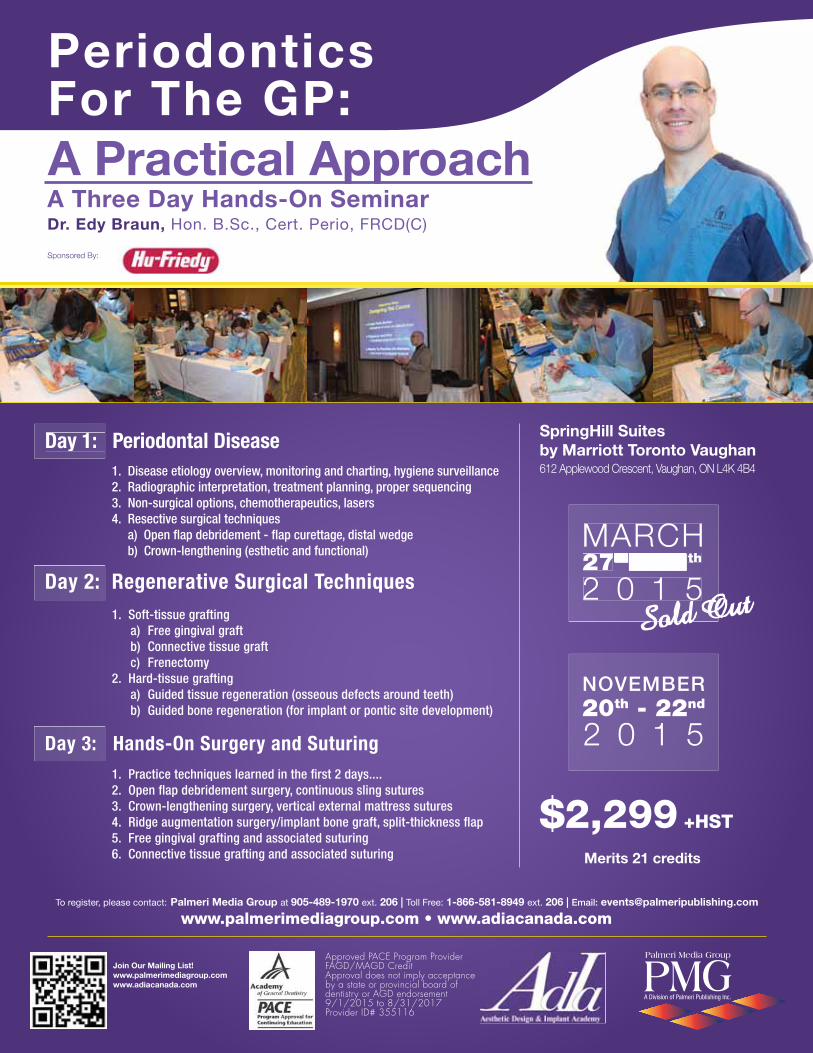
Periodontics For The GP:A Practical ApproachDr. Edy Braun, Hon. B.Sc., Cert. Perio, FRCD(C)A Three Day Hands-On Seminar
1. Disease etiology overview, monitoring and charting, hygiene surveillance2. Radiographic interpretation, treatment planning, proper sequencing3. Non-surgical options, chemotherapeutics, lasers4. Resective surgical techniques a) Open fl ap debridement - fl ap curettage, distal wedge b) Crown-lengthening (esthetic and functional)
1. Soft-tissue grafting a) Free gingival graft b) Connective tissue graft c) Frenectomy2. Hard-tissue grafting a) Guided tissue regeneration (osseous defects around teeth) b) Guided bone regeneration (for implant or pontic site development)
1. Practice techniques learned in the fi rst 2 days....2. Open fl ap debridement surgery, continuous sling sutures3. Crown-lengthening surgery, vertical external mattress sutures4. Ridge augmentation surgery/implant bone graft, split-thickness fl ap5. Free gingival grafting and associated suturing6. Connective tissue grafting and associated suturing
Day 1: Periodontal DiseaseDay 1:
Day 2: Regenerative Surgical Techniques
Day 3: Hands-On Surgery and Suturing
Approved PACE Program ProviderFAGD/MAGD CreditApproval does not imply acceptanceby a state or provincial board ofdentistry or AGD endorsement9/1/2015 to 8/31/2017Provider ID# 355116
To register, please contact: Palmeri Media Group at 905-489-1970 ext. 206 | Toll Free: 1-866-581-8949 ext. 206 | Email: [email protected]
SpringHill Suites by Marriott Toronto Vaughan612 Applewood Crescent, Vaughan, ON L4K 4B4
$2,299 +HST
MARCH27th - 29th
2 0 1 5
MARCH27th - 29th
2 0 1 5
NOVEMBER20th - 22nd
2 0 1 5
Sold Out
Merits 21 credits
Join Our Mailing List!www.palmerimediagroup.comwww.adiacanada.com
Sponsored By:

SPECTRUM DAY TORONTO
October 23, 2015
The Largest One-Day Techno-Clinical Event In Canada
To register, please call 905-489-1970 ext. 206/207 or toll free 1-866-581-8949 or email [email protected] / [email protected]
Hilton Toronto/Markham Suites Conference Centre & Spa8500 Warden AvenueMarkham, ON L6G 1A5
LOCATION:
Mark Your
Calendar!
www.spectrum-day.com
Included with your registration:Door Prizes (Limited to the first 50 attendees at the door)Chance to win tons of giveaways (Trips, Books, Prizes and more!)Access to over 65 Exhibitor boothsAccess to all educational lectures (excluding Core 1 Seminars, Dr. Gaum courses)Complimentary cocktails and hors d'oeuvres
Explore 10,000 Square Feet of Dental Innovation!
FREEPARKING(*Parking passes to be provided)
A CHANCE TO WINA TRIP TO THE MIDWINTER MEETING
*Conditions Apply. See An est.
Value of:
CHICAGOIN
$700.00
ADMISSIONWITH ADVANCED REGISTRATION

TIME TOPIC SPEAKER SPONSOR9:30am - 10:30am Screening For Sleep Breathing And Craniofacial Pain In Adults And Children June Williamson Steed, RDH Vector Diagnostics
10:30am - 11:00am Exhibit Hall [Donald Cousens 3] - BREAK
11:00am - 12:00pm Measuring Business Performance Dale TucciTucci Management
Consultants Inc
12:00pm - 1:00pm Exhibit Hall Opens [Donald Cousens 3] - Lunch For Purchase In Foyer
1:00pm - 2:00pm Whitening Essentials Beth Ryerse, RDH Spa-Dent
2:00pm - 4:00pm Five Behaviours Of A Cohesive Team Sandie BaillargeonDental Office Consulting
Services (DOCS)
SPECTRUM DAY TORONTO2 0 1 5 | C L I N I C A L P R O G R A M S @ A GLANCEFeatured Clinical Program [Room: Primrose]
TIME TOPIC SPEAKER SPONSOR9:00am - 12:00pm Have You Dabbled In Botox® Treatment? Want To Learn More? Dr. Warren Roberts BioResearch
Clinical Program [Room: Butternut / Holly]
WIDS / RDH Program [Room: Evergreen]
TIME TOPIC SPEAKER SPONSOR
9:30am - 11:00amIntroduction To Methodology & Instrumentation For Dental
Sleep Medicine: What Do These Signals Really Mean?Dr. Richard Bonato Braebon
11:00am - 12:00pm Practice Of Dental Medicine: Are You On Board? Dr. Jim Hyland, B.Sc. Oravital
12:00pm - 1:00pm Exhibit Hall Opens [Donald Cousens 3] - Lunch for Purchase In Foyer
1:00pm - 2:00pm Keeping It Simple With Implants Dr. Les KalmanImplant Direct
Kavo Kerr Group
2:00pm - 3:00pm Why Use Lasers In Dentistry?Dr. Stephan Gaines, B.Sc.,
FWCMIDBiolase
5:30pm - 6:00pm
5:30pm - 6:00pm
Exhibit Hall [Donald Cousens 3] - Cocktail Reception - Draw For Prizes
Exhibit Hall [Donald Cousens 3] - Cocktail Reception - Draw For Prizes
TIME TOPIC SPEAKER SPONSOR
9:30am - 10:30am Top Ten Tips To Practice Success Dale TucciTucci Management
Consultants Inc.
10:30am - 11:00am Exhibit Hall [Donald Cousens 3] - BREAK
11:00am - 12:00pm How To Think Like A Periodontist And Enhance Your Every-Day General Dentistry PracticeDr. Edy Braun, Hon. B.Sc.,
Cert. Perio, FRCD(C)Palmeri Media Group
12:00pm - 1:00pm Exhibit Hall Opens [Donald Cousens 3] - Lunch For Purchase In Foyer
1:00pm - 2:00pm Implants: Where To Begin Dr. Allen Aptekar, B.Sc. Implant Solutions
2:00pm - 3:00pm Know And Understand Your Numbers! Lori Margiotta Bookkeeper For Dentists
3:00pm - 4:00pm"Understanding the Bacterial Twitterverse" or "How microbes can immitate Zombies" -
Key Factors to Ensure Your Self-Protection and PreservationDean Swift Micrylium
Young Dentists Program [Room: Elm 2 / Primrose]
5:30pm - 6:00pm Exhibit Hall [Donald Cousens 3] - Cocktail Reception - Draw For Prizes

Canadian Journal of Restorative Dentistry & Prosthodontics/Journal canadien de dentisterie restauratrice et de prosthodontie — Vol. 8, No. 3 — Fall/automne 201542
Dr. Hubert Gaucher,DDS, MScD
Dr. Begüm Akkayan,DDS, PhD
Dental CAD/CAM — CAO/FAO dentaire
CAD/CAM Advances in Prosthodontics
Les progrès de la CAO/FAO en Prosthodontie
Highly acceptable clinical treatments of both fixed (FPD) and removable partial dentures (RPD) may beachieved with computer-aided design (CAD) / computer-aided manufacturing (CAM) technology.1 Laboratoryand in-office desktop and Intra-Oral Scanners (IOS) using 3D denture design software modules, advancedCAM block materials and 3D printing methods are rapidly replacing the conventional methods in dentalprosthesis fabrication. Direct metal sintering is another innovative manufacturing technology where digitalinformation is used to direct a focused laser beam to fuse metal powder in layers to fabricate a prosthesis.Laser milling applications are also becoming an alternative to conventional machine milling technologies.Continuous improvements in CAD/CAM technologies appear to be promising for ideal manufacturingparameters of achieving dental prostheses and it is very likely that wider acceptance of its use in dentistrywill occur in the near future with the motto of ‘adding instead of taking away’ when referring to 3Dprinting. The present literature reviews innovative dental technologies and advanced materials and theirintegration for the fabrication of CAD/CAM dental prostheses.
Abstract
Dental CAD/CAM: Technologies, Components, Systems, Materials Key words
Digital Workflow Options:CAD/CAM technologies have started a new age inrestorative and prosthetic dentistry by means ofstandardized production processes and efficient qualitymanagement. A prosthetic treatment should be durable,aesthetic, accurate and comfortable to fulfill its function.These requirements should be accomplished by anyprosthesis fabrication method. However, as digital dentaltechnology is developing it is constantly providing thepractitioner with innovative methods to practice in thefield of prosthodontics. In contemporary dentistry, theintegration to these digital era technologies is certainlyinevitable.
To date, the exponential increase in the applicationof computer-aided manufacturing in prosthodontics isattributed to continuous systems development andrefinement, greater ability for quality control, parallelmaterial development, and the possibility of virtual
evaluation.1,2 Dental CAD/CAM technology has beenbased on three major production stages. In the firststage, scanners are used to collect data that measurethree-dimensional jaw and tooth structures and transformthem into digital data sets. Then the design of theprosthesis is completed with customized computer aideddesign (CAD) software. In the third stage the data istransferred to a computer-aided manufacturing (CAM)software that controls the production unit. The CAMtechnology is based on two major approaches: thesubtractive and the additive methods. The differencesreside in the processing protocols, materials used, andtheir respective accuracy.
Three different production concepts are available forthe CAD/CAM fabrication in dentistry depending on thelocation of the components of the CAD/CAM systems.2
All components of the CAD/CAM system are located andlimited to the dental surgery in the chairside production

Canadian Journal of Restorative Dentistry & Prosthodontics/Journal canadien de dentisterie restauratrice et de prosthodontie — Vol. 8, No. 3 — Fall/automne 2015 43
Des traitements fort acceptables de prothèses partielles fixes et amovibles peuvent être réalisés à l’aide de la
technologie, y compris la conception assistée par ordinateur et l fabrication assistée par ordinateur (CAO/FAO).
Les balayeurs de table (scanners) en cabinet et en laboratoire dentaire, ou intra-oraux (SIO), utilisant des
modules de conception 3D pour prothèses, des blocs de matériaux avancés pour la FAO, ainsi que la technologie
imprimante 3D, remplacent rapidement les méthodes conventionnelles pour la fabrication des prothèses
dentaires. La fusion directe des métaux est une autre méthode de fabrication innovatrice où l’information
numérisée est utilisée pour diriger un faisceau laser permettant la fusion progressive des particules métalliques
pour fabriquer une prothèse. L’usinage dentaire au moyen d’un laser devient aussi une alternative aux technologies
d’usinage conventionnel. Les améliorations constantes des technologies CAO/FAO permettent d’entrevoir
l’obtention de paramètres optimaux en matière de fabrication de prothèses. Il est donc très probable que
l’acceptation accrue de cette technologie se répande à court terme en Dentisterie avec la devise «ajoutez au lieu
de soustraire» lorsqu’il s’agit de l’imprimante 3D. La présente littérature revoit les technologies innovatrices et
les matériaux avancés ainsi que leur intégration pour la réalisation de prothèses au moyen de la CAO/FAO.
Résumé
CAO/FAO dentaire: Technologies, Composantes, Systèmes, Matériaux Mots-clés
Dental CAD/CAM — CAO/FAO dentaire
concept also referred to as a “closed architecturesystem”. Historically, chairside production system wasthe first CAD/CAM system in dentistry and is currentlyavailable in its third product generation. A conventionalimpression is replaced by an intra-oral camera. Therestorations are fabricated at chairside without alaboratory procedure. This saves time and offers thepatient indirectly fabricated restorations at oneappointment. At present, only two “closed architecturesystems” are available. The Cerec IV system (Sirona,Bensheim, Germany) offers this possibility as well themore recently commercialized Planscan E4D (Planmeca,Helsinki, Finland) driven system.
The alternative digital workflow concept is thelaboratory production in which the traditional workingsequence is followed by the dentist and the laboratory.Both of the above “closed architecture systems” canalso be adapted to become what is commonly referredto as the “open architecture systems” described asfollows: In this scenatio, the dentist sends the digitalimpression by Internet to the laboratory where a mastercast is fabricated. The remaining CAD/CAM productionsteps are carried out entirely in the laboratory.4,5 Avariation of this digital laboratory workflow consists ofutilizing centralized production milling centers, forinstance the 3DCore Centres, for the CAM portion of theworkflow with the local laboratory completing therestorations using porcelain veneering in-house. In theevent the dentist is sending a conventional elastomericimpression to the local laboratory, these impressionscan be scanned either in the dental office or in thelaboratory, obtaining a digital model that can also beused to complete the CAD portion of the workflow before
forwarding these digital files to a CAM production centervia the internet. In summary, the produced data sets areforwarded by the local dental laboratory to the productioncenter for the restorations to be produced with aCAD/CAM device. The benefit of outsourcing CAMproduction is to be found in the small investmentrequirement, since only the digitalization tool (desktopscanner) and CAD software have to be purchased.6
1. ScannersIn the area of dental impressions, conventionalimpression taking methods are now progressivily giving way to digital impression techniques. Clinicianshave various options available with respect to takingimpressions. These systems are used to take a digitalimpression of the teeth instead of conventional chairsidematerials. The digital workflow and impressions havesignificantly helped the dentist and labs to establish a more efficient process and to avoid the wholeconventional impression taking experience for the patient.
Optical scanners are designed on the principle oftriangulation. The source of light and the receptor unitare in a definite angle in relationship to one another.Through this angle the computer can calculate a three-dimensional data set from the image on the receptorunit. White light projections or a laser beam can serveas a source of illumination.
Scanner, computer aided design software,computer aided manufacturing technology andmaterials for CAD/CAM processing are the basiccomponents of CAD/CAM systems.

Canadian Journal of Restorative Dentistry & Prosthodontics/Journal canadien de dentisterie restauratrice et de prosthodontie — Vol. 8, No. 3 — Fall/automne 201544
Redcam and Bluecam are the first two optical scanners of
the Cerec system. Development of Bluecam followed by Redcam,
included significant modifications such as the use of blue instead
of infrared light and a new lens configuration. However, the
Omnicam is the latest upgrade of the Cerec system. It is
differentiated from its predecessors in that it captures data
points on a three-dimensional plane with a live video image,
thus the problem of blurred pictures is eliminated. All the previous
Cerec systems captured data with static images requiring a
contrast medium and stitching. Omnicam is a powderless system
meaning that no powder has to be placed on the tooth
preparation. One of the limitations of the optical scanners of the
Cerec system were the so called ‘distal shadow phenomenon’.
The optical scanner projects a beam at a specific angle with
regard to the recording axis and creates a shadow distally to the
illuminated object; this shadow appears only at the distal surface.
Since the Omnicam uses a 3D video image capture, this parallax
error problem has all been eliminated. The camera technology
has improved so much that the unit is smaller, more ergonomic
and faster. It only takes a couple of seconds to take an image.8,9
Lava Scan ST (3M Espe), Trios (3Shape), Everest Scan (KaVo),
Es1 (Etkon) and iTero (Cadent) are, among others,10 the currently
available digital impression units with a stand-alone
configuration. The operator scans the prepared arch, the
opposing arch and the bite using the intraoral wand to take a
series of digital images or a digital video. Following the scan,
the clinician confirms that the preparation and the other relevant
areas on the digital model have been accurately recorded, and
that the occlusal clearance is appropriate for the material
selected. Then the data is sent through a wireless router to the
appropriate model facility or laboratory.8,9
Most developed systems offer the unique advantage of being
able to determine the exact occlusal clearance through the
use of a digital colored map. This is a huge advantage over the
traditional physical impression materials, where the exact
amount of occlusal reduction cannot, in some cases, be easily
verified chairside until after the case arrives at the dental
laboratory and is made ready for fabrication of the restoration.
Avoiding the need for a second preparation and impression
appointment due to lack of occlusal clearance has obvious
benefits for both the dentist and the patient.7 However,
caution should be taken by the dentist when using scanners
as undetectable preparation margins or subsections and
irregularities on the surface of the prepared tooth can be
inadequately recognized by many scanners. If sharp incisor and
occlusal edges are left, these details cannot be milled exactly.
Nevertheless, the rapid progress in technology will ensure that
taking the optical impressions will become practical in the clinic
in the near future.2 The stand-alone digital impression units are
well-suited for the clinician who wants to integrate digital
technologies into their practice, but wants to retain the options
and expertise which their dental laboratory can provide.7
2. Computer Assisted Design Software: CADDesign software developments can be traced to the inception
of the digital workflow originating from Prof. Dr. François Duret’s
Henson System (Lyon, France) in the ’80s. Remarkably, dental
technologists from this original team are still very much involved
in the present day CAD developments featured in the Dental
Wings Software (Montréal, Canada). This, in itself, reflects the
high level of professional commitment and perseverance master
technologists have invested in perfecting software interfaces
that today are advantageously replacing conventional dental
prosthetic methods. Dental Laboratories have a choice of CAD
technologies on the market and will voice their respective
preference based on many variants such as: accuracy, number
of features, user friendly interface, training/support and costs.
This remakable evolution and sophistication of design softwares
is also observed in the periodic updates offered by “closed
architecture systems” previously mentioned. All strive to become
more user friendly and intuitive in their applications allowing
all members of the dental team to participate in the use of
integrated chairside systems.
3. Computer Aided Manufacturing Technology: Available Alternatives
3.1 – Subtractive Technologies:3.1.1 Machine Milling:The conventional manufacture of model cast prostheses
represents a lot of work for the dental laboratory. The preparation
of the cast model and the wax modeling based on this procedure
often takes more than one hour and the complete casting
procedure including finishing is laborious and time-consuming.
In comparison to the conventional fabrication methods,
computer-aided manufacturing has the advantage of omitting
multiple error-introducing steps such as impression, waxing,
and casting. Furthermore, since modelling and production are
automated procedures, there is an overall reduction of fabrication
time and cost.1,2,12-15
The computer aided manufacturing (CAM) technology is based
on two major approaches: the subtractive and the additive
methods (Figure 1). The differences reside in the processing
protocols, materials used, and their respective accuracy.
Dental CAD/CAM — CAO/FAO dentaire

Canadian Journal of Restorative Dentistry & Prosthodontics/Journal canadien de dentisterie restauratrice et de prosthodontie — Vol. 8, No. 3 — Fall/automne 2015 45
Subtractive manufacturing method is based on milling the
work piece from a larger blank by a computer numeric controlled
(CNC) machine. The CAM software automatically translates the
CAD model into a tool path for the CNC machine. Due to the
unevenness of the features of dental restorations, the milling
machines combine burs with different shapes and sizes. The
dental CNC machines are composed of multiaxis milling devices
to facilitate the 3D milling of dental work pieces.12,16
The milling burs move in 3-axis milling systems. 180° rotation
of the blank is incorporated within the machine, allowing 3D
milling of the internal and external surfaces, producing
divergence and convergence of the milled surfaces. However,
as the movement is restricted to the milling tool, a large
prosthesis cannot be produced by 3-axis machines.2,12,16 The
4-axis machines allow for blank movements in an additional
axis. This is a useful feature for milling a large blank and
producing a long span framework. Most recently introduced
is the 5-axis machines which enables a rotating path of the
milling tool or the blank. This feature facilitates the production
of complex geometries and smooth external surfaces.17
Manufacturing deficiencies such as porosities and inhomogeneous
consistencies are reduced to a minimum as the restorations
are milled from an industrial grade blank.14
A long list of CAM materials are available for processing by
CAD/CAM devices depending on the production system. Some
milling devices are designed to be used for a wide range of
CAD/CAM blocks; others are specifically designed for the
fabrication of a certain material. The following CAD/CAM
materials are used for the different stages of the fabrication of
dental prostheses. Figure 2 illustrates photos of a variety of
CAD/CAM blocks available on the dental market.
3.1.2 Laser Milling: Laser ablation technologyAs a recently introduced dental milling option, it is described as
a “closed loop 3D process” using a laser beam to remove
material from polymers, ceramics, or glass ceramic blocks.
Dental restorations are thereby created from an open STL file.
Obviously, there is no toll wear or breakage, no cooling fluids
to manage and important reduced operating costs to be had.
The process bypasses the final crystalization steps for glass
ceramics allowing for high resolution precision manufacturing
of thin edges.11
3.2 Additive Manufacturing MethodIn prosthodontics, additive manufacturing methods are creating
entirely new ways of thinking in terms of design, production,
workflow and the products. 3D printing takes the efficiencies of
digital design to the production stage. 3D printing extrudes
material from a heated nozzle that solidifies as soon as it is
deposited on the manufacturing platform. There are a range of
materials that can be used for 3D dental printing. This includes
thermoplastic materials, such as waxes, resins, or fused
filament. Alternatively, liquid ceramic or resin materials with a
binder can also be printed.67-69 By combining oral scanning, CAD
and 3D printing, dental labs can accurately and rapidly produce
dental models, facial prosthesis patterns, crowns, bridges and
even polymer based RPD’s.
Additive manufacturing is currently an exponentially growing
fabrication method and will most likely be used more frequently
in dentistry in the future as its accuracy and range of applications
develop. Additive methods have the advantage of producing
large work pieces with significant surface variation and
competitive accuracy that makes them suitable also for
manufacturing removable partial denture frameworks. Currently
there are 3 main additive manufacturing methods available for
use on the dental market; these are selective laser melting,
stereolithography and 3D printing. Regardless of the method,
all share the following features that distinguish them from
Dental CAD/CAM — CAO/FAO dentaire
Figure 1 — Schematic representation of Computer Aided ManufacturingTechnologies used for the fabrication of prosthesis.

Canadian Journal of Restorative Dentistry & Prosthodontics/Journal canadien de dentisterie restauratrice et de prosthodontie — Vol. 8, No. 3 — Fall/automne 201546
subtractive manufacturing: Incremental vertical object build-up,
no material wastage, large objects produced, passive production-
no force application and fine detail production.10,11,66,69
Selective laser sintering/melting produces a 3D model by
laser sintering or melting a powder, layer by layer using a laser
beam. The laser beam locally raises the temperature close to
the melting point of the metal particle, to avoid complete melting.
After each new powder layer application, the laser melting
process is repeated until the 3D object is completed. Selective
Laser melting is the only additive manufacturing method that is
available to produce RPD metal frameworks.70 Han et al.71
introduced a new CAD/CAM software design specifically for RPD
framework design and fabrication. Maxillary and mandibular
frameworks were designed, manufactured and fitted to dental
casts. Metal frameworks were produced using selective laser
melting manufacturing method. The frameworks were evaluated
by visual inspection and pressure evaluation for movement and
were reported as having satisfactory fit on the casts.
The CAD frameworks were also evaluated clinically in some
other clinical reports. The treatment plan for a 75-year-old woman
included the use of CAD/CAM technology to fabricate the metal
RPD framework in a clinical case report.14 Patient’s master casts
were scanned and a virtual design was created using the
software. The rapid prototype was directly manufactured from
chromium cobalt using selective laser melting technology. The
framework was fitted to the patient. The accuracy and quality of
fit was deemed to be clinically acceptable. Others21 also reported
on the fit of a CAD/CAM framework on a patient. CAD/CAM and
rapid prototyping technology were used to develop a pattern
that was cast and finished using conventional methods. After
trial fitting, the framework was judged to be clinically acceptable.
Laser melting produced significantly improved hardness
values for PFM Co-Cr dental alloys compared to casting or milling
technologies.74
Stereolithography is another additive manufacturing method that
produces solid layers using a concentrated ultraviolet (UV) light
beam moving on a curable liquid polymer pool. Upon the
polymerization of the first layer, the next layer is cured and the
process is repeated until the whole workpiece is completed. The
workpiece is then rinsed with a solvent and placed in an UV oven
to thoroughly cure the resin. The applications of stereolithography
in dentistry are production of surgical templates, facial prosthesis
patterns, occlusal splints, burnout resin patterns.12,16 Recently,
Wu et al.15 produced a 3D CAD wax framework by RP using the
stereolithography technique. The wax framework was cast with
Co-Cr alloy and the fit was reported satisfactory.
Milling accuracy is also dictated by materials’ properties
when subtractive method is utilized. High material hardness
means low machinability and more considerable involved
forces.21 Thus, the substractive system limits the milling process
of large sized workpieces like the frameworks of RPD’s. For this
reason the use of advanced polymers and additive method is a
successful alternative for the production of RPD’s compared to
subtractive manufacturing method.
3.2.1 – 3D Printing and the Production of Digitally Designed RPDsThere are a number of companies on the market integrated
with ‘Additive Manufacturing Methods’ that can be used for
different stages of removable partial dentures (RPD) fabrication
(Table 1). The initial production of digitally designed removable
denture were printed in wax and cast using conventional
methods.75 ‘Visijet’ material of 3D Solutions74 (TX, USA) and
‘True Cast’ material of Stratasys77 (MN, USA) produce digitally
fabricated waxup frameworks of RPD’s using their company’s
corresponding 3D Printing technologies. Stratasys also
introduced a photopolymer material ‘Polyjet’ to the dental market
to be used for RPD framework ready for casting. Proto300078
Dental CAD/CAM — CAO/FAO dentaire
Company Name Manufacturing Method Dental Materials Used Manufactured Dental Product/Products
3D Solutions (TX, USA) 3D Printing Visijet Wax up for RPD framework
DWS – DFAB (Vicenza, Italy)Desktop & Chairside
3D PrintingTemporis Light curable material
Envision TEC (Gladbeck, Germany) 3D Printing E Partial (EC 1000) Photopolymer RPD framework ready for casting
EOS GmbH (München, Germany) Direct Metal Laser Sintering EOS Cobaltchrome Metal framework of partial denture
Stratasys / Objet (MN, USA) 3D Printing True Cast PolyjetWax up for RPD framework
Photopolymer RPD framework ready for casting
Table 1 : Companies integrated with ‘Additive Manufacturing Methods’ that can be used for different stages of removable partial dentures (RPD) fabrication.

Canadian Journal of Restorative Dentistry & Prosthodontics/Journal canadien de dentisterie restauratrice et de prosthodontie — Vol. 8, No. 3 — Fall/automne 2015 47
is an Ontario, Canada based
3D Engineering company with
numerous partnerships that
integrate CAD-RPD software,
materials, milling, 3D Printers and
a strategic alliance with the
emerging worldwide 3DCore
CAD/CAM manufacturing centres.
The options of denture fabrication
has therefore been taken to the
next level with open-architecture
scanners with powerful new denture
design software capabilities and
with the introduction of new
materials to be used, to create a
fully digital, machine-produced
partial-denture product.
Alternatively, EOS GmbH79
(München, Germany) uses one of
the innovative additive manufactur -
ing processes. The freedom of
design of the modeling software
Dental CAD/CAM — CAO/FAO dentaire
Figure 2 — Some examples of the CAD/CAM blocks available on the dental market. 1. Glass based ceramic 2. Infiltration ceramic 3A. Bilayered zirconia 3B. Monolithic zirconia 4. Glass based leucite reinforced 5. Composite resin-temporary restoration 6. Metal blocks
7. Advanced polymer 8. Glass based lithium disilicate reinforced.
Canadian Journal of Restorative Dentistry & Prosthodontics/Journal canadien de dentisterie restauratrice et de prosthodontie — Vol. 8, No. 3 — Fall/automne 201548
can be used to full capacity and further implemented for
manufacturing. The technique uses 3D data source directly from
the patient’s mouth as the basis for the manufacturing process.
Software functions of the new CAMbridge 2012 RPD module
from 3Shape (Copenhagen, Denmark) is used. The resulting
virtual model is sliced and the digital information is utilized to
direct a focused laser beam to fuse metal powder in successive
layers until the prosthesis is complete. The technique has been
named as the Direct Metal Laser-Sintering (DMLS) process by
the company. This is a selective laser melting method. EOS
Cobaltchrome is the powder metal certified for this procedure.
It is a fine grained, biocompatible material free from nickel,
beryllium and cadmium producing a metal framework with a
fine-grain structure. It also allows the production of RPD’s to be
comparatively low cost.
The production of RPD’s via DMLS is very accurate, making
laboratory work easier. The denture is produced to an accuracy
of ±20 µm and is of a consistently high quality. The digital
production method saves a lot of time; the design for the RPD
is ready in around 15 minutes with only a few mouse clicks. As
a result, high-strength, rigid and filigree geometries can be
produced and casting errors can be eliminated.79
Also the EnvisionTEC Company80 (Gladbeck, Germany),
‘Perfactory 4 DDP’ 3D printers deliver labor and material cost
reduction as well as higher clinical quality and consistency
throughout the manufacturing process. One of the materials
they use is named as ‘E-Partial’. It is a photopolymer developed
for the creation of removable partial dentures. E-Partial exhibits
superior stiffness, with a tensile strength of 57 MPa, it is
resistant to higher temperatures. E-Partial material maintains
flexural strength to ensure clasp flex without breakage. The
stiffness of E-Partial allows for production of a very hard retention
grid and super tight thin clasps with the perfect fit every time
once casted from metal alloy.
In addition, DWS (Digital Wax Systems)/DFAB81 (Vicenza, Italy)
is another patented 3D Additive Manufacturing system. It is a
company developing stereolithographic technology. It is an
ultrafast, desktop size additive manufacturing system fully
compatible with the majority of intraoral scanners and open
dental CAD/CAM solutions. The most specific feature of the
system is that it has 2 versions; desktop and chairside and it is
the only chairside 3D Printer available on the market. The
compact form and the affordable price make DFAB desktop is
the ideal rapid manufacturing solution for dental clinics. High
quality, long-term temporary crown and bridge restorations can
be fabricated form the light curable ‘Temporis’ material during a
single appointment.
Of interest, AREVO Labs29 (CA, USA) is another company with
the patent pending 3D Printing technology combining the benefits
of printing complex geometries with reinforced materials that
have excellent resistance to high temperatures and chemicals.
The 3D printed parts optimized for superior properties result in
lighter but stronger parts with unmatched mechanical properties.
Although the company’s 3D printing process is not yet available
to the dental market, the company uses two advanced polymer
materials; KetaSpire PEEK (Solvay, Brussels) and AvaSpire PAEK
(Solvay, Brussels).
4. CAD/CAM Materials Used in Prosthodontics
4.1 Metal blocksChrome cobalt and titanium alloys are processed using dental
milling devices. Milling is anticipated to eliminate waxing,
investing, and casting of prostheses which are assumed to
improve the overall precision. However, studies have shown that
the restorations produced by conventional methods exhibited
better fit than milled restorations. Tan et al.18 found that milled
titanium crowns exhibited a vertical gap of 79.4 µm, while cast
noble metal crowns had vertical gap of 23.9 µm. In a similar
study, Han et al.19 compared the marginal accuracy of milled
titanium and cast titanium crowns. The cast titanium samples
displayed 52-76 µm marginal opening compared to CAD/CAM
titanium samples of 60-80 µm.
Procera Cobalt Chromium (Nobel Biocare, Göteborg,
Switzerland) Ceramill NP M (Girrbach, Pforzheim, Germany) and
Kera C-Disc (Eisenbacher, Main, Germany) are among the
chromium cobalt based CAM blocks whereas Procera Titanium
(Nobel Biocare, Göteborg, Switzerland) Ceramill Ti (Girrbach,
Pforzheim, Germany) and Kera Ti5-Disc ( Eisenbacher, Main,
Germany) are titanium blocks. Considering the difficulty of milling
in metal blocks and causing more excessive milling bur wear
and tool failure,20,21 metal blocks that are initially manufactured
in a partially sintered state were later developed. The
restorations are enlarged by the inLab CAD/CAM system; they
are individually processed and finally densely sintered when
partially sintered metal blocks are used. InCoris CC block
(Sirona, Bensheim, Germany) Ceramill Sintron (Girrbach,
Pforzheim, Germany) and Crypton blank (Degudent, Frankfurt,
Germany) are some of the examples in this group.
4.2 Composite Resin – Temporary Restoration BlocksResidue-free resin blocks can be milled instead of wax pattern
fabrication to be used for the lost wax technique in the casting
technology. The conventional wax-up methods require
Dental CAD/CAM — CAO/FAO dentaire

Canadian Journal of Restorative Dentistry & Prosthodontics/Journal canadien de dentisterie restauratrice et de prosthodontie — Vol. 8, No. 3 — Fall/automne 2015 49
considerable human intervention and manipulation of materials
that also exhibit dimensional shrinkage and/or expansion.
This can lead to increased processing errors; however the
implementation of automated design and fabrication techniques
would facilitate the production of more accurate reliable
prosthesis.12,22
Highly cross-linked microfilled acrylic polymer blocks are also
used for long-term temporary full and partial crowns and FDPs.
Vita CAD-Temp (Vita, Bad Sackingen, Germany), is offered as
a monocolor block that comes in four shades or a multicolor
form with four shade layers for more esthetic cases. Telio CAD
(Ivoclar Vivadent AG, Schaan, Liechtenstein) is another millable
cross-linked polymethyl methacrylate block. The block is a part
of the Telio system, which includes a self-curing composite,
desensitizer, and cement. It is available in five shades. Both
temporary materials are recommended by the manufacturers
for up to 1 year of clinical service.23
Fiber reinforced resin blocks developed by RTD company
(RTD, St.Egreve, France) are one of the recent developments in
CAM technology. These blocks are composed of 75% multiaxial
glass fiber and 25% epoxy resin matrix by weight. They are
indicated to be used for long term temporary crown, bridge and
post-core restoration alternatives. The manufacturer reports
biomechanical properties compatible with dentin, aesthetic
shades and low costs compared to ceramic blocks as the
benefits of their product.24
Everest C-Temp (Kavo, Biberach, Germany), Cercon base cast
(DeguDent, Hanau, Germany), ZENO PMMA (Wieland Dental,
Pforzheim, Germany) and ArtBlock Temp (Merz Dental,
Lütjenburg, Germany) are the other CAM resin blocks used for
temporary restorations.
4.3 Composite Resin – Permanent Restoration Blocks The industrial fabrication of the composite block potentially
offers a significant clinical advantage to what may be expected
of a typical composite that is placed incrementally and cured
intraorally. Paradigm MZ100 (3M ESPE) composite resin blocks
have been available for over ten years as permanent restoration
blocks.25 Fasbinder26 reveals a much different clinical result for
prepolymerized blocks of composite that are used by chairside
CAD/CAM systems in comparison to what many dentists would
believe to be the outcome for compositionally similar materials
used in a traditional restorative technique.
4.4 Advanced Polymer Blocks Poly ether ether keton (PEEK) (Juvora Dental Disc, Juvora
Dental Ltd., Lancashire, UK) is a polymer from the group
polyaryletherketone (PAEK) which is a relatively new family of
high-temperature thermoplastic polymer, consisting of aromatic
backbone and molecular chain, interconnected by ketone and
ether functional groups.27 PEEK material is an aromatic, semi-
crystalline polymer that has been in use as a material for
implants in different fields such as orthopedics and oral and
maxillofacial surgery for more than ten years already. The
thermoplastic production procedure ensures that virtually no
residual monomers are released. Due to its semi-crystalline
character, the material remains mechanically stable even in an
aggressive environment like the oral cavity.28 The material is
presented for the use of dental market with the motto of ‘Let us
define and customize not only the geometry, but also the density,
stiffness and elasticity or modulus of the final product as
desired’.29
This material has been used successfully for production of
RPD’s using subtractive manufacturing methods. PEEK blanks30
(Juvora Dental Disc, Juvora Dental Ltd., Lancashire, UK) are
available for CAD/CAM production since September 2012. The
blanks are sourced from biomaterials of the company Invibio
Biomaterials Solutions (Invibio Ltd, Lancashire, UK) that have
been successfully used in medical technology for more than a
decade. In a clinical case reported by Siewert28, 12 units, for a
4 implants-supporting framework was milled from PEEK material
(Juvora Dental Disc) in one-piece. The clinician described the
PEEK material suitable as a framework material due to its
superior physical features for the fabrication of removable
dentures and concluded that its use combined with the digital
workflow of CAD/CAM processing will produce efficient and
precise outcomes. In another case report by Siewert and
Rieger31, milled PEEK blanks were used as the framework
material for both the connecting bar in the maxilla and for the
occlusal screw-retained bridge in the mandible. CAD/CAM
assisted fabrications of the prostheses were performed and
the design data were transmitted to the milling machine. Milling
of the frameworks was accomplished from PEEK blanks. The
clinicians reported advantages of the treatment modality with
PEEK material as avoidance of a galvanic element by using a
non-conducting material, the high biocompatibility with the soft
tissue when in contact with pure, unfilled PEEK, shock-absorbing
properties to protect the implants, optimum elasticity and high
patient comfort due to the low prosthesis weight.
It is specified that the melting point of this material is high for
polymers (340°C) and allows repeated sterilization of frameworks.
The modulus of elasticity is 165 MPa, the elongation at break
amounts to 40%, meaning that a sample breaks when it is
stretched by 40% of its initial length.28 The consequence is that
Dental CAD/CAM — CAO/FAO dentaire

Canadian Journal of Restorative Dentistry & Prosthodontics/Journal canadien de dentisterie restauratrice et de prosthodontie — Vol. 8, No. 3 — Fall/automne 201550
the material is flexible under load without fracturing. In a study by
Tannous et al.32 retentive force of clasps made from thermoplastic
resins of PEEK were evaluated and they have concluded that
the retention of adequately designed resin clasps were within
the acceptable amount of retention after 15,000 joining and
separating cycles corresponding to 10 years of simulated use.
A PEEK disc was used for the fabrication of a telescopic
denture in a clinical case by Aumüller and Eiber-Fäth.33 The
primary telescope was made from zirconium oxide ceramic using
CAD/CAM. An impression was taken from the primary telescope
and then the master model was prepared and scanned and the
secondary construction was designed using CAD software. The
secondary structure was milled directly from the PEEK disc and
after milling it was reported that the fit was very good. The try-
in was found very satisfactory and a good and even friction was
provided. The authors reported that the patient was pleased
about the light weight and the simple, smooth and tilt-free
insertion of the prosthesis.
Due to the elasticity of the PEEK material that is similar to
that of cancellous bone, chewing forces acting on the restoration
are compensated. In contemporary applications, it is also
possible to use the PEEK material by additive manufacturing
methods of 3D printing for RPD’s. The benefits resulting from
the additive manufacturing methods and 3D printing can be
combined with those of the digital workflow, such as efficiency
and precision when utilizing PEEK material for the fabrication of
RPD’s. It is obvious that the high-performance polymers have a
great future potential with regard to their use as framework
materials for RPD’S.
BioHPP34 (High Performance Polymer) is another brand name
material which is also based on PEEK, developed by Bredent
(Senden, Germany). It is composed of equally distributed ceramic
filler of grain size of 0,3 to 0,5 µm in the partially crystalline
polymer matrix. The fine granularity of the filler is the basis for
the extremely good polishing properties. It is described as a
material particularly well suited for producing prosthetic
restorations; fixed dental prosthesis and telescopic works.
4.5 Nano-Ceramic BlocksNano-ceramic blocks are based on the integration of nano-
technology and ceramics. These blocks are an alternative,
monolithic esthetic material that has been recently introduced
in a block form for CAD/CAM fabrication using both chairside
and laboratory systems. A stated advantage of the material is
to offer easier clinical finishing and polishing, without the need
for a porcelain oven, with the strength, surface gloss and finish
retention similar to ceramic materials.23
Lava Ultimate (3M ESPE, Seefeld, Germany) and Vita Enamic
(Vita, Bad Sackingen, Germany) are two examples for this
category. The blocks consist of 20nm silica and 4 to 11 nm
zirconia particles agglomerated in clusters and embedded in a
highly cross-linked polymer matrix. With a flexural strength of
200 MPa, the nano-ceramic block has a higher initial strength
than feldspathic and leucite reinforced porcelain blocks,
as well as veneering porcelains for PFM crowns. The fracture
toughness of the nano-ceramic material is reported by the
manufacturer to be greater than feldspathic materials and direct
composites while being less brittle than feldspathic glass-
ceramics, and therefore less prone to fracture during try-in and
function.23,35,36
4.6. Glass based (Feldspathic) Ceramic BlocksGlass based systems are made from materials that contain
mainly silicon dioxide also known as silica. These blocks are
manufactured for the production of inlays, onlays, veneers,
partial crowns and full crowns. In addition to monochromatic
blocks, various manufacturers offer polychromatic blanks with
multicoloured layers for the purpose of full anatomical crowns.2
Some of the monochrome silica based ceramic examples are
VITABLOCS Mark II, (Vita, Bad Sackingen, Germany) , IPS e.max
CAD (Ivoclar Vivadent, Schaan, Liechtenstein) and Cerec Blocs
(Sirona, Bensheim, Germany).
CEREC Blocs C PC is a widely used example for polychromatic
silica based blocks optimized for the use in crowns with their
three different layers based on varying degrees of color
saturation they can be optimally adapted to characteristic color
gradients in restorations with respect to translucency and
intensity. No additional glazing is required, making very fast, full
ceramic chairside treatment possible and they are also easily
polished.8 VITABLOCS Triluxe/ Forte, VITABLOCS Realife (Vita,
Bad Sackingen, Germany) and IPS Empress CAD Multi (Ivoclar
Vivadent, Schaan, Liechtenstein) are the other polychromatic
CAM block manufacturers.37
4.7 Glass Based Blocks Reinforced by FillersThese CAM blocks contain varying amounts of different types of
crystals added to the glass composition. The primary crystal
types added as fillers are leucite and lithium-disilicate.38
4.7.1 Glass Based Leucite Reinforced Blocks The two leucite-reinforced glass ceramic materials are IPS
Empress CAD (Ivoclar Vivadent AG, Schaan, Liechtenstein) and
Paradigm C Blocks (3M Espe, Seefeld, Germany). The current
IPS Empress CAD blocks consist of 35% to 45% leucite-reinforced
Dental CAD/CAM — CAO/FAO dentaire

Canadian Journal of Restorative Dentistry & Prosthodontics/Journal canadien de dentisterie restauratrice et de prosthodontie — Vol. 8, No. 3 — Fall/automne 2015 51
glass ceramic with a fine particle size of 1 µm to 5 µm. The
blocks are available in nine shades in either a high translucency
or low translucency version. It is offered in a multicolor block
form.39 Paradigm C blocks consist of 30% leucite-reinforced glass
ceramic. The block is a radiopaque ceramic; however it is not
available in a polychromatic composition.40
4.7.2 Glass Based Lithium-disilicate Reinforced BlocksIPS e.max CAD blocks (Ivoclar Vivadent AG, Schaan,
Liechtenstein), also named as high-strength ceramics, was
introduced in 2006 as a lithium-disilicate CAD/CAM material
with a flexural strength of 360 MPa to 400 MPa, which is two to
three times the flexural strength of the glass ceramics.41-43
Blocks consist of 0.2 µm to 1 µm lithium meta-silicate crystals
with approximately 40% crystals by volume. It has gained
popularity for use as a monolithic restoration in chairside
CAD/CAM systems due to the enhanced strength.23
4.8 Infiltration ceramic blocksMilling blocks of infiltration ceramics are processed in porous,
chalky condition and then infiltrated with lanthanum glass. All
blanks for infiltration ceramics originate from the Vita In-Ceram
system (Vita) and are offered in three variations as In Ceram
Alumina (Vita, Bad Sackingen, Germany), In Ceram Spinell (Vita,
Bad Sackingen, Germany) and In-ceram Zirconia (Vita, Bad
Sackingen, Germany).2
4.9 Oxide High performance Ceramic BlocksAluminum oxide and zirconium oxide are also offered as blocks
for CAD/CAM technology.
4.9.1 Aluminum Oxide (Al2O3) BlocksThese blocks are in a pre-sintered phase and are then sintered
at a temperature of 1520°C. They are indicated as crown copings
in the anterior and posterior area, primary crowns and three-
unit anterior FPD frameworks.
InCoris AL (Sirona, Bensheim, Germany), In-ceram AL Block
(Vita, Bad Sackingen, Germany) and Procera AllCeram (Nobel
Biocare, Göteborg, Switzerland) are the examples for milled
aluminum oxide blocks.
Dental CAD/CAM — CAO/FAO dentaire
Canadian Journal of Restorative Dentistry & Prosthodontics/Journal canadien de dentisterie restauratrice et de prosthodontie — Vol. 8, No. 3 — Fall/automne 201552
4.9.2 Yttrium Stabilized Zirconium Oxide (ZrO2, Y-TZP)BlocksFirst introduced to dental clinics in the late 1990s, yttria-
stabilized tetragonal zirconia polycrystal (Y-TZP) has been
used alternative for metal frameworks of FDP’s due to its
biocompatibility and more favorable esthetics. In addition,
Y-TZP exhibits significantly higher flexural strength and toughness
than earlier known dental ceramics.44-46
Crystal phase transformations build up detrimental internal
stresses within the ZrO2 in the temperature range between
sintering and room temperature, thus making it unsuitable for
construction purposes. However, the addition of Y2O3 results in
a stabilizing tetragonal phase at room temperature which is
considerably more stable, tougher, and stronger. If a crack occurs
in Y-TZP material, the surrounding tetragonal crystals turn into
a monoclinic structure, resulting in a local volume increase in
the crack tip area, preventing further crack propagation.44,46
Y-TZP framework processing can take place in two different
density stages for dental use. In the first system, frameworks
with final dimensions can be milled directly from fully sintered
dense ceramic blocks. This system has the advantage of a
superior fit, because no shrinkage is involved, but has the
disadvantage of inferior machining associated with wear of the
tool.47 It is also time consuming and the surface temperature
rise during the long milling process will result with phase
transformation to monoclinic phase which exacerbate surface
microcracking and low thermal degradation.48,49 In the second
system, frameworks with enlarged dimensions can be milled
from partially-sintered blocks, followed by post-sintering at high
temperature to obtain a framework with final dimensions and
sufficient strength.47 The sintering procedure will cause about
25-30% shrinkage of the workpiece.48 This approach has the
advantages of quicker milling, reduce cutting forces, increase
the tool life, better surface quality, and no moisture absorption
of the zirconia blanks.48-50 This system is currently popular for
fabricating zirconia frameworks using the main CAD/CAM
systems available in the worldwide dental market.
Generally, a zirconia ceramic crown consists of a zirconia
core in the lower part which is further layered with esthetic
veneering ceramics to obtain the required esthetics.51 Many
factors must be considered during the fabrication of bi-layered
all-ceramic restoration such as design of the framework,
differences in thermal expansion coefficients, layering technique
and thickness of the core/veneering ceramic, and the expected
functional loads. Clinical reports of failed bilayered zirconia-
based restorations due to low bond strengths of veneering
porcelain to zirconia are commonly reported in the literature
that result in premature fractures, cracks and as chip-off
fractures.52-54 Guess et al53. reported the shear bond strength
of porcelain to metal as 27.6 MPa, whereas porcelain to zirconia
was 9.4–11.5 MPa. Rocha et al55. reported that an incomplete
bond between the veneer and zirconia framework increased the
stress in the ceramic veneer and induced veneer failure.
InCoris ZI ceramics56 (Sirona Dental Systems, Bensheim,
Germany) constitute blocks compromised of zirconium oxide
ceramics. The manufacturer reports their chemical composition
as ZrO2+HfO2+Y2O3>99%, Al2O3<0.5%, other oxides<0.5%.The
blocks are tinted in five increasing color intensity. Therefore it
is not necessary to carry out subsequent coloring using a
submersion solution or liners. A minimum wall thickness of
0.5 mm is recommended as a circular wall thickness in single
crowns. They have a fracture toughness of 5.9 MPa and their
bending strength is reported to be 1100>MPa by the
manufacturer.
Many other millable standard zirconia materials are available
such as Lava Frame (3M/ESPE, Seefeld, Germany), Cercon Base
(Degudent, Hanau-Wolfgang, Germany), Procera Zirconia (Nobel
Biocare, Göteborg, Sweden), IPS e.max ZIRCAD (Ivoclar Vivadent,
Schaan, Liechtenstein), DC-Zirkon, Precident DCS (DCS, Allschwil,
Switzerland), Everest 2S-ZH (Kavo Dental GmbH, Biberach,
Germany), and Prettau (Zirkonzahn GmbH, Bruneck, Italy).
Bilayered zirconia frameworks are usually veneered with the
layering technique. Nano-fluorapatite glass-ceramic (e.max
Ceram, Ivoclar Vivadent, Schaan, Liechtenstein) is used as the
veneering material for layering technique on ZrO2 blocks (IPS
e.max ZirCAD) of the Ivoclar Vivadent company. Press-on
technique was used in an attempt to improve the bonding
strength compared to the conventional layering technique.
Fluorapatite glass-ceramic pressable blocks (e.max ZirPress)
are used for this purpose as an alternative technique.57
The progress continued with the development of IPS e.max
CAD-on technique (Ivovlar Vivadent) as an alternative to both
layering and press-on technique. The CAD-on technique combined
the advantages of lithium disilicate glass ceramic (IPS e.max
CAD, Ivoclar Vivadent) with those of ZrO2 (IPS e.max ZirCAD,
Ivoclar Vivadent) in an innovative way and allowed single crowns
and up to 4-unit posterior bridges with outstanding strength to
be fabricated. An innovative fusion glass-ceramic; IPS e.max
CAD Crystall./Connect (Ivoclar Vivadent) is used for achieving
homogeneous glass-ceramic bond between two separately milled
structures; the ZrO2 framework and the veneering lithium
disilicate glass ceramic parts. The optimally coordinated
CAD-On technique allows a convenient fusion of the IPS e.max
ZirCAD framework and the IPS e.max CAD veneering structure.57
Dental CAD/CAM — CAO/FAO dentaire

SPECTRUMDAY TORONTOThe Largest One-Day Techno-Clinical Event in Canada
CATEGORY 1CORE COURSES2 @
Adverse OutcomesAdverse outcomes take many forms, from serious morbidity following treatment to unmet patient expectations in cosmetic procedures. They know no boundaries, and are common to all aspects specialities and modalities of dental treatment. The lecture will begin by defi ning adverse outcomes with clinical examples, and move into the prevention and management strategies. The intent is to minimize the impact of these events to both the practitioner and patient. The seed for an adverse event or outcome may be present even prior to the patient presenting themselves in your offi ce and can occur during or after treatment. The presentation will highlight informed consent, post-treatment management communication, and documentation strategies. The requirements under the Regulated Health Professions Act “RHPA”, prevailing court jurisprudence will be discussed, as will the appropriate use of the Apology Act. These adverse events can lead to patient loss, RCDSO complains and lawsuits. Further, the new paradigm, one of “shared decision-making”, “informed consent” and “informed refusal” will form a part of this session. The necessity of appropriate follow up care, referral and disclosure will be highlighted. Dentists should become well versed in these principles in order to survive in today’s increasingly diffi cult practice environment.
Upon completion of this session the participants will be better able to:Understand the need for appropriate communication strategiesBecome aware of the perils of using “Grey Market” dental materials and unlicensed or offshore dental laboratoriesKnow the principles of informed consent and informed refusalUnderstanding why documentation and disclosure are requiredRealize why appropriate and available after-hours care and available will resolve many patient issues with post treatment problemsEffectively appropriate referral strategies to minimize their riskDevelop pre-emptive and reactive solutions to most adverse events
New Approaches In Management Of Endodontic Pain – Making Sense Of The EvidenceThis evidence-based lecture presents a broad overview of contemporary literature, clinical and scientifi c experience in acute and chronic endodontic pain management. Comprehensive understanding of different pain mechanisms provides an ultimate way for managing dental pain emergencies and post-operative conditions. The latest information on pharmacological and technological approaches will be provided to help clinicians with successful management of different types of endodontic emergencies. What is the best time for treatment of irreversible pulpitis? Can we predict which patients are more likely to experience pain after an endodontic therapy? What doesn’t work for post-op pain? What are genetic determinants of pain? What is a personalized medicine concept and how to apply it in dentistry?
At the conclusion, participants should be able to:Discuss different pain mechanisms.Discuss the genetic determinants of pain.Apply a personalized medicine concept in dentistry.
October 23, 2015 At Hilton Toronto/Markham Suites Conference Centre & Spa
8500 Warden Avenue, Markham, ON L6G 1A5 Room: Markham Ballroom A
Dr. Lionel Lenkinski, Cert Endo
Pavel Cherkas, DMD, PhD, FRCD(C)
8:30AM - 11:30AM
1:00PM - 4:00PM
Dr. Bruce Glazer, BScD, Dip. Prosth
Dr. Ruslan Dorfman, PhD, MBA, MSc, BSc
For more information or to register, please contact: (905) 489-1970 ext. 206 / 207 | Toll Free: (866) [email protected] | [email protected]
$199 +HST $259 +HST
Early Bird Rate (Until Sept. 30, 2015)
Includes breakfast, breaks and lunch
Regular Rate (As of Oct. 1, 2015)
2 Courses for the price of 1! Register Now!
Approved PACE Program ProviderFAGD/MAGD CreditApproval does not imply acceptanceby a state or provincial board ofdentistry or AGD endorsement9/1/2015 to 8/31/2017Provider ID# 355116
Merits SIX Category 1 CE Credits
BACK BY POPULAR
DEMAND!
OUR CORE COURSES ARE CATEGORY 1 APPROVED BY THE RCDSO QA COMMITTEE

Canadian Journal of Restorative Dentistry & Prosthodontics/Journal canadien de dentisterie restauratrice et de prosthodontie — Vol. 8, No. 3 — Fall/automne 201554
Monolithic full-contour zirconia restorations have been
developed to avoid the chipping of the veneering porcelain and
improve the mechanical stability.58 The opacity of Y-TZP is
improved and the sensitive manual porcelain work is replaced
by digital procedures. However, there was concern about wear
of the opposing enamel, because the hardness of Y-TZP was
over double that of porcelain. According to the current studies,
polished zirconia appears to be wear-friendly with the opposing
enamel, even after simulated aging.59-60
InCoris TZI blocks61 (Sirona Dental Systems, Bensheim,
Germany) are translucent zirconia ceramic blocks initially
manufactured in a partially sintered state, intended for use in
manufacturing individually designed fully anatomical restorations
after milling and sintering. Their chemical composition is
reported to be ZrO2+HfO2+Y2O3>99.9%, Y2O3 5.4%, HfO2<5%,
Al2O3<0.005%, Fe2O3<0.02%, other oxides<0.2%. Compared to
bilayerd zirconia blocks of the manufacturer, they display higher
fracture toughness of 6.4 MPa and a lower bending strength
of 900> MPa. The aesthetic features of InCoris TZI enable
application as fully anatomical crowns and bridges. The
advantages of Incoris TZI include; high strength, resistance to
corrosion, high translucency and good biological compatibility
of the product.
BruxZir Solid Zirconia (Glidewell, California, USA) is one of
the most recent monolithic full-contour zirconia CAM materials
on the market which recently has also received Health Canada
Approval. This highly esthetic restorative material, which displays
natural-looking tooth shade characteristics, is specifically
designed to satisfy the esthetic and functional requirements of
the anterior region of the mouth. The block exhibits substantially
higher light transmission in the warm color wavelengths which
results in restorations that more closely match the vital
translucency of natural dentition. The manufacturer reports the
material to be an ideal solution for anterior and premolar single-
unit crowns, implants, as well as 3-unit bridges.62,63 Ceramill
zolid (Girrbach, Pforzheim, Germany), Lava Plus High Translucency
Zirconia ( 3M Espe, Seefeld, Germany) and Cercon ht (Degudent,
Hanau-Wolfgang, Germany) are some of the other monolithic
zirconia blocks available on the dental market.
An in vitro study64 evaluated the load-bearing capacity of four
different zirconia based crowns, including zirconia core with
veneer layer produced either by powder build-up or CAD/CAM
technique, glazed monolithic zirconia, and polished monolithic
zirconia. The results showed that zirconia in bilayer configuration
had significantly lower load-bearing capacity than the other
crowns’ design. However, the fracture loads presented by all
groups were between 2051.8 – 3492.5 N range, still higher
than maximum chewing forces which is expected to be around
700 N for healthy young adults.65
ConclusionAs computer-aided manufacturing technology continues to
develop rapidly and already became an integrated part of
dentistry, particularly in relation to their treatment simplicity and
production time, it is important for clinicians and technicians to
be familiar with the use of these systems. The new generation
of ceramic materials presents interesting options, both in terms
of material selection and in terms of fabrication techniques.
A closer understanding of the dynamics of the materials with
respect to design of the restoration and the intended use is
required to enable these restorations to perform productively.
The digital fabrication of dental prostheses relies heavily on
the successful combination of 3 main variables which can be
listed as; (1) availability and ease of use of customized CAD
software to design restorations and models from scanned data
(2) a wide variety of advanced materials (3) highly reliable
CAM technologies which replicate data accurately. Among
the computer aided manufacturing technologies, additive
manufacturing methods are creating entirely new ways of thinking
in terms of design, production, workflow and the products
themselves. It has been reported that they ensure greater
contour accuracy much more easily than milling and use a lot
less material. It is obvious that as more upgrades become
available, the digital fabrication of prostheses will be more
commonly preferred for their production. Further clinical trials
comparing these advanced materials and technologies are
needed to better guide clinicians in their evidence based
selection of CAD/CAM options.
About the authorsDr. Begüm Akkayan, DDS, PhD, is professor, Department of Prosthodontics, Faculty ofDentistry, Istanbul University,Istanbul, Turkey. Her fields of interest are esthetic postsystems, adhesives, and all-ceramic systems. She can be reachedat:[email protected].
Dr. Hubert Gaucher, DDS, MScDScientific Director, Canadian Dental Research Institute (CDRI), Québec, Canada
Conflict of Interests: The authors report no conflict of interest
The extensive Bibliography for this article can be accessed onthe digital version of the CJRDP at www.cardp.ca — page 71
L’importante bibliographie pour cet article peut être consultésur la version numérique du JCDRP à www.cardp.ca — page 71
Dental CAD/CAM — CAO/FAO dentaire
Comments/Commentaires

Incisal Edge Dental3410 Yonge Street, TorontoON., M4N 2M9
Telephone: 416.489.6533Toll free: 1.877.INCISALFax: 416.489.6541
Striving for perfection and achieving excellence
Certified Laboratory in the Following
� Nobel Biocare Implants� Nobel Biocare CAD/CAM� Straumann Implants� 3Shape CAD/CAM Digital Impression� Captek� Ivoclar Empress & E-max� Laser Welding� Graduate of Misch International Implant Institute
Products
� All Custom Dental Implants� Titanium� Zirconia
� PFM Crowns � Captek (Including Captek Bridges)
� All Ceramic Crowns� Alumina Procera� E-Max� Empress Esthetic� Zirconia
� Porcelain Veneers
� Combination Removable and Fixed Prosthesis
� Essix Retainers
� Sports Guards
� Night Guards� Dual Laminate� Hard & Soft
� Full Metal Crowns� Gold� Silver Palladium� Titanium
� Telio Temporary Crowns
Thomas Kitsos, RDTOwner and Principal
E-mail: [email protected]: www.incisaledge.ca

Canadian Journal of Restorative Dentistry & Prosthodontics/Journal canadien de dentisterie restauratrice et de prosthodontie — Vol. 8, No. 3 — Fall/automne 201556
Academy News / Nouvelles de l’Académie
In Memoriam : Dr. John Nasedkin –June 2, 1936 - May 7, 2015
CARDP – Fellow & Life Member
Dr. Nasedkin was born and raised in thesmall farming community of Beaverlodge,
Alberta. He aspired to Dentistry and graduatedwith a D.D.S. from the University of Alberta in1959 then obtained a Clinical Fellowship inDental Medicine in 1961 from the ForsythDental Centre in Boston; this was followed bythree years of clinical practice in London,England.
In 1979 John obtained his certification in Prosthodontics in BC. He received hisFellowship from the RCD in 2001 after which he became an examinerin Prosthodontics for the RCD for many years.
John loved his profession and was devoted to his patients. He wasactive in many Dental organizations and founded a very successful“Biology of Occlusion” program for the American Equilibration Society.John had a particular passion for teaching Occlusion and presentedprograms on Occlusion/TMJ Esthetics in Canada and abroad. He wasthe section editor of Cranio (The Journal of Craniomandibular Practice)and co-editor of the book, “Occlusion: The State of the Art”.
John retired from clinical practice in 1998 but continued to beinvolved in dentistry as a part-time instructor at the UBC Faculty ofDentistry and as a Practice Coach teaching individual Dentists toperform complex dental rehabilitations.
John was a very involved and loyal member of CARDP whovolunteered and presented at several CARDP Scientific Meetings. Hischeerful handshake and earnest smile will be missed.
He is survived by his wife Jay, his children Nicola and Andrew, andhis grandchildren.
God Bless.
Dr. Dennis NimchukVancouver, BCHonorary Member & Past PresidentCanadian Academy of Restorative Dentistry and Prosthodontics
Dr. Kelvin Afrashtehfar, Graduate Student/Research Assistant,Prosthodontics and Restorative Dentistry Division, Faculty of Dentistry,McGill University,Montréal, Qc,(Right) receives inApril 2014 theYoung AuthorAward ($1,000)from CARDP’s PastPresident Dr. JayMcMullen (Left).This originalscientific articlecan be accessedat www.cardp.ca(CJRDP Section,Vol.7, No.4, 36-43– Winter 2014)
CARDP/CJRDP – ACRDP/JCDRP2014 Young Author Award
CARDP MEMBERS: Drs. Doug Hamilton, Frances Power, Mary Currie,Kelvin Afrashtehfar (Award recipient), Harry Rosen (ProfessorEmeritus, McGill University) and Jay McMullan at Kelvin’s Montréalpresentation.
CARDP 2015 Dental GraduateAcademic Award / ACDRP PrixAcadémique2015
Dr. Meredith Lowe, 2015 DMD Graduate from the Faculty of Dentistry,Dalhousie University, Halifax, NS, is the recipient of the CARDP AcademicAward ($500) presented to the graduating student with the highest mark inRestorative Dentistry and Prosthodontics combined in her graduating class.(This CARDP Academic Award will be offered to all Canadian Dental Facultiesin 2016)

$2,395 +HSTIncludes a $660 Lab Fee
To register, please contact:Palmeri Media Group at 905-489-1970 ext. 206 | 207
Toll Free: 1-866-581-8949 ext. 206 | 207Email: [email protected] | [email protected]
21 credits for seminar attendance
You Won't Be Disappointed!
Orlando, FL
March 17 - 19Toronto, ON
April 15 - 17
Niagara-On-The-Lake, ON
July 8 - 10Toronto, ON
October 28 - 30
Register early for our 2016 dates and locations!
Oral Surgery For The GP:A Practical Approach
A 3 Day Hands-On Seminar
New Dates Available!
Course Materials include Oral Surgery book, video and Bovie Change-A-tip deluxe high-temp Cautery kit
Approved PACE Program ProviderFAGD/MAGD CreditApproval does not imply acceptanceby a state or provincial board ofdentistry or AGD endorsement9/1/2015 to 8/31/2017Provider ID# 355116
Lawrence I. GaumDDS, FADSA, FICD, FADI
TORONTO TORONTONIAGARA-ON-THE-LAKE
April 17-19, 2015
Springhill Suites by Marriott612 Applewood Crescent,
Vaughn, ON L4K 4B4
July 10-12, 2015
White Oaks Resort & Spa253 Taylor Road SS4
Niagara-on-the-Lake, ONL0S 1J0
October 23-25, 2015
Hilton Toronto / Markham Suites Conference Centre and Spa
8500 Warden Ave, Markham, ON L6G 1A5
April 17-19, 2015
Springhill Suites by Marriott612 Applewood Crescent,
Vaughn, ONL4K 4B4
October 23-25, 2015
Hilton Toronto / Markham SuitesConference Centre and Spa
8500 Warden Ave, Markham, ONL6G 1A5
July 10-12, 2015
White Oaks Resort & Spa253 Taylor Road SS4
Niagara-on-the-Lake, ONL0S 1J0
SOLD OUT! SOLD OUT! SOLD OUT!

Canadian Journal of Restorative Dentistry & Prosthodontics/Journal canadien de dentisterie restauratrice et de prosthodontie — Vol. 8, No. 3 — Fall/automne 201558
2015 Annual Scientific Meeting ProgramOctober 1th – 3rd, 2015, Toronto, Ontario
Inspiring Excellence
The 23rd Annual Scientific Meeting of the Canadian Academy ofRestorative Dentistry and Prosthodontics (CARDP) will be held
at the Hotel Intercontinental (Front Street) in Toronto on October 1-3rd, 2015. As President, it is my pleasure to extend a personalinvitation to you to attend and experience our unique blend ofsuperb continuing education, camaraderie and social events.
As befits the preeminent Canadian Dental organization promotingexcellence and education in Restorative Dentistry and Prostho -dontics our organizing committee has designed an outstanding anddiverse program that will include a hands-on component formembers on Thursday, five world class speakers on Friday as wellas fourteen leading clinicians addressing clinically relevant topicson Saturday. The educational program is well balanced with oursocial events ensuring a fantastic weekend.
CARDP is proud of our history and the continued dedication ofour members to the art and science of Restorative Dentistry andProsthodontics. It is our mission to promote excellence in all aspectsof our personal and professional lives. We are committed to thehighest standards of professional ethics and are leaders inpromoting unbiased, outstanding continuing education. Our academyjournal (The Canadian Journal of Re-storative Dentistry andProsthodontics – CJRDP) is peer-reviewed and read nationwide.Conference information as well as digital back issues of our journalcan be viewed on our website www.cardp.ca.
We look forward to seeing you in Toronto October 1-3rd, 2015.
Sincerely,Ian W. Tester DDS, MScCARDP President
Le 23e Congrès annuel de l’Académie canadienne de dentisterierestauratrice et de prosthodontie (ACDRP) aura lieu à l’Hôtel
Intercontinental (rue Front), à Toronto, du 1er au 3 octobre 2015. À titre de président, j’ai l’honneur de vous inviter personnellementà vivre une expérience enrichissante et prendre part à unecombinaison unique de superbes formations continues, dans unesprit de camaraderie, et d’événements sociaux.
À l’image de notre organisation dentaire, chef de file au Canadapour la promotion de l’excellence et de l’éducation dans le domainede la dentisterie restauratrice et de prosthodontie, notre comitéorganisateur a conçu un programme exceptionnel et diversifiéincluant un volet pratique pour les membres, le jeudi, cinqconférenciers de renommée internationale, le vendredi, et 14cliniciens réputés qui aborderont des sujets cliniques dignesd’intérêt, le samedi. Le programme éducatif sera agrémentéd’événements sociaux, vous promettant ainsi un week-endfantastique.
L’ACDRP est fière de notre histoire et du dévouement continude nos membres à l’art et à la science de la dentisterie restauratriceet de prosthodontie. Notre mission est de promouvoir l’excellencedans tous les aspects de nos vies personnelles et professionnelles.Nous avons pris l’engagement de mener nos activités en fonctiondes normes professionnelles et éthiques les plus rigoureuses etsommes leaders en matière de formation continue impartiale etd’exception. Le journal de l’académie (le journal canadien dedentisterie restauratrice et de prosthodontie – JCDRP) est révisépar des pairs et lu à l’échelle du pays. Vous pouvez obtenir plusd’information sur les conférences et consultez les éditionsnumériques précédentes de notre publication sur notre site Web, à l’adresse www.cardp.ca.
Au plaisir de vous rencontrer à Toronto du 1er au 3 octobre, 2015.
Veuillez agréer mes salutations distinguées,Ian W. Tester DDS, MScPrésident de l’ACDRP
FOR MORE INFORMATION AND REGISTRATION / POUR PLUS D’INFORMATION ET POUR S’INSCRIRE:
WWW.CARDP.CA

Canadian Journal of Restorative Dentistry & Prosthodontics/Journal canadien de dentisterie restauratrice et de prosthodontie — Vol. 8, No. 3 — Fall/automne 2015 59
2015 Annual Scientific Meeting ProgramOctober 1th – 3rd, 2015, Toronto, Ontario
Inspiring Excellence
Time Thursday, October 1st
9:00 am — 5:00 pmHands on Course — Dr. Basil MizrahiTopic: Acrylic Temporary Crowns. Advanced techniques to allow you to get more out of yourtemporary crowns and let them aid you in the treatment of complex cases.
Time Friday, October 2nd
8:20 am Dr. Ian Tester – CARDP President, Dr. Kim Parlett & Dr. Tony Mancuso – Scientific Program Chairs
8:30 am Dr. Terry Donovan — Clinical Analysis of Contemporary Ceramic Systems
9:30 am Dr. Kim Kutsch — Dental Caries: A Disease of Choice?
10:30 am — 11:00 am Refreshment Break with Sponsors – Exhibit Hall
11:00 am Dr. Glen Johnson — New Dental Adhesives and Crown Cements – What should you use and why?
12:00 Noon — 1:30 pm Luncheon with Sponsors — Exhibit Hall
1:30 pm Dr. Carlo Ercoli — Complex implant reconstructions: surgical and prosthetic perspectives
2:30 pm Dr. Basil Mizrahi — Biomechanical and Aesthetic Precision in Complex Fixed Prosthodontics
3:30 pm — 4:00 pm Refreshment Break with Sponsors — Exhibit Hall
4:00 pm — 5:00 pm Dr. Van Haywood — Tooth Bleaching Techniques: The pre-bleaching exam and single dark teeth
Time Saturday, October 3rd
8:30 am — 8:50 am Dr. Peter Fritz — Supportive Implant Therapy: A Step By Step Protocol To Maintaining Implants
8:50 am — 9:10 am Dr. Kristina Perschbacher — Leukoplakia: Hyperkeratosis to Carcinoma
9:10 am — 9:30 am Dr. Susanne Perschbacher — Panoramic radiography: shedding light on the shadows
9:30 am — 9:50 am Dr. David Psutka — TMJ Surgery. State of the Art and Science
9:50 am — 10:10 am Dr. Michelle Lee — Surgical Strategies to Manage the Labially Positioned Dental Implant
10:10 am — 11:00 am Refreshment Break with Sponsors – Exhibit Hall
11:00 am — 12:00 Noon Dr. Keith Phillips — Expedited Implant Strategies for the Terminal Dentition
12:00 pm — 12:30 pm Pre Lunch Reception with Sponsors
12:30 pm — 2:00 pm CARDP Members & Guest Luncheon
2:00 pm — 2:20 pm Dr. Oliver Pin Harry — Short Implants: Scientific Rationale and Clinical Applications
2:20 pm — 2:40 pm Dr. Izchak Barzilay — Wide body implants: what are they good for?
2:40 pm — 3:00 pm Dr. Jim Soltys — The 1 — wing inlay/Maryland bridge; an implant alternative
3:00 pm — 3:20 pm Dr. Nancy Dubois — Prosthodontic Treatment Planning for Obstructive Sleep Apnea (OSA) Patients
3:20 pm — 3:40 pm Dr. Brent Winnett — Screw vs. Cement-retained Implant Prosthodontics: the Practical Reality
3:40 pm — 4:00 pm Dr. Uche Odiatu — Exercise is Medicine: The right Rx for the Dental Professional
4:00 pm — 4:10 pm Dr. Ian Tester — Meeting ConclusionDr. Terry Koltek — Halifax 2016 Annual Meeting Announcement & Video

Canadian Journal of Restorative Dentistry & Prosthodontics/Journal canadien de dentisterie restauratrice et de prosthodontie — Vol. 8, No. 3 — Fall/automne 201560
Programme scientifique du Congrès annuel 2015Toronto, Ontario — 1er au 3 octobre
« Inspirer l’excellence »
Heure Jeudi 1er octobre
9 h — 17 hCours pratique — Dr Basil MizrahiSujet : Couronnes provisoires en acrylique. Technique avancée visant à rehausser et à maximiser lafonction des couronnes provisoires dans le traitement de cas complexes
Heure Friday, October 2nd
8 h 20 Dr Ian Tester – président de l’ACDRP, Dr Kim Parlett et Dr Tony Mancuso, coprésidents du programmescientifique
8 h 30 Dr Terry Donovan — Analyse clinique des systèmes céramiques contemporains
9 h 30 Dr Kim Kutsch — La carie dentaire : à qui la faute?
10 h 30 — 11 h Pause avec les commanditaires – Salle des exposants
11 h Dr Glen Johnson — Nouveaux ciments et agents de scellement dentaires — Lesquels devrait-onutiliser et pour quelles raisons?
12 h — 13 h 30 Dîner avec les commanditaires — Salle des exposants
13 h 30 Dr Carlo Ercoli — Reconstruction implantologique complexe : perspectives chirurgicales et prosthétiques
14 h 30 Dr Basil Mizrahi — Précision biomécanique et esthétique des prothèses fixes complexes
15 h 30 — 16 h Pause avec les commanditaires — Salle des exposants
16 h — 17 h Dr Van Haywood — Techniques de blanchiment dentaire : l’examen préliminaire et la dent noircie
Heure Samedi 3 octobre
8 h 30 — 8 h 50 Dr Peter Fritz — Soins de maintien des implants : Protocole étape par étape du maintien des implants
8 h 50 — 9 h 10 Dre Kristina Perschbacher — Leucoplasie : hyperkératose, signe avant-coureur du carcinome de lacavité buccale
9 h 10 — 9 h 30 Dre Susanne Perschbacher — Radiographie panoramique : toute la lumière sur les ombres
9 h 30 — 9 h 50 Dr David Psutka — Chirurgie de l’ATM : à la fine pointe de l’art et de la science
9 h 50 — 10 h 10 Dre Michelle Lee — Stratégies chirurgicales de gestion de l’implant dentaire en position labiale
10 h 10 — 11 h Pause avec les commanditaires – Salle des exposants
11 h —12 h Dr Keith Phillips — Stratégies d’implantologie accélérées pour les cas de dentition terminale
12 h —12 h 30 Réception avec les commanditaires précédant le dîner
12 h 30 — 14 h Dîner des membres de l’ACDRP et des invités
14 h —14 h 20 Dr Oliver Pin Harry — Implants courts : justifications scientifiques et applications cliniques
14 h 20 — 14 h 40 Dr Izchak Barzilay — Implants à corps large : quelles sont leurs applications?
14 h 40 — 15 h Dr Jim Soltys — Le pont Maryland à une aile : une solution de rechange à l’implant
15 h —15 h 20 Dre Nancy Dubois — Élaboration du plan de traitement prosthodontique pour les patients souffrantd’apnée obstructive du sommeil (AOS)
15 h 20 —15 h 40 Dr Brent Winnett — Prothèses implantaires vissées ou scellées : la réalité pratique
15 h 40 —16 h Dr Uche Odiatu — L’exercice est la meilleure des médecines : le remède des praticiens dentaires
16 h — 16 h 10 Dr Ian Tester — Conclusion de la réunionDr Terry Koltek — Annonce du Congrès annuel de 2016 à Halifax et vidéo

Canadian Journal of Restorative Dentistry & Prosthodontics/Journal canadien de dentisterie restauratrice et de prosthodontie — Vol. 8, No. 3 — Fall/automne 2015 61
2015 Annual Scientific Meeting ProgramOctober 1th – 3rd, 2015, Toronto, Ontario
Time WEDNESDAY SEPTEMBER 30TH 2015 LOCATION / SET UP
2015 CONFERENCE PROGRAM
Time WEDNESDAY, SEPTEMBER 30TH, 2015 LOCATION / SET UP
24 HRS Office Simcoe Room
4:00 PM — 5:30 PM Journal Meeting President’s Suite
6:00 PM — 11:00 PM CARDP Executive Dinner Meeting Sapphire Room- Upper Level – Boardroom 20
Time THURSDAY, OCTOBER 1ST, 2015 LOCATION / SET UP
24 HRS Office Simcoe Room
6:45 AM — 5:00 PM Full Day — Hands On Course Off Site U of T Sci Can — Kingsway RoomMeet in Lobby InterContinental Hotel at 8:00 am
7:30 AM — 4:00 PM Golf at Hamilton Golf and Country Club Meet in Lobby InterContinental Hotel 7:30 am
10:45 AM — 3:30 PM Sailing on Lake Ontario Meet in Lobby InterContinental Hotel 10:45 am
8:00 AM — 11:59 PM Scientific Set-up Ballroom “B”
8:00 AM — 6:00 PM Trade Show Set-up Ontario/Niagara & Lower Lobby
11:00 AM — 8:00 PM Registration Lower Lobby Foyer
6:00 PM — 10:00 PM Eat, Meet & Greet, Welcome Buffet withSponsors Ontario/Niagara & Lower Lobby
7:00 PM — Finish APC Executive Meeting High Park Room
5:00 PM — 7:00 PM APO Annual General Meeting – Reception Ballroom “A”
7:00 PM — 9:00 PM APO Annual General Meeting - Dinner Ballroom “A”
Time FRIDAY, OCTOBER 2ND, 2015 LOCATION / SET UP
24 HRS Office Simcoe Room
7:00 AM — 5:00 PM Registration Lower Lobby Foyer
7:00 AM — 8:15 AM Breakfast with Sponsors Ontario/Niagara & Lower Lobby
8:15 AM — 5:00 PM Scientific Sessions Ballroom “B”
12:00 AM — 4:00 PM Wine tasting paired with tapas at The St. Lawrence Market (Partners Program) Meet in Lobby InterContinental Hotel 12:00 pm
10:30 AM — 11:00 AM Break with Sponsors Ontario/Niagara & Lower Lobby
12:00 PM — 1:30 PM Lunch with Sponsors Ontario/Niagara & Lower Lobby
3:30 PM — 4:00 PM Break with Sponsors Ontario/Niagara & Lower Lobby
5:00 PM — 6:00 PM Wine and Cheese Reception with Sponsors Ontario/Niagara & Lower Lobby
7:00 PM Delegate, Speaker & Guest Dinner - CN Tower Meet in Lobby InterContinental Hotel 7:00 pm
L’Académie canadienne de dentisterie restauratrice et de prosthodontie est unprestataire reconnu de l’ADA CERP (American Dental Association ContinuingEducation Recognition Program). L’ADA CERP fournit un mécanisme de sélectionqui permet aux praticiens dentaires de trouver des prestataires de qualité en matière de formation dentaire continue. Il n’incombe pas à L’ADA CERPd’approuver les activités individuelles ou les instructeurs ni de garantirl’acceptation des crédits pour les heures de formation continue par les bureauxd’examen dentaire. L’Académie canadienne de dentisterie restauratrice et deprosthodontie accorde les crédits de formation continue suivants : 7 créditspour notre cours pratique d’une journée, 7,5 crédits pour assister aux conférencesdu vendredi, et 7 crédits pour assister aux conférences du samedi.
The Canadian Academy of Restorative Dentistry andProsthodontic is an ADA CERP Recognized Provider. ADACERP is a service of the American Dental Association toassist dental professionals in identifying quality providersof continuing dental education. ADA CERP does not approveor endorse individual activities or instructors, nor does itimply acceptance of credit hours by boards of dentistry.The Canadian Academy of Restorative Dentistry andProsthodontics designates this activity for our Full DayHands on Course 7, Friday lecture attendance 7.5, Saturdaylecture attendance 7 continuing education credits.

Canadian Journal of Restorative Dentistry & Prosthodontics/Journal canadien de dentisterie restauratrice et de prosthodontie — Vol. 8, No. 3 — Fall/automne 201562
Programme scientifique du Congrès annuel 2015Toronto, Ontario — 1er au 3 octobre
Heures Mercredi 30 Septembre Lieu / Salle
24 heures Bureau Salle Simcoe
16 h — 17 h 30 Réunion du journal Suite du président
18 h — 23 h Souper-réunion de la direction de l’ACDRP Salle Sapphire – Niveau supérieur
Heures JEUDI 1er OCTOBRE Lieu / Salle
24 heures Bureau Salle Simcoe
6 h 45 — 17 h Cours pratique – toute la journéeHors-site — University of Toronto Sci Can — Salle Kingsway Point de rencontre dans le hall del’hôtel InterContinental à 8 h
7 h 30 — 16 h Golf au Hamilton Golf and Country Club Point de rencontre dans le hall de l’hôtelInterContinental à 7 h 30
10 h 45 — 15 h 30 Voile sur le lac Ontario Point de rencontre dans le hall de l’hôtelInterContinental à 10 h 45
8 h — 23 h 59 Montage des présentations scientifiques Salle de bal « B »
8 h — 18 h Montage de l’exposition Ontario/Niagara et hall inférieur
11 h — 20 h Inscription Foyer du hall inférieur
18 h — 22 h Réception de bienvenue incluant lescommanditaires — Buffet Ontario/Niagara et hall inférieur
19h Souper-réunion de la direction de l’APC Salle High Park
17 h — 19 h Cocktail du APO AGM Salle de bal « A »
19 h — 21 h Souper-réunion de la direction de l’APO Salle de bal « A »
Heures VENDREDI 2 OCTOBRE Lieu / Salle
24 heures Bureau Salle Simcoe
7 h — 17 h Inscription Foyer du hall inférieur
7 h — 8 h 30 Déjeuner avec les commanditaires Ontario/Niagara et hall inférieur
8 h 15 — 17 h Sessions scientifiques Salle de bal « B »
12 h — 16 h Programme pour conjoints/invités Point de rencontre dans le hall de l’hôtelInterContinental à 12 h
10 h 30 — 11h Pause avec les commanditaires Ontario/Niagara et hall inférieur
12 h — 13 h 30 Dîner avec les commanditaires Ontario/Niagara et hall inférieur
15 h 30 — 16 h Pause avec les commanditaires Ontario/Niagara et hall inférieur
17 h — 18 h Dégustation de vins et fromages avec les commanditaires Ontario/Niagara et hall inférieur
19 h Souper à la tour du CN – Délégués, conférenciers et invités
Point de rencontre dans le hall de l’hôtelInterContinental à 19 h
Programme du Congrès 2015

Canadian Journal of Restorative Dentistry & Prosthodontics/Journal canadien de dentisterie restauratrice et de prosthodontie — Vol. 8, No. 3 — Fall/automne 2015 63
2015 Annual Scientific Meeting ProgramOctober 1th – 3rd, 2015, Toronto, Ontario
Time SATURDAY, OCTOBER 3RD, 2015 LOCATION / SET UP
2015 CONFERENCE PROGRAM
Heures SAMEDI 3 OCTOBRE Lieu / Salle
24 heures 17 h Bureau Salle Simcoe
7 h — 16 h Inscription Foyer du hall inférieur
7 h — 7 h 45 Déjeuner des membres de l’ACDRP AGM Salle de bal « A »
7 h 30 — 8 h 30 Déjeuner avec les commanditaires Ontario/Niagara et hall inférieur
8 h 30 — 16 h 10 Sessions scientifiques Salle de bal « B »
12 h 30 — 16 h Thé d’honneur « High Tea »au Windsor Arms Hotel
Point de rencontre dans le hall de l’hôtelInterContinental à 12 h 15
10 h 10 — 11 h Pause avec les commanditaires Ontario/Niagara et hall inférieur
12 h — 12 h 30 Réception avec les commanditaires précédant le dîner Ontario/Niagara et hall inférieur
12 h 30 — 14 h Dîner – Membres de l’ACDRP et invités Salle de bal « A »
16 h 30 — 18 h 30 APC – Assemblée générale annuelle et dîner Salle Kingsway Wentworth
18 h 30 — 19 h 30 Cocktail du président Foyer du hall inférieur
19 h 30 — Minuit Gala du président — Souper et danse Salle de bal « A »
Heures DIMANCHE 4 OCOTBRE Lieu / Salle
24 heures Bureau Salle Simcoe
9 h — 12 h Déjeuner-réunionsur les cliniques et les conférences Grenadier – Salle de réunion 15 (Boardroom)
Time SATURDAY, OCTOBER 3RD, 2015 LOCATION / SET UP
24 HRS Office Simcoe Room
7:00 AM — 4:00 PM Registration Lower Lobby Foyer
7:00 AM — 7:45 AM CARDP AGM Member Breakfast Ballroom “A”
7:30 AM — 8:30 AM Breakfast with Sponsors Ontario/Niagara & Lower Lobby
8:30 AM — 4:10 PM Scientific Sessions Ballroom “B”
10:10 AM — 11:00 AM Break with Sponsors Ontario/Niagara & Lower Lobby
12:00 PM — 12:30 PM Pre Lunch reception with Sponsors Ontario/Niagara & Lower Lobby
12:15 PM — 3:30 PM High Tea at the Windsor Arms Hotel Meet in Lobby Le Westin 12:15 PM
12:30 PM — 2:00 PM CARDP Member & Guest Lunch Ballroom “A”
4:30 PM — 6:30 PM APC – Annual General Meeting Kingsway & Wentworth Rooms
6:30 PM — 7:30 PM President’s Reception Lower Lobby Foyer
7:30 PM — 12:00 AM President’s Party — Dinner Dance Ballroom “A”
Time SUNDAY, OCOTBER 4TH, 2015 LOCATION / SET UP
24 HRS Office Simcoe Room
9:00 AM — 12:00 PM Clinic and Essay Meeting & Breakfast Grenadier – Boardroom 15
Programme scientifique du Congrès annuel 2015 — Programme du Congrès 2015

Canadian Journal of Restorative Dentistry & Prosthodontics/Journal canadien de dentisterie restauratrice et de prosthodontie — Vol. 8, No. 3 — Fall/automne 201564
Canadian Journal of Restorative Dentistry & ProsthodonticsThe official publication of the Canadian Academy of Restorative Dentistry and Prosthodontics
Journal canadien de dentisterie restauratrice et de prosthodontiePublication officielle de l’Académie canadienne de dentisterie restauratrice et de prosthodontie
JCDRPCJRDP
Call for PapersDemande de
communicationsL’ACDRP a conclu une entente de publication avec PalmeriPublishing Inc. Le journal de l’Académie (CJRDP/JCDRP) estpublié depuis 2008 et a une circulation de 7 000 à 13 000exemplaires. Il y a quatre parutions par année. La cédule deproduction 2015 du Journal est accessible à http://www.cardp.ca/sitedocs/2015%20CJRDP%20Production%20Schedule.pdf Les articles scientifiques font l’objet d’une revue par des pairs.Le Journal accueille des articles de ses membres, de dentistes et prothésistes dentaires invités ainsi que de l’Industrie dentaire.
Rédacteur en chef: Dr Hubert Gaucher
Rédacteurs associés: Drs Maureen Andrea, Emo Rajczak et Dennis Nimchuk
Rédacteurs de sections: Drs Kim Parlett, Ron Zokol, Yvan Fortin,Paresh Shah, Izchak Barzilay, Peter Walford et Allan Coopersmith
Liaison académique: Dr. Peter Taylor
I – Articles scientifiques: (Recherches originales, revues, rapportsde cas): Veuillez vous référer aux «Instructions aux auteurs» pour les détails. http://www.cardp.ca/sitedocs/CJRDP-Guidelines-PPI-PR1.pdf%2002-12.pdf
Pour le Rapport de cas, veuillez consulter le document suivant:http://www.cardp.ca/sitedocs/CJRDP-Case-Report-Authors.pdf
II – Nouvelles des membres: S.V.P. nous envoyer toute informationpertinente à la profession.
III – Bourse pour les jeunes auteurs: Les contributions financièrespermettront de remettre une bourse de 1 000$ à un dentiste ayantmoins de cinq ans de pratique et/ou à un(e) étudiant(e) diplômé(e)au Canada pour le meilleur article publié au cours de l’année.
IV – Bourses pour étudiant(e) en Médecine dentaire: Lescontributions financières permettront de remettre une bourse de 500$ à un étudiant ou étudiante en Médecine dentaire auCanada pour le meilleur article publié au cours de l’année.
V – Nouvelles de l’Industrie et Articles publicitaires: Les nouveauxproduits, technologies et services de l’Industrie sont présentésaux lecteurs utilisant des articles venant de l’Industrie et qui sontidentifés comme tels. Cette information est contenue dans les«Instructions aux auteurs» ci-haut ainsi que dans la TrousseMédia: http://www.cardp.ca/sitedocs/MediaKit-2015-email.pdf
Si vous avez des commentaires ou des suggestions ou si vousdésirez vous impliquer davantage dans notre Journal, veuillezcommuniquer avec le Rédacteur en chef:
Dr Hubert [email protected]él: (418) 658-9210télécopieur: (418) 658-5393
CARDP’s Executive Board has concluded a publishing agreement with Palmeri Publishing Inc. The Academy’s Journal(CJRDP/JCDRP) is published four times a year since 2008 with a circulation of 7,000 up to 13,000. The 2015 Journal ProductionSchedule is accessible at http://www.cardp.ca/sitedocs/2015%20CJRDP%20Production%20Schedule.pdf Scientific articles are Peer Reviewed. The Journal welcomes article contributions from its members, guest dentists and dental technologists as well as the dental Industry.
Editor-in-chief: Dr. Hubert Gaucher
Associate Editors: Drs. Maureen Andrea, Emo Rajczak and Dennis Nimchuk
Section Editors: Drs. Kim Parlett, Ian Tester, Ron Zokol, Yvan Fortin, Paresh Shah, Izchak Barzilay, Peter Walford, Allan Coopersmith and Mr. Paul Rotsaert
Academic Liaison: Dr. Peter Taylor
I – Scientific Articles: (Original Research Studies, Reviews, Case Reports): Please refer to these “Instructions to Authors” for details. www.cardp.ca/sitedocs/CJRDP-Guidelines-PPI-PR1.pdf%2002-12.pdf
For Case Reports please review this information:http://www.cardp.ca/sitedocs/CJRDP-Case-Report-Authors.pdf
II – Member News: Please forward any news of interest to theProfession.
III – Young Authors Awards Fund: Financial contributions to this fundwill recognize a dentist with 5 years’ experience or less or agraduate student in Canada, with a $1,000 award for the bestpublished article of the year.
IV – Dental Student Award Fund: Financial contributions to this fundwill recognize a dental student in Canada, who will receive a $500award for the best published article of the year.
V – Industry News and Product Profile Articles: New dental products,technologies and Industry services are presented to readers using articlesthat originate from the Industry and that are identified as such. Thisinformation is contained in the above “Instructions to Authors” and in thefollowing Journal Media Kit: http://www.cardp.ca/sitedocs/MediaKit-2015-email.pdf
If you have comments or suggestions about submissions or would like tobecome more involved with the Journal, please contact the Editor-in-Chief:
Dr. Hubert [email protected] Tel: (418) 658-9210Fax: (418) 658-5393

Canadian Journal of Restorative Dentistry & Prosthodontics/Journal canadien de dentisterie restauratrice et de prosthodontie — Vol. 8, No. 3 — Fall/automne 2015 65
Palmeri Media GroupSpecializes in Dental websites
WE PROVIDE ALL YOU NEED IN DIGITAL
� Website creation� Digital reprints� Targeted Email marketing� Digital editions
� Digital news letters delivery� Digital data� Email blast� Website hosting
For information contact Samira at 905.489.1970 Ext. 212 or email [email protected]

To register contact:
Palmeri Media GroupPhone : 905. 489.1970 Ext. 206/207
Fax Orders: 905. 489.1971
Email: [email protected]@palmeripublishing.com
A Practical Dental Solutions for Snoring and Sleep Apneawith Dr. Les Priemer
Date: Friday, November 13, 2015 - $299+ HST
Location: The Country Club20 Lloyd Street, Woodbridge, ON L4L 2B9
7 credits for seminar attendance
Are you interested in getting involved with the fastest growing specialty in medicine?
Have patients asked you for help with snoring and sleep apnea problems or CPAP alternatives?
It is time to begin your sleep medicine education journey
The course objectives:
• Pathogenesis and epidemiology of sleep related breathing disorders (snoring and obstructive sleep apnea)
• Medical consequences and protocols• Interpretation of sleep studies and communication
with the medical community• Marketing
ADIA members 10% discount
Sponsored by:
Approved PACE Program Provider FAGD/MAGD Credit Approval does not imply acceptance by a stateor provincial board of dentistry or AGD endorsement 9/1/2013 to 8/31/2015.Provider ID# 355116


PRETTAU ZIRCONIA For large bridges and implants®
Prettau® Zirconia enables full zirconia restorations with high biocompatibility for large bridges and implants. The Prettau® Zirconia process includes a plastic prototype, increasing control and predictability as occlusion and esthetics can be tested on the patient before the final model is created.
INNOVATIONS @
855 Broadview Avenue, Toronto ON M4K 3Z1Phone: 416.469.4121 Toll Free: 800.268.6771
[email protected] and content courtesy of Zirkonzahn Worldwide and DENTSPLY Implants
Implant Guided Surgery with SIMPLANT Case planning is done online with screen sharing. You will receive SurgiGuide, final abutments and temporaries at the time of surgery.
As a digitally advanced, innovative dental laboratory, Pro-Art can provide dentists with the support, knowledge and experience they need to achieve success for any restoration with any implant system and with their choice of material.
Whether you are restoring a fixed case, a fixed detachable or an over-denture case, Pro-Art leads the way to success.
To find out more about Prettau® Zirconia and Implant Guided Surgery call Pro-Art today.erial.
®

Canadian Journal of Restorative Dentistry & Prosthodontics/Journal canadien de dentisterie restauratrice et de prosthodontie — Vol. 8, No. 3 — Fall/automne 201569
Bibliography/Bibliographie – References/Références1. Felton D, Madison S, Kanoy E, Kantor M, Maryniuk G Long term effects of crown
preparation on pulp vitality. J Dent Res 1989; 68 (special issue): 1009.2. Seltzer S, Bender IB The Dental Pulp 3rd Edition pp 324–348 Philadelphia:
Lippincott 19843. Zach L, Pulp lability and repair: effect of restorative procedures. Oral Surg 1972; 33:
111–1214. Laforgia PD, Milano V, Morea C, Desiate A. Temperature change in the pulp chamber
during complete crown preparation. J Prosthet Dent 1991;65:56-61.5. Pashley DH, Clinical considerations of microleakage. J Endod 1990; 16: 70–776. Bergenholtz G, Iatrogenic injury to the pulp in dental procedures: aspects of
pathogenesis, management and preventive measures. Int Dent J 1991; 41: 99–110.7. Abou-Rass M The stressed pulp condition: An endodontic-restorative diagnostic
concept. J Prosthet Dent 1986; 48: 264–267.8. Lauer HC, Kraft E, Rothlauf W, Zwingers T. Effects of the temperature of cooling water
during high-speed and ultra high-speed tooth preparation.J Prosthet Dent1990;63:407-14.
9. Attrill DC, Davies RM, King TA, Dickinson MR, Blinkhorn AS. Thermal effects of the Er:YAGlaser on a simulated dental pulp: a quantitative evaluation of the effects of a waterspray. J Dent. 2004;32:35–40
10. Zach L, Cohen G. Pulp response to externally applied heat. Oral Surg Oral Med OralPathol. 1965;19:515–530.
11. Firoozmand L, Faria R, Araujo MA, di Nicoló R, Huthala MF. Temperature rise in cavitiesprepared by high and low torque handpieces and Er:YAG laser. Br Dent J. 2008;205:E1.discussion 28-29.
12. Baldissara, P., Catapano, S., Scotti, R. Clinical and histological evaluation of thermalinjury thresholds in human teeth: A preliminary study. J Oral Rehabil, 1997Nov;24)11):791-801
13. Watson TF, Flanagan D, Stone DG. High and low torque handpieces: cutting dynamics,enamel cracking and tooth temperature. Br Dent J. 2000;188:680–686.
14. Goldberg, M, Farges, J-C, et al Inflammatory and immunological aspects of dental pulprepair. Pharmacol Res. 2008 Aug. 58(2): 137-147
15. Mangala A. Nadkarni, Mary R. Simonian, Derek W. S. Harty, Hans Zoellner, Nicholas A.Jacques, Neil Hunter J Clin Microbiol. Lactobacilli are prominent in the initial stages ofpolmicrobial infection of dental pulp. 2010 May; 48(5): 1732–1740. Published online2010 March 3. doi: 10.1128/JCM.01912-09
16. Hayashi M, Fujitani M, Yamaki C, Momoi, Y. Wauys of enhancing pulp preservation bystepwise excavation a systemic review, J Dent. 2011 Feb; 39(2): 95-107
17. Knezević A, Tarle Z, Meniga A, Sutalo J, Pichler G. Influence of light intensity from differentcuring units upon composite temperature rise. J Oral Rehabil. 2005;32:362–367.
18. Dogan A, Hubbezoglu I, Dogan OM, Bolayir G, Demir H. Temperature rise induced byvarious light curing units through human dentin. Dent Mater J. 2009;28:253–260.
19. Santini A, Watterson C, Miletic V. Temperature rise within the pulp chamber duringcomposite resin polymerisation using three different light sources. Open Dent J.2008;2:137–141.
20. Tosun G, Usumez A, Yondem I, Sener Y. Temperature rise under normal and caries-affected primary tooth dentin disks during polymerization of adhesives and resin-containing dental materials. Dent Mater J. 2008;27:466–470.
21. Yang B, Flaim G, Dickens SH. Remineralization of human natural caries and artificialcaries-like lesions with an experimental whisker-reinforced ART composite. ActaBiomater. 2011;7:2303–2309.
22. Fanibunda KB. Thermal conductivity of normal and abnormal human dentine. Arch Oral Biol. 1975;20:457–459.
23. Masutani S, Setcos JC, Schinell RJ, Philips RW. Temperature rise during polymerizationof visible light-activated composite resins. Dent Mater. 1998;4(4):174–178.
24. Lloyd CH, Joshi A, McGlynn E. Temperature rises produced by light sources andcomposites during curing. Dent Mater. 1986;2(4):170–174.
25. Loney RW, Price RBT. Temperature transmission of High-output light-curing units throughdentin. Oper Dent. 2001;26(5):516–520.
26. Shortall AC, Harrington E. Temperature rise during polymerization of light-activated resincomposites. J Oral Rehabil. 1998;25(12):908–913
27. Guirald, R., Consani, S., et al Influence of light energy density on heat generation duringphotoactivation of dental composites with different dentin and composite thickness, J Appl Oral Sci. 2009 Aug: 17(4): 289-293
28. Hannig M., Bott B. In-vitro pulp chamber temperature rise during composite resinpolymerization with various light-curing sources. Dent. Mater. J. 1999;15:275–281.
29. Mohammed A. Wahbi,, F.A. Aalam, F.I. Fatiny, S.A. Radwan, I.Y. Eshan, and K.H. Al-Samadani Characterization of heat emission of light-curing units, Saudi Dent J. 2012Apr; 24(2)91-98
30. Santini A. Current status of visible light activation units and the curing of light-activatedresin-based composite materials. Dent. Update. 2010;37(4):214–216. 218–220, 223–227.
31. Schmalz G, Arenholt-Bindslev D. Biocompatibility of dental materials. Berlin: Springer-Verlag; 2009
32. Pontes EC, Soares DG, Bebling J, Costa CA. Cytotoxicity of resin-based luting cements topulp cells. Am J Dent. 2014 Oct;27(5):237-44
33. Chen RS, Liu CC, Tseng WY, Jeng JH, Lin Cp. Cytotoxicity of three dentin bonding agentson human dental pulp cells, J Dent. 2003 Mar;31(3):223-9.
34. Yoon TH, Lee YK, Lim BS, Kim CW. Degree of polymerization of resin composites bydifferent light sources. J Oral Rehabil. 2002;29:1165–1173.
35. Silikas N, Eliades G, Watts DC. Light intensity effects on resin-composite degree ofconversion and shrinkage strain. Dent Mater. 2000;16:292–296.
36. Ozturk B, Cobanoglu N, Cetin Ali, Gunduz B, Concersiondegrees of resin compositesusing different light sources. Eur J Dent. 2013 Jan; 7(1): 102-109
37. Kim MJ, Kim KH, Kim YK, Kwon TY, Degree of conversion of two dual-cured resin cementslight-irradiated through zirconia ceramic disks. J Adv Prosthodont. 2013 Nov; 5(4): 464-470.
38. Bachoo IK, Seymour D, Brunton PA. Biocompatible and bioactive replacement fordentine: is this a reality? The properties and uses of a novel calcium-based cement.British Dental Journal. 2013; 214(2):p. E5.
39. Zahnarztl Mitt. 1985 Jun 16;75(12):1366-7.40. Briso ALF, Rahal V, Mestrener SR, Dezan E., Jr Biological response of pulps submitted to
different capping materials. Braz Oral Res. 2006;20(3):219–225.41. Cristina da Silve Modena, K, Cassa-Apayco, L. et al. Cytotoxicity and Biocompatability of
direct and indirect pulp capping materials. J Appl Oral Sci. 2009 Dec; 17(6): 544-554.42. Willershausen B, Willershausen I, Ross A, et al. Retrospective study on direct pulp
capping with calcium hydroxide. Quintessence Int. 2011;42:165-171.43. Nosrat A, Peimani A, Asgary S. A preliminary report on histological outcome of pulpotomy
with endodontic biomaterials vs calcium hydroxide. Restorative Dentistry &Endodontics. 2013;38(4):227–233.
44. Furey A, Hjelmhaug J, Lobner D. Toxicity of flow line, durafill VS, and Dycal to dental pulpcells: effects of growth factors. Journal of Endodontics. 2010;36(7):1149–1153.
45. Poggio, C, Ariola, C, Beltrami R, Monaco A, Dagna A, Lombardini. Visai L.;Cytocompatability and antibacterial properties of capping materials:ScientificWorldJournal. 2014; : 181945
46. Willershausen B, Willershausen I, Ross A, et al. Retrospective study on direct pulpcapping with calcium hydroxide. Quintessence Int. 2011;42:165-171
47. Takita T, Hayashi M, Takeichi O, et al. Effect of mineral trioxide aggregate on proliferationof cultured human dental pulp cells. International Endodontic Journal.2006;39(5):415–422.
48. Bogen G, Kim JS, Bakland LK. Direct pulp capping with mineral trioxide aggregate: an
Continued from page 25 – References for:
Iatrogenically Induced Pulpitis as a Consequence of Operative Dentistry– Preventive and Remedial Considerations Pulpite induite latrogéniquement, comme une conséquence de la dentisterieopératoire –considérations préventives et correctives

Canadian Journal of Restorative Dentistry & Prosthodontics/Journal canadien de dentisterie restauratrice et de prosthodontie — Vol. 8, No. 3 — Fall/automne 2015 70
observational study. Journal of the American Dental Association. 2008;139(3):305–315.49. Torabinejad M, Parirokh M. Mineral trioxide aggregate: a comprehensive literature
review—Part II: leakage and biocompatibility investigations. J Endod. 2010;36:190-20250. Gandolfi MG, Siboni F, Prati C. Chemical-physical properties of TheraCal, a novel
light-curable MTA-like material for pulp capping. Int Endod J. 2012;45:571-579.51. Jung GY, Park YJ, Han JS. Effects of HA released calcium ion on osteoblast
differentiation. J Mater Sci Mater Med. 2010;21:1649-1654.52. Goldberg M, Smith AJ. Cells and extracellular matrices of dentin and pulp: a biological
basis for repair and tissue engineering. Crit Rev Oral Biol Med. 2004;15:13-27.53. Cannon M, Gerodias N, Viera A, Percinoto C, Jurado R.;Primate pulpal tealing after
exposure and TheraCal application. J Clin Pediatr Dent. 2014 Summer;38(4):333-754. Asma Qureshi, Soujanya E., Nandakumar, Pratapkumar, Sambashivarao, Recent Advances
in pulp capping materials: An overview; J Clin diagn Res. 2014 Jan; 8(1) 316-32155. Schröder U. Effect of an extra-pulpal blood clot on healing following experimental
pulpotomy and capping with calcium hydroxide. Odontol Revy. 1973;24:257-6856. .Tziafas D. The future role of a molecular approach to pulp-dentinal regeneration.
Caries Res. 2004;38:314-20. 57. de Sousa Costa C, Edwards C, Hanks C. Cytotoxic effects of cleansing solutions
recommended for chemical lavage of pulp exposures. American Journal of Dentistry.2001;14:25–30.
58. Hilton T.J. Keyes to clinical success with pulp capping: A review of the literature; Oper Dent. 2009 Sept-Oct; 34(5); 615-625.
59. Odabas M, Ertuk M, Cinar C, Tuzuner T, Tulunoglu O, Cytotoxicity of a new hemostaticagent on human pulp fibroblasts in vitro; Med Oral Patol Oral Cir Bucal. 2011 Jul 1:16(4)> 584-7
60. Rotstein I, Dankner E, Goldman A, Heling I, Stabholz A, Zalkind M. Histochemicalanalysis of dental hard tissues following bleaching. J Endod. 1996;22:23–25.
61. Crim GA. Prerestorative bleaching: effect on microleakage of Class V cavities.Quintessence Int. 1992;23:823–825.
62. Robertson WD, Melfi RC. Pulpal response to vital bleaching procedures. J Endod.1980;6:645–649.
63. Weerakoon AT, Meyers IA, Symons AL, Walsh LJ. Pulpal heat changes with newlydeveloped resin photopolymerization systems. Aust Endod J. 2002;28:108–111.
64. Liang S, Sa Y, Jiang T, Ma X, Xing W, Wang Z, et al. In vitro evaluation of halogen light-activated vs chemically activated in-office bleaching systems. Acta Odontol Scand.2013;71:1149–55.
65. Carrasco T, Carrasco-Guerisoli L, Roner I; In vitro study of the pulp chamber temperaturerise during light-activated bleaching; J appl Oral Sci 2008 Oct; 16(5) 355-59
66. Tredwin C, Naik S, Lewis N, Scully C; Hydrogen peroxide tooth whitening products: Reviewof adverse effects and safety issues: British Dental Journal , 2006: 200, 371-376
67. Gokay O, Mujdeci A, Algin E. In vitro peroxide penetration into the pulp chamber fromnewer bleaching products. Int Endod J. 2004;37:120–4.
68. Benetti AR, Valera MC, Mancini MN, Miranda CB, Balducci I. In vitro penetration ofbleaching agents into the pulp chamber. Int Endod J. 2004;37:120–4.
69. Hanks CT, Fat JC, Wataha JC, Corcoran JF. Cytotoxicity and dentin permeability ofcarbamide peroxide and hydrogen peroxide vital bleaching materials in vitro. J Dent Res. 1993;72:931–8.
70. Bharti R, Wadhwani K; Spectrophotometric evaluation of peroxide penetration into thepulp chamber from whitening strips and gel: An in vitro study: J conserve Dent. 2013Mar-Apr; 16(2): 131-34
71. Pohjola RM, Browning WD, Hackman ST, Myers ML, Downey MC. Sensitivity and toothwhitening agents. J Esthet Restor Dent. 2002;14(2):85–91.
72. Flávio Fernando Demarco, Sônia Saeger Meireles, Hugo Ramalho Sarmento, RaquelVenâncio Fernandes Dantas, Tatiana Botero, Sandra Beatriz Chaves Tarquinio Erosionand abrasion on dental structures undergoing at-home bleaching Clin Cosmet InvestigDent. 2011; 3: 45–52. Published online 2011 July 18. doi: 10.2147/CCIDEN.S15943
73. Coutinho DS, Silveira L, Jr, Nicolau RA, Zanin F, Brugnera A., Jr Comparison oftemperature increase in vitro human tooth pulp by light sources in the dental whiteningprocess. Laser Med Sci. 2009;24:179–185.
74. Sulieman M, MacDonald E, Rees JS, Addy M. Comparison of three in-office bleachingsystems based on 35% hydrogen peroxide with different light activators. Am J Dent.2005;18:194–197.
75. Joiner A. The bleaching of teeth: a review of the literature. J Dent. 2006;34:412–419.76. Gwinnett AJ, Matsui A. A study of enamel adhesives. The physical relationship between
enamel and adhesives. Arch Oral Biol. 1967;12:1615–1619.
77. Nakabayashi N, Kojima K, Masuhara E. The promotion of adhesion by the infiltration ofmonomers into tooth substrates. J Biomed Mater Sci. 1982;16:265–273.
78. Ivnyi I, Kispelyi B, Fazekas A, Rosivall L, Nyarasdy I; The effect of acid etching onvascular diameter of pulp-vessels in rat incisor (vitalmicroscopic study); Oper Dent.2001 May-Jun;26(3):248-52
79. Heblling J, Giro EM, Costa CA; Human pulp response after an adhesive systemapplication in deep cavities. J Dent. 1999 Nov;27(8):557-64
80. De Souza Costa CA, do Nascimento AB, Teixeira HM; Response of human pulps followingacid conditioning and application of a bonding agent in deep cavities. Dent Mater.2002 Nov;18(7):543-
81. Cavassim R, Leite FR, Zandim DL, Dantas AA, Sampaio JE. Smear layer removal forcollagen fiber exposure after citric acid conditionings: J Contemp Dent Pract. 2010 Dec 1;11(6):E001-8.
82. Aleo JJ, De Renzis FA, Farber PA, Varboncoeur AP. The presence and biologic activity ofcementum-bound endotoxin. J Periodontol. 1974;45(9):672–675.
83. Isik AG, Tarim B, Hafez AA, Yalçin FS, Onan U, Cox CF. A comparative scanning electronmicroscopic study on the characteristics of demineralized dentin root surface usingdifferent tetracycline HCl concentrations and application times. J Periodontol.2000;71:219–25.
84. Polson AM, Proye MP. Fibrin linkage: A precursor for new attachment. J Periodontol.1983;54:141–7.
85. Cavassim R, Leite F, Zandim D, Dantas A, Rached R, Sampaio C;Influence ofconcentration, time and method of application of citric acid and sodium citrate in rootconditioning Appl Oral Sci. 2012 May-Jun; 20(3): 376–383. doi: 10.1590/S1678-77572012000300014
86. Sterrett Jd, Dhillon M, Murphy JH; Citric acid demineralization of cementum and dentin:the effect of application pressure: J Clin Periodontol, 1995 Jun:22(6):434-41
87. Chan C, et al. Morphological alterations associated with the cytotoxic and citoestaticeffects of citric acid on culture human dental pulp cells .Journal of Endodontics. v. 25,n. 5, p. 354-358, May 1999.
88. Ibbott CG, Oles RD, Laverty WH. Effects of citric acid treatment on autogenous free graftcoverage of localized recession. J Periodontol. 1985;56:662–5.
89. Junior R, et al. Biocompatibility of citric acid in different concentrations – edemogenictest. Salusvita, Bauru, v. 22, n. 2, p. 181-190, 2003.
90. Cotton W, Siegel R. Pulp response to citric acid cavity cleanser. US Navy Med., v. 28, n. 7, p. 27-29, July 1977
91. Crigger M, Bogle G, Nilveus R, Egelberg J, Selvig D, The effect of topical citric acidapplication on the healing of experimental furcation defects in dogs: J Perio Res: Jun,2006 DOI: 10.1111/j.1600-0765.1978.tb00208.x
92. Ruggeri A Jr, Prati C, Mazzoni A, Nucci C, Di Lenarda R, Mazzotti G, Breschi L.Arch Effects ofcitric acid and EDTA conditioning on exposed root dentin: An immunnohistochemicalanalysis of collagen and proteoglycans: Oral Biol. 2007 Jan;52(1):1-8. Epub 2006 Nov 13.
93. Susin C., Haas A.N., Oppermann R.V., Haugejorden O., Albandar J.M. Gingival recession:epidemiology and risk indicators in a representative urban Brazilian population. J.Periodontol. 2004;75(10):1377–1386.
94. Shi Y, Wang J, Cao C. Clilnical studies on pulpitis and periapical periodontitis caused by traumatic occlusion. Zhonghua Kou Qiang Yi Xue Za Zhi. 1997 Jan:32(1):23-25
95. Stenvik A, Mjor IA. Pulp and dentine reactions to experimental tooth intrusion: A histological study of the initial changes. Am J Orthod. 1970;57:370–85.
96. Guevara M, McClugage SG. Effects of intrusive forces upon the microvasculature ofdental pulp. Angle Orthod. 1980;50:129–34.
97. Santamaria M, Jr, Milagres D, Stuani AS, Stuani MB, Ruellas AC. Initial changes in pulpalmicrovasculature during orthodontic tooth movement: A stereological study. Eur JOrthod. 2006;28:217–20.
98. 5. Ramazanzadeh BA, Sahhafian AA, Mohtasham N, Hassanzadeh N, Jahanbin A, ShakeriMT. Histological changes in human dental pulp following application of intrusive andextrusive orthodontic forces. J Oral Sci. 2009;51:109–15
99. Meeran N, Iatrogenic possibilities of orthodontic treatment and modalities ofprevention; J Orthod Scie, 2013 Jul-Sept: 2(3): 73-86
100. Kvinnsland S, Kristiansen A, Kninnsland I, Heyeras K; Effect of experimental traumaticocclusion on periodontal and pulpal blood flow: Acta Odont Scand: 1992, Vol 50, No. 4 P 211-219.
101. Liu H, Zhao Y, Li S, Traumatic occlusion due to pulpitis: Chinese J of Pros, 2001102. Shi Y, Wang J, Cao C., Clinical studies on pulpitis and periapical periodontitis caused
by traumatic occlusion. Zhonghua Kou Qiang Yi Xue Za Zhi. 1997 Jan;32(1):23-5.

Canadian Journal of Restorative Dentistry & Prosthodontics/Journal canadien de dentisterie restauratrice et de prosthodontie — Vol. 8, No. 3 — Fall/automne 201571
Bibliography/Bibliographie – References/Références1. Miyazaki T, Hotta Y. CAD/CAM systems available for the fabrication of crown and bridge
restorations. Australian Dent J 2011; 56: 97-106. 2. Beuer F, Schweiger J, Edelhoff. Digital dentistry: an overview of recent developments for
CAD/CAM generated restorations. British Dent J 2008; 204: 505-511. 3. Reiss B. Cerec standard 3-D occlusal contouring in comparison with the new biogeneric
occlusal morphing: a case report. Int J Comput Dent 2007; 10: 69-75.4. Raigrodski AJ. All-ceramic full-coverage restorations: concepts and guidelines for
material selection. Pract Proced Aesthet Dent 2005; 17: 249-256.5. Raigrodski AJ, Chiche GJ. The safety and efficacy of anterior ceramic fixed partial
dentures: a review of the literature. J Prosthet Dent 2001; 86: 520-525.6. Reich S, Wichmann M, Nkenke E, Proeschel P. Clinical fit of all-ceramic three-unit fixed
partial dentures, generated with three different CAD/CAM systems. Eur J Oral Sci 2005;113: 174-179.
7. Schoenbaum TR. Dentistry in the Digital Age: An Update. Dent Teamwork 2012; 5(9): 16-26.
8. http://www.sirona.com/en/products/digital-dentistry. Product manual info.9. Logozzo S, Zanetti EM, Franceschini G, Kilpelä A, Mäkynen A. Recent advances in dental
optics – Part I:3D intraoral scanners for restorative dentistry. Opt Laser Eng 2014; 54:203-221.
10. CJRDP, Fall 2014, Vol 7- 4: Editorial (www.cardp.ca)11. Dental Tribune, March 2015:
http://www.dentaltribune.com/articles/news/middleeast/22444_dental_wings_introduces_a_revolutionary_laser_milling_technology_for_dental_restorations_at_ids_2015.html
12. Abduo J, Lyons K, Bennamoun M. Trends in computer-aided manufacturing inprosthodontics: A review of the available streams. Int J Dent 2014; ID 783948.
13. Miyazaki T, Hotta Y, Kuni J, Kuriyama S, Tamaki Y. A review of dental CAD/CAM: currentstatus and future perspectives from 20 years of experience. Dent Mater J 2009; 28: 44-56.
14. Abduo J, Lyons K. Rationale for the use of CAD/CAM technology in implantprosthodontics. Int J Dent 2013; ID 768121.
15. Wu F, Wang X, Zhoa X. A study on the fabrication method of removable partial dentureframework by computer-aided design and rapid protyping. Rapid Prototyping J.2012;18: 318-323.
16. Chang CC, Lee MY, Wang SH. Digital denture manufacturing – an integrated technologiesof abrasive computer tomography; CNC machining and rapid prototyping. Int JAdvanced Manu Tech 2006; 31: 41-49.
17. Kanazawa M, Inokoshi M, Minakuchi S, Ohbayashi. A CAD/CAM system for fabricatingcomplete dentures. Dent Mater J 2011; 30: 93-96.
18. Tan PI, Gratton DG, Diaz-Arnold AM, Holmes DC. An in vitro comparison of verticalmarginal gaps of CAD/CAM titanium and conventional cast restorations. J Prosthodont2008; 17(5): 378-383.
19. Han HS, Yang HS, Lim HP, Park YJ. Marginal accuracy and internal fit of machine-milledand cast titanium crowns. J Prosthet Dent 2011; 106(3): 191-197.
20. Kikuchi M. The use of cutting temperature to evaluate the machinability of titaniumalloys. Acta Biomater 2008; 5 (2): 770-775.
21. Kikuchi M, Okuno O. Machinability evaluation of titanium alloys. Dent Mater 2004; 23 (1): 37-45.
22. Lang LA, Tulunoglu I. A critically appraised topic review of computer –aideddesign/computer-aided machining of removable partial denture frameworks. Dent Clin N Am 2014; 58:247-255.
23. Fasbinder DJ. Materials for Chairside CAD/CAM Restorations. Compendium 2010;31(9): 702-709.
24. RTD. CAD/CAM block product manual, Research and Development Scientific ServiceSt.Egreve, France.
25. Paradigm MZ100 Block. Technical product profile. 3M ESPE; 2000.26. Fasbinder DJ, Dennison JB, Heys D. Clinical Evaluation of CAD/CAM-Generated
Composite Inlays: Ten-Year Report, AADR 2011, Abstract #379.27. Kurtz SM, Devine JN. PEEK biomaterials in trauma, orthopedic, and spinal implants.
Biomaterials 2007; 28: 4845-69.28. Siewert B. Production of implant-supported bridges from PEEK blanks. Digital Dent
News 2013; 22-31.29. www.arevolabs.com. Product manual info.30. www.juvoradental.com. Product info manual.31. Siewert B, Rieger H. PEEK- A ‘new’ framework material for metal-free prosthetic
treatment. Quint Zahntech 2013; 39 (10): 2-11.32. Tannous F, Steiner M, Shahin R, Kern M. Retentive forces and fatigue resistance of
thermoplastic resin clasps. Dent Mater J 2012; 273-278.33. Aumüller R, Eiber-Fath I. Innovative, biocompatible and radiolucent. ZWP Zahnart
2013;10-11.34. www.bredent.com. Product manual info.35. Koller M, Arnetzl G, Holly L, Arnetzl G. Lava ultimate resin nano ceramic for
CAD/CAM:customization case study. Int J Comp Dent 2012;15(2):159.36. Zimmermann M, Mehl A, Reich S. New CAD/CAM materials and blocks for chairside
procedures. Int J Comp Dent 2013;16(2):173-181.37. Vita machinable polymers: Product information, Vita Zahnfabrik, 2010.38. Shenoy A, Shenoy N. Dental ceramics: An update. J Conserve Dent 2010; 13(4):
195-203.39. IPS Empress CAD Instructions for clinical use chairside. Ivoclar Vivadent Technical.
Ivocalr Vivadent, 2006. 40. Paradigm C. Technical product profile. 3M ESPE, 2006.41. IPS e.max lithium disilicate: The future of all-ceramic dentistry. Ivoclar publication
627329, 2009: 1-15.42. Sedda M, Del Siena F, Fabianelli A, Vichi A, Ferrari M. Flexural strength of chairside
CEREC CAD/CAM materials. J Dent Res. Abstract 129, 2010.43. Charlton DG, Robert HW, Tiba A. Measurement of select physical and mechanical
properties of 3 machinable ceramic materials. Quintessence Int 2008; 39(7): 573-579.44. Piconi C, Maccauro G. Zirconia as a ceramic biomaterial. Biomaterials 1999; 20: 1-25.45. Denry I, Kelly JR. State of the art of zirconia for dental applications. Dent Mater 2008;
24: 29-307.46. Al-Ameleh B, Lyons K, Swain M. Clinical trial in zirconia: a systematic review. J Oral
Rehabil 2010; 37: 641-52.47. Miyazaki T, Nakamura T, Matsumura H, Ban S, Kobayashi T. Current status of zirconia
restoration. J Prosth Res 2013; 57: 236-261.48. Denry I, Kelly JR. State of the art of zirconia for dental applications. Dent Mater 2008;
24 (3): 299-307.49. Wang H, Aboushelib MN, Feilzer AJ. Strength influencing variables on CAD/CAM zirconia
frameworks. Dent Mater 2008; 24(5): 633-638.50. Luthardt RG, Holzhüter MS, Rudolph H, Herold V, Walter MH. CAD/CAM-machining
effects on Y-TZP zirconia. Dent Mater 2004; 20: 655-662.51. Kim MJ, Oh SH, Kim JH, Ju SW, Seo DG, Jun SH, Ahn JS, Ryu JJ. Wear evaluation of the
human enamel opposing different Y-TZP dental ceramics and other porcelains. J Dent2012; 40: 979-988.
Continued from page 54 – References for:
CAD/CAM Advances in Prosthodontics
Les progrès de la CAO/FAO en Prosthodontie

Canadian Journal of Restorative Dentistry & Prosthodontics/Journal canadien de dentisterie restauratrice et de prosthodontie — Vol. 8, No. 3 — Fall/automne 2015 72
52. Beuer F, Schweiger J, Eichberger M, Kappert HF, Gernet W, Edelhoff D. High-strengthCAD/CAM-fabricated veneering material sintered to zirconia copings —a newfabrication mode for all-ceramic restorations. Dent Mater 2009; 25:121-128.
53. Guess PC, Kulis A, Witkowski S, Wolkewitz M, Zhang Y, Strub JR. Shear bond strengthsbetween different zirconia cores and veneering ceramics and their susceptibility tothermocycling. Dent Mater 2008; 24: 1556-1567.
54. Guazzato M, Proos K, Quach L, Swain MV. Strength, reliability and mode of fracture of bilayered porcelain/zirconia (Y-TZP) dental ceramics. Biomaterials 2004; 25: 5045-5052.
55. Rocha EP, Anchieta RB, Freitas AC Jr, de Almeida EO, Cattaneo PM, Chang Ko C.Mechanical behavior of ceramic veneer in zirconia-based restorations: A 3- dimensionalfinite element analysis using microcomputed tomography data. J Prosthet Dent 2011;105: 14-20.
56. In Coris ZI, Material Property Data, Sirona Dental Systems; Germany, 11.2010D3487.201.02.05.02.
57. IPS e.max CAD.On. Scientific Documentation Ivoclar Vivadent, Liechtenstein, 2010.58. Guess PC, Bonfante EA, Coelho P, Ferencz JL, Silva NR. All ceramic systems: laboratory
and clinical performance. Dent Clin North Am 2011; 55: 333-52.59. Preis V, Behr M, Kolbeck C, Hahnel S, Handel G, Rosentritt M. Wear performance of
substructure ceramics and veneering porcelain. Dent Mater 2011; 27: 796-804.60. Rosentritt M, Preis V, Behr M, Hahnel S, Handel G,Kolbeck C. Two-body wear of dental
porcelain and substructure oxide ceramics. Clin Oral Investig 2012; 16: 935-43.61. In Coris TZI, Material Property Data, Sirona Dental Systems; Germany, 12.2011
D3487.201.06.03.02.62. http://www.bruxzir.com. Product manual info.63. http://protecdental.com/product/bruxzir-anterior-restorations. Product manual info.64. Stimmelmayr M, Gueth JF, Edelhoff D, Naumann M. In vitro performance of full-contour
zirconia single crowns. Dent Mater 2012. 28 (4): 449-456. 65. Ferrario VF, Sforza C, Zanotti G, Tartaglia GM. Maximal bite forces in healthy young
adults as predicted by surface electromyography. J Dent 2004; 32(6): 451-457. 66. Lima JM, Anami LC, Araujo RM, Pavanelli CA. Removable partial dentures: use of
rapidprototyping. J Prosthodont 2014; 23: 588-91.67. Inokoshi M, Kanazawa M, Minakuchi S. Evaluation of a complete denture trial method
applying rapid prototyping. Dent Mater J 2012; 31: 40-46. 68. Silva NRF, Witck L, Coclho PG, Thompson VP, Rekow ED, Smay J. Additive CAD/CAM
process for dental prostheses. J Prosthodont 2011; 20: 93-96. 69. Noort Van R. The future of dental devices are digital. Dent Mater 2012; 28: 3-12. 70. Williams RJ, Bibb R, Eggbeer D. CAD/CAM fabricated removable partial denture alloy
frameworks. Pract Proced Aesthet Dent 2008; 20: 349-351. 71. Han J, Wang Y, Lü P. A preliminary report of designing removable partial denture
frameworks using a specifically developed software package. Int J Prosthodont 2010;23: 370-375.
72. Williams RJ, Bibb R, Eggbeer D, Collis J. Use of CAD/CAM technology to fabricate aremovable partial denture framework. J Prosthet Dent 2006; 96: 96-99.
73. Bibb RJ, Eggbeer D, Williams RJ, Woodward A. Trial fitting of a removable partial dentureframework made using computer-aided design and rapid prototyping techniques. J Eng Mech 2006; 220: 793-797.
74. Schaefer O, et al. Effect of CNC-milling on the marginal and internal fit of dentalceramics: a pilot study. Dent Mater. 2013 Aug;29(8):851-8
75. Lanier D. CAD/CAM Dentures? TechEdge–Inside Dent Tech 2011; 56-57.76. www.3dsystems.com/solutions/healthcare. Product manual info.77. www.stratasys.com/industries/dental. Product manual info.78. www.proto3000.com/dental-software-dental wings. Product manual info.79. www.eos.info. Product manual info.80. www. envisiontec.com/3d-printer. Product manual info.81. www.dwssystems.com. Product manual info.http://planmecacadcam.com/planscan-complete-system/