The OCEAN suite: core excitations
7
international tables 1 of 7 https://doi.org/10.1107/S157487072000333X Int. Tables Crystallogr. I (2020). ISSN 1574-8707 it.iucr.org Volume I, X-ray Absorption Spectroscopy and Related Techniques ISBN: 978-1-119-43394-1 Keywords: Bethe–Salpeter equation; core excitation; near-edge spectra; nonresonant inelastic X-ray scattering; OCEAN; resonant inelastic X-ray scattering; X-ray absorption. # 2020 International Union of Crystallography The OCEAN suite: core excitations Eric L. Shirley, a * John Vinson b and Keith Gilmore c,d,e a Sensor Science Division, National Institute of Standards and Technology, 100 Bureau Drive MS 8441, Gaithersburg, MD 20899-8441, USA, b Materials Measurement Science Division, National Institute of Standards and Technology, 100 Bureau Drive MS 8372, Gaithersburg, MD 20899-8372, USA, c Theory Group, European Synchrotron Radiation Facility, 71 Avenue des Martyrs, 38043 Grenoble, France, d Condensed Matter Physics and Materials Science Division, Brookhaven National Laboratory, Upton, NY 12973-5000, USA, and e Physics Department and IRIS Adlershof, Humboldt- Universita ¨ t zu Berlin, Zum Grossen Windkanal 6, 12489 Berlin, Germany. *Correspondence e-mail: [email protected] This chapter is a high-level description of a suite of programs denoted by the acronym OCEAN (Obtaining Core Excitation spectra Ab initio and with NBSE), where NBSE denotes the underlying NIST Bethe–Salpeter equation program. The chapter discusses the main computational steps, physical approximations and scope of user input, and presents various examples of calculated results. Likely improvements and extensions, and how to access OCEAN and its documentation, are also discussed. 1. Introduction OCEAN is a first-principles, pseudopotential-based tool for modelling core-level near-edge spectroscopies. Multiple scat- tering, another type of first-principles technique, is efficacious far above edges, spanning energy ranges that reveal structural information. Further, full-potential versions can also describe unoccupied electron states completely near X-ray edges. Pseudopotential-based tools such as OCEAN can also claim such completeness because all-electron counterparts to pseudized wavefunctions can be found as needed. Theoretical treatments of near-edge spectra also range from independent- electron types [for example density-functional theory (DFT)], which reflect density-of-states effects, to many-electron methods (for example configuration interaction), which include local correlation. OCEAN solves a form of the Bethe–Salpeter equation (BSE), i.e. an interacting electron- plus-hole picture of the core-excitation process, retaining some advantages of independent-electron methods and including some correlation effects. OCEAN generates core- excitation spectra from the outputs of plane-wave pseudo- potential calculations and solves the electron–core-hole pair equation of motion (EOM). OCEAN can treat valence excitations (not discussed here) and core excitations. Valence excitations also matter in resonant inelastic X-ray scattering (RIXS), which has an electron–valence-hole pair final state, versus the electron–core-hole pair X-ray absorption spectro- scopy (XAS) final state and RIXS intermediate state. Section 2 discusses OCEAN’s methodology. A large body of work has been perfomed using OCEAN and its predecessors. Section 3 gives examples of results. These include low-Z near- edge spectra, multiplet spectra in d 0 transition-metal (TM) compounds, spectra featuring electric dipole and quadrupole transitions in TM oxides, O 1s spectra for ice and liquid water, and results for nonresonant inelastic X-ray scattering (NRIXS, also known as X-ray Raman scattering for core excitations), a complement of electron energy-loss spectroscopy (EELS) for large momentum transfers. Section 4 discusses future devel-
Transcript of The OCEAN suite: core excitations

international tables
1 of 7 https://doi.org/10.1107/S157487072000333X Int. Tables Crystallogr. I (2020).
ISSN 1574-8707
it.iucr.org
Volume I, X-ray Absorption Spectroscopy and
Related Techniques
ISBN: 978-1-119-43394-1
Keywords: Bethe–Salpeter equation; core
excitation; near-edge spectra; nonresonant
inelastic X-ray scattering; OCEAN; resonant
inelastic X-ray scattering; X-ray absorption.
# 2020 International Union of Crystallography
The OCEAN suite: core excitations
Eric L. Shirley,a* John Vinsonb and Keith Gilmorec,d,e
aSensor Science Division, National Institute of Standards and Technology, 100 Bureau Drive MS 8441, Gaithersburg,
MD 20899-8441, USA, bMaterials Measurement Science Division, National Institute of Standards and Technology,
100 Bureau Drive MS 8372, Gaithersburg, MD 20899-8372, USA, cTheory Group, European Synchrotron Radiation
Facility, 71 Avenue des Martyrs, 38043 Grenoble, France, dCondensed Matter Physics and Materials Science Division,
Brookhaven National Laboratory, Upton, NY 12973-5000, USA, and ePhysics Department and IRIS Adlershof, Humboldt-
Universitat zu Berlin, Zum Grossen Windkanal 6, 12489 Berlin, Germany. *Correspondence e-mail: [email protected]
This chapter is a high-level description of a suite of programs denoted by the
acronym OCEAN (Obtaining Core Excitation spectra Ab initio and with NBSE),
where NBSE denotes the underlying NIST Bethe–Salpeter equation program.
The chapter discusses the main computational steps, physical approximations
and scope of user input, and presents various examples of calculated results.
Likely improvements and extensions, and how to access OCEAN and its
documentation, are also discussed.
1. Introduction
OCEAN is a first-principles, pseudopotential-based tool for
modelling core-level near-edge spectroscopies. Multiple scat-
tering, another type of first-principles technique, is efficacious
far above edges, spanning energy ranges that reveal structural
information. Further, full-potential versions can also describe
unoccupied electron states completely near X-ray edges.
Pseudopotential-based tools such as OCEAN can also claim
such completeness because all-electron counterparts to
pseudized wavefunctions can be found as needed. Theoretical
treatments of near-edge spectra also range from independent-
electron types [for example density-functional theory (DFT)],
which reflect density-of-states effects, to many-electron
methods (for example configuration interaction), which
include local correlation. OCEAN solves a form of the
Bethe–Salpeter equation (BSE), i.e. an interacting electron-
plus-hole picture of the core-excitation process, retaining
some advantages of independent-electron methods and
including some correlation effects. OCEAN generates core-
excitation spectra from the outputs of plane-wave pseudo-
potential calculations and solves the electron–core-hole pair
equation of motion (EOM). OCEAN can treat valence
excitations (not discussed here) and core excitations. Valence
excitations also matter in resonant inelastic X-ray scattering
(RIXS), which has an electron–valence-hole pair final state,
versus the electron–core-hole pair X-ray absorption spectro-
scopy (XAS) final state and RIXS intermediate state.
Section 2 discusses OCEAN’s methodology. A large body of
work has been perfomed using OCEAN and its predecessors.
Section 3 gives examples of results. These include low-Z near-
edge spectra, multiplet spectra in d0 transition-metal (TM)
compounds, spectra featuring electric dipole and quadrupole
transitions in TM oxides, O 1s spectra for ice and liquid water,
and results for nonresonant inelastic X-ray scattering (NRIXS,
also known as X-ray Raman scattering for core excitations), a
complement of electron energy-loss spectroscopy (EELS) for
large momentum transfers. Section 4 discusses future devel-

opments and foreseen limitations of OCEAN. One growth
area is including effects of nuclear motions on spectra. This
can include Franck–Condon and Jahn–Teller effects, but
thermal motion also matters, especially in low-Z compounds.
Section 5 indicates how to access OCEAN, which continues to
undergo updates, improvements and extensions, and is open-
source. Others are encouraged to contact the authors
regarding its use.
2. Overall flow of calculations
Calculations begin with a ground-state self-consistent-field
DFT calculation (Hohenberg & Kohn, 1964; Kohn & Sham,
1965). OCEAN next computes random-phase approximation
(RPA) or adiabatic local-density approximation (ALDA)
screening of a core hole, requiring input of the macroscopic
dielectric constant "1. Self-energy corrections may be applied
to the DFT band structure. OCEAN assumes that excitation
spectra are found using the BSE as an effective EOM for the
electron–hole pair. Strongly correlated systems could be
treated poorly within such a simple picture. The effective
Hamiltonian includes the screened potential of the core hole,
including its central and multipole parts (which account for
exchange and multiplet effects), spin, core-hole orbital
angular momentum and core-level spin–orbit interactions.
An atomic program within OCEAN (Shirley, 1991) facilitates
treatment of atomic effects and reconstruction of all-electron
wavefunctions. Knowledge of self-energy corrections, whether
full (Soininen et al., 2003), model (Fister et al., 2011) or on the
level of the multipole self-energy methodology (Kas et al.,
2007), can renormalize the band structure and introduce
lifetime broadening and satellite features into excitation
spectra in post-processing steps applied to output. Thus,
OCEAN’s calculations entail many effects: core-level elec-
tronic structure, a DFT description of the valence-level
electronic structure, the screened core-hole potential, self-
energy effects, the effective EOM of the electron–hole pair,
periodicity effects (real or artificial) and final-state valence-
hole effects in RIXS. Below we summarize these aspects,
emphasizing the main equations describing the physics.
2.1. Isolated-atom electronic structure
The atomic electronic structure program performs all-electron
and norm-conserving pseudopotential calculations. Other
options include relativistic versus nonrelativistic calculations
and Hartree–Fock versus LDA calculations. In pseudopoten-
tial calculations, electron partial waves with low angular
momentum l, up to l = 2 or l = 3, experience a different
potential in the core region and are solutions of the radial
Schrodinger equation with accurate scattering properties in
chemically relevant energy ranges. Because OCEAN works
within a pseudopotential context, users must provide pseudo-
potentials in the form Vl(r) to the atomic program, which can
calculate corresponding pseudized and all-electron partial
waves at any energy.
All-electron (ae) wavefunctions are reconstructed from
pseudized (ps) wavefunctions using ‘optimized projector
functions’ (OPFs), which are described elsewhere (Shirley,
2004) and inspired by the projector-augmented-wave method
(Blochl, 1994). The atomic program finds a few (about four)
normalized OPFs that best span partial waves within a user-
specified energy range and cutoff radius Rc that should enclose
pseudized regions and pertinent core orbitals. Spherical
harmonics times ae and ps projector functions, {fae(�, l, r)}
and {fps(�, l, r)}, form a basis for band states inside r < Rc. For a
site at the origin, ae and ps band states and coefficients for
basis functions are related by
Cnkð�; l;mÞ ¼RRc
0
dr r2R
d� sin �R
d’Y�lmð�; ’Þfpsð�; l; rÞ
� psnkðr; �; ’Þ
aenkðr; �; ’Þ ¼
P
l
P
m
P
�
Cnkð�; l;mÞfaeð�; l; rÞYlmð�; ’Þ
9>>>>>=
>>>>>;
:
ð1Þ
Reconstruction is performed when calculating transition-
matrix elements, the electron–core-hole interaction and (as
of version 2.0) calculating core-hole screening, the latter
typically increasing screening near the nucleus, presumably
because the reintroduction of radial nodes also reintroduces
radial antinodes.
2.2. Density-functional theory wavefunctions
OCEAN obtains ground-state electronic structure and related
quantities by using a plane-wave/pseudopotential program
[for example ABINIT (Gonze et al., 2009) or Quantum
ESPRESSO (Giannozzi et al., 2009)]. Bloch states are
calculated on a regular grid throughout the full Brillouin
zone. The grid can be displaced to preclude degeneracies
among k points and accelerate convergence versus grid
density.
2.3. Core-hole potential
The largest part of the electron–core-hole interaction is the
spherically symmetrical central potential. It is important to
screen the central potential accurately. This differs from the
valence-hole case, where a model dielectric function (Levine
& Louie, 1982) with local field effects (Hybertsen & Louie,
1988) suffices. Obviously, the atomic core environment is very
different from a nearly free-electron gas. OCEAN includes
screening by the excited site’s core electrons in the atomic
program and by valence electrons in situ. If the Green’s
function is
Gðx; x0; EÞ ¼P
n;k
nkðxÞ �nkðx
0Þ
E � Enk � i�; ð2Þ
where Enk is a band energy and �i� reflects state occupancy,
then the bare and full ALDA polarization functions are
�0ðx; x0Þ ¼ 2Rþ1
� 1
dt
2�½Gðx; x0; EF þ itÞ�
2
�ðx; x0Þ ¼ �0ðx; x0Þ þR
d3x00R
d3x000�0ðx; x00Þ½vðx00; x000Þ
þKxcðx00Þ�3ðx00 � x000Þ��ðx000; x0Þ
9>>=
>>;
: ð3Þ
international tables
Int. Tables Crystallogr. I (2020). Eric L. Shirley et al. � The OCEAN suite 2 of 7
Here x and x0 are a real-space coordinate, Kxc is a local vertex
correction and EF is the Fermi energy. The RPA omits Kxc,
retaining only the Coulomb interaction v. The function �
screens the core hole (already screened by core electrons) plus
a sphere of charge � e with radius�0.2 nm. A model screens a
compensating sphere of charge +e. Quadrature facilitates
radial and angular integrations (Shirley, 2006).
Multipole interactions lead to multiplet effects and a
repulsive exchange part. Slater Fk and Gk integrals are
calculated in the atomic program for all-electron OPFs. A
scaling-factor parameter can be affixed, for example 0.83
improves the results in first-row TM systems. It may be
possible to compute solid-state (although not atomic)
contributions to this factor using screening calculations within
OCEAN.
2.4. Self-energy effects
DFT band gaps and widths differ from those revealed using
photoemission and inverse photoemission, which is a quite
systematic effect (Shirley, 1998a). OCEAN allows one to
amend the band structure suitably, typically by relating
quasiparticle (qp) and DFT electron-level energies according
to the form
Eqp ¼ EDFT þ�v þ svðEDFT � EFÞ
Eqp ¼ EDFT þ�c þ scðEDFT � EFÞ
�
ð4Þ
for occupied (v) and unoccupied (c) states, respectively,
allowing rigid shifts �v, �c and stretches sv, sc of bands. The
actual band shifts and stretches can be measured or calculated
(Hedin, 1999). State-dependent and energy-dependent self-
energy corrections can also be incorporated into an OCEAN
calculation (Vinson & Rehr, 2012).
2.5. Electron–core-hole two-particle EOM
OCEAN considers electron–hole pair states of the form
jN;L;mh; �h; n; k; �ei ¼ aynk;�e
aNLmh�hj�0i: ð5Þ
The operators act on the ground state to create the electron–
hole pair. N, L, mh and �h are standard quantum numbers for
an atomic core level, while n, k and �e denote electron band
index, crystal momentum and spin, respectively. By including
core orbital angular momentum and all spin momentum
degrees of freedom, multiplet and spin–orbit effects are
included naturally. Thus, multiplet spectra can be computed
for nominally d0 systems such as Ti4+ in rutile, because there is
no other electron in the 3d subshell. Also, DFT includes the
ligand or crystal field automatically, unlike model multiplet
calculations.
A spectrum can be computed using Fermi’s golden rule. A
site contributes to the imaginary part of the dielectric function
at X-ray wavevector q and energy ! according to
�"2ðq; !Þ ¼4�
�= �0 Oy
1
! � HBSE þ i�O
����
�����0
� �
; ð6Þ
where � is the unit-cell volume. In XAS the operator that acts
on a core electron is
O ¼ e � rþi
2ðe � rÞðq � rÞ þ . . . : ð7Þ
The electron coordinate r is relative to the nucleus, while e and
q are the X-ray electric field and momentum. For NRIXS and
EELS, one has O / exp(iQ · r) for momentum transfer Q, and
the spectrum is related to the dynamic structure factor.
Spectra can be evaluated efficiently within the Haydock
recursion method (Vinson et al., 2011). Treating RIXS
requires evaluating
j�ð!Þi ¼ ð! � HBSE þ i�Þ� 1
Oj�0i; ð8Þ
such as by using the generalized minimal residual (GMRES)
approach (Saad & Schultz, 1986). The subsequent electron–
valence-hole pair can also be treated with OCEAN (Shirley et
al., 2001).
The effective Hamiltonian is HBSE = He + Hh + Hs.o. + HC +
HM. He includes the electron-band energy, Hh the average
core-level binding energy, Hs.o. core-level spin–orbit coupling,
HC the central potential of the core hole and HM multipolar
terms. The action of multipolar terms and spatially varying
parts of HC inside the OPF cutoff radius are performed within
the OPF basis. The remainder of HC is smooth, and its action is
evaluated on a real-space grid using fast Fourier transform
techniques. All of the above allow fast evaluation of HBSE
acting on an arbitrary state.
2.6. Periodicity, treatment of molecular systems and liquids
OCEAN employs periodic boundary conditions. Systems
lacking translational invariance (for example molecules,
liquids and surfaces) require the use of a large periodic box,
possibly with multiple atomic configurations (Vinson et al.,
2012; Niskanen et al., 2017; Petitgirard et al., 2019; Spieker-
mann et al., 2019). Additionally, nominally periodic systems
lose periodicity owing to thermal or zero-point motion
(Vinson et al., 2014). However, judicious sampling of atomic
international tables
3 of 7 Eric L. Shirley et al. � The OCEAN suite Int. Tables Crystallogr. I (2020).
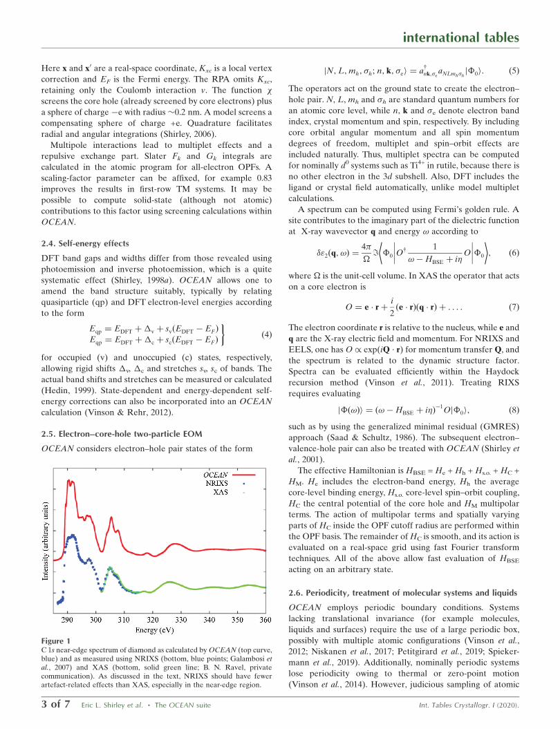
Figure 1C 1s near-edge spectrum of diamond as calculated by OCEAN (top curve,blue) and as measured using NRIXS (bottom, blue points; Galambosi etal., 2007) and XAS (bottom, solid green line; B. N. Ravel, privatecommunication). As discussed in the text, NRIXS should have fewerartefact-related effects than XAS, especially in the near-edge region.

displacements can render an accurate averaging of spectra
with only a few calculations.
3. Sample OCEAN results
OCEAN has been used to calculate near-edge core-excitation
spectra in a wide variety of systems, and RIXS spectra in a few
cases. Here, we present results for several different systems
and compare them with experimental results to illustrate the
reasonably expected accuracy of computed spectra.
3.1. Calculations in simple systems
Diamond is a simple system offering high-quality spectra. It is
easy to calculate spectra up to about 100 eV above the C 1s
edge including only 60 conduction bands with good Brillouin-
zone sampling. The most reliable spectroscopies at the C 1s
near edge are NRIXS and EELS, followed by XAS, which
might rely on electron-yield or fluorescence-yield signatures of
absorption and suffer from instrument carbon build-up. Fig. 1
shows near-edge spectra obtained using OCEAN, NRIXS
(Galambosi et al., 2007) and, above 315 eV, XAS (B. N. Ravel,
private communication). Despite the prediction of a low-lying
s-symmetry core-hole exciton (Jackson & Pederson, 1991),
EELS (Batson, 1993), NRIXS (Galambosi et al., 2007) and
calculations using the predecessor of OCEAN (Shirley, 1998b)
suggested otherwise.
3.2. Multiplet calculations
Vinson et al. (2011) and Vinson & Rehr (2012) consider a wide
range of 3d TM systems at the 2p edge of the TM species.
SrTiO3 and CaF2 serve as standard systems and allow
comparison to all-electron core BSE results (Laskowski &
Blaha, 2010; Gulans et al., 2014). Vinson and Rehr also
consider other systems, including metallic calcium. Results are
shown in Fig. 2.
3.3. Electric dipole plus quadrupole calculations
Spectra of TM compounds can be of interest at a TM 1s pre-
edge. OCEAN can treat such systems, which can feature
dipole-allowed 1s!np and quadrupole-allowed 1s!md
transitions, such as in rutile (Shirley, 2004). Perovskites such
as SrTiO3 and PbTiO3 are of interest (Woicik et al., 2007)
because 3d–4p mixing causes Eg-symmetry Ti 3d states to
acquire partial 4p character, giving rise to strong absorption
cross sections that help to reveal local atomic geometries.
3.4. Complex systems
Vinson et al. (2012) modelled the O 1s spectra of liquid water
and two ice phases as presented in Fig. 3. Calculations for the
liquid sample multiple molecular configurations. Care is
required to estimate core-level shifts for inequivalent sites,
such as oxygen sites in water, as discussed elsewhere
(Pasquarello et al., 1996).
3.5. Resonant inelastic X-ray scattering
Direct RIXS calculations are more involved because they
entail core-excited and valence-excited state calculations.
Diamond was an early subject of RIXS measurements (Ma et
al., 1992; Carlisle et al., 1999) and calculations (Shirley, 2000).
OCEAN is able to capture several of the changes in X-ray
emission for incident energy from 5 to 25 eV above the C 1s
edge.
international tables
Int. Tables Crystallogr. I (2020). Eric L. Shirley et al. � The OCEAN suite 4 of 7
Figure 3O 1s near-edge spectra in the Ih phase of ice and liquid water. Calculatedresults are shown by red solid lines. Measured results (blue dashed lines)were obtained using NRIXS (Pylkkanen et al., 2010) and XAS (Tse et al.,2008) in ice and scanning transmission X-ray microscopy in liquid water(Nilsson et al., 2010).
Figure 2The near-edge structure at the Ca 2p edge in metallic calcium and calciumfluoride. Calculations are shown by the red solid curve, and measure-ments are shown by blue dashed curves for calcium fluoride (de Groot etal., 1990) and metallic calcium (Fink et al., 1985).

3.6. Momentum-dependent results
LiF demonstrates the ability of NRIXS to probe excitations
of different symmetries. A small peak below the dipole-
allowed F 1s edge was attributed to a vibrationally allowed,
s-symmetry core-hole exciton. OCEAN and NRIXS (Hama-
lainen et al., 2002; Vinson et al., 2011) at various momentum
transfers confirmed this, as shown in Fig. 4. Finite-temperature
molecular-dynamics simulations provide snapshots of atomic
positions that make the pre-edge feature optically allowed
(Pascal et al., 2014). Others (Tse et al., 2014) have also
compared measured and calculated NRIXS of pressurized
silicon to study the pressure-induced insulator-to-metal
transition in this material.
4. Future directions
Working only within a single electron–hole pair picture
undermines OCEAN’s treatment of strongly correlated
systems, although its output might guide the development of
model Hamiltonians for such systems. Larger supercells can
lessen the effects of artificially imposed periodicity. Many
effects of atomic displacements and multi-electron excitations
are becoming treatable in a statistically averaged sense by
combining the results of different atomic configurations and/
or post-processing results.
4.1. Vibrational effects
Debye–Waller (DW) effects are ubiquitous in core spectra,
affecting electron-scattering processes because of displace-
ments of atoms from equilibrium positions, as has been
reviewed elsewhere (Rehr & Albers, 2000). Others (Story et
al., 2014) show how spectra can include some vibrational
effects to all orders using a cumulant approach and system-
specific knowledge of vibrational properties that is obtained
elsewhere. Near-edge features are also affected for reasons
outside the above DW treatment. One can also vary atomic
coordinates by sampling phonon modes or using molecular-
dynamics amenable to disordered systems (Vinson et al., 2014;
Brouder et al., 2010; Nemausat et al., 2015; Pascal et al., 2014;
Prendergast & Galli, 2006; Niskanen et al., 2017). Others (de
Groot et al., 1990) cite vibrational effects in d0 transition-metal
compounds for broadening in cases of strong ligand–TM
hybridization, for example E–e Jahn–Teller effects couple Eg
electron states and eg modes. OCEAN can help to determine
the coupling strength to use in effective Jahn–Teller Hamilto-
nians (Tinte & Shirley, 2008; Gilmore & Shirley, 2010) to
amend computed spectra. Still others (Zacharias & Giustino,
2016) have analysed valence edges, and core edges should also
be treatable.
4.2. Satellite effects
Multi-electron excitations broaden spectral features whenever
an electron and/or hole state is far from the Fermi level. The
‘electron–hole continuum’ part of the loss function smoothens
the onset of broadening (Soininen et al., 2003; Kas et al., 2007;
Fister et al., 2011). Multi-electron excitation also transfers
spectral weight to satellites, as largely captured by a cumulant
approach related to Hedin’s GW self-energy (Hedin, 1999).
This improves calculated photoemission (Guzzo et al., 2011;
Gumhalter et al., 2016; Lischner et al., 2015) and near-edge
(Kas et al., 2015) spectra. Others (Kas et al., 2016) have
presented a method that allows the inclusion of all losses,
including interference between intrinsic and extrinsic losses
because of the coupling of all particles to the valence electron
density. Including such effects should become a standard
aspect of calculations performed using tools such as
OCEAN.
The systems that OCEAN can treat are limited by our use of
the BSE. Many-electron effects are included only with a more
complete description of departures from the ground-state
wavefunction. However, even Coster–Kronig decay (Coster &
Kronig, 1935) and charge-transfer effects can be studied in
limited cases if response-theory analysis of the environment
facilitates an enhanced description of on-site excitations, with
adequate separability of excitations near a site versus at longer
range.
5. Access to OCEAN
Interested parties should access http://ocean-code.com, which
offers the source code and documentation. As of version 2,
OCEAN also incorporates valence BSE capabilities. Its
flexible input format allows future use with many DFT
programs. ABINIT and Quantum ESPRESSO, both of which
are open source, are already accommodated. Scaling of
computation time, memory and storage requirements, and a
parallelized version of OCEAN that should be particularly
helpful for large unit cells or large-scale structures have been
discussed elsewhere (Gilmore et al., 2015).
international tables
5 of 7 Eric L. Shirley et al. � The OCEAN suite Int. Tables Crystallogr. I (2020).
Figure 4F 1s near-edge spectra obtained by NRIXS for several momentumtransfers in LiF, as calculated (top) and measured (bottom; Hamalainen etal., 2002).

Acknowledgements
The OCEAN project has benefitted from many, including
L. X. Benedict, H. M. Lawler, J. A. Soininen, J. J. Rehr, J. J.
Kas, F. D. Vila, D. G. Prendergast, C. D. Pemmaraju and Y.
Liang. Some aided discussions and coordination, and others
provided key technical innovations. Benedict spearheaded the
valence excitation work, which was extended by Lawler and
repackaged for OCEAN. Soininen pioneered NRIXS and
EELS work and RPA core-hole screening. Kas and co-
workers shared innovations treating self-energy, Debye–
Waller and satellite effects in electron spectroscopies in
general.
References
Batson, P. E. (1993). Phys. Rev. Lett. 70, 1822–1825.Blochl, P. E. (1994). Phys. Rev. B, 50, 17953–17979.Brouder, C., Cabaret, D., Juhin, A. & Sainctavit, P. (2010). Phys. Rev.
B, 81, 115125.Carlisle, J. A., Shirley, E. L., Terminello, L. J., Jia, J. J., Callcott, T. A.,
Ederer, D. L., Perera, R. C. C. & Himpsel, F. J. (1999). Phys. Rev. B,59, 7433–7445.
Coster, D. & Kronig, R. De L. (1935). Physica, 2, 13–24.Fink, J., Muller-Heinzerling, T., Scheerer, B., Speier, W., Hillebrecht,
F. U., Fuggle, J. C., Zaanen, J. & Sawatzky, G. A. (1985). Phys. Rev.B, 32, 4899–4904.
Fister, T. T., Schmidt, M., Fenter, P., Johnson, C. S., Slater, M. D.,Chan, M. K. & Shirley, E. L. (2011). J. Chem. Phys. 135, 224513.
Galambosi, S., Soininen, J. A., Nygard, K., Huotari, S. & Hamalainen,K. (2007). Phys. Rev. B, 76, 195112.
Giannozzi, P., Baroni, S., Bonini, N., Calandra, M., Car, R., Cavazzoni,C., Ceresoli, D., Chiarotti, G. L., Cococcioni, M., Dabo, I., DalCorso, A., de Gironcoli, S., Fabris, S., Fratesi, G., Gebauer, R.,Gerstmann, U., Gougoussis, C., Kokalj, A., Lazzeri, M., Martin-Samos, L., Marzari, N., Mauri, F., Mazzarello, R., Paolini, S.,Pasquarello, A., Paulatto, L., Sbraccia, C., Scandolo, S., Sclauzero,G., Seitsonen, A. P., Smogunov, A., Umari, P. & Wentzcovitch,R. M. (2009). J. Phys. Condens. Matter, 21, 395502.
Gilmore, K. & Shirley, E. L. (2010). J. Phys. Condens. Matter, 22,315901.
Gilmore, K., Vinson, J., Shirley, E. L., Prendergast, D., Pemmaraju,C. D., Kas, J. J., Vila, F. D. & Rehr, J. J. (2015). Comput. Phys.Commun. 197, 109–117.
Gonze, X., Amadon, B., Anglade, P. M., Beuken, J. M., Bottin, F.,Boulanger, P., Bruneval, F., Caliste, D., Caracas, R., Cote, M.,Deutsch, T., Genovese, L., Ghosez, P., Giantomassi, M., Goedecker,S., Hamann, D. R., Hermet, P., Jollet, F., Jomard, G., Leroux, S.,Mancini, M., Mazevet, S., Oliveira, M. J. T., Onida, G., Pouillon, Y.,Rangel, T., Rignanese, G. M., Sangalli, D., Shaltaf, R., Torrent, M.,Verstraete, M. J., Zerah, G. & Zwanziger, J. W. (2009). Comput.Phys. Commun. 180, 2582–2615.
Groot, F. M. F. de, Fuggle, J. C., Thole, B. T. & Sawatzky, G. A. (1990).Phys. Rev. B, 41, 928–937.
Gulans, A., Kontur, S., Meisenbichler, C., Nabok, D., Pavone, P.,Rigamonti, S., Sagmeister, S., Werner, U. & Draxl, C. (2014). J.Phys. Condens. Matter, 26, 363202.
Gumhalter, B., Kovac, V., Caruso, F., Lambert, H. & Giustino, F.(2016). Phys. Rev. B, 94, 035103.
Guzzo, M., Lani, G., Sottile, F., Romaniello, P., Gatti, M., Kas, J. J.,Rehr, J. J., Silly, M. G., Sirotti, F. & Reining, L. (2011). Phys. Rev.Lett. 107, 166401.
Hamalainen, K., Galambosi, S., Soininen, J. A., Shirley, E. L., Rueff,J. P. & Shukla, A. (2002). Phys. Rev. B, 65, 155111.
Hedin, L. (1999). J. Phys. Condens. Matter, 11, R489–R528.Hohenberg, P. & Kohn, W. (1964). Phys. Rev. 136, B864–B871.
Hybertsen, M. S. & Louie, S. G. (1988). Phys. Rev. B, 37, 2733–2736.
Jackson, K. A. & Pederson, M. R. (1991). Phys. Rev. Lett. 67, 2521–2524.
Kas, J. J., Rehr, J. J. & Curtis, J. B. (2016). Phys. Rev. B, 94, 035156.Kas, J. J., Sorini, A. P., Prange, M. P., Cambell, L. W., Soininen, J. A. &
Rehr, J. J. (2007). Phys. Rev. B, 76, 195116.
Kas, J. J., Vila, F. D., Rehr, J. J. & Chambers, S. A. (2015). Phys. Rev.B, 91, 121112.
Kohn, W. & Sham, L. J. (1965). Phys. Rev. 140, A1133–A1138.
Laskowski, R. & Blaha, P. (2010). Phys. Rev. B, 82, 205104.
Levine, Z. H. & Louie, S. G. (1982). Phys. Rev. B, 25, 6310–6316.Lischner, J., Palsson, G. K., Vigil-Fowler, D., Nemsak, S., Avila, J.,
Asensio, M. C., Fadley, C. S. & Louie, S. G. (2015). Phys. Rev. B, 91,205113.
Ma, Y., Wassdahl, N., Skytt, P., Guo, J., Nordgren, J., Johnson, P. D.,Rubensson, J. E., Boske, T., Eberhardt, W. & Kevan, S. D. (1992).Phys. Rev. Lett. 69, 2598–2601.
Nemausat, R., Cabaret, D., Gervais, C., Brouder, C., Trcera, N.,Bordage, A., Errea, I. & Mauri, F. (2015). Phys. Rev. B, 92, 144310.
Nilsson, A., Nordlund, D., Waluyo, I., Huang, N., Ogasawara, H.,Kaya, S., Bergmann, U., Naslund, L. A., Ostrom, H., Wernet, P.,Andersson, K. J., Schiros, T. & Pettersson, L. G. M. (2010). J.Electron Spectrosc. Relat. Phenom. 177, 99–129.
Niskanen, J., Sahle, Ch. J., Gilmore, K., Uhlig, F., Smiatek, J. &Fohlisch, A. (2017). Phys. Rev. E, 96, 013319.
Pascal, T. A., Boesenberg, U., Kostecki, R., Richardson, T. J., Weng,T. C., Sokaras, D., Nordlund, D., McDermott, E., Moewes, A.,Cabana, J. & Prendergast, D. (2014). J. Chem. Phys. 140, 034107.
Pasquarello, A., Hybertsen, M. S. & Car, R. (1996). Phys. Rev. B, 53,10942–10950.
Petitgirard, S., Sahle, C. J., Weis, C., Gilmore, K., Spiekermann, G.,Tse, J. S., Wilke, M., Cavallari, C., Cerantola, V. & Sternemann, C.(2019). Geochem. Persp. Lett. 9, 32–37.
Prendergast, D. & Galli, G. (2006). Phys. Rev. Lett. 96, 215502.
Pylkkanen, T., Giordano, V. M., Chervin, J. C., Sakko, A., Hakala, M.,Soininen, J. A., Hamalainen, K., Monaco, G. & Huotari, S. (2010). J.Phys. Chem. B, 114, 3804–3808.
Rehr, J. J. & Albers, R. C. (2000). Rev. Mod. Phys. 72, 621–654.
Saad, Y. & Schultz, M. H. (1986). SIAM J. Sci. Stat. Comput. 7, 856–869.
Shirley, E. L. (1991). Thesis. University of Illinois at UrbanaChampaign, USA.
Shirley, E. L. (1998a). Phys. Rev. B, 58, 9579–9583.Shirley, E. L. (1998b). Phys. Rev. Lett. 80, 794–797.
Shirley, E. L. (2000). J. Electron Spectrosc. Relat. Phenom. 110–111,305–321.
Shirley, E. L. (2004). J. Electron Spectrosc. Relat. Phenom. 136, 77–83.Shirley, E. L. (2006). Ultramicroscopy, 106, 986–993.
Shirley, E. L., Soininen, J. A., Zhang, G. P., Carlisle, J. A., Callcott,T. A., Ederer, D. L., Terminello, L. J. & Perera, R. C. C. (2001). J.Electron Spectrosc. Relat. Phenom. 114–116, 939–946.
Soininen, J. A., Rehr, J. J. & Shirley, E. L. (2003). J. Phys. Condens.Matter, 15, 2573–2586.
Story, S. M., Kas, J. J., Vila, F. D., Verstraete, M. J. & Rehr, J. J. (2014).Phys. Rev. B, 90, 195135.
Spiekermann, G., Harder, M., Gilmore, K., Zalden, P., Sahle, Ch. J.,Petitgirard, S., Wilke, M., Biedermann, N., Weis, C., Morgenroth,W., Tse, J. S., Kulik, E., Nishiyama, N., Yavas, H. & Sternemann, C.(2019). Phys. Rev. X, 9, 011025.
Tinte, S. & Shirley, E. L. (2008). J. Phys. Condens. Matter, 20, 365221.
Tse, J. S., Hanfland, M., Flacau, R., Desgreniers, S., Li, Z., Mende, K.,Gilmore, K., Nyrow, A., Moretti Sala, M. & Sternemann, C. (2014).J. Phys. Chem. C, 118, 1161–1166.
Tse, J. S., Shaw, D. M., Klug, D. D., Patchkovskii, S., Vanko, G.,Monaco, G. & Krisch, M. (2008). Phys. Rev. Lett. 100, 095502.
Vinson, J., Jach, T., Elam, W. T. & Denlinger, J. D. (2014). Phys. Rev.B, 90, 205207.
international tables
Int. Tables Crystallogr. I (2020). Eric L. Shirley et al. � The OCEAN suite 6 of 7
Vinson, J., Kas, J. J., Vila, F. D., Rehr, J. J. & Shirley, E. L. (2012). Phys.Rev. B, 85, 045101.
Vinson, J. & Rehr, J. J. (2012). Phys. Rev. B, 86, 195135.Vinson, J., Rehr, J. J., Kas, J. J. & Shirley, E. L. (2011). Phys. Rev. B,
83, 115106.
Woicik, J. C., Shirley, E. L., Hellberg, C. S., Andersen, K. E.,Sambasivan, S., Fischer, D. A., Chapman, B. D., Stern, E. A.,Ryan, P., Ederer, D. L. & Li, H. (2007). Phys. Rev. B, 75,140103.
Zacharias, M. & Giustino, F. (2016). Phys. Rev. B, 94, 075125.
international tables
7 of 7 Eric L. Shirley et al. � The OCEAN suite Int. Tables Crystallogr. I (2020).