
The vertical lobe of cephalopods: an attractive brain structure for ...
Upload
ethan-griffinCategory
view
218download
3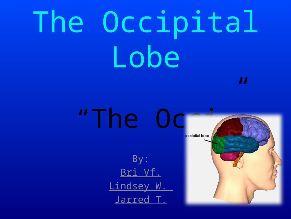
The Occipital Lobe
By:Bri Vf.
Lindsey W. Jarred T.
“The Occi”

Notes and VocabularyThe Occipital Lobe is the rearmost lobe in each cerebral
hemisphere of the brain. It contains the visual center of the brain.
- It is one of the main lobes/regions of the cerebral cortex- Main visual processing (color and face recognition)
- Occipital Cortex-(anatomy) a somewhat rounded subdivision of a bodily organ or part.

What body part does it control?
The occipital lobe is located at the back of our brain. It is responsible for receiving and processing visual information from
our eyes.
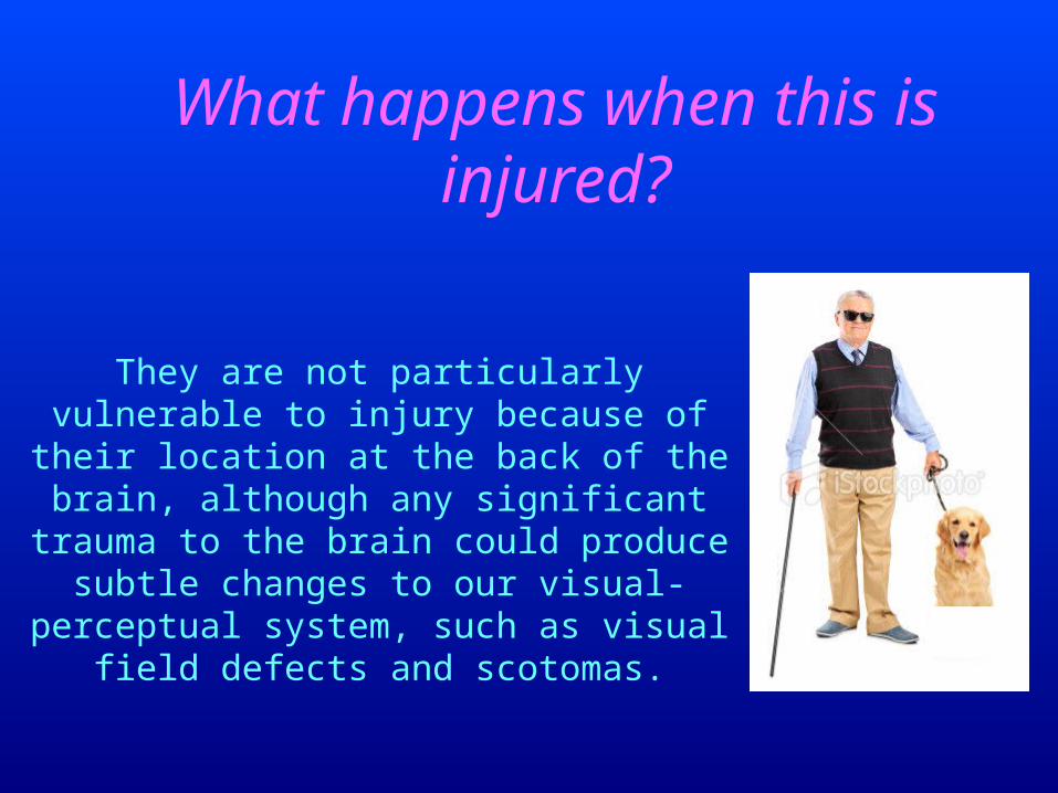
What happens when this is injured?
They are not particularly vulnerable to injury because of their location at the
back of the brain, although any significant trauma to the brain could produce subtle changes to our visual-perceptual system, such as visual field
defects and scotomas.

Visual Hallucination(Epilepsy)
Seizures can occur in the occipital lobe, which can cause visual hallucinations. The patient may see rapid
blinking, colored lights or flickering lights. Occipital lobe seizures, which the NYU Comprehensive Epilepsy Center states account for 5 to 10 percent of epilepsy
cases, can be triggered by flashing lights.
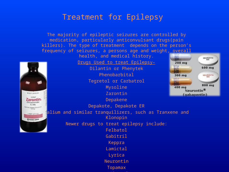
Treatment for Epilepsy
The majority of epileptic seizures are controlled by medication, particularly anticonvulsant drugs(pain killers). The type of treatment depends on the person’s
frequency of seizures, a persons age and weight, overall health, and medical history.Drugs Used to treat Epilepsy-
Dilantin or PhenytekPhenobarbital
Tegretol or CarbatrolMysolineZarontin
DepakeneDepakote, Depakote ER
Valium and similar tranquilizers, such as Tranxene and KlonopinNewer drugs to treat epilepsy include:
FelbatolGabitrilKeppra
LamictalLyrica
NeurontinTopamaxTrileptal
Zonegran

Activity!
Does Lincoln’s face look normal?
Does Lincoln’s face look normal?
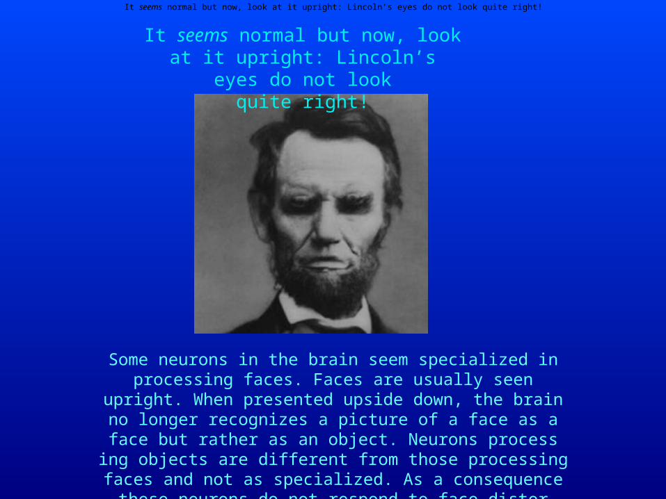
It seems nor mal but now, look at it upright: Lincoln’s eyes do not look quite right!
It seems nor mal but now, look at it upright: Lincoln’s eyes do not
look quite right!
Some neu rons in the brain seem spe cial ized in pro cess ing faces. Faces are usu ally seen upright. When pre sented upside down, the
brain no longer rec og nizes a pic ture of a face as a face but rather as an object. Neu rons pro cess ing objects are diff er ent from those pro
cess ing faces and not as spe cial ized. As a con se quence these neu rons do not respond to face dis tor tions as well. This explains why we miss
the weird eyes when the face is inverted.

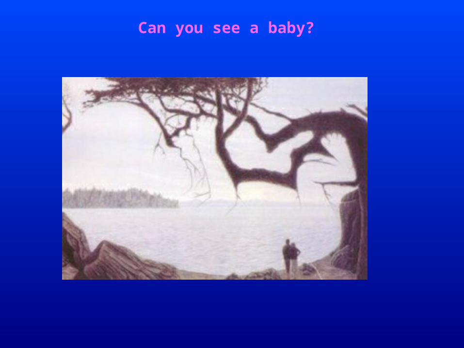
Another great exam ple of an illu sory con tour! The baby’s
head is on the left, the baby’s feet are against the trunk of the
tree on the right.

Stare at the yel low stripe in the mid dle of the fish in the pic ture below for about 10–20 sec.
Then move your gaze to the fish bowl.
Can you put the fish in the fishbowl?
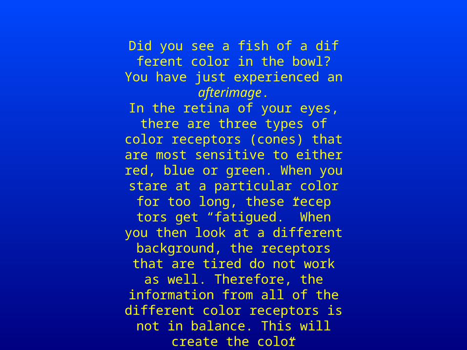
Did you see a fish of a diff er ent color in the bowl? You have just expe ri enced an
after im age.In the retina of your eyes, there are three types of color recep tors (cones) that are
most sen si tive to either red, blue or green. When you stare at a par tic u lar color for too long, these recep tors get
“fatigued.” When you then look at a diff er ent back ground, the recep tors that are
tired do not work as well. There fore, the infor ma tion from all of the diff er ent color recep tors is not in bal ance. This will cre ate
the color “afterimages.”

Quiz
1.What does the occipital lobe control and where is it located?
2.What part of the body does it operate?