The nucleoporin Nup214 sequesters CRM1 at the nuclear rim and
11
4409 Research Article Introduction Nuclear pore complexes (NPCs) are protein channels spanning the nuclear envelope to conduct macromolecular transport between the nucleus and the cytoplasm (reviewed by Suntharalingam and Wente, 2003). Genetic and biochemical studies in yeast and mammalian cells have identified ~30 nuclear pore proteins, which form phylogenetically conserved sub-complexes with defined composition and localization along the nuclear pore (Cronshaw et al., 2002; Rout et al., 2000). Although carrier-independent transport has been described for several proteins, most of the protein transport in or out of the nucleus relies on a family of soluble factors known as karyopherins. In facilitated transport events, importins and exportins recognize discrete signals on their cargoes and mediate either nuclear import or export. The karyopherin-cargo complex then binds to nucleoporins with phenylanine-glycine (FG) repeats, which may collectively provide a favorable environment for the passage of the complex (Becskei and Mattaj, 2005). Directionality is orchestrated by Ran, a small GTPase that is highly enriched in its GTP-bound form in the nucleus, where it dissociates the importin-cargo complexes and promotes the formation of cargo complexes with exportins (Weis, 2003). The exportin CRM1, recognizes and transports proteins bearing a leucine-rich nuclear export signal (Fornerod et al., 1997a; Fukuda et al., 1997; Stade et al., 1997). CRM1 binds cooperatively with RanGTP to the cargo to form a trimeric complex NES-RanGTP-CRM1, which then translocates through the NPC. Release of the export substrates in the cytoplasmic face of the NPC is accomplished by the RanGTPase-activating protein (RanGAP) and RanBP1, which hydrolyze RanGTP and dissociate the complex (Fornerod and Ohno, 2002; Kuersten et al., 2002). Many cell-signaling events culminate with the selective translocation of gene regulators in and out of the nucleus. Cytoplasmic anchoring and posttranslational modifications of either cargo molecules or transport mediators regulate the nuclear accumulation of signaling effectors leading to changes in gene expression (Kaffman and O’Shea, 1999; Xu and Massague, 2004). A less-explored mechanism involves the modification of individual nucleoporins and transport factors leading to alterations of transport rates (Kehlenbach and Gerace, 2000; Makhnevych et al., 2003). A paradigm for the ‘cytoplasmic anchoring’ mechanism derives from studies of NFB activation in flies and vertebrate systems. In Drosophila, Toll signaling determines dorsal-ventral axis specification in embryos and the activation of the innate immune responses in larvae and adults (Brennan and Anderson, 2004). Toll signaling upon bacterial infection, leads to the degradation of the IB homolog, Cactus, and the nuclear accumulation of the NFB proteins Dorsal and Dif. In mutant larvae lacking the nucleoporin Nup88, Cactus becomes degraded upon bacterial challenge but Dorsal and Dif fail to accumulate in the nucleus. Nuclear import of other transcription factors or artificial reporters bearing nuclear localization signals is not affected in CRM1-mediated protein export is an important determinant of the nuclear accumulation of many gene regulators. Here, we show that the NFB transcription factor Dorsal is a substrate of CRM1 and requires the nucleoporin Nup214 for its nuclear translocation upon signaling. Nup214 bound to CRM1 directly and anchored it to the nuclear envelope. In nup214 mutants CRM1 accumulated in the nucleus and NES-protein export was enhanced. Nup214 formed complexes with Nup88 and CRM1 in vivo and Nup214 protected Nup88 from degradation at the nuclear rim. In turn, Nup88 was sufficient for targeting the complex to the nuclear pores. Overexpression experiments indicated that Nup214 alone attracts a fraction of CRM1 to the nuclear envelope but does not interfere with NES-GFP export. By contrast, overexpression of the Nup214-Nup88 complex trapped CRM1 and Dorsal to cytoplasmic foci and inhibited protein export and immune response activation. We hypothesize that variation in levels of the Nup214-Nup88 complex at the pore changes the amount of NPC-bound CRM1 and influences the relative strength and duration of NFB signaling responses. Supplementary material available online at http://jcs.biologists.org/cgi/content/full/119/21/4409/DC1 Key words: Drosophila, Nucleoporins, Nup214, CRM1 export, NFB signaling, Nuclear transport Summary The nucleoporin Nup214 sequesters CRM1 at the nuclear rim and modulates NFB activation in Drosophila Nikos Xylourgidis*, Peggy Roth*, Nafiseh Sabri, Vasilios Tsarouhas and Christos Samakovlis ‡ Department of Developmental Biology, Wenner-Gren Institute, Stockholm University, S-10691, Stockholm, Sweden *These authors contributed equally to this work ‡ Author for correspondence (e-mail: [email protected]) Accepted 4 August 2006 Journal of Cell Science 119, 4409-4419 Published by The Company of Biologists 2006 doi:10.1242/jcs.03201 Journal of Cell Science
Transcript of The nucleoporin Nup214 sequesters CRM1 at the nuclear rim and
4409Research Article
IntroductionNuclear pore complexes (NPCs) are protein channels spanningthe nuclear envelope to conduct macromolecular transportbetween the nucleus and the cytoplasm (reviewed bySuntharalingam and Wente, 2003). Genetic and biochemicalstudies in yeast and mammalian cells have identified ~30nuclear pore proteins, which form phylogenetically conservedsub-complexes with defined composition and localizationalong the nuclear pore (Cronshaw et al., 2002; Rout et al.,2000). Although carrier-independent transport has beendescribed for several proteins, most of the protein transport inor out of the nucleus relies on a family of soluble factors knownas karyopherins. In facilitated transport events, importins andexportins recognize discrete signals on their cargoes andmediate either nuclear import or export. The karyopherin-cargocomplex then binds to nucleoporins with phenylanine-glycine(FG) repeats, which may collectively provide a favorableenvironment for the passage of the complex (Becskei andMattaj, 2005). Directionality is orchestrated by Ran, a smallGTPase that is highly enriched in its GTP-bound form in thenucleus, where it dissociates the importin-cargo complexes andpromotes the formation of cargo complexes with exportins(Weis, 2003). The exportin CRM1, recognizes and transportsproteins bearing a leucine-rich nuclear export signal (Fornerodet al., 1997a; Fukuda et al., 1997; Stade et al., 1997). CRM1binds cooperatively with RanGTP to the cargo to form atrimeric complex NES-RanGTP-CRM1, which then
translocates through the NPC. Release of the export substratesin the cytoplasmic face of the NPC is accomplished by theRanGTPase-activating protein (RanGAP) and RanBP1, whichhydrolyze RanGTP and dissociate the complex (Fornerod andOhno, 2002; Kuersten et al., 2002).
Many cell-signaling events culminate with the selectivetranslocation of gene regulators in and out of the nucleus.Cytoplasmic anchoring and posttranslational modifications ofeither cargo molecules or transport mediators regulate thenuclear accumulation of signaling effectors leading to changesin gene expression (Kaffman and O’Shea, 1999; Xu andMassague, 2004). A less-explored mechanism involves themodification of individual nucleoporins and transport factorsleading to alterations of transport rates (Kehlenbach andGerace, 2000; Makhnevych et al., 2003). A paradigm for the‘cytoplasmic anchoring’ mechanism derives from studies ofNF�B activation in flies and vertebrate systems. In Drosophila,Toll signaling determines dorsal-ventral axis specification inembryos and the activation of the innate immune responses inlarvae and adults (Brennan and Anderson, 2004). Toll signalingupon bacterial infection, leads to the degradation of the I�Bhomolog, Cactus, and the nuclear accumulation of the NF�Bproteins Dorsal and Dif. In mutant larvae lacking thenucleoporin Nup88, Cactus becomes degraded upon bacterialchallenge but Dorsal and Dif fail to accumulate in the nucleus.Nuclear import of other transcription factors or artificialreporters bearing nuclear localization signals is not affected in
CRM1-mediated protein export is an importantdeterminant of the nuclear accumulation of many generegulators. Here, we show that the NF��B transcriptionfactor Dorsal is a substrate of CRM1 and requires thenucleoporin Nup214 for its nuclear translocation uponsignaling. Nup214 bound to CRM1 directly and anchoredit to the nuclear envelope. In nup214 mutants CRM1accumulated in the nucleus and NES-protein export wasenhanced. Nup214 formed complexes with Nup88 andCRM1 in vivo and Nup214 protected Nup88 fromdegradation at the nuclear rim. In turn, Nup88 wassufficient for targeting the complex to the nuclear pores.Overexpression experiments indicated that Nup214 aloneattracts a fraction of CRM1 to the nuclear envelope but
does not interfere with NES-GFP export. By contrast,overexpression of the Nup214-Nup88 complex trappedCRM1 and Dorsal to cytoplasmic foci and inhibited proteinexport and immune response activation. We hypothesizethat variation in levels of the Nup214-Nup88 complex at thepore changes the amount of NPC-bound CRM1 andinfluences the relative strength and duration of NF��Bsignaling responses.
Supplementary material available online athttp://jcs.biologists.org/cgi/content/full/119/21/4409/DC1
Key words: Drosophila, Nucleoporins, Nup214, CRM1 export,NF�B signaling, Nuclear transport
Summary
The nucleoporin Nup214 sequesters CRM1 at thenuclear rim and modulates NF��B activation inDrosophilaNikos Xylourgidis*, Peggy Roth*, Nafiseh Sabri, Vasilios Tsarouhas and Christos Samakovlis‡
Department of Developmental Biology, Wenner-Gren Institute, Stockholm University, S-10691, Stockholm, Sweden*These authors contributed equally to this work‡Author for correspondence (e-mail: [email protected])
Accepted 4 August 2006Journal of Cell Science 119, 4409-4419 Published by The Company of Biologists 2006doi:10.1242/jcs.03201
Jour
nal o
f Cel
l Sci
ence

4410
the mutants (Uv et al., 2000). Surprisingly, Nup88 acts as anattenuator of CRM1-mediated protein export by anchoring afraction of CRM1 on the nuclear envelope (Roth et al., 2003)suggesting an additional mechanism controlling the nuclearaccumulation of NF�B proteins.
The nucleoporin CAN-Nup214 binds to Nup88 and wasoriginally identified as a putative oncogene, disrupted bychromosomal translocations associated with myeloidleukemias (Snow et al., 1987; von Lindern et al., 1992). It islocalized at the cytoplasmic side of the NPC (Kraemer et al.,1994; Pante et al., 1994) and forms complexes with at leastthree other nucleoporins, Nup88-Nup82p, RanBP2-Nup358and Nup62-Nsp1p (Belgareh et al., 1998; Bernad et al., 2004;Fornerod et al., 1997b). The binding of Nup214 to importin �and CRM1, and the phenotypic analysis of cells derived fromNup214-deficient mice or Xenopus nuclei depleted of Nup214,suggested a facilitative role for Nup214 in nucleocytoplasmictransport of proteins (Fornerod et al., 1997a; Moroianu et al.,1997). More recently, depletion of Nup214 in HeLa cellsrevealed a requirement for the nucleoporin in the nuclearexport of the 60S pre-ribosomal subunit but no major defectsin the export of NES-GFP reporters (Bernad et al., 2006). TheFG repeats of Nup214 have also been directly implicated in theshuttling of signaling effectors such as mitogen-activatedprotein kinase (ERK) and Smad2 without the need for transportreceptors (Matsubayashi et al., 2001; Xu et al., 2002).
Here we explore the interdependence of Nup214 and itspartner Nup88 at the NPC and the function of the complex inNES export and NF�B protein transport in Drosophila. We findthat Nup214 maintains high levels of Nup88 at the NPC andNup88 is required for targeting the complex to the pores.Nup214 can bind to CRM1 directly and is required foranchoring a portion of CRM1 to the NPC. In nup214 mutantsNES export is surprisingly enhanced and dosage-sensitivegenetic interactions between crm1 and nup214 mutants suggestthat Nup214 acts as CRM1 inhibitor. Dorsal and Diftranslocation upon bacterial infection is impaired in nup214mutants. We identified CRM1-dependent NES sequences inDorsal and found that it becomes mislocalized afteroverexpression of the two nucleoporins. We propose amechanism, where the amounts of the Nup214-Nup88 complexon the NPCs determine the levels of CRM1 export attenuationand thereby the degree of activation of NF�B downstreamgenes.
Journal of Cell Science 119 (21)
ResultsNup214 forms a complex with Nup88 and CRM1 inDrosophila embryosNup88 is required for the localization of Nup214 and a portionof CRM1 at the nuclear rim (Roth et al., 2003). Using yeasttwo-hybrid and in vitro binding assays, we first showed thatthe FG-rich, C-terminal part of Drosophila Nup214 bindsdirectly to CRM1 (supplementary material Fig. S1). To analyzethe complex in fly embryos, we prepared protein extracts fromthree embryonic stages and examined the relative amounts ofeach component in the complex by immunoprecipitations witha Nup214 antiserum. Nup214, Nup88 and CRM1 were thendetected with specific antibodies on western blots of the boundand unbound fractions (Fig. 1). All three proteins were foundin the precipitate, and although most Nup88 was precipitatedfrom the extract, only a subfraction of CRM1 was detected inthe complex (Fig. 1). In addition, the relative amounts ofCRM1 in the complex varied at different stages of embryonicdevelopment suggesting that the composition of the complexis dynamic. The variations in the relative amounts of CRM1bound to the nucleoporin complex at different developmentalstages suggest that the affinity of the export factor for the NPCmay be modulated during embryogenesis.
Interdependence of Nup214 and Nup88 at the nuclearrimTo investigate the function of Nup214, we generated deletionmutants by P-element transposon excision and characterizedtheir lesions. Homozygous animals from one of these strains(from now on referred to as nup214 mutants) lacked a 631 bpfragment including parts of the second and third exons of thegene and die as early third instar larvae. The expression of thefull-length nup214 cDNA clone, in nup214 larvae under thecontrol of the inducible hsp70 promoter partially rescues thelethality of the mutants and extends their life span to the latethird larval stage. Attempts to detect clones of nup214 cells,generated by mitotic recombination, in imaginal discs andadult tissues failed, suggesting an essential function of Nup214in cell division or cell survival. The phenotypic analysis ofnup214 was therefore conducted in early third instar mutantlarvae that lack zygotic Nup214 expression (Fig. 2A). nup214mutants develop normally up to this stage owing to a robustcontribution of maternal RNA and protein and do not show anygross defects in nuclear envelope integrity revealed by
Fig. 1. Drosophila Nup214 is in complex withNup88 and CRM1. The amounts of CRM1 bound tothe Nup214-Nup88 complex vary during flydevelopment. Nup214 antiserum was used tocoimmunoprecipitate protein complexes of 0-5, 5-10and 10-15 hour wild-type embryo extracts. Extracts,unbound fractions and precipitates (IP) wereanalyzed by labeling the western blots with Nup214,CRM1 and Nup88 antibodies. Lamin serves as aloading control. Total amounts of Nup214, Nup88and CRM1 appear unchanged in the extracts fromdifferent embryonic stages. The amount of co-immunoprecipitated CRM1 relative to Nup214 doesnot change in 0-5 hour and 5-10 hour embryoswhereas three times less CRM1 is brought down in10-15 hour embryos.
Jour
nal o
f Cel
l Sci
ence

4411Nup214 attenuates NES export
stainings with the nucleoporin marker mAb414 (Davisand Blobel, 1986) and lamin (Stuurman et al., 1995)antibodies (Fig. 2A). To confirm that the lethality andthe phenotypes in nup214 mutants are due to adisruption of only the nup214 gene, we also analyzednup214/Df(2R)3-70 mutant larvae. These animalsrevealed similar developmental and physiologicalphenotypes to the nup214 homozygous mutants,indicating that the defects in nup214 mutants are solelydue to the lack of zygotic Nup214 protein.
Staining of wild-type larvae with anti-Nup88 antiserarevealed a pronounced nuclear rim labeling, whichappeared diffuse and drastically reduced in nup214mutants (Fig. 2A). Western blots of protein extractsfrom staged wild-type, nup214 heterozygotes andnup214 homozygous larvae showed that Nup88 wasdecreased in heterozygotes and was undetectable in thenup214 homozygotes (Fig. 2B), whereas the nup88mRNA levels remained unchanged in nup214 mutantscompared to wild-type (Fig. 2C). This gradualreduction of Nup88 protein at decreasingconcentrations of Nup214 suggests a role for Nup214in titrating the amounts of Nup88 at the nuclear rim.Since the amount of the nup88 transcript is not affectedin nup214 larvae, Nup214 might function in promotingthe translation of Nup88 or protecting it fromdegradation.
To address the functional interplay between the twonucleoporins, we analyzed the Nup214-Nup88complex in S2 cells. We used double-stranded RNAinhibition (RNAi) to reduce Nup214 and analyzedNup88, Nup214 and �-Tubulin levels by westernblotting (Fig. 3A). The Nup88 antibody recognizes twobands in S2 cell extracts and we performed a nup88RNAi experiment to identify the Nup88-specific band(Fig. 3A, right panel). In cells treated with nup214dsRNA for 3 days, Nup214 levels decreased by 50%and after 6 days of nup214 RNAi they decreased by~90%. Similarly to mutant larvae, the relative amountof Nup88 was also decreased proportionally to thedecline of Nup214. To test if the reduction of Nup88 inthe absence of Nup214 is due to protein degradation,we added the proteasome inhibitor epoxomicin (Meng et al.,1999) to cells treated with nup214 dsRNA for three days in anattempt to restore the decrease of Nup88. Addition of theinhibitor compensated the reduction of Nup88 levels in cellslacking Nup214 (Fig. 3A) indicating that Nup214 protectsNup88 from proteasomal degradation at the nuclear pore.
Nup88 is required for the localization of Nup214 on the NPCbecause in mbo mutants Nup214 becomes nuclear (Roth et al.,2003). We further investigated the interdependence of the twocomponents of the complex in vivo, by heat-shock-inducedoverexpression of each of the nucleoporins in the wild type andmutants lacking Nup88 or Nup214, followed by detection ofboth proteins with specific antibodies. Overexpression ofNup88 in wild-type larvae resulted in a punctuated cytoplasmicstaining without affecting the localization of endogenousNup214 (Fig. 3B). By contrast, Nup214 expressed under thecontrol of the same promoter, was enriched only around therim, suggesting that Nup88 at the NPC has the capacity to bindexcessive Nup214 and anchor it to the pore (Fig. 3B). hsp70-
driven Nup214 in mbo mutants accumulated in the nucleus,highlighting the anticipated role of Nup88 in anchoringNup214 at the NPC (Fig. 3B). Nup214 was also nuclear whenit was expressed in nup214 mutants, which also lack Nup88(Fig. 3B). By contrast, overexpression of Nup88 in nup214larvae resulted in a distinct nuclear rim staining indicating thatNup88 contains all the necessary sequences for targeting theNPC (Fig. 3B). The punctate Nup88 staining in these animalsappeared much reduced in comparison with the levels ofoverexpressed Nup88 in the wild type, arguing again thatNup214 is required post translationally for the accumulation ofhigh levels of Nup88. We also co-expressed Nup88 andNup214 in nup214 mutants. In these experiments, the relativeamounts of the proteins varied in individual cells of the nup214larvae (supplementary material Fig. S2). In cells expressinghigh levels of Nup88 and low amounts of Nup214 after heatshock, the complex was strictly localized at the NPC. Bycontrast, in cells expressing relatively low levels of Nup88 andhigh amounts of Nup214, the complex was found in the
Fig. 2. Nup88 is degraded in nup214 mutants. (A) Nup214, mAb414, laminand Nup88 localization (red) in fat body cells of wild-type and nup214mutant larvae. Nuclei are visualized by DAPI in the adjacent panel.Confocal sections are shown in the insets. Bars, 35 �m. (B) Western blot ofprotein extract from wild-type, heterozygotes and nup214 mutant larvaeprobed with Nup88 antibody. �-tubulin provides a loading control. Nup88is reduced in heterozygote animals and totally abolished in the homozygousmutant. (C) RT-PCR for mbo on poly(A)+-purified larval extracts of wild-type (lanes 1-4), P-element (l(2)10444) (lanes 5-8) and nup214 mutants(lanes 9-12). (Fourfold serial dilutions of mRNA from each genotype wereused in the RT-PCR step.) rp49 provides a quantitative control. The mRNAlevels of mbo remain the same in wild-type and mutant larvae.
Jour
nal o
f Cel
l Sci
ence

4412
nucleus. The overexpression analysis suggests that the stabilityand the localization of the complex may depend on the relativeamounts of each constituent.
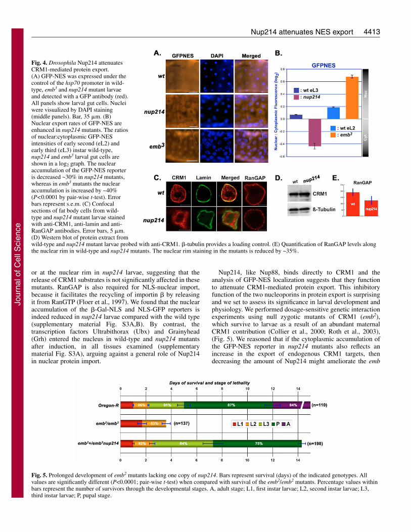
nup214 mutants show enhanced CRM1-mediatednuclear exportThe cytoplasmic accumulation of a GFP-NES reporter isincreased in mbo mutants and this phenotype could bereverted by LMB treatment of the animals, suggesting thatthe nucleoporin acts as an inhibitor of CRM1-mediatedprotein export (Roth et al., 2003). To investigate whetherNup214 has a similar function in protein export, we expressedGFP-NES under the control of the inducible hsp70 promoterin wild-type, CRM1 (emb3) (Roth et al., 2003) and nup214mutant larvae. GFP expression in wild-type and nup214larvae served as controls for the assay (supplementarymaterial Fig. S3A). After a 1-hour induction and 3 hoursrecovery, we examined the levels and the localization of thereporter in larval gut and fat body tissues using a GFPantibody. GFP-NES fusions were detected in both thecytoplasm and nucleus of wild-type tissues (Fig. 4A). In emb3
(CRM1) mutants, the GFP-NES reporter accumulated insidethe nucleus, indicating that it is recognized as a CRM1substrate (Fig. 4A). In nup214 mutants the distribution ofGFP-NES was predominantly cytoplasmic, suggesting thatCRM1-mediated protein export might be enhanced in theabsence of Nup214 (Fig. 4A). To measure the increasedcytoplasmic accumulation of GFP-NES in nup214 mutants,we compared the ratios of nuclear versus cytoplasmic GFP-NES intensities in nup214 and wild-type early third instarlarvae. GFP-NES nuclear accumulation was decreased by
30% in nup214, whereas the nuclear accumulation of thesame reporter was increased by 40% in emb3 mutantscompared with the wild type (Fig. 4B).
To explore the mechanism underlying the inhibitory role ofNup214 on nucleocytoplasmic export, we analyzed thesubcellular localization of CRM1 in the mutants. In severalwild-type tissues, a substantial part of CRM1 staining wasfound along the nuclear rim and at lower levels in the nucleus(Fig. 4C). By contrast, in nup214 mutants, most of the CRM1staining at the nuclear rim was absent, and the protein appearedto be concentrated inside the nucleus (Fig. 4C). Western blotsrevealed that the total amount of CRM1 was similar in bothwild-type and nup214 extracts (Fig. 4D) suggesting a role ofNup214 in tethering CRM1 at the nuclear pore.
RanGAP together with RanBP1 promotes the hydrolysis ofRanGTP and subsequent dissociation of the trimeric CRM1-NES-RanGTP export complex in the cytoplasm (Askjaer et al.,1999). It is localized at the cytoplasmic fibrils of the NPCthrough its association with RanBP2-Nup358 (Hopper et al.,1990; Matunis et al., 1996). Staining of wild-type tissuesrevealed a distinct accumulation of RanGAP at the nuclear rim,whereas in nup214 mutants the staining appeared punctuatedand reduced by ~30% (Fig. 4C,E). Thus, Nup214 is alsorequired to maintain RanGAP on the cytoplasmic side of theNPC. Because the localization of Nup358 on the filamentsdepends on Nup214, the mislocalization of RanGAP in themutants may be a secondary phenotype caused by the reductionof RanBP2-Nup358 at the NPC (Bernad et al., 2004). Thisfunction would also predict a facilitating role for Nup214 inthe release of export substrates from CRM1. However, we didnot detect any accumulation of GFP-NES either in the nucleus
Journal of Cell Science 119 (21)
Fig. 3. Nup214 and Nup88 areinterdependent. (A) Nup88degradation in nup214 RNAi cells isprevented by epoxomicin.Drosophila S2 cells were transfectedwith dsRNA against nup214. On day3 post transfection, cells weretreated with epoxomicin (or DMSO)for 0, 8 or 16 hours. Cell lysateswere analyzed by western blot withNup214 or Nup88 antibodies. �-tubulin served as a loading control.The arrow indicates the Nup88-specific band. The graph shows thelevels of Nup88 (grey) and Nup214(black) normalized against �-tubulin. The western blot to the rightrepresents a control for thespecificity of the Nup88 antibody.Drosophila S2 cells were exposedfor 4 days to nup88 RNAi. TheNup88-specific band (arrow) isabsent in the RNAi cells. (B) Nup88anchors Nup214 at the nuclear rim.Either Nup214 or Nup88 wasexpressed by the hsp70 promoter inwild-type, mbo or nup214 mutantlarvae. The images show confocalsections of malpighian tube nucleistained with anti-Nup214 and anti-Nup88. Bars, 5 �m.
Jour
nal o
f Cel
l Sci
ence
4413Nup214 attenuates NES export
or at the nuclear rim in nup214 larvae, suggesting that therelease of CRM1 substrates is not significantly affected in thesemutants. RanGAP is also required for NLS-nuclear import,because it facilitates the recycling of importin � by releasingit from RanGTP (Floer et al., 1997). We found that the nuclearaccumulation of the �-Gal-NLS and NLS-GFP reporters isindeed reduced in nup214 larvae compared with the wild type(supplementary material Fig. S3A,B). By contrast, thetranscription factors Ultrabithorax (Ubx) and Grainyhead(Grh) entered the nucleus in wild-type and nup214 mutantsafter induction, in all tissues examined (supplementarymaterial Fig. S3A), arguing against a general role of Nup214in nuclear protein import.
Nup214, like Nup88, binds directly to CRM1 and theanalysis of GFP-NES localization suggests that they functionto attenuate CRM1-mediated protein export. This inhibitoryfunction of the two nucleoporins in protein export is surprisingand we set to assess its significance in larval development andphysiology. We performed dosage-sensitive genetic interactionexperiments using null zygotic mutants of CRM1 (emb2),which survive to larvae as a result of an abundant maternalCRM1 contribution (Collier et al., 2000; Roth et al., 2003),(Fig. 5). We reasoned that if the cytoplasmic accumulation ofthe GFP-NES reporter in nup214 mutants also reflects anincrease in the export of endogenous CRM1 targets, thendecreasing the amount of Nup214 might ameliorate the emb
Fig. 4. Drosophila Nup214 attenuatesCRM1-mediated protein export.(A) GFP-NES was expressed under thecontrol of the hsp70 promoter in wild-type, emb3 and nup214 mutant larvaeand detected with a GFP antibody (red).All panels show larval gut cells. Nucleiwere visualized by DAPI staining(middle panels). Bar, 35 �m. (B)Nuclear export rates of GFP-NES areenhanced in nup214 mutants. The ratiosof nuclear:cytoplasmic GFP-NESintensities of early second (eL2) andearly third (eL3) instar wild-type,nup214 and emb3 larval gut cells areshown in a log2 graph. The nuclearaccumulation of the GFP-NES reporteris decreased ~30% in nup214 mutants,whereas in emb3 mutants the nuclearaccumulation is increased by ~40%(P<0.0001 by pair-wise t-test). Errorbars represent s.e.m. (C) Confocalsections of fat body cells from wild-type and nup214 mutant larvae stainedwith anti-CRM1, anti-lamin and anti-RanGAP antibodies. Error bars, 5 �m.(D) Western blot of protein extract fromwild-type and nup214 mutant larvae probed with anti-CRM1. �-tubulin provides a loading control. (E) Quantification of RanGAP levels alongthe nuclear rim in wild-type and nup214 mutants. The nuclear rim staining in the mutants is reduced by ~35%.
Fig. 5. Prolonged development of emb2 mutants lacking one copy of nup214. Bars represent survival (days) of the indicated genotypes. Allvalues are significantly different (P<0.0001; pair-wise t-test) when compared with survival of the emb2/emb2 mutants. Percentage values withinbars represent the number of survivors through the developmental stages. A, adult stage; L1, first instar larvae; L2, second instar larvae; L3,third instar larvae; P, pupal stage.
Jour
nal o
f Cel
l Sci
ence
4414
mutant phenotypes. emb larvae die at the 2nd instar stage withunderdeveloped anterior spiracles. Removal of one copy ofnup214 or the mbo gene from emb homozygous animalsprolonged their life span drastically and also allowed thedevelopment and eversion of the anterior spiracles (Fig. 5).Thus, halving the amount of Nup214 can partially rescue allthe defects caused by the reduction of CRM1 in the larva,offering genetic evidence that highlights the role of Nup214 asan antagonist of CRM1 in the animal.
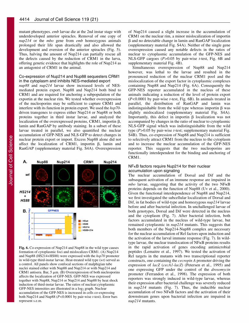
Co-expression of Nup214 and Nup88 sequesters CRM1in the cytoplasm and inhibits NES-mediated exportnup88 and nup214 larvae show increased levels of NES-mediated protein export. Nup88 and Nup214 both bind toCRM1 and are required for anchoring a subpopulation of theexportin at the nuclear rim. We tested whether overexpressionof the nucleoporins may be sufficient to capture CRM1 andinterfere with its function in protein export. We used the hsp70-driven transgenes to express either Nup214 or Nup88 or bothproteins together in third instar larvae, and analyzed thelocalization of the overexpressed proteins, CRM1, importin �,lamin and RanGAP by antibody staining. In a subset of theselarvae treated in parallel, we also quantified the nuclearaccumulation of GFP-NES and NLS-GFP to detect changes innuclear protein export or import. Excess Nup88 alone did notaffect the localization of CRM1, importin �, lamin andRanGAP (supplementary material Fig. S4A). Overexpression
of Nup214 caused a slight increase in the accumulation ofCRM1 on the nuclear rim, a minor mislocalization of importin� and no detectable change in lamin and RanGAP localization(supplementary material Fig. S4A). Neither of the single geneoverexpression caused any notable defects in the ratios ofnuclear to cytoplasmic accumulation of the GFP-NES andNLS-GFP cargoes (P>0.05 by pair-wise t-test, Fig. 6B andsupplementary material Fig. 4B).
Simultaneous overexpression of Nup88 and Nup214however, was lethal to the larvae and resulted in thepronounced reduction of the nuclear CRM1 pool and themislocalization of the export factor in cytoplasmic complexescontaining Nup88 and Nup214 (Fig. 6A). Consequently theGFP-NES reporter accumulated in the nucleus of theseanimals indicating a reduction of the level of protein export(P<0.0001 by pair-wise t-test, Fig. 6B). In animals treated inparallel, the distribution of RanGAP and lamin wasindistinguishable from the wild type whereas importin � wasslightly mislocalized (supplementary material Fig. S4A).Importantly, this defect in importin � localization was notaccompanied by changes in the ratio of nuclear to cytoplasmicNLS-GFP signal which was indistinguishable from the wildtype (P>0.05 by pair-wise t-test; supplementary material Fig.S4B). Thus, co-expression of Nup88 and Nup214 is sufficientto selectively attract CRM1 from the nucleus to the cytoplasmand to increase the nuclear accumulation of the GFP-NESreporter. This suggests that the two nucleoporins arefunctionally interdependent for the binding and anchoring ofCRM1.
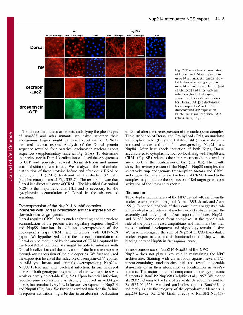
NF�B factors require Nup214 for their nuclearaccumulation upon signalingThe nuclear accumulation of Dorsal and Dif and thesubsequent activation of an immune response are impaired inmbo larvae, suggesting that the activity of the two NF�Bproteins depends on the function of Nup88 (Uv et al., 2000).Given the functional interdependence of Nup88 and Nup214,we first investigated the subcellular localization of Dorsal andDif, in fat bodies of wild-type and homozygous nup214 larvaebefore and after bacterial infection. In unchallenged larvae ofboth genotypes, Dorsal and Dif were detected in the nucleusand the cytoplasm (Fig. 7). After bacterial infection, bothfactors accumulated in the nucleus of wild-type larvae, butremained cytoplasmic in nup214 mutants. This suggests thatboth members of the Nup214-Nup88 complex are necessaryfor the nuclear accumulation of Rel factors upon induction andthe activation of the larval immune response (Fig. 7). In wild-type larvae, the nuclear translocation of NF�B proteins resultsin the rapid activation of genes encoding antimicrobialpeptides (Lemaitre et al., 1997). We tested the activation ofRel targets in the mutants with two transcriptional reporterconstructs, one containing the cecropin A promoter driving theexpression of lacZ (cecA1-lacZ) (Petersen et al., 1995) andone expressing GFP under the control of the drosomycinpromoter (Ferrandon et al., 1998). The expression of bothreporters was strongly induced in wild-type larvae, whereastheir expression after bacterial challenge was severely reducedin nup214 mutants (Fig. 7). Thus, the inducible nuclearaccumulation of two NF�B factors and the activation of theirdownstream genes upon bacterial infection are impaired innup214 mutants.
Journal of Cell Science 119 (21)
Fig. 6. Co-expression of Nup214 and Nup88 in the wild type causesformation of cytoplasmic foci and mislocalizes CRM1. (A) Nup214and Nup88 (HS214+HS88) were expressed with the hsp70 promoterin wild-type third-instar larvae. Heat-treated wild type (wt) served asa control. All panels show confocal sections of malphigian tubenuclei stained either with Nup88 and Nup214 or with Nup214 andCRM1 antisera. Bar, 5 �m. (B) Overexpression of both nucleoporinsaffects the localization of GFP-NES. GFP-NES was expressedtogether with Nup88, Nup214 or Nup214 and Nup88 by heat shockinduction of third-instar larvae. The ratios of nuclear:cytoplasmicGFP-NES intensities are illustrated in a log2 graph. Nuclearaccumulation of the GFP-NES is increased in cells co-expressingboth Nup214 and Nup88 (P<0.0001 by pair-wise t-test). Error barsrepresent s.e.m.
Jour
nal o
f Cel
l Sci
ence
4415Nup214 attenuates NES export
To address the molecular defects underlying the phenotypesof nup214 and mbo mutants we asked whether theirendogenous targets might be direct substrates of CRM1-mediated nuclear export. Analysis of the Dorsal proteinsequence revealed four putative leucine-rich nuclear exportsequences (supplementary material Fig. S5A). To determinetheir relevance in Dorsal localization we fused these sequencesto GFP and generated several Dorsal deletion and aminoacid substitution constructs. We analyzed the subcellulardistribution of these proteins before and after crm1 RNAi orleptomycin B (LMB) treatment of transfected S2 cells(supplementary material Fig. S5B,C). The results indicate thatDorsal is a direct substrate of CRM1. The identified C-terminalNES4 is the major functional NES and is necessary for thecytoplasmic accumulation of Dorsal in the absence ofsignaling.
Overexpression of the Nup214-Nup88 complexinterferes with Dorsal localization and the expression ofdownstream target genesDorsal requires CRM1 for its nuclear shuttling and the nuclearaccumulation of the protein after signaling requires Nup214and Nup88 function. In addition, overexpression of thenucleoporins traps CRM1 and interferes with GFP-NESexport. We hypothesized that if the nuclear accumulation ofDorsal can be modulated by the amount of CRM1 captured bythe Nup88-214 complex, we might be able to interfere withDorsal localization and the activation of the immune responsethrough overexpression of the nucleoporins. We first analyzedthe expression levels of the inducible drosomycin-GFP reporterin wild-type larvae and animals overexpressing Nup214-Nup88 before and after bacterial infection. In unchallengedlarvae of both genotypes, expression of the two reporters wasweak or barely detectable (Fig. 8A). Upon bacterial infection,reporter-gene expression was strongly induced in wild-typelarvae, but remained very low in larvae overexpressing Nup214and Nup88 (Fig. 8A). We further examined whether the failurein reporter activation might be due to an aberrant localization
of Dorsal after the overexpression of the nucleoporin complex.The distribution of Dorsal and Grainyhead (Grh), an unrelatedtranscription factor (Bray and Kafatos, 1991), was analyzed inuntreated larvae and animals overexpressing Nup214 andNup88. After heat shock induction of both Nups, Dorsalaccumulated to cytoplasmic foci co-localizing with Nup88 andCRM1 (Fig. 8B), whereas the same treatment did not result inany defects in the localization of Grh (Fig. 8B). The resultsshow that overexpression of the Nup214-Nup88 complex canselectively trap endogenous transcription factors and CRM1and suggest that alterations in the levels of CRM1 bound to thecomplex may modulate the expression of Rel target genes uponactivation of the immune response.
DiscussionThe cytoplasmic filaments of the NPC extend ~40 nm from thenuclear envelope (Goldberg and Allen, 1993; Jarnik and Aebi,1991). Functional analysis of their constituents suggests a rolein the cytoplasmic release of nuclear export substrates and theassembly and docking of nuclear import complexes. Nup214and Nup88 homologues form complexes at the cytoplasmicside of the pores in yeast, amphibians and mammals but theirroles in animal development and physiology remain elusive.We have investigated the role of Nup214 in CRM1-mediatednuclear export in vivo and its functional relationship with itsbinding partner Nup88 in Drosophila larvae.
Interdependence of Nup214-Nup88 at the NPCNup214 does not play a key role in maintaining the NPCarchitecture. Staining with an antibody against several FG-repeat-containing nucleoporins did not reveal detectableabnormalities in their abundance or localization in nup214mutants. The major structural component of the cytoplasmicfilaments is RanBP2-Nup358 (Delphin et al., 1997; Walther etal., 2002). Owing to the lack of a specific detection reagent forRanBP2-Nup358, we used antibodies against RanGAP, toindirectly assess the integrity of the cytoplasmic filaments innup214 larvae. RanGAP binds directly to RanBP2(Nup358)
Fig. 7. The nuclear accumulationof Dorsal and Dif is impaired innup214 mutants. All panels showfat bodies of wild-type (wt) andnup214 mutant larvae, before (notchallenged) and after bacterialinfection (bact. challenged)stained with specific antibodiesfor Dorsal, Dif, �-galactosidasefor cecropin-lacZ or GFP fordrosomycin-GFP expression.Nuclei are visualized with DAPI(blue). Bars, 35 �m.
Jour
nal o
f Cel
l Sci
ence

4416
and this binding is required for its accumulation at the nuclearrim. RanGAP was still localized along the nuclear envelope,but the staining appeared ~35% weaker and punctuated,suggesting that the Nup214-Nup88 complex does not playa major role in maintaining RanBP2(Nup358) on thecytoplasmic filaments.
The interdependence of Nup88 and Nup214 at the NPChowever, has been consistently observed in all species inwhich it has been analyzed. Nup88 is undetectable in cellsderived from Nup214-deficient embryos (Fornerod et al.,1997b) and RNAi inhibition of each of the genes in tissueculture results in reduced protein levels for the other (Bernadet al., 2004). In addition, yeast cells with a temperature-sensitive mutation of Nup82p, show a reduction of Nup159pfrom the nuclear rim (Belgareh et al., 1998). The molecularmechanisms underlying this interplay remain unknown.Nup88 is undetectable in nup214 mutants whereas the levelsof the nup88 transcript remain constant. In tissue cultureRNAi experiments, and in nup214 heterozygotes andhomozygous mutants, the reduction of Nup88 is proportionalto the amount of Nup214. In addition, epoxomycin can inhibitthe Nup88 reduction caused by the inactivation of Nup214.Since Nup214 and Nup88 bind to each other directly (Roth etal., 2003), the results suggest that Nup214 binding of Nup88at the NPC protects it from proteasome degradation. Thisprotection mechanism may involve interference with thedegradation of Nup88 selectively at the pore, because Nup88
appears to be a stable protein in the cytoplasm whenoverexpressed.
In mbo/nup88 mutants Nup214 detaches from the nuclearrim and localizes within the nucleus (Roth et al., 2003).Overexpression of Nup88 in nup214 mutants, lackingendogenous Nup214 and Nup88, results in accumulation of theoverexpressed protein at the nuclear rim indicating that Nup88alone is sufficient to target the complex to the pore. The highlevels of overexpressed Nup88 in nup214 mutants might beexplained by a protective function of minimal residual amountsof Nup214 or by the inability of the protein degradationmachinery to cope with the overproduced Nup88. The analysissuggests an intriguing posttranslational mechanism for theinterdependence of the two nucleoporins. Nup88 alone issufficient to associate with the NPC and this association is aprerequisite for the localization of Nup214 to the nuclearmembrane. In turn, the binding of Nup214 increases thestability of Nup88 proteins at the nuclear envelope and maythereby increase the potential of additional Nup88 moleculesto associate with the NPC. In sporadic cells of nup214 mutantsthat expressed high concentrations of Nup88 and low amountsof Nup214 after heat shock, the complex was localized alongthe nuclear envelope. By contrast, in cells expressing relativelylow Nup88 and high Nup214 levels, localization of thecomplex became nuclear. This distinct distribution of theproteins correlating with the relative expression levels ofthe two Nups in different cells, suggests that localization of
Journal of Cell Science 119 (21)
Fig. 8. Co-expression of Nup214and Nup88 in the wild type issufficient to mislocalizeendogenous targets. Nup214 andNup88 (HS214+HS88) expressionwas driven by the hsp70 promoterin wild-type third instar larvae.Heat-treated wild-type (wt) servedas a control. (A) Overexpressionof Nup214 and Nup88 interfereswith the expression of adrosomycin-GFP (dromGFP)reporter. All panels show fatbodies, before (not challenged)and after bacterial infection (bact.challenged) stained with GFPantibody. The images forHS214+HS88 unchallenged andinfected larval fat bodies wereacquired at three times lowerintensity for Nup214 labelingcompared with the wild type.(B) Confocal sections ofmalpighian tube nuclei stainedwith Nup88, Nup214, Dorsal,CRM1 or Grainyhead antiserum.Co-expression of Nup214 andNup88 can mislocalize Dorsal butnot Grainyhead. Bars, 35 �m (A);5 �m (B).
Jour
nal o
f Cel
l Sci
ence

4417Nup214 attenuates NES export
the complex is dynamic and depends on the relativeconcentrations of the two nucleoporins.
The Nup214-Nup88 complex attenuates NES-proteinexportNup214 binds to CRM1 and the cytoplasmic accumulation ofthe GFP-NES reporter is increased in nup214 mutants, whereasCRM1 is mislocalized from the NPC. These phenotypessuggest that Nup214 acts as an inhibitor of NES-mediatedexport. The physiological significance of the inhibitoryfunction of Nup214 on CRM1 export is further emphasized bythe phenotype caused by the reduction of Nup214 in crm1mutants. Removal of one chromosomal copy of nup214 inemb2 mutants, which die as second instar larvae, allows theseanimals to proceed into pupariation and to develop adultstructures. This extension of the life span of emb2 mutantssuggests that anchoring of CRM1 to the NPC is a generalmechanism that limits CRM1 activity. The endogenoussubstrates of CRM1 required for progression through the larvalstages and pupariation remain to be identified.
The phenotypes of Nup214 mutants are identical to thepreviously described CRM1-export defects of mbo/nup88larvae (Roth et al., 2003). However, overexpression of Nup88alone in wild-type animals did not affect CRM1 localizationand overexpression of Nup214 only caused a minor enrichmentof CRM1 concentration on the nuclear rim and did not interferewith NES export. The co-expression of both nucleoporinsunder the control of the heat-shock promoter resulted in thegross mislocalization of CRM1 from the nuclear envelope anddisrupted GFP-NES export. Thus, the Nup214-Nup88 complexis necessary and sufficient to tether a fraction of CRM1 andattenuate protein export.
Why would nuclear pore components negatively regulateCRM1 function? One possible explanation is that exportcomplex formation depends on the levels of CRM1 in thenucleoplasm. The binding affinity of CRM1 to natural NESsvaries, and cargoes with low affinity NESs may be exportedless efficiently (Engelsma et al., 2004; Henderson andEleftheriou, 2000; Kutay and Guttinger, 2005). Theintroduction of an artificial, high-affinity NES disrupts CRM1export indicating that natural NESs are selected for theirweaker affinity for the export factor (Engelsma et al., 2004).Removal of the Nup214-Nup88 sub-complex from the poreincreases the nuclear concentration of CRM1 and it would alsoincrease nuclear export of cargoes with low-affinity NESs.Tethering or releasing the NPC-bound fraction of the exportfactor may provide the means for controlling the nuclearconcentration of proteins carrying low-affinity export signals.
Nup214-Nup88 functions in NF�B translocation and theactivation of immune responsesDorsal contains a functional NLS embedded in its Rel-homology domain. This sequence is sufficient to target a �-galactosidase reporter into the nucleus and is required for thenuclear accumulation of Dorsal during embryogenesis (Drier etal., 1999; Govind et al., 1996). Owing to the phenotypes ofnup214 larvae in the nuclear accumulation of import reporterswe cannot exclude the fact that the defects of nup214 mutantsin the activation of immune responses may be partly due to areduction in the nuclear import levels of Dorsal and Dif. Wefavor the hypothesis that the nup214 phenotype in NF�B
translocation is primarily due to increased levels of proteinexport. Mutations inactivating Nup88, the partner of Nup214,disrupt NF�B translocation and show concurrent enhancedlevels of NES-mediated protein export but do not exhibit anydetectable effects on the nuclear import of the same reporters(Roth et al., 2003). In addition, we identified a functional,CRM1-dependent NES, required for the cytoplasmicaccumulation of Dorsal. This NES4 motif is deleted inhypomorphic dorsal alleles expressing truncated forms of theprotein and causing an extended nuclear gradient in the embryo.These mutants still retain their Cactus-binding domain and theirphenotypes become further enhanced by reduction of cactusactivity, suggesting that CRM1 export is an additional noveldeterminant of Dorsal localization and activation in S2 cells andthe embryo (Isoda et al., 1992; Rushlow et al., 1989). Therequirement of the Nup214-Nup88 complex for the fullactivation of the immune response and its inhibitory functionon CRM1 export in larvae suggest that the amplitude andduration of Toll signaling may be influenced by the export ratesof Dorsal and Dif. The interference of the complex with NF�Blocalization and activity upon overexpression, further suggeststhat changes in the relative amounts of the nucleoporin complexand the fraction of CRM1 bound to it, may provide a regulatorynode for the nuclear concentration of Dorsal and Dif. Variationsin the NPC-bound CRM1 pool could be accomplished in twoways: First by modifications of Nup88 or/and Nup214, whichcould influence their binding capacity to CRM1. Variations inthe affinity of the nucleoporin complex for CRM1 could explainthe changes in the amounts of co-precipitated CRM1, whereasthe amounts of Nup88, Nup214 and CRM1 remain constant inextracts from 5-10 and 10-15 hour Drosophila embryos. Nup88phosphorylation has been detected in Xenopus oocytes (Bernadet al., 2004), and such modifications might influence the affinityof CRM1 for the complex. Alternatively, transcriptional controlof Nup88 during fly development might also influence the levelsof Nup214 and CRM1 in turn, at the NPC. The steady-stateratios of Nup88 to Nup214 have been determined by proteomicstudies of yeast and rat liver NPCs and revealed a 2:1 and an8:1 excess of Nup88, respectively (Cronshaw et al., 2002; Routet al., 2000). Changes of the wild-type stoichiometry byoverexpression of the two nucleoporins leads to lethality inDrosophila larvae and apoptosis and G0 arrest in human cells(Boer et al., 1998). The zygotic expression pattern of Nup88 inDrosophila is tissue- and stage-specific (Roth et al., 2003; Uvet al., 2000). The interdependence of Nup214 and Nup88 atthe NPC may provide an elegant titration mechanism thatcontinuously monitors the structure and function of theNup214-Nup88 complex and the amount of CRM1 bound to it.
Materials and MethodsDrosophila strainsNup214 in Drosophila is encoded by the CG3820 gene and the enhancer trapinsertion l(2)10444 was identified as a P-element lethal mutation, where thetransposon was inserted into the nup214 gene at nucleotide position 122 after thetranslational initiation site of the predicted open reading frame (Flybase/BDGP).100 excision strains deriving from l(2)10444 were generated as described(Robertson et al., 1988) and balanced over CyO ftz-LacZ and CyO GFP. The P-element excision generated several homozygous viable strains and the lethalnup214. This excision allele failed to complement Df(2R)3-70 (Flybase/BDGP).The following fly strains were used: mbo-1 (Uv et al., 2000), hs-nup88 (Uv et al.,2000), hs-grh (Uv et al., 1994), hs-ubx (White and Wilcox, 1984), hs-NLS-GFP andhs-GFP-NES (Roth et al., 2003), cecropin-lacZ (Petersen et al., 1995) anddrosomycin-GFP (Ferrandon et al., 1998). The hs-nup214 transgenic fly strains weregenerated by P-element-mediated transformation (Spradling, 1986). nup214 was
Jour
nal o
f Cel
l Sci
ence

4418
amplified from full-length cDNA using LA polymerase (TAKARA) for 12 cyclesand cloned into the BglII and XbaI sites of pCaSpeR-hs.
Genetic interactions were assessed by examining the lethal phase of animals ofthe following genotypes: Oregon-R, emb2/emb2, nup214 emb2/emb2 and emb2/emb2;Dfmbo (Dfmbo corresponds to Df(3R)Kar-Sz29f, Flybase).
Sequencing and RT-PCRFor sequencing of the nup214 excision allele, genomic DNA from wild-type andhomozygous mutant larvae (Gloor et al., 1993) was amplified with the High FidelityPCR system (Roche) using primer pair: 5�-TGCGATTGAATTCGAAGGAT-3�and 3�-CAGCCTTGGGAGCATTTAGA-5�. PCR reactions were analyzed byelectrophoresis in 1% agarose gel and purified using the QIAquick PCR PurificationKit (Qiagen). Sequencing reactions were performed with the same primer pair orwith the internal primer: 5�-TCAAGCTAAGTTGGTTTCTTTT-3� using BigDyeTerminator v3.0 sequencing kit (Applied Biosystems).
Larval mRNA was extracted and annealed to magnetic oligo(dT) coupled beads(Dynabeads) according to the manufacturer’s instructions. Reverse transcription wasperformed using SuperScriptII (Invitrogen). Serial dilutions were used as templatefor PCR amplification with specific primers for: C-terminal mbo (5�-GTTA-ACCAGCCCATCTTGGCGG-3� and 3�-TTAGATGCCAACGATTTTATT-5�) (25cycles) and rp49 (5�-TGACCATCCGCCCAGCATACA-3� and 3�-TCTCGCCGCA-GTAAAC-5�) (22 cycles). The products were analyzed by electrophoresis andvisualized with ethidium bromide.
Binding assaysAll constructs were generated by ten-cycle PCR amplification from cDNA clones.Fragments were cloned either into pGEX-5x (Amersham Bioscience) or in pRSET(Invitrogen). GST and His6 fusion proteins were expressed in the bacterial strainBL21pLys. Bacteria were harvested and sonicated in a buffer containing 150 mMNaCl, 16 mM Na2HPO4, 4 mM NaH2PO4, 1% NP40 and 1 mM DTT. The boundproteins were analyzed on western blots using anti-RGS-His (Qiagen) diluted1:2000 and anti-GST (1:1000; Santa Cruz Biotechnology). For the yeast two-hybridsystem PCR fragments were cloned into either pAct2 (GAL4 DNA-binding domainvector; HA tagged; TRP selectable marker) or pAS1 (GAL4 activation domainvector; HA tagged; LEU2 selectable marker). The different constructs were pair-wise introduced in the yeast strain PJ69-4A (James et al., 1996) by LiActransformation method (Gietz and Schiestl, 1995) and grown at 30°C for 3-4 days.Transformants were selected on SD-TRP–LEU– plates. Protein was extracted frompositive clones as described in (Adams et al., 1997). Expression of the differentfusion proteins was detected on western blots using an anti-hemagglutinin (HA)antibody (1:2000; Babco). Interaction was scored by growth on SD-TRP–LEU–ADE– plates and �-galactosidase assays were performed as described(Adams et al., 1997).
Cell culture and RNAi treatmentS2 cells were cultured at 25°C in Schneider’s Drosophila medium (PAN)supplemented with 10% heat-inactivated FCS (Invitrogen), 50 �g/ml gentamycin,50 U/ml penicillin, 50 �g/ml streptomycin (PAN) and 2 mM glutamine (Invitrogen).700 bp fragments of nup214, nup88 and emb/crm1 were amplified by PCR usingthe nucleotides: 5�-TTAATACGACTCACTATAGGGAGAAAGCCTGATGACA-CTGAGC-3� and 5�-TTAATACGACTCACTATAGGGAGAGGAGTCGGTCAAT-GTATCC-3�; 5�-TTAATACGACTCACTATAGGGAGAACTACAAGGATGGCA-AGCC-3� and 5�-TTAATACGACTCACTATAGGGAGACAACACAGAGTCGT-CAACC-3�; TTAATACGACTCACTATAGGGAGATCGCAAATCTTGCGACGC-3� and 5�-TTAATACGACTCACTATAGGGAGATGTCAATGATGAATGTCTCC-3�. These were then used as templates to produce dsNup214, dsNup88 anddsCRM1/emb RNA respectively with the MEGAscript RNAi kit (Ambion). ThedsRNA treatment was performed as described (Clemens et al., 2000). For the Nup88degradation assay, Nup214 RNAi cells at day 3 post treatment were incubated with20 �M epoxomicin (Calbiochem).
Site-directed mutagenesis and live cell imagingMutations of the Dorsal NES3 and NES4 were created using the QuikChangemutagenesis kit (Stratagene). For both NESs the first two large hydrophobic residueswere changed to alanines. NES3, L553SNL556NNPFTM, was changed toA553SNA556NNPFTM and NES4, DL669QI671SNLSIS, was changed toDA669QA671SNLSIS. LMB was used at the final concentration of 10 ng/ml for 2hours. For protein expression in Drosophila S2 cells the different constructs werecloned either in pAc5.1/V5-His or pMT/V5-His (Invitrogen). S2 cells weretransfected with the TransFastTM Transfection Reagent according to themanufacturer’s instructions (Promega). For staining of DNA in living cells, Hoechst33342 (Sigma) was added to the culture medium at final concentration of 4 �M.Cells at day 4 and day 6 were viewed with a Leica DM IRB inverted Fluorescencemicroscope. Images were acquired using a Leica DC300F CCD camera andprocessed with Image Viewer 5.02 software (Kodak).
Immunostaining of larvae and Drosophila S2 cellsLarvae were heat induced for 1 hour at 37°C and analyzed after 2 or 3 hours.
Antibody staining of larval tissues was performed as described (Patel, 1994). Dorsaland Dif translocation experiments were done as described (Uv et al., 2000).Drosophila S2 cells were directly fixed on a poly-L-lysine (Sigma)-coated coverslipin 4% freshly prepared paraformaldehyde in phosphate-buffered saline (PBS) for30 minutes at room temperature. After fixing, the cells were washed in PBS andpermeabilized in 0.1% Triton X-100 in PBS for 5 minutes. Cells were preincubatedwith PBS 0.1% Triton X-100 and 0.5% BSA (Albumin, Bovine, Sigma) for 30minutes at room temperature followed by incubation with primary antibodiesovernight at 4°C. After rinsing and preincubation with PBS 0.1% Triton X-100 and0.5% BSA for 30 minutes, the cells were incubated with secondary antibody for 2hours at room temperature, washed and incubated with DAPI (0.4 �g/ml) for 5minutes. After rinsing, the cells were mounted in 70% glycerol containing 2.5%DABCO (1,4 diazabicyclo[2.2.2]octane; Sigma).
The following primary antibodies were used: anti-Nup214 (Roth et al., 2003)diluted 1:10,000, anti-CRM1 1:1000 (Roth et al., 2003); anti-GFP 1:1000(Molecular Probes); anti-lamin Dm1 (ADL84) 1:500 (Stuurman et al., 1995); anti-Ubx 1:20 (White and Wilcox, 1984); anti-Grh 1:5 (Bray and Kafatos, 1991); anti-RanGAP 1:1000 (Merrill et al., 1999); anti-Nup88 1:100 (Uv et al., 2000), mAb4141:5000 (Babco), anti-�-Gal 1:2000 (Promega), anti-Dorsal 1:1000 (Gillespie andWasserman, 1994), anti-Dif 1:300 (Cantera et al., 1999), C-terminal anti-His 1:2000(Invitrogen), anti-V5 1:2000 (Invitrogen) and anti-Ketel (Lippai et al., 2000) 1:1000.Secondary antibodies conjugated to Alexa Fluor 488 (Molecular Probes) or Cy3(Jackson Laboratories) were used as recommended.
Confocal images were acquired with a LSM 510 laser-scanning microscope(Zeiss). Quantitative image analysis was performed with the LSM 5 ver3 software(Zeiss). Fluorescent images were recorded on a Zeiss fluorescence microscopeequipped with (Axio Cam HRc) CCD camera. GFP intensities were measured froma selected area in the cytoplasm and an area of the same size in the nucleus(visualized by DAPI staining) by using the Volocity 2.0.1 software (Improvision).The ratio of nuclear/cytoplasmic fluorescent signal was determined from �25 cells.
Immunoprecipitation and western blotsImmunoprecipitations from embryonic extracts were performed as described(Edwards et al., 1997) except for the lysis buffer (10 mM Tris-HCl pH 8.0, 140 mMNaCl, 1.5 mM MgCl2, 1% NP-40, protease inhibitor cocktail (Boehringer), 5 mMpyrophosphate, 10 mM NaF, 10 mM �-glycerol phosphate, 5 mM sodium vanadate).The following antibody dilutions were used in western blots: anti-�-tubulin(Amersham) (1:1000), anti-lamin Dm1 (ADL84) 1:500 (Stuurman et al., 1995),anti-Nup214 (Roth et al., 2003) 1:1000 and anti-Nup88 (Uv et al., 2000) 1:1000and anti-CRM1 (Roth et al., 2003) 1:1000. Signals on western blots were quantifiedwith the Fuji Image Gauge V3.45 software.
We thank J. Szabad (University of Szeged, Hungary), P. A. Fisher(Stony Brook University, Stony Brook, NY), B. Ganetzky (Universityof Wisconsin, Madison, WI) for reagents and K. Senti and N. Visa forhelpful comments on the manuscript. This work was supported bygrants from the Cancerfonden and Wallenberg Consortium North toC.S.
ReferencesAdams, A., Gottschling, D. E., Kaiser, C. A. and Stearns, T. (1997). Methods in Yeast
Genetics: A Cold Spring Harbor Laboratory Course Manual. Cold Spring Harbor:Cold Spring Harbor Laboratory Press.
Askjaer, P., Bachi, A., Wilm, M., Bischoff, F. R., Weeks, D. L., Ogniewski, V., Ohno,M., Niehrs, C., Kjems, J., Mattaj, I. W. et al. (1999). RanGTP-regulated interactionsof CRM1 with nucleoporins and a shuttling DEAD-box helicase. Mol. Cell. Biol. 19,6276-6285.
Becskei, A. and Mattaj, I. W. (2005). Quantitative models of nuclear transport. Curr.Opin. Cell Biol. 17, 27-34.
Belgareh, N., Snay-Hodge, C., Pasteau, F., Dagher, S., Cole, C. N. and Doye, V. (1998).Functional characterization of a Nup159p-containing nuclear pore subcomplex. Mol.Biol. Cell 9, 3475-3492.
Bernad, R., van der Velde, H., Fornerod, M. and Pickersgill, H. (2004).Nup358/RanBP2 attaches to the nuclear pore complex via association with Nup88 andNup214/CAN and plays a supporting role in CRM1-mediated nuclear protein export.Mol. Cell. Biol. 24, 2373-2384.
Bernad, R., Engelsma, D., Sanderson, H., Pickersgill, H. and Fornerod, M. (2006).The Nup214/Nup88 nucleoporin subcomplex is required for CRM1 mediated 60Spreribosomal nuclear export. J. Biol. Chem. 281, 19378-19386.
Boer, J., Bonten-Surtel, J. and Grosveld, G. (1998). Overexpression of the nucleoporinCAN/NUP214 induces growth arrest, nucleocytoplasmic transport defects, andapoptosis. Mol. Cell. Biol. 18, 1236-1247.
Bray, S. J. and Kafatos, F. C. (1991). Developmental function of Elf-1: an essentialtranscription factor during embryogenesis in Drosophila. Genes Dev. 5, 1672-1683.
Brennan, C. A. and Anderson, K. V. (2004). Drosophila: the genetics of innate immunerecognition and response. Annu. Rev. Immunol. 22, 457-483.
Cantera, R., Roos, E. and Engstrom, Y. (1999). Dif and cactus are colocalized in thelarval nervous system of Drosophila melanogaster. J. Neurobiol. 38, 16-26.
Journal of Cell Science 119 (21)
Jour
nal o
f Cel
l Sci
ence

4419Nup214 attenuates NES export
Clemens, J. C., Worby, C. A., Simonson-Leff, N., Muda, M., Maehama, T.,Hemmings, B. A. and Dixon, J. E. (2000). Use of double-stranded RNA interferencein Drosophila cell lines to dissect signal transduction pathways. Proc. Natl. Acad. Sci.USA 97, 6499-6503.
Collier, S., Chan, H. Y., Toda, T., McKimmie, C., Johnson, G., Adler, P. N., O’Kane,C. and Ashburner, M. (2000). The Drosophila embargoed gene is required for larvalprogression and encodes the functional homolog of schizosaccharomyces Crm1.Genetics 155, 1799-1807.
Cronshaw, J. M., Krutchinsky, A. N., Zhang, W., Chait, B. T. and Matunis, M. J.(2002). Proteomic analysis of the mammalian nuclear pore complex. J. Cell Biol. 158,915-927.
Davis, L. I. and Blobel, G. (1986). Identification and characterization of a nuclear porecomplex protein. Cell 45, 699-709.
Delphin, C., Guan, T., Melchior, F. and Gerace, L. (1997). RanGTP targets p97 toRanBP2, a filamentous protein localized at the cytoplasmic periphery of the nuclearpore complex. Mol. Biol. Cell 8, 2379-2390.
Drier, E. A., Huang, L. H. and Steward, R. (1999). Nuclear import of the DrosophilaRel protein Dorsal is regulated by phosphorylation. Genes Dev. 13, 556-568.
Edwards, D. N., Towb, P. and Wasserman, S. A. (1997). An activity-dependent networkof interactions links the Rel protein Dorsal with its cytoplasmic regulators.Development 124, 3855-3864.
Engelsma, D., Bernad, R., Calafat, J. and Fornerod, M. (2004). Supraphysiologicalnuclear export signals bind CRM1 independently of RanGTP and arrest at Nup358.EMBO J. 23, 3643-3652.
Ferrandon, D., Jung, A. C., Criqui, M., Lemaitre, B., Uttenweiler-Joseph, S.,Michaut, L., Reichhart, J. and Hoffmann, J. A. (1998). A drosomycin-GFP reportertransgene reveals a local immune response in Drosophila that is not dependent on theToll pathway. EMBO J. 17, 1217-1227.
Floer, M., Blobel, G. and Rexach, M. (1997). Disassembly of RanGTP-karyopherin betacomplex, an intermediate in nuclear protein import. J. Biol. Chem. 272, 19538-19546.
Fornerod, M. and Ohno, M. (2002). Exportin-mediated nuclear export of proteins andribonucleoproteins. Results Probl. Cell Differ. 35, 67-91.
Fornerod, M., Ohno, M., Yoshida, M. and Mattaj, I. W. (1997a). CRM1 is an exportreceptor for leucine-rich nuclear export signals. Cell 90, 1051-1060.
Fornerod, M., van Deursen, J., van Baal, S., Reynolds, A., Davis, D., Murti, K. G.,Fransen, J. and Grosveld, G. (1997b). The human homologue of yeast CRM1 is ina dynamic subcomplex with CAN/Nup214 and a novel nuclear pore component Nup88.EMBO J. 16, 807-816.
Fukuda, M., Asano, S., Nakamura, T., Adachi, M., Yoshida, M., Yanagida, M. andNishida, E. (1997). CRM1 is responsible for intracellular transport mediated by thenuclear export signal. Nature 390, 308-311.
Gietz, R. D. and Schiestl, R. H. (1995). Transforming yeast with DNA. Methods Mol.Cell. Biol. 5, 255-269.
Gillespie, S. K. and Wasserman, S. A. (1994). Dorsal, a Drosophila Rel-like protein, isphosphorylated upon activation of the transmembrane protein Toll. Mol. Cell. Biol. 14,3559-3568.
Gloor, G. B., Preston, C. R., Johnson-Schlitz, D. M., Nassif, N. A., Phillis, R. W.,Benz, W. K., Robertson, H. M. and Engels, W. R. (1993). Type I repressors of Pelement mobility. Genetics 135, 81-95.
Goldberg, M. W. and Allen, T. D. (1993). The nuclear pore complex: three-dimensionalsurface structure revealed by field emission, in-lens scanning electron microscopy, withunderlying structure uncovered by proteolysis. J. Cell Sci. 106, 261-274.
Govind, S., Drier, E., Huang, L. H. and Steward, R. (1996). Regulated nuclear importof the Drosophila rel protein dorsal: structure-function analysis. Mol. Cell. Biol. 16,1103-1114.
Henderson, B. R. and Eleftheriou, A. (2000). A comparison of the activity, sequencespecificity, and CRM1-dependence of different nuclear export signals. Exp. Cell Res.256, 213-224.
Hopper, A. K., Traglia, H. M. and Dunst, R. W. (1990). The yeast RNA1 gene productnecessary for RNA processing is located in the cytosol and apparently excluded fromthe nucleus. J. Cell Biol. 111, 309-321.
Isoda, K., Roth, S. and Nusslein-Volhard, C. (1992). The functional domains of theDrosophila morphogen dorsal: evidence from the analysis of mutants. Genes Dev. 6,619-630.
James, P., Halladay, J. and Craig, E. A. (1996). Genomic libraries and a host straindesigned for highly efficient two- hybrid selection in yeast. Genetics 144, 1425-1436.
Jarnik, M. and Aebi, U. (1991). Toward a more complete 3-D structure of the nuclearpore complex. J. Struct. Biol. 107, 291-308.
Kaffman, A. and O’Shea, E. K. (1999). Regulation of nuclear localization: a key to adoor. Annu. Rev. Cell Dev. Biol. 15, 291-339.
Kehlenbach, R. H. and Gerace, L. (2000). Phosphorylation of the nuclear transportmachinery down-regulates nuclear protein import in vitro. J. Biol. Chem. 275, 17848-17856.
Kraemer, D., Wozniak, R. W., Blobel, G. and Radu, A. (1994). The human CANprotein, a putative oncogene product associated with myeloid leukemogenesis, is anuclear pore complex protein that faces the cytoplasm. Proc. Natl. Acad. Sci. USA 91,1519-1523.
Kuersten, S., Arts, G. J., Walther, T. C., Englmeier, L. and Mattaj, I. W. (2002).Steady-state nuclear localization of exportin-t involves RanGTP binding and twodistinct nuclear pore complex interaction domains. Mol. Cell. Biol. 22, 5708-5720.
Kutay, U. and Guttinger, S. (2005). Leucine-rich nuclear-export signals: born to be weak.Trends Cell Biol. 15, 121-124.
Lemaitre, B., Reichhart, J. M. and Hoffmann, J. A. (1997). Drosophila host defense:differential induction of antimicrobial peptide genes after infection by various classesof microorganisms. Proc. Natl. Acad. Sci. USA 94, 14614-14619.
Lippai, M., Tirian, L., Boros, I., Mihaly, J., Erdelyi, M., Belecz, I., Mathe, E., Posfai,J., Nagy, A., Udvardy, A. et al. (2000). The Ketel gene encodes a Drosophilahomologue of importin-beta. Genetics 156, 1889-1900.
Makhnevych, T., Lusk, C. P., Anderson, A. M., Aitchison, J. D. and Wozniak, R. W.(2003). Cell cycle regulated transport controlled by alterations in the nuclear porecomplex. Cell 115, 813-823.
Matsubayashi, Y., Fukuda, M. and Nishida, E. (2001). Evidence for existence of anuclear pore complex-mediated, cytosol-independent pathway of nuclear translocationof ERK MAP kinase in permeabilized cells. J. Biol. Chem. 276, 41755-41760.
Matunis, M. J., Coutavas, E. and Blobel, G. (1996). A novel ubiquitin-like modificationmodulates the partitioning of the Ran-GTPase-activating protein RanGAP1 betweenthe cytosol and the nuclear pore complex. J. Cell Biol. 135, 1457-1470.
Meng, L., Mohan, R., Kwok, B. H., Elofsson, M., Sin, N. and Crews, C. M. (1999).Epoxomicin, a potent and selective proteasome inhibitor, exhibits in vivoantiinflammatory activity. Proc. Natl. Acad. Sci. USA 96, 10403-10408.
Merrill, C., Bayraktaroglu, L., Kusano, A. and Ganetzky, B. (1999). TruncatedRanGAP encoded by the Segregation Distorter locus of Drosophila. Science 283, 1742-1745.
Moroianu, J., Blobel, G. and Radu, A. (1997). RanGTP-mediated nuclear export ofkaryopherin alpha involves its interaction with the nucleoporin Nup153. Proc. Natl.Acad. Sci. USA 94, 9699-9704.
Pante, N., Bastos, R., McMorrow, I., Burke, B. and Aebi, U. (1994). Interactions andthree-dimensional localization of a group of nuclear pore complex proteins. J. CellBiol. 126, 603-617.
Patel, N. H. (1994). Imaging neuronal subsets and other cell types in whole-mountDrosophila embryos and larvae using antibody probes. In Drosophila melanogaster:Practical uses in Cell and Molecular Biology (ed S. B. Goldstein and E. A. Fyrberg),pp. 45-487. Cambridge, MA: Academic Press.
Petersen, U. M., Bjorklund, G., Ip, Y. T. and Engstrom, Y. (1995). The dorsal-relatedimmunity factor, Dif, is a sequence-specific trans-activator of Drosophila Cecropingene expression. EMBO J. 14, 3146-3158.
Robertson, H. M., Preston, C. R., Phillis, R. W., Johnson-Schlitz, D. M., Benz, W. K.and Engels, W. R. (1988). A stable genomic source of P element transposase inDrosophila melanogaster. Genetics 118, 461-470.
Roth, P., Xylourgidis, N., Sabri, N., Uv, A., Fornerod, M. and Samakovlis, C.(2003). The Drosophila nucleoporin DNup88 localizes DNup214 and CRM1 on thenuclear envelope and attenuates NES-mediated nuclear export. J. Cell Biol. 163, 701-706.
Rout, M. P., Aitchison, J. D., Suprapto, A., Hjertaas, K., Zhao, Y. and Chait, B. T.(2000). The yeast nuclear pore complex: composition, architecture, and transportmechanism. J. Cell Biol. 148, 635-651.
Rushlow, C. A., Han, K., Manley, J. L. and Levine, M. (1989). The graded distributionof the dorsal morphogen is initiated by selective nuclear transport in Drosophila. Cell59, 1165-1177.
Snow, C. M., Senior, A. and Gerace, L. (1987). Monoclonal antibodies identify a groupof nuclear pore complex glycoproteins. J. Cell Biol. 104, 1143-1156.
Spradling, A. C. (1986). P element-mediated transformation. In Drosophila: A practicalApproach (ed. D. B. Roberts). Oxford: Oxford University Press.
Stade, K., Ford, C. S., Guthrie, C. and Weis, K. (1997). Exportin 1 (Crm1p) is anessential nuclear export factor. Cell 90, 1041-1050.
Stuurman, N., Maus, N. and Fisher, P. A. (1995). Interphase phosphorylation of theDrosophila nuclear lamin: site- mapping using a monoclonal antibody. J. Cell Sci. 108,3137-3144.
Suntharalingam, M. and Wente, S. R. (2003). Peering through the pore: nuclear porecomplex structure, assembly, and function. Dev. Cell 4, 775-789.
Uv, A. E., Thompson, C. R. and Bray, S. J. (1994). The Drosophila tissue-specific factorGrainyhead contains novel DNA-binding and dimerization domains which areconserved in the human protein CP2. Mol. Cell. Biol. 14, 4020-4031.
Uv, A. E., Roth, P., Xylourgidis, N., Wickberg, A., Cantera, R. and Samakovlis, C.(2000). members only encodes a Drosophila nucleoporin required for rel protein importand immune response activation. Genes Dev. 14, 1945-1957.
von Lindern, M., Fornerod, M., van Baal, S., Jaegle, M., de Wit, T., Buijs, A. andGrosveld, G. (1992). The translocation (6;9), associated with a specific subtype ofacute myeloid leukemia, results in the fusion of two genes, dek and can, and theexpression of a chimeric, leukemia-specific dek-can mRNA. Mol. Cell. Biol. 12, 1687-1697.
Walther, T. C., Pickersgill, H. S., Cordes, V. C., Goldberg, M. W., Allen, T. D.,Mattaj, I. W. and Fornerod, M. (2002). The cytoplasmic filaments of the nuclearpore complex are dispensable for selective nuclear protein import. J. Cell Biol. 158,63-77.
Weis, K. (2003). Regulating access to the genome: nucleocytoplasmic transportthroughout the cell cycle. Cell 112, 441-451.
White, R. A. and Wilcox, M. (1984). Protein products of the bithorax complex inDrosophila. Cell 39, 163-171.
Xu, L. and Massague, J. (2004). Nucleocytoplasmic shuttling of signal transducers. Nat.Rev. Mol. Cell Biol. 5, 209-219.
Xu, L., Kang, Y., Col, S. and Massague, J. (2002). Smad2 nucleocytoplasmic shuttlingby nucleoporins CAN/Nup214 and Nup153 feeds TGFbeta signaling complexes in thecytoplasm and nucleus. Mol. Cell 10, 271-282.
Jour
nal o
f Cel
l Sci
ence