The new england journal medicine - Welcome — UNC … new england journal of medicine 2 n engl j...
12
n engl j med 360;22 nejm.org may 28, 2009 1 The new england journal of medicine established in 1812 may 28, 2009 vol. 360 no. 22 Radiofrequency Ablation in Barrett’s Esophagus with Dysplasia Nicholas J. Shaheen, M.D., M.P.H., Prateek Sharma, M.D., Bergein F. Overholt, M.D., Herbert C. Wolfsen, M.D., Richard E. Sampliner, M.D., Kenneth K. Wang, M.D., Joseph A. Galanko, Ph.D., Mary P. Bronner, M.D., John R. Goldblum, M.D., Ana E. Bennett, M.D., Blair A. Jobe, M.D., Glenn M. Eisen, M.D., M.P.H., M. Brian Fennerty, M.D., John G. Hunter, M.D., David E. Fleischer, M.D., Virender K. Sharma, M.D., Robert H. Hawes, M.D., Brenda J. Hoffman, M.D., Richard I. Rothstein, M.D., Stuart R. Gordon, M.D., Hiroshi Mashimo, M.D., Ph.D., Kenneth J. Chang, M.D., V. Raman Muthusamy, M.D., Steven A. Edmundowicz, M.D., Stuart J. Spechler, M.D., Ali A. Siddiqui, M.D., Rhonda F. Souza, M.D., Anthony Infantolino, M.D., Gary W. Falk, M.D., Michael B. Kimmey, M.D., Ryan D. Madanick, M.D., Amitabh Chak, M.D., and Charles J. Lightdale, M.D. Abstract The authors’ affiliations are listed in the Appendix. Address reprint requests to Dr. Shaheen at the Center for Esophageal Diseases and Swallowing, University of North Carolina School of Medicine, CB 7080, Chapel Hill, NC 27599-7080, or at [email protected]. N Engl J Med 2009;360:xxx-xx. Copyright © 2009 Massachusetts Medical Society. Background Barrett’s esophagus, a condition of intestinal metaplasia of the esophagus, is associ- ated with an increased risk of esophageal adenocarcinoma. The condition may prog- ress through stages of dysplasia before cancer. We assessed whether an endoscopic intervention, radiofrequency ablation, could eradicate dysplastic Barrett’s esophagus and decrease the rate of neoplastic progression. Methods In a multicenter, sham-controlled trial, we randomly assigned 127 patients with dys- plastic Barrett’s esophagus in a 2:1 ratio to receive either radiofrequency ablation (ablation group) or a sham procedure (control group). Randomization was stratified according to the grade of dysplasia (low-grade or high-grade) and the length of Barrett’s esophagus (<4 cm or 4 to 8 cm). Primary outcomes at 12 months included the complete eradication of dysplasia and intestinal metaplasia. Secondary outcomes included progression to more severe dysplasia or cancer and adverse events. Results In the intention-to-treat analyses, among patients with low-grade dysplasia, complete eradication of dysplasia occurred in 90.5% of those in the ablation group, as com- pared with 22.7% of those in the control group (P<0.001). Among patients with high- grade dysplasia, complete eradication occurred in 81.0% of those in the ablation group, as compared with 19.0% of those in the control group (P<0.001). Overall, 77.4% of patients in the ablation group had complete eradication of intestinal metaplasia, as compared with 2.3% of those in the control group (P<0.001). Patients in the abla- tion group had less disease progression (3.6% vs. 16.3%, P = 0.03) and fewer cancers (1.2% vs. 9.3%, P = 0.045). Patients reported having more chest pain after the ablation procedure than after the sham procedure. In the ablation group, one patient had upper gastrointestinal hemorrhage, and five (6.0%) patients had esophageal stricture. Conclusions In patients with dysplastic Barrett’s esophagus, radiofrequency ablation was associated with a high rate of complete eradication of both dysplasia and intestinal metaplasia and a reduced risk of disease progression. (ClinicalTrials.gov number, NCT00282672.)
Transcript of The new england journal medicine - Welcome — UNC … new england journal of medicine 2 n engl j...

n engl j med 360;22 nejm.org may 28, 2009 1
The new england journal of medicineestablished in 1812 may 28, 2009 vol. 360 no. 22
Radiofrequency Ablation in Barrett’s Esophagus with DysplasiaNicholas J. Shaheen, M.D., M.P.H., Prateek Sharma, M.D., Bergein F. Overholt, M.D., Herbert C. Wolfsen, M.D.,
Richard E. Sampliner, M.D., Kenneth K. Wang, M.D., Joseph A. Galanko, Ph.D., Mary P. Bronner, M.D., John R. Goldblum, M.D., Ana E. Bennett, M.D., Blair A. Jobe, M.D., Glenn M. Eisen, M.D., M.P.H.,
M. Brian Fennerty, M.D., John G. Hunter, M.D., David E. Fleischer, M.D., Virender K. Sharma, M.D., Robert H. Hawes, M.D., Brenda J. Hoffman, M.D., Richard I. Rothstein, M.D., Stuart R. Gordon, M.D.,
Hiroshi Mashimo, M.D., Ph.D., Kenneth J. Chang, M.D., V. Raman Muthusamy, M.D., Steven A. Edmundowicz, M.D., Stuart J. Spechler, M.D., Ali A. Siddiqui, M.D., Rhonda F. Souza, M.D.,
Anthony Infantolino, M.D., Gary W. Falk, M.D., Michael B. Kimmey, M.D., Ryan D. Madanick, M.D., Amitabh Chak, M.D., and Charles J. Lightdale, M.D.
A bs tr ac t
The authors’ affiliations are listed in the Appendix. Address reprint requests to Dr. Shaheen at the Center for Esophageal Diseases and Swallowing, University of North Carolina School of Medicine, CB 7080, Chapel Hill, NC 27599-7080, or at [email protected].
N Engl J Med 2009;360:xxx-xx.Copyright © 2009 Massachusetts Medical Society.
Background
Barrett’s esophagus, a condition of intestinal metaplasia of the esophagus, is associ-ated with an increased risk of esophageal adenocarcinoma. The condition may prog-ress through stages of dysplasia before cancer. We assessed whether an endoscopic intervention, radiofrequency ablation, could eradicate dysplastic Barrett’s esophagus and decrease the rate of neoplastic progression.
Methods
In a multicenter, sham-controlled trial, we randomly assigned 127 patients with dys-plastic Barrett’s esophagus in a 2:1 ratio to receive either radiofrequency ablation (ablation group) or a sham procedure (control group). Randomization was stratified according to the grade of dysplasia (low-grade or high-grade) and the length of Barrett’s esophagus (<4 cm or 4 to 8 cm). Primary outcomes at 12 months included the complete eradication of dysplasia and intestinal metaplasia. Secondary outcomes included progression to more severe dysplasia or cancer and adverse events.
Results
In the intention-to-treat analyses, among patients with low-grade dysplasia, complete eradication of dysplasia occurred in 90.5% of those in the ablation group, as com-pared with 22.7% of those in the control group (P<0.001). Among patients with high-grade dysplasia, complete eradication occurred in 81.0% of those in the ablation group, as compared with 19.0% of those in the control group (P<0.001). Overall, 77.4% of patients in the ablation group had complete eradication of intestinal metaplasia, as compared with 2.3% of those in the control group (P<0.001). Patients in the abla-tion group had less disease progression (3.6% vs. 16.3%, P = 0.03) and fewer cancers (1.2% vs. 9.3%, P = 0.045). Patients reported having more chest pain after the ablation procedure than after the sham procedure. In the ablation group, one patient had upper gastrointestinal hemorrhage, and five (6.0%) patients had esophageal stricture.
Conclusions
In patients with dysplastic Barrett’s esophagus, radiofrequency ablation was associated with a high rate of complete eradication of both dysplasia and intestinal metaplasia and a reduced risk of disease progression. (ClinicalTrials.gov number, NCT00282672.)

T h e n e w e ngl a nd j o u r na l o f m e dic i n e
n engl j med 360;22 nejm.org may 28, 20092
Barrett’s esophagus is defined as metaplasia of the esophageal epithelium, with normal squamous epithelium replaced
by columnar epithelium containing goblet cells, also known as intestinal metaplasia (Fig. 1A).1 This change is associated with gastroesophageal ref lux disease.2 Approximately 10% of patients with chronic reflux have Barrett’s esophagus,3,4 and the prevalence of the condition in a recent population study was 1.6%.5 The condition is as-sociated with an increased risk of esophageal ad-enocarcinoma.6,7 The incidence of this once rare cancer has increased by more than 500% since the 1970s.8 The cancer remains highly lethal, with a 5-year survival rate of less than 15%.9
Barrett’s esophagus with no histologic evidence of cellular atypia is classified as nondysplastic in-testinal metaplasia. However, intestinal metaplasia cells may develop progressively more abnormal features, ranging from low-grade dysplasia to high-grade dysplasia. Longitudinal studies have shown that most cases of Barrett’s esophagus do not progress beyond nondysplastic intestinal meta-plasia or transient low-grade dysplasia.10,11 How-ever, in cases of progression to high-grade dyspla-sia, the risk of esophageal cancer may be more than 10% per patient-year.12-14 The optimal care of patients with dysplastic Barrett’s esophagus is unclear.
We performed a multicenter, randomized trial of endoscopic radiofrequency ablative therapy ver-sus a sham procedure. Both study groups under-went intensive endoscopic surveillance to moni-tor progression of the disease and response to therapy.
Me thods
Study Design
At 19 sites in the United States, we recruited pa-tients who were between 18 and 80 years of age and who had endoscopically evident, non-nodular, dysplastic Barrett’s esophagus of no more than 8 cm in length. For patients with high-grade dys-plasia, we additionally required negative results on endoscopic ultrasonography for lymphadenopa-thy and esophageal-wall abnormalities within 12 months before enrollment. Previous endoscopic mucosal resection was permissible 8 weeks or more before study entry if subsequent endoscopy showed non-nodular dysplasia. Exclusion criteria were pregnancy, active esophagitis or stricture pre-cluding passage of the endoscope, a history of
esophageal cancer, esophageal varices, uncon-trolled coagulopathy, or a life expectancy of less than 2 years, as judged by the site investigator. Cancer risks and conventional treatment options (including esophagectomy in patients with high-grade dysplasia) were reviewed with all patients, who provided written informed consent.
Patients were randomly assigned in a 2:1 ratio to receive either radiofrequency ablation (ablation group) or a sham endoscopic procedure (control group). Randomization was stratified according to the grade of dysplasia (low-grade or high-grade) and the length of Barrett’s esophagus (<4 cm or 4 to 8 cm), as viewed on endoscopy. All patients underwent upper endoscopy, esophageal intuba-tion with a study catheter, measurement of the esophageal inner diameter,15 and periprocedural assignment to a study group with the use of a computer-generated block-randomization sequence. Among patients in the ablation group, the entire segment of Barrett’s esophagus was ablated. Among those in the control group, the study cath-eter was removed and the procedure was termi-nated.
Patients in the ablation group could receive up to four ablation sessions, performed at base-line and at 2, 4, and 9 months. Patients with low-grade dysplasia underwent biopsy procedures at 6 and 12 months; those with high-grade dyspla-sia underwent such procedures at 3, 6, 9, and 12 months. Endoscopic biopsies were performed with maximum-capacity or jumbo forceps in four quad-rants every 1 cm throughout the original length of Barrett’s esophagus; in addition, directed biop-sies were performed at sites with any visible ab-normalities. After completion of all 12-month assessments, patients in the control group were offered open-label radiofrequency ablation. All patients received 40 mg of esomeprazole (which was provided by AstraZeneca) twice daily through-out the trial.
The protocol was approved by the ethics com-mittee at each study center. The trial was per-formed in accordance with the provisions of the Declaration of Helsinki. An independent data and safety monitoring committee monitored the tri-al. The academic investigators collected data at each study site, and the sponsor, BÂRRX Medi-cal, managed the database. At the completion of the study, the database was transferred to the au-thors and the independent study statistician with concealment of study-group assignments. The statistician and the lead academic author analyzed

R adiofrequency Ablation in Barrett’s Esophagus
n engl j med 360;22 nejm.org may 28, 2009 3
the data and vouch for the completeness and ac-curacy of the analyses.
Primary and Secondary Outcomes
There were three primary outcome variables: the proportion of patients with low-grade dysplasia who had complete eradication of dysplasia at 12 months, the proportion of patients with high-grade dysplasia who had complete eradication of dysplasia at 12 months, and the proportion of all patients who had complete eradication of intesti-nal metaplasia at 12 months.
Secondary outcome variables included the pro-portion of patients who had progression of dys-plasia, including progression of low-grade dys-plasia to high-grade dysplasia or to esophageal cancer and progression of high-grade dysplasia to esophageal cancer; the proportion of biopsy sam-ples from each study group that were free of in-testinal metaplasia at 12 months, stratified ac-cording to dysplasia grade; the discomfort level of patients, as assessed with the use of a 14-day
diary of daily symptoms, scored on a 100-point visual-analogue scale; and the proportion of pa-tients who reported adverse events.
Endoscopic Intervention
Patients who were assigned to receive radiofre-quency ablation were treated with a circumferen-tial ablation device (HALO360, BÂRRX Medical) (Fig. 1B, 1C, and 1D). The ablation catheter incor-porated a cylindrical balloon that was inflated, bringing the electrodes into contact with the esoph-ageal lining. A preset amount of energy was then delivered (12 J and 40 W per square centimeter). For Barrett’s esophagus segments that were more
Figure 1. Radiofrequency Ablation in the Treatment of Barrett’s Esophagus.
In Panel A, an endoscopic photograph shows the distal esophagus in a patient with Barrett’s esophagus, taken from the midesophagus toward the gastroesophageal junction. The pale mucosa in the foreground is normal squamous epithelium, whereas the salmon-colored tis-sue is the Barrett’s tissue. In Panel B, a circumferential radiofrequency-ablation balloon is inflated to demon-strate the bipolar electrode array. The balloon is covered by a 3-cm-long bipolar electrode array with 60 tightly spaced electrodes encircling it. In Panel C, a deflated ra-diofrequency-ablation balloon is positioned in a seg-ment of Barrett’s esophagus to be treated. Once the balloon is inflated, it will be used to treat the Barrett’s tissue that comes into contact with the electrode. In Panel D, the immediate treatment effect after circumfer-ential ablation is shown, with all epithelium within the treatment zone removed. In Panel E, a focal radiofre-quency-ablation device is shown on the distal end of the endoscope. The upper surface (20 mm by 13 mm) con-tains the electrode array. When the endoscope is de-flected upward, the platform pivots, bringing the elec-trode into apposition with the epithelium. This device has the same electrode spacing and delivers the same energy density and power density as the circumferential device. In Panel F, an endoscopic photograph shows a small residual island of Barrett’s tissue (circled) 2 months after primary circumferential ablation, with the majority of tissue having reverted to neosquamous epithelium. In Panel G, the same small residual island is visible immediately after application of focal radiofre-quency ablation. The residual island is now encom-passed in a rectangular area of white coagulum.
AUTHOR
FIGURE
JOB: ISSUE:
4-CH/T
RETAKE 1st
2nd
SIZE
ICM
CASE
EMail LineH/TCombo
Revised
AUTHOR, PLEASE NOTE: Figure has been redrawn and type has been reset.
Please check carefully.
REG F
FILL
TITLE3rd
Enon ARTIST:
Shaheen
1a-g
5-28-09
mleahy
36022
A B
C D
E F
G
22p3

T h e n e w e ngl a nd j o u r na l o f m e dic i n e
n engl j med 360;22 nejm.org may 28, 20094
than 3 cm in length, the catheter was repositioned, and the remaining Barrett’s esophagus was ab-lated in 3-cm increments. The catheter was with-drawn, coagulative debris was cleaned from the ablation zone and electrodes, and the abnormal tissue was again ablated (for details, see the vid-eo clip in the Supplementary Appendix, available with the full text of this article at NEJM.org).
Patients who received radiofrequency ablation and had residual Barrett’s esophagus at subse-quent visits were treated with a focal ablation de-vice (HALO90) (Fig. 1E, 1F, and 1G). Ablation was applied to the residual Barrett’s esophagus twice, followed by removal of coagulum from the treat-ment area and the electrodes. Two additional treat-ments were then delivered, totaling four applica-tions per session.
Histologic Analysis
Samples from eligible patients with a diagnosis of dysplastic Barrett’s esophagus underwent review by a study pathologist at a central laboratory. If the readings were concordant, the patient was deemed to be eligible for the study and was as-signed an entry grade of dysplasia. If the readings were discordant, a second masked review was per-formed, with assignment by concordance.
At follow-up biopsy sessions, tissues were fixed in formalin, stained with hematoxylin and eo-sin, and interpreted by pathologists at the cen-tral laboratory with the use of standardized cri-teria.16 Each biopsy specimen was assessed for tissue type and the presence or absence of sub-squamous intestinal metaplasia, defined as in-testinal metaplasia beneath a layer of squamous epithelium. Each biopsy specimen containing intestinal metaplasia was assessed for the worst histologic grade in the sample (nondysplastic in-testinal metaplasia, indefinite for dysplasia, low-grade dysplasia, high-grade dysplasia, or cancer). All samples with dysplasia underwent a confir-matory masked review by a second pathologist and, in cases of disagreement, a third review with assignment by concordance. The worst histologic grade that was identified was the overall histo-logic grade for that session.
Statistical Analysis
Power calculations were performed for the pri-mary outcome variables with the use of estimates from cohort studies of ablative therapy and re-ports of the natural history of dysplastic Barrett’s
esophagus. We assumed that 30% of the patients in the control group would have no dysplasia at 1 year and that 5% would have no intestinal meta-plasia. The study was designed to have statistical power of no less than 80% to detect a difference of 50% in the proportion of patients with com-plete eradication of dysplasia and a difference of 45% in the proportion of patients with complete eradication of intestinal metaplasia between the ablation group and the control group on the ba-sis of a two-sided test with a significance level of 0.05. Calculations allowed for a dropout rate of 15 to 20%.
The study population for the primary inten-tion-to-treat analysis included all patients who underwent randomization. In this analysis, pa-tients who were lost to follow-up were regarded as having had a failure of treatment for the pri-mary outcome. A secondary per-protocol analysis was performed in patients who completed the 12-month visit. Fisher’s exact test and Student’s t-test were used to compare baseline variables. Fisher’s exact test was used to assess differences between the two study groups in eradication of dysplasia and intestinal metaplasia at 12 months. Because of a non-normal distribution, chest-pain scores were compared with the use of the Wilcox-on rank-sum test, and medians were reported.
In the intention-to-treat population, we calcu-lated how many patients would need to be treated to prevent one outcome failure, according to the variable being assessed. Logistic regression was used to control for other risk factors and assess for predictors of response to therapy. To account for correlation in cases in which there were re-peated measures from the same patient, general-ized estimating equations were used. For all out-comes, a two-sided P value of less than 0.05 was considered to indicate statistical significance, and no adjustments were made for multiple compari-sons. All analyses were performed with the use of SAS software, version 9.0 (SAS Institute).
R esult s
Patients
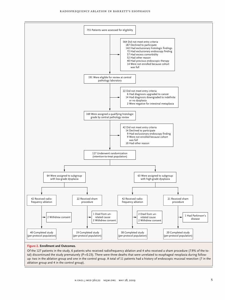
Of the 755 patients screened, 191 fulfilled study criteria and provided histologic specimens for re-view (Fig. 2). Of these patients, 127 underwent randomization; 64 were deemed to have low-grade dysplasia, and 63 were deemed to have high-grade dysplasia. Of the total group, 84 were assigned to
R adiofrequency Ablation in Barrett’s Esophagus
n engl j med 360;22 nejm.org may 28, 2009 5
39p6
127 Underwent randomization(intention-to-treat population)
169 Were assigned a qualifying histologicgrade by central pathology review
42 Did not meet entry criteria14 Declined to participate9 Had exclusionary endoscopy finding9 Were not enrolled because cohort
was full10 Had other reason
1 Had Parkinson’sdisease
2 Died from un-related cause
2 Withdrew consent
1 Died from un-related cause
2 Withdrew consent2 Withdrew consent
64 Were assigned to subgroupwith low-grade dysplasia
63 Were assigned to subgroupwith high-grade dysplasia
42 Received radio-frequency ablation
22 Received shamprocedure
42 Received radio-frequency ablation
21 Received shamprocedure
40 Completed study(per-protocol population)
19 Completed study(per-protocol population)
38 Completed study(per-protocol population)
20 Completed study(per-protocol population)
AUTHOR:
FIGURE:
JOB: ISSUE:
4-CH/T
RETAKE
SIZE
ICM
CASE
EMail LineH/TCombo
Revised
AUTHOR, PLEASE NOTE: Figure has been redrawn and type has been reset.
Please check carefully.
REG F
Enon
1st2nd
3rd
Shaheen
2 of 3
05-28-09
ARTIST: ts
36022
191 Were eligible for review at centralpathology laboratory
22 Did not meet entry criteria6 Had diagnosis upgraded to cancer
14 Had diagnosis downgraded to indefiniteor no dysplasia
2 Were negative for intestinal metaplasia
755 Patients were assessed for eligibility
564 Did not meet entry criteria187 Declined to participate142 Had exclusionary histologic findings72 Had exclusionary endoscopy finding57 Had excess comorbidity52 Had other reason40 Had previous endoscopic therapy14 Were not enrolled because cohort
was full
Figure 2. Enrollment and Outcomes.
Of the 127 patients in the study, 6 patients who received radiofrequency ablation and 4 who received a sham procedure (7.9% of the to-tal) discontinued the study prematurely (P = 0.23). There were three deaths that were unrelated to esophageal neoplasia during follow-up: two in the ablation group and one in the control group. A total of 11 patients had a history of endoscopic mucosal resection (7 in the ablation group and 4 in the control group).

T h e n e w e ngl a nd j o u r na l o f m e dic i n e
n engl j med 360;22 nejm.org may 28, 20096
the ablation group and 43 to the control group. The baseline characteristics of the patients in the two study groups, as stratified according to the level of dysplasia, did not differ significantly, ex-cept for an elevated body-mass index (BMI) among patients with high-grade dysplasia in the control group (Table 1).
Primary and Secondary Outcomes
Regardless of the level of dysplasia, patients who received radiofrequency ablation were significantly more likely than those in the control group to
achieve complete eradication of dysplasia (Fig. 3 and Table 2). Among patients with low-grade dys-plasia, complete eradication of dysplasia occurred in 90.5% of the patients assigned to the ablation group, as compared with 22.7% of those assigned to the control group (P<0.001). Among patients with high-grade dysplasia, complete eradication of dysplasia occurred in 81.0% of the patients assigned to the ablation group, as compared with 19.0% of those assigned to the control group (P<0.001). Among all patients regardless of the grade of dysplasia, complete eradication of all in-
Table 1. Baseline Characteristics of the Patients, According to Grade of Dysplasia.*
Variable High-Grade Dysplasia (N = 63) Low-Grade Dysplasia (N = 64)
Radiofrequency Ablation(N = 42)
Sham Procedure(N = 21)
Radiofrequency Ablation(N = 42)
Sham Procedure(N = 22)
Age — yr
Mean 65.9±1.4 67.3±1.8 66.3±1.4 64.6±1.9
Range 49–80 54–80 41–79 45–78
Sex — no. (%)
Female 5 (11.9) 0 9 (21.4) 3 (13.6)
Male 37 (88.1) 21 (100) 33 (78.6) 19 (86.4)
Race or ethnic group — no. (%)†
White 38 (90.5) 21 (100.0) 40 (95.2) 22 (100.0)
Black 2 (4.8) 0 1 (2.4) 0
Latino 2 (4.8) 0 1 (2.4) 0
Body-mass index
Mean 27.8±0.7 31.7±1.3‡ 29.2±0.8 30.9±1.2
Range 21.3–38.3 23.4–46.8 18.9–44.0 21.5–41.3
Length of Barrett’s esophagus — cm
Mean 5.3±0.3 5.3±0.5 4.6±0.4 4.6±0.5
Range 1.0–8.0 1.0–8.0 0.5–8.0 0.5–8.0
Subsquamous intestinal metaplasia — no. (%) 10 (23.8) 3 (14.3) 11 (26.2) 8 (36.4)
Multifocal dysplasia — no. (%) 33 (78.6) 18 (85.7) 32 (76.2) 13 (59.1)
Time since diagnosis of Barrett’s esophagus — yr
Mean 4.7±0.6 4.2±1.4 5.8±0.7 5.2±1.0
Range 0.2–13.9 0.1–27.0 0.2–22.9 0.2–15.9
Time since diagnosis of dysplasia — yr
Mean 2.1±0.4 1.3±0.6 2.2±0.5 2.4±0.6
Range 0.1–12.4 0.1–12.2 0.1–11.9 0.1–9.4
Current use of aspirin or NSAID — no. (%) 18 (42.9) 12 (57.1) 20 (47.6) 7 (31.8)
* Plus–minus values are means ±SE. The body-mass index is the weight in kilograms divided by the square of the height in meters. Percentages may not total 100 because of rounding. NSAID denotes nonsteroidal antiinflammatory drug.
† Race or ethnic group was self-reported.‡ P<0.05 for the comparison between the ablation group and the control group among patients with high-grade dysplasia.

R adiofrequency Ablation in Barrett’s Esophagus
n engl j med 360;22 nejm.org may 28, 2009 7
testinal metaplasia occurred in 77.4% of the pa-tients assigned to the ablation group, as compared with 2.3% of those assigned to the control group (P<0.001).
Among patients in the ablation group, the rate of complete eradication of intestinal metaplasia among patients with high-grade dysplasia (73.8%) was similar to that among those with low-grade dysplasia (81.0%, P = 0.44). Logistic-regression modeling using study group, age, BMI, length of Barrett’s esophagus, and time since the diagno-sis of Barrett’s esophagus as predictor variables and the three primary outcome variables as re-sponse variables showed that the strong relation-ship between study-group assignment and erad-ication of dysplasia and intestinal metaplasia was not attenuated by adjustment for other risk fac-tors (P<0.001 for study-group assignment in all three models) (for details, see the Supplementary Appendix).
Disease Progression
Patients who were assigned to the control group were more likely to have disease progression (16.3%) than were those in the ablation group (3.6%, P = 0.03). Among patients with high-grade dyspla-sia, 19.0% of those in the control group had pro-gression to esophageal cancer, as compared with 2.4% of those in the ablation group (P = 0.04). Among all patients, esophageal cancer developed in significantly more patients in the control group than in the ablation group (9.3% vs. 1.2%, P = 0.045). Of the four patients in the control group in whom esophageal cancer developed, two had intramu-cosal carcinoma, and two had T1 lesions; the sin-gle patient with esophageal cancer in the ablation group had intramucosal carcinoma.
Safety and Tolerability
A total of 298 treatments were performed in 84 patients in the ablation group (mean, 3.5 treat-ments per patient). All procedures were performed on an outpatient basis with the use of intravenous sedation (narcotic with benzodiazepine in 70% of procedures and propofol in 30%). Median pro-cedure times were 36 minutes (interquartile range, 29 to 45) for circumferential radiofrequency abla-tion and 26 minutes (interquartile range, 19 to 40) for focal radiofrequency ablation.
Three serious adverse events possibly or prob-ably associated with the study occurred in the ablation group and none in the control group
(P = 0.55). The events were one episode of upper gastrointestinal hemorrhage in a patient receiv-ing antiplatelet therapy for heart disease, which was treated endoscopically; one overnight hospi-talization for new-onset chest pain 8 days after radiofrequency ablation; and one overnight hos-pitalization for chest discomfort and nausea im-mediately after radiofrequency ablation. No per-forations or procedure-related deaths occurred. Two extraesophageal incident cancers were diag-nosed during follow-up (one gastric cancer in the ablation group and one ocular melanoma in the control group).
After the initial treatment, the degree of chest discomfort on day 1 was higher in the ablation group than in the control group (median, 23 vs. 0 on a 100-point visual-analogue scale; P<0.001). By day 8, the median chest-discomfort score had returned to 0. By comparison, because of the lo-calized nature of focal ablation, the median day 1 score after subsequent focal radiofrequency abla-tion was 0.
Among patients in the ablation group, esoph-ageal stricture (defined as endoscopically identi-fied narrowing of the esophagus with or without dysphagia) developed in five patients (6.0%). All five patients underwent successful endoscopic dil-atation (mean, 2.6 sessions).
22p3
100
Perc
ent o
f Pat
ient
s w
ith C
ompl
ete
Erad
icat
ion
of In
test
inal
Met
apla
sia
or D
yspl
asia
at 1
2 M
o 80
60
40
20
0Complete
eradicationof intestinalmetaplasia
(all patients)
Completeeradicationof dysplasia(low-gradedysplasia)
Completeeradicationof dysplasia(high-gradedysplasia)
Intention-to-Treat Comparison Groups
AUTHOR:
FIGURE:
JOB:
4-CH/T
RETAKE
SIZE
ICM
CASE
EMail LineH/TCombo
Revised
AUTHOR, PLEASE NOTE: Figure has been redrawn and type has been reset.
Please check carefully.
REG F
Enon
1st2nd
3rd
Shaheen
3 of 3
05-28-09
ARTIST: ts
36022 ISSUE:
Control Ablation
P<0.001
P<0.001
P<0.001
Figure 3. Primary Outcomes in the Intention-to-Treat Analysis.
The primary outcomes were complete histologic eradication of intestinal metaplasia in all patients and complete eradication of dysplasia in the sub-group with low-grade dysplasia and in the subgroup with high-grade dys-plasia at 12 months.

T h e n e w e ngl a nd j o u r na l o f m e dic i n e
n engl j med 360;22 nejm.org may 28, 20098
Biopsy Analysis
From baseline to 12 months, 13,573 biopsy spec-imens were collected (9517 in the ablation group and 4056 in the control group). Among 1260 sam-ples from patients with low-grade dysplasia in the ablation group, 1228 (97.5%) were free of intesti-nal metaplasia at 12 months, as compared with 313 of 550 samples (56.9%) in the control group (Table 2). Among 1464 samples from patients with high-grade dysplasia in the ablation group, 1442
(98.5%) were free of intestinal metaplasia at 12 months, as compared with 360 of 614 samples (58.6%) in the control group (P<0.001 for all pair-wise comparisons after accounting for intrapa-tient correlation).
Subsquamous Intestinal Metaplasia
At baseline, 25.2% of the patients had evidence of subsquamous intestinal metaplasia (20.6% of those with high-grade dysplasia and 29.7% of those
Table 2. Primary and Secondary Outcomes at Follow-up.*
Outcome and AnalysisRadiofrequency
AblationSham
ProcedureRelative Risk
(95% CI) P ValueNo. Needed
to Treat†
no./total no. (%)
Primary outcome
Complete eradication of intestinal metaplasia (all patients)
Intention-to-treat 65/84 (77.4) 1/43 (2.3) 33.3 (4.8–231.7) <0.001 1.3
Per-protocol 65/78 (83.3) 1/39 (2.6) 32.5 (4.6–225.5) <0.001 1.2
Complete eradication of dysplasia (low-grade dysplasia)
Intention-to-treat 38/42 (90.5) 5/22 (22.7) 4.0 (1.8–10.7) <0.001 1.5
Per-protocol 38/40 (95.0) 5/19 (26.3) 3.6 (1.7–7.7) <0.001 1.5
Complete eradication of dysplasia (high-grade dysplasia)
Intention-to-treat 34/42 (81.0) 4/21 (19.0) 4.2 (1.7–10.4) <0.001 1.6
Per-protocol 34/38 (89.5) 4/20 (20.0) 4.5 (1.8–10.8) <0.001 1.4
Secondary outcomes
Complete eradication of intestinal metaplasia (high-grade dysplasia)
Intention-to-treat 31/42 (73.8) 0/21 ND <0.001 1.4
Per-protocol 31/38 (81.6) 0/20 ND <0.001 1.2
Complete eradication of intestinal metaplasia (low-grade dysplasia)
Intention-to-treat 34/42 (81.0) 1/22 (4.5) 17.8 (2.6–121.5) <0.001 1.3
Per-protocol 34/40 (85.0) 1/19 (5.3) 16.1 (2.4–109.3) <0.001 1.3
Complete eradication of dysplasia (all patients)
Intention-to-treat 72/84 (85.7) 9/43 (20.9) 4.1 (2.3–7.4) <0.001 1.5
Per-protocol 72/78 (92.3) 9/39 (23.1) 4.0 (2.2–7.1) <0.001 1.4
Progression of dysplasia
Any 3/84 (3.6) 7/43 (16.3) 0.2 (0.1–0.8) 0.03 7.9
Low-grade to high-grade 2/42 (4.8) 3/22 (13.6) 0.3 (0.1–1.9) 0.33 11.3
Low-grade to cancer 0/42 0/22 ND ND NA
High-grade to cancer 1/42 (2.4) 4/21 (19.0) 0.1 (0.01–1.0) 0.04 6.0
High-grade or low-grade to cancer 1/84 (1.2) 4/43 (9.3) 0.1 (0.01–1.1) 0.045 12.3
Biopsy specimen free of intestinal metaplasia at 12 mo
All patients 2670/2724 (98.0) 673/1164 (57.8) 1.7 (1.6–1.8) <0.001 NA
Low-grade-dysplasia subgroup 1228/1260 (97.5) 313/550 (56.9) 1.7 (1.6–1.8) <0.001 NA
High-grade-dysplasia subgroup 1442/1464 (98.5) 360/614 (58.6) 1.7 (1.6–1.8) <0.001 NA

R adiofrequency Ablation in Barrett’s Esophagus
n engl j med 360;22 nejm.org may 28, 2009 9
with low-grade dysplasia) (Table 1). At 12 months, subsquamous intestinal metaplasia occurred in 5.1% of the patients in the ablation group and in 40.0% of those in the control group (P<0.001).
Predictors of Response to Therapy
Bivariate analysis showed that patients in the ab-lation group who achieved complete eradication of intestinal metaplasia were on average younger, had shorter-length Barrett’s esophagus, had a lower BMI, and had a shorter history of dysplasia than did those who did not have a complete response. However, in multivariate analysis, none of these factors reached statistical significance (for details, see the model in the Supplementary Appendix).
Discussion
Despite the large number of patients with Bar-rett’s esophagus and the remarkable increase in the incidence of esophageal adenocarcinoma in the past 30 years, the optimal management strat-egy for dysplastic Barrett’s esophagus has not been defined. Although professional guidelines endorse various strategies,17,18 the relative safety and ef-
ficacy of these interventions remain unclear. In uncontrolled studies, excellent results have been noted in patients with high-grade dysplasia who were treated with esophagectomy,19 intensive en-doscopic surveillance,11 and ablative therapy.20 Because of the morbidity and mortality associat-ed with more invasive treatments, such as abla-tive therapy and surgery, a more conservative ap-proach has been advocated by some investigators,11 who recommend more invasive therapies only for patients with high-grade dysplasia that progress-es to cancer.
In our study, we compared outcomes in pa-tients treated with radiofrequency ablation with those in patients treated with a sham procedure, with all patients undergoing intensive endoscopic surveillance. Our data show that most patients who were treated with radiofrequency ablation had complete eradication of intestinal metapla-sia and dysplasia and a decreased risk of disease progression at 12 months. Our a priori plan was to analyze the eradication of dysplasia, stratified according to the grade of dysplasia at baseline, on the basis of the hypothesis that high-grade dysplasia might be more difficult to eradicate than
Table 2. (Continued.)
Outcome and AnalysisRadiofrequency
AblationSham
ProcedureRelative Risk
(95% CI) P ValueNo. Needed
to Treat†
no./total no. (%)
Secondary outcomes
Chest-pain score on day 1‡
All patients <0.001 NA
No. of patients 81 40
Median 23 0
Interquartile range 0–51 0–0
Low-grade dysplasia <0.001 NA
No. of patients 40 20
Median 26 0
Interquartile range 4–48 0–0
High-grade dysplasia <0.001 NA
No. of patients 41 20
Median 22 0
Interquartile range 0–57 0–0
* NA denotes not applicable, and ND not done.† The number needed to treat refers to the number of patients who would need to be treated with radiofrequency ablation to prevent one out-
come failure (the inverse of the absolute risk reduction).‡ Chest pain was measured on a visual-analogue scale of 0 to 100, with higher scores indicating a greater severity of pain.

T h e n e w e ngl a nd j o u r na l o f m e dic i n e
n engl j med 360;22 nejm.org may 28, 200910
low-grade dysplasia. However, we observed simi-lar rates of complete eradication of dysplasia in both subgroups (90.5% in patients with low-grade dysplasia and 81.0% in those with high-grade dys-plasia). These high rates of complete eradication were associated with a decreased incidence of progression of dysplasia and a decreased risk of esophageal cancer in the ablation group, as com-pared with the control group.
Our finding of a decreased incidence of can-cer in the ablation group should be viewed with caution. Cancers occurred in only 5 patients (1 of 84 in the ablation group and 4 of 43 in the control group), so the shift of a single incident cancer would have resulted in a loss of statistical signifi-cance. Radiofrequency ablation was associated with a transient increase in chest pain, with a median resolution of pain by day 8, and a rate of serious adverse events that did not differ signifi-cantly from that in the control group.
In a study by Overholt and colleagues,12,21 pa-tients with high-grade dysplasia were randomly assigned to recieve either photodynamic therapy or endoscopic surveillance, with the absence of high-grade dysplasia at any time during the 18- month follow-up as a primary outcome. The in-vestigators observed no high-grade dysplasia in 77% of the patients receiving photodynamic ther-apy and in 39% of the patients in the control group, with a decreased risk of esophageal cancer in the photodynamic-therapy group. Among pa-tients receiving such therapy, esophageal stricture developed in 36% of the patients, and 69% had a photosensitivity reaction to the chemosensitiz-ing agent.
In our study, among patients with high-grade dysplasia, the 1-year incidence of esophageal can-cer in the control group (19.0%) was higher than that reported in some previous studies.11,13,22 There are several possible explanations for this difference. Our study incorporated a central pa-thology laboratory and required concurrence of two pathologists for entry. It is likely that this method resulted in the exclusion of patients with equivocal diagnoses of high-grade dysplasia, leav-ing a subgroup with more severe cellular atypia. A previous study with similar histologic require-ments for entry likewise reported a high incidence of cancer.12 In addition, our rigorous biopsy pro-tocol probably provided more sensitive early de-tection of cancer than would standard-of-care endoscopy.23 Finally, the patients in our study were
referred to tertiary care centers and may have dif-fered in substantial ways from patients with Bar-rett’s esophagus in the community.
The strengths of our study include rigorous masking of study-group assignments, expert his-tologic analysis of biopsy samples, and a low, nondifferential loss to follow-up. Our study also had several limitations. We used eradication of intestinal metaplasia and dysplasia, along with neoplastic progression, as surrogate markers for death from cancer, even though long-term data demonstrating an association between eradication of intestinal metaplasia and a decreased risk of cancer are sparse.21 Second, the study duration was 1 year. Although other data suggest that reversion to neosquamous epithelium after radiofrequency ablation is durable,24 it is not clear whether the results of the study will persist. Third, because of stratified randomization according to the degree of dysplasia and our 2:1 ratio for assignment of patients to the ablation group and the control group, the number of patients in some groups was small. Fourth, since our study did not com-pare radiofrequency ablation with other interven-tions, such as photodynamic therapy and esopha-gectomy, we cannot determine which of these interventions is superior. Finally, whether our re-sults can be generalized to community-practice settings is unknown.
The risk of subsquamous intestinal metapla-sia after ablative therapy is a concern for all abla-tive techniques.25 However, the malignant poten-tial of subsquamous intestinal metaplasia is unknown. In our study, subsquamous intestinal metaplasia was quite common in patients (25.2%) before enrollment and, similar to previous re-ports,20,26 was low after radiofrequency ablation (5.1%). Although our biopsy regimen was aggres-sive, it is possible that some patients had unde-tected subsquamous intestinal metaplasia.
Because we sought to define the efficacy of radiofrequency ablation for the spectrum of dys-plasia, we enrolled patients with both low-grade dysplasia and high-grade dysplasia. However, the implications of these two diagnoses are markedly different. Low-grade dysplasia implies a risk of progression to cancer of less than 1% per patient-year,10 whereas the risk associated with high-grade dysplasia may be higher by a factor of 10.13,22 In making decisions about the management of pre-cancerous conditions, clinicians, patients, and policymakers consider possible benefits and risks

R adiofrequency Ablation in Barrett’s Esophagus
n engl j med 360;22 nejm.org may 28, 2009 11
of competing strategies. Because high-grade dys-plasia has a more ominous natural history than low-grade dysplasia (or nondysplastic intestinal metaplasia), greater risks and costs are tolerable. For less severe disease, the safety profile and as-sociated costs become increasingly important. Detailed consideration of these trade-offs is be-yond the scope of this study. Regardless, both of the dysplasia subgroups showed high rates of reversion to squamous epithelium after radiofre-quency ablation and reduced rates of disease pro-gression with few serious adverse effects, suggest-ing that the application of ablative therapy in patients with low-grade dysplasia is worth further investigation and consideration.
In conclusion, in this multicenter, randomized, sham-controlled study of radiofrequency ablation in patients with dysplastic Barrett’s esophagus, there was a high rate of complete eradication of dysplasia and intestinal metaplasia and decreased disease progression in patients in the ablation group, as compared with the control group.
Supported by BÂRRX Medical. Study medication was provided by AstraZeneca. Statistical analysis and data management were supported by a grant (P30 DK034987) from the National Insti-tutes of Health.
Presented in part at Digestive Diseases Week, 2008, San Diego, CA, May 17–22, 2008.
Dr. Shaheen reports receiving grant support from AstraZene-ca, TAP/Takeda, CSA Medical, and Procter & Gamble, consulting fees from CSA Medical, TAP/Takeda, and AstraZeneca, and lec-ture fees from AstraZeneca; Dr. P. Sharma, receiving grant sup-port from Given Imaging and Olympus and consulting fees from
AstraZeneca, Santarus, and TAP/Takeda; Dr. Overholt, receiving lecture fees from BÂRRX Medical; Dr. Sampliner, receiving con-sulting fees from TAP/Takeda; Dr. Wang, receiving grant sup-port from BÂRRX Medical and lecture fees from Olympus, Fuji-non, and Cook; Dr. Jobe, receiving grant support from EndoGastric Solutions; Dr. Eisen, receiving grant support from AstraZeneca and TAP/Takeda, consulting fees from AstraZeneca and Pfizer, and lecture fees from AstraZeneca, TAP/Takeda, and Given Imaging; Dr. Fennerty, receiving consulting fees from AstraZeneca, Novartis, Merck, and Santarus; Dr. Hunter, receiv-ing grant support from Cook; Dr. V.K. Sharma, receiving lecture fees from BÂRRX Medical; Dr. Hoffman, receiving grant sup-port from AstraZeneca, Bristol-Myers Squibb, Salix, Lantheus Medical Imaging, and Otsuka Medical Research and lecture fees from Given Imaging; Dr. Rothstein, receiving consulting and lecture fees from BÂRRX Medical, Ethicon Endosurgery, and Olympus; Dr. Mashimo, receiving consulting fees from Meda-corp and lecture fees from TAP/Takeda; Dr. Chang, receiving grant support and consulting and lecture fees, having an equity interest in BÂRRX Medical, and receiving royalties from the BÂRRX Medical HALO90 device; Dr. Muthusamy, receiving lec-ture fees from BÂRRX Medical; Dr. Edmunowicz, receiving consulting fees from Boston Scientific and Olympus and lecture fees from BÂRRX Medical; Dr. Spechler, receiving grant support and consulting fees from AstraZeneca and TAP/Takeda; Dr. Souza, receiving grant support from AstraZeneca and consult-ing fees from AstraZeneca and TAP/Takeda; Dr. Infantolino, re-ceiving grant support from Boston Scientific and consulting and lecture fees from BÂRRX Medical, AstraZeneca, Santarus, Ab-bott, UCB, Centocor, and CSA Medical; Dr. Falk, receiving grant support and consulting and lecture fees from AstraZeneca, grant support from Given Imaging, and consulting fees from Olympus, Nycomed, and Ethicon Endosurgery; Dr. Madanick, receiving lecture fees from BÂRRX Medical and AstraZeneca; and Dr. Lightdale, receiving grant support from BÂRRX Medical and Boston Scientific and consulting fees from BÂRRX Medical and AstraZeneca. No other potential conflict of interest relevant to this article was reported.
We thank Drs. Evan S. Dellon and Tara Rubinas for their work on the video accompanying this article.
AppendixThe authors’ affiliations are as follows: the University of North Carolina at Chapel Hill, Chapel Hill (N.J.S., J.A.G., R.D.M.); Veterans Affairs Medical Center and University of Kansas School of Medicine, Kansas City (P.S.); Gastrointestinal Associates, Knoxville, TN (B.F.O.); Mayo Clinic Jacksonville, Jacksonville, FL (H.C.W.); South Arizona Veterans Affairs Health Care System, Tucson (R.E.S.); Mayo Clinic Rochester, Rochester, MN (K.K.W.); Cleveland Clinic, Cleveland (M.P.B., J.R.G., A.E.B., G.W.F.); Oregon Health and Sciences University, Portland (B.A.J., G.M.E., M.B.F., J.G.H.); Mayo Clinic Arizona, Scottsdale (D.E.F., V.K.S.); Medical University of South Carolina, Charleston (R.H.H., B.J.H.); Dartmouth-Hitchcock Medical Center, Lebanon, NH (R.I.R., S.R.G.); Veterans Affairs Boston Healthcare System, West Roxbury, MA (H.M.); University of California, Irvine, Orange (K.J.C., V.R.M.); Washington University Medical Center, St Louis (S.A.E.); University of Texas Southwestern Medical Center, Dallas (S.J.S., A.A.S., R.F.S.); Thomas Jefferson University, Philadelphia (A.I.); Tacoma Digestive Disease Research Center, Tacoma, WA (M.B.K.); University Hospitals–Case Medical Center, Cleveland (A.C.); and Columbia University Medical Center, New York (C.J.L.).
References
Spechler SJ. Barrett’s esophagus. 1. N Engl J Med 2002;346:836-42.
Winters C Jr, Spurling TJ, Chobanian 2. SJ, et al. Barrett’s esophagus: a prevalent, occult complication of gastroesophageal reflux disease. Gastroenterology 1987;92: 118-24.
Abrams JA, Fields S, Lightdale CJ, 3. Neugut AI. Racial and ethnic disparities in the prevalence of Barrett’s esophagus among patients who undergo upper endos-copy. Clin Gastroenterol Hepatol 2008; 6:30-4.
Westhoff B, Brotze S, Weston A, et al. 4. The frequency of Barrett’s esophagus in high-risk patients with chronic GERD. Gastrointest Endosc 2005;61:226-31.
Ronkainen J, Aro P, Storskrubb T, et al. 5. Prevalence of Barrett’s esophagus in the general population: an endoscopic study. Gastroenterology 2005;129:1825-31.
Drewitz DJ, Sampliner RE, Garewal 6. HS. The incidence of adenocarcinoma in Barrett’s esophagus: a prospective study of 170 patients followed 4.8 years. Am J Gastroenterol 1997;92:212-5.
O’Connor JB, Falk GW, Richter JE. 7. The incidence of adenocarcinoma and dysplasia in Barrett’s esophagus: report on the Cleveland Clinic Barrett’s Esopha-gus Registry. Am J Gastroenterol 1999; 94:2037-42.
Pohl H, Welch HG. The role of over-8. diagnosis and reclassification in the marked increase of esophageal adenocarcinoma incidence. J Natl Cancer Inst 2005;97: 142-6.
Eloubeidi MA, Mason AC, Desmond 9. RA, El-Serag HB. Temporal trends (1973-

n engl j med 360;22 nejm.org may 28, 200912
R adiofrequency Ablation in Barrett’s Esophagus
1997) in survival of patients with esopha-geal adenocarcinoma in the United States: a glimmer of hope? Am J Gastroenterol 2003;98:1627-33.
Sharma P, Falk GW, Weston AP, Reker 10. D, Johnston M, Sampliner RE. Dysplasia and cancer in a large multicenter cohort of patients with Barrett’s esophagus. Clin Gastroenterol Hepatol 2006;4:566-72.
Schnell TG, Sontag SJ, Chejfec G, et al. 11. Long-term nonsurgical management of Barrett’s esophagus with high-grade dys-plasia. Gastroenterology 2001;120:1607-19.
Overholt BF, Lightdale CJ, Wang KK, et 12. al. Photodynamic therapy with porfimer sodium for ablation of high-grade dyspla-sia in Barrett’s esophagus: international, partially blinded, randomized phase III trial. Gastrointest Endosc 2005;62:488-98. [Erratum, Gastrointest Endosc 2006;63: 359.]
Reid BJ, Levine DS, Longton G, Blount 13. PL, Rabinovitch PS. Predictors of progres-sion to cancer in Barrett’s esophagus: baseline histology and flow cytometry identify low- and high-risk patient sub-sets. Am J Gastroenterol 2000;95:1669-76.
Rastogi A, Puli S, El-Serag HB, Bansal 14. A, Wani S, Sharma P. Incidence of esopha-geal adenocarcinoma in patients with Barrett’s esophagus and high-grade dys-
plasia: a meta-analysis. Gastrointest En-dosc 2008;67:394-8.
Gondrie JJ, Pouw RE, Sondermeijer 15. CM, et al. Stepwise circumferential and focal ablation of Barrett’s esophagus with high-grade dysplasia: results of the first prospective series of 11 patients. Endos-copy 2008;40:359-69.
Montgomery E, Bronner MP, Gold-16. blum JR, et al. Reproducibility of the di-agnosis of dysplasia in Barrett esophagus: a reaffirmation. Hum Pathol 2001;32:368-78.
Wang KK, Sampliner RE. Updated 17. guidelines 2008 for the diagnosis, sur-veillance and therapy of Barrett’s esopha-gus. Am J Gastroenterol 2008;103:788-97.
Playford RJ. New British Society of 18. Gastroenterology (BSG) guidelines for the diagnosis and management of Barrett’s oesophagus. Gut 2006;55:442.
Williams VA, Watson TJ, Herbella FA, 19. et al. Esophagectomy for high grade dys-plasia is safe, curative, and results in good alimentary outcome. J Gastrointest Surg 2007;11:1589-97.
Ganz RA, Overholt BF, Sharma VK, et 20. al. Circumferential ablation of Barrett’s esophagus that contains high-grade dys-plasia: a U.S. multicenter registry. Gastro-intest Endosc 2008;68:35-40.
Overholt BF, Wang KK, Burdick JS, et 21.
al. Five-year efficacy and safety of photo-dynamic therapy with Photofrin in Bar-rett’s high-grade dysplasia. Gastrointest Endosc 2007;66:460-8.
Buttar NS, Wang KK, Sebo TJ, et al. 22. Extent of high-grade dysplasia in Barrett’s esophagus correlates with risk of adeno-carcinoma. Gastroenterology 2001;120: 1630-9.
Das D, Ishaq S, Harrison R, et al. 23. Management of Barrett’s esophagus in the UK: overtreated and underbiopsied but improved by the introduction of a na-tional randomized trial. Am J Gastroen-terol 2008;103:1079-89.
Fleischer DE, Overholt BF, Sharma 24. VK, et al. Endoscopic ablation of Barrett’s esophagus: a multicenter study with 2.5-year follow-up. Gastrointest Endosc 2008; 68:867-76.
Hornick JL, Blount PL, Sanchez CA, et 25. al. Biologic properties of columnar epithe-lium underneath reepithelialized squamous mucosa in Barrett’s esophagus. Am J Surg Pathol 2005;29:372-80.
Sharma VK, Wang KK, Overholt BF, 26. et al. Balloon-based, circumferential, en-doscopic radiofrequency ablation of Bar-rett’s esophagus: 1-year follow-up of 100 patients. Gastrointest Endosc 2007;65: 185-95.Copyright © 2009 Massachusetts Medical Society.
full text of all journal articles on the world wide web
Access to the complete text of the Journal on the Internet is free to all subscribers. To use this Web site, subscribers should go to the Journal’s home page (NEJM.org) and register by entering their names and subscriber numbers as they appear on their mailing labels. After this one-time registration, subscribers can use their passwords to log on for electronic access to the entire Journal from any computer that is connected to the Internet. Features include a library of all issues since January 1993 and abstracts since January 1975, a full-text search capacity, and a personal archive for saving articles and search results of interest. All articles can be printed in a format that is virtually identical to that of the typeset pages. Beginning 6 months after publication, the full text of all Original Articles and Special Articles is available free to nonsubscribers.