The neurobiology of itchpcpr.pitt.edu/wp-content/uploads/2018/02/Ikoma-et-al2c-2006.pdf · skin,...
13
*Department of Dermatology, Kyoto University, Shogin- Kawahara-cho 54, Sakyo-ku, Kyoto 606-8507, Japan. ‡ Department of Dermatology, IZKF Münster, and Boltzmann Institute for Immunobiology of the Skin, § Department of Dermatology, University Hospital, Von-Esmarch-Str. 58, 48149 Münster, Germany. || Departments of Dermatology, Neurobiology and Anatomy, Wake Forest University School of Medicine, Winston-Salem, North Carolina 27157, USA. ¶ Department of Anaesthesiology, Mannheim, University of Heidelberg, Theodor Kutzer Ufer 1–3, 68135 Manheim, Germany. Correspondence to M.S. e-mail: martin.schmelz@ anaes.ma.uni-heidelberg.de doi:10.1038/nrn1950 Central sensitization Plastic changes in the CNS (adaptive or pathological) that lead to enhanced responses and/or lower thresholds. Historically, the sensations of itch and pain have been regarded as closely related; weak activation of noci- ceptors has been proposed to mediate itch, and stronger activation results in weak pain — the ‘intensity theory’ 1 . Current data in rodents are still compatible with this idea 2 . However, the identification of primary afferent neurons in humans 3 and spinal projection neurons in cats 4 that respond to histamine application with a simi- lar time course as the itch sensation clearly favours the ‘specificity theory’, which stipulates that separate sets of neurons mediate pruritus and pain. However, these his- tamine-sensitive neurons also respond to capsaicin, an algogen (a pain-producing substance), and could there- fore be termed ‘itch selective’ rather than ‘itch-specific’ 5 . Research into itch has tried to identify specific pruritic mediators and mechanisms as opposed to algesic ones. In contrast to the separated neuronal pathways, the results have revealed that a broad overlap exists between pain and itch processing in terms of mediators, mechanisms and even therapeutic approaches: protease-activated receptor 2 (PAR 2 ) and members of the transient receptor potential (TRP) family have been implicated as targets for both algesic and pruritic mediators, and sensitiza- tion of peripheral nerve endings by nerve growth fac- tor (NGF) is a known pathophysiological mechanism in both chronic itch and pain. There are strikingly similar patterns of central sensitization between allodynia and punctate hyperalgesia for pain, as well as between alloknesis and punctate hyperknesis for pruritus. These similarities are already being translated into similar therapeutic approaches, such as gabapentin treatment or the use of antidepressants against neuropathic pain and chronic itch. In this review, we first introduce the definition and clinical classification of itch. We then focus on the inter- action between itch and pain on a peripheral and central level. These interactions will be specified in terms of the mediators involved and their activity in pain and itch processing. Evolving similarities in the patterns of peripheral and central sensitization, and their therapeu- tic implications, are also highlighted. This functional approach is followed by a detailed analysis of the role of selected mediator systems, including PARs, TRP channels, opioids and cannabinoids in the modulation of itch, but also pain. Finally, the neuronal basis for the itch sensation is discussed, with a description of hista- mine-dependent and histamine-independent primary afferent fibres, spinal projection neurons and the cen- tral processing of itch. We also discuss results of central imaging experiments that visualize the similarities and differences between itch and pain processing. For each aspect we not only focus on the particular relevance of mediators and mechanisms for itch, but also emphasize the complex interaction between pruri- tus and pain processing, including typical antagonistic modulation and unexpected similarities. Itch: definition and classification Itch was defined more than 340 years ago by the German physician Samuel Hafenreffer as an “unpleas- ant sensation that elicits the desire or reflex to scratch”. This definition is still valid, but for many reasons we differentiate between acute and chronic pruritus, and it is understood that chronic itch is a complex, unpleas- ant sensory experience with many similarities to pain. Both sensations are multidimensional with sensory The neurobiology of itch Akihiko Ikoma*, Martin Steinhoff ‡ , Sonja Ständer § , Gil Yosipovitch || and Martin Schmelz ¶ Abstract | The neurobiology of itch, which is formally known as pruritus, and its interaction with pain have been illustrated by the complexity of specific mediators, itch-related neuronal pathways and the central processing of itch. Scratch-induced pain can abolish itch, and analgesic opioids can generate itch, which indicates an antagonistic interaction. However, recent data suggest that there is a broad overlap between pain- and itch-related peripheral mediators and/or receptors, and there are astonishingly similar mechanisms of neuronal sensitization in the PNS and the CNS. The antagonistic interaction between pain and itch is already exploited in pruritus therapy, and current research concentrates on the identification of common targets for future analgesic and antipruritic therapy. REVIEWS NATURE REVIEWS | NEUROSCIENCE VOLUME 7 | JULY 2006 | 535
Transcript of The neurobiology of itchpcpr.pitt.edu/wp-content/uploads/2018/02/Ikoma-et-al2c-2006.pdf · skin,...
*Department of Dermatology, Kyoto University, Shogin-Kawahara-cho 54, Sakyo-ku, Kyoto 606-8507, Japan. ‡Department of Dermatology, IZKF Münster, and Boltzmann Institute for Immunobiology of the Skin, §Department of Dermatology, University Hospital, Von-Esmarch-Str. 58, 48149 Münster, Germany. ||Departments of Dermatology, Neurobiology and Anatomy, Wake Forest University School of Medicine, Winston-Salem, North Carolina 27157, USA. ¶Department of Anaesthesiology, Mannheim, University of Heidelberg, Theodor Kutzer Ufer 1–3, 68135 Manheim, Germany. Correspondence to M.S. e-mail: [email protected]:10.1038/nrn1950
Central sensitizationPlastic changes in the CNS (adaptive or pathological) that lead to enhanced responses and/or lower thresholds.
Historically, the sensations of itch and pain have been regarded as closely related; weak activation of noci-ceptors has been proposed to mediate itch, and stronger activation results in weak pain — the ‘intensity theory’1. Current data in rodents are still compatible with this idea2. However, the identification of primary afferent neurons in humans3 and spinal projection neurons in cats4 that respond to histamine application with a simi-lar time course as the itch sensation clearly favours the ‘specificity theory’, which stipulates that separate sets of neurons mediate pruritus and pain. However, these his-tamine-sensitive neurons also respond to capsaicin, an algogen (a pain-producing substance), and could there-fore be termed ‘itch selective’ rather than ‘itch-specific’5. Research into itch has tried to identify specific pruritic mediators and mechanisms as opposed to algesic ones. In contrast to the separated neuronal pathways, the results have revealed that a broad overlap exists between pain and itch processing in terms of mediators, mechanisms and even therapeutic approaches: protease-activated receptor 2 (PAR2) and members of the transient receptor potential (TRP) family have been implicated as targets for both algesic and pruritic mediators, and sensitiza-tion of peripheral nerve endings by nerve growth fac-tor (NGF) is a known pathophysiological mechanism in both chronic itch and pain. There are strikingly similar patterns of central sensitization between allodynia and punctate hyperalgesia for pain, as well as between alloknesis and punctate hyperknesis for pruritus. These similarities are already being translated into similar therapeutic approaches, such as gabapentin treatment or the use of antidepressants against neuropathic pain and chronic itch.
In this review, we first introduce the definition and clinical classification of itch. We then focus on the inter-action between itch and pain on a peripheral and central level. These interactions will be specified in terms of the mediators involved and their activity in pain and itch processing. Evolving similarities in the patterns of peripheral and central sensitization, and their therapeu-tic implications, are also highlighted. This functional approach is followed by a detailed analysis of the role of selected mediator systems, including PARs, TRP channels, opioids and cannabinoids in the modulation of itch, but also pain. Finally, the neuronal basis for the itch sensation is discussed, with a description of hista-mine-dependent and histamine-independent primary afferent fibres, spinal projection neurons and the cen-tral processing of itch. We also discuss results of central imaging experiments that visualize the similarities and differences between itch and pain processing.
For each aspect we not only focus on the particular relevance of mediators and mechanisms for itch, but also emphasize the complex interaction between pruri-tus and pain processing, including typical antagonistic modulation and unexpected similarities.
Itch: definition and classificationItch was defined more than 340 years ago by the German physician Samuel Hafenreffer as an “unpleas-ant sensation that elicits the desire or reflex to scratch”. This definition is still valid, but for many reasons we differentiate between acute and chronic pruritus, and it is understood that chronic itch is a complex, unpleas-ant sensory experience with many similarities to pain. Both sensations are multidimensional with sensory
The neurobiology of itchAkihiko Ikoma*, Martin Steinhoff‡, Sonja Ständer§, Gil Yosipovitch|| and Martin Schmelz¶
Abstract | The neurobiology of itch, which is formally known as pruritus, and its interaction with pain have been illustrated by the complexity of specific mediators, itch-related neuronal pathways and the central processing of itch. Scratch-induced pain can abolish itch, and analgesic opioids can generate itch, which indicates an antagonistic interaction. However, recent data suggest that there is a broad overlap between pain- and itch-related peripheral mediators and/or receptors, and there are astonishingly similar mechanisms of neuronal sensitization in the PNS and the CNS. The antagonistic interaction between pain and itch is already exploited in pruritus therapy, and current research concentrates on the identification of common targets for future analgesic and antipruritic therapy.
R E V I E W S
NATURE REVIEWS | NEUROSCIENCE VOLUME 7 | JULY 2006 | 535
AllodyniaThe perception of a stimulus as painful when previously the same stimulus was reported to be non-painful.
Punctate hyperalgesiaType of central sensitization for pain in which the pain elicited by punctate mechanical stimuli is more prolonged and stronger than normally experienced.
AlloknesisType of central sensitization for itch in which touch triggers the sensation of itch.
Punctate hyperknesisType of central sensitization for itch in which the itch evoked by punctate mechanical stimuli is more prolonged and stronger than normally experienced.
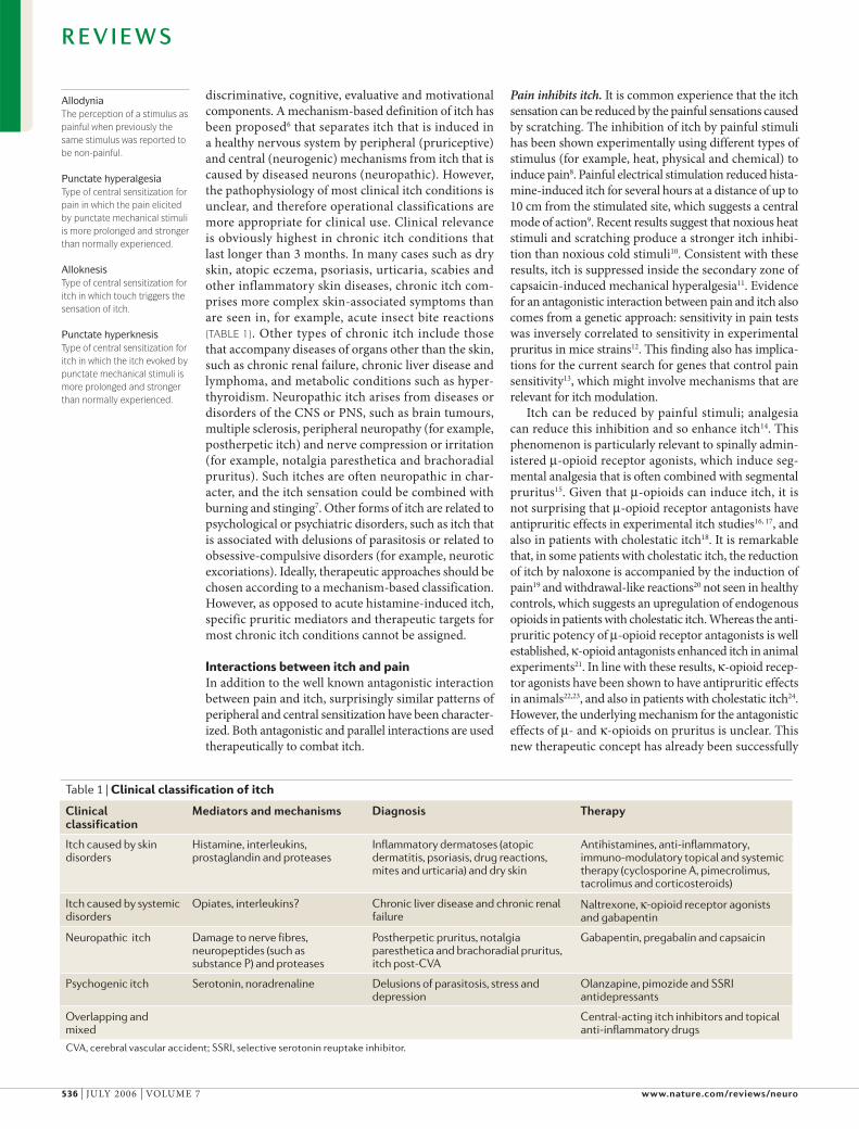
discriminative, cognitive, evaluative and motivational components. A mechanism-based definition of itch has been proposed6 that separates itch that is induced in a healthy nervous system by peripheral (pruriceptive) and central (neurogenic) mechanisms from itch that is caused by diseased neurons (neuropathic). However, the pathophysiology of most clinical itch conditions is unclear, and therefore operational classifications are more appropriate for clinical use. Clinical relevance is obviously highest in chronic itch conditions that last longer than 3 months. In many cases such as dry skin, atopic eczema, psoriasis, urticaria, scabies and other inflammatory skin diseases, chronic itch com-prises more complex skin-associated symptoms than are seen in, for example, acute insect bite reactions (TABLE 1). Other types of chronic itch include those that accompany diseases of organs other than the skin, such as chronic renal failure, chronic liver disease and lymphoma, and metabolic conditions such as hyper-thyroidism. Neuropathic itch arises from diseases or disorders of the CNS or PNS, such as brain tumours, multiple sclerosis, peripheral neuropathy (for example, postherpetic itch) and nerve compression or irritation (for example, notalgia paresthetica and brachoradial pruritus). Such itches are often neuropathic in char-acter, and the itch sensation could be combined with burning and stinging7. Other forms of itch are related to psychological or psychiatric disorders, such as itch that is associated with delusions of parasitosis or related to obsessive-compulsive disorders (for example, neurotic excoriations). Ideally, therapeutic approaches should be chosen according to a mechanism-based classification. However, as opposed to acute histamine-induced itch, specific pruritic mediators and therapeutic targets for most chronic itch conditions cannot be assigned.
Interactions between itch and painIn addition to the well known antagonistic interaction between pain and itch, surprisingly similar patterns of peripheral and central sensitization have been character-ized. Both antagonistic and parallel interactions are used therapeutically to combat itch.
Pain inhibits itch. It is common experience that the itch sensation can be reduced by the painful sensations caused by scratching. The inhibition of itch by painful stimuli has been shown experimentally using different types of stimulus (for example, heat, physical and chemical) to induce pain8. Painful electrical stimulation reduced hista-mine-induced itch for several hours at a distance of up to 10 cm from the stimulated site, which suggests a central mode of action9. Recent results suggest that noxious heat stimuli and scratching produce a stronger itch inhibi-tion than noxious cold stimuli10. Consistent with these results, itch is suppressed inside the secondary zone of capsaicin-induced mechanical hyperalgesia11. Evidence for an antagonistic interaction between pain and itch also comes from a genetic approach: sensitivity in pain tests was inversely correlated to sensitivity in experimental pruritus in mice strains12. This finding also has implica-tions for the current search for genes that control pain sensitivity13, which might involve mechanisms that are relevant for itch modulation.
Itch can be reduced by painful stimuli; analgesia can reduce this inhibition and so enhance itch14. This phenomenon is particularly relevant to spinally admin-istered µ-opioid receptor agonists, which induce seg-mental analgesia that is often combined with segmental pruritus15. Given that µ-opioids can induce itch, it is not surprising that µ-opioid receptor antagonists have antipruritic effects in experimental itch studies16, 17, and also in patients with cholestatic itch18. It is remarkable that, in some patients with cholestatic itch, the reduction of itch by naloxone is accompanied by the induction of pain19 and withdrawal-like reactions20 not seen in healthy controls, which suggests an upregulation of endogenous opioids in patients with cholestatic itch. Whereas the anti-pruritic potency of µ-opioid receptor antagonists is well established, κ-opioid antagonists enhanced itch in animal experiments21. In line with these results, κ-opioid recep-tor agonists have been shown to have antipruritic effects in animals22,23, and also in patients with cholestatic itch24. However, the underlying mechanism for the antagonistic effects of µ- and κ-opioids on pruritus is unclear. This new therapeutic concept has already been successfully
Table 1 | Clinical classification of itch
Clinical classification
Mediators and mechanisms Diagnosis Therapy
Itch caused by skin disorders
Histamine, interleukins, prostaglandin and proteases
Inflammatory dermatoses (atopic dermatitis, psoriasis, drug reactions, mites and urticaria) and dry skin
Antihistamines, anti-inflammatory, immuno-modulatory topical and systemic therapy (cyclosporine A, pimecrolimus, tacrolimus and corticosteroids)
Itch caused by systemic disorders
Opiates, interleukins? Chronic liver disease and chronic renal failure
Naltrexone, κ-opioid receptor agonists and gabapentin
Neuropathic itch Damage to nerve fibres, neuropeptides (such as substance P) and proteases
Postherpetic pruritus, notalgia paresthetica and brachoradial pruritus, itch post-CVA
Gabapentin, pregabalin and capsaicin
Psychogenic itch Serotonin, noradrenaline Delusions of parasitosis, stress and depression
Olanzapine, pimozide and SSRI antidepressants
Overlapping and mixed
Central-acting itch inhibitors and topical anti-inflammatory drugs
CVA, cerebral vascular accident; SSRI, selective serotonin reuptake inhibitor.
R E V I E W S
536 | JULY 2006 | VOLUME 7 www.nature.com/reviews/neuro
IontophoresisA non-invasive method of propelling high concentrations of a charged substance, normally medication or bioactive-agents, transdermally by repulsive electromotive force using a small electrical charge applied to an iontophoretic chamber containing a similarly charged active agent and its vehicle.
tested in patients with chronic itch using a recently developed κ-opioid receptor agonist25. Buprenorphine, a substance combining µ-opioid antagonistic and κ-opioid agonistic effects, has recently been found to be effective in the treatment of chronic intractable itch26.
Peripheral sensitization. In pain research, the sensitiza-tion of nociceptive nerve endings has been recognized as a major mechanism for inflammatory pain27. Classic inflammatory mediators such as bradykinin, serotonin (5-hydroxytryptamine; 5-HT) and prostaglandins, which are released in a wide range of painful and pruritic inflammatory conditions, have been shown to not only activate ‘itch fibres’ (pruriceptors)12 but also acutely sen-sitize nociceptors28. In addition to acute sensitization29, neurotrophins cause lasting structural changes (sprout-ing) of nociceptors. The expression of NGF is high in injured and inflamed tissues, and activation of the NGF receptor tyrosine kinase TrkA on nociceptive neurons triggers and potentiates pain signalling by multiple mechanisms30.
The sprouting of epidermal nerve fibres that is initi-ated by increased NGF is not only found in combina-tion with localized pain and hyperalgesia, such as vulvar dysesthesia31, but also in atopic dermatitis32. In addition, remarkably increased serum levels of NGF and substance P have been found to correlate with the severity of the disease in atopic dermatitis33. The sources of NGF were mainly keratinocytes and mast cells34. Increased fibre density and higher local NGF concentrations were also found in patients with pruritic contact dermatitis35, and increased NGF and TrkA immunoreactivity was detected in prurigo nodularis36 and also in pruritic lesions of patients with psoriasis37. These similarities between localized painful and pruritic lesions might suggest that similar mechanisms of neuronal sprouting and sensitiza-tion exist for both pain and pruritus on a peripheral level. Anti-NGF strategies have already been used in animal pain models38 and in patients with pains39. Therapeutic anti-NGF approaches against pruritus have been tested only in animal models of atopic dermatitis40. In NC/Nga mice (an atopic dermatitis mouse model), the role of increased epidermal NGF expression in abnormal itch perception has been validated41.
NGF is known to upregulate neuropeptides, espe-cially substance P and calcitonin-gene-related peptide (CGRP)42. Substance P has been found to have an important role in the induction of pain and hyperalge-sia in rodents43, although there is little evidence for the clinical analgesic efficacy of the antagonists of its receptor neurokinin 1 (NK1)
44. There is no confirmation that sub-stance P is an acute pruritogen in humans45, but it might contribute to itch by increasing neuronal sensitization and through its long-term interaction with mast cells46. A sensitizing effect on nociceptors has also been found for CGRP in rodents13,47, but its role in pruritus is unclear48. Interestingly, in NC/Nga mice, substance P concentra-tions are elevated and CGRP levels are reduced compared with that in the controls49. Given that sensitivity to pain induced by heat correlates to CGRP concentrations13, and pain sensitivity negatively correlates to itch sensitivity in
animal models12, it could be speculated that CGRP has a greater role in nociception and substance P has a greater role in itch.
Central sensitization. Noxious input to the spinal cord is known to provoke central sensitization50, which consists of allodynia and punctate hyperalgesia. Two types of mechanical hyperalgesia can be differentiated. In allo-dynia, touch that is normally painless in the uninjured surroundings of a trauma can trigger painful sensations — a phenomenon also referred to as touch- or brush-evoked hyperalgesia. Although this sensation is medi-ated by myelinated mechanoreceptor units, it requires the ongoing activity of primary afferent C-nociceptors51. The second type of mechanical hyperalgesia results in slightly painful pin prick stimulation being perceived as more painful in the secondary zone around a focus of inflammation. This type has been called punctate hyper-algesia, and does not require ongoing activity of primary nociceptors for its maintenance. Punctate hyperalgesia can persist for hours after a trauma, usually much longer than touch- or brush-evoked hyperalgesia52.
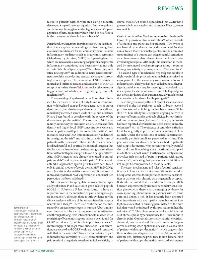
A strikingly similar pattern of central sensitization is observed in the itch pathway: touch- or brush-evoked pruritus around an itching site has been termed ‘itchy skin’53,54. Like allodynia, it requires ongoing activity in primary afferents and is probably elicited by low thresh-old mechanoreceptors (A-fibres)54,55. Also, hyperknesis has been reported after histamine iontophoresis in healthy volunteers14 (FIG. 1). The existence of central sensitization for itch can greatly improve our understanding of clini-cal itch. Under the conditions of central sensitization, normally painful stimuli are perceived as itching. This phenomenon has already been described in patients with atopic dermatitis, who perceive normally painful electrical stimuli as itching when the stimuli are applied inside their lesional skin56. Furthermore, acetylcholine provokes itch instead of pain in patients with atopic dermatitis57, indicating that pain-induced inhibition of itch might be compromised in these patients.
The exact mechanisms and roles of central sensitiza-tion for itch in specific clinical conditions still need to be explored, whereas the importance of central sensitiza-tion in patients with chronic pain is generally accepted. It should be noted that, in addition to the parallels between experimentally induced secondary sensitiza-tion phenomena, there is also emerging evidence for corresponding phenomena in patients with chronic pain and chronic itch. It has recently been reported that, in patients with neuropathic pain, histamine ion-tophoresis resulted in burning pain instead of the pure itch that would be induced by this procedure in healthy volunteers58,59. This phenomenon is of special interest as it shows spinal hypersensitivity to C-fibre input in chronic pain. Conversely, normally painful electrical, chemical, mechanical and thermal stimulation is per-ceived as itching when applied in or close to lesional skin of patients with atopic dermatitis60, which suggests that there is also spinal hypersensitivity to C-fibre input in chronic itch. Histamine prick tests in non-lesional skin of patients with atopic dermatitis provoked less intense
R E V I E W S
NATURE REVIEWS | NEUROSCIENCE VOLUME 7 | JULY 2006 | 537
Nociceptors Pruriceptors
IL-8
IL-6TNF
C
Touch-evoked allodyniaPunctate hyperalgesiaAlgogen-induced itch
Touch-evoked alloknesisPunctate hyperknesisHistamine-induced pain
AβAδ
Immune cells(T cells, mast cells)
Bloodvessels
CGRP
CGRP
CGRP
SP
SP
ATP
ACh NGF
Endothelin
Epidermis
Keratinocyte
IL-31Histamine
Tryptase
NGFH+
itching compared with healthy controls. However, when the stimulus was applied inside their lesions, itch rat-ings were enhanced and lasted for an extended period of time, whereas the axon-reflex erythema was still smaller compared with the controls55. Therefore, in addition to peripheral sensitization, there is evidence for a central sensitization of itch in chronic pruritus.
Neuropathic pain medication for chronic itch. The simi-lar patterns of central sensitization in itch and pain have led to antipruritic therapeutic approaches using drugs that are usually prescribed for neuropathic pain. So far, there have been no controlled studies; however, anecdotal reports show success with carbamazepine, gabapentin
and the recently developed pregabalin61. Gabapentin and pregabalin inhibit the α2δ-subunit of voltage-dependent Ca2+ channels62. Gabapentin has also proved to be effec-tive for the treatment of neuropathic pruritus, particu-larly in the case of brachioradial pruritus and multiple sclerosis-related itch63, 64. Gabapentin seems to alter the sensation of itch, as well as pruritus associated with nerve damage in cutaneous and systemic diseases65.
Antidepressants have been shown to reduce pruritus associated with some psychiatric diseases66. However, in a placebo-controlled trial, paroxetine also reduced itch in patients who did not have a psychiatric disorder67. Although selective serotonin reuptake inhibitors (SSRIs) are not commonly used against itch, third generation antidepressants might be more effective antipruritics. Mirtazapine is an antagonist of central presynaptic α2-adrenergic inhibitory autoreceptors and heteroreceptors, and a potent antagonist of the 5-HT2 and 5-HT3 recep-tors, and of the histamine 1 (H1) receptor68. It has been shown to be effective when used to treat pruritus associ-ated with malignant cholestasis, lymphoma and uraemia, as well as itch related to inflammatory skin diseases that are resistant to high doses of sedating antihistamines69,70. It is interesting to note that a similar pattern of analgesic activity was found in animal experiments: amtryptiline and mirtazapine reduced mechanical hyperalgesia in a rat chronic constriction model, whereas the SSRI citalo-pram was ineffective71. The parallel occurrence of neuro-pathic pain and neuropathic postherpetic itch72 provides further evidence for an intimate link between these two phenomena, although clinical data on the therapeutic implications are still rare.
Modulators for itch and painInstead of having a clear set of separate specific media-tors for pain and itch, most receptor systems have a prominent role in both. This also includes the interac-tion between the keratinocytes and nerve endings that modulate pain and itch.
Histamine. Histamine is the best-known pruritogen and has been regarded as a main target for antipruritic therapies. Among histamine receptors, H1 receptors have been presumed to be important in histamine-induced reactions. Indeed, histamine-induced itch, wheal and axon-reflex flare in human skin are almost completely suppressed by inhibitors of H1 receptors, although local erythema remains73. However, inhibitors of H1 receptors are successfully applied to only a few pruritic diseases (for example, certain subtypes of urticaria), and they are ineffective in many other diseases, such as atopic derma-titis74. Other histamine receptors such as H4 receptors could have an independent role in pruritus, as has been shown for histamine-induced itch in mice75. However, as the species differences are large, the role of H4 receptors in human pruritic diseases remains to be elucidated.
Interleukins. The crosstalk between immune cells and neurons has been in the focus of pain research dur-ing recent years76. However, until recently, no specific candidate could explain neuronal sensitization for itch.
Figure 1 | Mediators and the sensitization pattern of nociceptive and pruriceptive neurons. Sensitizing and activating mediators in the skin are shown for primary afferent fibres involved itch (red) and pain processing (blue). Predominantly pruritic mediators are shown in red, algogenic mediators are shown in blue; mediators equally involved in pain and itch are shown in yellow. Note that different classes of fibres subserve pain (mechano-sensitive polymodal nociceptors and mechano-insensitive ‘sleeping’ nociceptors200) and itch (histamine-sensitive mechano-insensitive pruriceptors3, and probably mechano-sensitive pruriceptors158). In the spinal cord, noxious input can induce central sensitization for pain and pruriceptive input can provoke central sensitization for itch. Of note is the corresponding pattern of central sensitization to touch by amyloid-β (Aβ) fibres (allodynia versus alloknesis), by Aδ fibres (punctate hyperalgesia versus punctate hyperknesis) and by C-fibres (histamine-induced pain versus algogen-induced itch). ACh, acetylcholine; CGRP, calcitonin-gene-related protein; H+, hydrogen ion; IL, interleukin; NGF, nerve growth factor; SP, substance P; TNF, tumour necrosis factor.
R E V I E W S
538 | JULY 2006 | VOLUME 7 www.nature.com/reviews/neuro
Supernatants of mitogen-stimulated leukocytes, which were pruritic in patients with atopic dermatitis but not in controls, contained larger amounts of interleukin 2 (IL-2) and IL-6 (REF. 77). However, no correlation between IL-6-like content and itch intensity was found in atopic der-matitis78. IL-6 and IL-6 receptors are expressed in nerve and Schwann cells79, and IL-6-like immunoreactivity was increased in nerve fibres of patients with positive epicuta-neous patch tests and prurigo nodularis. This might indi-cate that IL-6 has a role in the pathophysiology of some types of itch. However, the discovery that IL-31 induces pruritic dermatitis in mice47,80 has finally provided a candidate for itch-specific sensitization. Expression and protein content of IL-31 are increased in an atopic mouse model81 and correlates to itch behaviour82. Moreover, this increase was already verified in patients with atopic der-matitis83. These results suggest a special role for IL-31 in pruritus. However, studies in pain models are still lacking and therefore a similar sensitizing role in pain induction cannot be ruled out.
Protease-activated receptors. The hypothesis that pro-teases are involved in pain and pruritic pathways is rather old84. Exogenous agents (for example, microbes or plants), as well as several inflammatory cells, can induce the production of sufficient amounts of proteases, which in turn activate protease-activated receptors (PARs). The expression and function of PARs vary among tissues and neuronal cells. PAR2 has a role in the regulation of vas-cular tone, various pro- and anti-inflammatory effects, and is pronociceptive in models of somatic and visceral pain85–87. PAR1 has been implicated in haemostasis and platelet signalling, has pro-inflammatory effects and can induce analgesia from either peripheral or spinal activation88,89. PAR1 and PAR2 are functional in primary afferent neurons and modulate nociception90. PAR2 elic-its hyperalgesia to both thermal and mechanical stimuli through an NK1-receptor-dependent mechanism85,87. By contrast, PAR1 mediates an analgesic response to both mechanical and thermal stimuli under normal and inflammatory conditions88 — probably by modulating ascending nociceptive transmission in a fashion similar to that of opioid receptors — which results in analgesia.
The cloning of PARs, especially PAR2, has shed new light on proteases as signalling molecules in nociception. Functional PAR2 is mainly expressed in small-diameter sensory neurons and induces neurogenic inflammation90, and dermal PAR2 stimulation induces c-Fos expression in the superficial laminae of the dorsal horn91. PAR2 is ulti-mately involved in inflammatory somatic85 and visceral87 hyperalgesia, thereby probably contributing to acute and chronic pain conditions89. With respect to pruritus, sev-eral proteases are upregulated during inflammation and can produce itching (for a review, see REF. 92). In humans, PAR2 is upregulated in itchy dermatoses, and PAR2 agon-ists induce pruritus in these patients. It is of note that concentrations of tryptase are enhanced in patients with atopic dermatitis, whereas histamine concentrations are unchanged, which suggests that PAR2 — which is similar to histamine receptors — is a receptor for ‘itchy’ proteases in atopic dermatitis93. For PAR1, both antihyperalgesic
activity in peripheral inflammatory pain models88 and hyperalgesic effects in neuropathic pain models94 have been shown. A question that remains unanswered is that of the role of PAR4, a receptor for leukocyte-derived cathepsin G, in inflammatory nociception95. So, proteases and PARs can directly activate both the itch and the pain pathway, and further in vitro and in vivo studies are required to finally determine their clinical role in nocic-eption and pruritus.
Transient receptor potential receptors. TRP receptors that sense ‘hot’ and ‘cold’ represent another receptor family involved in itch and pain. The first and best-known is the capsaicin receptor TRP vanilloid receptor 1 (TRPV1). TRPV1 expression in humans has been described on primary afferent neurons, keratinocytes, dendritic cells and mast cells, although so far its functionality has not been verified in all cells96–98. Certain itch and pain mediators — such as capsaicin or eicosanoids, histamine, ATP and various neurotrophins73,99–102 — activate or sensitize the non-selective, calcium-permeable TRPV1 (REFS 99,103–105). TRPV1 activation is related to action potential generation and neuropeptide release105, but pro-longed calcium influx can also desensitize the primary afferents106. Interestingly, TRPV1 acts synergistically with PAR2 and the substance P receptor NK1R, which might have implications for the sensitization to itch and pain. Moreover, TRPV1 can activate the release of cytokines, which are involved in pruritus107,108.
Topical application of TRPV1 agonists has been pro-posed for pruritic lesions, but also for painful skin; for example, in postherpetic neuralgia109. On chronic stimu-lation, TRPV1 desensitizes in a Ca2+-dependent manner and leaves the nociceptive and pruriceptive neurons inactive. Moreover, neuropeptides such as substance P are depleted from the sensory nerve fibres; the axonal transport of both neuropeptides and NGF in the periph-ery is slower. Clinically, the first days of the therapy are accompanied by a burning sensation and neurogenic inflammation, followed by a lasting depression of pain and itch. Also, other members of the TRP family that are activated in warm (TRPV2, TRPV3 and TRPV4) or cold (TRPM8 and TRPA1) temperature ranges might be involved in modulation of pruritus110 (TABLE 2; FIG. 2). In particular, activation of neuronal ‘cold receptors’111,112 might be beneficial for the treatment of pruritus, as there is evidence for antipruritic effects of cold. TRPM8 can be activated by cold (that is, 8–28 ºC) or by menthol and icilin113 (for a review, see REF. 110), which might therefore be used therapeutically for cold-mediated suppression of itch114. TRPA1 is co-expressed with TRPV1 in rat dorsal root ganglion neurons115 and can be activated by noxious cold (≤17 ºC). It is responsive to mustard oil, cinnamon oils and raw garlic, and can be sensitized by camphor, endocannabinoids and bradykinin112,116. However, no direct link to the pruritus pathway has been described.
Recent findings have revealed that TRPV ion channels not only contribute to the thermal activation of sensory neurons, but are also thermosensory receptors for epithe-lial cells117. The important question of whether — and if so, how — TRPV1-activated keratinocytes communicate
R E V I E W S
NATURE REVIEWS | NEUROSCIENCE VOLUME 7 | JULY 2006 | 539
with sensory nerves is currently unanswered. Potential signalling molecule candidates are endothelin, ATP and endorphins117 (FIG. 1; TABLE 2). Human epidermal and hair follicle keratinocytes express functional TRPV1 (REF. 98), and its stimulation leads to the release of prostaglandin E2 (PGE2) and IL-8, which suggests that TRPV1 is involved in skin inflammation following thermal injury107. It is also assumed that TRPV1 modulates keratinocyte prolif-eration, differentiation and apoptosis98. Camphor, which induces a warm sensation on topical application118 and acts as an antipruritic agent, activates TRPV3 (REF. 119), but also TRPV1 (REF. 120). Together with TRPV4, TRPV3 is expressed on keratinocytes and mediates warm-induced currents121,122. Therefore, keratinocytes have the potential to participate in temperature transduction; however, their significance in this process still needs to be assessed.
Opioids and cannabinoids. Opioids are potent neuro-transmitters, hormones and immunomodulators123 and can be divided into three classes: endorphins, dynor-phins and enkephalins. They exert their effects through the activation of µ-, κ-, and δ-opioid receptors, which are widely distributed in the CNS and PNS124,125. In the periphery, opioid receptor mRNA is associated with up to 90% of substance P-containing dorsal root ganglion cells, which indicates a role in nociception, as well as neurogenic inflammation126. Activation of opioid recep-tors reduces neuronal excitability through the inhibition of voltage-dependent Ca2+ channels and adenyl cyclase,
and activation of K+ channels127. Regarding the primary afferent neurons in the skin, this reduced excitability by the activation of opioid receptors would lead to an inhibition of pain and itch123.
The psychotropic, but also analgesic, effects of can-nabinoids have been recognized for centuries. They bind to two G-protein-coupled receptors, the cannabinoid receptors CB1 and CB2 (REFS 128–130). CB1 receptors are mainly localized in the CNS, but they are also expressed in primary afferent neurons131–134. CB2 receptors are predominantly found in the periphery; for example, on T lymphocytes and mast cells135,136, but also on rat spinal cord and human sensory nerve fibres137. Endogenous cannabinoids belong to a group of fatty acid amines, the N-acetylethanolamines (NAEs), with N-arachidonoyleth-anolamine (anandamide) and N-palmitoylethanolamine (PEA, palmidrol, N-(2-hydroxyethyl) hexadecanamide) being the most abundant138,139. Biochemical evidence indicates that endogenous NAEs are produced and released by animal and human neurons139, and by human epidermal keratinocytes140,141. On local application, can-nabinoids have analgesic and antipruritic effects in acute pain and itch models142–145; moreover, there is growing evidence for the suppression of itch by topical cannabi-noid receptor agonists under clinical conditions. Direct activation of CB1 and CB2 (but also activity at TRPV1), a new cannabinoid-receptor ‘CB2-like receptor’146 and non-receptor-mediated effects are discussed as possible itch-suppressing mechanisms.
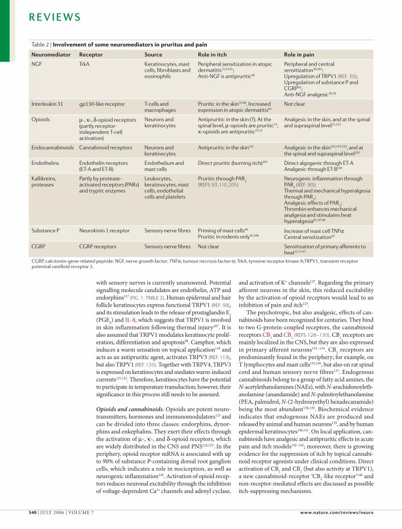
Table 2 | Involvement of some neuromediators in pruritus and pain
Neuromediator Receptor Source Role in itch Role in pain
NGF TrkA Keratinocytes, mast cells, fibroblasts and eosinophils
Peripheral sensitization in atopic dermatitis33,34,41;Anti-NGF is antipruritic40
Peripheral and central sensitization30,201;Upregulation of TRPV1 (REF. 30);Upregulation of substance P and CGRP42;Anti-NGF analgesic38,39
Interleukin 31 gp130-like receptor T-cells and macrophages
Pruritic in the skin47,80. Increased expression in atopic dermatitis83
Not clear
Opioids µ-, κ-, δ-opioid receptors (partly receptor-independent T-cell activation)
Neurons and keratinocytes
Antipruritic in the skin (?). At the spinal level, µ-opioids are pruritic15, κ-opioids are antipruritic23,25
Analgesic in the skin, and at the spinal and supraspinal level15,123
Endocannabinoids Cannabinoid receptors Neurons and keratinocytes
Antipruritic in the skin145 Analgesic in the skin142,144,150, and at the spinal and supraspinal level202
Endothelins Endothelin receptors (ET-A and ET-B)
Endothelium and mast cells
Direct pruritic (burning itch)203 Direct algogenic through ET-AAnalgesic through ET-B204
Kallikreins, proteases
Partly by protease-activated receptors (PARs) and tryptic enzymes
Leukocytes, keratinocytes, mast cells, endothelial cells and platelets
Pruritic through PAR2 (REFS 93,110,205)
Neurogenic inflammation through PAR2 (REF. 90);Thermal and mechanical hyperalgesia through PAR2;Analgesic effects of PAR1;Thrombin enhances mechanical analgesia and stimulates heat hyperalgesia85–87,89
Substance P Neurokinin 1 receptor Sensory nerve fibres Priming of mast cells46
Pruritic in rodents only45,206Increase of mast cell TNFαCentral sensitization43
CGRP CGRP receptors Sensory nerve fibres Not clear Sensitization of primary afferents to heat12,13,47
CGRP, calcitonin-gene-related peptide; NGF, nerve growth factor; TNFα, tumour necrosis factor-α; TrkA, tyrosine receptor kinase A;TRPV1, transient receptor potential vanilloid receptor 1.
R E V I E W S
540 | JULY 2006 | VOLUME 7 www.nature.com/reviews/neuro
Time (min)Time (min)
Dis
char
ge fr
eque
ncy
(Hz)
Dis
char
ge fr
eque
ncy
(Hz)
0
5
10
0
5
10
0–2 2 4 6 8 10 12 14 –2 0 2 4 6 8
Time (min)
Pain
rati
ng (%
VA
S)
5
10
15
20
25
30
–2 0 2 4 6 8
f Pain sensation (human)
b Pruriceptive projection neuron (cat) e Polymodal projection neuron (cat)
Time (min)
Dis
char
ge fr
eque
ncy
(Hz)
0.01
0.1
1
10
–2 0 2 4 6 8
d Polymodal primary afferent (human)
Time (min)
Dis
char
ge fr
eque
ncy
(Hz)
0.01
0.1
1
10
0–2 2 4 6 8 10 12 14
a Pruriceptive afferent (human)
Time (min)
Itch
rati
ng (%
VA
S)
5
10
15
20
25
30
0–2 2 4 6 8 10 12 14
c Itch sensation (human)
The cannabinoid and opioid systems share neuro-anatomical, neurochemical and pharmacological features. There is growing evidence for a crosstalk between the endogenous cannabinoid and opioid systems, including induction of the release of opioid peptides by cannabi-noids, or endocannabinoids by opioids147–149. Although this connection was first shown in the CNS, recent data suggest that the receptors and agonists interact on the cutaneous level. Neuronal- or immune cell-derived cannabinoids can stimulate the release of β-endorphin from keratinocytes through CB2 activation, thereby allowing a combination of inhibitory cannabinoids and opioids to modulate the excit-ability of local nociceptive nerve endings150. It is of note that the nociceptive effects can be blocked by antibodies to β-endorphin, or in µ-opioid receptor-deficient mice150. These data clearly indicate a crucial role of keratinocyte-derived opioids in nociception.
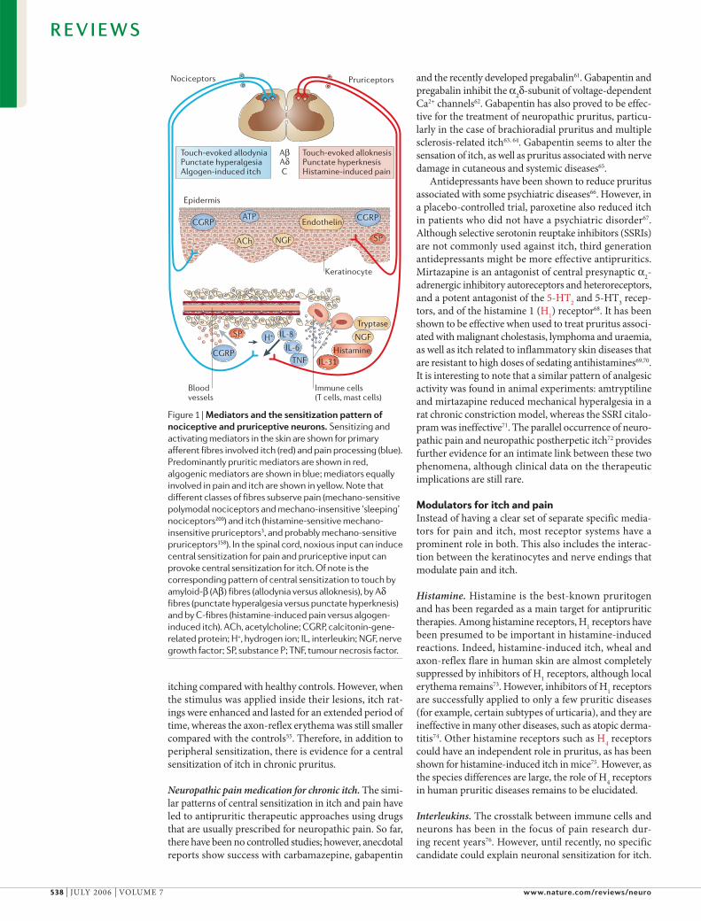
Neuronal pathways for itchThe selective histamine-sensitive neuronal pathway can be correlated with histamine-induced itch, which supports the specificity theory of itch. However, additional hista-mine-independent itch pathways are crucially required to explain key aspects of clinical itch conditions.
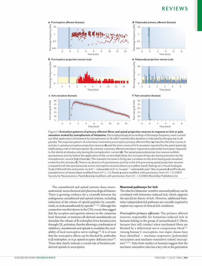
Pruriceptive primary afferents. The primary afferent neurons responsible for histamine-induced itch in humans belong to the group of unmyelinated C-fibres, because they still conduct when myelinated fibres are blocked by a differential nerve compression block151. Among human C-nociceptors, two major classes have been identified — mechano-responsive (polymodal) nociceptors and mechano-insensitive (silent) nocicep-tors152,153. Data from studies in humans suggest that the mechano-insensitive class has a key role in the generation
Figure 2 | Activation patterns of primary afferent fibres and spinal projection neurons in response to itch or pain sensation evoked by iontophoresis of histamine. Electrophysiological recordings of discharge frequency were carried out after application of histamine by iontophoresis at 20 milli Coulombs (the duration is indicated by the grey bar in all panels). The response pattern of a mechano-insensitive pruriceptive primary afferent fibre (a) matches the time course of activity in spinal pruriceptive projection neurons (b) and the time course of itch sensation reported by the participants (c), implicating a role in itch perception. By contrast, a primary afferent mechano-responsive polymodal nociceptor responds to the identical stimulus only during the iontophoretic current (d). The spinal polymodal projection neuron exhibits spontaneous activity before the application of the current (dark blue), but increases firing rate during activation by the iontophoretic current (light blue) (e). | The transient increase in firing rate correlates to the short lasting pain sensation evoked by this stimulus (f). There is an absence of spontaneous activity in the itch processing spinal projection neurons compared with the spontaneously active nociceptive neurons (shown as a yellow band). Ratings on a Visual Analogue Scale (VAS) with the end points ‘no itch’ – ‘unbearable itch’ or ‘no pain’ – ‘unbearable pain’. Bars in panels d and f indicate standard error of means (data modified from REFS 5,23). Panels a and c modified, with permission, from REF. 3 © (1997) Society for Neuroscience. Panels b and e modified, with permission, from REF. 4 © (2001) Macmillan Publishers Ltd.
R E V I E W S
NATURE REVIEWS | NEUROSCIENCE VOLUME 7 | JULY 2006 | 541
Positron emission tomography(PET). In vivo imaging technique used for diagnostic examination that involves the acquisition of physiological images based on the detection of positrons, which are emitted from a radioactive substance previously administered to the patient.
Functional MRI(fMRI). Technique that allows the spatial investigation of central neuronal activation by the measurement of the secondary increase of perfusion following neuronal activity.
of the axon-reflex erythema and in mechanical sensi-tization154. Histamine-sensitive C-fibres that respond in parallel to the itch sensation have been identified in the class of mechano-insensitive C-fibres in healthy vol-unteers3 and in patients with itch155 (FIG. 2). By contrast, the most common polymodal nociceptors only show spurious histamine responses and are related to pain processing15,156. The histamine-sensitive pruriceptors have large innervation territories in the skin of the foot and lower leg (with a maximum diameter of roughly 10 cm)3, which fit to the poor two point-discrimination for histamine-induced itch157.
New itch fibres. It is obvious that histamine-dependent itch, although of major importance as an experimental itch model, does not have the same role in clinical itch conditions. Mechanically induced itch or itch without accompanying flare reaction, which are both commonly observed clinically, cannot be explained by histamine-sensitive pruriceptors. Indeed, there is growing evidence for the existence of histamine-independent types of itch fibres, which could explain itch without accompanying axon-reflex erythma158. Itch can be generated without flare reaction by the cowhage spicules containing the protease mucunain159, and also by papain160. Activation of mechano-sensitive C-fibres by cowhage has already been reported161,162. It can therefore be expected that pruriceptive nerve fibres also have different classes, similar to the C-nociceptors that can be differenti-ated in mechano-sensitive and mechano-insensitive units (FIG. 1). Different classes of pruriceptors could also account for the various submodalities of pruritus reported by patients163,164, which are well known in the field of pain research165.
Spinal projection neurons. Histamine-sensitive neurons have also been studied by recording dorsal horn neurons in the cat, and represent a small fraction (about 5%) of spinothalamic projection neurons in the cat4. These neu-rons are similar to the primary afferent fibres in that they are mechanically insensitive and respond to histamine with a typical prolonged activation4. Therefore, there is a distinct neuronal pathway for the processing of his-tamine-induced itch consisting of specialized primary afferent and spinal projection neurons. There is still an open question regarding the specificity of pruriceptors, because their responses to capsaicin or mustard oil do not support the theory of specificity for pruritus, and instead support the selectivity theory5. However, as cap-saicin can also provoke itch on topical application166 it is unclear whether the lack of specificity is to be attributed to capsaicin or to the pruriceptors. It might take some time to resolve this issue because of the elaborate tech-niques required to identify pruriceptive fibres and their low prevalence4,167.
In contrast to spinothalamic projection neurons involved in pain processing, the pruriceptive projection neurons do not exhibit spontaneous activity (FIG. 2). It has been speculated that pain processing spinal neurons exert tonic inhibition to silence the pruriceptive neurons168. Thereby itch could be generated by downregulating
the pain-induced inhibition of pruritic neurons in the spinal cord in the absence of activity in primary afferent pruriceptive neurons. The itch-selective spinal neurons form a distinct pathway projecting from lamina I of the spinal cord to the ventrocaudal part of the nucleus medialis dorsalis (MDvc); the MDvc has projections to the anterior cingulate and dorsal insular cortex4,169.
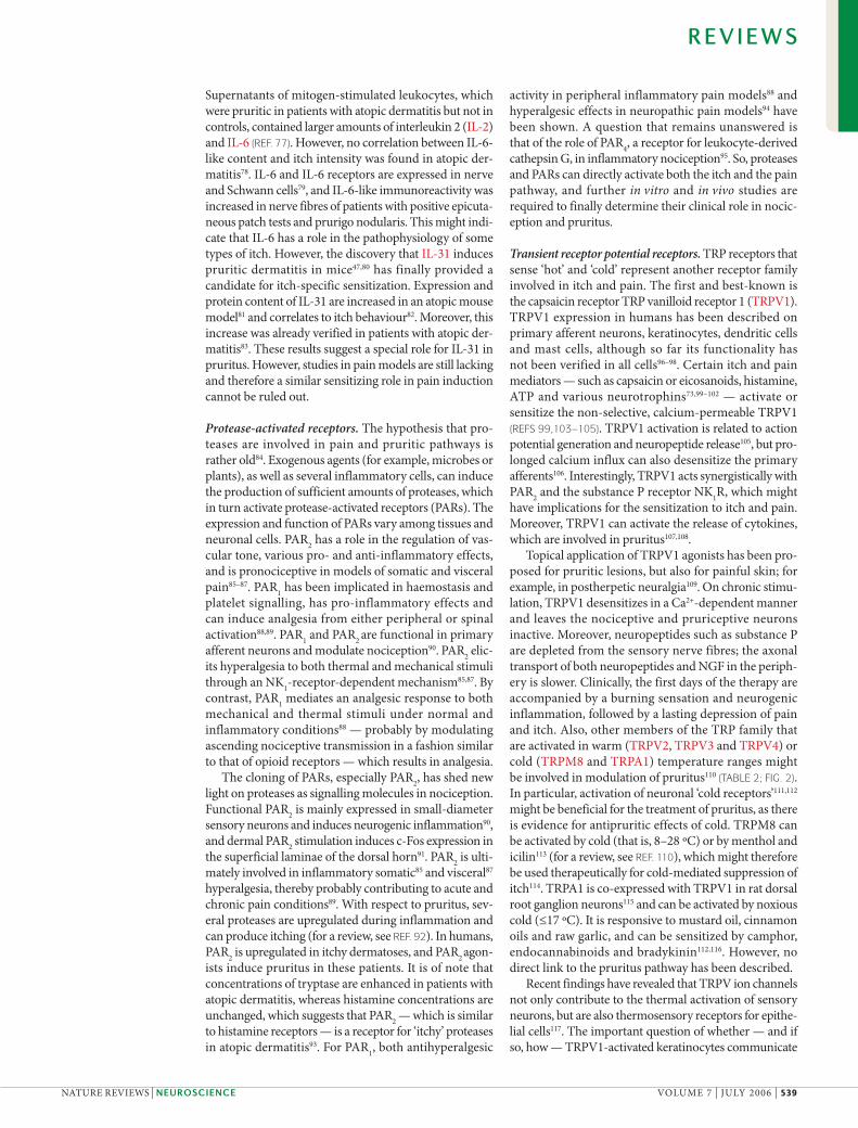
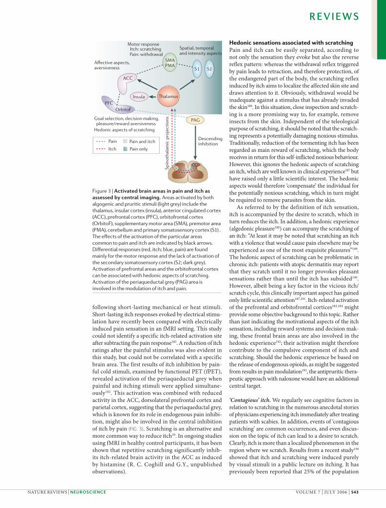
Central processing. Itch and pain are complex sensa-tions consisting of sensory discriminative, affective and motivational components. Brain imaging studies in the early 1990s have significantly widened our knowledge and understanding of the complexity of pain presenta-tion in the brain, trying to correlate different aspects of the pain sensation to particular brain areas170. It is generally accepted that spatial, temporal and intensity aspects of pain perception are processed in the primary and secondary somatosensory cortex (S1 and S2, respec-tively), whereas the anterior cingulate cortex (ACC) and insular cortex are involved in the affective-motivational component171,172. Somatotopic representation of pain is reported for the S1173, but might be much clearer in the dorsal posterior insula174. This would confirm a different central projection pathway for pain primary projections to the dorsal posterior insula as part of a basic interoceptive cortex with homeostatic function175. Moreover, the thalamus, prefrontal cortex, premotor areas176,177 and cerebellum are most commonly activated by painful stimulation178.
Some central imaging studies on itch have been carried out using positron emission tomography (PET) and functional MRI (fMRI). An early PET study179 found significant activation in the prefrontal cortex, premotor areas and ACC in response to itch. More recent stud-ies180,181 using histamine pricks or injections showed similar activation of the premotor area, as well as the prefrontal area and S1. Also, with the less noxious ion-tophoresis of histamine, a similar dose-related activation pattern was induced in the ACC, premotor cortex and the posterior parietal cortex182.
fMRI studies have provided further information on itch processing in the brain with improved tem-poral and spatial resolution183,184. In these studies, as above, the premotor areas, prefrontal cortex, ACC and cerebellum were activated. When comparing the brain areas involved in itch and pain processing (FIG. 3), a large overlap between the two sensations was identified, with no obvious itch-specific activa-tion pattern. Differences mainly relate to the lack of S2 activation and the predominant activation of ipsilateral motor areas in pruritus. The activa-tion of ipsilateral motor areas can be correlated to the planning of the scratch response directed to the stimulated limb, as opposed to the pain response in which contralateral motor activation is required to withdraw the stimulated limb.
However, this comparison is based on only a few studies and is to be regarded as preliminary. Some of the differences might also relate to variant experimen-tal protocols involving either the prolonged time course of histamine-induced itch or acute pain responses
R E V I E W S
542 | JULY 2006 | VOLUME 7 www.nature.com/reviews/neuro
Affective aspects,aversiveness
Motor responseSpatial, temporaland intensity aspects
Descendinginhibition
Goal selection, decision making, pleasure/reward aversiveness
Itch: scratching
Hedonic aspects of scratching
Spin
otha
lam
ic p
roje
ctio
ns
SMAPMA
S1 S2
PAG
PFCOrbitoF
ACC
Insula Thalamus
Pain
Itch
Pain: withdrawal
Pain and itch
Pain only
following short-lasting mechanical or heat stimuli. Short-lasting itch responses evoked by electrical stimu-lation have recently been compared with electrically induced pain sensation in an fMRI setting. This study could not identify a specific itch-related activation site after subtracting the pain response185. A reduction of itch ratings after the painful stimulus was also evident in this study, but could not be correlated with a specific brain area. The first results of itch inhibition by pain-ful cold stimuli, examined by functional PET (fPET), revealed activation of the periaqueductal grey when painful and itching stimuli were applied simultane-ously182. This activation was combined with reduced activity in the ACC, dorsolateral prefrontal cortex and parietal cortex, suggesting that the periaqueductal grey, which is known for its role in endogenous pain inhibi-tion, might also be involved in the central inhibition of itch by pain (FIG. 3). Scratching is an alternative and more common way to reduce itch10. In ongoing studies using fMRI in healthy control participants, it has been shown that repetitive scratching significantly inhib-its itch-related brain activity in the ACC as induced by histamine (R. C. Coghill and G.Y., unpublished observations).
Hedonic sensations associated with scratchingPain and itch can be easily separated, according to not only the sensation they evoke but also the reverse reflex pattern: whereas the withdrawal reflex triggered by pain leads to retraction, and therefore protection, of the endangered part of the body, the scratching reflex induced by itch aims to localize the affected skin site and draws attention to it. Obviously, withdrawal would be inadequate against a stimulus that has already invaded the skin186. In this situation, close inspection and scratch-ing is a more promising way to, for example, remove insects from the skin. Independent of the teleological purpose of scratching, it should be noted that the scratch-ing represents a potentially damaging noxious stimulus. Traditionally, reduction of the tormenting itch has been regarded as main reward of scratching, which the body receives in return for this self-inflicted noxious behaviour. However, this ignores the hedonic aspects of scratching an itch, which are well known in clinical experience187 but have raised only a little scientific interest. The hedonic aspects would therefore ‘compensate’ the individual for the potentially noxious scratching, which in turn might be required to remove parasites from the skin.
As referred to by the definition of itch sensation, itch is accompanied by the desire to scratch, which in turn reduces the itch. In addition, a hedonic experience (algedonic pleasure188) can accompany the scratching of an itch: “At least it may be noted that scratching an itch with a violence that would cause pain elsewhere may be experienced as one of the most exquisite pleasures”189. The hedonic aspect of scratching can be problematic in chronic itch: patients with atopic dermatitis may report that they scratch until it no longer provokes pleasant sensations rather than until the itch has subsided190. However, albeit being a key factor in the vicious itch/scratch cycle, this clinically important aspect has gained only little scientific attention187,191. Itch-related activation of the prefrontal and orbitofrontal cortices183,184 might provide some objective background to this topic. Rather than just indicating the motivational aspects of the itch sensation, including reward systems and decision mak-ing, these frontal brain areas are also involved in the hedonic experience192; their activation might therefore contribute to the compulsive component of itch and scratching. Should the hedonic experience be based on the release of endogenous opioids, as might be suggested from results in pain modulation193, the antipruritic thera-peutic approach with naloxone would have an additional central target.
‘Contagious’ itch. We regularly see cognitive factors in relation to scratching in the numerous anecdotal stories of physicians experiencing itch immediately after treating patients with scabies. In addition, events of ‘contagious scratching’ are common occurrences, and even discus-sion on the topic of itch can lead to a desire to scratch. Clearly, itch is more than a localized phenomenon in the region where we scratch. Results from a recent study194 showed that itch and scratching were induced purely by visual stimuli in a public lecture on itching. It has previously been reported that 25% of the population
Figure 3 | Activated brain areas in pain and itch as assessed by central imaging. Areas activated by both algogenic and pruritic stimuli (light grey) include the thalamus, insular cortex (insula), anterior cingulated cortex (ACC), prefrontal cortex (PFC), orbitofrontal cortex (OrbitoF), supplementary motor area (SMA), premotor area (PMA), cerebellum and primary somatosensory cortex (S1) . The effects of the activation of the particular areas common to pain and itch are indicated by black arrows. Differential responses (red, itch; blue, pain) are found mainly for the motor response and the lack of activation of the secondary somatosensory cortex (S2; dark grey). Activation of prefrontal areas and the orbitofrontal cortex can be associated with hedonic aspects of scratching. Activation of the periaqueductal grey (PAG) area is involved in the modulation of itch and pain.
R E V I E W S
NATURE REVIEWS | NEUROSCIENCE VOLUME 7 | JULY 2006 | 543
are conscious that scratching an itchy area can produce the sensation of itch elsewhere — the so-called ‘referred itch’195. Infectious scratching has also been observed in monkeys196, but the underlying mechanism is not clear. A possible neural substrate is ‘mirror neurons’197, which are active on executing a certain motor action, but also on viewing others performing the same action. They have been implicated in imitation and learning198. No data on central activation for contagious itching are available, but for a similar phenomenon, contagious yawning, a classical imitation pattern with activation of the human mirror neuron system has been proposed but not verified199.
Conclusions and future perspectivesItch and pain are different sensations that are processed by distinct sets of neurons. Histamine-sensitive pruri-ceptors have been characterized that can explain acute histamine-induced itch. However, they cannot account entirely for the clinically more relevant histamine-inde-pendent types of chronic itch. Therefore, additional classes of pruriceptors will have to be characterized.
Complex interactions exist between pain and itch, with itch inhibition by painful stimuli such as scratch-ing being used in everyday life to combat itch. Another example of the antagonistic interaction is itch induced by analgesic opioids. More than just being a side effect of opioid therapy, this antagonistic interaction is thera-peutically exploited by µ-opioid receptor antagonists acting as antipruritics under central itch conditions. In addition to the antagonistic interaction, there is a broad overlap between relevant mediator systems in pain and itch, including NGF, TRPV1 and PARs. Peripheral mechanisms of itch and pain are so closely related that even similar therapeutic strategies are being exploited, for example, anti-NGF.
Parallels between pain and itch processing are even more evident in the pattern of central sensitization. Touch- and pin prick-induced pain (allodynia and punctate hyperalgesia) correlate to touch- and pin prick-induced itch (alloknesis and punctate hyperknesis). The similarities of these symptoms have led researchers to successfully apply validated therapeutic approaches from the pain field — such as gabapentin for neuropathic pain — in itch therapy. Moreover, the knowledge about similar mechanisms of sensitization could also spark the interest of pain researchers in chronic pruritic diseases to study neuronal sensitization mechanisms. It remains to be established whether central processes leading to
memory formation in chronic pain are also of clinical relevance for chronic itch.
Central imaging techniques like PET and fMRI have identified the pattern of central activation that correlates to acute itch. Central areas involved in itch processing include the dorsal posterior insula, ACC and PFC, thalamus and premotor areas. So, there is considerable similarity to areas involved in pain processing. More pronounced ipsilateral activation of motor areas in itch might relate to the planning of the scratch response, whereas so far the background for the lack of activation of the S2 in itch is not clear. Clinically important, itch not only has aversive dimensions but also has a hedonic component, which might be a major drive for compulsive scratching. Teleologically, this aspect is of considerable interest, as the reward of scratching could compensate the organism for its potentially damaging effects.
New candidates for itch-specific mediator systems have emerged and will be tested for clinical significance in humans. Although data on histamine H4 receptors are currently restricted to mice, data on IL-31 are much broader and are highly promising. The interaction between inflammatory cells and neurons has gained major attention for peripheral, but also central, sensitiza-tion mechanisms. Recent work has highlighted the role of keratinocyte–neuron interactions in the generation of itch. In this interaction, keratinocytes can be the source and recipient of various sensitizing mediators, and might even be involved in the transduction process through their TRP receptors. Classical inflammatory signs (swelling, redness and warmth) can fail to appear after the release of keratinocyte-derived inflammatory mediators, because of the special environment in the vessel-free epidermis. Instead, disturbed barrier function, dry skin and itch can result. Therefore, the study of keratinocyte–neuron inter-actions is of considerable interest for further investigation of pruritic mechanisms in the skin. Central imaging tech-niques and experimental protocols for the study of itch have been developed. In the future, patient-based research in fMRI settings and interaction studies between itch and pain will further clarify the brain mechanisms of chronic itch and its inhibition. Finally, enormous progress has been achieved in the development of clinically relevant animal models for itch. Although no animal model exists so far for systemic itch (for example, hepatic or renal itch), the acute chemical stimulation models have been further refined. Now animal models for atopic dermatitis and dry skin are available, which will facilitate translational aspects to humans.
1. v. Frey, M. Zur Physiologie der Juckempfindung. Arch. Neerl. Physiol. 7, 142–145 (1922).
2. Nojima, H. et al. Opioid modulation of scratching and spinal c-fos expression evoked by intradermal serotonin. J. Neurosci. 23, 10784–10790 (2003).c-Fos expression as objective evidence for differential pain and itch behaviour in animals.
3. Schmelz, M., Schmidt, R., Bickel, A., Handwerker, H. O. & Torebjörk, H. E. Specific C-receptors for itch in human skin. J. Neurosci. 17, 8003–8008 (1997).
4. Andrew, D. & Craig, A. D. Spinothalamic lamina 1 neurons selectively sensitive to histamine: a central neural pathway for itch. Nature Neurosci. 4, 72–77 (2001).
References 3 and 4 are pioneering works on the identification of an itch-selective neuronal pathway.
5. McMahon, S. B. & Koltzenburg, M. Itching for an explanation. Trends. Neurosci. 15, 497–501 (1992).
6. Twycross, R. et al. Itch: scratching more than the surface. QJM. 96, 7–26 (2003).
7. Bernhard, J. D. Itch and pruritus: what are they, and how should itches be classified? Dermatol. Ther. 18, 288–291 (2005).
8. Ward, L., Wright, E. & McMahon, S. B. A comparison of the effects of noxious and innocuous counterstimuli on experimentally induced itch and pain. Pain 64, 129–138 (1996).
9. Nilsson, H. J., Levinsson, A. & Schouenborg, J. Cutaneous field stimulation (CFS): a new powerful method to combat itch. Pain 71, 49–55 (1997).
10. Yosipovitch, G., Fast, K. & Bernhard, J. D. Noxious heat and scratching decrease histamine-induced itch and skin blood flow. J. Invest. Dermatol. 125, 1268–1272 (2005).
11. Brull, S. J., Atanassoff, P. G., Silverman, D. G., Zhang, J. & LaMotte, R. H. Attenuation of experimental pruritus and mechanically evoked dysesthesiae in an area of cutaneous allodynia. Somatosens. Mot. Res. 16, 299–303 (1999).
12. Green, A. D., Young, K. K., Lehto, S. G., Smith, S. B. & Mogil, J. S. Influence of genotype, dose and sex on
R E V I E W S
544 | JULY 2006 | VOLUME 7 www.nature.com/reviews/neuro
pruritogen-induced scratching behaviour in the mouse. Pain 10 May 2006 (doi:10.1016/j.pain.2006.03.023).Pioneering work on the genetic approach to itch research.
13. Mogil, J. S. et al. Variable sensitivity to noxious heat is mediated by differential expression of the CGRP gene. Proc. Natl Acad. Sci. USA 102, 12938–12943 (2005).
14. Atanassoff, P. G. et al. Enhancement of experimental pruritus and mechanically evoked dysesthesiae with local anesthesia. Somatosens. Mot. Res. 16, 291–298 (1999).
15. Andrew, D., Schmelz, M. & Ballantyne, J. C. in Progress in Pain Research and Management (eds Dostrovsky, J. O., Carr, D. B. & Koltzenburg, M.) 213–226 (IASP Press, Seattle, 2003).
16. Heyer, G., Dotzer, M., Diepgen, T. L. & Handwerker, H. O. Opiate and H1 antagonist effects on histamine induced pruritus and alloknesis. Pain 73, 239–243 (1997).
17. Ko, M. C., Song, M. S., Edwards, T., Lee, H. & Naughton, N. N. The role of central µ opioid receptors in opioid-induced itch in primates. J. Pharmacol. Exp. Ther. 310, 169–176 (2004).
18. Bergasa, N. V. The pruritus of cholestasis. J. Hepatol. 43, 1078–1088 (2005).
19. McRae, C. A. et al. Pain as a complication of use of opiate antagonists for symptom control in cholestasis. Gastroenterology 125, 591–596 (2003).
20. Jones, E. A., Neuberger, J. & Bergasa, N. V. Opiate antagonist therapy for the pruritus of cholestasis: the avoidance of opioid withdrawal-like reactions. QJM 95, 547–552 (2002).
21. Kamei, J. & Nagase, H. Norbinaltorphimine, a selective κ-opioid receptor antagonist, induces an itch-associated response in mice. Eur. J. Pharmacol. 418, 141–145 (2001).
22. Ko, M. C. et al. Activation of κ-opioid receptors inhibits pruritus evoked by subcutaneous or intrathecal administration of morphine in monkeys. J. Pharmacol. Exp. Ther. 305, 173–179 (2003).
23. Wakasa, Y. et al. Inhibitory effects of TRK-820 on systemic skin scratching induced by morphine in rhesus monkeys. Life Sci. 75, 2947–2957 (2004).
24. Kjellberg, F. & Tramer, M. R. Pharmacological control of opioid-induced pruritus: a quantitative systematic review of randomized trials. Eur. J. Anaesthesiol. 18, 346–357 (2001).
25. Wikstrom, B. et al. κ-opioid system in uremic pruritus: multicenter, randomized, double-blind, placebo-controlled clinical studies. J. Am. Soc. Nephrol. 16, 3742–3747 (2005).
26. Dawn, A. G. & Yosipovitch, G. Butorphanol for treatment of intractable pruritus. J. Am. Acad. Dermatol. 54, 527–531 (2006).
27. Reeh, P. W. & Kress, M. Effects of Classical Algogens. Semin. Neurosci. 7, 221–226 (1995).
28. Kidd, B. L. & Urban, L. A. Mechanisms of inflammatory pain. Br. J. Anaesth. 87, 3–11 (2001).
29. Zhang, X., Huang, J. & McNaughton, P. A. NGF rapidly increases membrane expression of TRPV1 heat-gated ion channels. EMBO J. 24, 4211–4223 (2005).
30. Hefti, F. F. et al. Novel class of pain drugs based on antagonism of NGF. Trends Pharmacol. Sci. 27, 85–91 (2005).
31. Bohm-Starke, N., Hilliges, M., Falconer, C. & Rylander, E. Increased intraepithelial innervation in women with vulvar vestibulitis syndrome. Gynecol. Obstet. Invest. 46, 256–260 (1998).
32. Urashima, R. & Mihara, M. Cutaneous nerves in atopic dermatitis — a histological, immunohistochemical and electron microscopic study. Virchows Arch. Int. J. Pathol. 432, 363–370 (1998).
33. Toyoda, M. et al. Nerve growth factor and substance P are useful plasma markers of disease activity in atopic dermatitis. Br. J. Dermatol. 147, 71–79 (2002).
34. Groneberg, D. A. et al. Gene expression and regulation of nerve growth factor in atopic dermatitis mast cells and the human mast cell line-1. J. Neuroimmunol. 161, 87–92 (2005).References 33 and 34 provide convincing evidence for clinically relevant NGF increases in atopic dermatitis.
35. Kinkelin, I., Motzing, S., Koltenzenburg, M. & Brocker, E. B. Increase in NGF content and nerve fiber sprouting in human allergic contact eczema. Cell Tissue Res. 302, 31–37 (2000).
36. Johansson, O., Liang, Y. & Emtestam, L. Increased nerve growth factor- and tyrosine kinase A-like immunoreactivities in prurigo nodularis skin — an exploration of the cause of neurohyperplasia. Arch. Dermatol. Res. 293, 614–619 (2002).
37. Choi, J. C., Yang, J. H., Chang, S. E. & Choi, J. H. Pruritus and nerve growth factor in psoriasis. Korean J. Dermatol. 43, 769–773 (2005).
38. Halvorson, K. G. et al. A blocking antibody to nerve growth factor attenuates skeletal pain induced by prostate tumor cells growing in bone. Cancer Res. 65, 9426–9435 (2005).
39. Lane, N. et al. RN624 (Anti-NGF) improves pain and function in subjects with moderate knee osteoarthritis: a Phase I study. Osteoarthritis — clinical aspects. Proceedings American College of Rheumatology Abstr. 765 (2005).
40. Takano, N., Sakurai, T. & Kurachi, M. Effects of anti-nerve growth factor antibody on symptoms in the NC/Nga mouse, an atopic dermatitis model. J. Pharmacol. Sci. 99, 277–286 (2005).References 39 and 40 provided the first evidence for therapeutic efficacy of anti-NGF strategies in itch and pain.
41. Tanaka, A. & Matsuda, H. Expression of nerve growth factor in itchy skins of atopic NC/NgaTnd mice. J. Vet. Med. Sci. 67, 915–919 (2005).
42. Verge, V. M., Richardson, P. M., Wiesenfeld-Hallin, Z. & Hokfelt, T. Differential influence of nerve growth factor on neuropeptide expression in vivo: a novel role in peptide suppression in adult sensory neurons. J. Neurosci. 15, 2081–2096 (1995).
43. Laird, J. M., Roza, C., De Felipe, C., Hunt, S. P. & Cervero, F. Role of central and peripheral tachykinin NK1 receptors in capsaicin-induced pain and hyperalgesia in mice. Pain 90, 97–103 (2001).
44. Hill, R. NK1 (substance P) receptor antagonists — why are they not analgesic in humans? Trends Pharmacol. Sci. 21, 244–246 (2000).
45. Weidner, C. et al. Acute effects of substance P and calcitonin gene-related peptide in human skin — a microdialysis study. J. Invest. Dermatol. 115, 1015–1020 (2000).
46. Yosipovitch, G., Greaves, M. & Schmelz, M. Itch. Lancet 361, 690–694 (2003).
47. Sun, R. Q. et al. Calcitonin gene-related peptide receptor activation produces PKA- and PKC-dependent mechanical hyperalgesia and central sensitization. J. Neurophysiol. 92, 2859–2866 (2004).
48. Ekblom, A., Lundeberg, T. & Wahlgren, C. F. Influence of calcitonin gene-related peptide on histamine- and substance P-induced itch, flare and weal in humans. Skin Pharmacol. 6, 215–222 (1993).
49. Katsuno, M. et al. Neuropeptides concentrations in the skin of a murine (NC/Nga mice) model of atopic dermatitis. J. Dermatol. Sci. 33, 55–65 (2003).
50. Koltzenburg, M. Neural mechanisms of cutaneous nociceptive pain. Clin. J. Pain 16, S131–S138 (2000).
51. Torebjörk, H. E., Schmelz, M. & Handwerker, H. O. Functional properties of human cutaneous nociceptors and their role in pain and hyperalgesia (eds Cervero & Belmonte, C.) 349–369 (Oxford Univ. Press, 1996).
52. LaMotte, R. H., Shain, C. N., Simone, D. A. & Tsai, E. F. P. Neurogenic hyperalgesia psychophysical studies of underlying mechanisms. J. Neurophysiol. 66, 190–211 (1991).
53. Bickford, R. G. L. Experiments relating to itch sensation, its peripheral mechanism and central pathways. Clin. Sci. 3, 377–386 (1938).
54. Simone, D. A., Alreja, M. & LaMotte, R. H. Psychophysical studies of the itch sensation and itchy skin (‘alloknesis’) produced by intracutaneous injection of histamine. Somatosens. Mot. Res. 8, 271–279 (1991).A detailed psychophysical investigation of central sensitization for itch.
55. Heyer, G., Ulmer, F. J., Schmitz, J. & Handwerker, H. O. Histamine-induced itch and alloknesis (itchy skin) in atopic eczema patients and controls. Acta Derm. Venereol. (Stockh.) 75, 348–352 (1995).
56. Nilsson, H. J. & Schouenborg, J. Differential inhibitory effect on human nociceptive skin senses induced by local stimulation of thin cutaneous fibers. Pain 80, 103–112 (1999).
57. Vogelsang, M., Heyer, G. & Hornstein, O. P. Acetylcholine induces different cutaneous sensations in atopic and non-atopic subjects. Acta Derm. Venereol. 75, 434–436 (1995).
58. Birklein, F., Claus, D., Riedl, B., Neundorfer, B. & Handwerker, H. O. Effects of cutaneous histamine application in patients with sympathetic reflex dystrophy. Muscle Nerve 20, 1389–1395 (1997).
59. Baron, R., Schwarz, K., Kleinert, A., Schattschneider, J. & Wasner, G. Histamine-induced itch converts into pain in neuropathic hyperalgesia. Neuroreport 12,
3475–3478 (2001).References 58 and 59 were the first descriptions of central sensitization for normally pruritic histamine in chronic pain patients, and introduced evidence for central sensitization for C-fibre input in humans.
60. Ikoma, A. et al. Painful stimuli evoke itch in patients with chronic pruritus: central sensitization for itch. Neurology 62, 212–217 (2004).
61. Summey, B. T., Jr. & Yosipovitch, G. Pharmacologic advances in the systemic treatment of itch. Dermatol. Ther. 18, 328–332 (2005).
62. Rogawski, M. A. & Loscher, W. The neurobiology of antiepileptic drugs for the treatment of nonepileptic conditions. Nature Med. 10, 685–692 (2004).
63. Bueller, H. A., Bernhard, J. D. & Dubroff, L. M. Gabapentin treatment for brachioradial pruritus. J. Eur. Acad. Dermatol. Venereol. 13, 227–228 (1999).
64. Winhoven, S. M., Coulson, I. H. & Bottomley, W. W. Brachioradial pruritus: response to treatment with gabapentin. Br. J. Dermatol. 150, 786–787 (2004).
65. Yesudian, P. D. & Wilson, N. J. Efficacy of gabapentin in the management of pruritus of unknown origin. Arch. Dermatol. 141, 1507–1509 (2005).
66. Gupta, M. A. & Guptat, A. K. The use of antidepressant drugs in dermatology. J. Eur. Acad. Dermatol. Venereol. 15, 512–518 (2001).
67. Zylicz, Z., Krajnik, M., Sorge, A. A. & Costantini, M. Paroxetine in the treatment of severe non-dermatological pruritus: a randomized, controlled trial. J. Pain Symptom Manage. 26, 1105–1112 (2003).
68. Anttila, S. A. & Leinonen, E. V. A review of the pharmacological and clinical profile of mirtazapine. CNS Drug Rev. 7, 249–264 (2001).
69. Davis, M. P., Frandsen, J. L., Walsh, D., Andresen, S. & Taylor, S. Mirtazapine for pruritus. J. Pain Symptom Manage. 25, 288–291 (2003).
70. Hundley, J. L. & Yosipovitch, G. Mirtazapine for reducing nocturnal itch in patients with chronic pruritus: a pilot study. J. Am. Acad. Dermatol. 50, 889–891 (2004).
71. Bomholt, S. F., Mikkelsen, J. D. & Blackburn-Munro, G. Antinociceptive effects of the antidepressants amitriptyline, duloxetine, mirtazapine and citalopram in animal models of acute, persistent and neuropathic pain. Neuropharmacology 48, 252–263 (2005).
72. Oaklander, A. L., Bowsher, D., Galer, B., Haanpää, M. & Jensen, M. P. Herpes zoster itch: preliminary epidemiologic data. J. Pain 4, 338–343 (2003).
73. Morita, E., Matsuo, H. & Zhang, Y. Double-blind, crossover comparison of olopatadine and cetirizine versus placebo: suppressive effects on skin response to histamine iontophoresis. J. Dermatol. 32, 58–61 (2005).
74. Klein, P. A. & Clark, R. A. An evidence-based review of the efficacy of antihistamines in relieving pruritus in atopic dermatitis. Arch. Dermatol. 135, 1522–1525 (1999).
75. Bell, J. K., Mcqueen, D. S. & Rees, J. L. Involvement of histamine H4 and H1 receptors in scratching induced by histamine receptor agonists in Balb C mice. Br. J. Pharmacol. 142, 374–380 (2004).Introduces a new and powerful candidate for an itch-specific mediator system in rodents.
76. Sommer, C. & Kress, M. Recent findings on how proinflammatory cytokines cause pain: peripheral mechanisms in inflammatory and neuropathic hyperalgesia. Neurosci. Lett. 361, 184–187 (2004).
77. Cremer, B., Heimann, A., Dippel, E. & Czarnetzki, B. M. Pruritogenic effects of mitogen stimulated peripheral blood mononuclear cells in atopic eczema. Acta Derm. Venereol. (Stockh.) 75, 426–428 (1995).
78. Lippert, U. et al. Role of antigen-induced cytokine release in atopic pruritus. Int. Arch. Allergy Immunol. 116, 36–39 (1998).
79. Grothe, C. et al. Expression of interleukin-6 and its receptor in the sciatic nerve and cultured Schwann cells: relation to 18-kD fibroblast growth factor-2. Brain Res. 885, 172–181 (2000).
80. Dillon, S. R. et al. Interleukin 31, a cytokine produced by activated T cells, induces dermatitis in mice. Nature Immunol. 5, 752–760 (2004).Introduces a new and powerful candidate for an itch-specific mediator system, with major implications for human diseases.
81. Takaoka, A. et al. Expression of IL-31 gene transcripts in NC/Nga mice with atopic dermatitis. Eur. J. Pharmacol. 516, 180–181 (2005).
82. Takaoka, A. et al. Involvement of IL-31 on scratching
R E V I E W S
NATURE REVIEWS | NEUROSCIENCE VOLUME 7 | JULY 2006 | 545
behavior in NC/Nga mice with atopic-like dermatitis. Exp. Dermatol. 15, 161–167 (2006).
83. Bilsborough, J. et al. IL-31 is associated with cutaneous lymphocyte antigen-positive skin homing T cells in patients with atopic dermatitis. J. Allergy Clin. Immunol. 117, 418–425 (2006).
84. Biro, T. et al. How best to fight that nasty itch — from new insights into the neuroimmunological, neuroendocrine, and neurophysiological bases of pruritus to novel therapeutic approaches. Exp. Dermatol. 14, 225 (2005).
85. Vergnolle, N. et al. Proteinase-activated receptor-2 and hyperalgesia: a novel pain pathway. Nature Med. 7, 821–826 (2001).
86. Vergnolle, N. The enteric nervous system in inflammation and pain: the role of proteinase-activated receptors. Can. J. Gastroenterol. 17, 589–592 (2003).
87. Coelho, A. M., Vergnolle, N., Guiard, B., Fioramonti, J. & Bueno, L. Proteinases and proteinase-activated receptor 2: a possible role to promote visceral hyperalgesia in rats. Gastroenterology 122, 1035–1047 (2002).
88. Asfaha, S., Brussee, V., Chapman, K., Zochodne, D. W. & Vergnolle, N. Proteinase-activated receptor-1 agonists attenuate nociception in response to noxious stimuli. Br. J. Pharmacol. 135, 1101–1106 (2002).
89. Vergnolle, N., Wallace, J. L., Bunnett, N. W. & Hollenberg, M. D. Protease-activated receptors in inflammation, neuronal signaling and pain. Trends Pharmacol. Sci. 22, 146–152 (2001).
90. Steinhoff, M. et al. Agonists of proteinase-activated receptor 2 induce inflammation by a neurogenic mechanism. Nature Med. 6, 151–158 (2000).Presents a new concept of the role of proteases in neurogenic inflammation.
91. Dai, Y. et al. Proteinase-activated receptor 2-mediated potentiation of transient receptor potential vanilloid subfamily 1 activity reveals a mechanism for proteinase-induced inflammatory pain. J. Neurosci. 24, 4293–4299 (2004).
92. Paus, R., Schmelz, M., Biro, T. & Steinhoff, M. Scratching the brain for more effective itch therapy — frontiers in pruritus research. J. Clin. Invest. 116, 1174–1186 (2006).
93. Steinhoff, M. et al. Proteinase-activated receptor-2 mediates itch: a novel pathway for pruritus in human skin. J. Neurosci. 23, 6176–6180 (2003).
94. Narita, M. et al. Protease-activated receptor-1 and platelet-derived growth factor in spinal cord neurons are implicated in neuropathic pain after nerve injury. J. Neurosci. 25, 10000–10009 (2005).
95. Houle, S., Papez, M. D., Ferazzini, M., Hollenberg, M. D. & Vergnolle, N. Neutrophils and the kallikrein-kinin system in proteinase-activated receptor 4-mediated inflammation in rodents. Br. J. Pharmacol. 146, 670–678 (2005).
96. Bodo, E. et al. Vanilloid receptor-1 (VR1) is widely expressed on various epithelial and mesenchymal cell types of human skin. J. Invest. Dermatol. 123, 410–413 (2004).
97. Inoue, K. et al. Functional vanilloid receptors in cultured normal human epidermal keratinocytes. Biochem. Biophys. Res. Commun. 291, 124–129 (2002).
98. Stander, S. et al. Expression of vanilloid receptor subtype 1 in cutaneous sensory nerve fibers, mast cells, and epithelial cells of appendage structures. Exp. Dermatol. 13, 129–139 (2004).
99. Hwang, S. W. et al. Direct activation of capsaicin receptors by products of lipoxygenases: endogenous capsaicin-like substances. Proc. Natl Acad. Sci. USA 97, 6155–6160 (2000).
100. Chuang, H. H. et al. Bradykinin and nerve growth factor release the capsaicin receptor from PtdIns(4, 5)P2-mediated inhibition. Nature 411, 957–962 (2001).
101. Shin, J. et al. Bradykinin-12-lipoxygenase-VR1 signaling pathway for inflammatory hyperalgesia. Proc. Natl Acad. Sci. USA 99, 10150–10155 (2002).
102. Mohapatra, D. P. & Nau, C. Desensitization of capsaicin-activated currents in the vanilloid receptor TRPV1 is decreased by the cyclic AMP-dependent protein kinase pathway. J. Biol. Chem. 278, 50080–50090 (2003).
103. Cortright, D. N. & Szallasi, A. Biochemical pharmacology of the vanilloid receptor TRPV1. An update. Eur. J. Biochem. 271, 1814–1819 (2004).
104. Caterina, M. J. et al. The capsaicin receptor: a heat-activated ion channel in the pain pathway. Nature 389, 816–824 (1997).
105. Caterina, M. J. & Julius, D. The vanilloid receptor: a molecular gateway to the pain pathway. Annu. Rev. Neurosci. 24, 487–517 (2001).
106. Koplas, P. A., Rosenberg, R. L. & Oxford, G. S. The role of calcium in the desensitization of capsaicin responses in rat dorsal root ganglion neurons. J. Neurosci. 17, 3525–3537 (1997).
107. Southall, M. D. et al. Activation of epidermal vanilloid receptor-1 induces release of proinflammatory mediators in human keratinocytes. J. Pharmacol. Exp. Ther. 304, 217–222 (2003).Presents a new concept of the role of keratinocytes in inflammation and nociception.
108. Bodo, E. et al. A hot new twist to hair biology: involvement of vanilloid receptor-1 (VR1/TRPV1) signaling in human hair growth control. Am. J. Pathol. 166, 985–998 (2005).
109. Meier, T. et al. Efficacy of lidocaine patch 5% in the treatment of focal peripheral neuropathic pain syndromes: a randomized, double-blind, placebo-controlled study. Pain 106, 151–158 (2003).
110. Steinhoff, M. et al. Neurophysiological, neuroimmunological and neuroendocrine basis of pruritus. J. Invest. Dermatol. (in the press).
111. Peier, A. M. et al. A TRP channel that senses cold stimuli and menthol. Cell 108, 705–715 (2002).
112. Story, G. M. et al. ANKTM1, a TRP-like channel expressed in nociceptive neurons, is activated by cold temperatures. Cell 112, 819–829 (2003).
113. Wei, E. T. & Seid, D. A. AG-3–5: a chemical producing sensations of cold. J. Pharm. Pharmacol. 35, 110–112 (1983).
114. Patapoutian, A., Peier, A. M., Story, G. M. & Viswanath, V. ThermoTRP channels and beyond: mechanisms of temperature sensation. Nature Rev. Neurosci. 4, 529–539 (2003).
115. Kobayashi, K. et al. Distinct expression of TRPM8, TRPA1, and TRPV1 mRNAs in rat primary afferent neurons with αδ/c-fibers and colocalization with trk receptors. J. Comp. Neurol. 493, 596–606 (2005).
116. Reid, G. ThermoTRP channels and cold sensing: what are they really up to? Pflugers Arch. 451, 250–263 (2005).
117. Lee, H. & Caterina, M. J. TRPV channels as thermosensory receptors in epithelial cells. Pflugers Arch. 451, 160–167 (2005).
118. Green, B. G. Sensory characteristics of camphor. J. Invest. Dermatol. 94, 662–666 (1990).
119. Peier, A. M. et al. A heat-sensitive TRP channel expressed in keratinocytes. Science 296, 2046–2049 (2002).
120. Xu, H., Blair, N. T. & Clapham, D. E. Camphor activates and strongly desensitizes the transient receptor potential vanilloid subtype 1 channel in a vanilloid-independent mechanism. J. Neurosci. 25, 8924–8937 (2005).
121. Chung, M. K., Lee, H., Mizuno, A., Suzuki, M. & Caterina, M. J. TRPV3 and TRPV4 mediate warmth-evoked currents in primary mouse keratinocytes. J. Biol. Chem. 279, 21569–21575 (2004).
122. Moqrich, A. et al. Impaired thermosensation in mice lacking TRPV3, a heat and camphor sensor in the skin. Science 307, 1468–1472 (2005).Evidence for a role of keratinocytes in the transduction process.
123. Stein, C., Schafer, M. & Machelska, H. Attacking pain at its source: new perspectives on opioids. Nature Med. 9, 1003–1008 (2003).
124. Steinhoff, M. et al. Modern aspects of cutaneous neurogenic inflammation. Arch. Dermatol. 139, 1479–1488 (2003).
125. Stander, S. et al. Neurophysiology of pruritus: cutaneous elicitation of itch. Arch Dermatol. 139, 1463–1470 (2003).
126. Minami, M., Maekawa, K., Yabuuchi, K. & Satoh, M. Double in situ hybridization study on coexistence of µ-, δ- and κ-opioid receptor mRNAs with preprotachykinin A mRNA in the rat dorsal root ganglia. Brain Res. Mol. Brain Res. 30, 203–210 (1995).
127. Satoh, M. & Minami, M. Molecular pharmacology of the opioid receptors. Pharmacol. Ther. 68, 343–364 (1995).
128. Devane, W. A., Dysarz, F. A., Johnson, M. R., Melvin, L. S. & Howlett, A. C. Determination and characterization of a cannabinoid receptor in rat brain. Mol. Pharmacol. 34, 605–613 (1988).
129. Matsuda, L. A., Lolait, S. J., Brownstein, M. J., Young, A. C. & Bonner, T. I. Structure of a cannabinoid receptor and functional expression of the cloned cDNA. Nature 346, 561–564 (1990).
130. Munro, S., Thomas, K. L. & Abu-Shaar, M. Molecular characterization of a peripheral receptor for cannabinoids. Nature 365, 61–65 (1993).
131. Pertwee, R. G. Evidence for the presence of CB1 cannabinoid receptors on peripheral neurones and for the existence of neuronal non-CB1 cannabinoid receptors. Life Sci. 65, 597–605 (1999).
132. Coutts, A. A., Irving, A. J., Mackie, K., Pertwee, R. G. & Anavi-Goffer, S. Localisation of cannabinoid CB(1) receptor immunoreactivity in the guinea pig and rat myenteric plexus. J. Comp. Neurol. 448, 410–422 (2002).
133. Ahluwalia, J., Urban, L., Bevan, S., Capogna, M. & Nagy, I. Cannabinoid 1 receptors are expressed by nerve growth factor- and glial cell-derived neurotrophic factor-responsive primary sensory neurones. Neuroscience. 110, 747–753 (2002).
134. Stander, S., Schmelz, M., Metze, D., Luger, T. & Rukwied, R. Distribution of cannabinoid receptor 1 (CB1) and 2 (CB2) on sensory nerve fibers and adnexal structures in human skin. J. Dermatol. Sci. 38, 177–188 (2005).
135. Galiegue, S. et al. Expression of central and peripheral cannabinoid receptors in human immune tissues and leukocyte subpopulations. Eur. J. Biochem. 232, 54–61 (1995).
136. Facci, L. et al. Mast cells express a peripheral cannabinoid receptor with differential sensitivity to anandamide and palmitoylethanolamide. Proc. Natl Acad. Sci. USA 92, 3376–3380 (1995).
137. Zhang, J. et al. Induction of CB2 receptor expression in the rat spinal cord of neuropathic but not inflammatory chronic pain models. Eur. J. Neurosci. 17, 2750–2754 (2003).
138. Re, G., Barbero, R., Miolo, A. & Di, M. V. Palmitoylethanolamide, endocannabinoids and related cannabimimetic compounds in protection against tissue inflammation and pain: potential use in companion animals. Vet. J. 29 Nov 2005 (doi:10.1016/j.pain.2005.10.003).
139. Di, M., V, Bifulco, M. & De Petrocellis, L. The endocannabinoid system and its therapeutic exploitation. Nature Rev. Drug Discov. 3, 771–784 (2004).
140. MacCarrone, M. et al. The endocannabinoid system in human keratinocytes. Evidence that anandamide inhibits epidermal differentiation through CB1 receptor-dependent inhibition of protein kinase C, activation protein-1, and transglutaminase. J. Biol. Chem. 278, 33896–33903 (2003).
141. Oddi, S. et al. Confocal microscopy and biochemical analysis reveal spatial and functional separation between anandamide uptake and hydrolysis in human keratinocytes. Cell. Mol. Life Sci. 62, 386–395 (2005).
142. Johanek, L. M. & Simone, D. A. Activation of peripheral cannabinoid receptors attenuates cutaneous hyperalgesia produced by a heat injury. Pain 109, 432–442 (2004).
143. Diaz-Laviada, I. & Ruiz-Llorente, L. Signal transduction activated by cannabinoid receptors. Mini Rev. Med. Chem. 5, 619–630 (2005).
144. Rukwied, R., Watkinson, A., McGlone, F. & Dvorak, M. Cannabinoid agonists attenuate capsaicin-induced responses in human skin. Pain 102, 283–288 (2003).
145. Dvorak, M., Watkinson, A., McGlone, F. & Rukwied, R. Histamine induced responses are attenuated by a cannabinoid receptor agonist in human skin. Inflamm. Res 52, 238–245 (2003).
146. Begg, M. et al. Evidence for novel cannabinoid receptors. Pharmacol. Ther. 106, 133–145 (2005).
147. Fattore, L. et al. Cannabinoids and reward: interactions with the opioid system. Crit. Rev. Neurobiol. 16, 147–158 (2004).
148. Vigano, D., Rubino, T. & Parolaro, D. Molecular and cellular basis of cannabinoid and opioid interactions. Pharmacol. Biochem. Behav. 81, 360–368 (2005).
149. Vigano, D. et al. Molecular mechanisms involved in the asymmetric interaction between cannabinoid and opioid systems. Psychopharmacology (Berl). 182, 527–536 (2005).
150. Ibrahim, M. M. et al. CB2 cannabinoid receptor activation produces antinociception by stimulating peripheral release of endogenous opioids. Proc. Natl Acad. Sci. USA. 102, 3093–3098 (2005).Analgesic effects of CB2 agonists were shown here to be based on peripheral release of endogenous opioids; therefore, this indirect effect has to be discussed.
151. Koltzenburg, M., Handwerker, H. O. & Torebjörk, H. E. The ability of humans to localise noxious stimuli. Neurosci. Lett. 150, 219–222 (1993).
R E V I E W S
546 | JULY 2006 | VOLUME 7 www.nature.com/reviews/neuro
152. Schmidt, R. et al. Novel classes of responsive and unresponsive C nociceptors in human skin. J. Neurosci. 15, 333–341 (1995).
153. Weidner, C. et al. Functional attributes discriminating mechano-insensitive and mechano-responsive C nociceptors in human skin. J. Neurosci. 19, 10184–10190 (1999).
154. Klede, M., Handwerker, H. O. & Schmelz, M. Central origin of secondary mechanical hyperalgesia. J. Neurophysiol. 90, 353–359 (2003).
155. Schmelz, M. et al. Active ‘itch fibers’ in chronic pruritus. Neurology 61, 564–566 (2003).
156. Schmelz, M. et al. Chemical response pattern of different classes of C-nociceptors to pruritogens and algogens. J. Neurophysiol. 89, 2441–2448 (2003).Prostaglandin induced activation is found only in histamine-sensitive pruriceptors — this is evidence for itch-specific neurons.
157. Wahlgren, C. F. & Ekblom, A. Two-point discrimination of itch in patients with atopic dermatitis and healthy subjects. Acta Derm. Venereol. (Stockh.) 76, 48–51 (1996).
158. Ikoma, A., Handwerker, H., Miyachi, Y. & Schmelz, M. Electrically evoked itch in humans. Pain 113, 148–154 (2005).
159. Shelley, W. B. & Arthur, R. P. Mucunain, the active pruritogenic proteinase of cowhage. Science 122, 469–470 (1955).
160. Hägermark, O. Influence of antihistamines, sedatives, and aspirin on experimental itch. Acta Derm. Venereol. 53, 363–368 (1973).
161. Tuckett, R. P. & Wei, J. Y. Response to an itch-producing substance in cat. II. Cutaneous receptor populations with unmyelinated axons. Brain Res. 413, 95–103 (1987).
162. Wei, J. Y. & Tuckett, R. P. Response of cat ventrolateral spinal axons to an itch-producing stimulus (cowhage). Somatosens. Mot. Res. 8, 227–239 (1991).
163. Darsow, U. et al. New aspects of itch pathophysiology: component analysis of atopic itch using the ‘Eppendorf Itch Questionnaire’. Int. Arch. Allergy Immunol. 124, 326–331 (2001).
164. Yosipovitch, G. et al. Itch characteristics in Chinese patients with atopic dermatitis using a new questionnaire for the assessment of pruritus. Int. J. Dermatol. 41, 212–216 (2002).
165. Melzack, R. The McGill Pain Questionnaire: major properties and scoring methods. Pain 1, 277–299 (1975).
166. Green, B. G. & Shaffer, G. S. The sensory response to capsaicin during repeated topical exposures: differential effects on sensations of itching and pungency. Pain 53, 323–334 (1993).
167. Simone, D. A. et al. Comparison of responses of primate spinothalamic tract neurons to pruritic and algogenic stimuli. J. Neurophysiol. 91, 213–222 (2004).
168. Schmelz, M. A neural pathway for itch. Nature Neurosci. 4, 9–10 (2001).
169. Craig, A. D. How do you feel? Interoception: the sense of the physiological condition of the body. Nature Rev. Neurosci. 3, 655–666 (2002).
170. Treede, R. D., Kenshalo, D. R., Gracely, R. H. & Jones, A. The cortical representation of pain. Pain 79, 105–111 (1999).
171. Vogt, B. A. Pain and emotion interactions in subregions of the cingulate gyrus. Nature Rev. Neurosci. 6, 533–544 (2005).
172. Bushnell, M. C. & Apkarian, A. V. in Wall and Melzack’s Textbook of Pain (eds McMahon, S. B. & Koltzenburg, M.) 107–124 (Churchill Livingstone, Edinburgh, 2005).
173. Apkarian, A. V., Bushnell, M. C., Treede, R. D. & Zubieta, J. K. Human brain mechanisms of pain perception and regulation in health and disease. Eur. J. Pain. 9, 463–484 (2005).
174. Brooks, J. C., Zambreanu, L., Godinez, A., Craig, A. D. & Tracey, I. Somatotopic organisation of the human insula to painful heat studied with high resolution functional imaging. Neuroimage. 27, 201–209 (2005).
175. Craig, A. D. A new view of pain as a homeostatic emotion. Trends Neurosci. 26, 303–307 (2003).Basic consideration of the role of pain (and itch) with major implications for central imaging.
176. Kwan, C. L., Crawley, A. P., Mikulis, D. J. & Davis, K. D. An fMRI study of the anterior cingulate cortex and surrounding medial wall activations evoked by noxious cutaneous heat and cold stimuli. Pain. 85, 359–374 (2000).
177. Moulton, E. A., Keaser, M. L., Gullapalli, R. P. & Greenspan, J. D. Regional intensive and temporal patterns of functional MRI activation distinguishing noxious and innocuous contact heat. J. Neurophysiol. 93, 2183–2193 (2005).
178. Brooks, J. & Tracey, I. From nociception to pain perception: imaging the spinal and supraspinal pathways. J. Anat. 207, 19–33 (2005).
179. Hsieh, J. C. et al. Urge to scratch represented in the human cerebral cortex during itch. J. Neurophysiol. 72, 3004–3008 (1994).Pioneering work on central imaging of pruritus.
180. Drzezga, A. et al. Central activation by histamine-induced itch: analogies to pain processing: a correlational analysis of O-15 H2O positron emission tomography studies. Pain 92, 295–305 (2001).
181. Darsow, U. et al. Processing of histamine-induced itch in the human cerebral cortex: a correlation analysis with dermal reactions. J. Invest. Dermatol. 115, 1029–1033 (2000).
182. Mochizuki, H. et al. Imaging of central itch modulation in the human brain using positron emission tomography. Pain 105, 339–346 (2003).
183. McGlone, F., Rukwied, R., Howard, M. & Hitchcock, D. in Itch — Basic Mechanisms and Therapy (eds. Yosipovitch, G., Greaves, M. W., Fleischer, A. B. & McGlone, F.) 51–62 (Marcel Dekker Inc, New York, Basel, 2004).
184. Walter, B. et al. Brain activation by histamine prick test-induced itch. J. Invest. Dermatol. 125, 380–382 (2005).
185. Ikoma, A. et al. Differential activation in the secondary somatosensory cortex by electrically evoked itch and pain: a human functional MRI study. Soc. Neurosci. Abstr. 53.11 (2005).
186. Schmelz, M. & Handwerker, H. O. in Textbook of Pain (eds. McMahon, S. B. & Koltzenburg, M.) 219–227 (Elsevier, Philadelphia, 2006).
187. Ayres, S. J. The fine art of scratching. JAMA 189, 1003–1007 (1964).
188. Kepecs, J. G. & Robin, M. Studies on itching. Psychosom. Med. 17, 87–95 (1955).
189. Bishop, G. H. Skin as organ of senses with special reference to itching sensation. J. Invest. Dermatol. 11, 143–154 (1948).
190. Aoki, T. ‘Pleasure of scratch’ is a complex sensation of itch and pain. 2nd International Workshop for the Study of Itch, 23–25 Oct (2003).
191. Laihinen, A. Assessment of psychiatric and psychosocial factors disposing to chronic outcome of dermatoses. Acta Derm. Venereol. Suppl (Stockh). 156, 46–48 (1991).
192. Kringelbach, M. L. The human orbitofrontal cortex: linking reward to hedonic experience. Nature Rev. Neurosci. 6, 691–702 (2005).
193. Zubieta, J. K. et al. Regional µ opioid receptor regulation of sensory and affective dimensions of pain. Science 293, 311–315 (2001).
194. Niemeier, V., Kupfer, J. & Gieler, U. Observations during an itch inducing lecture. Dermatol. Psychosom. 1, 15–18 (2000).
195. Evans, P. R. Referred itch (Mitempfindungen). Br. Med. J. 2, 839–841 (1976).
196. Nakayama, K. Observing conspecifics scratching induces a contagion of scratching in Japanese monkeys (Macaca fuscata). J. Comp. Psychol. 118, 20–24 (2004).
197. Rizzolatti, G. & Craighero, L. The mirror-neuron system. Annu. Rev. Neurosci. 27, 169–192 (2004).
198. Iacoboni, M. Neural mechanisms of imitation. Curr. Opin. Neurobiol. 15, 632–637 (2005).
199. Schurmann, M. et al. Yearning to yawn: the neural basis of contagious yawning. Neuroimage. 24, 1260–1264 (2005).
200. Schmelz, M., Schmidt, R., Handwerker, H. O. & Torebjörk, H. E. Encoding of burning pain from capsaicin-treated human skin in two categories of unmyelinated nerve fibres. Brain 123, 560–571 (2000).
201. Dyck, P. J. et al. Intradermal recombinant human nerve growth factor induces pressure allodynia and lowered heat pain threshold in humans. Neurology 48, 501–505 (1997).
202. Iversen, L. Cannabis and the brain. Brain 126, 1252–1270 (2003).
203. Katugampola, R., Church, M. K. & Clough, G. F. The neurogenic vasodilator response to endothelin-1: a study in human skin in vivo. Exp. Physiol. 85, 839–846 (2000).
204. Nicol, G. D. ET — phone the pain clinic. Trends Neurosci. 27, 177–180 (2004).
205. Shimada, S. G., Shimada, K. A. & Collins, J. G. Scratching behavior in mice induced by the proteinase-activated receptor-2 agonist, SLIGRL-NH2. Eur. J. Pharmacol. 530, 281–283 (2006).
206. Andoh, T., Nagasawa, T., Satoh, M. & Kuraishi, Y. Substance P induction of itch-associated response mediated by cutaneous NK1 tachykinin receptors in mice. J. Pharmacol. Exp. Ther. 286, 1140–1145 (1998).
Competing interests statementThe authors declare no competing financial interests.
DATABASESThe following terms in this article are linked online to:Entrez Gene: http://www.ncbi.nlm.nih.gov/entrez/query.fcgi?db=gene5-HT2 | CB1 | CB2 | H1 |H4 | IL-2 | IL-6 | IL-8 | IL-31 | TrkA | TRPA1 | TRPM8 | TRPV1 | TRPV2 | TRPV3 | TRPV4 Access to this links box is available online.
R E V I E W S
NATURE REVIEWS | NEUROSCIENCE VOLUME 7 | JULY 2006 | 547