THE NERVOUS SYSTEMaromalyne.com/wp-content/uploads/2015/05/The-Nervous-System-Co… · The nervous...
17
Aromalyne Training Level 3 Diploma in Aromatherapy (VTCT) 1 Christina Lyne Ltd©2017 LEVEL 3 DIPLOMA IN AROMATHERAPY MODULE 12 KNOWLEDGE OF ANATOMY, PHYSIOLOGY & PATHOLOGY FOR COMPLEMENTARY THERAPIES THE NERVOUS SYSTEM COURSE MANUAL CHRISTINA LYNE [email protected]
Transcript of THE NERVOUS SYSTEMaromalyne.com/wp-content/uploads/2015/05/The-Nervous-System-Co… · The nervous...

Aromalyne Training Level 3 Diploma in Aromatherapy (VTCT)
1 Christina Lyne Ltd©2017
LEVEL 3 DIPLOMA IN AROMATHERAPY
MODULE 12
KNOWLEDGE OF ANATOMY, PHYSIOLOGY & PATHOLOGY FOR
COMPLEMENTARY THERAPIES
THE NERVOUS SYSTEM
COURSE MANUAL
CHRISTINA LYNE [email protected]
Aromalyne Training Level 3 Diploma in Aromatherapy (VTCT)
2 Christina Lyne Ltd©2017
THE NERVOUS SYSTEM
Of all the body systems, the nervous system and the endocrine system share the greatest responsibility for maintaining homeostasis. Together, they control many vital aspects of body function. The nervous system responds rapidly to help adjust body processes by means of nerve impulses, while the endocrine responds more slowly and influences the operation of specific cells throughout the body by releasing hormones into the bloodstream. Response to changes in the internal environment regulates essential involuntary functions such as breathing and blood pressure. Response to changes in the external environment maintains such things as posture. The nervous system has three basic functions:
1. Sensory functions: to detect stimuli – both inside and outside the body
2. Integrative functions: to process and interpret sensory stimuli
3. Motor functions: to respond to the stimuli The nervous system is the main communicating system for the body. There are three parts:
• The central nervous system - consisting of the brain and the spinal cord.
• The peripheral nervous system - consisting of all other nerves outside the
CNS - 31 pairs of spinal nerves, 12 pairs of cranial nerves.
• The autonomic nervous system - consisting of the sympathetic and para-
sympathetic nerves.
There are 2 main types of nerve cell:
1. Neurons - these cells conduct nerve impulses. They act as links in a chain, each one passing information or instruction to the next until this reaches the brain or part of the body in question. A neuron receives and sends nerve impulses within the body.
2. Neuroglia - these are the supporting cells. They do not conduct nerve impulses; instead, they support, nourish and protect the neurons. Neuroglia are a lot smaller than neurons and there are a lot more of them. The five main types of neuroglia are astrocytes, microglia, oligodendrocytes and ependymal cells, found in the CNS (central nervous system). Schwann cells are also neuroglia, but are found in the PNS (peripheral nervous system).

Aromalyne Training Level 3 Diploma in Aromatherapy (VTCT)
3 Christina Lyne Ltd©2017
NEURONS
A neuron is commonly referred to as a nerve cell. It is made up of a cell body containing a nucleus, one axon and many dendrites (nerve fibres). The axon is covered by a fatty, protective covering called a myelin sheath. Bundles of axons are bound together by connective tissue to form nerves. Nerves need a continuous supply of oxygen and glucose to survive. They cannot divide and reproduce. They can only be repaired if damaged. Neurons are responsible for the sensory, integrative and motor functions of the nervous system. They differ in size and shape and their main function is to transmit impulses (electrical signals) to, from or within the brain.
THE STRUCTURE OF A NEURON
• CELL BODY: the centre of the neuron, with a nucleus and cytoplasm
• DENDRITES: nerve fibres, like branches, which transmit nerve impulses to the cell body. Most neurons have several dendrites.
• AXON: a long single nerve fibre, which transmits nerve impulses away from the cell body; neurons generally have only one axon.
• MYELIN SHEATH: made of a white, fatty substance, this sheath covers the axon/nerve. Its main function is protection. It insulates the axon and helps speed up nerve conduction (the speed at which messages are transmitted).
• SCHWANN CELLS: produce part of the myelin sheath around a single axon of a PNS neuron. They help in the regeneration of PNS axons.

Aromalyne Training Level 3 Diploma in Aromatherapy (VTCT)
4 Christina Lyne Ltd©2017
• NEURILEMMA: a fine, delicate membrane which surrounds axons and helps regenerate nerve cells; only found in peripheral nerves and not in the brain or spinal cord.
• NODES OF RANVIER: gaps occur in the myelin sheath along the nerve. They ensure the impulses are carried quickly from neuron to neuron.
• AXON TERMINALS: these are distal terminations of the branches of an axon. They are separated from neighbouring neurones by a small gap called the synapse, across which impulses are sent. The terminals pass on the nerve impulse to the dendrites of the next neuron.
• SYNAPSE: the point where one neuron meets another. A chemical messenger (neurotransmitter) fills the gap between one neuron and the next, and enables the impulse to be transmitted.
STRUCTURE OF A NERVE
A nerve consists of a bundle of nerve fibres surrounded by connective tissue: The:
Endoneurium surrounds each individual nerve fibre
Perineurium surrounds groups of nerve fibres called fascicles
Epineurium binds the fascicles together and is the outer covering of the nerve

Aromalyne Training Level 3 Diploma in Aromatherapy (VTCT)
5 Christina Lyne Ltd©2017
WHAT IS A NERVE IMPULSE?
A nerve impulse is a signal that passes down a neuron. Nerve impulses do not continually run along each nerve. These impulses are caused by chemical changes in the cell body. Electrical charges sweep along the axon at up to 100 metres per second. Nerve impulses can be initiated in response to stimuli from:
• Outside the body - touch
• Inside the body - a thought may trigger a voluntary movement; a change in carbon dioxide in the blood alters breathing
TRANSMISSION OF A NERVE IMPULSE
A neuron has two main characteristics:
Irritability – this is the ability to respond to a stimulus and convert it into an impulse.
Conductivity – this is the ability to transmit an impulse from a neuron to another neuron, muscle or gland.
A neuron responds to a stimulus and converts it into an impulse via electrical means. It then conducts the impulse chemically to another neuron. Nerve impulses are transmitted electrochemically.
NEUROTRANSMITTERS - chemicals that act as messengers
A neuron communicates with other cells via impulses, which occur when the nerve cell is stimulated. Reaching a synapse, an impulse causes the release of a neurotransmitter, which diffuses across the gap and binds to the receptor on the receiving neurons membrane, where it acts as a stimulus for that neuron. Examples of neurotransmitters include: acetylcholine, dopamine, noradrenaline (which is both a neurotransmitter and a hormone)
WHAT IS A SYNAPSE? It is the point (gap) where one neuron meets another.
SYNAPSES
Neuron synapses can be either electrical or chemical and are the structures that allow neurons in the body to communicate with each other and, essentially, to other areas of the body. Signalling between pre-synaptic and post-synaptic neurons takes place via neuron synapses:
In an electrical synapse, the pre-synaptic and post-synaptic cell membranes are connected by channels that are capable of passing electrical current, causing voltage changes in the pre-synaptic cell to induce voltage changes in the post-synaptic cell. Electrical synapses conduct nerve impulses faster and are often found in neural systems that require the fastest possible response, such as defensive

Aromalyne Training Level 3 Diploma in Aromatherapy (VTCT)
6 Christina Lyne Ltd©2017
reflexes. An important characteristic of electrical synapses is that most of the time, they are bidirectional, i.e. they allow impulse transmission in either direction.
At a chemical synapse, the pre-synaptic neuron releases a chemical called a neurotransmitter that binds to receptors located in the post-synaptic cell, usually embedded in the plasma membrane. The plasma membranes do not touch – they are separated by the synaptic cleft, a tiny space filled with extracellular fluid. Neurotransmitters must then be cleared out of the synapse efficiently so that the synapse can be ready to function again as soon as possible. It is quickly removed either by diffusion, enzyme breakdown or is actively transported back into the cells where it is recycled. Because this process has to take place, chemical synapses send signals more slowly than electrical synapses. Chemical synapses can only send impulses in one direction.
THE SPEED OF NERVE TRANSMISSION
Most neurons are surrounded by a myelin sheath - a covering composed of lipid and protein that insulates the axon and increases the speed of nerve impulse
conduction. Axons with such a covering are called myelinated neurons.
Myelin sheaths can deteriorate and they form hardened scars or plaque. If myelin degrades, conduction of signals along the nerve can be impaired or even lost, and the nerve eventually withers. Axons with no myelin covering are called
unmyelinated neurons. Speed of nerve transmission depends on the diameter of the nerve fibre, how much myelin is present and a warm temperature.
GROWTH & REPAIR OF NERVES
Neurons reach maturity a few weeks after birth. When they die they cannot be
replaced. If they are damaged they have very little capability to replicate or repair themselves. Damage can be caused by various reasons: nutritional deficiencies infections trauma – head injuries ageing hypoglycaemia poisons e.g. organic lead, drugs, alcohol
In the peripheral nervous system (PNS), if axons or dendrites are crushed or only partially cut, they may slowly regenerate if the cell body, the Schwann cells and myelin sheath have not been damaged. The Schwann cells on either side of an injured site multiply by mitosis, grow toward each other, and may form a regeneration tube across the injured area. The tube guides new growth across the injured area. New axons cannot grow if the gap at the site of injury is too large or if the gap becomes filled with scar tissue.
Aromalyne Training Level 3 Diploma in Aromatherapy (VTCT)
7 Christina Lyne Ltd©2017
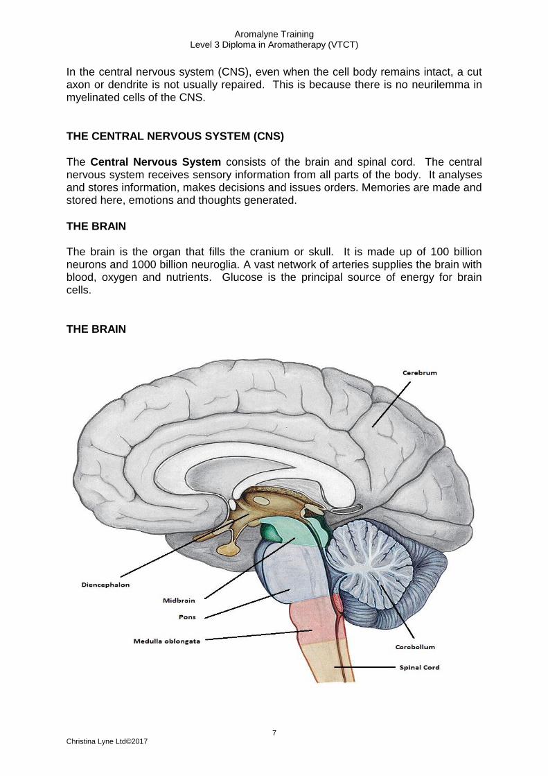
In the central nervous system (CNS), even when the cell body remains intact, a cut axon or dendrite is not usually repaired. This is because there is no neurilemma in myelinated cells of the CNS.
THE CENTRAL NERVOUS SYSTEM (CNS)
The Central Nervous System consists of the brain and spinal cord. The central nervous system receives sensory information from all parts of the body. It analyses and stores information, makes decisions and issues orders. Memories are made and stored here, emotions and thoughts generated.
THE BRAIN The brain is the organ that fills the cranium or skull. It is made up of 100 billion neurons and 1000 billion neuroglia. A vast network of arteries supplies the brain with blood, oxygen and nutrients. Glucose is the principal source of energy for brain cells.
THE BRAIN

Aromalyne Training Level 3 Diploma in Aromatherapy (VTCT)
8 Christina Lyne Ltd©2017
The four principal parts of the brain are:
1. Cerebrum - the largest part of the brain. A deep fissure called the longitudinal fissure separates the cerebrum into two halves – the right and left hemisphere. They are both joined internally by a large bundle of nerves. The cerebrum’s outer superficial layer is made up of ridges and grooves. This layer is called the cerebral cortex and it consists of grey matter. Underneath the grey matter sits an inner layer consisting of white matter, the limbic system and the basal ganglia. Each hemisphere has four main lobes names after the bones that cover them: frontal, parietal, temporal and occipital lobes. It has many areas relating to different functions. These include:
• Sensory areas – for all conscious activities such as touch, taste, smell, hearing, sight
• Motor areas – for all voluntary muscular movements
• Association areas - that have control over intelligence, reasoning, storing memories, personality / emotion, speech, recognising objects / sounds, writing, etc
2. Cerebellum - is found below and at the back of the cerebrum. It is the second largest area of the brain. Its functions are: producing smooth, coordinated movements – such as throwing a ball, dancing regulating posture and balance - such as riding a bike or walking a tightrope
3. Brain Stem – situated at the top of the spinal cord. Its main function is to relay motor and sensory impulses between the spinal cord and the other parts of the brain. It consists of 3 structures:
- Medulla Oblongata - this is the lowest part of the brain stem and sits at the top of the spinal cord. It is a continuation of the spinal cord and it controls the following four vital centres:
• Cardiac centre: controls the rate and force of heart contraction.
• Respiratory centre: controls the rate and depth of breathing.
• Vasomotor centre: controls the constriction and dilation of blood vessels.
• Reflex centre: responds to irritants thereby controlling vomiting, coughing, sneezing, swallowing and hiccupping.
- Pons Varolii - forms a bridge between the brain and the spinal cord. It connects the medulla oblongata with the upper parts of the brain stem. Its function is: to transmit messages between the spinal cord, cerebrum and cerebellum
- Midbrain - this is the smallest region of the brain and contains reflex centres that control many important functions such as: movements of the eyes, head and neck in response to visual and other stimuli

Aromalyne Training Level 3 Diploma in Aromatherapy (VTCT)
9 Christina Lyne Ltd©2017
movements of the head and trunk in response to auditory stimuli areas that control subconscious muscle activities
4. Diencephalon - is located in the middle of the brain. It consists of 3 structures:
- Hypothalamus
This is a small but important structure that is located at the base of the brain above the pituitary. It controls the activities of the autonomic nervous system. It helps to maintain a constant internal environment in the body (homeostasis). It has the following functions: Controls body temperature Stimulates appetite, thirst and fullness Regulates circadian rhythm (sleep patterns) Regulates emotional and behavioural patterns - eg aggression, pleasure, pain Controls / regulates the autonomic nervous system Controls / houses the pituitary and pineal endocrine glands
- Thalamus
This structure is an oval mass of grey matter that sits above the midbrain. It serves as the main relay station for sensory impulses to the cerebral cortex. Sensory impulses include those of hearing, touch, pressure, vibration, pain, taste, cold, heat and vision. It also allows crude appreciation of some sensations such as pain, temperature, pressure and has some functions in memory.
- Epithalamus This structure houses the pineal gland and the choroid plexus which is the group of capillaries that makes the cerebrospinal fluid.
STRUCTURES THAT PROTECT THE BRAIN
Cranial Meninges
As well as being protected by the cranium, the brain is further protected by the three layers of membrane composed of connective tissue - lying just under the skull. They are: the outer meninge is called the dura mater the middle meninge is called the arachnoid the inner meninge is called the pia mater.

Aromalyne Training Level 3 Diploma in Aromatherapy (VTCT)
10 Christina Lyne Ltd©2017
Cerebrospinal Fluid
This is a clear, colourless, watery fluid that contains proteins, glucose, lactic acid, urea, cations, anions and a few white blood cells. It is formed by filtration and secreted into each ventricle of the brain by choroid plexuses (specialised capillaries). It fills and continually circulates in and around the four cavities of the brain and spinal cord. Its functions are:
• to protect the brain and spinal cord, forming a cushion between the bony cavities and the nerves and acting as a shock absorber.
• to keep the pressure around the brain and spinal cord constant.
• to transport nutrients to and remove waste and toxic substances produced by the brain and spinal cord cells.
• to provide the correct chemical environment in which neurons can function
• to maintain homeostasis Cerebrospinal Fluid is obtained through a simple and safe process known as a Lumbar Puncture (LP). The sample is quick and involves only the slightest discomfort. There is no risk of paralysis. The very small needle that is used to draw the fluid is never in contact with the spinal cord. The person giving the Cerebrospinal Fluid sample can lie comfortably on his or her side or sit during the procedure. The skin is swabbed with a cleansing solution and a local anaesthetic is injected under the skin where the needle will be placed, making the area totally numb. A small needle is inserted into the lower back, below where the spinal cord itself ends in the spinal cavity, in between the bones of the spine to reach the fluid. The needle is not inserted into the bones of the spine. Only about 3 tablespoons of fluid are drawn out from the spinal cavity.
Ventricles The brain is not entirely solid. It contains four ventricles (cavities) which connect with each other, and with the space around the brain and the spinal cord. Cerebrospinal fluid flows through and fills these cavities.
The effects of poisons on the Central Nervous System
Exposure to chemicals, environmental toxins and drugs are damaging to the central nervous system. Neuron function may be disturbed either by damage to the neuron itself or be secondary to dysfunction of other organs i.e. the liver or the kidneys. The outcome depends on the toxicity of the substance, the dose and the duration of exposure. This may range from minor short-term neurological disturbance i.e. hypoglycaemia in diabetes mellitus, to encephalopathy, which may cause coma and death as a consequence of liver failure.

Aromalyne Training Level 3 Diploma in Aromatherapy (VTCT)
11 Christina Lyne Ltd©2017
SPINAL CORD
This is the other main part of the central nervous system. It is a long mass of nerve tissue and it extends from the medulla oblongata through the spinal vertebrae ending just above the second lumbar vertebra. It has two main functions that help maintain homeostasis:
• It serves as a conduction pathway for nerve impulses going to and from the brain. Sensory impulses travel to the brain on ascending tracts in the cord. Motor impulses travel to the body on descending tracts in the cord.
• It serves as a reflex centre. The reflex arc is the functional unit of the nervous system. Reflexes are responses to stimuli that do not require conscious thought and consequently, they occur more quickly than reactions that require thought processes. For example, with the withdrawal reflex, the reflex action withdraws the affected part before the body is aware of the pain. Many reflexes are mediated in the spinal cord without going to the higher brain centres. (see additional notes on reflexes, reflex actions and reflex arcs)
Structure of the Spinal Cord
The spinal cord contains two types of matter: white and grey. Each has different functions and contains different structures:
Grey Matter – consists of cell bodies, unmyelinated axons, dendrites of mixed and motor neurons. Its function is to receive and integrate information.
White Matter – consists of tracts of myelinated fibres. Its function is to transport impulses between the brain and the periphery.
Spinal Meninges
Like the brain, the spinal cord is protected by the spinal meninges. They are: the dura mater – the outer covering the arachnoid - the middle covering the pia mater - the thin, inner covering
SPINAL NERVES There are 31 pairs of spinal nerves that originate in the spinal cord. These nerves form part of the PNS and connect the CNS to receptors in the muscles and glands. They are named and grouped after the vertebrae to which they are connected and supply all the areas of the body below the neck: Cervical: 8 pairs Thoracic: 12 pairs Lumbar: 5 pairs Sacral: 5 pairs

Aromalyne Training Level 3 Diploma in Aromatherapy (VTCT)
12 Christina Lyne Ltd©2017
Coccygeal: 1 pair Spinal nerves contain sensory nerves and motor nerves – this makes them mixed nerves. The function of spinal nerves is to carry impulses to and from the body. Each nerve is attached to the spinal cord by a posterior root and an anterior root. Both roots emerge from the vertebral column to form the nerve trunk. These divide into several rami i.e. nerve branches that extend in different directions.
• The posterior ramus supplies the deep muscles and skin of the back
• The anterior ramus supplies the superficial back muscles, all the upper and lower limbs, and the lateral and anterior trunk
With the exception of the thoracic spinal nerves, the anterior rami do not extend directly to the body structures they supply. Instead they form networks on either side of the body. These networks are called plexuses (see spinal plexuses).
THE PERIPHERAL NERVOUS SYSTEM (PNS)
The peripheral nervous system concerns all the nervous system outside the central nervous system and contains motor, sensory and mixed nerves which transmit information to and from the body and brain.
Motor (efferent) nerves carry motor nerve impulses away from the brain, through the spinal cord to structures called effectors. These are skeletal muscles and glands (called effectors). The impulses will stimulate them into carrying out their activities.
Sensory (afferent) nerves carry sensory nerve impulses towards the brain and spinal cord via sensory nerve endings. Sensory nerves will transmit sensations such as heat, cold, pain, taste, smell, sight and hearing from the sense organs, muscles, joints, skin and viscera.
Mixed nerves - so called, when both sensory and motor nerves are enclosed within the same sheath of connective tissue. They run to and from a particular region of the body. An example of a mixed nerve is: all the spinal nerves.
CRANIAL NERVES Cranial nerves are considered part of the PNS. There are 12 pairs of cranial nerves that originate inside the brain. They supply the head, heart, respiratory and digestive organs (via the vagus nerve). Most of the cranial nerves contain sensory and motor nerves and so are mixed nerves. The function of cranial nerves is to carry impulses to and from the brain.

Aromalyne Training Level 3 Diploma in Aromatherapy (VTCT)
13 Christina Lyne Ltd©2017
SPINAL PLEXUSES Branches of some of the spinal nerves form networks of nerves. These networks are called plexuses and all the nerves emerging from a particular plexus will innervate specific structures. These large plexuses are formed on either side of the vertebral column. They are the:
PLEXUS
BODY AREAS SERVED
IMPORTANT NERVES
Cervical
the skin and muscles of the head, neck, upper part of the shoulder and the diaphragm
Phrenic nerve – supplies motor fibres to the diaphragm
Brachial
the shoulder and upper limbs
Axillary nerve – supplies the deltoid and teres minor muscles
Musculocutaneous nerve – supplies the flexors of the arm
Median nerve – supplies the muscles on the anterior aspect of the arm and forearm
Radial nerve – supplies the muscles on the posterior aspect of the arm and forearm
Ulnar nerve – supplies some of the muscles of the forearm and most of the muscles of the hand
Lumbar
the abdominal wall, external genitals and part of the lower limbs
Femoral nerve – supplies the flexor muscles of the thigh and extensor muscles of the leg and the skin over parts of the thigh, leg and foot
Obturator nerve – supplies the adductor muscles of the leg and skin over the medial aspect of the thigh
Sacral
supplying the buttocks, perineum and lower limbs
Sciatic nerve – descends through the thigh and splits into the tibial and common peroneal nerves
The Peripheral Nervous System (PNS) is subdivided into the Somatic Nervous System (SNS) and the Autonomic Nervous System (CNS)
SOMATIC NERVOUS SYSTEM (SNS)
This is the system that allows us to control our skeletal muscles. It contains:
Sensory neurons that convey information from the skin and special sense receptors to the CNS
Motor neurons that conduct impulses from the CNS to the skeletal muscles only.

Aromalyne Training Level 3 Diploma in Aromatherapy (VTCT)
14 Christina Lyne Ltd©2017
We do not always have voluntary control of our skeletal muscles. Sometimes they contract involuntarily through what is called a reflex arc (see further notes)
THE AUTONOMIC NERVOUS SYSTEM (controls involuntary actions)
The autonomic nervous system supplies nerves to all the internal organs of the body and to all the blood vessels. It controls all processes of smooth muscle, cardiac muscle and glands. Its actions are not controlled by the brain or will but are completely reflex and involuntary (heart, lungs, etc). Thus the brain does not know, and nor do we, when the actions happen. This system is divided into two parts: Sympathetic - speeds up / stimulates body activity Parasympathetic - slows down / inhibits body activity The sympathetic and parasympathetic systems work together to regulate the internal functions of the body, even though their actions oppose each other. Every organ in the body has a sympathetic and parasympathetic nerve supply.
SYMPATHETIC NERVOUS SYSTEM
The sympathetic nervous system consists of ganglia (a collection of nerve cells) which are situated in front of the vertebral column and travel the length of the body from the thoracic to the lumbar region. This system prepares the body to deal with exciting and stressful situations. The effects of sympathetic stimulation are:
• to release noradrenaline which prepares the body for action, ie the ‘ fight or flight ‘ response.
• to increase heart rate, making the heart beat faster.
• to dilate the arteries, thereby increasing blood supply to the heart muscles.
• to dilate the blood vessels enabling the skeletal muscles to work better.
• to restricting the flow of digestive juices.
• to stimulate the secretion of sweat
PARASYMPATHETIC NERVOUS SYSTEM The parasympathetic nervous system creates the conditions needed for rest, sleep and generally slowing the body down. It rebalances the body systems. The body prepares itself for inactivity, conservation and the restoration of energy. The only body systems that are not affected are the digestion and the genito-urinary. The effects of parasympathetic stimulation are as follows: The bronchi constrict Slows down the action of the heart

Aromalyne Training Level 3 Diploma in Aromatherapy (VTCT)
15 Christina Lyne Ltd©2017
Skeletal blood vessels constrict Pupils constrict Peristalsis is increased The secretion of insulin and digestive juices are increased The parasympathetic system consists of the vagus, oculomotor, facial and glosso - pharyngeal nerves. One that deserves special mention here is the vagus nerve because it is the longest of the cranial nerves, and it branches most extensively. It emerges from the medulla oblongata, passes through the neck and chest to the abdomen, and has branches to most of the major organs in the body.
Parts of the body controlled by the vagus nerve are: Larynx Pharynx Oesophagus Bile Duct Trachea Gall Bladder Ureters Kidneys Spleen Intestines Pancreas Lungs Heart Stomach Liver
WHAT ARE REFLEXES?
A reflex is a rapid response to a stimulus in the nervous system. It is instant, involuntary and mostly protective. During a reflex action, the body reacts without waiting for us to think. Reflexes are usually triggered by simple nervous pathways called reflex arcs. Many of these involve the spinal cord, but not the brain.
REFLEX ACTIONS (an immediate motor response to a sensory stimulus) Reflexes have to be fast, so their signals travel along the shortest pathway. A delay could cause a serious injury to occur. For example, if you tread on a sharp object, receptors in the foot detect pain caused by the sharp object and send a signal to the spinal cord. Only later do signals reach the brain.
REFLEX ARC
When we stand on something sharp, we move the foot very quickly. We don't have to think about it - it feels like it happens even before you feel the pain. This is because the process is a reflex and doesn't involve the brain.

Aromalyne Training Level 3 Diploma in Aromatherapy (VTCT)
16 Christina Lyne Ltd©2017
Receptors and Effectors The diagram shows someone stepping on a drawing pin. Receptors in the skin of the foot will register the fact that this has happened and will send a signal along a sensory neuron to the spinal cord. Inside the spinal cord an interneuron will transfer the nerve signal to another nerve cell. This cell is a motor neuron and it carries the signal to the muscles in the leg. The leg muscles will contract and, hopefully, the foot will be moved away from the source of pain.
VISCERAL PAIN Physical pain is the awareness of a displeasing and uncomfortable physical sensory stimulus. The human body experiences three types of physical pain, which are called somatic, neuropathic and visceral. Of these, visceral pain is the most common type. Viscera refers to internal parts of the body that are enclosed in a cavity, so visceral pain is the pain felt when internal organs and body tissues are injured or inflamed. This includes the heart and lungs found in the thoracic cavity, the reproductive organs and bladder found in the pelvic cavity, the digestive organs, spleen, and kidneys found in the abdominal cavity.
Visceral pain is a dull pain from within caused by infiltration, expansion, perforation, blockage, stretching, or irritation of internal organs. Visceral pain is often described as pressure or a squeezing sensation which radiates throughout a cavity. The level of intensity can vary from mild to excruciating, however, depending on the malady causing the pain receptors to alert the brain that an issue exists. Though pain is unpleasant, it is a necessary element in the diagnosis and treatment of disease and disorders.
REFERRED PAIN
Referred pain is a phenomenon in which a pathology in one area of the body causes pain in another location. The classic example of referred pain is the pain in the left arm and neck associated with a heart attack or angina episode. In this case, the pain is actually occurring in the heart, but the patient experiences the pain in another location. While referred pain has been documented and studied extensively, researchers are not entirely sure about what causes it.

Aromalyne Training Level 3 Diploma in Aromatherapy (VTCT)
17 Christina Lyne Ltd©2017
Some types of referred pain, such as the arm and neck pain from heart attacks, are well known, and if a patient comes to a doctor with this sort of pain, the doctor may order tests on the heart. In other cases, a doctor may not immediately connect a report of pain with specific cases of referred pain, in which case treatment may be delayed. At times, this can be very problematic for the patient, because medical conditions tend to grow worse when no treatment is offered.
DERMATOMES
A dermatome is an area of skin that is mainly supplied by a single spinal nerve. There are 30 pairs of dermatomes in the body, from the skull to the toes, and each can be traced back to a specific nerve root. Each of these nerves relays sensation (including pain) from a particular region of skin to the brain.
Along the torso, the dermatomes look like horizontal bands, with each band corresponding to a particular nerve root. The arms and legs have longitudinal bands, which explains why pain sometimes shoots down an arm or a leg, because it is following the dermatome. Cervical, thoracic, lumbar, and sacral nerves all supply nerve fibres to various dermatomes on the body.