The Nervous System: Neural Tissue - Cabrillo Collegepdarcey/Bio 4/Fall 2011/Class Notes... · The...
7
1 The Nervous System: Neural Tissue Chapter 13 Human Anatomy, 3rd edition Prentice Hall, © 2001 Introduction • Nervous system = control center & communications network • Functions – Stimulates movements – Maintains homeostasis (with endocrine system) Human Anatomy, 3rd edition Prentice Hall, © 2001 Organization of the Nervous System Functional Classification of the Peripheral Nervous System • Sensory (afferent) division • Nerve fibers that carry information to the central nervous system • Motor (efferent) • Nerve fibers that carry information from the central nervous system Marieb, Essentials of Human Anatomy and Physiology , 8 th edition, 2006 Histology of Nervous Tissue • 2 types of cells – Neurons • Structural & functional part of nervous system • Specialized functions – Neuroglia (glial cells) • Gli = glue • Support & protection of nervous system Neuroglia • Neuroglia of CNS – Astrocytes – Oligodendrocytes – Microglia – Ependymal cells
Transcript of The Nervous System: Neural Tissue - Cabrillo Collegepdarcey/Bio 4/Fall 2011/Class Notes... · The...
1
The Nervous System: Neural Tissue
Chapter 13
Human Anatomy, 3rd edition Prentice Hall, © 2001
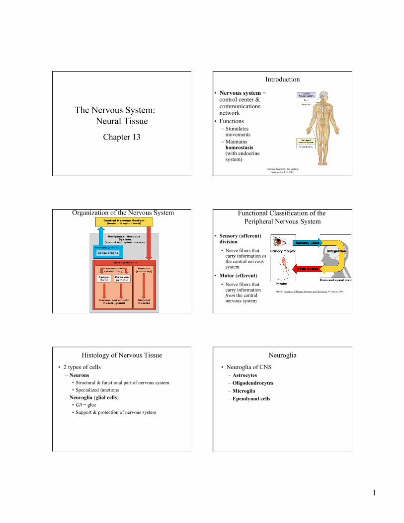
Introduction
• Nervous system = control center & communications network
• Functions – Stimulates
movements – Maintains
homeostasis (with endocrine system)
Human Anatomy, 3rd edition Prentice Hall, © 2001
Organization of the Nervous System Functional Classification of the Peripheral Nervous System
• Sensory (afferent) division • Nerve fibers that
carry information to the central nervous system
• Motor (efferent) • Nerve fibers that
carry information from the central nervous system
Marieb, Essentials of Human Anatomy and Physiology, 8th edition, 2006
Histology of Nervous Tissue
• 2 types of cells – Neurons
• Structural & functional part of nervous system • Specialized functions
– Neuroglia (glial cells) • Gli = glue • Support & protection of nervous system
Neuroglia
• Neuroglia of CNS – Astrocytes – Oligodendrocytes – Microglia – Ependymal cells

2
Neuroglia of CNS • Astrocytes
• Form the blood-brain barrier
• Structural framework for CNS
• Repair damaged neural tissue
• Control the interstitial environment of the brain
Marieb, Essentials of Human Anatomy and Physiology, 8th edition, 2006
Neuroglia of CNS
• Oligodendrocytes
• Produce myelin sheath around nerve fibers in the central nervous system
Marieb, Essentials of Anatomy and Physiology, 8th edition, 2006
Neuroglia of CNS
• Microglia • Spider-like
phagocytes • Dispose of debris
Marieb, Essentials of Human Anatomy and Physiology, 8th edition, 2006
Neuroglia of CNS
• Ependymal cells • Line ventricles of the brain and spinal cord • Secrete cerebrospinal fluid
Marieb, Essentials of Human Anatomy and Physiology, 8th edition, 2006
Human Anatomy, 3rd edition Prentice Hall, © 2001
Neuroglia of CNS Neuroglia of PNS • Schwann cells
– Form myelin sheaths of PNS • Satellite cells
Marieb, Essentials of Anatomy and Physiology, 8th edition, 2006

3
Human Anatomy, 3rd edition Prentice Hall, © 2001
Neurons • Function
– Conduct electrical impulses
• Structure – Cell body
• Nucleus with nucleolus
• Cytoplasm (perikaryon)
– Cytoplasmic processes
• Dendrites • Axon Human Anatomy, 3rd edition
Prentice Hall, © 2001
Axon Structure • Long, specialized
– Collaterals = branches – Telodendria = termination of axons & collaterals
• Cytoplasm = axoplasm • Plasma membrane = axolemma
Human Anatomy, 3rd edition Prentice Hall, © 2001
Anatomy of a Neuron Nerve Fibers of the PNS
• An axon and its sheaths – Myelinated axon
• Axon is surrounded by a myelin sheath – Unmyelinated axon
• Axon has no myelin sheath
Myelin • White matter of
nerves, brain, spinal cord
• Composed primarily of phospholipids
• Production – Developing Schwann
cells wind around axon
• Neurilemma – Peripheral
cytoplasmic layer of the Schwann cell enclosing the myelin sheath Marieb, Essentials of Anatomy and Physiology, 8th edition, 2006
Human Anatomy, 3rd edition Prentice Hall, © 2001
A Myelinated Axon • Function of myelin
– Increases speed of impulse conduction
– Insulation and maintenance of axon
• Nodes of Ranvier – Unmyelinated gaps
between segments of myelin
– Impulses “jump” from node to node

4
Human Anatomy, 3rd edition Prentice Hall, © 2001
Nerve Fibers of the CNS
• Unmyelinated • Myelinated
– Production of myelin is from oligodendrocytes
– Nodes of Ranvier are less numerous
Human Anatomy, 3rd edition Prentice Hall, © 2001
Structural Classification of Neurons • Based on the number of cytoplasmic processes
Human Anatomy, 3rd edition Prentice Hall, © 2001
Functional Classification of Neurons • Based on the direction of impulse transmission
– Sensory neurons – Motor neurons – Interneurons (association)
Nerve Impulse
• A change in charge that travels as a wave along the membrane of a neuron
• Called an action potential • Depends on the movement of sodium ions
(Na+) and potassium ions (K+) between the interstitial fluid and the inside of the neuron.
Human Anatomy, 3rd edition Prentice Hall, © 2001
Resting Potential • Sodium ions are in large concentration along the
outside of the cell membrane • Potassium ions are in large concentration along
the inside of the cell membrane
Beginning of a Nerve Impulse • Requires a stimulus of adequate strength • Membrane is irritable
– Neuron may respond to a stimulus and convert it to an impulse.
• When? – If above threshold
5
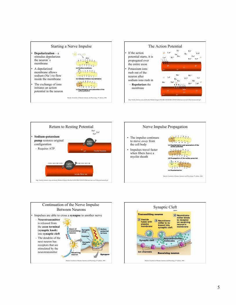
Starting a Nerve Impulse
• Depolarization – a stimulus depolarizes the neuron’s membrane
• A depolarized membrane allows sodium (Na+) to flow inside the membrane
• The exchange of ions initiates an action potential in the neuron
Marieb, Essentials of Human Anatomy and Physiology, 8th edition, 2006
The Action Potential • If the action
potential starts, it is propogated over the entire axon
• Potassium ions rush out of the neuron after sodium ions rush in – Repolarizes the
membrane
http://faculty.clintoncc.suny.edu/faculty/Michael.Gregory/files/Bio%20102/Bio%20102%20lectures/nervous%20system/neuron6.gif
Return to Resting Potential
• Sodium-potassium pump restores original configuration – Requires ATP
http://faculty.clintoncc.suny.edu/faculty/Michael.Gregory/files/Bio%20102/Bio%20102%20lectures/nervous%20system/neuron6.gif
Nerve Impulse Propagation
• The impulse continues to move away from the cell body
• Impulses travel faster when fibers have a myelin sheath
Marieb, Essentials of Human Anatomy and Physiology, 8th edition, 2006
Continuation of the Nerve Impulse Between Neurons
• Impulses are able to cross a synapse to another nerve – Neurotransmitter
is released from the axon terminal (synaptic knob) into synaptic cleft
– The dendrite of the next neuron has receptors that are stimulated by the neurotransmitter
Marieb, Essentials of Human Anatomy and Physiology, 8th edition, 2006
Synaptic Cleft
Marieb, Essentials of Human Anatomy and Physiology, 8th edition, 2006

6
Postsynaptic Membrane Receptor
Marieb, Essentials of Human Anatomy and Physiology, 8th edition, 2006
Human Anatomy, 3rd edition Prentice Hall, © 2001
Synapse
Human Anatomy, 3rd edition Prentice Hall, © 2001
Neural Regeneration After Injury
Human Anatomy, 3rd edition Prentice Hall, © 2001
Neural Regeneration
Human Anatomy, 3rd edition Prentice Hall, © 2001
Neural Regeneration
Human Anatomy, 3rd edition Prentice Hall, © 2001
Neural Regeneration

7
Nerves • Neurons are bundled
into fasciculi which are bundled into nerves. – Endoneurium
surrounds each nerve fiber (axon)
– Groups of fibers are bound into fascicles
• Surrounded by the perineurium
– Fascicles are bound together into a nerve • Surrounded by
the epineurium Marieb, Essentials of Human Anatomy and Physiology, 8th edition, 2006