The Nervous System: General and Special...
47
© 2012 Pearson Education, Inc. 18 The Nervous System: General and Special Senses
Transcript of The Nervous System: General and Special...
© 2012 Pearson Education, Inc.
18 The Nervous System: General and Special Senses
© 2012 Pearson Education, Inc.
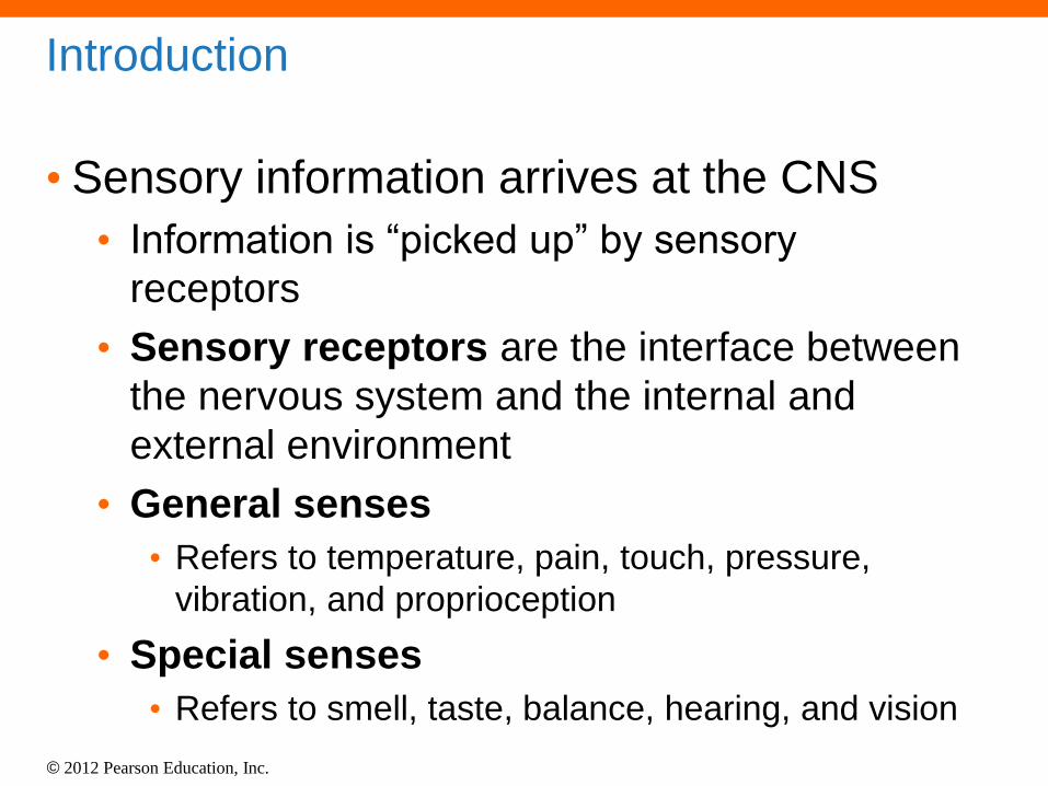
Introduction
• Sensory information arrives at the CNS
• Information is “picked up” by sensory
receptors
• Sensory receptors are the interface between
the nervous system and the internal and
external environment
• General senses
• Refers to temperature, pain, touch, pressure,
vibration, and proprioception
• Special senses
• Refers to smell, taste, balance, hearing, and vision
© 2012 Pearson Education, Inc.
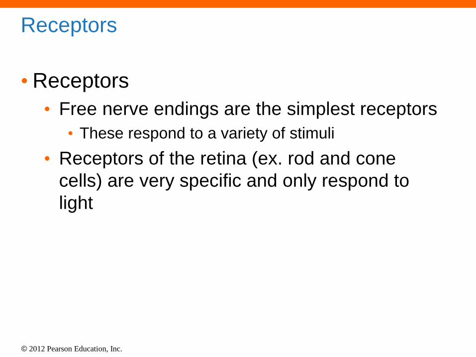
Receptors
• Receptors
• Free nerve endings are the simplest receptors
• These respond to a variety of stimuli
• Receptors of the retina (ex. rod and cone
cells) are very specific and only respond to
light

© 2012 Pearson Education, Inc.
The General Senses
• Classification of the General Senses
• Nociceptors: respond to the sensation of
pain
• Thermoreceptors: respond to changes in
temperature
• Mechanoreceptors: activated by physical
distortion of cell membranes
• Chemoreceptors: monitor the chemical
composition of body fluids
© 2012 Pearson Education, Inc.
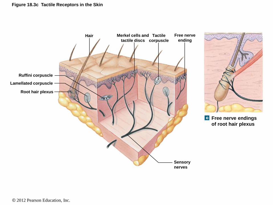
Figure 18.3c Tactile Receptors in the Skin
Hair
Root hair plexus
Lamellated corpuscle
Ruffini corpuscle
Merkel cells and
tactile discs
Tactile
corpuscle
Free nerve
ending
Sensory
nerves
Free nerve endings
of root hair plexus

© 2012 Pearson Education, Inc.
The General Senses
• Nociceptors
• Known as pain receptors
• Associated with free nerve endings
• difficult to “pinpoint” the location of the origin of
the pain
© 2012 Pearson Education, Inc.
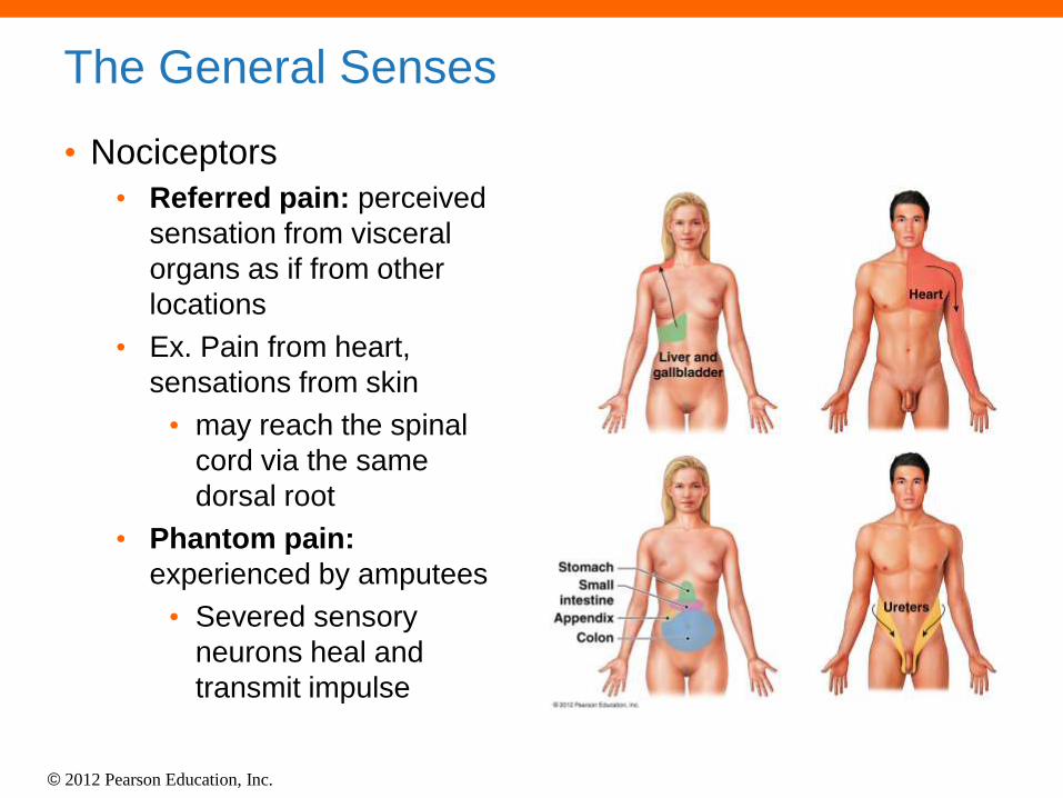
The General Senses
• Nociceptors
• Referred pain: perceived
sensation from visceral
organs as if from other
locations
• Ex. Pain from heart,
sensations from skin
• may reach the spinal
cord via the same
dorsal root
• Phantom pain:
experienced by amputees
• Severed sensory
neurons heal and
transmit impulse
© 2012 Pearson Education, Inc.
The General Senses
• Thermoreceptors
• Found in the dermis, skeletal muscles, liver,
and hypothalamus
• Cold receptors are more numerous than hot
receptors
• Exist as free nerve endings
© 2012 Pearson Education, Inc.
The General Senses
• Mechanoreceptors
• Receptors that are sensitive to stretch,
compression, twisting, or distortion of the
plasmalemmae
• There are three types
• Tactile receptors- Provide sensations of touch,
pressure, and vibrations
• Baroreceptors- Stretch receptors that monitor
changes in the stretch of organs
• Proprioceptors- Monitor the position of joints,
tension in the tendons and ligaments, and the
length of muscle fibers upon contraction
© 2012 Pearson Education, Inc.
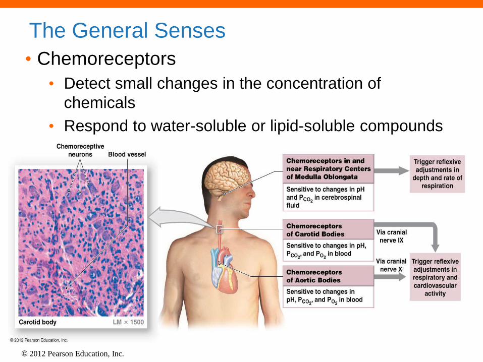
The General Senses
• Chemoreceptors
• Detect small changes in the concentration of
chemicals
• Respond to water-soluble or lipid-soluble compounds

© 2012 Pearson Education, Inc.
The Special Senses
• The special senses include:
• Olfaction (smell)
• Gustation (taste)
• Equilibrium
• Hearing
• Vision

© 2012 Pearson Education, Inc.
Olfaction (Smell)
• The olfactory
epithelium consists of: •Olfactory receptors
•Supporting cells
•Basal cells

© 2012 Pearson Education, Inc.
Pathway for Olfaction:
© 2012 Pearson Education, Inc.
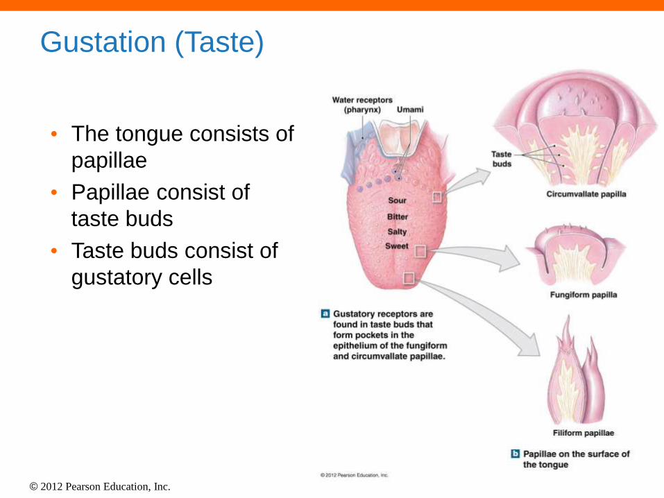
Gustation (Taste)
• The tongue consists of
papillae
• Papillae consist of
taste buds
• Taste buds consist of
gustatory cells

© 2012 Pearson Education, Inc.

© 2012 Pearson Education, Inc.
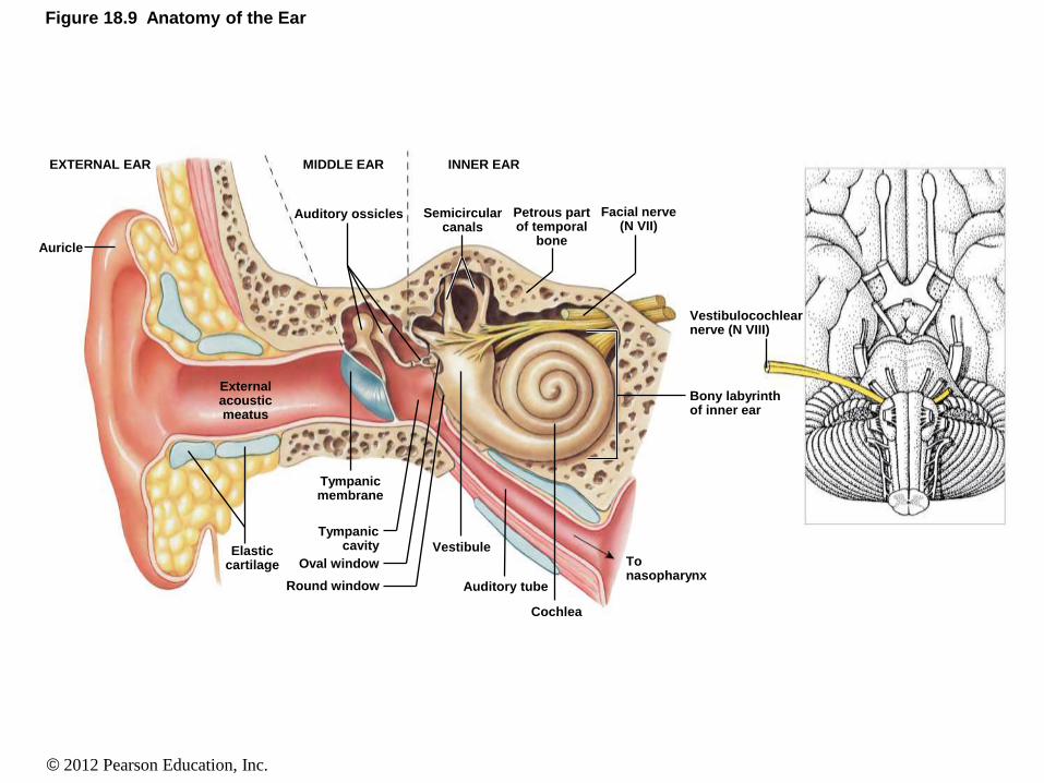
Equilibrium and Hearing • Structures of the ear are involved in balance and
hearing
• The ear is subdivided into three regions
• External ear
• Middle ear
• Inner ear
© 2012 Pearson Education, Inc.
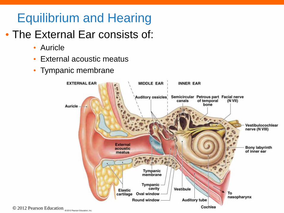
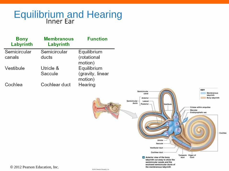
Equilibrium and Hearing
• The External Ear consists of: • Auricle
• External acoustic meatus
• Tympanic membrane
© 2012 Pearson Education, Inc.
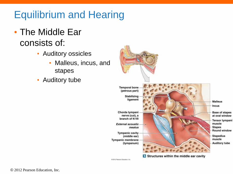
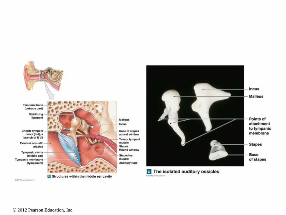
Equilibrium and Hearing
• The Middle Ear
consists of: • Auditory ossicles
• Malleus, incus, and
stapes
• Auditory tube
© 2012 Pearson Education, Inc.
© 2012 Pearson Education, Inc.
Equilibrium and Hearing

© 2012 Pearson Education, Inc.
Figure 18.12a Semicircular Canals and Ducts
Anterior view of the bony
labyrinth cut away to show the
semicircular canals and the
enclosed semicircular ducts of
the membranous labyrinth
Cochlear duct
Vestibular duct
Saccule
Utricle
Tympanic
duct
Organ of
Corti
Cochlea
Endolymphatic sac
Maculae
Cristae within ampullae
Bony labyrinth
Membranous
labyrinth
KEY
Vestibule
Anterior
Lateral
Posterior
Semicircular
canal
Semicircular
ducts

© 2012 Pearson Education, Inc.
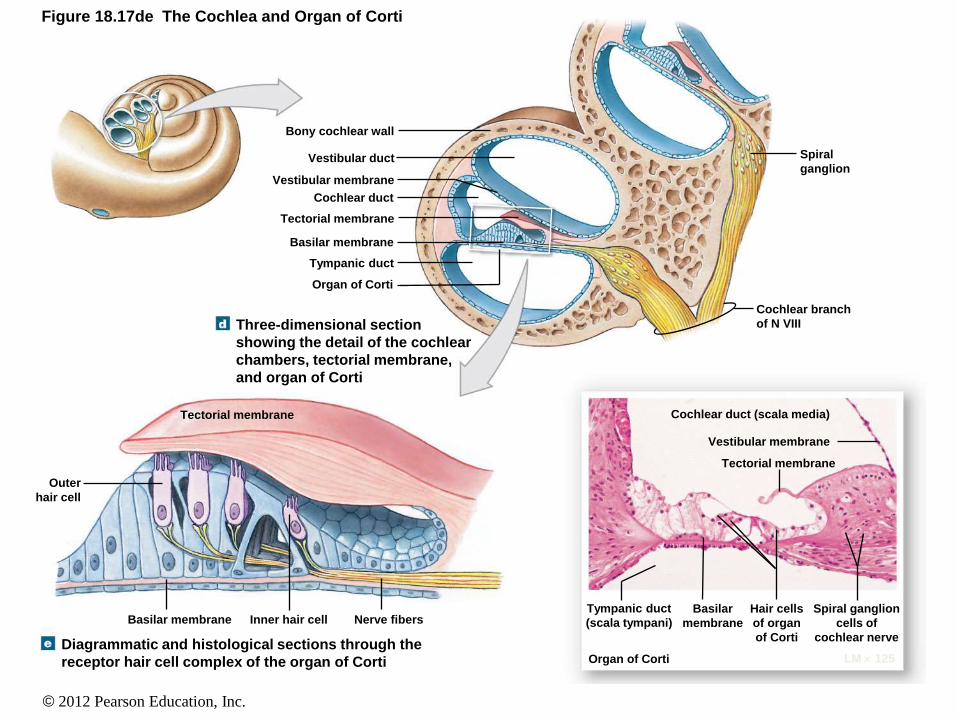
Spiral organ of corti = hearing
© 2012 Pearson Education, Inc.
Figure 18.17de The Cochlea and Organ of Corti
Diagrammatic and histological sections through the
receptor hair cell complex of the organ of Corti
Three-dimensional section
showing the detail of the cochlear
chambers, tectorial membrane,
and organ of Corti
Bony cochlear wall
Vestibular duct
Vestibular membrane
Cochlear duct
Tectorial membrane
Basilar membrane
Tympanic duct
Organ of Corti
Spiral
ganglion
Cochlear branch
of N VIII
Cochlear duct (scala media)
Vestibular membrane
Tectorial membrane
Organ of Corti LM 125
Tympanic duct
(scala tympani)
Basilar
membrane
Hair cells
of organ
of Corti
Spiral ganglion
cells of
cochlear nerve
Tectorial membrane
Outer
hair cell
Basilar membrane Inner hair cell Nerve fibers

© 2012 Pearson Education, Inc.
Equilibrium and Hearing

© 2012 Pearson Education, Inc.

© 2012 Pearson Education, Inc.
Mechanics of Equilibrium
• Complex process
• Requires input from sensory neurons of the
vestibular organ of both ears
• Other sources of equilibrium information
are:
• Tactile receptors
• Proprioceptors of tendons, muscles, joints
© 2012 Pearson Education, Inc.
Figure 18.9 Anatomy of the Ear
EXTERNAL EAR MIDDLE EAR INNER EAR
Auricle
Auditory ossicles Semicircular canals
Petrous part of temporal
bone
Facial nerve (N VII)
External acoustic meatus
Elastic cartilage
Tympanic membrane
Tympanic cavity
Oval window
Round window
Vestibule
Auditory tube
Cochlea
To nasopharynx
Bony labyrinth of inner ear
Vestibulocochlear nerve (N VIII)
© 2012 Pearson Education, Inc.
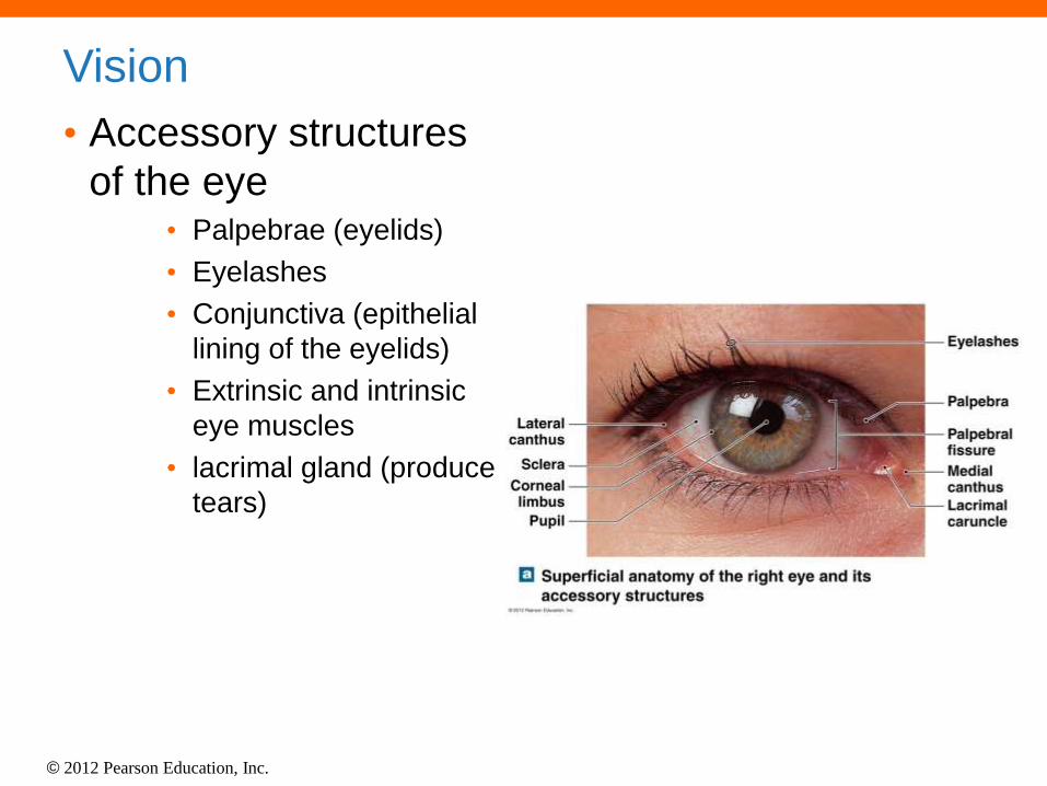
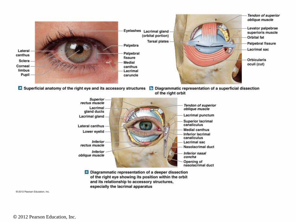
Vision
• Accessory structures
of the eye • Palpebrae (eyelids)
• Eyelashes
• Conjunctiva (epithelial
lining of the eyelids)
• Extrinsic and intrinsic
eye muscles
• lacrimal gland (produce
tears)
© 2012 Pearson Education, Inc.
© 2012 Pearson Education, Inc.
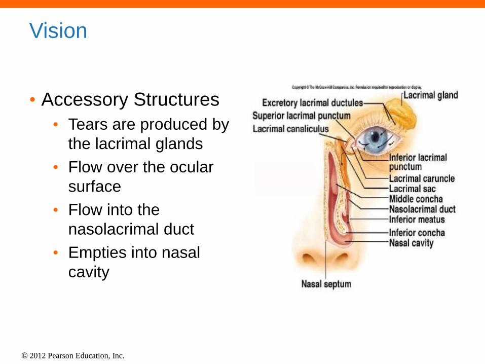
Vision
• Accessory Structures
• Tears are produced by
the lacrimal glands
• Flow over the ocular
surface
• Flow into the
nasolacrimal duct
• Empties into nasal
cavity
© 2012 Pearson Education, Inc.
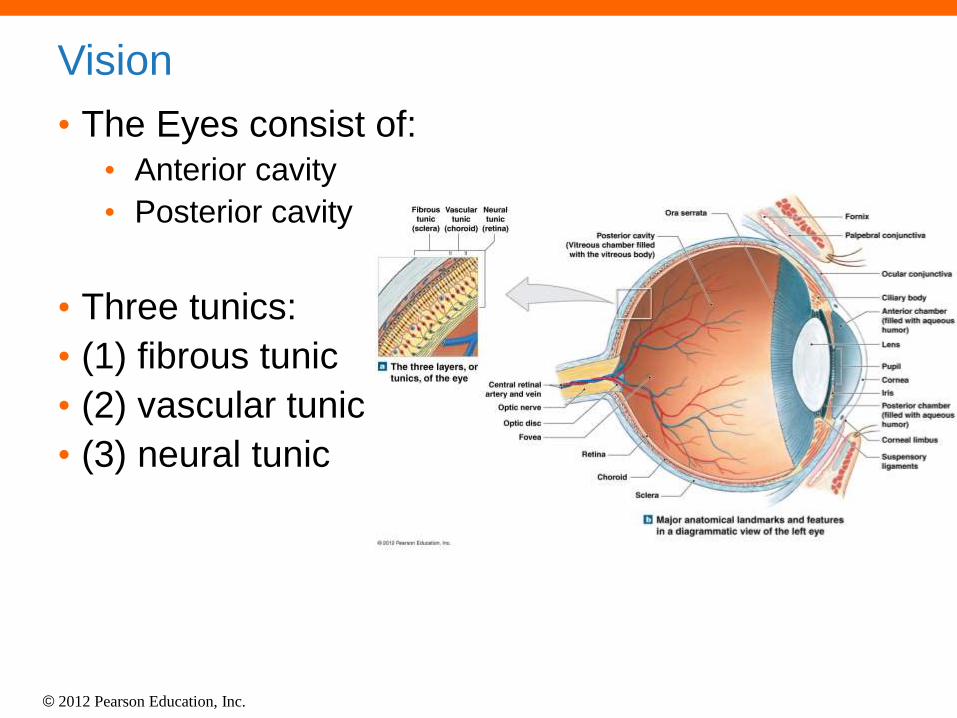
Vision
• The Eyes consist of:
• Anterior cavity
• Posterior cavity
• Three tunics:
• (1) fibrous tunic
• (2) vascular tunic
• (3) neural tunic
© 2012 Pearson Education, Inc.
© 2012 Pearson Education, Inc.
Functions of the Eyeball
• Transmission of light rays
• Refraction of light rays (bending)
• Accommodation of the lens (to focus)
• Constriction of the pupil (regulate light)
• Convergence of the eyeballs (depth
perception)
© 2012 Pearson Education, Inc.
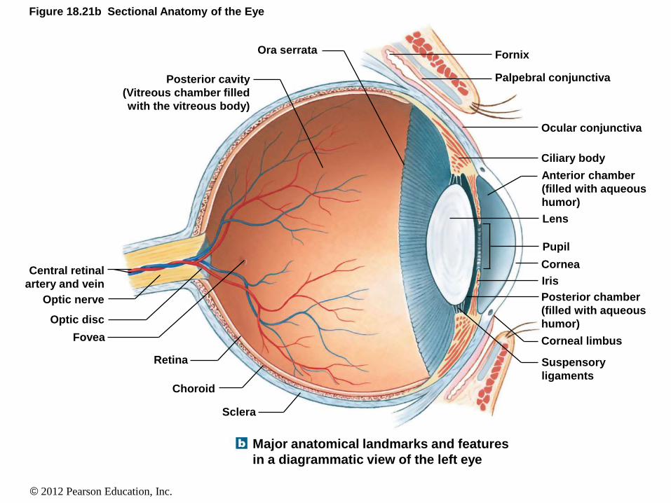
Figure 18.21b Sectional Anatomy of the Eye
Major anatomical landmarks and features
in a diagrammatic view of the left eye
Central retinal
artery and vein
Optic nerve
Optic disc
Fovea
Retina
Choroid
Sclera
Posterior cavity
(Vitreous chamber filled
with the vitreous body)
Ora serrata Fornix
Palpebral conjunctiva
Ocular conjunctiva
Ciliary body
Anterior chamber
(filled with aqueous
humor)
Lens
Pupil
Cornea
Iris
Posterior chamber
(filled with aqueous
humor)
Corneal limbus
Suspensory
ligaments
© 2012 Pearson Education, Inc.
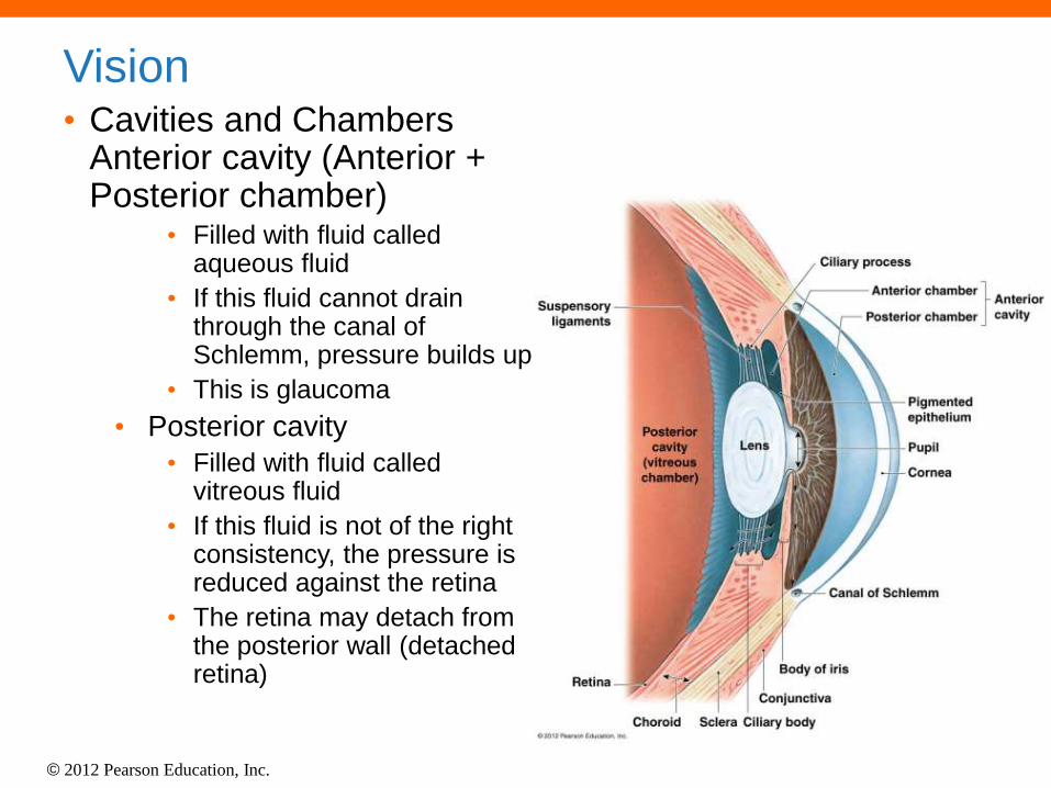
Vision • Cavities and Chambers
Anterior cavity (Anterior + Posterior chamber)
• Filled with fluid called aqueous fluid
• If this fluid cannot drain through the canal of Schlemm, pressure builds up
• This is glaucoma
• Posterior cavity
• Filled with fluid called vitreous fluid
• If this fluid is not of the right consistency, the pressure is reduced against the retina
• The retina may detach from the posterior wall (detached retina)
© 2012 Pearson Education, Inc.
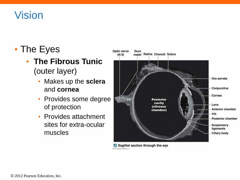
Vision
• The Eyes
• The Fibrous Tunic
(outer layer)
• Makes up the sclera
and cornea
• Provides some degree
of protection
• Provides attachment
sites for extra-ocular
muscles

© 2012 Pearson Education, Inc.
Vision
• The Eyes
• The Vascular Tunic
(middle layer)
• Consists of blood
vessels, lymphatics,
and intrinsic eye
muscles
• Regulates the amount
of light entering the eye
• Secretes and reabsorbs
aqueous humor
• Controls the shape of
the lens
• Includes the iris, ciliary
body, and the choroid
© 2012 Pearson Education, Inc.
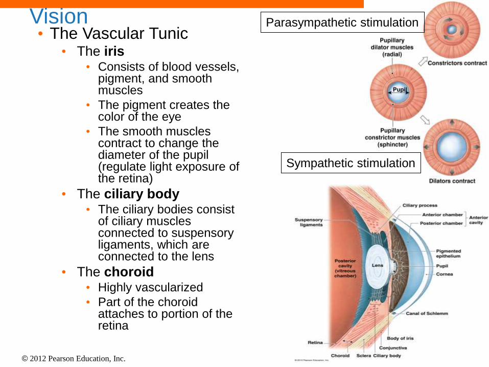
Vision • The Vascular Tunic
• The iris • Consists of blood vessels,
pigment, and smooth muscles
• The pigment creates the color of the eye
• The smooth muscles contract to change the diameter of the pupil (regulate light exposure of the retina)
• The ciliary body • The ciliary bodies consist
of ciliary muscles connected to suspensory ligaments, which are connected to the lens
• The choroid • Highly vascularized
• Part of the choroid attaches to portion of the retina
Sympathetic stimulation
Parasympathetic stimulation

© 2012 Pearson Education, Inc.
Vision
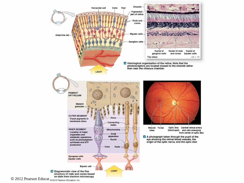
• The Eyes • The Neural Tunic
(inner layer) • Also called the retina
• Innermost layer of the eye
• Made of two layers: (pigmented layer – outer layer) and (neural layer – inner layer)
• Retina cells: rods (night vision) and cones (color vision)

© 2012 Pearson Education, Inc.
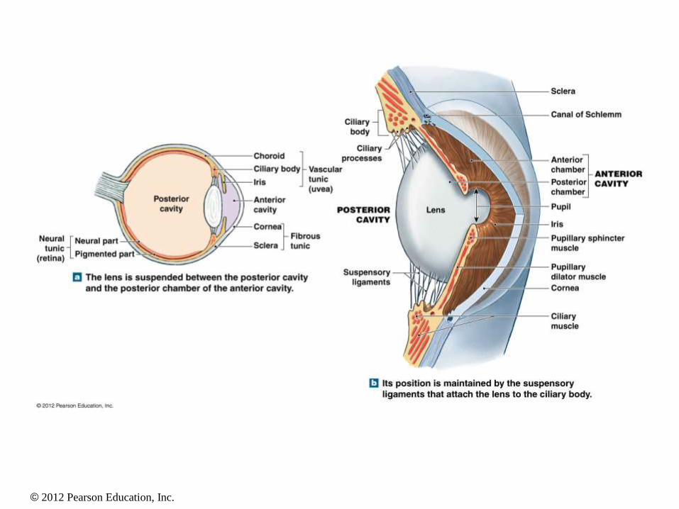
Figure 18.22a The Lens and Chambers of the Eye
The lens is suspended between the posterior cavity
and the posterior chamber of the anterior cavity.
Pigmented part
Neural part Neural
tunic
(retina)
Posterior
cavity
Choroid
Ciliary body
Iris
Vascular
tunic
(uvea)
Anterior
cavity
Cornea
Sclera Fibrous
tunic
© 2012 Pearson Education, Inc.
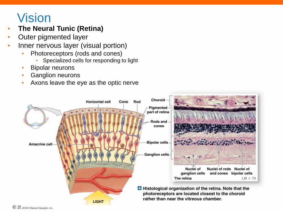
• The Neural Tunic (Retina)
• Outer pigmented layer
• Inner nervous layer (visual portion) • Photoreceptors (rods and cones)
• Specialized cells for responding to light
• Bipolar neurons
• Ganglion neurons
• Axons leave the eye as the optic nerve
Vision
© 2012 Pearson Education, Inc.
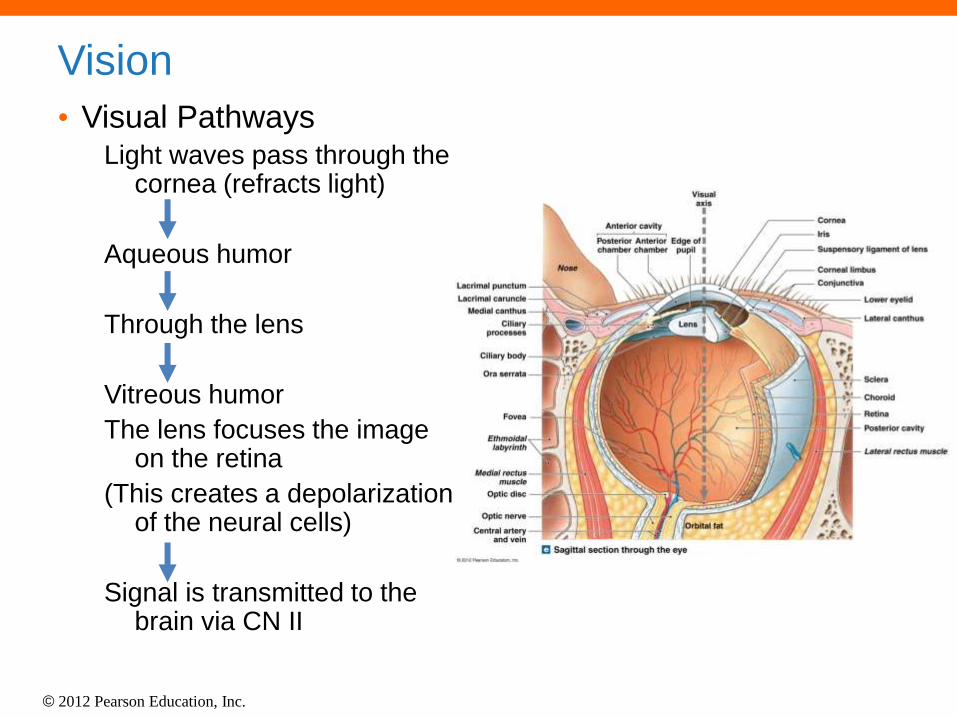
Vision
• Visual Pathways Light waves pass through the
cornea (refracts light)
Aqueous humor
Through the lens
Vitreous humor
The lens focuses the image on the retina
(This creates a depolarization of the neural cells)
Signal is transmitted to the brain via CN II
© 2012 Pearson Education, Inc.
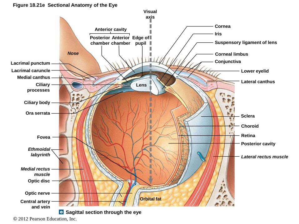
Figure 18.21e Sectional Anatomy of the Eye
Sagittal section through the eye
Orbital fat Central artery
and vein
Medial rectus
muscle
Ethmoidal
labyrinth
Optic nerve
Optic disc
Fovea
Ora serrata
Ciliary body
Lens Ciliary
processes
Medial canthus
Lacrimal caruncle
Lacrimal punctum
Nose
Anterior cavity
Posterior
chamber
Anterior
chamber
Edge of
pupil
Visual
axis
Cornea
Iris
Suspensory ligament of lens
Corneal limbus
Conjunctiva
Lower eyelid
Lateral canthus
Sclera
Choroid
Retina
Posterior cavity
Lateral rectus muscle

© 2012 Pearson Education, Inc.
• Rod Cells • Dim light
• Do not distinguish color
• Low resolution
• Cone Cells • Bright light
• High resolution
• Color detection
• Optic Disc = Blind spot (0% rods and cones) • An area lacking photoreceptors
• If an object is focused on this area, vision does not occur
• Optic nerve is attached to eyeball at this location
• Fovea Centralis = 100% cones • The best color vision is when an object is focused on the fovea
centralis
Vision
© 2012 Pearson Education, Inc.
© 2012 Pearson Education, Inc.
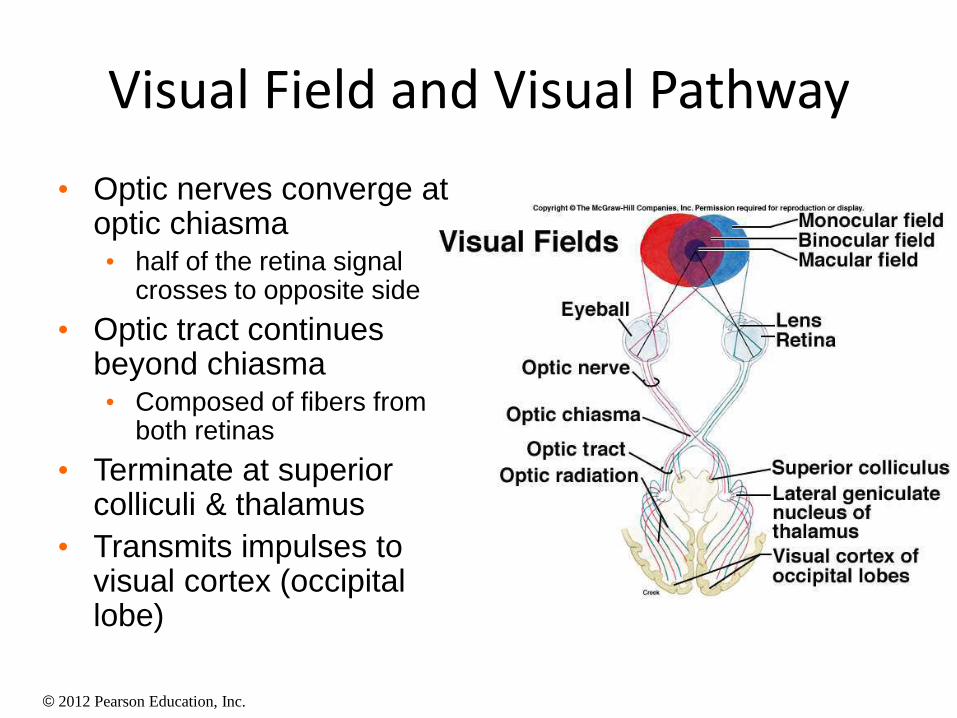
Visual Field and Visual Pathway
• Optic nerves converge at optic chiasma • half of the retina signal
crosses to opposite side
• Optic tract continues beyond chiasma • Composed of fibers from
both retinas
• Terminate at superior colliculi & thalamus
• Transmits impulses to visual cortex (occipital lobe)
© 2012 Pearson Education, Inc.
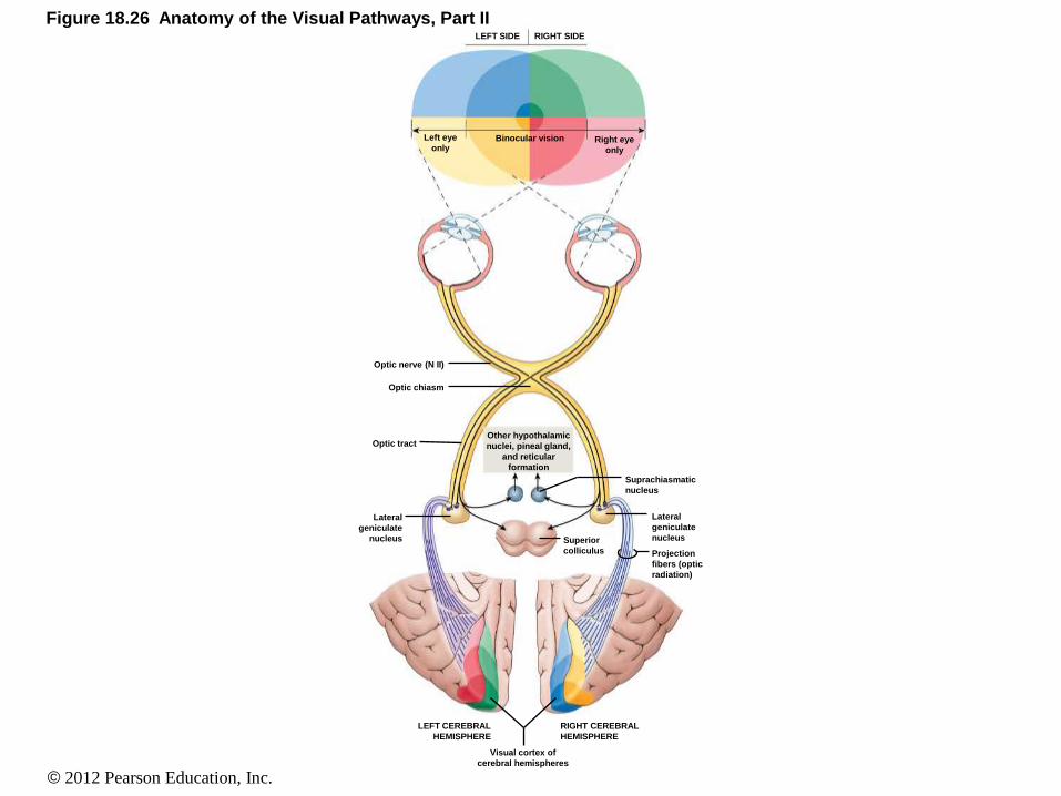
Figure 18.26 Anatomy of the Visual Pathways, Part II LEFT SIDE RIGHT SIDE
Left eye
only Right eye
only
Binocular vision
Optic nerve (N II)
Optic chiasm
Optic tract Other hypothalamic
nuclei, pineal gland,
and reticular
formation
Suprachiasmatic
nucleus
Superior
colliculus
Lateral
geniculate
nucleus
Projection
fibers (optic
radiation)
Lateral
geniculate
nucleus
RIGHT CEREBRAL
HEMISPHERE
LEFT CEREBRAL
HEMISPHERE
Visual cortex of
cerebral hemispheres