The myxomycete genus Schenella: morphological and … · 2017-02-15 · The myxomycete genus...
11
139 Mycologia, 97(1), 2005, pp. 139–149. q 2005 by The Mycological Society of America, Lawrence, KS 66044-8897 The myxomycete genus Schenella: morphological and DNA sequence evidence for synonymy with the gasteromycete genus Pyrenogaster Arturo Estrada-Torres Centro de Investigaciones en Ciencias Biolo ´gicas, Universidad Auto ´noma de Tlaxcala, Apdo. Postal 183. Tlaxcala 90000, Me ´xico Thomas W. Gaither Department of Biology, Slippery Rock University, Slippery Rock, Pennsylvania 16507-1325 Dennis L. Miller 1 Department of Molecular and Cell Biology F03.1, University of Texas at Dallas, Richardson, Texas 75080 Carlos Lado Real Jardı ´n Bota ´nico, CSIC. Plaza de Murillo 2, 28014 Madrid, Espan ˜a Harold W. Keller Department of Biology, Central Missouri State University, Warrensburg, Missouri 64093 Abstract: The genus Schenella has proven difficult to classify since its description as a new genus in 1911. Macbride placed it with the Myxomycetes but it was unclear with which myxomycete, if any, it should be grouped. Recent identification of abundant samples of Schenella has aided a re-evaluation of its classifica- tion as a myxomycete. Morphological evidence based on light and scanning electron microscopy of re- cently collected specimens and on the type specimen of Macbride suggested that it might be synonymous with the gasteromycete Pyrenogaster. Analysis of DNA sequences from freshly isolated samples indicates that the genus Schenella is related closely to an anciently diverged, monophyletic group of fungi that includes several gasteromycete genera, among them Geastrum, Sphaerobolus and Pseudocolus. Comparisons of the morphology and DNA sequences of authentically identified specimens of Pyrenogaster atrogleba indicate that it is synonymous with Schenella simplex. The no- menclatural implications of this discovery are dis- cussed. Key words: Eumycetozoa, gasteromycete, molec- ular systematics, Myxomycetes, plasmodial slime molds, Pyrenogaster, Schenella Accepted for publication 20 August 2004. 1 Corresponding author. E-mail: [email protected] INTRODUCTION Schenella simplex was described and illustrated as a new genus and species by Thomas H. Macbride (1911). A single collection gathered in Aug 1903 on a decaying pine log in Yosemite Valley, California, was designated as the holotype. Macbride expressed doubt about this taxon in the first sentence of his brief paper: ‘‘This following description and accom- panying figures are submitted to mycologists partly for the sake of eliciting information.’’ Macbride fur- ther noted that, ‘‘If a slime-mould, the species is re- ferable to the family Dianemaceae and is akin to those in which the capillitial threads pass from side to side of the fructification, attached at each end.’’ This unique combination of characters—aethalioid fructifications, simple capillitial threads twisted to- gether to form vertical columns attached to the outer peridium and at the base of the fructification along with tiny, smooth, spherical spores, 5–6 mm diam— were the stated hallmarks of this enigmatic taxon. The status of Schenella simplex as a myxomycete was so doubtful that Macbride omitted the taxon from his 1922 monograph (Macbride 1922), nor was it in- cluded in the Lister monograph (Lister 1925). In the 1934 monograph by Macbride and Martin (1934) Schenella simplex again was included. Com- mentary after the species description again casts doubt on the status of this taxon: ‘‘A very curious form, of somewhat doubtful affinities and placed in the Stemonitaceae on the basis of the dark violet brown spores and the dark capillitium. There is no suggestion of columellae in the pillar-like columns. To the naked eye it suggests an amaurochaete.’’ Spores were described as ‘‘rather coarsely tubercu- late’’ and not smooth. Hagelstein (1944) made reference to Schenella sim- plex in the Amaurochaetaceae but again expressed doubt about its status: ‘‘. . . might appropriately be placed in this family if conclusively proven to be a form of Mycetozoa. The original description and fig- ures are not impressive, and clearly indicate the au- thor was uncertain.’’ Schenella was not included in his key to the genera. Martin (1949) included a note about Schenella in the Stemonitaceae that, ‘‘if it is a myxomycete, (it) would be placed in this family close to Amaurochae- te.’’ A dichotomous key to 10 genera in the family
Transcript of The myxomycete genus Schenella: morphological and … · 2017-02-15 · The myxomycete genus...
139
Mycologia, 97(1), 2005, pp. 139–149.q 2005 by The Mycological Society of America, Lawrence, KS 66044-8897
The myxomycete genus Schenella: morphological and DNA sequence evidencefor synonymy with the gasteromycete genus Pyrenogaster
Arturo Estrada-TorresCentro de Investigaciones en Ciencias Biologicas,Universidad Autonoma de Tlaxcala, Apdo. Postal183. Tlaxcala 90000, Mexico
Thomas W. GaitherDepartment of Biology, Slippery Rock University,Slippery Rock, Pennsylvania 16507-1325
Dennis L. Miller1
Department of Molecular and Cell Biology F03.1,University of Texas at Dallas, Richardson,Texas 75080
Carlos LadoReal Jardın Botanico, CSIC. Plaza de Murillo 2,28014 Madrid, Espana
Harold W. KellerDepartment of Biology, Central Missouri StateUniversity, Warrensburg, Missouri 64093
Abstract: The genus Schenella has proven difficult toclassify since its description as a new genus in 1911.Macbride placed it with the Myxomycetes but it wasunclear with which myxomycete, if any, it should begrouped. Recent identification of abundant samplesof Schenella has aided a re-evaluation of its classifica-tion as a myxomycete. Morphological evidence basedon light and scanning electron microscopy of re-cently collected specimens and on the type specimenof Macbride suggested that it might be synonymouswith the gasteromycete Pyrenogaster. Analysis of DNAsequences from freshly isolated samples indicates thatthe genus Schenella is related closely to an ancientlydiverged, monophyletic group of fungi that includesseveral gasteromycete genera, among them Geastrum,Sphaerobolus and Pseudocolus. Comparisons of themorphology and DNA sequences of authenticallyidentified specimens of Pyrenogaster atrogleba indicatethat it is synonymous with Schenella simplex. The no-menclatural implications of this discovery are dis-cussed.
Key words: Eumycetozoa, gasteromycete, molec-ular systematics, Myxomycetes, plasmodial slimemolds, Pyrenogaster, Schenella
Accepted for publication 20 August 2004.1 Corresponding author. E-mail: [email protected]
INTRODUCTION
Schenella simplex was described and illustrated as anew genus and species by Thomas H. Macbride(1911). A single collection gathered in Aug 1903 ona decaying pine log in Yosemite Valley, California, wasdesignated as the holotype. Macbride expresseddoubt about this taxon in the first sentence of hisbrief paper: ‘‘This following description and accom-panying figures are submitted to mycologists partlyfor the sake of eliciting information.’’ Macbride fur-ther noted that, ‘‘If a slime-mould, the species is re-ferable to the family Dianemaceae and is akin tothose in which the capillitial threads pass from sideto side of the fructification, attached at each end.’’This unique combination of characters—aethalioidfructifications, simple capillitial threads twisted to-gether to form vertical columns attached to the outerperidium and at the base of the fructification alongwith tiny, smooth, spherical spores, 5–6 mm diam—were the stated hallmarks of this enigmatic taxon.The status of Schenella simplex as a myxomycete wasso doubtful that Macbride omitted the taxon fromhis 1922 monograph (Macbride 1922), nor was it in-cluded in the Lister monograph (Lister 1925).
In the 1934 monograph by Macbride and Martin(1934) Schenella simplex again was included. Com-mentary after the species description again castsdoubt on the status of this taxon: ‘‘A very curiousform, of somewhat doubtful affinities and placed inthe Stemonitaceae on the basis of the dark violetbrown spores and the dark capillitium. There is nosuggestion of columellae in the pillar-like columns.To the naked eye it suggests an amaurochaete.’’Spores were described as ‘‘rather coarsely tubercu-late’’ and not smooth.
Hagelstein (1944) made reference to Schenella sim-plex in the Amaurochaetaceae but again expresseddoubt about its status: ‘‘. . . might appropriately beplaced in this family if conclusively proven to be aform of Mycetozoa. The original description and fig-ures are not impressive, and clearly indicate the au-thor was uncertain.’’ Schenella was not included in hiskey to the genera.
Martin (1949) included a note about Schenella inthe Stemonitaceae that, ‘‘if it is a myxomycete, (it)would be placed in this family close to Amaurochae-te.’’ A dichotomous key to 10 genera in the family
140 MYCOLOGIA
fails to include Schenella. Martin (1961) described asecond species of Schenella, S. microspora, after re-examination of the type of S. simplex. He concludedthat Schenella was a valid genus in the Stemonitaceae,closely allied to Amaurochaete. Martin re-interpretedthe morphology of this taxon as a pseudoaethalium,not an aethalium, with the sporangia arranged ver-tically much as in Dictydiaethalium. Each column(sporangium) appeared to have a distinct top withan individual peridium and a coiled, twisted mass ofpseudocapillitial threads attached also at the base ofthe fructification. There was no trace of a columella.The capillitial system consisted of a bundle of threadswithin each sporangium. Martin (1961) emended thegeneric and species description, emphasizing thedark capillitium and dark spores as the salient mor-phological characters for assignment to the Stemon-itaceae close to Amaurochaete. Spore diameter of S.simplex was given as 5–6 mm and for S. microspora 3.5–4(4.5) mm. Martin (1961) also noted that spores weresmall for a myxomycete but no smaller than otherwell known species. This statement is supported byexamples such as Stemonitis microsperma B. Ing, andS. smithii Macbr., with the smallest spore diametersknown in the Myxomycetes (Martin and Alexopoulos1969).
Nannenga-Bremekamp (1967) used capillitialcharacteristics to transfer Schenella from the Stemon-itaceae to its own monogeneric family, the Schenel-laceae. This transfer was based exclusively on the lit-erature because no specimens were examined.
The description of a second species in the genus,Schenella microspora, by Martin (1961) seems to haveconvinced other authors as to its identity as a myxo-mycete and recognition of a separate family, Sche-nellaceae (Martin and Alexopolous 1969, Nannenga-Bremekamp 1974, Farr 1976, Martin et al 1983,Hawksworth et al 1995).
Rammeloo (1985) examined spores of the typespecimen of S. simplex using scanning electron mi-croscopy, expressing doubt that the taxon was amyxomycete because the spores possessed a pore sim-ilar to that of some taxa in the fungal order Agari-cales. One field collection from Mexico more re-cently was identified as Schenella simplex and com-pared with the type of S. simplex. Rodrıguez-Palmaand Estrada-Torres (1996) collected this specimen inthe Abies religiosa forest in Malintzi National Park,Tlaxcala, Mexico.
This paper reports comparisons of additional spec-imens from Cofre de Perote National Park, Veracruz,Mexico, identified as S. simplex, previously knownonly from its type locality in California. Evidencefrom light microscopy and scanning electron micros-copy indicates that the Mexican specimens are iden-
tical to the S. simplex type. Morphological data, aswell as DNA sequence evidence from the Mexicanspecimens, also indicate that S. simplex is related toan anciently diverged group of gasteromycete fungiand should not be classified as a myxomycete. Fur-ther, morphological characterization indicates that S.simplex is synonymous with the gasteromycete fungus,Pyrenogaster atrogleba (Zeller) L.S. Domınguez & Cas-tellano. In support of this, DNA sequences obtainedfrom authentically identified P. atrogleba specimensare identical to DNA sequences from the Mexicanspecimens of S. simplex.
Preliminary work on the status of Schenella hasbeen published as abstracts (Gaither and Keller 1997,Estrada-Torres et al 2002, Gaither and Keller 2002).
MATERIALS AND METHODS
Recent field collections of S. simplex were examined fromtwo Mexican states, Tlaxcala and Veracruz. Representativespecimens are deposited in the TLXM herbarium withsome duplicates at MA-Fungi. Specimens were washed withalcohol and distilled water to eliminate as many spores aspossible. Freehand transverse sections were made of perid-ial fragments for microscopic examination and then rinsedand stained with a 1% aqueous solution of methylene bluefollowing the technique described by Largent et al (1977).Permanent slides of these sections or of complete fruitingbodies were made in Hoyer’s medium. Microscopic exami-nation was made with a light microscope equipped with dif-ferential interference contrast imaging (Nomarski optics)or with bright field light microscopy. Fruiting bodies weremounted in either distilled water, clear lactophenol or Hoy-er’s medium. Selected specimens were photographed un-der 6003 or 10003 under the oil immersion lens usingeither a Nikon E-600 microscope or an Olympus PM10 pho-tomicrographic system. All SEM preparations used the crit-ical point drying technique. SEM images were recorded onPolaroid 55 film using either of these SEMs: AMRAY 1200at 35 kV, a JEOL JSM-35C, or a JEOL T-300 at 15 kV. Colorstandards followed the ISCC-NBS Color Name Charts Illus-trated with Centroid Colors (Anon. 1976).
Spore germination was observed using a suspension ofspores in sterile distilled water placed in a depression mi-croscope slide. Hanging drop spore preparations usingsealed cover glasses over a depression microscope slide wereobserved periodically up to 1 wk. Spore cultures were pre-pared by spreading spores uniformly using a sterile glassrod with 0.1 ml aliquots of a spore suspension on the sur-face of these culture media: 2% water agar, potato-dextroseagar and a modification of Melin & Norkrans medium forectomycorrhizal fungi (Marx 1969).
Specimens examined.—Schenella simplex T.H. Macbr., Typespecimen. USA. CALIFORNIA: Yosemite Valley, on a decay-ing pine log, Aug 1903, T.H. Macbride (HOLOTY PE,BPI839197). MEXICO. TLAXCALA: Huamantla municipal-ity, Malintzi National Park, canada central, 198149290N,978599380W, 3300 m, 13 Apr 1990, on leaves of Alnus sp. in
141ESTRADA-TORRES ET AL: THE MYXOMYCETE GENUS SCHENELLA
Abies-Pinus forest, A. Estrada-Torres & M. Rodrıguez-Palma,TLXM sub RP-1775. VERACRUZ: Perote municipality, Cof-re de Perote National Park, 198319110N, 978099280W, 3390m, 9 Mar 1998, on trunks, needles and wood of Abies reli-giosa, and bryophytes, A. Estrada-Torres, C. Lado, M. Rod-rıguez & A. Varela, TLXM sub ET 4029, 4030, 4031, 4032,4033, 4034, 4035, 4036; ibıdem, 1 Mar 2001, on bryophytes,A. Estrada-Torres & M. Ramırez-Ortega, TLXM sub ET7511, 7512. Pyrenogaster atrogleba (Zeller) L.S. Domınguez& Castellano: ITALY: Ravenna, under Pinus pinea, 18 Dec1993, leg. A. Randi, MA-Fungi 32070. MEXICO. MORE-LOS: Lagunas de Zempoala National Park, under Abies, 12Oct 1965, ENCB sub G. Guzman 4946 (as Radiigera atrogle-ba Zeller). TLAXCALA: Huamantla municipality, MalintziNational Park, canada central, 198149290N, 978599380W,3300 m, under Abies religiosa, 9 Mar 1990, TLXM sub A.Kong Luz 1338 (as Radiigera atrogleba). USA. WASHING-TON: Pend Orielle County, USA. Pend Orielle County Park,along U.S. 2, under Pseudotsuga menziesii, and other coni-fers, 6 Oct 2000, M. Castellano, collector, collection numberTrappe 25561 (OSC80968).
Molecular techniques.—DNA isolation. Total cellular DNAwas released from the Mexican S. simplex specimens by sus-pending a small portion of the material in BEST (0.1% bo-vine serum albumin, 60 mM EDTA, 300 mM sucrose, 20mM Tris-HCL, pH 7.4) and disrupting membranes by theaddition of sodium dodecyl sulfate (SDS) to a concentra-tion of 2%. The lysate was centrifuged at 8000 3 g 10 minat 4 C. After centrifugation the supernatant was extractedwith an equal volume of phenol and then with an equalvolume of chloroform. Total nucleic acids were precipitatedby the addition of 0.2 volumes of 3M sodium acetate, pH7.8 and 3 volumes of 95% ethanol. Nucleic acids were pel-leted by centrifugation at 12 000 3 g 10 min at 4 C. Afterdecanting the supernatant ethanol, the pellet was dried invacuo and resuspended in TE buffer (10 mM Tris-HCl, pH8; 1 mM EDTA).
Oligonucleotide primers. Primers used for the polymerasechain reaction (PCR), and DNA sequence analysis were syn-thesized commercially (Sigma-Genosys).
Mitochondrial SSU primers
Mt SSU Bam HI 59 CGGGATCCAGCAGCCGCGGTAA 39Mt SSU PstI 59 AACTGCAGTCGAATTAAACCACAT 39Fungal 59 mtSSU 59 CTGGTGCCAGAAGACTCGGT 39Fungal 39 mtSSU 59 GTACTCACAAGGCGGAATGG 39
Nuclear SSU primers
Nuc SSU BamHI 59 CGGGATCCAGCACCCGCGGTAA 39Nuc SSU EcoRI 59 GGAATTCGTCAAATTAAGCCGCAGG 39
Polymerase chain reaction. Total nucleic acids were resus-pended in TE (10mM Tris-HCl, pH 8; 1 mM EDTA), andRNA was removed by digestion with DNAase-free RNAase A(Sigma Chemical Co.) at 37 C for 30 min. The DNA wasused as template for PCR (Saiki et al 1988) with the set ofeither SSU mitochondrial primers or the SSU nuclear prim-ers to amplify the core region of the small subunit ribosom-al RNA from the mitochondria or the nucleus respectively.The amplification was performed in 10 mM Tris-HCl, pH
8.3; 50 mM KCl; 2.5 mM MgCl2; 200 mM deoxynucleotidetriphosphates; 2.5 units Taq DNA polymerase (Fisher Bio-tech). The thermal cycle regimen consisted of 94 C for 1min, 55 C for 2 min and 72 C for 2 min for 35 cycles. Duringthe final cycle an extension of 10 min at 72 C was per-formed and the reaction products were slowly cooled to 4C for complete annealing.
Recovery of PCR amplification products. PCR amplificationproducts were separated from the PCR primers by electro-phoresis in 1% agarose-TAE (40 mM Tris-acetate, pH 7.9, 1mM EDTA) gels. The amplification products were excisedfrom the gels and removed from the agarose by binding toglassmilk (GENECLEAN II kit, BIO101).
Sequence analysis. DNA amplification products recoveredfrom agarose were sequenced directly. Both strands of theamplification products were sequenced individually usingeach of the PCR primers. Sequences of the amplificationproducts were determined using the cycle sequencing, dye-labeled terminator procedure (ABI PRISM, Applied Biosys-tems). Thirteen pmol of primer in 4 mL of water was ex-tended on a template of 80 ng of PCR amplification prod-uct in 4 mL of water using ampliTaq DNA Polymerase FS inthe presence of Big Dye terminator mix. Thermocyclingconditions were 90 C for 30 s, 50 C for 15 s and 60 C for3 min. Sequence data were analyzed using the MicroGeniesequence analysis package (Queen and Korn 1984).
Phylogenetic analysis. DNA sequences were aligned ini-tially using the multiple alignment algorithm of MicroGeniesequence software (Queen and Korn 1984) and then man-ually adjusted. Regions of length variation were omitted inthe alignment. Maximum parsimony trees were producedfrom alignments using the DNAPARS algorithm from thePHYLIP package, version 3.57c (Felsenstein 1995). Distancematrices were produced from pairwise comparisons ofaligned sequences or by using the maximum likelihood(ML) option of the DNADIST algorithm of PHYLIP onmultiple alignments. Trees were produced from these ma-trices using the FITCH algorithm of PHYLIP. Bootstrap val-ues were determined using the SEQBOOT algorithm ofPHYLIP.
Nucleotide sequence accession numbers. The mtDNA andnuclear DNA core SSU rRNA sequences from S. simplexhave been submitted to GenBank under accession numbersAY573059 and AY573060 respectively.
RESULTS
Description of specimens identified as Schenella simplexfrom Mexico (FIGS. 1–16).—Fruiting bodies pulvinate,sessile, slightly adhering to the substratum, spreadingover 1–5 cm, grayish (265. med. Gy), formed by cy-lindrical cavities of 5–6 3 0.5 mm, concrescent, close-ly compacted in a palisade layer, divided by peridialplates (FIGS. 14, 15). Hypothallus lacking, not ob-served on the substratum. Peridium thick, evanescentor persistent at the apex when remaining as a cap,grayish, opaque, brown by transmitted light, formedby irregular cells, branched and intermixed with hy-
142 MYCOLOGIA
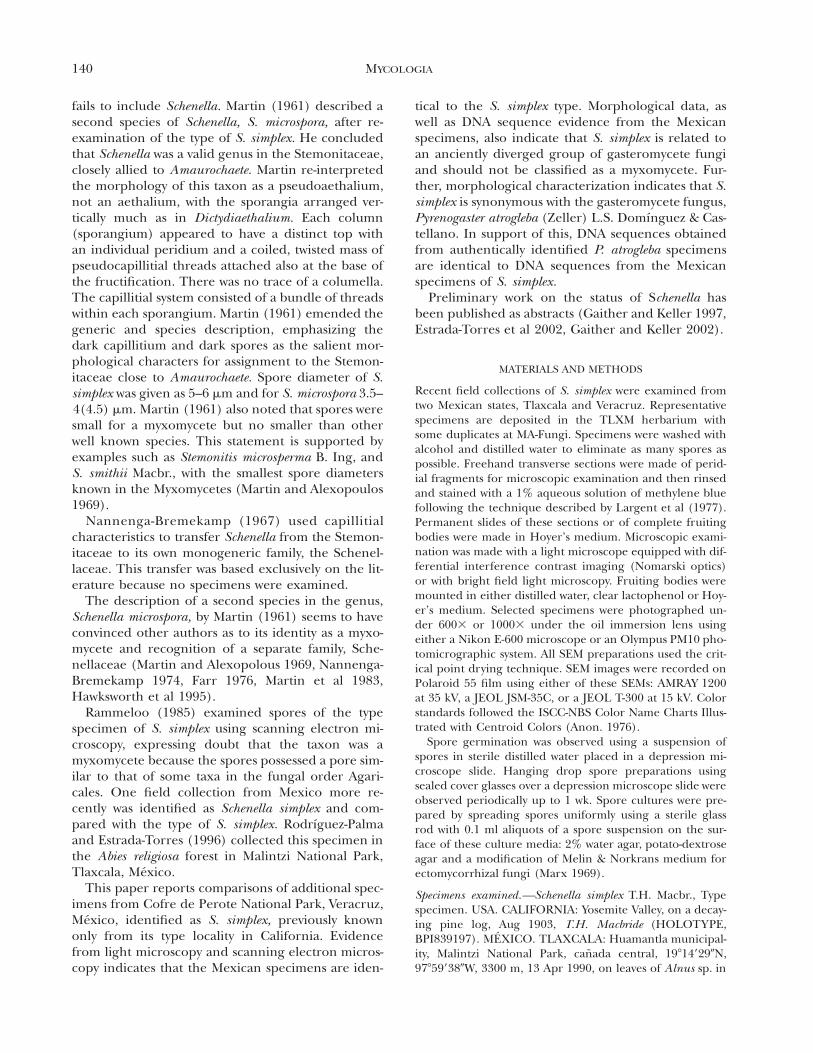
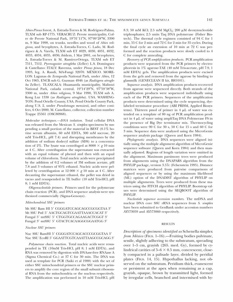
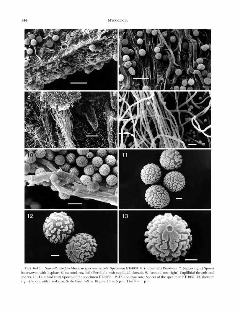
phae; peridial plates limiting the cylindrical cavitiesformed by a compact system of colorless hyphae in-terwoven with brown and granular hyphae (conduc-tive hyphae) when seen with light microscopy, 2.4–4mm diam; peridium persistent and coriaceous at theirbase, areolate, olive-brown (95. m. Ol Br-96. d. Ol Br)by transmitted light. A stratification of four differentlayers of cells can be observed (FIG. 1), the external10–15 mm thick, formed by crystalline deposits (bi-refringent by polarized light) and with some emer-gent fibulate hyphae, a second compacted stratum of10–15 mm thick, formed by irregular cells (FIGS. 3,4), a third stratum 30–35 mm thick, formed by irreg-ular or subglobose cells, 8–24 3 4–9.6 mm, interwo-ven, and finally a compact stratum, 5–10 mm thick,formed by hyphae with irregular and branched cells(FIGS. 3, 4); jointly the layers form a pseudoparen-chymatous tissue easy to observe by transverse cutswith LM (FIG. 1) which freely absorb methylene blue.The cellular nature of the peridium can be observedby SEM (FIG. 6). Capillitium composed of elastic,brownish gray (63. l. brGy-64. br Gray), brightthreads in groups of helical filaments (FIG. 8) thatoccupy almost all of the cavity of the peridiole, andjoined to the extremities, elastic at maturity and pro-cumbent; threads single (FIG. 9) or branched at theends, smooth, bright, grayish yellow (91. d. gy. Y) toolive brown (95. m. Ol Br) by transmitted light, 2.4–4.8 mm diam, thick-walled (up to 1.5 mm; FIG. 5),occasionally septate (FIG. 16), arising from irregularand branched cells of the inner layer of the peridium(FIG. 5). Spores free, dark brown (62. d. gy. Br-65. brBlack) in mass, dark yellowish brown (78. d. y Br-75.deep y Br) to dark olive brown (96. d. Ol Br) bytransmitted light, subglobose to slightly elliptical,5.6–6.4 3 4.4–5.6 mm, thick walled, 1.0–1.5 mm, palerat the end with a pore or a scar, the pore circularand depressed, the ornamentation consists of dense,irregular warts, confluent into a subreticulum, fusedand smaller toward the pore or scar forming radialcrests as seen with SEM (FIGS. 10–13). Based on thesecharacteristics, these specimens were tentatively iden-tified as S. simplex.
Germination and culture.—Spore germination or pro-duction of myxamoebae or hyphal filaments were notobserved in water or in selected media.
Commentary.—The microscopic characters are remi-niscent in color, form and habit of some species ofmyxomycetes (Amaurochaete), but the spore orna-mentation and the scar, which is discernible by LM(Rammeloo 1985) and clearly visible by SEM, havenot been seen in any myxomycete.
Analysis of Schenella simplex type specimen.—To con-firm that the Mexican specimens were S. simplex, par-allel analyses of the type specimen for Schenella sim-plex were done with light and scanning electron mi-croscopy.
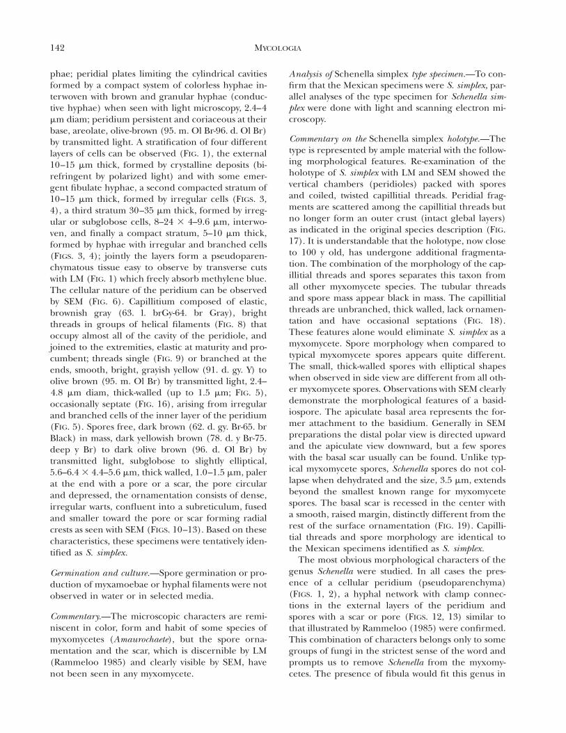
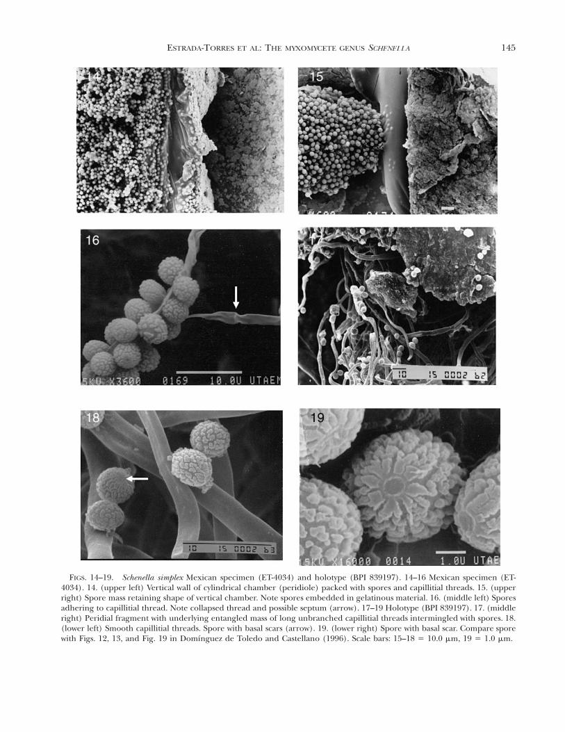
Commentary on the Schenella simplex holotype.—Thetype is represented by ample material with the follow-ing morphological features. Re-examination of theholotype of S. simplex with LM and SEM showed thevertical chambers (peridioles) packed with sporesand coiled, twisted capillitial threads. Peridial frag-ments are scattered among the capillitial threads butno longer form an outer crust (intact glebal layers)as indicated in the original species description (FIG.17). It is understandable that the holotype, now closeto 100 y old, has undergone additional fragmenta-tion. The combination of the morphology of the cap-illitial threads and spores separates this taxon fromall other myxomycete species. The tubular threadsand spore mass appear black in mass. The capillitialthreads are unbranched, thick walled, lack ornamen-tation and have occasional septations (FIG. 18).These features alone would eliminate S. simplex as amyxomycete. Spore morphology when compared totypical myxomycete spores appears quite different.The small, thick-walled spores with elliptical shapeswhen observed in side view are different from all oth-er myxomycete spores. Observations with SEM clearlydemonstrate the morphological features of a basid-iospore. The apiculate basal area represents the for-mer attachment to the basidium. Generally in SEMpreparations the distal polar view is directed upwardand the apiculate view downward, but a few sporeswith the basal scar usually can be found. Unlike typ-ical myxomycete spores, Schenella spores do not col-lapse when dehydrated and the size, 3.5 mm, extendsbeyond the smallest known range for myxomycetespores. The basal scar is recessed in the center witha smooth, raised margin, distinctly different from therest of the surface ornamentation (FIG. 19). Capilli-tial threads and spore morphology are identical tothe Mexican specimens identified as S. simplex.
The most obvious morphological characters of thegenus Schenella were studied. In all cases the pres-ence of a cellular peridium (pseudoparenchyma)(FIGS. 1, 2), a hyphal network with clamp connec-tions in the external layers of the peridium andspores with a scar or pore (FIGS. 12, 13) similar tothat illustrated by Rammeloo (1985) were confirmed.This combination of characters belongs only to somegroups of fungi in the strictest sense of the word andprompts us to remove Schenella from the myxomy-cetes. The presence of fibula would fit this genus in
143ESTRADA-TORRES ET AL: THE MYXOMYCETE GENUS SCHENELLA
FIGS. 1–5. Schenella simplex Mexican specimen (ET-4036). 1. (top panel) Pseudoparenchymatous tissue with a stratificationof different layers of cells. 2. (middle left) Middle stratum of the peridium with globose cells. 3–4. (middle right, lower left)Irregular cells of the peridium. 5. (lower right) Capillitial thread branched at the end. Scale bars 5 10.0 mm.
144 MYCOLOGIA
FIGS. 6–13. Schenella simplex Mexican specimens. 6–9. Specimen ET-4031. 6. (upper left) Peridium. 7. (upper right) Sporesinterwoven with hyphae. 8. (second row left) Peridiole with capillitial threads. 9. (second row right) Capillitial threads andspores. 10–11. (third row) Spores of the specimen ET-4036. 12–13. (bottom row) Spores of the specimen ET-4031. 13. (bottomright) Spore with basal scar. Scale bars: 6–9 5 10 mm, 10 5 5 mm, 11–13 5 1 mm.
145ESTRADA-TORRES ET AL: THE MYXOMYCETE GENUS SCHENELLA
FIGS. 14–19. Schenella simplex Mexican specimen (ET-4034) and holotype (BPI 839197). 14–16 Mexican specimen (ET-4034). 14. (upper left) Vertical wall of cylindrical chamber (peridiole) packed with spores and capillitial threads. 15. (upperright) Spore mass retaining shape of vertical chamber. Note spores embedded in gelatinous material. 16. (middle left) Sporesadhering to capillitial thread. Note collapsed thread and possible septum (arrow). 17–19 Holotype (BPI 839197). 17. (middleright) Peridial fragment with underlying entangled mass of long unbranched capillitial threads intermingled with spores. 18.(lower left) Smooth capillitial threads. Spore with basal scars (arrow). 19. (lower right) Spore with basal scar. Compare sporewith Figs. 12, 13, and Fig. 19 in Domınguez de Toledo and Castellano (1996). Scale bars: 15–18 5 10.0 mm, 19 5 1.0 mm.
146 MYCOLOGIA
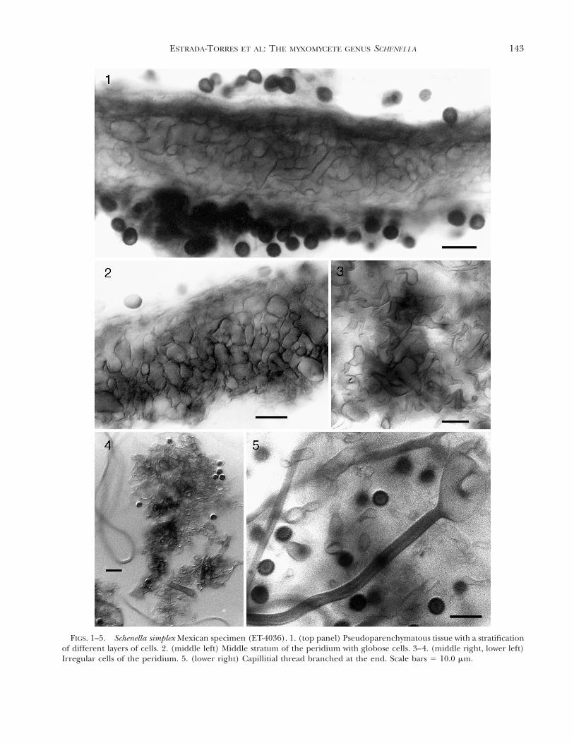
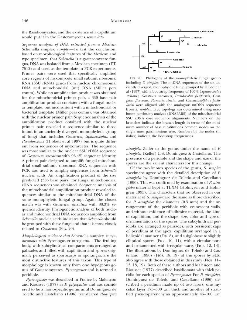
FIG. 20. Phylogeny of the monophyletic fungal groupincluding S. simplex. The mtDNA sequences of the six an-ciently diverged, monophyletic fungi grouped by Hibbett etal (1997) with a bootstrap frequency of 100% (Sphaerobolusstellatus, Geastrum saccatum, Pseudocolus fusiformis, Gom-phus floccosus, Ramaria stricta, and Clavariadelphus pistil-laris) were aligned with the analogous mtDNA sequencefrom S. simplex. Tree topology was determined using max-imum parsimony analysis (DNAPARS) of the mitochondrialSSU rDNA core sequence alignments. Numbers on thebranches indicate the branch length in terms of the mini-mum number of base substitutions between nodes on thesingle most parsimonious tree. Numbers by the nodes (initalics) indicate the bootstrap frequencies.
the Basidiomycetes, and the existence of a capillitiumwould put it in the Gasteromycetes sensu lato.
Sequence analysis of DNA extracted from a MexicanSchenella simplex sample.—To test the conclusion,based on morphological features of the Mexican andtype specimen, that Schenella is a gasteromycete fun-gus, DNA was isolated from a Mexican specimen (ET-7512) and used as the template in PCR experiments.Primer pairs were used that specifically amplifiedcore regions of myxomycete small subunit ribosomalRNA (SSU rRNA) genes from nuclear chromosomalDNA and mitochondrial (mt) DNA (Miller perscomm). While no amplification product was obtainedfor the mitochondrial primer pair, a 639 base pairamplification product consistent with a fungal nucle-ar template, but inconsistent with a mitochondrial orbacterial template (Miller pers comm), was obtainedwith the nuclear primer pair. Sequence analysis of theamplification product obtained with the nuclearprimer pair revealed a sequence similar to thosefound in an anciently diverged, monophyletic groupof fungi that includes Geastrum, Sphaerobolus andPseudocolous (Hibbett et al 1997) but is quite differ-ent from sequences of myxomycetes. The sequencewas most similar to the nuclear SSU rDNA sequenceof Geastrum saccatum with 96.4% sequence identity.A primer pair designed to amplify fungal mitochon-drial small subunit ribosomal RNA sequences withPCR was used to amplify sequences from Schenellanucleic acids. An amplification product of the sizepredicted (383 base pairs) for fungal mitochondrialrDNA sequences was obtained. Sequence analysis ofthe mitochondrial amplification product revealed se-quences similar to the mitochondrial DNA of thesame monophyletic fungal group. Again the closestmatch was with Geastrum saccatum with 88.3% se-quence identity. Phylogenetic analysis of both nucle-ar and mitochondrial DNA sequences amplified fromSchenella nucleic acids indicates that Schenella shouldbe grouped with these fungi and that it is most closelyrelated to Geastrum (FIG. 20).
Morphological evidence that Schenella simplex is syn-onymous with Pyrenogaster atrogleba.—The fruitingbody, with subcylindrical compartments arranged aspalisades and filled with capillitium and spores orig-inally perceived as sporocarps or sporangia, are themost distinctive features of this taxon. This type ofmorphology is known only from one hypogeous ge-nus of Gasteromycetes, Pyrenogaster and is termed aperidiole.
Pyrenogaster was described in France by Malenconand Riousset (1977) as P. pityophilus and was consid-ered to be a monospecific genus until Domınguez deToledo and Castellano (1996) transferred Radiigera
atrogleba Zeller to the genus under the name of P.atrogleba (Zeller) L.S. Domınguez & Castellano. Thepresence of a peridiole and the shape and size of thespores are the salient characters for this change.
Of the two known species of the genus, S. simplexspecimens agree with the detailed description of P.atrogleba by Domınguez de Toledo and Castellano(1996). This was confirmed by examination of P. atro-gleba material kept at TLXM (Holmgren and Holm-gren 1995). The characters that we observed in ourmaterial of S. simplex are the same as those describedfor P. atrogleba: the diameter (0.5 mm) and the ar-rangement of the peridiole with evanescent walls,and without evidence of adhesive material, the kindof capillitium, and the shape, size, color and type ofornamentation of the spores. The subcylindrical per-idiola are arranged as palisades, with persistent capsof peridium at the apex, capillitium arranged in ahelicoidal manner (FIG. 8), and subglobose to slightlyelliptical spores (FIGS. 10, 11), with a circular poreand ornamented with irregular warts (FIGS. 12, 13).The illustrations by Domınguez de Toledo and Cas-tellano (1996) (FIGS. 18, 19) of the spores by SEMalso agree with those obtained in this study (FIGS. 11–13, 18, 19). Both of these authors and Malencon andRiousset (1977) described basidiomata with thick pe-ridia for each species of Pyrenogaster. For P. atrogleba,Domınguez de Toledo and Castellano (1996) de-scribed a peridium made up of two layers, one my-celial layer 175–500 mm thick and another of strati-fied pseudoparenchyma approximately 45–100 mm
147ESTRADA-TORRES ET AL: THE MYXOMYCETE GENUS SCHENELLA
thick. Of these layers, the field specimens had onlythe innermost stratum of the second layer (see FIG.1) illustrated by Domınguez de Toledo and Castel-lano (1996, Fig. 1J) due to deterioration of the ma-terial. The characteristics of the strata observed arehighlighted in the description. We have compared anexsiccatum of P. atrogleba from the same locality ofthe first record of S. simplex (Rodrıguez-Palma andEstrada-Torres 1996), another from Morelos, Mexicoand another from Italy, all coincide with S. simplex inthe characters mentioned above.
One character not pointed out by Domınguez deToledo and Castellano (1996) is the presence of theirregular branched tips of the capillitial filaments(FIG. 5) that mingle with the cells of the internal lay-er of the peridium and are distinguishable fromthem by their thick walls. These already had beendescribed, however, by Malencon and Riousset(1977) and illustrated for the other species of thegenus. These also were present in the field collec-tions of S. simplex.
The interpretation of S. simplex as a myxomycetecould be due to the advanced maturity of the fungusand its fragmentation, which reveals structures ofsimilar appearance to myxomycetes such as capilli-tium, dark spores and peridium. Malencon andRiousset (1977) noted the difficulty in identifying anyvery mature stages of the fungus. The appearance ofthis hypogeous fungus on the surface of logs, deadwood and bryophyte layers covering the ground is anenigma, perhaps explainable by the activities of someanimal.
DNA sequence evidence that Schenella simplex is syn-onymous with Pyrenogaster atrogleba.—To confirmthe supposition that Schenella simplex is synonymouswith Pyrenogaster atrogleba, DNA was isolated from au-thentically identified P. atrogleba specimens (Trappe25561, OSC 80968) from M. Castellano and used asthe template in PCR reactions with the nuclearmtSSU primers and, in a separate reaction, with thefungal mtSSU primers. Amplification products of asize identical to those produced from the MexicanSchenella simplex samples (639 and 383 base pairsfrom nuclear and mitochondrial primer pairs respec-tively) were obtained. These amplification productswere isolated and sequenced. These sequences werecompared with those obtained from the MexicanSchenella simplex specimen and found to be identical.
DISCUSSION
Taxonomy.—Morphological and molecular evidenceleads us to conclude that S. simplex and P. atroglebaare identical and should be considered synonymous.
From a nomenclatural point of view, the genus Sche-nella is transferred from the Myxomycetes to the Gas-teromycetes and given its priority (art. 11.3 Interna-tional Code of Botanical Nomenclature) over Pyren-ogaster, these synonyms and combinations are pro-posed:
Schenella T. Macbr., Mycologia 3(1):39. 19115 Pyrenogaster Malencon & Riousset, Bull. Soc. Mycol.
France 93: 289. 1977, syn. nov.Generitypus: Schenella simplex T. Macbr., Mycologia
3(1):39. 1911.Generitypus specimen: BPI 839197. USA. Califor-
nia, Yosemite Valley, Aug 1903, on a decaying pinelog, T.H. Macbride.
Schenella simplex T. Macbr., Mycologia 3(1):39. 19115 Radiigera atrogleba Zeller, Mycologia 36(6):634. 1944,
syn. nov., [ Pyrenogaster atrogleba (Zeller) L.S.Domınguez & Castellano, Mycologia 88(5):866.1996.
Typus.- BPI 839197. USA. California, Yosemite Valley,Aug 1903, on a decaying pine log, T.H. Macbride,holotypus.
Schenella pityophilus (Malencon & Riousset) Estrada& Lado, comb. nov.[ Pyrenogaster pityophilus Malencon & Riousset, Bull.
Soc. Mycol. France 93:290. 1977 [basion.].Typus.—MPU (in coll. G.J.L. Malencon). FRANCE:
near Aigues-Mortes, Mar-May 1976, under Pinus pi-nea needles, L. Riousset, holotypus (Isotypus atOSC).
Schenella romana (Quadracia) Estrada & Lado, comb.nov.[ Radiigera romana Quadracia, Mycotaxon 58:336
(1996) [basion.]; [ Pyrenogaster romana (Quadracia)Calonge, Bol. Soc. Micol. Madrid 22:108. 1997)
Typus—ROHB (1325 LQ). ITALY: Roma, Orto Bo-tanico, 22 Jan 1922, under Cupressus sempervirens, L.Quadracia, holotypus.
The identity of S. microspora (G.W. Martin, Mycol-ogia 53[1]: 29. 1961, BPI 839199, U.S.A.: CALIFOR-NIA, Big Basin State Park, San Mateo County, 26 Au-gust 1957, on fallen trunk of Sequoia sempervirens[Lamb.] Endl., G.W. Martin 6547) still has to be clar-ified. Schenella microspora was studied using LM andSEM, but these results will be published elsewhere.There are still some questions if this taxon is synon-ymous with P. atrogleba (Gaither and Keller 2002,Gaither and Keller 2003).
148 MYCOLOGIA
Spore germination.—Further evidence of the fungalnature of Schenella might have been provided by ger-mination of the spores from the Mexican specimens.Spores did not germinate in either media for ecto-mycorrhizal fungi (potato-dextrose agar or modifiedMelin & Norkrans medium) or on 2% water agar.Most ectomycorrhizal fungal species such as Pyreno-gaster have a low percentage of germination on arti-ficial laboratory media (Molina & Palmer 1982), al-though the use of active charcoal to remove inhibi-tors or the presence of some stimulators (living or-ganisms or chemical substances) can greatly improvethe success of these procedures (Fries 1977, Ali andJackson 1989, Iwase 1991). It also is relevant thatspores of truffle-like fungi, such as Pyrenogaster, seemto be activated by passage through the intestine ofmycophagous animals (Fogel and Trappe 1978).These two observations might explain why Schenellaspores were not germinated using the techniques de-scribed. Germination of Schenella spores using alter-nate techniques or media is the goal of future ex-periments.
Ecology.—The basidiomata studied were found on di-verse substrata such as trunks, needles and fallenbranches of conifers or bryophyte layers growing onthe ground. These habitats are typical of many spe-cies of myxomycetes. This hypogeous taxon appar-ently is dug up by an animal and transported toabove ground sites so that its appearance on thesesubstrata is accidental.
As with other hypogeous Gasteromycetes, S. sim-plex must be dug up and carried away to be eaten bysmall rodents, a fact that favors its dispersal as hasbeen demonstrated for other species of hypogeousectomycorrhizal fungi (Maser et al 1978, Malajczuket al 1987, Trappe 1988). The abundance of fruitingbodies (in 400 m2 about 20 were observed) and theway some were found, on trunks more than 1 mabove ground or on mounds covered with bryo-phytes, supports this hypothesis. Furthermore rodentfeces occasionally were observed alongside fruitingbody remains.
None of the field collections contained completebasidiomata and in the majority of cases they werecomposed of spore masses and capillitium. This canbe reconciled with the differential use of the fruitingbody by animals (Cork and Kenagy 1989). Rodentsfeed on the fleshy parts of the fungus such as thecolumella and external layers of the peridium. As thecolumella and probably other fleshy parts of the un-ripe fungus are consumed the fruiting body is in-verted leaving the outer peridium against the sub-strate and the base of the peridiole in the upper part,exposing the capillitium. Because the capillitium is
elastic, it gives the procumbent appearance of somemyxomycetes such as Comatricha longa Peck or spe-cies of Amaurochaete.
The fact that animals dig up and consume thewhole fruiting structure appears to be one of the rea-sons why, in spite of intense searching in one locality,it was found again only after 3 y. It also is interestingto note that all collections of S. simplex (includingthose identified as Radiigera atrogleba) from Tlaxcalaand Veracruz were collected during the end of thedry season (March and April) (Tejeda-Martınez et al1989), when most of the mushroom species had notyet fruited in this area, indicating a peculiar phenol-ogy of the species. Furthermore in 1998 the collec-tion of specimens in Cofre de Perote, one of the mostproductive localities, coincided with the dry periodwhich that year was aggravated by the effects of ElNino.
The distribution of S. simplex is wider than wasonce thought since it was reported as Radiigera atro-gleba from southwestern Canada (British Columbia),from USA (California, Colorado, Idaho, Montana,Oregon, Utah and Washington), from Mexico (BajaCalifornia Norte and Morelos), from Sweden and It-aly (Zeller 1944, Guzman 1971, Kers 1976, Ayala andGuzman 1984, Domınguez de Toledo and Castellano1996, Calonge 1997) and as S. simplex from USA(California) and Mexico (Tlaxcala and Veracruz)(Macbride 1911, Rodrıguez-Palma and Estrada-Tor-res 1996).
Considering the ectomycorrhizal character of mosthypogeous species, this species appears to be exclu-sively associated with conifers of such diverse generaas Abies, Picea, Pinus, Pseudotsuga, Thuja and Tsuga,although Kers (1976) also cited it under Fraxinus ex-celsior.
ACKNOWLEDGMENTS
We thank the curators of herbaria BPI, ENCB and OSC forthe loan of specimens. This research project was financedin part by a grant to HWK from the National Science Foun-dation, Biotic Surveys and Inventories Program, awardnumber DEB-0079058, and to AET and CL by the SpanishGovernment, through the grant REN2002–00445.
LITERATURE CITED
Ali NA, Jackson RM. 1989. Stimulation of germination ofspores of some ectomycorrhizal fungi by other micro-organisms. Mycol Res 93:182–186.
Anonymous. 1976. ISCC-NBS color-name charts illustratedwith centroid colors. Washington, DC: Inter-SocietyColor Council, National Bureau of Standards. 4 p 1 18pl.
Ayala N, Guzman G. 1984. Los hongos de la penınsula de
149ESTRADA-TORRES ET AL: THE MYXOMYCETE GENUS SCHENELLA
Baja California. I. Las especies conocidas. Bol Soc MexMic 19:73–91.
Calonge FD. 1997. Notes on the genera Pyrenogaster andRadiigera (Gasteromycetes). Bol Soc Micol Madrid 22:105–112.
Cork SJ, Kenagy GJ. 1989. Nutritional value of a hypogeousfungus for a forest-dwelling ground squirrel. Ecology70:577–586.
Domınguez de Toledo LS, Castellano MA. 1996. A revisionof the genus Radiigera and Pyrenogaster. Mycologia 88:863–884.
Estrada-Torres A, Gaither TW, Miller DL, Lado C, KellerHW. 2002. The myxomycete genus Schenella: morpho-logical and molecular evidence for the gasteromycetegenus Pyrenogaster. In: Rammeloo J, Bogaerts A, eds.Fourth International Congress on Systematics andEcology of Myxomycetes, Abstracts. Scripta Bot Belg22:5.
Farr ML. 1976. Myxomycetes. Flora Neotrop 16:1–304.Felsenstein J. 1995. PHYLIP (Phylogeny Inference Pack-
age), version 3.57c, Department of Genetics, Universityof Washington, Seattle.
Fogel R, Trappe J. 1978. Fungal consumption (mycophagy)in small mammals. Northwest Science 52:1–31.
Fries N. 1977. Germination of Laccaria laccata spores invitro. Mycologia 69:848–850.
Gaither TW, Keller HW. 1997. The mystery remains: whatis Schenella? Tex J Micros 28(2):44.
, . 2002. Microscopic evidence supporting thecogeneric status of Schenella simplex and S. microspora.In: Rammeloo J, Bogaerts A, eds. Fourth InternationalCongress on Systematics and Ecology of Myxomycetes,Abstracts. Scripta Bot Belg 22:34.
, . 2003. Light and scanning electron micros-copy of the myxomycete species Schenella microsporaand S. simplex: Morphological evidence for a gastroidbasidiomycete. Inoculum 54:21.
Guzman G. 1971. Notas sobre los generos Radiigera y Me-sophelliopsis en Mexico. Bol Soc Mex Mic 5:7–11.
Hagelstein R. 1944. The mycetozoa of North America. Min-eola, New York. 306 p.
Hawksworth DL, Kirk PM, Sutton BC, Pegler DN. 1995. Ain-sworth & Bisby’s Dictionary of the Fungi. 8th ed. NewYork: French & European Publications Inc. 615 p.
Hibbett DS, Pine EM, Langer E, Langer G, Donoghue MJ.1997. Evolution of gilled mushrooms and puffballs in-ferred from ribosomal DNA sequences. Proc Natl AcadSci USA 94:12002–12006.
Holmgren PK, Holmgren NH. 1995. Additions to IndexHerbariorum (Herbaria), Edition 8-Fourth Series. Tax-on 44:251–266.
Iwase K. 1991. Induction of basidiospore germination bygluconic acid in the ectomycorrhizal fungus Tricholomarobustum. Can J Bot 70:1234–1238.
Kers LE. 1976. Radiigera Zeller, a genus of Gasteromycetesnew to Europe. Bot Notiser 129:173–178.
Largent D, Johnson D, Watling R. 1977. How to identifymushrooms to genus III: Microscopic features. Eureka,California: Mad River Press. 148 p.
Lister A. 1925. A Monograph of the Mycetozoa Being a De-
scriptive Catalogue of the Species in the Herbarium ofthe British Museum. 3rd ed. Revised by G. Lister. Brit-ish Museum of Natural History. London. 296 p.
Macbride TH. 1911. A new genus of Myxomycetes? Mycol-ogia 3:39.
. 1922. The North American Slime-Moulds. 2nd ed.New York: Macmillian Co. 299 p.
, Martin GW. 1934. The Myxomycetes. New York:Macmillan Co. 339 p.
Malajczuk N, Trappe JM, Molina R. 1987. Interrelationshipsamong some ectomycorrhizal trees, hypogeous fungiand small mammals: Western Australian and north-western American parallels. Aust J Ecol 12:53–55.
Malencon G, Riousset L. 1977. Pyrenogaster pityophilus G.Malencon et L. Riousset, nouveau genre et nouvelleespece de gasteromycete (Geastraceae). Bull Soc MycolFrance 93:289–311.
Martin GW. 1949. Myxomycetes. N Am Flora 1:1–190.. 1961. The genus Schenella. Mycologia 53:25–30., Alexopoulos CJ. 1969. The Myxomycetes. Iowa City,
Iowa: University of Iowa Press. 501 p., , Farr ML. 1983. The genera of Myxomy-
cetes. Iowa City, Iowa: University of Iowa Press. 102 p.Marx DH. 1969. The influence of ectotrophic mycorrhizal
fungi on the resistance of pine roots to pathogenic in-fections. I. Antagonism of mycorrhizal fungi to rootpathogenic fungi and bacteria. Phytopathol 59:153–163.
Maser C, Trappe JM, Nussbaum RA. 1978. Fungal-smallmammal interrelationships with emphasis on Oregonconiferous forests. Ecology 59:799–809.
Molina R, Palmer G. 1982. Isolation, maintenance, andpure culture manipulation of ectomycorrhizal fungi.In: Schenck NC, ed. Methods and principles of mycor-rhizal research. Minnesota: The American Phytopath-ological Society. p 115–129.
Nannenga-Bremekamp NE. 1967. Notes on Myxomycetes.XII. A revision of the Stemonitales. Proc Kon Ned AkadWetensch C 70:201–216.
. 1974. De Nederlandse Myxomyceten. Biblioth KonNederl Natuurhist Ver 18:1–440.
Queen C, Korn LJ. 1984. A comparative sequence analysisprogram for the IBM personal computer. Nucleic AcidsRes 12:581–599.
Rammeloo J. 1985. Schenella simplex Macbride. In: Ram-meloo J, ed. Icones Mycolgicae 93–110. Pl. 110. Nation-ale Plantetuin van Belgie, Meise.
Rodrıguez-Palma M, Estrada-Torres A. 1996. Some Stemon-itales (Myxomycetes) from the state of Tlaxcala, Mexi-co. Mycotaxon 60:79–102.
Saiki RK, Gelfand DH, Stoffel S, Scharf SJ, Higuchi R, HornGT, Mullis KB, Erlich HA. 1988. Primer-directed enzy-matic amplification of DNA with a thermostable DNApolymerase. Science 239:487–491.
Tejeda-Martınez A, Acevedo F, Jauregui E. 1989. Atlas cli-matico del estado de Veracruz. Universidad Veracru-zana, Xalapa, Veracruz.
Trappe JM. 1988. Lessons from alpine fungi. Mycologia 80:1–10.
Zeller SM. 1944. Representatives of the Mesophelliaceae inNorth America. Mycologia 36:627–637.