The Mouth, Pharynx, and Esophagus - OpenStax CNX4.pdf/the... · The Mouth, Pharynx, and Esophagus...
22
* † • • • • * †
Transcript of The Mouth, Pharynx, and Esophagus - OpenStax CNX4.pdf/the... · The Mouth, Pharynx, and Esophagus...

OpenStax-CNX module: m46511 1
The Mouth, Pharynx, and Esophagus∗
OpenStax College
This work is produced by OpenStax-CNX and licensed under the
Creative Commons Attribution License 3.0†
Abstract
By the end of this section, you will be able to:
• Describe the structures of the mouth, including its three accessory digestive organs
• Group the 32 adult teeth according to name, location, and function
• Describe the process of swallowing, including the roles of the tongue, upper esophageal sphincter,
and epiglottis
• Trace the pathway food follows from ingestion into the mouth through release into the stomach
In this section, you will examine the anatomy and functions of the three main organs of the upperalimentary canal�the mouth, pharynx, and esophagus�as well as three associated accessory organs�thetongue, salivary glands, and teeth.
1 The Mouth
The cheeks, tongue, and palate frame the mouth, which is also called the oral cavity (or buccal cavity).The structures of the mouth are illustrated in Figure 1 (Mouth ).
At the entrance to the mouth are the lips, or labia (singular = labium). Their outer covering is skin,which transitions to a mucous membrane in the mouth proper. Lips are very vascular with a thin layer ofkeratin; hence, the reason they are "red." They have a huge representation on the cerebral cortex, whichprobably explains the human fascination with kissing! The lips cover the orbicularis oris muscle, whichregulates what comes in and goes out of the mouth. The labial frenulum is a midline fold of mucousmembrane that attaches the inner surface of each lip to the gum. The cheeks make up the oral cavity'ssidewalls. While their outer covering is skin, their inner covering is mucous membrane. This membraneis made up of non-keratinized, strati�ed squamous epithelium. Between the skin and mucous membranesare connective tissue and buccinator muscles. The next time you eat some food, notice how the buccinatormuscles in your cheeks and the orbicularis oris muscle in your lips contract, helping you keep the food fromfalling out of your mouth. Additionally, notice how these muscles work when you are speaking.
The pocket-like part of the mouth that is framed on the inside by the gums and teeth, and on the outsideby the cheeks and lips is called the oral vestibule. Moving farther into the mouth, the opening betweenthe oral cavity and throat (oropharynx) is called the fauces (like the kitchen "faucet"). The main open areaof the mouth, or oral cavity proper, runs from the gums and teeth to the fauces.
When you are chewing, you do not �nd it di�cult to breathe simultaneously. The next time you have foodin your mouth, notice how the arched shape of the roof of your mouth allows you to handle both digestionand respiration at the same time. This arch is called the palate. The anterior region of the palate serves asa wall (or septum) between the oral and nasal cavities as well as a rigid shelf against which the tongue can
∗Version 1.4: Jun 28, 2013 10:24 am -0500†http://creativecommons.org/licenses/by/3.0/
http://cnx.org/content/m46511/1.4/

OpenStax-CNX module: m46511 2
push food. It is created by the maxillary and palatine bones of the skull and, given its bony structure, isknown as the hard palate. If you run your tongue along the roof of your mouth, you'll notice that the hardpalate ends in the posterior oral cavity, and the tissue becomes �eshier. This part of the palate, known asthe soft palate, is composed mainly of skeletal muscle. You can therefore manipulate, subconsciously, thesoft palate�for instance, to yawn, swallow, or sing (see Figure 1 (Mouth )).
Mouth
Figure 1: The mouth includes the lips, tongue, palate, gums, and teeth.
A �eshy bead of tissue called the uvula drops down from the center of the posterior edge of the softpalate. Although some have suggested that the uvula is a vestigial organ, it serves an important purpose.
http://cnx.org/content/m46511/1.4/

OpenStax-CNX module: m46511 3
When you swallow, the soft palate and uvula move upward, helping to keep foods and liquid from enteringthe nasal cavity. Unfortunately, it can also contribute to the sound produced by snoring. Two muscular foldsextend downward from the soft palate, on either side of the uvula. Toward the front, the palatoglossalarch lies next to the base of the tongue; behind it, the palatopharyngeal arch forms the superior andlateral margins of the fauces. Between these two arches are the palatine tonsils, clusters of lymphoid tissuethat protect the pharynx. The lingual tonsils are located at the base of the tongue.
2 The Tongue
Perhaps you have heard it said that the tongue is the strongest muscle in the body. Those who stake thisclaim cite its strength proportionate to its size. Although it is di�cult to quantify the relative strengthof di�erent muscles, it remains indisputable that the tongue is a workhorse, facilitating ingestion, mechan-ical digestion, chemical digestion (lingual lipase), sensation (of taste, texture, and temperature of food),swallowing, and vocalization.
The tongue is attached to the mandible, the styloid processes of the temporal bones, and the hyoid bone.The hyoid is unique in that it only distantly/indirectly articulates with other bones. The tongue is positionedover the �oor of the oral cavity. A medial septum extends the entire length of the tongue, dividing it intosymmetrical halves.
Beneath its mucous membrane covering, each half of the tongue is composed of the same number andtype of intrinsic and extrinsic skeletal muscles. The intrinsic muscles (those within the tongue) are thelongitudinalis inferior, longitudinalis superior, transversus linguae, and verticalis linguae muscles. Theseallow you to change the size and shape of your tongue, as well as to stick it out, if you wish. Having such a�exible tongue facilitates both swallowing and speech.
As you learned in your study of the muscular system, the extrinsic muscles of the tongue are the mylo-hyoid, hyoglossus, styloglossus, and genioglossus muscles. These muscles originate outside the tongue andinsert into connective tissues within the tongue. The mylohyoid is responsible for raising the tongue, thehyoglossus pulls it down and back, the styloglossus pulls it up and back, and the genioglossus pulls it forward.Working in concert, these muscles perform three important digestive functions in the mouth: (1) positionfood for optimal chewing, (2) gather food into a bolus (rounded mass), and (3) position food so it can beswallowed.
The top and sides of the tongue are studded with papillae, extensions of lamina propria of the mucosa,which are covered in strati�ed squamous epithelium (Figure 2 (Tongue )). Fungiform papillae, which aremushroom shaped, cover a large area of the tongue; they tend to be larger toward the rear of the tongue andsmaller on the tip and sides. In contrast, �liform papillae are long and thin. Fungiform papillae contain tastebuds, and �liform papillae have touch receptors that help the tongue move food around in the mouth. The�liform papillae create an abrasive surface that performs mechanically, much like a cat's rough tongue thatis used for grooming. Lingual glands in the lamina propria of the tongue secrete mucus and a watery serous�uid that contains the enzyme lingual lipase, which plays a minor role in breaking down triglycerides butdoes not begin working until it is activated in the stomach. A fold of mucous membrane on the underside ofthe tongue, the lingual frenulum, tethers the tongue to the �oor of the mouth. People with the congenitalanomaly ankyloglossia, also known by the non-medical term �tongue tie,� have a lingual frenulum that is tooshort or otherwise malformed. Severe ankyloglossia can impair speech and must be corrected with surgery.
http://cnx.org/content/m46511/1.4/

OpenStax-CNX module: m46511 4
Tongue
Figure 2: This superior view of the tongue shows the locations and types of lingual papillae.
3 The Salivary Glands
Many small salivary glands are housed within the mucous membranes of the mouth and tongue. Theseminor exocrine glands are constantly secreting saliva, either directly into the oral cavity or indirectly throughducts, even while you sleep. In fact, an average of 1 to 1.5 liters of saliva is secreted each day. Usually justenough saliva is present to moisten the mouth and teeth. Secretion increases when you eat, because salivais essential to moisten food and initiate the chemical breakdown of carbohydrates. Small amounts of salivaare also secreted by the labial glands in the lips. In addition, the buccal glands in the cheeks, palatal glandsin the palate, and lingual glands in the tongue help ensure that all areas of the mouth are supplied withadequate saliva.
3.1 The Major Salivary Glands
Outside the oral mucosa are three pairs of major salivary glands, which secrete the majority of saliva intoducts that open into the mouth:
• The submandibular glands, which are in the �oor of the mouth, secrete saliva into the mouththrough the submandibular ducts.
http://cnx.org/content/m46511/1.4/

OpenStax-CNX module: m46511 5
• The sublingual glands, which lie below the tongue, use the lesser sublingual ducts to secrete salivainto the oral cavity.
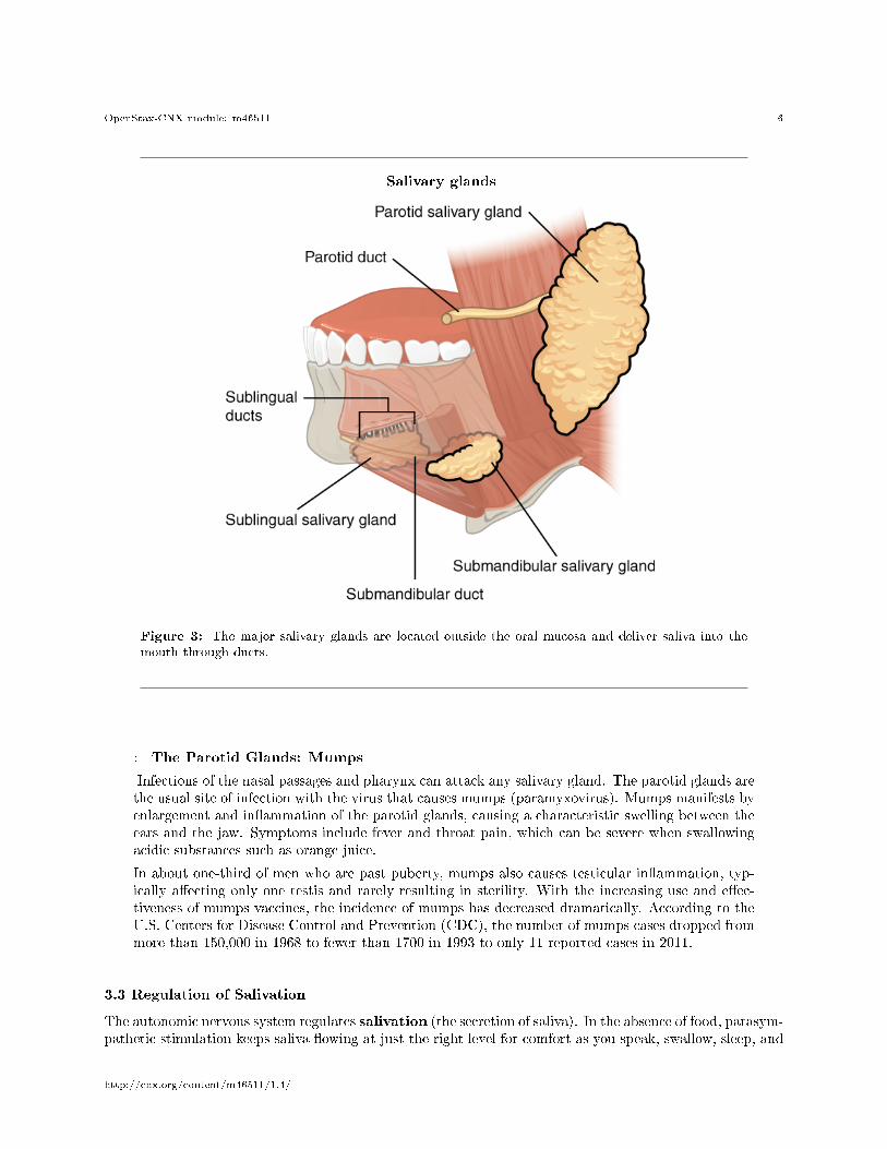
• The parotid glands lie between the skin and the masseter muscle, near the ears. They secrete salivainto the mouth through the parotid duct, which is located near the second upper molar tooth (Figure 3(Salivary glands )).
3.2 Saliva
Saliva is essentially (95.5 percent) water. The remaining 4.5 percent is a complex mixture of ions, glycopro-teins, enzymes, growth factors, and waste products. Perhaps the most important ingredient in salvia from theperspective of digestion is the enzyme salivary amylase, which initiates the breakdown of carbohydrates.Food does not spend enough time in the mouth to allow all the carbohydrates to break down, but salivaryamylase continues acting until it is inactivated by stomach acids. Bicarbonate and phosphate ions functionas chemical bu�ers, maintaining saliva at a pH between 6.35 and 6.85. Salivary mucus helps lubricate food,facilitating movement in the mouth, bolus formation, and swallowing. Saliva contains immunoglobulin A,which prevents microbes from penetrating the epithelium, and lysozyme, which makes saliva antimicrobial.Saliva also contains epidermal growth factor, which might have given rise to the adage �a mother's kiss canheal a wound.�
Each of the major salivary glands secretes a unique formulation of saliva according to its cellular makeup.For example, the parotid glands secrete a watery solution that contains salivary amylase. The submandibularglands have cells similar to those of the parotid glands, as well as mucus-secreting cells. Therefore, salivasecreted by the submandibular glands also contains amylase but in a liquid thickened with mucus. Thesublingual glands contain mostly mucous cells, and they secrete the thickest saliva with the least amount ofsalivary amylase.
http://cnx.org/content/m46511/1.4/
OpenStax-CNX module: m46511 6
Salivary glands
Figure 3: The major salivary glands are located outside the oral mucosa and deliver saliva into the
mouth through ducts.
: The Parotid Glands: Mumps
Infections of the nasal passages and pharynx can attack any salivary gland. The parotid glands arethe usual site of infection with the virus that causes mumps (paramyxovirus). Mumps manifests byenlargement and in�ammation of the parotid glands, causing a characteristic swelling between theears and the jaw. Symptoms include fever and throat pain, which can be severe when swallowingacidic substances such as orange juice.
In about one-third of men who are past puberty, mumps also causes testicular in�ammation, typ-ically a�ecting only one testis and rarely resulting in sterility. With the increasing use and e�ec-tiveness of mumps vaccines, the incidence of mumps has decreased dramatically. According to theU.S. Centers for Disease Control and Prevention (CDC), the number of mumps cases dropped frommore than 150,000 in 1968 to fewer than 1700 in 1993 to only 11 reported cases in 2011.
3.3 Regulation of Salivation
The autonomic nervous system regulates salivation (the secretion of saliva). In the absence of food, parasym-pathetic stimulation keeps saliva �owing at just the right level for comfort as you speak, swallow, sleep, and
http://cnx.org/content/m46511/1.4/

OpenStax-CNX module: m46511 7
generally go about life. Over-salivation can occur, for example, if you are stimulated by the smell of food,but that food is not available for you to eat. Drooling is an extreme instance of the overproduction of saliva.During times of stress, such as before speaking in public, sympathetic stimulation takes over, reducing sali-vation and producing the symptom of dry mouth often associated with anxiety. When you are dehydrated,salivation is reduced, causing the mouth to feel dry and prompting you to take action to quench your thirst.
Salivation can be stimulated by the sight, smell, and taste of food. It can even be stimulated by thinkingabout food. You might notice whether reading about food and salivation right now has had any e�ect onyour production of saliva.
How does the salivation process work while you are eating? Food contains chemicals that stimulate tastereceptors on the tongue, which send impulses to the superior and inferior salivatory nuclei in the brain stem.These two nuclei then send back parasympathetic impulses through �bers in the glossopharyngeal and facialnerves, which stimulate salivation. Even after you swallow food, salivation is increased to cleanse the mouthand to water down and neutralize any irritating chemical remnants, such as that hot sauce in your burrito.Most saliva is swallowed along with food and is reabsorbed, so that �uid is not lost.
4 The Teeth
The teeth, or dentes (singular = dens), are organs similar to bones that you use to tear, grind, and otherwisemechanically break down food.
4.1 Types of Teeth
During the course of your lifetime, you have two sets of teeth (one set of teeth is a dentition). Your 20deciduous teeth, or baby teeth, �rst begin to appear at about 6 months of age. Between approximatelyage 6 and 12, these teeth are replaced by 32 permanent teeth. Moving from the center of the mouthtoward the side, these are as follows (Figure 4 (Permanent and Deciduous Teeth )):
• The eight incisors, four top and four bottom, are the sharp front teeth you use for biting into food.• The four cuspids (or canines) �ank the incisors and have a pointed edge (cusp) to tear up food. These
fang-like teeth are superb for piercing tough or �eshy foods.• Posterior to the cuspids are the eight premolars (or bicuspids), which have an overall �atter shape
with two rounded cusps useful for mashing foods.• The most posterior and largest are the 12 molars, which have several pointed cusps used to crush
food so it is ready for swallowing. The third members of each set of three molars, top and bottom,are commonly referred to as the wisdom teeth, because their eruption is commonly delayed until earlyadulthood. It is not uncommon for wisdom teeth to fail to erupt; that is, they remain impacted. Inthese cases, the teeth are typically removed by orthodontic surgery.
http://cnx.org/content/m46511/1.4/

OpenStax-CNX module: m46511 8
Permanent and Deciduous Teeth
Figure 4: This �gure of two human dentitions shows the arrangement of teeth in the maxilla and
mandible, and the relationship between the deciduous and permanent teeth.
http://cnx.org/content/m46511/1.4/

OpenStax-CNX module: m46511 9
4.2 Anatomy of a Tooth
The teeth are secured in the alveolar processes (sockets) of the maxilla and the mandible. Gingivae (com-monly called the gums) are soft tissues that line the alveolar processes and surround the necks of the teeth.Teeth are also held in their sockets by a connective tissue called the periodontal ligament.
The two main parts of a tooth are the crown, which is the portion projecting above the gum line, andthe root, which is embedded within the maxilla and mandible. Both parts contain an inner pulp cavity,containing loose connective tissue through which run nerves and blood vessels. The region of the pulp cavitythat runs through the root of the tooth is called the root canal. Surrounding the pulp cavity is dentin, abone-like tissue. In the root of each tooth, the dentin is covered by an even harder bone-like layer calledcementum. In the crown of each tooth, the dentin is covered by an outer layer of enamel, the hardestsubstance in the body (Figure 5 (The Structure of the Tooth )).
Although enamel protects the underlying dentin and pulp cavity, it is still nonetheless susceptible tomechanical and chemical erosion, or what is known as tooth decay. The most common form, dental caries(cavities) develops when colonies of bacteria feeding on sugars in the mouth release acids that cause softtissue in�ammation and degradation of the calcium crystals of the enamel. The digestive functions of themouth are summarized in Table 1.
The Structure of the Tooth
Figure 5: This longitudinal section through a molar in its alveolar socket shows the relationships
between enamel, dentin, and pulp.
http://cnx.org/content/m46511/1.4/

OpenStax-CNX module: m46511 10
Digestive Functions of the Mouth
Structure Action Outcome
Lips and cheeks Con�ne food between teeth
Food is chewed evenly dur-ing mastication
Salivary glands Secrete saliva
Moisten and lubricate thelining of the mouth andpharynxMoisten, soften, and dis-solve foodClean the mouth and teethSalivary amylase breaksdown starch
Tongue's extrinsic muscles Move tongue sideways, and inand out Manipulate food for chew-
ingShape food into a bolusManipulate food for swal-lowing
Tongue's intrinsic muscles Change tongue shape
Manipulate food for swal-lowing
Taste buds Sense food in mouth and sensetaste Nerve impulses from taste
buds are conducted to sali-vary nuclei in the brainstem and then to salivaryglands, stimulating salivasecretion
continued on next page
http://cnx.org/content/m46511/1.4/
OpenStax-CNX module: m46511 11
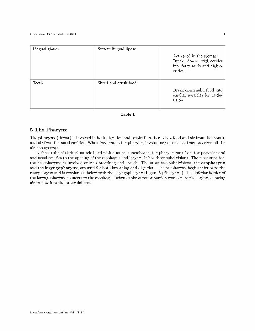
Lingual glands Secrete lingual lipase
Activated in the stomachBreak down triglyceridesinto fatty acids and diglyc-erides
Teeth Shred and crush food
Break down solid food intosmaller particles for deglu-tition
Table 1
5 The Pharynx
The pharynx (throat) is involved in both digestion and respiration. It receives food and air from the mouth,and air from the nasal cavities. When food enters the pharynx, involuntary muscle contractions close o� theair passageways.
A short tube of skeletal muscle lined with a mucous membrane, the pharynx runs from the posterior oraland nasal cavities to the opening of the esophagus and larynx. It has three subdivisions. The most superior,the nasopharynx, is involved only in breathing and speech. The other two subdivisions, the oropharynxand the laryngopharynx, are used for both breathing and digestion. The oropharynx begins inferior to thenasopharynx and is continuous below with the laryngopharynx (Figure 6 (Pharynx )). The inferior border ofthe laryngopharynx connects to the esophagus, whereas the anterior portion connects to the larynx, allowingair to �ow into the bronchial tree.
http://cnx.org/content/m46511/1.4/

OpenStax-CNX module: m46511 12
Pharynx
Figure 6: The pharynx runs from the nostrils to the esophagus and the larynx.
Histologically, the wall of the oropharynx is similar to that of the oral cavity. The mucosa includesa strati�ed squamous epithelium that is endowed with mucus-producing glands. During swallowing, theelevator skeletal muscles of the pharynx contract, raising and expanding the pharynx to receive the bolusof food. Once received, these muscles relax and the constrictor muscles of the pharynx contract, forcing thebolus into the esophagus and initiating peristalsis.
http://cnx.org/content/m46511/1.4/

OpenStax-CNX module: m46511 13
Usually during swallowing, the soft palate and uvula rise re�exively to close o� the entrance to thenasopharynx. At the same time, the larynx is pulled superiorly and the cartilaginous epiglottis, its mostsuperior structure, folds inferiorly, covering the glottis (the opening to the larynx); this process e�ectivelyblocks access to the trachea and bronchi. When the food �goes down the wrong way,� it goes into thetrachea. When food enters the trachea, the reaction is to cough, which usually forces the food up and outof the trachea, and back into the pharynx.
6 The Esophagus
The esophagus is a muscular tube that connects the pharynx to the stomach. It is approximately 25.4 cm(10 in) in length, located posterior to the trachea, and remains in a collapsed form when not engaged inswallowing. As you can see in Figure 7 (Esophagus ), the esophagus runs a mainly straight route throughthe mediastinum of the thorax. To enter the abdomen, the esophagus penetrates the diaphragm through anopening called the esophageal hiatus.
6.1 Passage of Food through the Esophagus
The upper esophageal sphincter, which is continuous with the inferior pharyngeal constrictor, controlsthe movement of food from the pharynx into the esophagus. The upper two-thirds of the esophagus consistsof both smooth and skeletal muscle �bers, with the latter fading out in the bottom third of the esophagus.Rhythmic waves of peristalsis, which begin in the upper esophagus, propel the bolus of food toward thestomach. Meanwhile, secretions from the esophageal mucosa lubricate the esophagus and food. Food passesfrom the esophagus into the stomach at the lower esophageal sphincter (also called the gastroesophagealor cardiac sphincter). Recall that sphincters are muscles that surround tubes and serve as valves, closing thetube when the sphincters contract and opening it when they relax. The lower esophageal sphincter relaxesto let food pass into the stomach, and then contracts to prevent stomach acids from backing up into theesophagus. Surrounding this sphincter is the muscular diaphragm, which helps close o� the sphincter whenno food is being swallowed. When the lower esophageal sphincter does not completely close, the stomach'scontents can re�ux (that is, back up into the esophagus), causing heartburn or gastroesophageal re�uxdisease (GERD).
http://cnx.org/content/m46511/1.4/

OpenStax-CNX module: m46511 14
Esophagus
Figure 7: The upper esophageal sphincter controls the movement of food from the pharynx to the
esophagus. The lower esophageal sphincter controls the movement of food from the esophagus to the
stomach.
6.2 Histology of the Esophagus
The mucosa of the esophagus is made up of an epithelial lining that contains non-keratinized, strati�edsquamous epithelium, with a layer of basal and parabasal cells. This epithelium protects against erosion
http://cnx.org/content/m46511/1.4/

OpenStax-CNX module: m46511 15
from food particles. The mucosa's lamina propria contains mucus-secreting glands. The muscularis layerchanges according to location: In the upper third of the esophagus, the muscularis is skeletal muscle. In themiddle third, it is both skeletal and smooth muscle. In the lower third, it is smooth muscle. As mentionedpreviously, the most super�cial layer of the esophagus is called the adventitia, not the serosa. In contrast tothe stomach and intestines, the loose connective tissue of the adventitia is not covered by a fold of visceralperitoneum. The digestive functions of the esophagus are identi�ed in Table 2.
Digestive Functions of the Esophagus
Action Outcome
Upper esophageal sphincter relaxation Allows the bolus to move from the laryngopharynxto the esophagus
Peristalsis Propels the bolus through the esophagus
Lower esophageal sphincter relaxation Allows the bolus to move from the esophagus intothe stomach and prevents chime from entering theesophagus
Mucus secretion Lubricates the esophagus, allowing easy passage ofthe bolus
Table 2
7 Deglutition
Deglutition is another word for swallowing�the movement of food from the mouth to the stomach. Theentire process takes about 4 to 8 seconds for solid or semisolid food, and about 1 second for very soft foodand liquids. Although this sounds quick and e�ortless, deglutition is, in fact, a complex process that involvesboth the skeletal muscle of the tongue and the muscles of the pharynx and esophagus. It is aided by thepresence of mucus and saliva. There are three stages in deglutition: the voluntary phase, the pharyngealphase, and the esophageal phase (Figure 8 (Deglutition )). The autonomic nervous system controls the lattertwo phases.
http://cnx.org/content/m46511/1.4/

OpenStax-CNX module: m46511 16
Deglutition
Figure 8: Deglutition includes the voluntary phase and two involuntary phases: the pharyngeal phase
and the esophageal phase.
7.1 The Voluntary Phase
The voluntary phase of deglutition (also known as the oral or buccal phase) is so called because you cancontrol when you swallow food. In this phase, chewing has been completed and swallowing is set in motion.The tongue moves upward and backward against the palate, pushing the bolus to the back of the oral cavityand into the oropharynx. Other muscles keep the mouth closed and prevent food from falling out. At thispoint, the two involuntary phases of swallowing begin.
7.2 The Pharyngeal Phase
In the pharyngeal phase, stimulation of receptors in the oropharynx sends impulses to the deglutition center(a collection of neurons that controls swallowing) in the medulla oblongata. Impulses are then sent backto the uvula and soft palate, causing them to move upward and close o� the nasopharynx. The laryngealmuscles also constrict to prevent aspiration of food into the trachea. At this point, deglutition apnea takesplace, which means that breathing ceases for a very brief time. Contractions of the pharyngeal constrictormuscles move the bolus through the oropharynx and laryngopharynx. Relaxation of the upper esophagealsphincter then allows food to enter the esophagus.
http://cnx.org/content/m46511/1.4/

OpenStax-CNX module: m46511 17
7.3 The Esophageal Phase
The entry of food into the esophagus marks the beginning of the esophageal phase of deglutition and theinitiation of peristalsis. As in the previous phase, the complex neuromuscular actions are controlled bythe medulla oblongata. Peristalsis propels the bolus through the esophagus and toward the stomach. Thecircular muscle layer of the muscularis contracts, pinching the esophageal wall and forcing the bolus forward.At the same time, the longitudinal muscle layer of the muscularis also contracts, shortening this area andpushing out its walls to receive the bolus. In this way, a series of contractions keeps moving food toward thestomach. When the bolus nears the stomach, distention of the esophagus initiates a short re�ex relaxationof the lower esophageal sphincter that allows the bolus to pass into the stomach. During the esophagealphase, esophageal glands secrete mucus that lubricates the bolus and minimizes friction.
: Watch this animation1 to see how swallowing is a complex processthat involves the nervous system to coordinate the actions of upper respiratory and digestive ac-tivities. During which stage of swallowing is there a risk of food entering respiratory pathways andhow is this risk blocked?
8 Chapter Review
In the mouth, the tongue and the teeth begin mechanical digestion, and saliva begins chemical digestion. Thepharynx, which plays roles in breathing and vocalization as well as digestion, runs from the nasal and oralcavities superiorly to the esophagus inferiorly (for digestion) and to the larynx anteriorly (for respiration).During deglutition (swallowing), the soft palate rises to close o� the nasopharynx, the larynx elevates, andthe epiglottis folds over the glottis. The esophagus includes an upper esophageal sphincter made of skeletalmuscle, which regulates the movement of food from the pharynx to the esophagus. It also has a loweresophageal sphincter, made of smooth muscle, which controls the passage of food from the esophagus to thestomach. Cells in the esophageal wall secrete mucus that eases the passage of the food bolus.
9 Interactive Link Questions
Exercise 1 (Solution on p. 20.)
Watch this animation2 to see how swallowing is a complex process that involves the nervoussystem to coordinate the actions of upper respiratory and digestive activities. During which stageof swallowing is there a risk of food entering respiratory pathways and how is this risk blocked?
10 Review Questions
Exercise 2 (Solution on p. 20.)
Which of these ingredients in saliva is responsible for activating salivary amylase?
1http://openstaxcollege.org/l/swallowing2http://openstaxcollege.org/l/swallowing
http://cnx.org/content/m46511/1.4/

OpenStax-CNX module: m46511 18
a. mucusb. phosphate ionsc. chloride ionsd. urea
Exercise 3 (Solution on p. 20.)
Which of these statements about the pharynx is true?
a. It extends from the nasal and oral cavities superiorly to the esophagus anteriorly.b. The oropharynx is continuous superiorly with the nasopharynx.c. The nasopharynx is involved in digestion.d. The laryngopharynx is composed partially of cartilage.
Exercise 4 (Solution on p. 20.)
Which structure is located where the esophagus penetrates the diaphragm?
a. esophageal hiatusb. cardiac ori�cec. upper esophageal sphincterd. lower esophageal sphincter
Exercise 5 (Solution on p. 20.)
Which phase of deglutition involves contraction of the longitudinal muscle layer of the muscularis?
a. voluntary phaseb. buccal phasec. pharyngeal phased. esophageal phase
11 Critical Thinking Questions
Exercise 6 (Solution on p. 20.)
The composition of saliva varies from gland to gland. Discuss how saliva produced by the parotidgland di�ers in action from saliva produced by the sublingual gland.
Exercise 7 (Solution on p. 20.)
During a hockey game, the puck hits a player in the mouth, knocking out all eight of his mostanterior teeth. Which teeth did the player lose and how does this loss a�ect food ingestion?
Exercise 8 (Solution on p. 20.)
What prevents swallowed food from entering the airways?
Exercise 9 (Solution on p. 20.)
Explain the mechanism responsible for gastroesophageal re�ux.
Exercise 10 (Solution on p. 20.)
Describe the three processes involved in the esophageal phase of deglutition.
12 References
van Loon FPL, Holmes SJ, Sirotkin B, Williams W, Cochi S, Hadler S, Lindegren ML. Morbidity and Mortal-ity Weekly Report: Mumps surveillance � United States, 1988�1993 [Internet]. Atlanta, GA: Center for Dis-ease Control; [cited 2013 Apr 3]. Available from: http://www.cdc.gov/mmwr/preview/mmwrhtml/00038546.htm3
3http://www.cdc.gov/mmwr/preview/mmwrhtml/00038546.htm
http://cnx.org/content/m46511/1.4/

OpenStax-CNX module: m46511 19
.
http://cnx.org/content/m46511/1.4/

OpenStax-CNX module: m46511 20
Solutions to Exercises in this Module
to Exercise (p. 17)Answers may vary.to Exercise (p. 17)Cto Exercise (p. 18)Bto Exercise (p. 18)Ato Exercise (p. 18)Dto Exercise (p. 18)Parotid gland saliva is watery with little mucus but a lot of amylase, which allows it to mix freely withfood during mastication and begin the digestion of carbohydrates. In contrast, sublingual gland saliva has alot of mucus with the least amount of amylase of all the salivary glands. The high mucus content serves tolubricate the food for swallowing.to Exercise (p. 18)The incisors. Since these teeth are used for tearing o� pieces of food during ingestion, the player will needto ingest foods that have already been cut into bite-sized pieces until the broken teeth are replaced.to Exercise (p. 18)Usually when food is swallowed, involuntary muscle contractions cause the soft palate to rise and close o�the nasopharynx. The larynx also is pulled up, and the epiglottis folds over the glottis. These actions blocko� the air passages.to Exercise (p. 18)If the lower esophageal sphincter does not close completely, the stomach's acidic contents can back up intothe esophagus, a phenomenon known as GERD.to Exercise (p. 18)Peristalsis moves the bolus down the esophagus and toward the stomach. Esophageal glands secrete mucusthat lubricates the bolus and reduces friction. When the bolus nears the stomach, the lower esophagealsphincter relaxes, allowing the bolus to pass into the stomach.
Glossary
De�nition 1: bolusmass of chewed food
De�nition 2: cementumbone-like tissue covering the root of a tooth
De�nition 3: crownportion of tooth visible superior to the gum line
De�nition 4: cuspid(also, canine) pointed tooth used for tearing and shredding food
De�nition 5: deciduous toothone of 20 �baby teeth�
De�nition 6: deglutitionthree-stage process of swallowing
De�nition 7: denstooth
http://cnx.org/content/m46511/1.4/

OpenStax-CNX module: m46511 21
De�nition 8: dentinbone-like tissue immediately deep to the enamel of the crown or cementum of the root of a tooth
De�nition 9: dentitionset of teeth
De�nition 10: enamelcovering of the dentin of the crown of a tooth
De�nition 11: esophagusmuscular tube that runs from the pharynx to the stomach
De�nition 12: faucesopening between the oral cavity and the oropharynx
De�nition 13: gingivagum
De�nition 14: incisormidline, chisel-shaped tooth used for cutting into food
De�nition 15: labiumlip
De�nition 16: labial frenulummidline mucous membrane fold that attaches the inner surface of the lips to the gums
De�nition 17: laryngopharynxpart of the pharynx that functions in respiration and digestion
De�nition 18: lingual frenulummucous membrane fold that attaches the bottom of the tongue to the �oor of the mouth
De�nition 19: lingual lipasedigestive enzyme from glands in the tongue that acts on triglycerides
De�nition 20: lower esophageal sphinctersmooth muscle sphincter that regulates food movement from the esophagus to the stomach
De�nition 21: molartooth used for crushing and grinding food
De�nition 22: oral cavity(also, buccal cavity) mouth
De�nition 23: oral vestibulepart of the mouth bounded externally by the cheeks and lips, and internally by the gums and teeth
De�nition 24: oropharynxpart of the pharynx continuous with the oral cavity that functions in respiration and digestion
De�nition 25: palatoglossal archmuscular fold that extends from the lateral side of the soft palate to the base of the tongue
De�nition 26: palatopharyngeal archmuscular fold that extends from the lateral side of the soft palate to the side of the pharynx
De�nition 27: parotid glandone of a pair of major salivary glands located inferior and anterior to the ears
De�nition 28: permanent toothone of 32 adult teeth
De�nition 29: pharynxthroat
http://cnx.org/content/m46511/1.4/

OpenStax-CNX module: m46511 22
De�nition 30: premolar(also, bicuspid) transitional tooth used for mastication, crushing, and grinding food
De�nition 31: pulp cavitydeepest portion of a tooth, containing nerve endings and blood vessels
De�nition 32: rootportion of a tooth embedded in the alveolar processes beneath the gum line
De�nition 33: salivaaqueous solution of proteins and ions secreted into the mouth by the salivary glands
De�nition 34: salivary amylasedigestive enzyme in saliva that acts on starch
De�nition 35: salivary glandan exocrine gland that secretes a digestive �uid called saliva
De�nition 36: salivationsecretion of saliva
De�nition 37: soft palateposterior region of the bottom portion of the nasal cavity that consists of skeletal muscle
De�nition 38: sublingual glandone of a pair of major salivary glands located beneath the tongue
De�nition 39: submandibular glandone of a pair of major salivary glands located in the �oor of the mouth
De�nition 40: tongueaccessory digestive organ of the mouth, the bulk of which is composed of skeletal muscle
De�nition 41: upper esophageal sphincterskeletal muscle sphincter that regulates food movement from the pharynx to the esophagus
De�nition 42: voluntary phaseinitial phase of deglutition, in which the bolus moves from the mouth to the oropharynx
http://cnx.org/content/m46511/1.4/