The molecular phylogeny of the type-species of Oodinium Chatton, 1912 (Dinoflagellata: Oodiniaceae),...
11
The molecular phylogeny of the type-species of Oodinium Chatton, 1912 (Dinoflagellata: Oodiniaceae), a highly divergent parasitic dinoflagellate with non-dinokaryotic characters Fernando Go ´mez • Alf Skovgaard Received: 10 October 2014 / Accepted: 20 November 2014 Ó Springer Science+Business Media Dordrecht 2014 Abstract Oodinium pouchetii (Lemmermann, 1899) Chatton, 1912, the first described parasitic dinoflagel- late, is the type of the Oodiniaceae Chatton, 1920. In the taxonomical schemes, this family of metazoan parasites includes Amyloodinium Brown & Hovasse, 1946 and Piscinoodinium Lom, 1981 that are respon- sible of important damages in fish aquaculture. Species of Oodinium Chatton, 1912 have unique characteristics such as the possession of both non- dinokaryotic and dinokaryotic nuclei within the life- cycle, and the absence of the transversal (cingulum) and longitudinal (sulcus) surface grooves in the parasitic stage. We provide the first molecular data for the genus Oodinium from specimens of O. pouchetii infecting the chordate Oikopleura sp. (Tu- nicata: Appendicularia) off the coasts of Brazil. Although O. pouchetii lacks dinokaryotic characters in the parasitic stage, the SSU rDNA phylogeny revealed that it forms a distinct fast-evolved clade that branches among the dinokaryotic dinoflagellates. However, there is no clear relationship with other dinoflagellates. Hence, the taxonomic affinity of the family Oodiniaceae is unclear at the moment. Introduction A large number of dinoflagellates are known to parasitise marine vertebrates and invertebrates (e.g. Chatton, 1920; Cachon & Cachon, 1987; Shields, 1994). The first parasitic dinoflagellate to be described was Oodinium pouchetii (Lemmermann, 1899) Chat- ton, 1912 (= Gymnodinium pulvisculus (Ehrenberg, 1832) Stein, 1878 sensu Pouchet, 1885), an ectopar- asite on the tail of the tunicate appendicularian Oikopleura dioica Fol (see Pouchet, 1884, 1885). The genus Oodinium Chatton, 1912 also includes another parasite of appendicularians, O. fritillariae Chatton, 1912, the parasite of annelids O. dogielii J. Cachon & M. Cachon, 1971, the parasites of chaetog- naths O. jordanii McLean & Nielsen, 1989 and O. inlandicum Horiguchi & Ohtsuka, 2001, and an undescribed species that parasitises ctenophores and cnidarians (Chatton, 1912; Cachon & Cachon, 1971; Electronic supplementary material The online version of this article (doi:10.1007/s11230-014-9538-8) contains supple- mentary material, which is available to authorized users. F. Go ´mez (&) Laboratory of Plankton Systems, Oceanographic Institute, University of Sa ˜o Paulo, Prac ¸a do Oceanogra ´fico 191, Cidade Universita ´ria, Butanta ˜, Sa ˜o Paulo 05508-900, Brazil e-mail: fernando.gomez@fitoplancton.com A. Skovgaard Department of Veterinary Disease Biology, Faculty of Health and Medical Sciences, University of Copenhagen, Stigbøjlen 7, 1870 Frederiksberg C, Denmark Present Address: A. Skovgaard Department of Environmental, Social and Spatial Change, University of Roskilde, 4000 Roskilde, Denmark 123 Syst Parasitol (2015) 90:125–135 DOI 10.1007/s11230-014-9538-8
-
Upload
sarah-cobb -
Category
Documents
-
view
22 -
download
7
description
Oodinium pouchetii (Lemmermann, 1899)Chatton, 1912, the first described parasitic dinoflagellate,is the type of the Oodiniaceae Chatton, 1920. Inthe taxonomical schemes, this family of metazoanparasites includes Amyloodinium Brown & Hovasse,1946 and Piscinoodinium Lom, 1981 that are responsibleof important damages in fish aquaculture.Species of Oodinium Chatton, 1912 have uniquecharacteristics such as the possession of both nondinokaryoticand dinokaryotic nuclei within the lifecycle,and the absence of the transversal (cingulum)and longitudinal (sulcus) surface grooves in theparasitic stage. We provide the first molecular datafor the genus Oodinium from specimens of O.pouchetii infecting the chordate Oikopleura sp. (Tunicata:Appendicularia) off the coasts of Brazil.Although O. pouchetii lacks dinokaryotic charactersin the parasitic stage, the SSU rDNA phylogenyrevealed that it forms a distinct fast-evolved clade thatbranches among the dinokaryotic dinoflagellates.However, there is no clear relationship with otherdinoflagellates. Hence, the taxonomic affinity of thefamily Oodiniaceae is unclear at the moment.
Transcript of The molecular phylogeny of the type-species of Oodinium Chatton, 1912 (Dinoflagellata: Oodiniaceae),...

The molecular phylogeny of the type-species of OodiniumChatton, 1912 (Dinoflagellata: Oodiniaceae), a highlydivergent parasitic dinoflagellate with non-dinokaryoticcharacters
Fernando Gomez • Alf Skovgaard
Received: 10 October 2014 / Accepted: 20 November 2014
� Springer Science+Business Media Dordrecht 2014
Abstract Oodinium pouchetii (Lemmermann, 1899)
Chatton, 1912, the first described parasitic dinoflagel-
late, is the type of the Oodiniaceae Chatton, 1920. In
the taxonomical schemes, this family of metazoan
parasites includes Amyloodinium Brown & Hovasse,
1946 and Piscinoodinium Lom, 1981 that are respon-
sible of important damages in fish aquaculture.
Species of Oodinium Chatton, 1912 have unique
characteristics such as the possession of both non-
dinokaryotic and dinokaryotic nuclei within the life-
cycle, and the absence of the transversal (cingulum)
and longitudinal (sulcus) surface grooves in the
parasitic stage. We provide the first molecular data
for the genus Oodinium from specimens of O.
pouchetii infecting the chordate Oikopleura sp. (Tu-
nicata: Appendicularia) off the coasts of Brazil.
Although O. pouchetii lacks dinokaryotic characters
in the parasitic stage, the SSU rDNA phylogeny
revealed that it forms a distinct fast-evolved clade that
branches among the dinokaryotic dinoflagellates.
However, there is no clear relationship with other
dinoflagellates. Hence, the taxonomic affinity of the
family Oodiniaceae is unclear at the moment.
Introduction
A large number of dinoflagellates are known to
parasitise marine vertebrates and invertebrates (e.g.
Chatton, 1920; Cachon & Cachon, 1987; Shields,
1994). The first parasitic dinoflagellate to be described
was Oodinium pouchetii (Lemmermann, 1899) Chat-
ton, 1912 (= Gymnodinium pulvisculus (Ehrenberg,
1832) Stein, 1878 sensu Pouchet, 1885), an ectopar-
asite on the tail of the tunicate appendicularian
Oikopleura dioica Fol (see Pouchet, 1884, 1885).
The genus Oodinium Chatton, 1912 also includes
another parasite of appendicularians, O. fritillariae
Chatton, 1912, the parasite of annelids O. dogielii J.
Cachon & M. Cachon, 1971, the parasites of chaetog-
naths O. jordanii McLean & Nielsen, 1989 and O.
inlandicum Horiguchi & Ohtsuka, 2001, and an
undescribed species that parasitises ctenophores and
cnidarians (Chatton, 1912; Cachon & Cachon, 1971;
Electronic supplementary material The online version ofthis article (doi:10.1007/s11230-014-9538-8) contains supple-mentary material, which is available to authorized users.
F. Gomez (&)
Laboratory of Plankton Systems, Oceanographic Institute,
University of Sao Paulo, Praca do Oceanografico 191,
Cidade Universitaria, Butanta, Sao Paulo 05508-900,
Brazil
e-mail: [email protected]
A. Skovgaard
Department of Veterinary Disease Biology, Faculty of
Health and Medical Sciences, University of Copenhagen,
Stigbøjlen 7, 1870 Frederiksberg C, Denmark
Present Address:
A. Skovgaard
Department of Environmental, Social and Spatial Change,
University of Roskilde, 4000 Roskilde, Denmark
123
Syst Parasitol (2015) 90:125–135
DOI 10.1007/s11230-014-9538-8

McLean & Nielsen, 1989; Mills & McLean, 1991;
Horiguchi & Ohtsuka, 2001). The oodinids that
parasitise fishes have been removed from the genus
Oodinium. Oodinium pillularis Schaperclaus, 1954
and O. limneticum Jacobs, 1946 have been transferred
to the genus Piscinoodinium Lom, 1981 and O.
ocellatum Brown, 1931 has been transferred to
Amyloodinium Brown & Hovasse, 1946, together with
the parasite of salps Amyloodinium amylaceum (Barg-
oni, 1894) Brown & Hovasse, 1946 (see Brown &
Hovasse, 1946; Lom, 1981). Oodinium cyprinodon-
tum Lawler, 1967 has been transferred to Crepidoodi-
nium Lom & Lawler, 1981, a genus that includes other
two species (Lom et al., 1993). Another parasite of
fishes, the type-species of the monotypic genus
Oodinioides Reichenbach-Klinke, 1970, is also clas-
sified within the family Oodiniaceae Chatton, 1920
(see Cachon & Cachon, 1987). Species of the genera
that infect fishes cause great damage in aquaculture
(Lauckner, 1984).
Historically, most species of parasitic dinoflagel-
lates have been classified as members of the orders
Blastodiniales or Syndiniales (Fensome et al., 1993).
Molecular phylogeny has demonstrated that Blastodi-
niales is an artificial assemblage with several of its
members now distributed among the dinokaryotic
dinoflagellates (Litaker et al., 1999; Saldarriaga et al.,
2001; Kuhn & Medlin, 2005; Levy et al., 2007;
Skovgaard et al., 2007; Gomez et al., 2009a; Coats
et al., 2010; Gomez & Skovgaard, 2014). Even genera
such as Amyloodinium and Piscinoodinium, classified
within the family Oodiniaceae, belong to different
phylogenetic clades. Amyloodinium ocellatum
branches with thecate dinoflagellates, the fish-killer
pfiesterids and cryptoperidiniopsoids, the parasites of
diatoms of the genus Paulsenella Chatton, 1920 and
the parasites of tintinnid ciliates of the genus Tintinn-
ophagus Coats, 2010 (see Litaker et al., 1999; Kuhn &
Medlin, 2005; Coats et al., 2010). Piscinoodinium spp.
branch with the so-called ‘thin-walled’ dinoflagellates
with mutualistic symbionts of reef-forming inverte-
brates and planktonic rhizarians (Pelagodinium Siano,
Montresor, Probert & de Vargas, 2010; Symbiodinium
Freudenthal, 1962), and free-living photosynthetic
species (Polarella Montresor, Procaccini & Stoecker,
1999 and Biecheleria Moestrup, Lindberg & Daugb-
jerg, 2009) (see Levy et al., 2007; Siano et al., 2010).
Whether one or both of the clades Amyloodinium-
pfiesterids or Piscinoodinium-‘thin-walled’ dinoflagellates
belongs to the family Oodiniaceae depends on the
phylogenetic position of the type Oodinium pouchetii.
However, up to date no sequence from the type-genus
of Oodiniaceae is available.
The heterotrophic dinoflagellates of the genus
Oodinium have been the subject of numerous cyto-
logical investigations that revealed the possession of
both non-dinokaryotic and dinokaryotic nuclei within
the life-cycle (Chatton, 1920; Hovasse, 1935; Cachon
& Cachon, 1971, 1977; McLean & Nielsen, 1989;
Horiguchi & Ohtsuka, 2001). During the early vege-
tative stage, the cell is ovoid and attached to its host by
a peduncle through which adsorption of metabolites
takes place (Cachon & Cachon, 1971). It lacks typical
dinoflagellate characters such as flagella and trans-
verse or longitudinal grooves. On the other hand, it has
organelles such as trichocysts and a pusule. At this
stage, the parasite and its nucleus grow tremendously
without any cellular division. The cell covering of the
trophont comprises thecal plates. However, assign-
ment of these plates into conventional Kofoid’s
tabulation system is difficult, due to the lack of
conventional reference points for the recognition of
plate series (Hovasse, 1935; Horiguchi & Ohtsuka,
2001). All these unique characters make Oodinium a
key genus for inferring dinoflagellate evolution.
During plankton surveys along the coasts of Brazil,
specimens of the appendicularian Oikopleura sp.
infected with Oodinium pouchetii were collected. This
study describes the life-cycle, with the first micrographs
of the dinospores, and provides the first molecular data
for O. pouchetii, the type-species of the family Oodi-
niaceae and the first described parasitic dinoflagellate.
Materials and methods
Sampling and isolation of materials
Specimens of O. pouchetii were isolated in the South
Atlantic Ocean at two locations off the coast of Sao
Paulo State, Brazil, at Sao Sebastiao Channel
(23�5004.0500S, 45�24028.820W) in July 2013 and off
Ubatuba (23�32020.1500S, 45�5058.9400W) in June
2014. The specimens were collected from the surface
using a phytoplankton net (20 lm mesh size). The live,
concentrated samples were examined in Utermohl
chambers at magnification of 920 with an inverted
microscope (Eclipse TS-100, Nikon, Tokyo) and
photographed with digital camera (Cyber-shot DSC-
126 Syst Parasitol (2015) 90:125–135
123
W300, Sony, Tokyo) mounted on the microscope
eyepiece. Trophonts that had detached before the first
division were micropipetted individually with a fine
capillary into a clean chamber and washed several
times in a series of drops of 0.2 lm-filtered and
sterilised seawater. Finally, trophonts of O. pouchetii
were placed in 0.2 ml Eppendorf tubes filled with
several drops of absolute ethanol. We used naturally
detached trophonts instead of cutting the attachment
peduncle of trophonts still attached to the host. In the
case of other ectoparasites that do not naturally
detached from the host (i.e. species of Ellobiopsis
Caullery, 1910), this manipulation increases the
probability of contamination with DNA from the
damaged host (Gomez et al., 2009a). Samples were
kept at room temperature and in darkness until
molecular analysis could be performed.
Life-cycle observations
After obtaining samples for DNA analysis, specimens
of O. pouchetii were used to investigate cell division,
and the morphology and behaviour of the dinospores.
Recently detached trophonts prior the first division
were individually placed in Utermohl chambers with
0.2 lm-filtered seawater, and periodically observed
under the microscope. Other recently detached troph-
onts were individually placed in 12-well tissue culture
plates with 0.2 lm-filtered seawater. In order to have
controlled environmental conditions, the plates were
placed in an incubator used for microalgae culturing,
at 23�C, 100 lmol photons m-2s-1 from cool-white
tubes and 12:12 h Light:Dark photoperiod. The cell
division and the swarmers were periodically photo-
graphed under light microscope.
PCR amplification and sequencing
The sample tubes containing specimens of O. pouch-
etii in ethanol were centrifuged and dried by placing
them overnight in a desiccator at room temperature.
Then 30 ll of sterile DNase-free water was added to
each sample tube and the samples were sonicated
through three 10-second pulses at an output setting of
1.0 (Coats et al., 2010) using a Virsonic 600 sonicator
(SP Scientific, Gardiner, NY) equipped with a micro-
tip. Ten microlitres of the crude cell lysate was used
for each polymerase chain reaction (PCR) amplifica-
tion. SSU rDNA was amplified using the primers
EukA and EukB (Medlin et al., 1988). A single PCR
was in one case (isolate #58) sufficient for obtaining a
PCR product suitable for sequencing. In two cases
(isolates #23 and #54), a nested PCR was performed
using 0.5 ll of the initial PCR product as template for a
second PCR with the newly-designed primers Ask1F
(5’-GAT TAA GCC ATG CAT GTC TCA G-3’) and
Ask2R (5’-GAA ACC TTG TTA CGA CTT CTC
C-3’). All PCR amplifications were performed in 25 ll
reaction volumes containing 1.25 unit of Biotaq
polymerase (Bioline Reagents Limited, London,
UK), buffer supplied with the polymerase, MgCl2 at
3.0 mM, dNTPs at 1.6 mM, and the forward and
reverse primers at 1.0 mM. The PCR was run in a
T100TM Thermal Cycler (Bio-Rad Laboratories, Her-
cules, CA) under the following conditions: initial
denaturation (94�C/2 min); 35 cycles of denaturation
(94�C/15 s), annealing (57�C/30 s), and extension
(72�C/2 min); final extension (72�C/7 min). Nested
PCR was run as touchdown PCR for 30 cycles: initial
denaturation (94�C/2 min); then 10 cycles of ‘touch-
down’ PCR with denaturation (94�C/15 s), 10 anneal-
ing steps (30 s) decreasing from 65 down to 56�C (1�C
decrease with each cycle), and extension (72�C/2
min); followed by 20 cycles of denaturation (94�C/15
sec), annealing (55�C/30 sec), and extension (72�C/2
min); and final extension (72�C/7 min). PCR products
were purified using Illustra GFX PCR DNA and Gel
Purification Kit (GE Healthcare, Little Chalfont, UK)
and sequenced bi-directionally with an ABI3730xl
sequencer (Macrogen Europe, Amsterdam, The Neth-
erlands) using the same primers as used for PCR and
additional internal primers [ND2F, ND7R, ND9R
(Ekelund et al., 2004); 528f (Elwood et al., 1985);
1209f (Giovannoni et al., 1988)]. Sequence reads were
aligned and assembled using the software ChromasPro
1.75 (Technelysium, Brisbane, Australia).
Phylogenetic analyses
Three phylogenetic analyses were performed based on
nearly complete SSU rDNA sequences. One analysis
(Alveolata tree) was based on an alignment of 73
sequences for species of the major alveolate groups
plus four cercozoan sequences serving as the out-
group. The second analysis (Dinokaryota tree) com-
prised sequences for dinokaryotes most similar to O.
pouchetii as identified through BLAST search (http://
blast.ncbi.nlm.nih.gov/Blast.cgi; Altschul et al.,
1997). Furthermore, sequences of a wide selection of
dinokaryotes were included, aiming at including spe-
cies of all mutualist symbiotic and parasitic
Syst Parasitol (2015) 90:125–135 127
123
dinokaryote genera for which sequences were avail-
able. Two perkinsid and two syndinean sequences
were used as outgroups. The final matrix contained 64
sequences. The Dinokaryota tree analysis was repe-
ated with the addition of the two shorter O. pouchetii
sequences and three short sequences with highest
similarity to O. pouchetii according to a BLAST
search.
Sequences were aligned using Clustal X v2.1
(Larkin et al., 2007) and non-informative sites were
removed using Gblocks (Castresana, 2000) with
parameters set for less stringent conditions (minimum
number of sequences for a flanking position: 28;
minimum length of a block: 5; allow gaps in half
positions). Final alignments of the SSU rDNA
sequences spanned over 1,630 and 1,737 positions
(Alveolata and Dinokaryota trees, respectively).
Bayesian phylogenetic trees were constructed with
MrBayes v3.2 (Huelsenbeck & Ronquist, 2001).
MrBayes settings for the best-fit model (GTR?I?G)
were selected by AIC in MrModeltest 2.3 (Nylander,
2004). Four simultaneous Monte Carlo Markov chains
were run from random trees for a total of 2,000,000
generations in two parallel runs. A tree was sampled
every 100 generations, and the first 2,000 trees (burn-
in) were discarded before calculating posterior prob-
abilities and constructing Bayesian consensus trees.
The newly-generated sequences were deposited in
DDBJ/EMBL/GenBank under accession numbers
KM879217–KM879219.
Results
Life-cycle
Specimens of the appendicularian Oikopleura sp. were
observed with ectoparasitic trophonts of O. pouchetii.
The tails of the appendicularians contained up to
sixteen trophonts of different sizes (Fig. 1A–B).
Larger trophonts (up to 200 lm) showed dark brown
pigmentation and an oval shape (Fig. 1C–D); smaller
specimens were pyriform with yellowish pigmentation
(Fig. 1A–B; see video at http://youtu.be/DlCPkpK7
oSM). In younger specimens, the nucleus was distally
placed (opposite side of the attachment peduncle),
while the nucleus was centrally located in the larger
specimens. These large trophonts with dark brown
pigmentation were usually found detached from the
host at the bottom of the settling chambers; these were
isolated for PCR analysis. There was a high proportion
of detached mature trophonts on the bottom of the
settling chambers compared with those still attached to
the hosts. This may suggest that the manipulation due
to plankton net sampling and/or the stress or damage
of the captured hosts induce the final of the trophic life
when the parasite leaves its host by breaking its
peduncle attachment. The rupture of the stalk always
preceded sporogenesis. When the plankton sample
was allowed to settle for several hours, aggregations
of c.200 sporocysts were observed (Fig. 1Q–R).
Detached trophonts were individually placed in fil-
tered seawater in order to observe the different steps of
the sporogenesis process avoiding the influence of
other organisms and under controlled environmental
conditions. The divisions proceeded without inter-
ruption to form progressively smaller sporocysts that
eventually became liberated as flagellated dinospores
after 8–12 hours (Fig. 1F–W). The palintomic sporo-
genesis began with the retraction of the cell cytoplasm
(Fig. 1F, H). The cleavage of the trophont was longi-
tudinal (slightly oblique to the axis of the attachment
peduncle) (Fig. 1G, I). In the first generation, the two
daughter cells sometimes remained inside the theca
(Fig. 1K) and later they left behind an empty theca
(Fig. 1J). Further divisions always occurred outside
the theca. The nucleus divided before the cytoplasmic
furrow reached the nuclear level. The cell cytokinesis
began before the complete division of the nuclei
(Fig. 1L, M). Different steps of the development of the
first generation of daughter cells are shown in
Figs. 1G, I, K–O and the second generation of
daughter cells are illustrated in Fig. 1P–O. The dark
brown pigmentation of the adult trophont was pro-
gressively diluted along the successive divisions. This
suggested that this pigmented substance is a reserve
compound consumed during the sporogenesis. The
sporocyst generations followed one after the other
without any compensating growth until swarmers with
dinoflagellate characteristics appeared. The sporo-
genic divisions proceeded without pause and were
synchronic in the first generations. However, this
synchrony was lost in the last steps of the sporogenesis
(Fig. 1Q, S). This made it difficult to account the final
number of infective dinospores produced by each
trophont because the dinospores, swarmers, began to
swim when sporocysts were yet devoid of flagella. An
aggregation of c.200 immotile sporocysts was
128 Syst Parasitol (2015) 90:125–135
123

observed after eight hours at a temperature of 23�C
(Fig. 1R). In the last step, these immotile sporocysts
divided again and formed a couple of dinospores each
that developed two flagella. The couple of dinospores
remained together, and began to move the flagella
(Fig. 1S, T). The transversal flagellum first vibrated
Fig. 1 Life-cycle stages of the ectoparasite Oodinium pouchetii from the coast of Brazil. Ectoparasitic phase of young trophonts
infecting Oikopleura sp. (A–E) and phase of multiplication (palintomic sporogenesis) (D–W). A, B, Young trophonts; C–E, Mature
trophont; F–H, Recently detached trophonts (note the retraction of the cytoplasm inside the theca and the large round nucleus); G, I, K,
L–O. First generation of daughter cells; G–I, The cleavage is longitudinal, slightly oblique to the axis of the attachment peduncle. The
daughter cells and the nuclei are oval; J, Empty theca; K–O, Division of the first generation; P, Note that cell division begins with
segmentation of the cell; P, Q, Second generation of daughter cells; R, Sporocysts of the penultimate generation; S, Sporocysts of the
penultimate generation (large) and sporocysts of the last generation that formed each a pair of swarmers. The inset shows the last
division; T–W, Recently formed dinospores, swarmers, forming couples; U, V, Note the longitudinal flagellum (lf), and the transversal
flagellum (tf) that vibrates outside the cingulum. Scale-bars: E–R, 20 lm; S–W, 10 lm
Syst Parasitol (2015) 90:125–135 129
123
outside the cingulum, but eventually beat inside the
cingulum when the cells began to swim (Fig. 1U–W;
see video at http://youtu.be/DlCPkpK7oSM). The
swarmers began to disperse while other dinospores were
yet devoid of flagella. The small dinospores swam with
the typical dinoflagellate rotation, following a straight
trajectory at different levels in the settling chamber, and
with sudden accelerations. This feature made it difficult
to record the trajectories at high magnification. We
counted c.200 immotile sporocysts from each detached
trophont. Subsequently each immotile sporocyst divi-
ded into two swarmers. With this last division the total
number of swarmers from each detached trophont
would be 512, implying nine successive binary divisions
(29 = 512). The swarmers (c.9 lm long and c.7 lm wide)
showed a hemispherical contour of the hyposome and an
episome more conical with a round apex, and a well-
marked cingulum. The swarmers contained small yel-
low-greenish body inclusions, and a round nucleus
located in the centre of the cell (Fig. 1T–W).
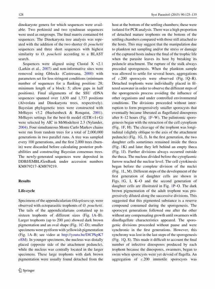
Molecular phylogeny
Three partial SSU rDNA sequences (up to 1,713 bp)
for O. pouchetii were obtained: one sequence from
detached trophonts isolated at Sao Sebastiao Channel
on 10 July 2013 (isolate #23; KM879217), and two
sequences from detached trophonts isolated off Uba-
tuba on 6 June 2014 (isolate #54; KM879218) and on
13 June 2014 (isolate #58; KM878219). The three
sequences were identical. A BLAST search was
conducted on the new sequences to find related
sequences in the GenBank database. Initial BLAST
comparisons showed that, with the exception of the
partial environmental sequence (643 bp; EF539018,
retrieved from the western Pacific coast, which shared
99% identity with O. pouchetii), the closest identified
relatives in the database were dinokaryotes such as the
thecate parasites Blastodinium spp. (JX473665 and
DQ317538) and Duboscquodinium collinii Grasse,
1952 (HM483399), and free-living thecate species
[e.g. Peridinium umbonatum Stein, 1883 (GU001637)
and Scrippsiella trochoidea (Stein, 1883) Balech ex
Loeblich III, 1965 (HM483396)]. However, similari-
ties were low in all cases (84%).
We studied the phylogenetic position of O. pouch-
etii in three SSU rDNA phylogenetic trees. The first
tree contained diverse representatives of the alveolate
lineages, with ciliates, apicomplexans, perkinsids,
euduboscquellids (Marine Alveolate Group I), syn-
dineans (Marine Alveolate Group II) and dinokary-
otes. This tree (Supplementary file 1, Fig. S1) placed
unequivocally O. pouchetii within the dinokaryotic
lineage. Then, we studied the phylogenetic position of
O. pouchetii using a dataset including a variety of
dinoflagellate sequences, especially with representa-
tives of the parasitic dinokaryotes, including the
oodinioid dinoflagellates (species of Amyloodinium
and Piscinoodinium), other parasitic dinoflagellates,
and a diverse representation of the dinokaryotic
lineages, and rooted using perkinsid and syndinean
sequences as an outgroup (Fig. 2). We carried out an
additional analysis that also included shorter
sequences, especially partial SSU rDNA sequences
of environmental clones and other known dinoflagel-
lates (Supplementary file 1, Fig. S2). The clade of O.
pouchetii in the resulting tree is included as an inset in
Fig. 2.
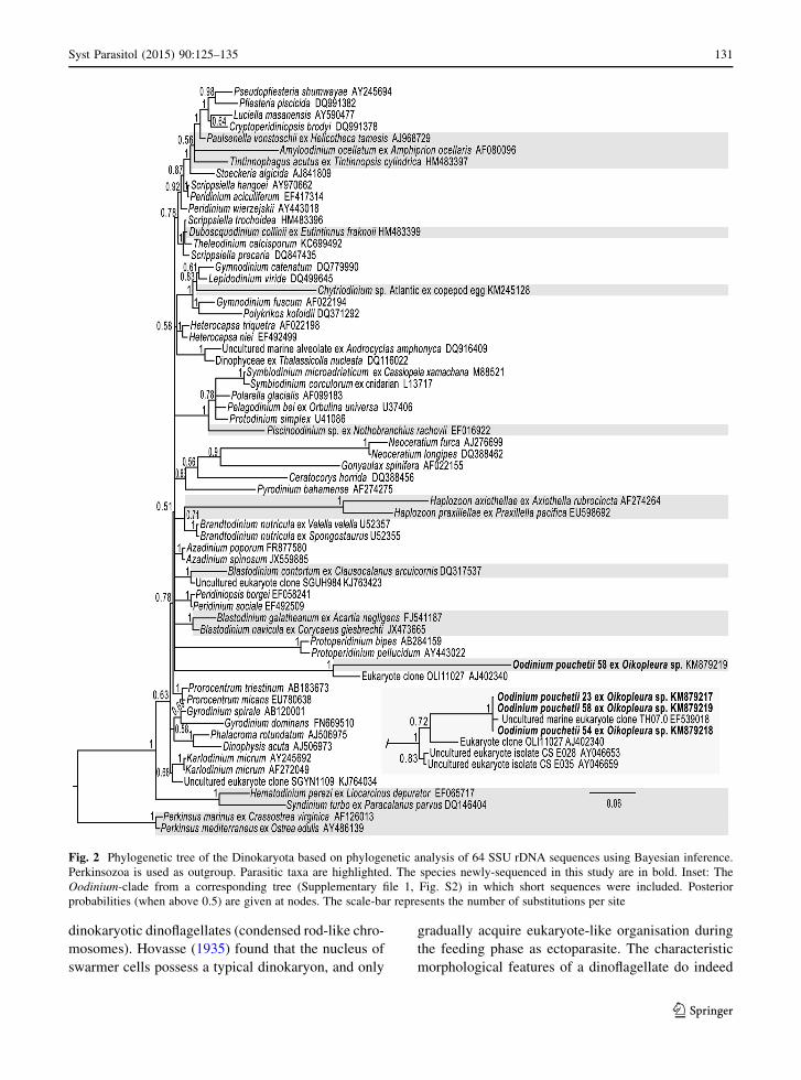
In the Bayesian consensus tree (Fig. 2), the SSU
rDNA phylogeny revealed that sequences for Oodini-
um spp. formed a distinct clade among the dinokaryotic
dinoflagellates. The three sequences for O. pouchetii
formed a highly supported lineage (maximum poster-
ior probability value) with an environmental sequence
(EF539018) from the port of Hong Kong, Western
Pacific. In the tree containing short sequences (Sup-
plementary file 1, Fig. S2), this lineage branched with
strong support with an environmental sequence from
the sub-surface waters of the equatorial Pacific
(AJ402340), and two environmental sequences
(AY046653; AY046659) from the surface waters of
the South Pacific Ocean. The Oodinium clade branched
within the dinokaryotic dinoflagellates. However, it
was not possible to find any close genetically charac-
terised relatives of the family Oodiniaceae.
Discussion
The life-cycle of the trophont and the morphology of
the dinospores of O. pouchetii studied here coincide
with the original description of O. pouchetii. Pouchet
(1885, figure 27) illustrated a pair of recently formed
dinospores with long flagella and a cell contour similar
to those observed in the present specimens (Fig. 1T–
U). Chatton (1920) demonstrated that along the
growth of the Oodinium trophont, it progressively
losses the typical nuclear characteristic of the
130 Syst Parasitol (2015) 90:125–135
123
dinokaryotic dinoflagellates (condensed rod-like chro-
mosomes). Hovasse (1935) found that the nucleus of
swarmer cells possess a typical dinokaryon, and only
gradually acquire eukaryote-like organisation during
the feeding phase as ectoparasite. The characteristic
morphological features of a dinoflagellate do indeed
Fig. 2 Phylogenetic tree of the Dinokaryota based on phylogenetic analysis of 64 SSU rDNA sequences using Bayesian inference.
Perkinsozoa is used as outgroup. Parasitic taxa are highlighted. The species newly-sequenced in this study are in bold. Inset: The
Oodinium-clade from a corresponding tree (Supplementary file 1, Fig. S2) in which short sequences were included. Posterior
probabilities (when above 0.5) are given at nodes. The scale-bar represents the number of substitutions per site
Syst Parasitol (2015) 90:125–135 131
123
disappear during the vegetative stage and can only be
seen again in the last stages of sporogenesis. Conse-
quently, O. pouchetii alternates between ultrastructure
features of basal dinoflagellates and dinokaryotes.
Also the trophonts of Noctiluca scintillans (Macart-
ney, 1810) Kofoid, 1920 do not retain the shared
characters of typical dinoflagellates such as a trans-
verse flagellum and permanently condensed chromo-
somes. The swarmers of species of Noctiluca Suriray,
1836 maintain the dinokaryotic characteristics includ-
ing two grooves, slightly differentiated flagella and
condensed chromosomes (Fukuda & Endoh, 2006).
Species of Noctiluca and other noctilucoids (species of
Spatulodinium J. Cachon & M. Cachon, 1968; Kofo-
idinium Pavillard, 1928) do not branch within the
dinokaryotic lineage (Gomez et al., 2010). Despite the
absence of dinokaryotic characters in the parasitic
phase, the molecular phylogeny reveals that O.
pouchetii is truly a member of the dinokaryotic
lineage.
The type of life-cycle has traditionally been used
for classification of the parasitic dinoflagellates into
families (Fensome et al., 1993). However, molecular
phylogeny has revealed that species of closely related
genera (i.e. Dissodinium Klebs, 1916 and Chytriodi-
nium Chatton, 1912) have different life-cycles
(Gomez et al., 2009a). The morphology of dinospores
seems to be more informative for the classification of
these parasites. However, it is not always easy to
observe the detailed morphology of the small swar-
mers when compared with the trophonts. For example,
before Skovgaard et al. (2007), the thecate swarmers
of Blastodinium Chatton, 1906 were usually referred
to as naked, gymnodinioid dinoflagellates (Chatton,
1920; Taylor, 2004). The swarmers of Amyloodinium
ocellatum were first described as naked gymnodinioid
dinoflagellate (Brown, 1934; Lom, 1981), but further
analysis with scanning electron microscopy revealed
that the swarmers were thecate (Landsberg et al.,
1994). Paulsenella vonstoschii Drebes & Schnepf
branches within a clade of thecate dinoflagellate.
However, the thecal plates have not yet been reported
(Kuhn & Medlin, 2005). Due to instrumental con-
straints, we were not able to discern the tabulation of
the dinospores of O. pouchetii. The thecal plates of the
trophonts of this species consist of four equatorial
series and are thin, with no visible ornamentation
(Hovasse, 1935). The assignment of these plates into
conventional Kofoid’s tabulation system is difficult,
due to the lack of conventional reference points (apical
pore plate, cingulum or sulcus) for the recognition of
plate series.
To date, Amyloodinium is still classified within the
family Oodiniaceae, but we have here shown that it is
distantly related to the type-species of the family.
Hence, O. pouchetii remains the only morphologically
identified member of Oodiniaceae with a sequence in
the GenBank database. Several environmental
sequences are related to O. pouchetii and thus may
represent potential unidentified members of the Oodi-
niaceae (Fig. 2; Supplementary file 1, Fig. S2).
Assuming that these environmental clones are not
artefacts and correspond to true organisms, unidenti-
fied relatives of Oodinium spp. are present in different
marine habitats. An environmental sequence from
coastal eutrophic waters unequivocally corresponded
to O. pouchetii (see Cheung et al., 2008). Sequences
for relatives of O. pouchetii were retrieved from
surface and subsurface waters of the oligotrophic
waters of the Pacific Ocean (Moon-van der Staay et al.,
2001; Lie et al., 2014). We do not know whether these
environmental clones branching with O. pouchetti are
parasitic or free-living organisms. Our results suggest
that there are dinoflagellates belonging to the family
Oodiniaceae that have not yet been morphologically
and ecologically characterised.
We must conclude that the parasite of fishes,
Amyloodinium and Piscinoodinium, should not be
placed in the family Oodiniaceae. In the classical
taxonomical schemes, Fensome et al. (1993) grouped
under the Oodiniaceae ectoparasites with suboval to
fusiform in outline trophonts, with a well-developed
peduncle or invasive organ that consists of a complex
rhizoid-like absorptive structure and with reproduc-
tion by palintomic sporogenesis. Another character of
this family is the possession of both non-dinokaryotic
and dinokaryotic nuclei within the life-cycle (Fen-
some et al., 1993). There is no evidence for the
presence of non-dinokaryotic nuclei in species of
Amyloodinium and Piscinoodinium. The thecate Am-
yloodinium ocellatum branched with other parasites
such as Paulsenella vonstoschii and Tintinnophagus
acutus Coats (as in Coats et al., 2010). The trophont of
O. pouchetii is also covered by thecal plates (Hovasse,
1935), and we can expect a relationship between these
taxa. Amyloodinium ocellatum can be distinguished
from O. pouchetii by the possession of rhizoid- and
root-like processes for attachment and by the
132 Syst Parasitol (2015) 90:125–135
123
production of starch grains. Species of the genus
Oodinium, on the other hand, are characterised by the
possession of a disk, rather than rhizoids, for attach-
ment and by the lack of starch grains (Brown &
Hovasse, 1946). Several of the congeneric species that
parasitise other metazoan groups show marked mor-
phological differences in comparison with the type-
species O. pouchetii (see McLean & Nielsen, 1989;
Mills & McLean, 1991; Horiguchi & Ohtsuka, 2001).
This raises questions of the correct generic affiliation
of these species. Other parasitic genera with some
morphological resemblance such as Cachonella Rose
& Cachon, 1952, Crepidoodinium, Protoodinium
Hovasse, 1935 or Oodinioides also lack molecular
information. Unfortunately, the strong bias in the
availability of sequences towards photosynthetic and
cultivable dinoflagellates hinders the advances in
dinoflagellate phylogeny. The percentage of parasitic
dinoflagellates for which at least one DNA sequence is
available is very low (7%; Gomez, 2014). Proper
taxon sampling is one of the greatest challenges to our
understanding of the phylogenetic relationships of the
parasitic dinoflagellates.
Analyses of environmental marine rDNA
sequences have revealed an extensive diversity of
ribotypes related to dinoflagellates, especially Syndi-
niales, widely distributed throughout the oceans
(Lopez-Garcıa et al., 2001; Guillou et al., 2008). The
closest relatives to the dinoflagellates, apicomplexans
and perkinsids, and nearly all the basal dinoflagellates
(Syndiniales, euduboscquellids and ellobiopsids), are
parasites. These basal dinoflagellates lack the charac-
teristics of dinokaryotic dinoflagellates such as the
condensed chromosomes in interphase. The propor-
tion of parasites is low (3%) among the core
dinoflagellates (Gomez, 2012). In our phylogenetic
analyses, we have included the available sequences for
other parasitic dinokaryotes. The parasite species of
the genera Amyloodinium, Tintinnophagus, Piscino-
odinium, Duboscquodinium Grasse, 1952, Chytriodi-
nium and Dissodinium branched with strong support in
different clades dominated by free-living dinoflagel-
lates (Litaker et al., 1999; Kuhn & Medlin, 2005; Levy
et al., 2007; Gomez et al., 2009a, b; Coats et al., 2010).
The phylogenetic position of Blastodinium remains
unclear with a weak relationship with peridinioid
clades (Skovgaard et al., 2007, 2012), and Blastodi-
nium is not always a monophyletic group in SSU
rDNA phylogenies (Coats et al., 2008; Skovgaard &
Salomonsen, 2009). With exception of Amyloodinium/
Paulsenella and Chytriodinium/Dissodinium, the
clades of parasitic dinokaryotes are not related to
each other.
Haplozoon Dogiel, 1906 and Oodinium have in
common that they comprise both fast-evolved dinok-
aryotes without any close known relatives (Fig. 2; see
also Saldarriaga et al., 2001; Rueckert & Leander,
2008). We observed in the SSU rDNA phylogeny that
the sequences for species of Amyloodinium, Tintinn-
ophagus, Piscinoodinium, Chytriodinium, Haplozoon
and Oodinium have long branches when compared
with the typical short-branched species of the main
clades of the core dinoflagellates. This has been
considered with caution because the branches of these
parasites are shorter in ingroup trees with a rich
taxonomic sampling of their relatives (Gomez &
Skovgaard, 2014). A longer branch usually is inter-
preted as either a longer time period since that taxon
split from the rest of the organisms in the tree or faster
evolutionary change in a lineage. We can only
hypothesise accelerated rates of evolutionary change
in parasitic dinoflagellates when compared with their
free-living counterparts.
Oodinium pouchetii is obviously highly derived not
only in relation to its morphology, but also to SSU
rDNA sequence. In this, as well as in many other
parasitic dinoflagellates, the detached trophont, its
sporogenesis stages and the small dinospores are only
recognised during routine phytoplankton analysis by
well-trained observers. Researchers focused on plank-
ton metazoans usually preserve the samples in formalin
and the parasites are unrecognisable due to fixation-
induced distortion (Skovgaard & Saiz, 2006). Conse-
quently, the abundance and role of Oodinium spp. in the
world oceans remains understudied.
Acknowledgements F.G. was supported by the Brazilian
Conselho Nacional de Desenvolvimento Cientıfico e Tecnologico
(grant no. BJT 370646/2013-14). A.S. was supported through the
project IMPAQ - IMProvement of AQuaculture high quality fish fry
production, funded by the Danish Council for Strategic Research
(Grant No. 10-093522).
References
Altschul, S. F., Madden, T. L., Schaffer, A. A., Zhang, J., Zhang,
Z., Miller, W., & Lipman, D. J. (1997). Gapped BLAST
and PSI-BLAST: a new generation of protein database
search programs. Nucleic Acids Research, 25, 3389–3402.
Syst Parasitol (2015) 90:125–135 133
123
Brown, E. M. (1934). On Oodinium ocellatum Brown, a para-
sitic dinoflagellate causing epidemic disease in marine fish.
Proceedings of the Zoological Society of London, Part, 3,
583–607.
Brown, E. M., & Hovasse, R. (1946). Amyloodinium ocellatum
(Brown), a peridinian parasitic on marine fishes. A com-
plementary study. Proceedings of the Zoological Society of
London, 116, 33–46.
Cachon, J., & Cachon, M. (1971). Ultrastructures du genre
Oodinium Chatton. Differentiations cellulaires en rapport
avec la vie parasitaire. Protistologica, 7, 153–169.
Cachon, J., & Cachon, M. (1977). Observations on the mitosis
and on the chromosome evolution during the life-cycle of
Oodinium, a parasitic dinoflagellate. Chromosoma, 60,
237–251.
Cachon, J., & Cachon, M. (1987). Parasitic dinoflagellates. In:
Taylor, F. J. R. (Ed.) The Biology of Dinoflagellates. Botan-
ical Monographs, Vol. 21. Oxford: Blackwell, pp. 571–610.
Castresana, J. (2000). Selection of conserved blocks from
multiple alignments for their use in phylogenetic analysis.
Molecular Biology and Evolution, 17, 540–552.
Chatton, E. (1912). Diagnoses preliminaires de Peridiniens
parasites nouveaux. Bulletin de la Societe zoologique de
France, 37, 85–93.
Chatton, E. (1920). Les Peridiniens parasites. Morphologie,
reproduction, ethologie. Archives de Zoologie Experi-
mentale et Generale, 59, 1–475.
Cheung, M. K., Chu, K. H., Li, C. P., Kwan, H. S., & Wong, C.
K. (2008). Genetic diversity of picoeukaryotes in a semi-
enclosed harbour in the subtropical western Pacific.
Aquatic Microbial Ecology, 53, 295–305.
Coats, D. W., Bachvaroff, T. R., Handy, S. M., Kim, S. Y.,
Garate-Lizarraga, I., & Delwiche, C. F. (2008). Prevalence
and phylogeny of parasitic dinoflagellates (genus Blasto-
dinium) infecting copepods in the Gulf of California.
CICIMAR Oceanides, 23, 67–77.
Coats, D. W., Kim, S., Bachvaroff, T. R., Handy, S. M., &
Delwiche, C. F. (2010). Tintinnophagus acutus n. g., n. sp.
(Phylum Dinoflagellata), an ectoparasite of the ciliate
Tintinnopsis cylindrica Daday 1887, and its relationship to
Duboscquodinium collini Grasse 1952. Journal of
Eukaryotic Microbiology, 57, 468–682.
Ekelund, F., Daugbjerg, N., & Fredslund, K. (2004). Phylogeny
of Heteromita, Cercomonas and Thaumatomonas based on
SSU rDNA sequences, including the description of Neoc-
ercomonas jutlandica sp. nov., gen. nov. European Journal
of Protistology, 40, 119–135.
Elwood, H. J., Olsen, G. J., & Sogin, M. L. (1985). The small-
subunit ribosomal RNA gene sequences from the hypo-
trichous ciliates Oxytricha nova and Stylonychia pustulata.
Molecular Biology and Evolution, 2, 399–410.
Fensome, R. A., Taylor, F. J. R., Norris, G., Sarjeant, W. A. S.,
Wharton, D. I., & Williams, G. L. (1993). A classification
of living and fossil dinoflagellates. Micropaleontology,
special publication number, 7, 1–351.
Fukuda, Y., & Endoh, H. (2006). New details from the complete
life cycle of the red-tide dinoflagellate Noctiluca scintillans
(Ehrenberg) McCartney. European Journal of Protistol-
ogy, 42, 209–219.
Giovannoni, S. J., DeLong, E. F., Olsen, G. J., & Pace, N. R.
(1988). Phylogenetic group-specific oligodeoxynucleotide
probes for identification of single microbial cells. Journal
of Bacteriology, 170, 720–726.
Gomez, F. (2012). A quantitative review of the lifestyle, habitat
and trophic diversity of dinoflagellates (Dinoflagellata,
Alveolata). Systematics and Biodiversity, 10, 267–275.
Gomez, F. (2014). Problematic biases in the availability of
molecular markers in protists: The example of the dino-
flagellates. Acta Protozoologica, 53, 63–75.
Gomez, F., Lopez-Garcıa, P., Nowaczyk, A., & Moreira, D.
(2009a). The crustacean parasites Ellobiopsis Caullery,
1910 and Thalassomyces Niezabitowski, 1913 form a
monophyletic divergent clade within the Alveolata. Sys-
tematic Parasitology, 74, 65–74.
Gomez, F., Moreira, D., & Lopez-Garcıa, P. (2009b). Life cycle
and molecular phylogeny of the dinoflagellates Chytriod-
inium and Dissodinium, ectoparasites of copepod eggs.
European Journal of Protistology, 45, 260–270.
Gomez, F., Moreira, D., & Lopez-Garcıa, P. (2010). Molecular
phylogeny of noctilucoid dinoflagellates (Noctilucales,
Dinophyta). Protist, 161, 466–478.
Gomez, F., & Skovgaard, A. (2014). Molecular phylogeny of the
parasitic dinoflagellate Chytriodinium within the Gymn-
odinium clade (Gymnodiniales, Dinophyceae). Journal of
Eukaryotic Microbiology (in press) doi:10.1111/jeu.12180.
Guillou, L., Viprey, M., Chambouvet, A., Welsh, R. M., Kirk-
ham, A. R., Massana, R., Scanlan, D. J., & Worden, A. Z.
(2008). Widespread occurrence and genetic diversity of
marine parasitoids belonging to Syndiniales (Alveolata).
Environmental Microbiology, 10, 3349–3365.
Horiguchi, T., & Ohtsuka, S. (2001). Oodinium inlandicum sp.
nov. (Blastodiniales, Dinophyta), a new ectoparastic
dinoflagellate infecting a chaetognath. Sagitta crassa.
Plankton Biology and Ecology, 48, 85–95.
Hovasse, R. (1935). Deux Peridiniens parasites convergents:
Oodinium poucheti (Lemm.), Protoodinium chattoni gen.
nov. sp. nov. Bulletin Scientifique de la France et de la
Belgique, 69, 59–86.
Huelsenbeck, J. P., & Ronquist, F. (2001). MRBAYES:
Bayesian inference of phylogenetic trees. Bioinformatics,
17, 754–755.
Kuhn, S. F., & Medlin, L. K. (2005). The systematic position of
the parasitoid marine dinoflagellate Paulsenella vonsto-
schii (Dinophyceae) inferred from nuclear-encoded small
subunit ribosomal DNA. Protist, 156, 393–398.
Landsberg, J. H., Steidinger, K. A., Blakesley, B. A., & Zon-
dervan, R. L. (1994). Scanning electron microscope study
of dinospores of Amyloodinium cf. ocellatum, a pathogenic
dinoflagellate parasite of marine fish, and comments on its
relationship to the peridiniales. Diseases of Aquatic
Organisms, 20, 23–32.
Larkin, M. A., Blackshields, G., Brown, N. P., Chenna, R.,
McGettigan, P. A., McWilliam, H., Valentin, F., Wallace,
I. M., Wilm, A., Lopez, R., Thompson, J. D., Gibson, T. J.,
& Higgins, D. G. (2007). Clustal W and Clustal X version
2.0. Bioinformatics, 23, 2947–2948.
Lauckner, G. (1984). Diseases caused by protophytans (algae).
In Kinne, O. (Ed.) Diseases of marine animals, Vol. IV,
Part 1, Pisces. Hamburg: Biologische Anstalt Helgoland,
pp. 169–179.
Levy, M. G., Litaker, R. W., Goldstein, R. J., Dykstra, M. J.,
Vandersea, M. W., & Noga, E. J. (2007). Piscinoodinium, a
134 Syst Parasitol (2015) 90:125–135
123

fish-ectoparasitic dinoflagellate, is a member of the class
Dinophyceae, subclass Gymnodiniphycidae: convergent
evolution with Amyloodinium. Journal of Parasitology, 93,
1006–1015.
Lie, A. A., Liu, Z., Hu, S. K., Jones, A. C., Kim, D. Y.,
Countway, P. D., Amaral-Zettler, L. A., Cary, S. C., Sherr,
E. B., Sherr, B. F., Gast, R. J., & Caron, D. A. (2014).
Investigating microbial eukaryotic diversity from a global
census: Insights from a comparison of Pyrotag and full-
length sequences of 18S rRNA genes. Applied Environ-
mental Microbiology, 80, 4363–4373.
Litaker, R. W., Tester, P. A., Haugen, E. M., Colorni, A., Levy,
M. G., & Noga, E. J. (1999). The phylogenetic relationship
of Pfiesteria piscicida, cryptoperidiniopsoid sp., Amylo-
odinium ocellatum and a Pfiesteria-like dinoflagellate to
other dinoflagellates and apicomplexans. Journal of Phy-
cology, 35, 1379–1389.
Lom, J. (1981). Fish invading dinoflagellates: A synopsis of
existing and newly proposed genera. Folia Parasitologica,
28, 3–11.
Lom, J., Rohde, K., & Dykova, I. (1993). Crepidoodinium
australe n. sp., an ectocommensal dinoflagellate from the
gills of Sillago ciliata, an estuarine fish from the New
South Wales coast of Australia. Diseases of Aquatic
Organisms, 15, 63–72.
Lopez-Garcıa, P., Rodrıguez-Valera, F., Pedros-Alio, C., &
Moreira, D. (2001). Unexpected diversity of small
eukaryotes in deep-sea Antarctic plankton. Nature, 409,
603–607.
McLean, N., & Nielsen, C. (1989). Oodinium jordani n. sp., a
dinoflagellate (Dinoflagellata: Oodinida) ectoparasitic on
Sagitta elegans (Chaetognatha). Diseases of Aquatic
Organisms, 7, 61–66.
Medlin, L., Elwood, H. J., Stickel, S., & Sogin, M. L. (1988).
The characterization of enzymatically amplified eukaryotic
16S-like rRNA-coding regions. Gene, 71, 491–499.
Mills, C. E., & McLean, N. (1991). Ectoparasitism by a dino-
flagellate (Dinoflagellata: Oodinidae) on 5 ctenophores
(Ctenophora) and a hydromedusa (Cnidaria). Diseases of
Aquatic Organisms, 10, 211–216.
Moon-van der Staay, S. Y., De Wachter, R., & Vaulot, D.
(2001). Oceanic 18S rDNA sequences from picoplankton
reveal unsuspected eukaryotic diversity. Nature, 409,
607–610.
Nylander, J. A. A. (2004). MrModeltest v2. Program distributed
by the author. http://www.abc.se/*nylander/mrmodeltest2/
mrmodeltest2.html.
Pouchet, G. (1884). Sur un Peridinien parasite. Comptes Rendus
de l’Academie des Sciences, Paris, 98, 1345–1346.
Pouchet, G. (1885). Contribution a l’histoire des Peridiniens
marins. Journal de l’Anatomie et de la Physiologie Nor-
males et Pathologiques de l’Homme et des Animaux, Paris,
21, 28–88.
Rueckert, S., & Leander, B. S. (2008). Morphology and
molecular phylogeny of Haplozoon praxillellae n. sp.
(Dinoflagellata): a novel intestinal parasite of the maldanid
polychaete Praxillella pacifica Berkeley. European Jour-
nal of Protistology, 44, 299–307.
Saldarriaga, J. F., Taylor, F. J. R., Keeling, P. J., & Cavalier-
Smith, T. (2001). Dinoflagellate nuclear SSU rRNA phy-
logeny suggests multiple plastid losses and replacements.
Journal of Molecular Evolution, 53, 204–213.
Shields, J. D. (1994). The parasitic dinoflagellates of marine
crustaceans. Annual Review of Fish Diseases, 4, 241–271.
Siano, P., Montresor, M., Not, F., & Vargas, C. (2010).
Pelagodinium gen. nov. and P. beii comb. nov., a dino-
flagellate symbiont of planktonic Foraminifera. Protist,
161, 385–399.
Skovgaard, A., & Saiz, E. (2006). Seasonal occurrence and role
of protistan parasites in coastal marine zooplankton.
Marine Ecology Progress Series, 327, 37–49.
Skovgaard, A., Massana, R., & Saiz, E. (2007). Parasitic species
of the genus Blastodinium (Blastodiniphyceae) are peridi-
nioid dinoflagellates. Journal of Phycology, 43, 553–560.
Skovgaard, A., & Salomonsen, X. M. (2009). Blastodinium
galatheanum sp. nov. (Dinophyceae) a parasite of the
planktonic copepod Acartia negligens (Crustacea, Calan-
oida) in the central Atlantic Ocean. European Journal of
Phycology, 44, 425–438.
Skovgaard, A., Karpov, S. A., & Guillou, L. (2012). The para-
sitic dinoflagellates Blastodinium spp. inhabiting the gut of
marine, planktonic copepods: morphology, ecology, and
unrecognized species diversity. Frontiers in Microbiology,
3, 305.
Taylor, F. J. R. (2004). Illumination or confusion? Dinoflagel-
late molecular phylogenetic data viewed from a primarily
morphological standpoint. Phycological Research, 52,
308–324.
Syst Parasitol (2015) 90:125–135 135
123


![Martin Chatton arXiv:2004.10884v1 [eess.IV] 22 Apr 2020](https://static.fdocuments.in/doc/165x107/617c332a0e238a11096b5f98/martin-chatton-arxiv200410884v1-eessiv-22-apr-2020.jpg)