The Molecular Mechanism of Substrate Engagement and ...
13
The Molecular Mechanism of Substrate Engagement and Immunosuppressant Inhibition of Calcineurin Simina Grigoriu 1,2 , Rachel Bond 3 , Pilar Cossio 4 , Jennifer A. Chen 3 , Nina Ly 3 , Gerhard Hummer 4 , Rebecca Page 2 , Martha S. Cyert 3 *, Wolfgang Peti 1,5 * 1 Department of Molecular Pharmacology, Physiology and Biotechnology, Brown University, Providence, Rhode Island, United States of America, 2 Department of Molecular Biology, Cell Biology and Biochemistry, Brown University, Providence, Rhode Island, United States of America, 3 Department of Biology, Stanford University, Stanford, California, United States of America, 4 Laboratory of Chemical Physics, National Institute of Diabetes and Digestive and Kidney Diseases, National Institutes of Health, Bethesda, Maryland, United States of America, 5 Department of Chemistry, Brown University, Providence, Rhode Island, United States of America Abstract Ser/thr phosphatases dephosphorylate their targets with high specificity, yet the structural and sequence determinants of phosphosite recognition are poorly understood. Calcineurin (CN) is a conserved Ca 2+ /calmodulin-dependent ser/thr phosphatase and the target of immunosuppressants, FK506 and cyclosporin A (CSA). To investigate CN substrate recognition we used X-ray crystallography, biochemistry, modeling, and in vivo experiments to study A238L, a viral protein inhibitor of CN. We show that A238L competitively inhibits CN by occupying a critical substrate recognition site, while leaving the catalytic center fully accessible. Critically, the 1.7 A ˚ structure of the A238L-CN complex reveals how CN recognizes residues in A238L that are analogous to a substrate motif, ‘‘LxVP.’’ The structure enabled modeling of a peptide substrate bound to CN, which predicts substrate interactions beyond the catalytic center. Finally, this study establishes that ‘‘LxVP’’ sequences and immunosuppressants bind to the identical site on CN. Thus, FK506, CSA, and A238L all prevent ‘‘LxVP’’-mediated substrate recognition by CN, highlighting the importance of this interaction for substrate dephosphorylation. Collectively, this work presents the first integrated structural model for substrate selection and dephosphorylation by CN and lays the groundwork for structure-based development of new CN inhibitors. Citation: Grigoriu S, Bond R, Cossio P, Chen JA, Ly N, et al. (2013) The Molecular Mechanism of Substrate Engagement and Immunosuppressant Inhibition of Calcineurin. PLoS Biol 11(2): e1001492. doi:10.1371/journal.pbio.1001492 Academic Editor: Gregory A. Petsko, Brandeis University, United States of America Received November 1, 2012; Accepted January 10, 2013; Published February 26, 2013 This is an open-access article, free of all copyright, and may be freely reproduced, distributed, transmitted, modified, built upon, or otherwise used by anyone for any lawful purpose. The work is made available under the Creative Commons CC0 public domain dedication. Funding: The work was supported by grant R01NS056128 from the National Institute of Neurological Disorders and Stroke to W.P., grant R01GM098482 from the National Institute of General Medicine and an American Cancer Society research scholar grant RSG-08-067-01-LIB to R.P., and grant R01GM48729 from the National Institute of General Medicine to M.S.C. P.C. and G.H. were supported by the Intramural Research Program of the National Institute of Diabetes and Digestive and Kidney Diseases, National Institutes of Health. R.B. was funded by Departmental NIH training grant T32-GM007276. The funders had no role in study design, data collection and analysis, decision to publish, or preparation of the manuscript. Competing Interests: The authors have declared that no competing interests exist. Abbreviations: AID, auto-inhibitory domain; CN, Calcineurin; CNA, calcineurin catalytic A subunit; CNB, calcineurin regulatory B subunit; CSA, cyclosporin A; DFT, density functional theory; ITC, isothermal titration calorimetry; MD, Molecular Dynamics; NFAT, nuclear factor of activated T-cells; pNpp, p-nitrophenyl phosphate; PP1, Protein Phosphatase 1; PP2B, Protein Phosphatase 2B; PP3, Protein Phosphatase 3; PPP, phosphoprotein phosphatase; RII, RII regulatory subunit of PKA. * E-mail: [email protected] (MSC); [email protected] (WP) Introduction The vast majority of eukaryotic proteins are phosphorylated, and this modification rapidly and reversibly modulates protein dynam- ics, interactions, activities, localization, and/or stability [1]. This essential regulation is carried out by the opposing activities of a large array of protein kinases, and a surprisingly small cadre of phosphoprotein phosphatases. Despite decades of investigation, basic questions about how these phosphatases act on phosphosites that share little similarity in primary sequence remain unanswered [2]. Here, we unveil a key mechanism of substrate recognition by calcineurin (CN) [3], the highly conserved Ca 2+ /calmodulin- activated ser/thr phosphatase [also called Protein Phosphatase 2B (PP2B) or Protein Phosphatase 3 (PP3)]; establish that structurally unrelated inhibitors of CN specifically disrupt this interaction; and show that substrates engaged at this site have additional interactions to orient the phosphosite toward the catalytic center of the enzyme. CN is ubiquitously expressed and is particularly abundant in the brain. By dephosphorylating a variety of protein substrates in response to Ca 2+ signals, CN regulates development, learning and memory, cardiac function, and the immune response [3]. One of the best-studied activities of CN is its dephosphorylation of the nuclear factor of activated T-cell family of transcription factors (NFATc1-c4), which allows NFAT to translocate to the nucleus where it induces the expression of genes required for T-cell activation [4]. Because inhibition of NFAT signaling suppresses T- cell activation, natural products that specifically inhibit CN, cyclosporin A (CSA) and FK506, are widely prescribed as immunosuppressants to prevent posttransplant organ rejection [5]. CN is a member of the PPP family of protein phosphatases, which also includes PP1 and PP2A, among others. These enzymes contain structurally related catalytic domains, which rely on coordinated metal ions to directly bind phosphate and hydrolyze phosphoserine/phosphothreonine [2]. Despite their similarities, substrate recognition by these phosphatases is distinct, and natural products selectively inhibit each enzyme. In contrast to PP1 and PP2A, whose catalytic subunits combine with different regulatory subunits to create a suite of distinct holoenzymes [2], CN is always PLOS Biology | www.plosbiology.org 1 February 2013 | Volume 11 | Issue 2 | e1001492
Transcript of The Molecular Mechanism of Substrate Engagement and ...
The Molecular Mechanism of Substrate Engagement andImmunosuppressant Inhibition of CalcineurinSimina Grigoriu1,2, Rachel Bond3, Pilar Cossio4, Jennifer A. Chen3, Nina Ly3, Gerhard Hummer4,
Rebecca Page2, Martha S. Cyert3*, Wolfgang Peti1,5*
1 Department of Molecular Pharmacology, Physiology and Biotechnology, Brown University, Providence, Rhode Island, United States of America, 2 Department of
Molecular Biology, Cell Biology and Biochemistry, Brown University, Providence, Rhode Island, United States of America, 3 Department of Biology, Stanford University,
Stanford, California, United States of America, 4 Laboratory of Chemical Physics, National Institute of Diabetes and Digestive and Kidney Diseases, National Institutes of
Health, Bethesda, Maryland, United States of America, 5 Department of Chemistry, Brown University, Providence, Rhode Island, United States of America
Abstract
Ser/thr phosphatases dephosphorylate their targets with high specificity, yet the structural and sequence determinants ofphosphosite recognition are poorly understood. Calcineurin (CN) is a conserved Ca2+/calmodulin-dependent ser/thrphosphatase and the target of immunosuppressants, FK506 and cyclosporin A (CSA). To investigate CN substraterecognition we used X-ray crystallography, biochemistry, modeling, and in vivo experiments to study A238L, a viral proteininhibitor of CN. We show that A238L competitively inhibits CN by occupying a critical substrate recognition site, whileleaving the catalytic center fully accessible. Critically, the 1.7 A structure of the A238L-CN complex reveals how CNrecognizes residues in A238L that are analogous to a substrate motif, ‘‘LxVP.’’ The structure enabled modeling of a peptidesubstrate bound to CN, which predicts substrate interactions beyond the catalytic center. Finally, this study establishes that‘‘LxVP’’ sequences and immunosuppressants bind to the identical site on CN. Thus, FK506, CSA, and A238L all prevent‘‘LxVP’’-mediated substrate recognition by CN, highlighting the importance of this interaction for substratedephosphorylation. Collectively, this work presents the first integrated structural model for substrate selection anddephosphorylation by CN and lays the groundwork for structure-based development of new CN inhibitors.
Citation: Grigoriu S, Bond R, Cossio P, Chen JA, Ly N, et al. (2013) The Molecular Mechanism of Substrate Engagement and Immunosuppressant Inhibition ofCalcineurin. PLoS Biol 11(2): e1001492. doi:10.1371/journal.pbio.1001492
Academic Editor: Gregory A. Petsko, Brandeis University, United States of America
Received November 1, 2012; Accepted January 10, 2013; Published February 26, 2013
This is an open-access article, free of all copyright, and may be freely reproduced, distributed, transmitted, modified, built upon, or otherwise used by anyone forany lawful purpose. The work is made available under the Creative Commons CC0 public domain dedication.
Funding: The work was supported by grant R01NS056128 from the National Institute of Neurological Disorders and Stroke to W.P., grant R01GM098482 from theNational Institute of General Medicine and an American Cancer Society research scholar grant RSG-08-067-01-LIB to R.P., and grant R01GM48729 from theNational Institute of General Medicine to M.S.C. P.C. and G.H. were supported by the Intramural Research Program of the National Institute of Diabetes andDigestive and Kidney Diseases, National Institutes of Health. R.B. was funded by Departmental NIH training grant T32-GM007276. The funders had no role in studydesign, data collection and analysis, decision to publish, or preparation of the manuscript.
Competing Interests: The authors have declared that no competing interests exist.
Abbreviations: AID, auto-inhibitory domain; CN, Calcineurin; CNA, calcineurin catalytic A subunit; CNB, calcineurin regulatory B subunit; CSA, cyclosporin A; DFT,density functional theory; ITC, isothermal titration calorimetry; MD, Molecular Dynamics; NFAT, nuclear factor of activated T-cells; pNpp, p-nitrophenyl phosphate;PP1, Protein Phosphatase 1; PP2B, Protein Phosphatase 2B; PP3, Protein Phosphatase 3; PPP, phosphoprotein phosphatase; RII, RII regulatory subunit of PKA.
* E-mail: [email protected] (MSC); [email protected] (WP)
Introduction
The vast majority of eukaryotic proteins are phosphorylated, and
this modification rapidly and reversibly modulates protein dynam-
ics, interactions, activities, localization, and/or stability [1]. This
essential regulation is carried out by the opposing activities of a large
array of protein kinases, and a surprisingly small cadre of
phosphoprotein phosphatases. Despite decades of investigation,
basic questions about how these phosphatases act on phosphosites
that share little similarity in primary sequence remain unanswered
[2]. Here, we unveil a key mechanism of substrate recognition by
calcineurin (CN) [3], the highly conserved Ca2+/calmodulin-
activated ser/thr phosphatase [also called Protein Phosphatase 2B
(PP2B) or Protein Phosphatase 3 (PP3)]; establish that structurally
unrelated inhibitors of CN specifically disrupt this interaction; and
show that substrates engaged at this site have additional interactions
to orient the phosphosite toward the catalytic center of the enzyme.
CN is ubiquitously expressed and is particularly abundant in the
brain. By dephosphorylating a variety of protein substrates in
response to Ca2+ signals, CN regulates development, learning and
memory, cardiac function, and the immune response [3]. One of
the best-studied activities of CN is its dephosphorylation of the
nuclear factor of activated T-cell family of transcription factors
(NFATc1-c4), which allows NFAT to translocate to the nucleus
where it induces the expression of genes required for T-cell
activation [4]. Because inhibition of NFAT signaling suppresses T-
cell activation, natural products that specifically inhibit CN,
cyclosporin A (CSA) and FK506, are widely prescribed as
immunosuppressants to prevent posttransplant organ rejection [5].
CN is a member of the PPP family of protein phosphatases,
which also includes PP1 and PP2A, among others. These enzymes
contain structurally related catalytic domains, which rely on
coordinated metal ions to directly bind phosphate and hydrolyze
phosphoserine/phosphothreonine [2]. Despite their similarities,
substrate recognition by these phosphatases is distinct, and natural
products selectively inhibit each enzyme. In contrast to PP1 and
PP2A, whose catalytic subunits combine with different regulatory
subunits to create a suite of distinct holoenzymes [2], CN is always
PLOS Biology | www.plosbiology.org 1 February 2013 | Volume 11 | Issue 2 | e1001492

composed of a catalytic A subunit (CNA), bound to a regulatory B
subunit (CNB) that binds four Ca2+ ions [6]. The C-terminus of
CNA also contains a calmodulin-binding domain and an auto-
inhibitory domain (AID), which regulate CN activity. Under basal
Ca2+ conditions, the AID interacts with the catalytic center and
prevents dephosphorylation. During signaling, increased Ca2+
levels cause Ca2+-loaded calmodulin to bind CNA, which displaces
the AID from the catalytic site and stimulates CN phosphatase
activity [7].
Despite the critical biological importance of CN, the molecular
mechanisms that allow CN to recognize and dephosphorylate
specific protein substrates are still not well understood. Some but
not all substrates contain a short CN-binding motif, termed
‘‘PxIxIT,’’ for its consensus sequence in NFATc1-c4 [8]. This
sequence, which also occurs in scaffold proteins—that is, AKAP79
[9]—binds to CNA at a groove distal from the catalytic center.
PxIxIT peptides bind with equal affinities to the inactive or active
form of CN [10]. Because they do not occlude or alter the CN
active site, they fail to inhibit dephosphorylation of either a model
phosphopeptide substrate or a small molecule, p-nitrophenyl
phosphate (pNpp) [11,12]. Thus, while this interaction can
improve dephosphorylation efficiency by tethering a substrate to
CN, it is not essential to the mechanism of dephosphorylation.
There has been limited insight into which substrate features do
influence catalysis. Short peptides are not efficiently acted upon by
CN; however, phosphopeptides that contain a basic residue at the
23 position relative to the phosphosite show 4-fold better
dephosphorylation [13]. Examination of one protein substrate,
the RII regulatory subunit of PKA, defined a 19 mer as the
smallest peptide that was robustly dephosphorylated (Km = 26 mM;
Vmax = 1.7 mmol min21 mg21), and showed that N-terminal
residues (DLDV) lying 10 amino acids upstream of the
phosphosite, were critical for substrate recognition [14]. Subse-
quent studies of the NFAT family (NFATc1–c4) similarly
identified a conserved CN-interaction site in these proteins,
‘‘WLxVP,’’ which interacts with Ca2+/calmodulin-activated CN,
and contributes to efficient dephosphorylation [8]. However, this
sequence is significantly displaced from and C-terminal to CN-
regulated phosphosites. Thus, the molecular details of the
‘‘WLxVP’’–CN interaction and its role in dephosphorylation are
still unclear.
Finally, CN is inhibited by the fungal-derived immunosuppres-
sant drugs CSA and FK506, which bind the immunophilin
proteins cyclophilin and FKBP, respectively, and engage CN as
drug-immunophilin complexes. Structural analyses revealed that,
unlike the AID, these drug-immunophilin complexes do not target
the active site, but instead bind in a pocket ,30 A away at the
interface of the CNA/CNB subunits [6,15,16]. However, while
pNpp can be readily dephosphorylated by CN:drug-immunophilin
complexes, CN phosphoprotein and phosphopeptide substrates
cannot [17], suggesting that the presence of the drug-immuno-
philin complexes impedes substrate/CN binding [12]. The
detailed mechanism by which immunosuppressants achieve CN
inhibition is still unclear [18,19].
Here, we describe the first high-resolution structure of CN
bound to a physiological binding partner: the protein inhibitor
A238L from African swine fever virus (ASFV), a highly virulent
double-stranded DNA virus that infects domestic pigs in Africa
and Europe. Upon infection, A238L suppresses the host immune
response by inhibiting both NFkB and CN. While A238L contains
a ‘‘PxIxIT’’-type anchoring sequence, the molecular mechanism
by which A238L inhibits the enzyme is unknown [20]. Our studies
demonstrate that A238L competitively inhibits CN and that
residues in A238L (‘‘FLCV’’) directly compete with substrates for
binding to a substrate-recognition cleft, the WLxVP cleft, in
activated CN. Our 1.7 A crystal structure of the CN-A238L
complex reveals the molecular interactions that mediate this key
element of substrate recognition and surprisingly shows that
A238L effectively inhibits CN not by blocking and occluding the
active site, which is fully accessible in the complex, but instead by
binding and blocking the WLxVP substrate recognition groove.
This structure enabled molecular dynamics (MD) modeling of a
minimal substrate, the RII peptide, bound to CN. The model
reveals that interactions at the hydrophobic substrate-recognition
groove are augmented by charged interactions at position 23
upstream of the phosphosite [13]. These studies provide the first
structural insights into active site substrate engagement for any
ser/thr phosphatase. Thus, this work elucidates a key mechanism
by which CN recognizes substrates and provides structural insights
into the presentation of phosphosites to the active site during
dephosphorylation. Furthermore, the CN-A238L structure also
unequivocally shows that the LxVP sequence and immunosup-
pressants bind to the identical site on CN. Thus, these studies
establish the mechanism of action of these drugs and lay the
foundation for renewed efforts in the structure-based targeted
design of novel CN inhibitors.
Results
A238L Engages CN Via Both an LxVP and a PxIxITDocking Motif
To understand how substrates engage CN, we investigated
A238L (aa 157–239, Malawi LIL20-1 strain), a protein inhibitor
from the African swine fever virus (ASFV) that suppresses the host
immune response by inhibiting both NFkB and CN [20]. Two
substrate motifs, PxIxIT and LxVP, have been shown to bind to
CN [8]. While it was known that a C-terminal fragment of A238L
contains a PxIxIT motif that anchors the protein to CN [21], we
showed that excess amounts of a peptide that encodes the LxVP
Author Summary
Transplantation medicine was revolutionized by the intro-duction of the immunosuppressant drugs cyclosporin A andFK506 that prevent rejection of transplanted organs by therecipient’s immune system. These drugs work by inhibitingcalcineurin, a conserved protein phosphatase. Calcineurinregulates the immune response by dephosphorylating andactivating the members of the NFAT family of transcriptionfactors, which in turn activate genes required for theantigen-dependent stimulation of T-cells. Despite its bio-logical and clinical importance, we have only a limitedunderstanding of how calcineurin and other proteinphosphatases interact with their substrates and targetspecific phosphorylated residues for dephosphorylation.Here, we determined the structure of calcineurin in complexwith A238L, a viral peptide inhibitor of its function. Thisstudy shows that the viral peptide inhibits calcineurin notby targeting its active site but rather by occupying twocritical substrate-binding regions of calcineurin (distantfrom each other and from the active site), therebypreventing its interaction with protein substrates. Thesefindings allow us to present the first computational modelof calcineurin bound to a phospho-substrate at its activesite. Furthermore, by elucidating the structural basis for oneparticular mode of substrate–calcineurin interaction, thisstudy reveals that both this viral peptide and immunosup-pressant drugs inhibit calcineurin by blocking substrateaccess to a single critical region of the enzyme.
Structure of the Calcineurin-A238L Complex
PLOS Biology | www.plosbiology.org 2 February 2013 | Volume 11 | Issue 2 | e1001492
docking motif from NFATc1, but not a mutant (LxVPc1mut
LAVPRAAAA) also disrupted A238L-CN binding (Figure S1A).
This establishes that A238L also contains an LxVP motif. The
only sequence in A238L157–239 that is similar to the LxVP motif is
LCVK (Figure 1A). To verify that this sequence binds CN, we
generated a GST fusion protein containing the LCVK sequence
from A238L (but lacking the PKIIIT sequence) and showed that it
forms a complex with recombinant CN that is disrupted by the
addition of a peptide encoding the LxVP motif from NFATc1, but
not a mutant version of this peptide (Figure 1B). In contrast, a
GST-fusion protein with a mutated sequence (FLCVKRAACAA)
did not bind CN (Figure 1B). Next, we expressed full-length and
truncated forms of A238L in yeast as GST fusions and measured
their ability to inhibit expression of a CN-dependent reporter
gene, CDRE-lacZ [22]. This analysis showed that a shorter A238L
fragment, A238L200–239, which includes both CN binding motifs
(206PKIIIT211 and 229LCVK232), is sufficient to inhibit CN
(Figure 1C). Moreover, this same fragment competitively inhibits
dephosphorylation of an RII phosphopeptide substrate by CN
with a Ki of 0.37 nM (Figures 1D and S1B; RII contains an LxVP
motif) [14]. A238L200–239 also forms a very tight complex with
CN, with a dissociation constant (KD) of 4 nM as determined
using isothermal titration calorimetry (ITC) measurements
(Figures 1E and S2, and Table 1). In agreement with this result,
the CN-A238L complex is a stable trimer (CNA1–370,B1–170-
A238L200–239; hereafter referred to as CN-A238L), as evidenced
by the elution of the complex in a single peak during size exclusion
chromatography at the expected elution volume for a 67.2 kDa
trimeric complex. Taken together, these observations demonstrate
that the competitive protein inhibitor A238L200–239, hereafter
referred to as A238L, binds tightly to CN using both a PxIxIT-
type anchoring sequence and an LxVP motif.
Structure of the CN-A238L Heterodimer/InhibitorComplex
To elucidate the molecular mechanism of A238L binding and
inhibition, we determined the 1.7 A crystal structure of the CN-
A238L heterodimer/inhibitor complex (CNA/CNB/A238L). The
structure of the CNA/B heterodimer in complex with A238L is
virtually identical to CNA/B heterodimer structures from previous
reports [6,9,15,16,23–25]. CNA residues 1–13, CNB residues 1–
5/161–170, and A238L residues 200–204/235–239 were not
visible in the electron density map and thus were not modeled.
The absence of electron density for A238L residues 200–204 (N-
terminal to the PxIxIT motif) and A238L residues 235–239 (C-
terminal to the LxVP motif) suggests that these regions remain
flexible upon complex formation and do not contribute to CN
binding. A238L binds CN in a largely extended conformation,
stretching from the PxIxIT binding site to the CNA/CNB
interface and then looping back along the CNA/B interface to
occupy the newly identified LxVP binding pocket (Figure 2A, B).
As a consequence, the CN-A238L interaction buries 3,083 A2 of
solvent accessible surface area (SASA). Because A238L potently
inhibits CN, it was predicted that A238L would bind and occlude
the CN active site. However, the structure reveals that the CN
active site is fully accessible in the CN-A238L complex (Figure 2C,
left). Moreover, the key catalytic residues are structurally invariant
when compared with those in previously determined CN
structures [6,16,24], suggesting that CN in the CN-A238L
complex is catalytically active (Figure 2C, right). The ability of
CN to dephosphorylate small molecule substrates was confirmed
using p-nitrophenyl phosphate (pNpp) dephosphorylation assays
(Figure 2D). In fact, A238L and A238LPKIIITmut both increased
the rate of pNpp hydrolysis compared to untreated CN, consistent
with previously reported rate increases by both a peptide
containing the LxVP sequence from NFATc1 as well as by the
immunosuppressants FK506 and CSA [12,17]. Thus, the CN-
A238L complex retains full catalytic activity. Together, these data
show that A238L does not inhibit CN by blocking the catalytic
center, but instead utilizes an alternative mechanism.
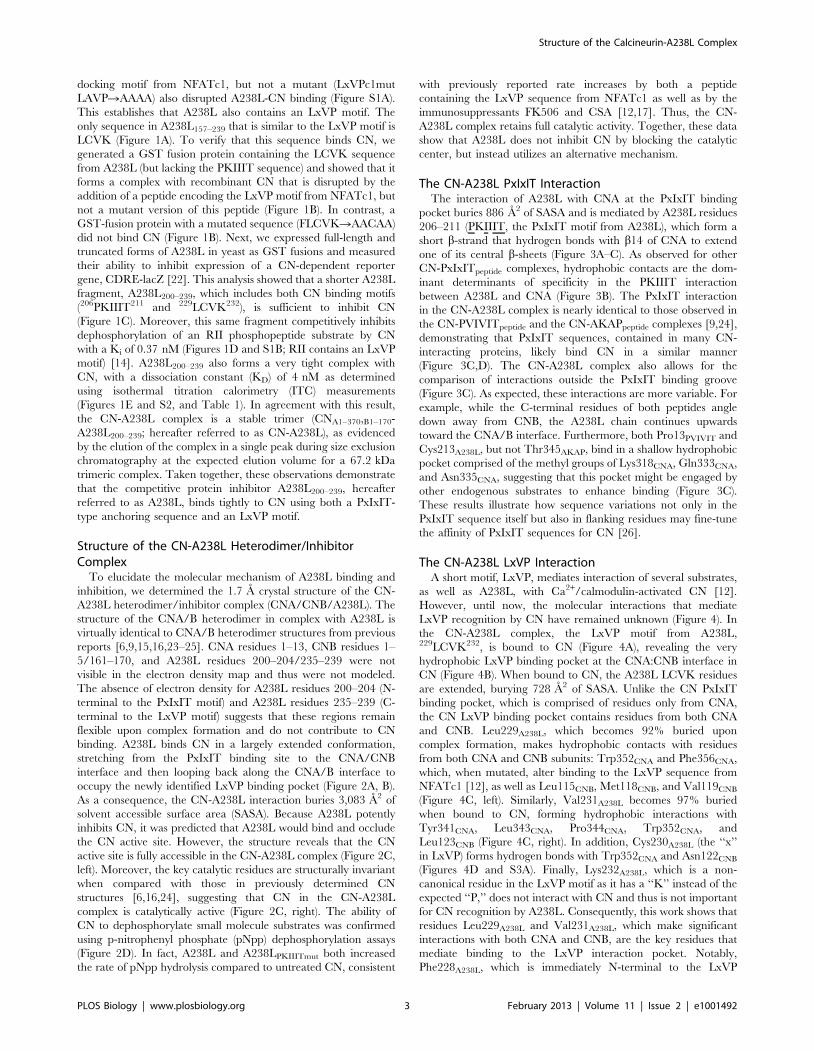
The CN-A238L PxIxIT InteractionThe interaction of A238L with CNA at the PxIxIT binding
pocket buries 886 A2 of SASA and is mediated by A238L residues
206–211 (PKIIIT, the PxIxIT motif from A238L), which form a
short b-strand that hydrogen bonds with b14 of CNA to extend
one of its central b-sheets (Figure 3A–C). As observed for other
CN-PxIxITpeptide complexes, hydrophobic contacts are the dom-
inant determinants of specificity in the PKIIIT interaction
between A238L and CNA (Figure 3B). The PxIxIT interaction
in the CN-A238L complex is nearly identical to those observed in
the CN-PVIVITpeptide and the CN-AKAPpeptide complexes [9,24],
demonstrating that PxIxIT sequences, contained in many CN-
interacting proteins, likely bind CN in a similar manner
(Figure 3C,D). The CN-A238L complex also allows for the
comparison of interactions outside the PxIxIT binding groove
(Figure 3C). As expected, these interactions are more variable. For
example, while the C-terminal residues of both peptides angle
down away from CNB, the A238L chain continues upwards
toward the CNA/B interface. Furthermore, both Pro13PVIVIT and
Cys213A238L, but not Thr345AKAP, bind in a shallow hydrophobic
pocket comprised of the methyl groups of Lys318CNA, Gln333CNA,
and Asn335CNA, suggesting that this pocket might be engaged by
other endogenous substrates to enhance binding (Figure 3C).
These results illustrate how sequence variations not only in the
PxIxIT sequence itself but also in flanking residues may fine-tune
the affinity of PxIxIT sequences for CN [26].
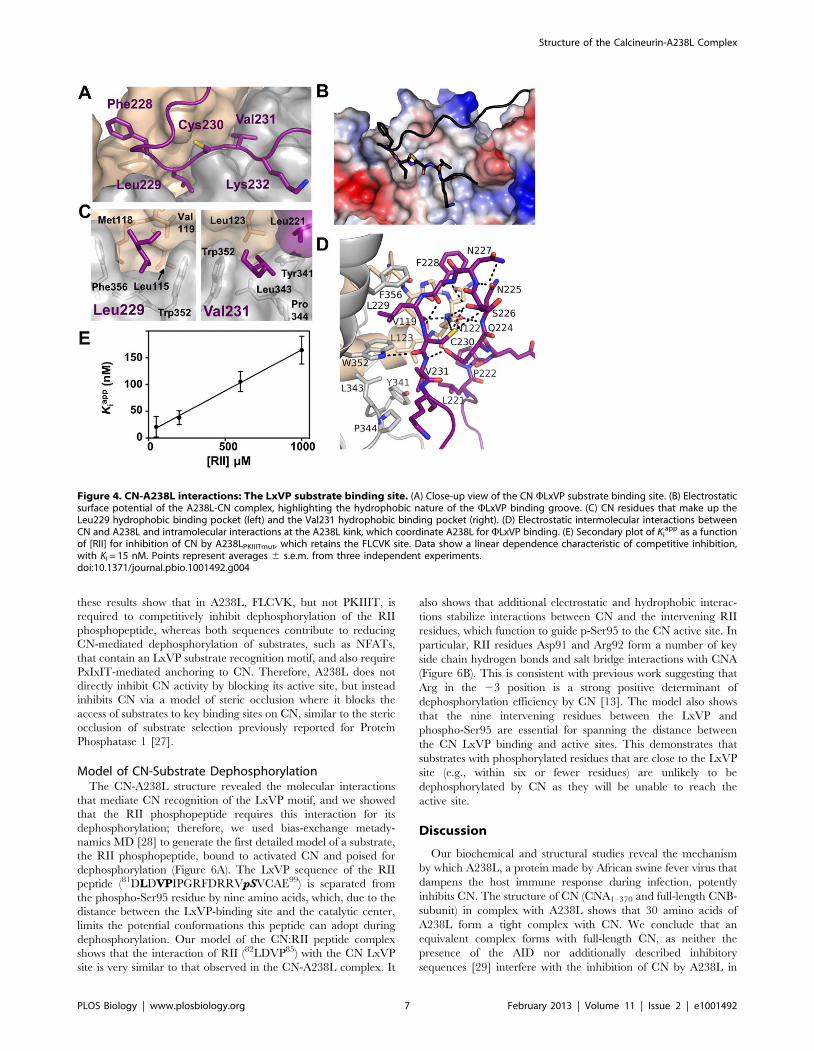
The CN-A238L LxVP InteractionA short motif, LxVP, mediates interaction of several substrates,
as well as A238L, with Ca2+/calmodulin-activated CN [12].
However, until now, the molecular interactions that mediate
LxVP recognition by CN have remained unknown (Figure 4). In
the CN-A238L complex, the LxVP motif from A238L,229LCVK232, is bound to CN (Figure 4A), revealing the very
hydrophobic LxVP binding pocket at the CNA:CNB interface in
CN (Figure 4B). When bound to CN, the A238L LCVK residues
are extended, burying 728 A2 of SASA. Unlike the CN PxIxIT
binding pocket, which is comprised of residues only from CNA,
the CN LxVP binding pocket contains residues from both CNA
and CNB. Leu229A238L, which becomes 92% buried upon
complex formation, makes hydrophobic contacts with residues
from both CNA and CNB subunits: Trp352CNA and Phe356CNA,
which, when mutated, alter binding to the LxVP sequence from
NFATc1 [12], as well as Leu115CNB, Met118CNB, and Val119CNB
(Figure 4C, left). Similarly, Val231A238L becomes 97% buried
when bound to CN, forming hydrophobic interactions with
Tyr341CNA, Leu343CNA, Pro344CNA, Trp352CNA, and
Leu123CNB (Figure 4C, right). In addition, Cys230A238L (the ‘‘x’’
in LxVP) forms hydrogen bonds with Trp352CNA and Asn122CNB
(Figures 4D and S3A). Finally, Lys232A238L, which is a non-
canonical residue in the LxVP motif as it has a ‘‘K’’ instead of the
expected ‘‘P,’’ does not interact with CN and thus is not important
for CN recognition by A238L. Consequently, this work shows that
residues Leu229A238L and Val231A238L, which make significant
interactions with both CNA and CNB, are the key residues that
mediate binding to the LxVP interaction pocket. Notably,
Phe228A238L, which is immediately N-terminal to the LxVP
Structure of the Calcineurin-A238L Complex
PLOS Biology | www.plosbiology.org 3 February 2013 | Volume 11 | Issue 2 | e1001492

binding motif, also contributes to CN binding (Figure 4A, B),
fitting into a deep pocket formed by the loops connecting EF-
hands 1 and 2 and EF-hands 3 and 4 of CNB. This results in a
25% increase in the SASA buried at this site (FLCVK buries
911 A SASA). Critically, multiple LxVP sites are immediately
preceded by an aromatic residue (Phe or Tyr), which may act as a
binding strength enhancer, as mutation of this aromatic residue
weakens the LxVP interaction [12]. Thus, a subset of LxVP sites,
including the one in A238L, is best described as WLxVP.
A238L Residues Also Contribute to the LxVP BindingPocket
The WLxVP binding groove is comprised of residues from both
CNA and CNB, in the CN-A238L complex. Unexpectedly, however,
Figure 1. A238L interacts with CN via an LxVP and a PxIxIT motif. (A) C-terminal residues (200–239) of A238L showing putative docking siteFLCVK (aa 228–232). Underlined residues were fused to GST. (B) Recombinant CN was incubated with GST fused to 15 amino acids encoding the LxVPmotif of NFATc1 or the FLCVK sequence in A238L. CN co-purifies with both motifs; this interaction is disrupted by incubation with excess peptideLxVPc1 encoding the LxVP motif from NFATc1, but not LxVPmut. CN fails to co-purify with GST fused to mutated FLCVK sequence (FLCVK mutated toAACAA). (C) b-galactosidase activity of extracts from yeast strains that harbor 2xCDRE-lacZ, a CN-dependent reporter gene, and GST or GST-A238Ltruncations are shown. We added 50 mM CaCl2 to the cell culture 2.5 h before harvesting to induce CN-dependent activation of the Crz1transcription factor ([22]; see also Text S1). Error bars indicate 6 s.d. from three independent experiments. (D) Secondary plot of Ki
app as a function of[RII] for A238L200–239 inhibition of CN. Data show a linear dependence characteristic of competitive inhibition, with Ki = 0.37 nM. Ki
app values wereobtained from the nonlinear fit of Figure S1B. Points represent averages 6 s.e.m. (E) Isothermal titration calorimetry confirming that purifiedA238L200–239 binds to CN.doi:10.1371/journal.pbio.1001492.g001
Table 1. Thermodynamic parameters and dissociation (KD) and inhibition (Ki) constants for CNA1–391/B1–170 with A238L200–239 wild-type, and A238L PxIxIT and LxVP mutants, derived from ITC experiments at 25uC or enzyme assays performed with RII at 37uC.
Complex KD (nM) DH (kcal?mol21) 2TDS (kcal?mol21) DG (kcal?mol21) Ki (nM)
CN:A238LWT 461 240.364.9 28.764.9 211.560.2 0.3760.03
CN:A238LPKIIITmut. 624626 234.360.9 25.960.9 28.560.0 1561
CN:A238LFLCVKmut. 803626 214.060.1 5.760.1 28.360.0 7,70063,000
Thermodynamic and dissociation constant data represent mean values 6 one s.d. for triplicate measurements except A238LWT, which was performed 5 times. Inhibitionconstants are mean values 6 one s.e.m. from three independent experiments.doi:10.1371/journal.pbio.1001492.t001
Structure of the Calcineurin-A238L Complex
PLOS Biology | www.plosbiology.org 4 February 2013 | Volume 11 | Issue 2 | e1001492

a few A238L residues also contribute to the WLxVP binding pocket.
Specifically, A238L, which projects upwards from the PxIxIT site
towards the CNB interface, forms a tight 180u turn at Asn227A238L
(Figures 4D and S3A). This kink is essential for WLxVP binding, as it
redirects the WLxVP sequence (FLCVK) back toward its docking site
on CN at the CNA/B interface. This enables the A238L residues that
immediately precede the WLxVP to interact directly with the CN-
bound WLxVP residues and contribute to the WLxVP binding site.
Thus, in addition to the multiple interactions observed with residues
from CNA and CNB, Val231A238L (the ‘‘V’’ in the LxVP motif) also
makes intramolecular hydrophobic contacts with Leu221A238L. In
addition, the side chain Gln224A238L hydrogen bonds with the
backbone amide of Val231A238L (Figures 4D and S3A). Therefore,
residues from all three proteins—CNA, CNB, and A238L—function
to keep Val231A238L occluded from solvent in the bound conforma-
tion. In addition, although Cys230A238L is the ‘‘x’’ in the LxVP motif,
this residue also makes multiple intramolecular interactions that help
stabilize the A238L bound conformation. Specifically, the amide
nitrogen of Cys230A238L hydrogen bonds with the carbonyl oxygen of
Phe228A238L. Cys230A238L also forms thiol hydrogen bonds with the
amide nitrogen of S226A238L and the carbonyl of N225A238L. These
intramolecular interactions explain why Cys230A238L is still nearly
completely buried (70%) in the CN-A238L complex even though its
side chain points away from the LxVP docking groove. Thus,
although it is not yet known how similar the CN-LxVP interaction of
CN-A238L is with that of other LxVP docking motifs from substrates,
our structure suggests that residues flanking the LxVP sequence may
also modulate the affinity of this motif for CN.
Although the most extensive interactions between A238L and CN
occur at the PxIxIT and WLxVP binding grooves, additional, largely
polar intra- and intermolecular interactions outside of these docking
sites also contribute to A238L binding. For example, the interactions
that stabilize the A238L kink (the 180u tight turn at Asn227A238L,
which enables the rest of A238L to point back toward the LxVP
Figure 2. The CN active site is fully accessible in the CN-A238L complex. (A) Overview of the CN-A238L complex. CNA (gray, surfacerepresentation) interacts via helix a14 with CNB (beige, surface representation). The CNA active site is highlighted in cyan, with PxIxIT and LxVPbinding pockets shown in yellow and green, respectively. A238L is shown as a cartoon representation (purple), with the PxIxIT (PKIIIT) and LxVP(LCVK) substrate binding motifs highlighted as sticks. (B) Cartoon representation of the CN-A238L complex. Structure is rotated 120u about the x-axisrelative to (A); all colors as in (A); blue spheres representing four Ca2+ atoms in CNB. CNA b-sheet1 is shown in dark grey. The N-terminal b-strandformed by the A238L PxIxIT motif complements b-strand14 of CNA and thus extends b-sheet1. (C, left) CN-A238L illustrated as in (A), with thedistances between the CN active site and the PxIxIT and LxVP binding grooves indicated by arrows (,32 A and ,31 A, respectively). (C, right)Overlap of the catalytic residues from the CN-A238L complex (black), apo-CN (cyan, PDBID 1AUI), CN-AKAPpeptide (dark blue, PDBID 3LL8), CN-PVIVITpeptide (green, PDBID 2P6B), CN-Cyclosporin (light blue, PDBID 1M63), and CN-FK506 (pink, PDBID 1TCO). Catalytic residues of CNA are shownas sticks and labeled (Asp90, His92, Asp118, Asn150, His199, His281). Fe3+ and Zn2+ ions from 1AUI are shown as spheres. (D) The rate of pNpphydrolysis was measured in the presence of A238L (grey), A238LPKIIITmut (yellow), or A238LFLCVKmut (green). Assays were performed in triplicate using10 mM pNpp, and error bars indicate one s.d.doi:10.1371/journal.pbio.1001492.g002
Structure of the Calcineurin-A238L Complex
PLOS Biology | www.plosbiology.org 5 February 2013 | Volume 11 | Issue 2 | e1001492

binding pocket) are mediated by an extensive network of more than 10
hydrogen bonds, the center of which is Asn122CNB (Figures 4D and
3SA). The side chain amide nitrogen and carbonyl of Asn122CNB form
hydrogen bonds with the backbone carbonyl and backbone amide
nitrogen of two residues that border the kink, L229A238L and
Asn225A238L, respectively. Similarly, A238L residues C-terminal to
the PxIxIT motif (212GCEDNVY218) also interact with CNA through
main chain/side chain side chain/side chain hydrogen bonds (Figure
S3B). Although the two dominant A238L:CN PxIxIT and LxVP
interfaces together bury 1,797 A2 SASA, these additional mostly polar
interactions also contribute to the CN-A238L interface, burying
1,286 A2 SASA.
A238L Inhibits CN by Interfering with SubstrateRecognition
We next sought to determine the relative importance of the
PxIxIT and LxVP sites for CN binding. Using ITC and two
A238L mutants (a PxIxIT motif mutant, with PKIIIT mutated to
AKAIAA and a WLxVP motif mutant, with FLCVK mutated to
AACAA; A238LPKIIITmut and A238LFLCVKmut, respectively), we
found that both the PxIxIT and WLxVP sequences are important
for CN binding, as the PxIxIT and WLxVP mutations increase the
KD similarly by ,150-fold and ,200-fold, respectively (Table 1
and Figure S2). Next, we investigated the contribution of the
individual A238L motifs toward the inhibition of CN.
A238LPKIIITmut, which still contains the FLCVK sequence,
competitively inhibits dephosphorylation of RII by CN with
Ki = 15 nM (Table 1 and Figure 4E) [14]. In contrast,
A238LFLCVKmut, which still contains the PKIIIT sequence,
inhibited CN very poorly, with Ki = 7,700 nM, a 20,000-fold
decrease in inhibitor efficacy compared to wild-type A238L
(Table 1 and Figures 3E and S4). In addition, when expressed in
HEK293T cells in which NFAT signaling had been stimulated,
both A238L mutants reduced the activity of an NFAT-dependent
reporter gene. At roughly equal levels of protein expression,
neither A238LFLCVKmut nor A238LPKIIITmut were as effective as
wt-A238L in NFAT inhibition (Figure 5A, B). Taken together,
Figure 3. CN-A238L interactions: The PxIxIT substrate binding site. (A) Close-up view of the CN PxIxIT substrate binding site. A238L PKIIIT isshown as magenta sticks and labeled; CNA is shown as a grey surface. (B) Same view as (A), with a transparent CNA surface. Individual CNA residuesthat participate in the interaction with PKIIITA238L are shown as grey sticks. (C) Superposition of the PKIIITA238L motif (purple) with a synthetic PVIVITpeptide (orange) and the IAIIIT CN docking site of AKAP79 (green) bound to CN. Pro13PVIVIT and Cys213A238L are shown as sticks. (D) Overlay as in (C)but illustrated as sticks. Corresponding ‘‘variable’’ residues Ile209A238L, Val9PVIVIT, and Ile341AKAP79 (the second ‘‘x’’ in PxIxIT) participate in the samehydrophobic interaction with Val328CNA (dotted lines). (E) Secondary plot of Ki
app as a function of [RII] for inhibition of CN by A238LFLCVKmut, whichretains the PKIIIT site. Data show a linear dependence characteristic of competitive inhibition, with Ki = 7,70063,000 nM. Ki
app values were obtainedfrom the nonlinear fit of Figure S4A. Points represent averages 6 s.e.m.doi:10.1371/journal.pbio.1001492.g003
Structure of the Calcineurin-A238L Complex
PLOS Biology | www.plosbiology.org 6 February 2013 | Volume 11 | Issue 2 | e1001492
these results show that in A238L, FLCVK, but not PKIIIT, is
required to competitively inhibit dephosphorylation of the RII
phosphopeptide, whereas both sequences contribute to reducing
CN-mediated dephosphorylation of substrates, such as NFATs,
that contain an LxVP substrate recognition motif, and also require
PxIxIT-mediated anchoring to CN. Therefore, A238L does not
directly inhibit CN activity by blocking its active site, but instead
inhibits CN via a model of steric occlusion where it blocks the
access of substrates to key binding sites on CN, similar to the steric
occlusion of substrate selection previously reported for Protein
Phosphatase 1 [27].
Model of CN-Substrate DephosphorylationThe CN-A238L structure revealed the molecular interactions
that mediate CN recognition of the LxVP motif, and we showed
that the RII phosphopeptide requires this interaction for its
dephosphorylation; therefore, we used bias-exchange metady-
namics MD [28] to generate the first detailed model of a substrate,
the RII phosphopeptide, bound to activated CN and poised for
dephosphorylation (Figure 6A). The LxVP sequence of the RII
peptide (81DLDVPIPGRFDRRVpSVCAE99) is separated from
the phospho-Ser95 residue by nine amino acids, which, due to the
distance between the LxVP-binding site and the catalytic center,
limits the potential conformations this peptide can adopt during
dephosphorylation. Our model of the CN:RII peptide complex
shows that the interaction of RII (82LDVP85) with the CN LxVP
site is very similar to that observed in the CN-A238L complex. It
also shows that additional electrostatic and hydrophobic interac-
tions stabilize interactions between CN and the intervening RII
residues, which function to guide p-Ser95 to the CN active site. In
particular, RII residues Asp91 and Arg92 form a number of key
side chain hydrogen bonds and salt bridge interactions with CNA
(Figure 6B). This is consistent with previous work suggesting that
Arg in the 23 position is a strong positive determinant of
dephosphorylation efficiency by CN [13]. The model also shows
that the nine intervening residues between the LxVP and
phospho-Ser95 are essential for spanning the distance between
the CN LxVP binding and active sites. This demonstrates that
substrates with phosphorylated residues that are close to the LxVP
site (e.g., within six or fewer residues) are unlikely to be
dephosphorylated by CN as they will be unable to reach the
active site.
Discussion
Our biochemical and structural studies reveal the mechanism
by which A238L, a protein made by African swine fever virus that
dampens the host immune response during infection, potently
inhibits CN. The structure of CN (CNA1–370 and full-length CNB-
subunit) in complex with A238L shows that 30 amino acids of
A238L form a tight complex with CN. We conclude that an
equivalent complex forms with full-length CN, as neither the
presence of the AID nor additionally described inhibitory
sequences [29] interfere with the inhibition of CN by A238L in
Figure 4. CN-A238L interactions: The LxVP substrate binding site. (A) Close-up view of the CN WLxVP substrate binding site. (B) Electrostaticsurface potential of the A238L-CN complex, highlighting the hydrophobic nature of the WLxVP binding groove. (C) CN residues that make up theLeu229 hydrophobic binding pocket (left) and the Val231 hydrophobic binding pocket (right). (D) Electrostatic intermolecular interactions betweenCN and A238L and intramolecular interactions at the A238L kink, which coordinate A238L for WLxVP binding. (E) Secondary plot of Ki
app as a functionof [RII] for inhibition of CN by A238LPKIIITmut, which retains the FLCVK site. Data show a linear dependence characteristic of competitive inhibition,with Ki = 15 nM. Points represent averages 6 s.e.m. from three independent experiments.doi:10.1371/journal.pbio.1001492.g004
Structure of the Calcineurin-A238L Complex
PLOS Biology | www.plosbiology.org 7 February 2013 | Volume 11 | Issue 2 | e1001492

vivo. As anticipated, A238L interacts with the PxIxIT binding
groove on CNA. However, we discovered that A238L also binds
CN in additional surface grooves, the most important of which is
the LxVP-binding pocket. Remarkably, while A238L is a potent
inhibitor of CN, the CN catalytic center is fully accessible and
active, as confirmed by our biochemical findings that show that
A238L-bound CN rapidly dephosphorylates the small molecule
substrate pNpp. In contrast, the CN-A238L complex is unable to
dephosphorylate a peptide substrate derived from the RII subunit
of protein kinase A, which contains an LxVP sequence, or a
protein substrate, NFAT, which contains an LxVP sequence and a
PxIxIT anchoring sequence. Collectively, these structural and
biochemical data show that A238L inhibits CN function through a
model of ‘‘steric inhibition,’’ in which A238L effectively inhibits
CN function, not by directly blocking the active site of the
phosphatase, but instead by occupying two critical docking
grooves and sterically occluding CN from interacting with
substrates. To our best knowledge, this mechanism of altered
substrate dephosphorylation by steric occlusion of substrate
binding sites has only been directly observed in one other ser/
thr protein phosphatase, that of PP1, which, like CN, is one of the
key members of the PPP family [27]. Thus, our results establish
that this mechanism, whereby enzyme activity is modulated via
substrate access rather than through active site inhibition or
allostery, is likely utilized by the entire PPP family.
Although the structure of CN bound to a bona fide substrate has
yet to be determined, the CN-A238L structure and our CN-RII
substrate model significantly advance our understanding of how
CN uses two complementary strategies to recognize substrates.
First, the PxIxIT anchoring motif is used by protein scaffolds,
inhibitors, and some substrates to form a stable interaction with
CN by docking to a site that is available regardless of the activation
state of the enzyme. For PxIxIT-containing substrates, the strength
of this anchoring modulates the Ca2+-concentration dependence
of their dephosphorylation, but does not directly contribute to
recognition of phosphosites during the dephosphorylation reaction
[26,30]. Second, CN overcomes the limited specificity of its
catalytic site by recognizing specific residues, such as ‘‘WLxVP,’’
which are distal to the phosphosite. This interaction is likely
required for the dephosphorylation of multiple substrates, as it is
this site that is targeted by multiple inhibitors, including A238L
and the immunosuppressants CSA and FK506 (Figure 6C).
This work also reveals interactions that contribute to phospho-
site selection by CN. The ‘‘WLxVP’’ binding site is now
molecularly defined as a hydrophobic binding surface composed
of residues from the CNA and CNB subunits, which becomes
accessible after binding of Ca2+-loaded calmodulin displaces the
C-terminal AID domain from the active site [12]. Our structure
and the RII model show that the substrate residue dephosphor-
ylated by CN (hereafter referred to as pS/pT) must be a minimum
of 9–15 residues away from the WLxVP sequence, and be in an
extended conformation in order for the pS/pT residue to reach
the catalytic site. In fact, the pS/pT residues in the RII peptide
and the substrate RCAN1 are only 9 and 10 residues C-terminal
to the ‘‘P’’ in their LxVP motifs, respectively (Figure 6A, B, D)
[14,31]. Our model also shows that additional electrostatic
interactions, especially at basic residue at position 23, help orient
the phosphosite in RII toward the catalytic center. In addition, the
C-terminus of the RII peptide lies in a groove identified in other
PPPs, namely PP1, to be a substrate recognition groove
(hydrophobic groove in PP1), suggesting that this groove functions
in a similar manner in CN and thus likely for the entire family of
PPPs [2,32].
While our model provides fundamental insights into phospho-
site selection by CN for those substrates in which the phosphosite
is 9–10 residues C-terminal to LxVP sequence, other surface
grooves near the catalytic center are likely also important for
substrate recognition. This is because the spacing between
experimentally determined ‘‘LxVP’’ and phosphosites substrates
is quite variable. For example, substrates such as NFAT and
KSR2 have pS/pT sites that are 10–100 s of residues away from
the known LxVP sequences [33–37]. In these cases, one or several
pS/pT sites are found in long, extended sequences that are
predicted to be unstructured, suggesting that these regions are
dynamic when bound to CN and that interactions at the LxVP
and PxIxIT sites tether the substrate to CN allowing the
phosphosite(s) to encounter the catalytic site and be dephosphor-
ylated (Figure 6D). A detailed understanding of how phosphosites
that are distal from the LxVP and PxIxIT motifs engage the CN
catalytic center is an active area of investigation.
Finally, these studies also provide critical new insights into the
mechanism by which the immunosuppressants CSA and FK506
Figure 5. A238L inhibits CN by interfering with substratedocking. (A) NFAT-dependent transcription was measured in stimu-lated HEK293T cells co-transfected with an NFAT-luciferase reporterplasmid and a plasmid expressing SV5-PK-tagged A238L wild-type ormotif mutant proteins, or vector alone. Data are means 6 s.e.m. fromthree independent experiments. (B) Transiently transfected SV5-PK-tagged A238L protein expression levels were analyzed by immuno-blotting. The increased electrophoretic mobility of A238LPKIIITmut
relative to wt-A238L may be due to changes in a posttranslationalmodification that is known to affect the electrophoretic mobility ofA238L [55].doi:10.1371/journal.pbio.1001492.g005
Structure of the Calcineurin-A238L Complex
PLOS Biology | www.plosbiology.org 8 February 2013 | Volume 11 | Issue 2 | e1001492

Figure 6. Potential interaction modes of CN substrates/regulators with CN. (A) The CN-RII peptide complex obtained by MD. Colors as inFigure 2A, C. CN is shown in surface representation and the RII peptide in dark green with the LxVP motif (LDVP) and phospho-Ser95 as green sticks.LDVP is bound to the LxVP binding pocket (light green), and phospho-Ser95 is bound in the CN active site (cyan). (B) Electrostatic interactionsbetween CN and the RII peptide. The CN electrostatic surface has positively and negatively charged areas colored blue and red, respectively. The LxVPmotif and residues in RII that participate in polar interactions with CN are shown as green sticks. (C) Features of selected CN substrates and regulators,including substrates tested in this work (NFAT, Crz1, and the RII peptide). PxIxIT and LxVP motifs are highlighted in yellow and green, respectively,with intervening residues in grey. Regions containing S-T residues that are dephosphorylated by CN are pink. (D) Potential modes of interaction of CNwith various binding partners. CN is shown in grey, with the active site in cyan, the PxIxIT docking site in yellow, and the LxVP docking site in green.CN binding partners are shown in blue, with PxIxIT and LxVP motifs in purple and phosphorylated regions shown as red circles. The residues betweenthe two CN docking motifs, or between one docking motif and regions dephosphorylated by CN, are represented as coils, as they are predicted to beunstructured in solution. A238L is the CN-A238L crystal structure.doi:10.1371/journal.pbio.1001492.g006
Structure of the Calcineurin-A238L Complex
PLOS Biology | www.plosbiology.org 9 February 2013 | Volume 11 | Issue 2 | e1001492
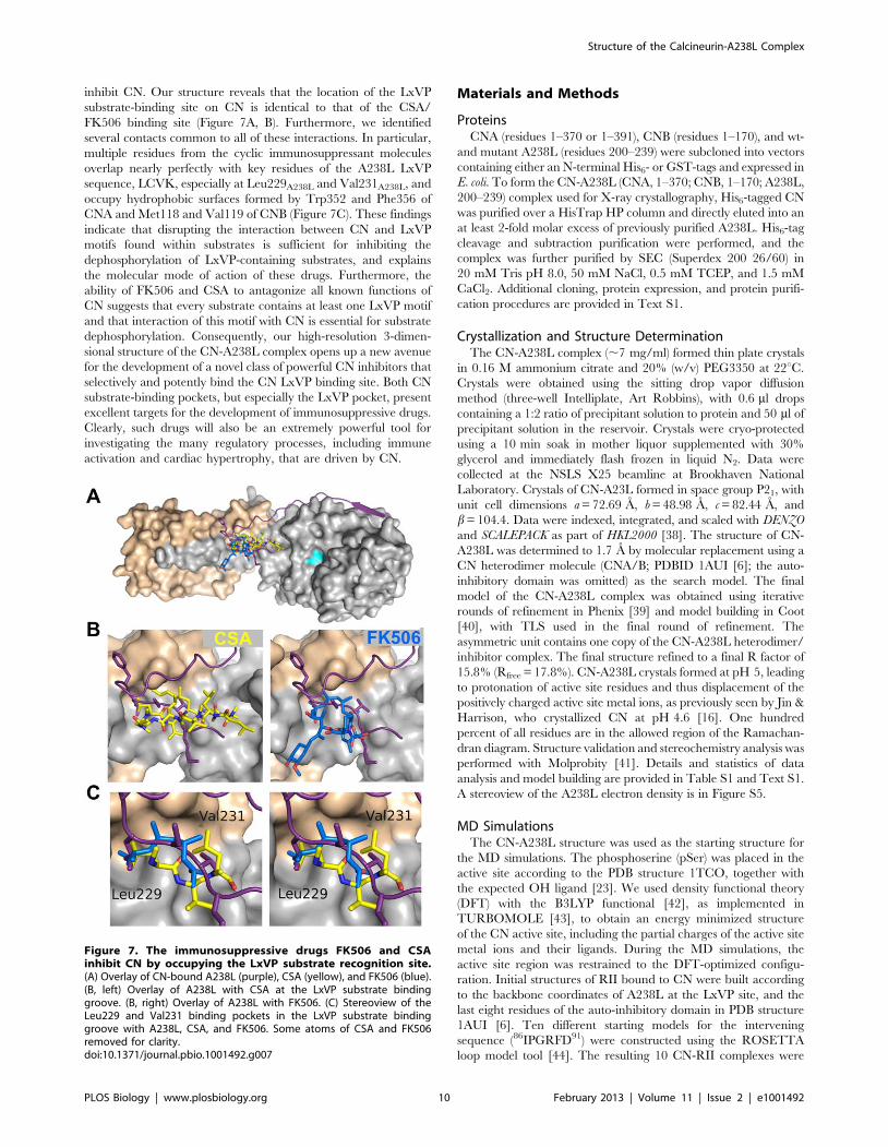
inhibit CN. Our structure reveals that the location of the LxVP
substrate-binding site on CN is identical to that of the CSA/
FK506 binding site (Figure 7A, B). Furthermore, we identified
several contacts common to all of these interactions. In particular,
multiple residues from the cyclic immunosuppressant molecules
overlap nearly perfectly with key residues of the A238L LxVP
sequence, LCVK, especially at Leu229A238L and Val231A238L, and
occupy hydrophobic surfaces formed by Trp352 and Phe356 of
CNA and Met118 and Val119 of CNB (Figure 7C). These findings
indicate that disrupting the interaction between CN and LxVP
motifs found within substrates is sufficient for inhibiting the
dephosphorylation of LxVP-containing substrates, and explains
the molecular mode of action of these drugs. Furthermore, the
ability of FK506 and CSA to antagonize all known functions of
CN suggests that every substrate contains at least one LxVP motif
and that interaction of this motif with CN is essential for substrate
dephosphorylation. Consequently, our high-resolution 3-dimen-
sional structure of the CN-A238L complex opens up a new avenue
for the development of a novel class of powerful CN inhibitors that
selectively and potently bind the CN LxVP binding site. Both CN
substrate-binding pockets, but especially the LxVP pocket, present
excellent targets for the development of immunosuppressive drugs.
Clearly, such drugs will also be an extremely powerful tool for
investigating the many regulatory processes, including immune
activation and cardiac hypertrophy, that are driven by CN.
Materials and Methods
ProteinsCNA (residues 1–370 or 1–391), CNB (residues 1–170), and wt-
and mutant A238L (residues 200–239) were subcloned into vectors
containing either an N-terminal His6- or GST-tags and expressed in
E. coli. To form the CN-A238L (CNA, 1–370; CNB, 1–170; A238L,
200–239) complex used for X-ray crystallography, His6-tagged CN
was purified over a HisTrap HP column and directly eluted into an
at least 2-fold molar excess of previously purified A238L. His6-tag
cleavage and subtraction purification were performed, and the
complex was further purified by SEC (Superdex 200 26/60) in
20 mM Tris pH 8.0, 50 mM NaCl, 0.5 mM TCEP, and 1.5 mM
CaCl2. Additional cloning, protein expression, and protein purifi-
cation procedures are provided in Text S1.
Crystallization and Structure DeterminationThe CN-A238L complex (,7 mg/ml) formed thin plate crystals
in 0.16 M ammonium citrate and 20% (w/v) PEG3350 at 22uC.
Crystals were obtained using the sitting drop vapor diffusion
method (three-well Intelliplate, Art Robbins), with 0.6 ml drops
containing a 1:2 ratio of precipitant solution to protein and 50 ml of
precipitant solution in the reservoir. Crystals were cryo-protected
using a 10 min soak in mother liquor supplemented with 30%
glycerol and immediately flash frozen in liquid N2. Data were
collected at the NSLS X25 beamline at Brookhaven National
Laboratory. Crystals of CN-A23L formed in space group P21, with
unit cell dimensions a = 72.69 A, b = 48.98 A, c = 82.44 A, and
b = 104.4. Data were indexed, integrated, and scaled with DENZO
and SCALEPACK as part of HKL2000 [38]. The structure of CN-
A238L was determined to 1.7 A by molecular replacement using a
CN heterodimer molecule (CNA/B; PDBID 1AUI [6]; the auto-
inhibitory domain was omitted) as the search model. The final
model of the CN-A238L complex was obtained using iterative
rounds of refinement in Phenix [39] and model building in Coot
[40], with TLS used in the final round of refinement. The
asymmetric unit contains one copy of the CN-A238L heterodimer/
inhibitor complex. The final structure refined to a final R factor of
15.8% (Rfree = 17.8%). CN-A238L crystals formed at pH 5, leading
to protonation of active site residues and thus displacement of the
positively charged active site metal ions, as previously seen by Jin &
Harrison, who crystallized CN at pH 4.6 [16]. One hundred
percent of all residues are in the allowed region of the Ramachan-
dran diagram. Structure validation and stereochemistry analysis was
performed with Molprobity [41]. Details and statistics of data
analysis and model building are provided in Table S1 and Text S1.
A stereoview of the A238L electron density is in Figure S5.
MD SimulationsThe CN-A238L structure was used as the starting structure for
the MD simulations. The phosphoserine (pSer) was placed in the
active site according to the PDB structure 1TCO, together with
the expected OH ligand [23]. We used density functional theory
(DFT) with the B3LYP functional [42], as implemented in
TURBOMOLE [43], to obtain an energy minimized structure
of the CN active site, including the partial charges of the active site
metal ions and their ligands. During the MD simulations, the
active site region was restrained to the DFT-optimized configu-
ration. Initial structures of RII bound to CN were built according
to the backbone coordinates of A238L at the LxVP site, and the
last eight residues of the auto-inhibitory domain in PDB structure
1AUI [6]. Ten different starting models for the intervening
sequence (86IPGRFD91) were constructed using the ROSETTA
loop model tool [44]. The resulting 10 CN-RII complexes were
Figure 7. The immunosuppressive drugs FK506 and CSAinhibit CN by occupying the LxVP substrate recognition site.(A) Overlay of CN-bound A238L (purple), CSA (yellow), and FK506 (blue).(B, left) Overlay of A238L with CSA at the LxVP substrate bindinggroove. (B, right) Overlay of A238L with FK506. (C) Stereoview of theLeu229 and Val231 binding pockets in the LxVP substrate bindinggroove with A238L, CSA, and FK506. Some atoms of CSA and FK506removed for clarity.doi:10.1371/journal.pbio.1001492.g007
Structure of the Calcineurin-A238L Complex
PLOS Biology | www.plosbiology.org 10 February 2013 | Volume 11 | Issue 2 | e1001492

inserted into rectangular boxes (868612 nm3) with 22,881 TIP3P
water molecules [45] and eight Na+ ions each for electro-
neutrality. MD simulations were performed using GRO-
MACSv4.5.3 [46], the Amber ff99SB-ILDN force field [47],
particle mesh Ewald summation [48] using a 0.12 nm grid
spacing, a time step of 2 fs and a 0.9 nm real-space cutoff at a
constant temperature [49] of 300 K and at a pressure [50,51] of
one bar. After 2 ns of equilibration of each of the 10 starting
models, bias-exchange metadynamics [28] using the
PLUMED1.3.0 plugin [52] was used to accelerate the conforma-
tional sampling. In 9 of the 10 replicas, the Y dihedral angles of
RII residues Ile86, Pro87, Arg89, Phe90, Asp91, Arg92, Arg93,
Cys97, and Ala98, respectively, were biased, while replica 10 was
kept unbiased. To ensure RII stayed bound to CN during the
conformational sampling, the Fe3+-pSer and LxVP interactions
were harmonically restrained. Convergence was reached after
1.15 ms (115 ns/replica), as judged by the free energy profiles of
the biased Y. The RII structures of the unbiased replica were
clustered according to a 1.2 A backbone root-mean-square-
distance (RMSD) threshold. The most populated cluster contains
58% of all structures, from which a representative was selected
with the lowest Coulomb energy. A different clustering technique
using reweighting of the biased replicas [53] led to similar results.
Isothermal Titration CalorimetryITC experiments were performed at 25uC using a VP-ITC
microcalorimeter (GE Healthcare). All protein samples were
equilibrated in ITC buffer (20 mM Tris pH 7.5, 150 mM NaCl,
1.5 mM CaCl2, 0.5 mM TCEP). Wild-type or mutated A238L
was titrated into CNA1–391/B1–170. Titrant (10 mL per injection)
was injected into the sample cell over a period of 20 s with a 250 s
interval between titrations to allow for complete equilibration and
baseline recovery. Twenty-eight injections were delivered during
each experiment, and the solution in the sample cell was stirred at
307 rpm to ensure rapid mixing. Data were analyzed with one set
of sites binding model, based on the 1:1 stoichiometry observed in
the crystal structure, using Origin 7.0 (OriginLab).
CN Activity Assays with RIIThe rate of RII phosphopeptide dephosphorylation by CN was
determined by measuring the total phosphate released over four
time points (total 5–20 min). Reaction rates were linear over this
time period and constituted less than 1% of product formation.
The 50 ml reactions contained assay buffer (50 mM Tris pH 7.4,
100 mM NaCl, 6 mM MgCl2, 0.5 mM CaCl2, 0.1% PEG 3250,
0.5 mM DTT), 10 nM CN, wt- or mutant A238L (0–10 mM), and
50–1,000 mM RII phosphopeptide. Reactions were performed at
37uC and were initiated by the addition of 106 RII and
terminated using 100 ml Biomol Green Reagent (Enzo Life
Sciences). After color development for 20 min, absorbance was
measured at 595 nm and compared to phosphate standards of
known concentration to determine the amount of phosphate
released. Kinetic constants were determined with GraphPad Prism
by fitting the points to the Morrison model for tight-binding
inhibitors using nonlinear regression analysis [23]. Competitive
and noncompetitive models were compared using the corrected
Akaike information criterion, AICc [54]. Phosphorylated RII was
synthesized by the Tufts University Core Facility.
CN Activity Assays with pNppThe rate of CN hydrolysis of pNpp was measured in a continuous
assay by monitoring the production of pNP at 415 nm. Reaction
rates were linear for at least 1 h and reaction progress was ,0.1%.
The 100 ml reactions contained assay buffer (100 mM Tris pH 7.5,
100 mM NaCl, 0.4 mM CaCl2, 100 mg/ml BSA, 1 mM MnCl2,
0.5 mM DTT), 10–20 nM truncated CN, wt or mutant A238L (0–
8 mM), and 10 mM pNpp. Experiments were performed at room
temperature. Standards of known pNp concentration were used to
convert absorbance units to pNp concentration.
GST Pull-Down and Competition AssaysCells extracts were prepared by resuspending cell pellets in lysis
buffer (50 mM Tris, pH 7.4, 100 mM NaCl, 2 mM EDTA,
2 mM EGTA, 5 mM DTT, 1 mM PMSF, 5 mg/ml each
pepstatin, leupeptin, aprotinin, and benzamidine) and lysed by
sonication. Cell debris was pelleted by centrifugation (20,0006 g,
20 min), and clarified lysate was brought to 0.1% Tween-20 and
stored in aliquots at 280uC. For GST-peptide fusion experiments,
50–200 mg cell extracts containing GST or GST fusion proteins
were bound to glutathione sepharose 4B beads (GE Healthcare),
washed 36 with wash buffer (10 mM Tris, pH 8.0, 110 mM
KOAc, 2 mM MgOAc, 0.1% Tween-20), incubated with 200 mg
CNA1–391/B1–370 cell extract with or without 200 mM competing
peptide, and finally washed 36 with wash buffer (containing
competing peptides if present in the previous step). For GST-
CNA1 experiments involving S-A238L157–239, 20 mg cell extracts
containing co-expressed GST-CNA1 and CNB1 were bound to
beads as described above, then incubated with 350 ng purified S-
A238L157–239 in the presence or absence of competing peptide. In
both experiments, bound proteins were eluted by boiling samples
in Laemmli buffer. Peptides for competition assays were synthe-
sized by the Tufts University Core Facility. The amino acid
sequences were: LxVPc1, DQYLAVPQHPYQWAK; LxVPmut,
DQYAAAAQHPYQWAK; PVIVIT, GPHPVIVITGPHEE; and
PVIVITscrambled, GPIVPIHVTHPGEE.
Accession NumbersThe structure factors and coordinates for the CN-A238L
complex have been deposited with the Protein Databank with
accession number 4F0Z.
Supporting Information
Figure S1 A238L binds CN via a PxIxIT and LxVP motif. (A)
Recombinant S-tagged A238L157–239 incubated with GST-CNA
and CNB co-purifies with GST-CNA (lane 5). Incubation with
excess peptides encoding the LxVP site from NFATc1 (lane 1) or
the high-affinity PxIxIT peptide PVIVIT (lane 3) interferes with
A238L-CNA binding. Control peptides (lanes 2 and 4) do not
interfere with binding. (B) Plot of CN rate as a function of
A238L200–239 concentration at different RII concentrations
ranging from 50–1,000 mM. Curve fit obtained by nonlinear
regression using the Morrison equation to account for tight
binding inhibition. Error bars indicate one s.d. from three
independent experiments.
(TIF)
Figure S2 Role of the PxIxIT and LxVP sites in the CN-A238L
interaction. Raw isothermal titration calorimetry data (upper
panels) and derived binding isotherm plotted versus the molar
ratio of titrant fit using a one-site model (lower panels) for CNA1–
391/B1–170 titrated with: (A) WT A238L, (B) A238L PxIxIT mutant
(PKIIIT mutated to AKAIAA), and (C) A238L LxVP mutant
(FLCVK mutated to AACAA). Thermodynamic data and KD
values are summarized in Table 1.
(TIF)
Figure S3 A238L-CN polar interactions. (A) Stereo-view of
the FLCVKA238L interface. CN residues participating in the
Structure of the Calcineurin-A238L Complex
PLOS Biology | www.plosbiology.org 11 February 2013 | Volume 11 | Issue 2 | e1001492
interaction are shown as grey (CNA) or beige (CNB) sticks, with
A238L residues shown in purple. The multiple intra- and
intermolecular hydrogen bonds that stabilize the A238L kink are
shown as black dotted lines. (B) A238L residues immediately C-
terminal to the 206PKIIIT211 motif, 212GCEDNVY218, are
illustrated as sticks and labeled. CNA residues that interact with
these A238L residues are also shown as sticks (black). Hydrogen
bonds/salt bridge interactions are indicated by black dashed lines.
(TIF)
Figure S4 A238L LxVP motif mutant weakly inhibits RII
dephosphorylation. (A) Dose-response plot of CN rate as a function
of A238LFLCVKmut at different RII concentrations ranging from 50–
1,000 mM. Curves were fit by nonlinear regression using the
Morrison equation. Error bars indicate one s.d. from three
independent experiments. (B) Plot of CN rate as a function of
[RII]. Data fit the Michaelis-Menten model for competitive
inhibition. Points represent averages 6 s.d. from three independent
experiments. Concentrations of A238LFLCVKmut are indicated.
(TIF)
Figure S5 Stereoview of the A238L electron density. (A) Sigma
2mFo-DFc electron density map of A238L contoured at 1s to
1.70 A (blue mesh). A238L shown as magenta sticks with the
PxIxIT and LxVP motifs in green. (B) Close-up stereoview of the
A238L LxVP motif, with LxVP residues labeled.
(TIF)
Table S1 Data collection and refinement statistics.
(DOCX)
Text S1 Supplementary materials and methods.
(DOCX)
Acknowledgments
The authors thank Dr. M. Allaire (National Synchrotron Light Source,
NSLS) for his support at NSLS beamline X25. Use of the NSLS at
Brookhaven National Laboratory was supported by the U.S. Department
of Energy, Office of Science, Office of Basic Energy Sciences under
contract no. DE-AC02-98CH10886. MD calculations were performed on
the Biowulf computing cluster at NIH. The authors thank Dr. Linda Dixon
for providing information and materials relating to A238L and Dan
Herschlag, Jagoree Roy, and Evan Guiney for helpful discussion and
reading of the manuscript. We thank Dara Dowlatshahi for technical
advice and Dr. V. Kaila (National Institutes of Health) for his support with
the quantum mechanical calculations of the model substrate.
Author Contributions
The author(s) have made the following declarations about their
contributions: Conceived and designed the experiments: SG RB PC GH
RP MSC WP. Performed the experiments: SG RB NL JC PC RP WP.
Analyzed the data: SG RB PC GH RP MSC WP. Wrote the paper: SG RB
PC GH RP MSC WP.
References
1. Olsen JV, Blagoev B, Gnad F, Macek B, Kumar C, et al. (2006) Global, in vivo,
and site-specific phosphorylation dynamics in signaling networks. Cell 127: 635–
648.
2. Shi Y (2009) Serine/threonine phosphatases: mechanism through structure. Cell139: 468–484.
3. Aramburu J, Rao A, Klee CB (2000) Calcineurin: from structure to function.Curr Top Cell Regul 36: 237–295.
4. Crabtree GR, Schreiber SL (2009) SnapShot: Ca2+-calcineurin-NFAT
signaling. Cell 138: 210, 210.e211.
5. Musson RE, Smit NP (2011) Regulatory mechanisms of calcineurin phosphatase
activity. Curr Med Chem 18: 301–315.
6. Kissinger CR, Parge HE, Knighton DR, Lewis CT, Pelletier LA, et al. (1995)
Crystal structures of human calcineurin and the human FKBP12-FK506-calcineurin complex. Nature 378: 641–644.
7. Yang SA, Klee CB (2000) Low affinity Ca2+-binding sites of calcineurin B
mediate conformational changes in calcineurin A. Biochemistry 39: 16147–
16154.
8. Roy J, Cyert MS (2009) Cracking the phosphatase code: docking interactionsdetermine substrate specificity. Sci Signal 2: re9.
9. Li H, Pink MD, Murphy JG, Stein A, Dell’acqua ML, et al. (2012) Balancedinteractions of calcineurin with AKAP79 regulate Ca(2+)-calcineurin-NFAT
signaling. Nat Struct Mol Biol 19: 337–345.
10. Garcia-Cozar FJ, Okamura H, Aramburu JF, Shaw KT, Pelletier L, et al. (1998)
Two-site interaction of nuclear factor of activated T cells with activatedcalcineurin. J Biol Chem 273: 23877–23883.
11. Aramburu J, Yaffe MB, Lopez-Rodriguez C, Cantley LC, Hogan PG, et al.
(1999) Affinity-driven peptide selection of an NFAT inhibitor more selective
than cyclosporin A. Science 285: 2129–2133.
12. Rodriguez A, Roy J, Martinez-Martinez S, Lopez-Maderuelo MD, Nino-Moreno P, et al. (2009) A conserved docking surface on calcineurin mediates
interaction with substrates and immunosuppressants. Mol Cell 33: 616–626.
13. Donella-Deana A, Krinks MH, Ruzzene M, Klee C, Pinna LA (1994)
Dephosphorylation of phosphopeptides by calcineurin (protein phosphatase2B). Eur J Biochem 219: 109–117.
14. Blumenthal DK, Takio K, Hansen RS, Krebs EG (1986) Dephosphorylation ofcAMP-dependent protein kinase regulatory subunit (type II) by calmodulin-
dependent protein phosphatase. Determinants of substrate specificity. J BiolChem 261: 8140–8145.
15. Huai Q, Kim HY, Liu Y, Zhao Y, Mondragon A, et al. (2002) Crystal structureof calcineurin-cyclophilin-cyclosporin shows common but distinct recognition of
immunophilin-drug complexes. Proc Natl Acad Sci U S A 99: 12037–12042.
16. Jin L, Harrison SC (2002) Crystal structure of human calcineurin complexedwith cyclosporin A and human cyclophilin. Proc Natl Acad Sci U S A 99:
13522–13526.
17. Liu J, Farmer JD, Jr., Lane WS, Friedman J, Weissman I, et al. (1991)
Calcineurin is a common target of cyclophilin-cyclosporin A and FKBP-FK506complexes. Cell 66: 807–815.
18. Etzkorn FA, Chang ZY, Stolz LA, Walsh CT (1994) Cyclophilin residues that
affect noncompetitive inhibition of the protein serine phosphatase activity of
calcineurin by the cyclophilin.cyclosporin A complex. Biochemistry 33: 2380–
2388.
19. Salowe SP, Hermes JD (1998) Competitive and slow-binding inhibition of
calcineurin by drug6immunophilin complexes. Arch Biochem Biophys 355:
165–174.
20. Dixon LK, Abrams CC, Bowick G, Goatley LC, Kay-Jackson PC, et al. (2004)
African swine fever virus proteins involved in evading host defence systems. Vet
Immunol Immunopathol 100: 117–134.
21. Abrams CC, Chapman DA, Silk R, Liverani E, Dixon LK (2008) Domains
involved in calcineurin phosphatase inhibition and nuclear localisation in the
African swine fever virus A238L protein. Virology 374: 477–486.
22. Stathopoulos AM, Cyert MS (1997) Calcineurin acts through the CRZ1/TCN1-
encoded transcription factor to regulate gene expression in yeast. Genes Dev 11:
3432–3444.
23. Griffith JP, Kim JL, Kim EE, Sintchak MD, Thomson JA, et al. (1995) X-ray
structure of calcineurin inhibited by the immunophilin-immunosuppressant
FKBP12-FK506 complex. Cell 82: 507–522.
24. Li H, Zhang L, Rao A, Harrison SC, Hogan PG (2007) Structure of calcineurin
in complex with PVIVIT peptide: portrait of a low-affinity signalling interaction.
J Mol Biol 369: 1296–1306.
25. Takeuchi K, Roehrl MH, Sun ZY, Wagner G (2007) Structure of the
calcineurin-NFAT complex: defining a T cell activation switch using solution
NMR and crystal coordinates. Structure 15: 587–597.
26. Roy J, Li H, Hogan PG, Cyert MS (2007) A conserved docking site modulates
substrate affinity for calcineurin, signaling output, and in vivo function. Mol Cell
25: 889–901.
27. Ragusa MJ, Dancheck B, Critton DA, Nairn AC, Page R, et al. (2010)
Spinophilin directs protein phosphatase 1 specificity by blocking substrate
binding sites. Nat Struct Mol Biol 17: 459–464.
28. Piana S, Laio A (2007) A bias-exchange approach to protein folding. J Phys
Chem B 111: 4553–4559.
29. Perrino BA (1999) Regulation of calcineurin phosphatase activity by its
autoinhibitory domain. Arch Biochem Biophys 372: 159–165.
30. Muller MR, Sasaki Y, Stevanovic I, Lamperti ED, Ghosh S, et al. (2009)
Requirement for balanced Ca/NFAT signaling in hematopoietic and embryonic
development. Proc Natl Acad Sci U S A 106: 7034–7039.
31. Vega RB, Yang J, Rothermel BA, Bassel-Duby R, Williams RS (2002) Multiple
domains of MCIP1 contribute to inhibition of calcineurin activity. J Biol Chem
277: 30401–30407.
32. Peti W, Nairn AC, Page R (2012) Structural basis for protein phosphatase 1
regulation and specificity. FEBS J.
33. Dougherty MK, Ritt DA, Zhou M, Specht SI, Monson DM, et al. (2009) KSR2
is a calcineurin substrate that promotes ERK cascade activation in response to
calcium signals. Mol Cell 34: 652–662.
Structure of the Calcineurin-A238L Complex
PLOS Biology | www.plosbiology.org 12 February 2013 | Volume 11 | Issue 2 | e1001492

34. Kafadar KA, Cyert MS (2004) Integration of stress responses: modulation of
calcineurin signaling in Saccharomyces cerevisiae by protein kinase A. EukaryotCell 3: 1147–1153.
35. Kafadar KA, Zhu H, Snyder M, Cyert MS (2003) Negative regulation of
calcineurin signaling by Hrr25p, a yeast homolog of casein kinase I. Genes Dev17: 2698–2708.
36. Okamura H, Aramburu J, Garcia-Rodriguez C, Viola JP, Raghavan A, et al.(2000) Concerted dephosphorylation of the transcription factor NFAT1 induces
a conformational switch that regulates transcriptional activity. Mol Cell 6: 539–
550.37. Sopko R, Huang D, Preston N, Chua G, Papp B, et al. (2006) Mapping
pathways and phenotypes by systematic gene overexpression. Mol Cell 21: 319–330.
38. Otwinowski Z, Minor W (1997) Processing of X-ray diffraction data collected inoscillation mode. Methods in Enzym (part A) 276: 307–326.
39. Adams PD, Afonine PV, Bunkoczi G, Chen VB, Davis IW, et al. (2010)
PHENIX: a comprehensive Python-based system for macromolecular structuresolution. Acta Crystallogr D Biol Crystallogr 66: 213–221.
40. Emsley P, Cowtan K (2004) Coot: model-building tools for molecular graphics.Acta Crystallogr D Biol Crystallogr 60: 2126–2132.
41. Lovell SC, Davis IW, Arendall WB, 3rd, de Bakker PI, Word JM, et al. (2003)
Structure validation by Calpha geometry: phi,psi and Cbeta deviation. Proteins50: 437–450.
42. Becke AD (1993) Density-functional thermochemistry. III. The role of exactexchange. J Chem Phys 98: 5648–5652.
43. Ahlrichs R, Bar M, Haser M, Horn H, Kolmel C (1989) Electronic-structurecalculations on workstation computers: the program system TURBOMOLE.
Chem Phys Lett 162: 165–169.
44. Rohl CA, Strauss CE, Chivian D, Baker D (2004) Modeling structurally variableregions in homologous proteins with rosetta. Proteins 55: 656–677.
45. Jorgensen WL, Chandrasekhar J, Madura JD, Impey RW, Klein ML (1983)
Comparison of simple potential functions for simulation liquid water. J Chem
Phys 79: 926–935.
46. Hess B, Kutzner C, Spoel D, Lindahl E (2008) GROMACS 4: algorithms for
highly efficient, load-balanced, and scalable molecular simulation. J Chem
Theory Comput 4: 435–447.
47. Lindorff-Larsen K, Piana S, Palmo K, Maragakis P, Klepeis JL, et al. (2010)
Improved side-chain torsion potentials for the Amber ff99SB protein force field.
Proteins 78: 1950–1958.
48. Darden T, York D, Pedersen L (1993) Particle mesh Ewald: an N-log(N) method
for Ewald sums in large systems. J Chem Phys 98: 10089–10092.
49. Hoover WG (1985) Canonical dynamics: equilibrium phase-space distributions.
Phys Rev A 31: 1695–1697.
50. Andersen HC (1980) Molecular-Dynamics simulations at constant pressure and/
or temperature. J Chem Phys 72: 2384–2393.
51. Parrinello M, Rahman A (1981) Polymorphic transitions in single crystals: a new
molecular dynamics method. J Appl Phys 52: 7182–7190.
52. Bonomi M, Branduardi D, Bussi G, Camilloni C, Provasi D, et al. (2009)
PLUMED: a portable plugin for free-energy calculations with molecular
dynamics. Comput Phys Commun 180: 1961–1972.
53. Marinelli F, Pietrucci F, Laio A, Piana S (2009) A kinetic model of trp-cage
folding from multiple biased molecular dynamics simulations. PLoS Comput
Biol 5: e1000452. doi:10.1371/journal.pcbi.1000452
54. Akaike H (1974) A new look at the statistical model identification. IEEE
Transactions on Automatic Control 19: 716–723.
55. Tait SW, Reid EB, Greaves DR, Wileman TE, Powell PP (2000) Mechanism of
inactivation of NF-kappa B by a viral homologue of I kappa b alpha. Signal-
induced release of i kappa b alpha results in binding of the viral homologue to
NF-kappa B. J Biol Chem 275: 34656–34664.
Structure of the Calcineurin-A238L Complex
PLOS Biology | www.plosbiology.org 13 February 2013 | Volume 11 | Issue 2 | e1001492