
THE MODE OF ACTION OF 2,4-D IN COUNTER- …jcs.biologists.org/content/joces/45/1/257.full.pdfTHE...
12
J. CM Set. 45, 257-268 (1980) 257 Printed in Great Britain © Company of Biologists Limited 1980 THE MODE OF ACTION OF 2,4-D IN COUNTER- ACTING THE ELONGATION OF CARROT CELLS GROWN IN CULTURE CLIVE W. LLOYD, SUSAN B. LOWE AND GEOFFREY W. PEACE Biosciences Division, Unilever Research, Sharnbrook, Bedfordshire, England SUMMARY The growth regulator 2,4-D (2,4-dichlorophenoxyacetic acid) has been used to investigate the inter-relationship between cell elongation and cell division in carrot suspension cells. Maintained in 1 mg/1. 2,4-D, dividing populations of cells remain spheroidal and in clusters. But when subcultured into lower levels or zero, 2,4-D they increasingly elongate at the expense of division. Over the range of o to i - o mg/1. 2,4-D, elongation and division are therefore inversely related. However, by suppressing the mitogenic effect with FUdR it can be shown that cells do elongate in 1 nag/1. 2,4-D - a concentration which otherwise produces dividing, spheroidal cells. This indicates that mitogenic levels of 2,4-D do not perturb structures which support cellular elongation. This conclusion is confirmed by immuno- and electron-microscopy which show that development of elaborate arrays of cytoplasmic microtubules is unaffected by 1 mg/1. 2,4-D when FUdR is present. It is concluded that over the time periods under study here, 2,4-D regulates cell size (and shape) by stimulating growing cells to enter the division cycle and not by inhibiting elongation per se. INTRODUCTION Unlike the situation in animals, where cell migration often plays an important part in development, the positions of plant cells are fixed at division in the meristem and so other processes assume more significance in plant morphogenesis. Instead, the rate of cell division, the plane of division, cell enlargement and the shape that those enlarging cells adopt can be identified as major elements of plant growth and form. This can be seen in the apical regions of plants where unelongated cells of the meristem give rise to elongated, non-dividing cells behind. The molecular basis of this shape (and growth) transition is unknown but gradients of growth substances are often discussed as controlling the balance between division and differentiation (of which elongation is a part) (e.g. Barlow, 1976). In this investigation, a carrot cell suspension culture has been used to examine the effect of a growth substance 2,4-dichlorophenoxyacetic acid (2,4-D) on cell elongation. 2,4-D is added to tissue cultures to maintain division and dividing carrot suspensions are characterized by clumps of spheroidal cells as well as (if the culture is embryogenic) globular embryoids which do not progress towards the axial, torpedo-shaped stage in the presence of maintenance levels of 2,4-D (Halperin & Wetherell, 1964; Jones, 1974; Lloyd & Lowe, unpublished observations). But, development of the embryoidal axis is allowed by removal of 2,4-D and a less spectacular, parallel transition also occurs whereby small non-embryoidal clusters
Transcript of THE MODE OF ACTION OF 2,4-D IN COUNTER- …jcs.biologists.org/content/joces/45/1/257.full.pdfTHE...
J. CM Set. 45, 257-268 (1980) 257Printed in Great Britain © Company of Biologists Limited 1980
THE MODE OF ACTION OF 2,4-D IN COUNTER-ACTING THE ELONGATION OF CARROTCELLS GROWN IN CULTURE
CLIVE W. LLOYD, SUSAN B. LOWE AND GEOFFREY W. PEACEBiosciences Division, Unilever Research, Sharnbrook, Bedfordshire, England
SUMMARY
The growth regulator 2,4-D (2,4-dichlorophenoxyacetic acid) has been used to investigatethe inter-relationship between cell elongation and cell division in carrot suspension cells.Maintained in 1 mg/1. 2,4-D, dividing populations of cells remain spheroidal and in clusters.But when subcultured into lower levels or zero, 2,4-D they increasingly elongate at the expenseof division. Over the range of o to i-o mg/1. 2,4-D, elongation and division are thereforeinversely related. However, by suppressing the mitogenic effect with FUdR it can be shownthat cells do elongate in 1 nag/1. 2,4-D - a concentration which otherwise produces dividing,spheroidal cells. This indicates that mitogenic levels of 2,4-D do not perturb structures whichsupport cellular elongation. This conclusion is confirmed by immuno- and electron-microscopywhich show that development of elaborate arrays of cytoplasmic microtubules is unaffected by1 mg/1. 2,4-D when FUdR is present. It is concluded that over the time periods under studyhere, 2,4-D regulates cell size (and shape) by stimulating growing cells to enter the divisioncycle and not by inhibiting elongation per se.
INTRODUCTION
Unlike the situation in animals, where cell migration often plays an important partin development, the positions of plant cells are fixed at division in the meristem and soother processes assume more significance in plant morphogenesis. Instead, the rate ofcell division, the plane of division, cell enlargement and the shape that those enlargingcells adopt can be identified as major elements of plant growth and form. This canbe seen in the apical regions of plants where unelongated cells of the meristem giverise to elongated, non-dividing cells behind. The molecular basis of this shape (andgrowth) transition is unknown but gradients of growth substances are often discussedas controlling the balance between division and differentiation (of which elongationis a part) (e.g. Barlow, 1976). In this investigation, a carrot cell suspension culturehas been used to examine the effect of a growth substance 2,4-dichlorophenoxyaceticacid (2,4-D) on cell elongation. 2,4-D is added to tissue cultures to maintain divisionand dividing carrot suspensions are characterized by clumps of spheroidal cells as wellas (if the culture is embryogenic) globular embryoids which do not progress towardsthe axial, torpedo-shaped stage in the presence of maintenance levels of 2,4-D(Halperin & Wetherell, 1964; Jones, 1974; Lloyd & Lowe, unpublished observations).But, development of the embryoidal axis is allowed by removal of 2,4-D and a lessspectacular, parallel transition also occurs whereby small non-embryoidal clusters

258 C. W. Lloyd, S. B. Lowe and G. W. Peace
composed of spheroidal clusters fragment and form single, elongated cells. A non-embryogenic carrot suspension has therefore been deliberately chosen in order tostudy this simple shape transition which bears hallmarks of axial developmentnormally associated with more organized systems.
The shape of elongated plant cells is thought to be due to transversely arrangedcytoplasmic microtubules which somehow influence the deposition of cellulose in thewall. From the point of view of axial development it is interesting, therefore, thatHalperin & Jensen (1967) have observed microtubules to appear in carrot embryoidsupon the removal of 2,4-D - a step which permits formation of the embryoidal axis.Previously, the single, 'sausage-shaped' cells have been demonstrated, by immuno-fluorescence, to contain transverse hoops of microtubules (Lloyd et al. 1979 a, b),depolymerization of which, by colchicine, leads to the loss of asymmetric cell shape(Lloyd, Slabas, Powell & Lowe, 1980). In view of all this, it is conceivable that thegenerally isodiametric shape of dividing cell populations maintained in 2,4-D is dueto some effect of this growth regulator on the microtubular cytoskeleton.
The present observations are not, however, able to confirm any negative effect of2,4-D on microtubules and it is suggested instead that 2,4-D affects cell shape as anindirect consequence of its action as a mitogen; coupling growth to division abovethreshold levels but allowing enlargement to occur unimpeded by division when itsconcentration falls below such levels.
MATERIALS AND METHODS
Cell culture
The cell suspension used throughout this study was isolated originally from the secondaryphloem of roots of Daucus carota L. (cv. Scailet Nantes) and has been maintained for over 2years by subculturing in Murashige & Skoog's (GIBCO) medium containing 3 % (w/v)sucrose; 5% (v/v) coconut milk; 05 mg/1. kinetin and variable amounts of 2,4-dichloro-phenoxyacetic acid (2,4-D) as stated. Cultures were maintained at 26 °C on a 12-h dark/12-hlight regime, shaking at i2orev/min on a gyratory shaker. The cultures did not produceembryoids. We have obtained similar results, though, with embryogenic cultures but suchexperiments are complicated by the development of embryoids in the lower concentrations of2,4-D used.
Where used, fluorodeoxyuridine (FUdR, Sigma Chemical Co.) was dissolved in growthmedium and sterilized by nitration.
Microscopy
Cell length was measured by 2 techniques which gave equivalent results: first, by directmeasurement using a calibrated eye piece graticule in a Leitz Ortholux II microscope andsecondly, by using a Quantimet 720 Image Analyser coupled to the microscope. In the lattercase, the images of cells were edited with the light pen by tracing the lengths of cells fromrandomly selected fields and the histograms of length distribution were computed from thesedata using a specially modified programme. Cells wete prepared for this by fixation in 3 %(v/v) glutaraldehyde in cacodylate buffer, warming, making 10% in HC1 before storing atminus 20 °C. When required, an equal volume of 2 % chromic acid was added to thawed cellswhich were separated by passage through the barrel of a hypodermic syringe. To increase thecontrast of cells for Image Analysis, they were stained with 1 % (w/v) toluidine blue in a 1 %(w/v) solution of borax. 200 cells were measured at each of the 7 concentrations of 2,4-D.The experiment was repeated 4 times (s.E. 0-08-0-17).

Cell division versus elongation in plant cells 259
Cell multiplication
This was estimated by turbidimetry which (for this asynchronous, fine suspension) we haveascertained produces a linear relationship between cell number (as independently measured byhaemocytometry) and optical density. Each point was measured using 3 separate samples andthe experiment was repeated 3 times (s.E. 001-0-19).
Electron microscopy
Centrifuged cells were resuspended in 15 % (v/v) glutaraldehyde in 0-14 M sodium cacody-late buffer for 16 h. Cells were then washed 3 times, by centrifugation, over a 24-h period. Thecells were then postfixed in 1 % osmium tetroxide in cacodylate buffer for 1-5 h. They werenext washed twice in distilled water and stained with 2 % uranyl acetate for 1 h before beingdehydrated through ascending concentrations of ethanol. All steps were performed at roomtemperature. Samples were embedded in Spurr's low viscosity resin and ultrathin sections,stained with uranyl acetate in ethanol and Reynold's lead citrate, were examined in a JEOL 100Celectron microscope.
Immunofluorescence
Cells treated with FUdR were stained, 5 days later, with anti-tubulin and fluorescein-conjugated goat anti-rabbit antibodies as described previously (Lloyd et al. 1979a).
RESULTS
The effects of 2,4-Z) on cell division and cell elongation
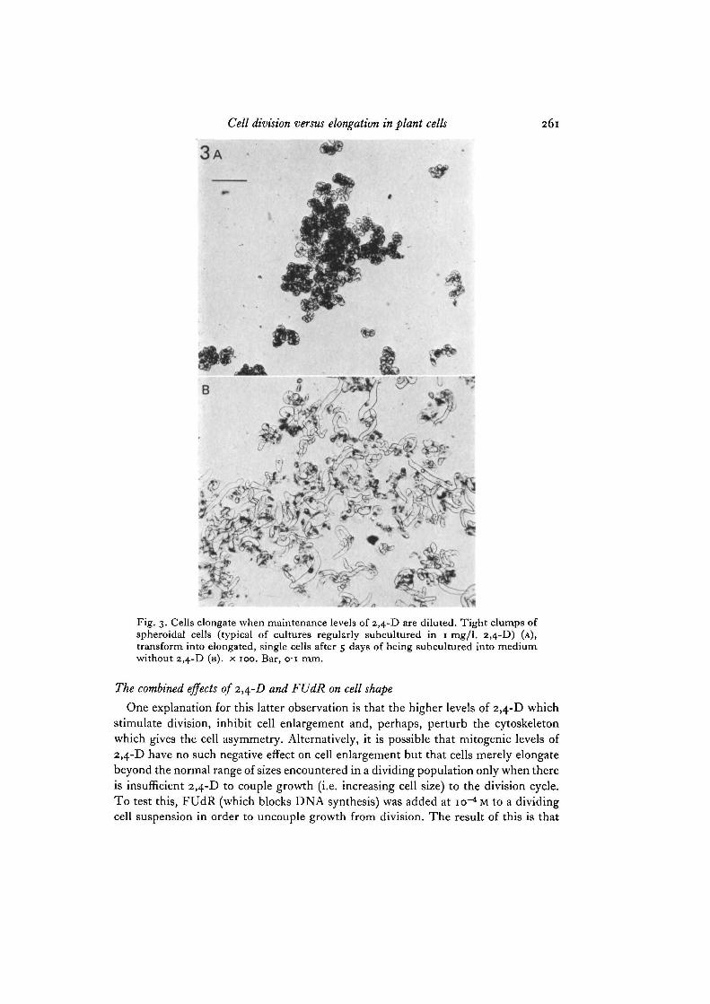
Elongated, sausage-shaped cells are a feature of many plant cell suspensions andexperience with this carrot culture showed that such cells were more abundant whenit was in the stationary phase of growth. To establish the relationship between growthconditions and the formation of such cells, cell length was measured after 5 days byexamining replicates of a suspension which had been maintained in 1 mg/1. 2,4-D,washed several times in medium without 2,4-D and then subcultured into freshmedium containing o, o-oi, o-i, i-o, 3-0, 5-0 or 25-0 mg/1. 2,4-D. Some elongatedcells are always found in stock cultures maintained by frequent subculture in 1 mg/1.2,4-D but the main characteristic of such cultures is the small, regular, clumps ofspheroidal cells and this is reflected in the Image Analysis histogram (see Fig. 1)which shows a narrow distribution for cell length. When washed into zero 2,4-D,however, cells elongate and become distributed over a broader range of lengths. Thiseffect depends upon the concentration of 2,4-D and Fig. 2 demonstrates that, onaverage, cells tend to be longer at lower levels of 2,4-D. That longer cells divide lessfrequently is also shown in this figure. The optimal concentration of 2,4-D for celldivision is 1 mg/1. - the concentration used for routine subculturing. Higher levels(e.g. 3 mg/1.) are somewhat less stimulatory for division but below the optimum thereis a sharp decrease in division rate and Fig. 2 displays the reciprocity between divisionand elongation over the range of o to i-omg/1. 2,4-D. The general appearance ofthese cultures is illustrated in Fig. 3 which shows the regular clumps of spheroidalcells at i-o mg/1. 2,4-D (Fig. 3 A) and the variety of elongated cell shapes encounteredin cultures maintained in zero 2,4-D (Fig. 3B).
These results suggest that 2,4-D strikes the balance between cell division and cellelongation.

260 C. W. Lloyd, S. B. Lowe and G. W. Peace
10
100 200 300 400 500 600
ion
CD
3
a&"o
30
20
10
200 300 400
Cell length, Mm
500—I—600
Fig. i. Distribution of the length of cells maintained in i-omg/1. 2,4-D (lowerhistogram) or subcultured into zero 2,4-D (upper histogram). Note that the narrowrange of cell sizes in i-o mg/1. 2,4-D broadens and that longer cells are encounteredafter 5 days in zero 2,4-D.
175
150
Ea.
t 125g
100
75
-25
- 2 0
1 5 1-10
0 1 2 32, 4-D, mg/l.
Fig. 2. Average cell length and cell multiplication as a function of the concentrationof 2,4-D. Over 5 days, cells multiply better at i-o mg/1. 2,4-D than in zero 2,4-Dbut over this range, the average cell length decreases as cell density increases.
Cell division versus elongation in plant cells 261
Fig. 3. Cells elongate when maintenance levels of 2,4-D are diluted. Tight clumps ofspheroidal cells (typical of cultures regularly subcultured in 1 mg/1. 2,4-D) (A),transform into elongated, single cells after 5 days of being subcultured into mediumwithout 2,4-D (B). x 100. Bar, o-i mm.
The combined effects of 2,4-D and FUdR on cell shape
One explanation for this latter observation is that the higher levels of 2,4-D whichstimulate division, inhibit cell enlargement and, perhaps, perturb the cytoskeletonwhich gives the cell asymmetry. Alternatively, it is possible that mitogenic levels of2,4-D have no such negative effect on cell enlargement but that cells merely elongatebeyond the normal range of sizes encountered in a dividing population only when thereis insufficient 2,4-D to couple growth (i.e. increasing cell size) to the division cycle.To test this, FUdR (which blocks DNA synthesis) was added at io~* M to a dividingcell suspension in order to uncouple growth from division. The result of this is that

C. W. Lloyd, S. B. Lowe and G. W. Peace
Fig. 4. Addition of FUdR to dividing cultures arrests cell multiplication and allowscells to elongate. Cells maintained in 1 mg/l. 2,4-D (A) elongate in the presence ofio~* M FUdR (B) even though the fresh medium also contained 1 mg/l. 2,4-D whichotherwise gives rise to non-elongate spheroidal cells. This effect of FUdR can beinhibited by adding thymidine at io~* M. X 240. Scale bar, c i mm.
the arrested cells elongate (Fig. 4A) (much in the way that cells do in stationary phasecultures or in low/no 2,4-D) even though the level of 2,4-D used (1 mg/l.) was suchthat spheroidal dividing cells were produced in controls without FUdR (Fig. 4B).This effect of FUdR can be bypassed and, hence, negated by adding thymidine atio~* M (which confirms that cell cycle arrest occurs at the level of de novo thymidinesynthesis) and has been performed in the presence of uridine at I O ^ M with noobservable differences (which argues against a spill-over effect on RNA metabolism).This demonstrates that cells elongate in some phase of the cell cycle up to the G^S

Cell division versus elongation in plant cells 263
boundary when prevented from progressing through 5-phase (and eventually on tomitosis/cytokinesis). In addition, these observations provide strong presumptiveevidence against mitogenic levels of the growth substance having any negative effecton structural elements which support directional enlargement.
The effect of 2,4-Z) on microtubules
Elongated carrot cells are known from previous studies to contain transverse hoopsof laterally associated microtubules (Lloyd et al. 1979a, b). These microtubulesprobably support the development of cell polarity and this has been demonstrated inother studies (Lloyd et al. 1980) in which depolymerization of microtubules bycolchicine results in loss of elongated shape. If 2,4-D were perturbing this micro-tubular cytoskeleton to form spheroidal, morphologically undifferentiated cells itcould be expected to be acting at one of several levels: microtubule polymerization;microtubule-microtubule interaction to form bundles; the formation of transversehoops from such bundled microtubules; the co-axial alignment of separate hoops.Electron-microscopic examination of thin sections shows that cells grown in 'higher'levels of 2,4-D contain abundant microtubules which have been seen in concen-trations of 2,4-D as high as 25 mg/1. In this case, the parallel arrangement of adjacentmicrotubules is also preserved. Without a long axis to help orientate such cells insection, it is difficult to reconstruct the presumed 3-dimensional pattern of micro-tubules. Sections are, however, regularly encountered which show microtubulessectioned in patterns consistent with a predominantly co-axial arrangement (Fig. 5).But the most convincing evidence that mitogenic 2,4-D does not affect any of thelevels of microtubular organization is shown in Fig. 6. In this micrograph, transversehoops of microtubules - of the kind seen in low 2,4-D controls - are found in a cellwhich elongated in 1 mg/1. 2,4-D when FUdR was added. There is nothing tosuggest, therefore, that higher, mitogenic levels of 2,4-D perturb the microtubularcytoskeleton or prevent the rearrangements which must occur to it during the transi-tion from spheroidal to elongated shape.
DISCUSSION
The results presented here indicate that for suspended carrot cells, cell divisionand cell elongation are reciprocally related and that the balance of this equation canbe variably adjusted by the growth regulator 2,4-D.
That 2,4-D should affect cell division was to be expected and has been describedpreviously for many cell types (e.g. Peaud-Lenoel, 1977), including sycamore cellsuspensions, in which threshold levels of this growth regulator are required to initiatemitosis (Leguay & Guern, 1977); below these levels the cells become quiescent in G1
and elongate (Street, Collin, Short & Simpkins, 1968). This also applies to carrot sinceNishi, Kato, Takahashi & Yoshida (1977) have shown that carrot suspension cells arearrested in Gx by the deprivation of 2,4-D. What is interesting about the present datais not that 2,4-D affects cell division but the reciprocity between division andelongation. In this discussion, we concentrate upon why such processes should bemutually excluding and in this respect the FUdR experiments are particularly

264 C. W. Lloyd, S. B. Lowe and G. W. Peace
Fig. 5. Thin section of a cell maintained in 1 mg/1. 2,4-D. Regularly spaced corticalmicrotubules run parallel to one another and show a pattern consistent with trans-verse banding. Arrowheads indicate groups of parallel cortical microtubules. x 25 500.Scale bar, 1 /im.

Cell division versus elongation in plant cells
Fig. 6. Immunofluorescent staining of microtubules. Spheroidal cells, maintainedin i mg/1. 2,4D, were treated with FUdR to inhibit entry into the division cyclewhereupon the cells elongate. The microtubules in these elongated cells were stainedby indirect immunofiuorescence. The pattern of transverse microtubules is similarto that previously reported foi carrot cells elongated in zero 2,4-D (Lloyd et al.1979a) which indicates that the presence of 1 mg/1. 2,4-D does not perturb thedevelopment of the interphase arrays of cortical microtubules which supportelongated shape, x 1000. Scale bar, 001 mm.
revealing, for this drug uncouples the 2 phenomena. Upon the addition of FUdR,cells begin to elongate. Elongation is not, therefore, directly dependent upon con-comitant DNA synthesis and in keeping with this, others have suggested that RNAand protein biosynthesis are more essential features of cell elongation (Street et al.1968; Nishi et al. 1977; Barlow, 1969).
Barlow (1969) previously used another inhibitor of DNA synthesis, hydroxyurea,to uncouple growth from division in roots of Zea mays, and his observation thatpolarity of growth was maintained under such circumstances is relevant here, for weobtain a similar result with FUdR for carrot cells in suspension. However, from thepoint of view of cell morphology, the intriguing part of the present study is not that aninhibitor of DNA synthesis leaves cell polarity undisturbed but that it presents adevice for demonstrating that cells are quite capable of elongating in the higher levelsof 2,4-D which, in the absence of FUdR, would have caused them to divide and remainunelongated. Clearly, such mitogenic levels of 2,4-D have no intrinsic negative effecton cell polarity. In the light of these, and other findings, the following scheme appearsto be the simplest explanation of the way in which cell division is related directly, andcell elongation, inversely, to the concentration of 2,4-D.
Provided the nutrient medium contains all appropriate factors, cells will increasein size. Whether or not the lower concentrations of 2,4-D used here accelerate thisincrease in size (elongation), they clearly do not stimulate cell division as much as dothe higher levels. It is therefore proposed that cells in Gx will, up to a point, continueto grow until some event or events (which requires 2,4-D to be present above acritical level) propels the cell out of this indeterminate phase into DNA synthesis and
18 CEL 45

266 C. W. Lloyd, S. B. Lowe and G. W. Peace
mitosis. In plants (Bayliss, 1975), as for animal cells (e.g. Smith & Martin, 1973), theperiod prior to DNA synthesis (Glt Go, A) appears to be the most variable phase ofthe cycle, whereas, once initiated, the sequence of events from DNA synthesis tomitosis is relatively constant. Our interpretation is that above some critical level of2,4-D cells continue to cycle (i.e. couple growth to division) whereas they grow, butdo not divide, when 2,4-D is diluted or metabolized to below this level or when the2 processes are metabolically uncoupled as with FUdR and hydroxyurea. In this way,the lower is the concentration of 2,4-D, the more likely is an increased residence timein Gx where the cell continues to grow in size.
As Hartwell, Culotti, Pringle & Reid (1974) have argued, a characteristic range ofcell sizes with upper and lower limits can be maintained by some mechanism whichcouples the 2 separate processes of cell growth and division which increase anddecrease cell mass in turn. It is in Gx that cells test their environment by way of itsability to sustain growth and measure it against some (as yet undetermined) internalstandard for division. In order to maintain the average cell size, recently divided cellsmust therefore grow to approximately double their size in the pre-replicative phasebefore entering upon a sequence of events which commits them to halving their sizeonce more. In view of the highly elongated cells in non-dividing cultures, size aloneis clearly not a trigger for division (although the mass:volume ratio of cells is low,due to vacuolation) so that cells continue to grow in the absence of mitogenic levels ofauxin. That a mitogenic factor (i.e. auxin) is required in plant cells to initiate divisionin addition to a presumably adequate nutrient supply is probably a reflexion of thesocial factors required by multicellular organisms to coordinate growth and division.In elongating tissues in vivo it follows that as cells are distanced by division fromconcentrated regions of division-initiating factors (meristems) they cease to cycle andbegin to elongate and differentiate at the expense of division. Gradients of such factorstherefore produce a graded series of growth states which reflects, to varying degrees,events seen in miniature in the individual cell cycle. In suspension cultures, nocontinuous gradients exist such as those postulated between the meristem and thezone of enlargement, although varying the levels of exogenous growth regulatorsprobably goes some way towards mimicking this process in culture.
Finally, it is probably important to know why, when cells enlarge, they adoptpolarized, cylindrical shapes instead of becoming non-polar, inflated spheres. Inflatedspheres can be formed from these carrot cells by depolymerizing the microtubuleswith colchicine (Lloyd et al. 1980), which supports the view that cell polarity isdependent upon the presence of transverse, cytoplasmic hoops of microtubules. Theprocess in which the microtubular cytoskeleton, characteristic of the elongated cell,evolves from that of the unelongated cell would therefore seem to be an importantfeature in the acquisition of cell (and tissue) polarity and is at the centre of ourinterest in 2,4-D as a modulator of growth and form. In principle, the ' elongated'cytoskeleton could evolve from that of unelongated cells in at least 2 different ways,depending on the nature of microtubular arrays in the latter. They could alreadyexist in transverse hoops or girdles, being a smaller version of the elongated cell'scytoskeleton, like a compressed spring prior to being sprung. Alternatively, it could

Cell division versus elongation in plant cells 267
be that microtubules are disorganized in rapidly dividing cells and that hoops onlybegin to form or tend towards a co-axial alignment when the cell begins to expandupon dilution of 2,4-D. However, we find no evidence for a breakdown in the micro-tubular cytoskeleton of rapidly dividing carrot cells and as far as we are able to judgefrom the limited view afforded by thin sections, parallel bands of microtubulesalready exist in some unelongated cells. This tends to fit the 'coiled spring' hypothesisand agrees with the observation (Gunning, Hardham & Hughes, 1978) that cyto-plasmic microtubules are randomly arranged in roots of Azolla for only a brief periodafter cytokinesis, until microtubules polymerized from separate microtubule-organizing centres can interdigitate to establish the transverse hoops which characterizecells in interphase. Further elongation of the cells is then suggested to be accompaniedby an interpolation of additional microtubules (Hardham & Gunning, 1979; Gunning& Hardham, 1979).
The main conclusion to be drawn from the present results is that the spectrum ofcell shapes and sizes produced by a graded series of the growth substance 2,4-D isunlikely to be produced by variable perturbation of morphogenetic arrays of micro-tubules but is a result of whether or not cells can limit the visible effects of enlarge-ment by entering the division cycle.
We thank Drs D. A. Rees, L. H. Jones and A. R. Slabas, for discussion and A. J. Powell forperforming the immunofluorescence work.
REFERENCESBARLOW, P. W. (1969). Cell growth in the absence of division in a root meristem. Planta 88,
215-223.BARLOW, P. W. (1976). Towards an understanding of the behaviour of root meristems. J. theor.
Biol. 57, 433-451.BAYLISS, M. W. (1975). The duration of the cell cycle of Daucus carota L. in vivo and in vitro.
Expl Cell Res. 92, 31-38.GUNNING, B. E. S. & HARDHAM, A. R. (1979). Microtubules and morphogenesis in plants.
Endeavour, New Series 3, 112-117.GUNNING, B. E. S., HARDHAM, A. R. & HUGHES, J. E. (1978). Evidence for initiation of micro-
tubules in discrete regions of the cell cortex in Azolla root-tip cells, and an hypothesis on thedevelopment of cortical arrays of microtubules. Planta 143, 161-179.
HALPERIN, W. & JENSEN, W. A. (1967). Ultrastructural changes during growth and embryo-genesis in carrot cell cultures. J. Ultrastruct. Res. 18, 428-443.
HALPERIN, W. & WETHERELL, D. F. (1964). Adventive embryony in tissue cultures of the wildcarrot, Daucus carota. Am. J. Bot. 51, 274-283.
HARDHAM, A. R. & GUNNING, B. E. S. (1979). Interpolation of microtubules into corticalarrays during cell elongation and differentiation in roots of Azolla pinnata. J. Cell Set. 37,411-442.
HARTWELL, L. H., CULOTTI, J., PRINGLE, J. R. & REID, B. J. (1974). Genetic control of the celldivision in yeast. Science, N. Y. 183, 46—51.
JONES, L. H. (1974). Factors influencing embryogenesis in carrot cultures (Daucus carota L.).Ann. Bot. 38, 1077-1088.
LEGUAY, J.-J. & GUERN, J. (1977). Quantitative effects of 2,4-dichlorophenoxyacetic acid ongrowth of suspension-cultured Acer pseudoplatanus cells. II. Influence of 2,4-D metabolismand intracellular pH on the control of cell division by intracellular 2,4-D concentration.PI. Physiol., Lancaster 60, 265-270.
18-2

268 C. W. Lloyd, S. B. Lowe and G. W. Peace
LLOYD, C. W., SLABAS, A. R., POWELL, A. J., MACDONALD, G. & BADLEY, R. A. (1979a).Cytoplasmic microtubules of higher plant cells visualised with anti-tubulin antibodies.Nature, Lond. 279, 239-241.
LLOYD, C. W., SLABAS, A. R., POWELL, A. J., MACDONALD, G., LOWE, S. B. & PEACE, G.(1979&). Microtubules in higher plant cells and protoplasts. In Proc. Vth int. ProtoplastSymp., Szeged, Hungary. Oxford: Pergamon.
LLOYD, C. W., SLABAS, A. R., POWELL, A. J. & LOWE, S. B. (1980). Microtubules, protoplastsand plant cell shape: an immunofluorescent study. Planta 147, 500—506.
NISHI, A., KATO, K., TAKAHASHI, M. & YOSHIDA, R. (1977). Partial synchronization of carrotcell culture by auxin deprivation. Physiol. PI. 39, 9-12.
PEAUD-LENOEL, C. (1977). The hormonal regulation of the cell division cycle. In Plant GrowthRegulation (ed. P. E. Pilet), pp. 240-248. Berlin: Springer.
SMITH, J. A. & MARTIN, L. (1973). Do cells cycle? Proc. natn.Acad. Sci. U.S.A. 70, 1263-1267.STREET, H. E., COLLIN, H. A., SHORT, K. & SIMPKINS, I. (1968). Hormonal control of cell
division and expansion in suspension cultures of Acer pseudoplatanus L.: the action o(kinetin. In Biochemistry and Physiology of Plant Growth Substances (ed. F. Wightman &G. Setterfield), pp. 489-504. Ottawa, Canada: Runge Press.
(Received 28 February 1980)


![[RKRD] 2,4 MB Bundesliga](https://static.fdocuments.in/doc/165x107/568bd6511a28ab20349b9fe9/rkrd-24-mb-bundesliga.jpg)


