The Microcirculation. The microcirculation Regulation of blood flow through tissues – Not all...
25
The Microcirculation
-
Upload
meredith-summers -
Category
Documents
-
view
234 -
download
4
Transcript of The Microcirculation. The microcirculation Regulation of blood flow through tissues – Not all...
The Microcirculation
The microcirculation
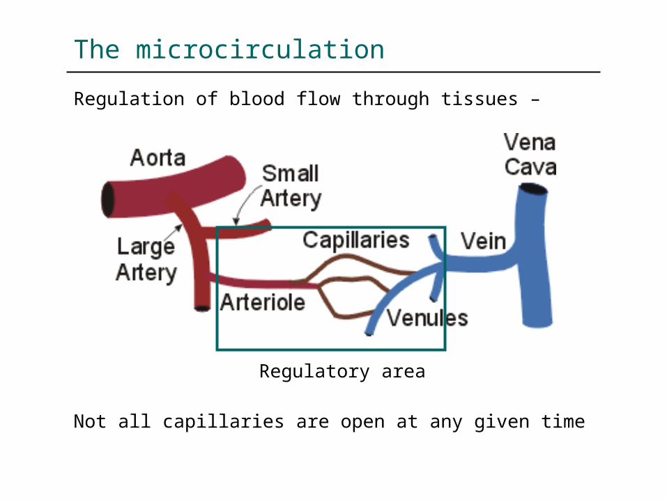
Regulation of blood flow through tissues –
Not all capillaries are open at any given time
Regulatory area

The microcirculation
Regulation of blood flow through tissues –
Primary arteriolesInnervated-sympathetic
Secondary & tertiary arteriolesLess smooth muscle, sparselyinnervated
Terminal arterioles Capillaries
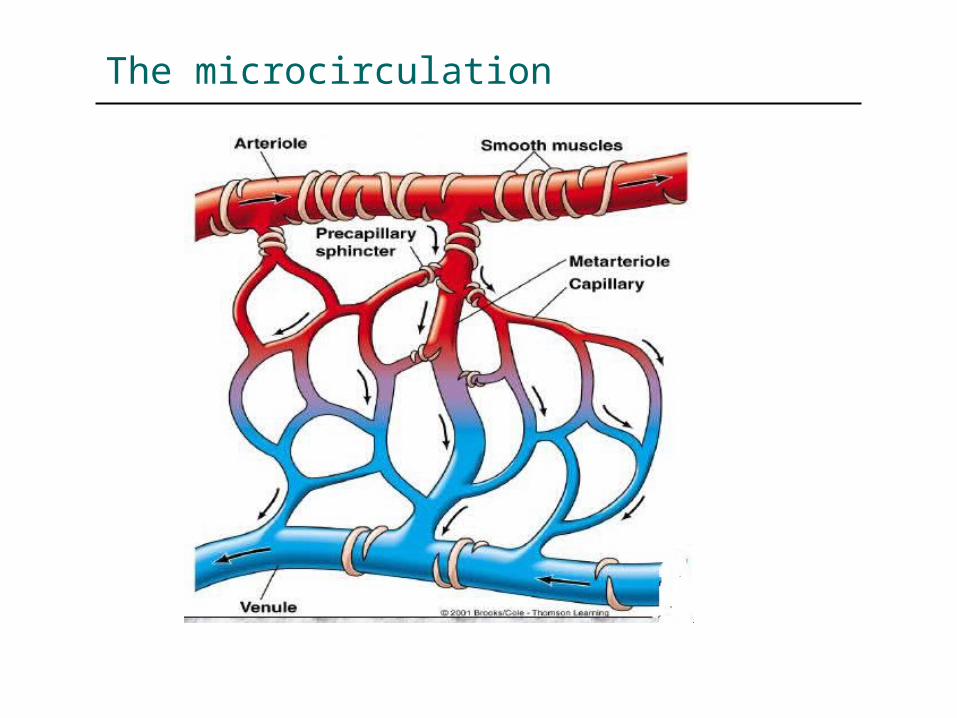
precapillary sphincter – at the start of capillary
The microcirculation

Blood flow is directly proportional to the metabolic demands and other needs of the tissues
For example, Blood flow through –Brain – 14%, 700ml/min, 50ml/min/100gHeart – 4%, 200ml/min, 70ml/min/100gKidneys – 22%, 1100ml/min, 360ml/min/100gLiver – 27%, 1350ml/min, 95ml/min/100gThyroid – 6%, 50ml/min, 160ml/min/100g
The microcirculation
Blood flow through tissues - 2 phases of regulation
Acute regulation/control Long term regulation
Acute control is achieved by local constriction or dilatation of arterioles, metarterioles, precapillary sphincters
The microcirculation - regulation

The microcirculation - acute regulation

Long term increase to a growing tissue/ tissue with increased demands over a period of days weeks due to generation of new vessels Angiogenesis – growth of new vessels Angiogenic factors – endothelial cell growth
factor, fibroblast growth factor, angiogenin
Collateral circulation Block in an artery/vein – development of a
new vascular channel allowing partial re-supply of blood to the affected tissue
The microcirculation – long term regulation
Exchange vessels There is a free exchange of water,
electrolytes, and small molecules between the intravascular and extravascular compartments of the body
The primary site of this exchange is capillaries and small post-capillary venules (sometimes grouped together and called "exchange vessels").
The exchange vessels
The exchange vessels
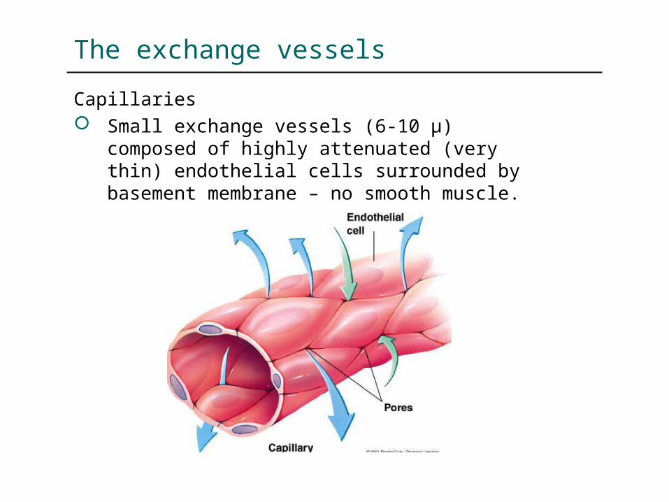
Capillaries Small exchange vessels (6-10 µ) composed of
highly attenuated (very thin) endothelial cells surrounded by basement membrane – no smooth muscle.

The exchange vessels
Capillaries
Three structural types

The exchange vessels
Capillaries - structural types Continuous (found in muscle, skin, lung,
central nervous system) – basement membrane is continuous and intercellular clefts are tight (i.e., have tight junctions); these capillaries have the lowest permeability
Fenestrated (found in exocrine glands, renal glomeruli, intestinal mucosa) – perforations (fenestrae) in endothelium result in relatively high permeability
Discontinuous (found in liver, spleen, bone marrow) – large intercellular gaps and gaps in basement membrane result in extremely high permeability
The exchange vessels
Capillaries Large surface area and relatively high
permeability (especially at intercellular clefts) to fluid and macromolecules make capillaries the primary site of exchange for fluid, electrolytes, gases, and macromolecules
In some organs, precapillary sphincters (a circular band of smooth muscle at entrance to capillary) can regulate the number of perfused capillaries
The exchange vessels
Venules Small exchange vessels (10-50 µ) composed
of endothelial cells surrounded by basement membrane (smallest postcapillary venules) and smooth muscle (larger venules)
Fluid and macromolecular exchange occur most prominently at venular junctions
Sympathetic innervation of larger venules can alter venular tone which plays a role in regulating capillary hydrostatic pressure
Fluid exchange
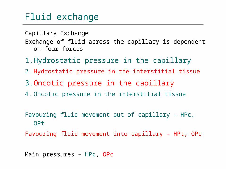
Capillary ExchangeExchange of fluid across the capillary is dependent on
four forces
1. Hydrostatic pressure in the capillary
2. Hydrostatic pressure in the interstitial tissue
3. Oncotic pressure in the capillary
4. Oncotic pressure in the interstitial tissue
Favouring fluid movement out of capillary – HPc, OPt
Favouring fluid movement into capillary – HPt, OPc
Main pressures – HPc, OPc

Fluid exchange
Starling Forces
The pressures that influence capillary exchange
Net driving force (net filtration pressure) is the arithmetic sum of all pressures

Fluid exchange
Capillary Exchange – (Systemic circulation)Pressures at the arterial and venous end of capillary (mmHg)
HPc 35 15
HPt 3 3
OPc 25 27
OPt 7 7
Arterial Venous
NFP + 14 - 8

Fluid exchange
Capillary Exchange The exchange of fluid across a capillary is
determined by hydrostatic and oncotic pressure gradients
Properties of of the capillary wall also influence fluid exchange - permeability and surface area of the capillaries
These relationships can be summarized by the following equation:JV = KF A [(PC – PT) – (pC - pT)][where, JV = rate of fluid movement, KF = capillary filtration constant, A = surface area for exchange, PC and PT = capillary and tissue hydrostatic pressures, and pC and pT = capillary and tissue oncotic pressures]
Fluid exchange
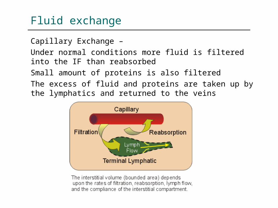
Capillary Exchange –Under normal conditions more fluid is filtered into the IF than reabsorbed Small amount of proteins is also filtered The excess of fluid and proteins are taken up by the lymphatics and returned to the veins
Fluid exchange
Terminal Lymphatics Composed of endothelium with intercellular gaps
surrounded by highly permeable basement membrane and are similar in size to venules – terminal lymphatics terminate as blind sacs
Larger lymphatics also have smooth muscle cells. Spontaneous and stretch-activated vasomotion is
present which serves to "pump" lymph Sympathetic nerves can modulate vasomotion and
cause contraction One-way valves direct lymph away from the tissue
and eventually back into the systemic circulation via the thoracic duct and subclavian veins (2-4 liters/day returned)

Fluid exchange
Mechanisms of fluid exchange
Fluid, electrolytes, gases, small and large molecular weight substances can transverse the capillary endothelium by several different mechanisms
Diffusuion – gases, lipid soluble substancesBulk flow – fluid and electrolytes
through pores Vesicular transport – macromolecules Active transport – not a major mechanism
Abnormalities of fluid exchange
Excess fluid accumulation in interstitial fluid
May occur due to a disturbance in the factors that influence filtration / reabsorption process
This condition is called ‘oedema’
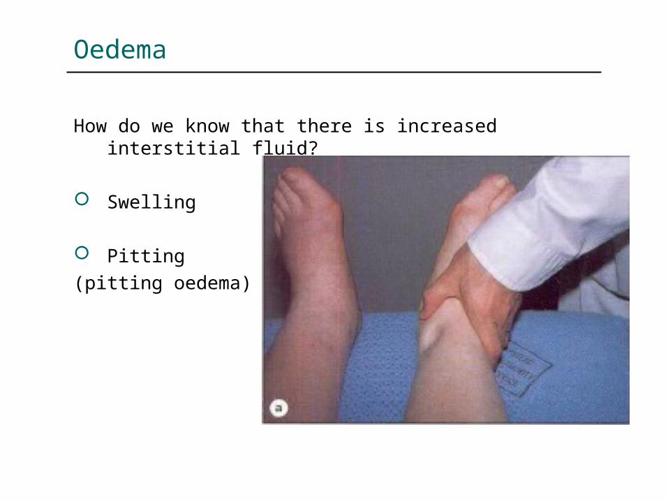
Oedema
How do we know that there is increased interstitial fluid?
Swelling
Pitting (pitting oedema)
Abnormalities of fluid exchange
Mechanisms of oedema
Disturbances in Starling forces Increased capillary hydrostatic pressure Decreased plasma oncotic pressure (other two forces are insignificant)
Increased capillary permeability
Lymphatic obstruction