The metabolism and toxicology of rutin on liver type cells
86
The metabolism and toxicology of rutin in liver-type cells Andrew Hoffman Statement of originality: I declare that, with the exception of any statements to the contrary, the contents of this report/dissertation are my own work, that the data presented herein has been obtained by experimentation and that no part of the report has been copied from previous reports/dissertations, books, manuscripts, research papers or the internet. 1
-
Upload
andrew-hoffman -
Category
Documents
-
view
100 -
download
2
Transcript of The metabolism and toxicology of rutin on liver type cells

The metabolism and toxicology of rutin in liver-type cells Andrew Hoffman
Statement of originality:I declare that, with the exception of any statements to the contrary, the contents of this report/dissertation are my own work, that the data presented herein has been obtained by experimentation and that no part of the report has been copied from previous reports/dissertations, books, manuscripts, research papers or the internet.
Signed.............................................. Print name................................................................Date.........................................................
1

The metabolism and toxicology of rutin in liver-type cells Andrew Hoffman
Contents1. Abstract.............................................................................................................................................3
Abbreviations....................................................................................................................................3
2. Introduction.......................................................................................................................................4
3. Methods..........................................................................................................................................11
3.1 – Materials.................................................................................................................................11
3.2 – Making up the Media for the growing of the cells..................................................................11
3.3 – Splitting the cells.....................................................................................................................12
3.4 - Preparing the compounds.......................................................................................................13
3.5 – Preparing the dilutions............................................................................................................14
3.6 – Dosing the cells.......................................................................................................................17
3.7 – MTT Protocol - Running the MTT Assay..................................................................................18
4 .Results.............................................................................................................................................19
4.1 - Short Term assay (HepG2 4000 cells per well [48 hours]).......................................................19
4.2 - Long Term Assay (HepG2 2000 cells per well [1 week (168 hours)]).......................................19
4.3 - Short Term Assay Repeat (HepG2 4000 cells per well [48 hours])...........................................20
4.4 - Long Term Assay Repeat (HepG2 2000 cells per well [1 week (168 hours)])...........................20
5. Discussion........................................................................................................................................22
Results obtained..............................................................................................................................22
Problems which may have occurred and changes which could be made........................................24
Alternative approaches to the methodology...................................................................................25
MTT Assay....................................................................................................................................25
S9 Fraction...................................................................................................................................27
6. Acknowledgments...........................................................................................................................31
7. References.......................................................................................................................................32
8. Appendix..........................................................................................................................................36
Ethics Form..................................................................................................................................66
2

The metabolism and toxicology of rutin in liver-type cells Andrew Hoffman
1. Abstract
Rutin is a type of flavonoid which are a group of plant metabolites. It is found predominantly
in fruit, vegetables and in around 70 different types of plant. For medicinal use, it is found
mainly in buckwheat. A study was conducted to look at the possible anti-cancerous effects
of rutin on hepatocellular carcinoma cells (the HepG2 cell line). Out of two repeated tests,
the results were fairly inconclusive as 50% of the results found no particular correlation
between the percentage of cell viability (how many cells survived) and the concentration of
rutin. The results also showed a high EC50 in pharmacological terms. The following also
contains critical analysis of other methods which could have been used to study these
effects.
3
Abbreviations17-β-E2 - Endogenousestrogen17-β-estradiolADME – Absorption, Distribution, Metabolism and ExcretionCYP – Cytochrome P450 enzymeCPZ - ChlorpromazineFBS – Foetal bovine serumMTT - 3-(4,5-dimethylthiazol-2-yl)-2,5-diphenyltetrazolium bromideNADPH - Nicotinamide adenine dinucleotide phosphatePES – Phenazine ethyl sulphatePMS - Phenazine methyl sulphateRedox – Reduction and OxidationRNS – Reactive Nitrogen SpeciesRPMI 1640 – Roswell Park Memorial Institute MediumS9 Fraction – post-mitochondrial supernatant fraction

The metabolism and toxicology of rutin in liver-type cells Andrew Hoffman
2. Introduction
Rutin is a flavonoid, which are a type of plant metabolite. When they were first discovered,
flavonoids were referred to as Vitamin P (due to their effect on vascular capillary
permeability). Rutin itself is found in many different fruits and vegetables and in over 70
different types of plant (Chua, 2013) but is most notably found for medicinal use in
Buckwheat (Fagopyrum esculentum Moench) (Kreft, et al., 1999). There is even a slight
chance that rutin can be found in Tobacco leaves (Fathiazad, et al., 2006). Rutin is the
joining of quercetin and rutinoside by a glycosidic bond, as shown in figure 1.
Figure 1 – Skeletal structure of a rutin molecule (quercetin-3-O-rutinoside).
It was believed that rutin strengthens the blood vessels and that because of this feature, it
could be used in treatment of varicose veins, internal bleeding, haemorrhoids and
haemorrhagic strokes. Since then, rutin has been acknowledged for its many medicinal
properties that it could be used for, detailed below.
Rutin has huge antioxidant capabilities as it is a wonderful free radical inhibitor (Korkmaz &
Kolankaya, 2010). Free radicals are created in vivo during respiration in mitochondria and
4

The metabolism and toxicology of rutin in liver-type cells Andrew Hoffman
are also found to be released by peroxisomes. Free radicals are known to be a catalyst for
certain redox reactions in the body and are generated either due to a persons’ diet being
unhealthy or as a result of an external stimuli, such as an infection. Excessive production of
these free radicals can lead to destruction of cells and, ultimately, the damage of tissues and
organs. High concentrations of naturally found rutin (buckwheat hull) have been found to
diminish the amount of NO2- and NO3
- (RNS) (Khan, et al., 2009). When investigated at as
little as 0.05 mg/mL, rutin showed an inhibition total of around 90%, almost as powerful as
Vitamin C which was at an inhibition rate of around 93% (Yang, et al., 2008).
Rutin also been proven to have anti-inflammation properties (Umar, et al., 2012).
Inflammation is an autonomic response to an injury that an organism might sustain. COX-2
is one of the main catalysts for the production of prostaglandins which induce the
inflammatory response. Although the inflammatory response is autonomic, it is not always
best for the body as inflammation can bring multiple problems, for example rheumatoid
arthritis and kidney failure. Rutin was proposed as a molecule to block the COX-2 pathway
(Guardia, et al., 2001) and at a concentration of 80µm there was significant inhibition on
macrophages as an in vitro model. Acting on a mouse for an in vivo model, rutin showed the
same inhibition properties when given at a 6mg dose (Shen, et al., 2002).
With its many properties, rutin is also considered to be a potent anti-adipogenic (Choi, et al.,
2006). Fatty liver is a treatable and manageable problem if and when a controlled diet and
exercise regime is implemented. If nothing is done to treat fatty liver, it can cause a whole
host of problems for the organism. In a particular study of the effect of flavonoids on fatty
liver (Choi, et al., 2006), rutin was introduced into the diet of mice that were given a high fat
diet. The mice given a high fat diet including rutin (25/ 50mg per kg of body weight on a
5

The metabolism and toxicology of rutin in liver-type cells Andrew Hoffman
daily basis) were found to have a reduced fat build up than those which had a high fat only
diet.
Previous studies on plant metabolites have shown that the compounds have similar
structures to hormones which regulate the endocrine system in a mammalian system.
Namely they have been recognised as phytoestrogens, a molecule with a similar structure to
estrogen (Guo, et al., 2012).Rutin has a similar structure to that of 17-β-E2,
endogenousestrogen17-β-estradiol (shown in figure 2), so it is possible that rutin could be
used to bind to an estrogen receptor, which would normally be occupied by 17-β-E2, and
prospectively act as estrogen (Tham, et al., 1998).
Figure 2 – Skeletal structure of 17- β-E2,
endogenousestrogen17-β-estradiol. Compared to rutin, the structures are similar due to the
3 planar benzene rings.
Flavonoids have been thought to have anti-cancerous properties and have been proven, on
an in vitro model, to inhibit a multitude of cancerous cell lines and ultimately stop and/or
minimize tumour growth and even cause it to regress (Van der Logt, et al., 2003). It has
been suggested that the size regression could be due to the inhibition of certain DNA
topoisomerases, namely topoisomerase I and topoisomerase II, which are involved in the
marking of DNA damage and chromosomal damage (Cantero, et al., 2006).
6

The metabolism and toxicology of rutin in liver-type cells Andrew Hoffman
Rutin has many other medicinal properties, although not much about the precise
mechanisms are known. Other medicinal properties include; anti-diabetes (Hao, et al.,
2012), renal protection (Kamalakkannan & Stanley Mainzen Prince, 2006), anti-asthma
(Jung, et al., 2007), gastroprotective (La Casa, et al., 2000), neuroprotection
(Tonjaroenbuangam, et al., 2011), cardioprotection (Annapurna, et al., 2009), Osteoarthritis
(only when used in combination with trypsin and bromelain) (Klein & Kullich, 2000) and
mucositis, a painful side effect of cancer treatment, characterized by the swelling and ulcer
formation in the mouth or lining of the digestive tract. It can even be used in veterinary
practice to treat animals which are suffering from idiopathic chylothorax (Kopco, 2005). It is
suggested that rutin is metabolised by microflora in the gut (Kuhnau, 1976) into smaller
metabolites including; quercetin, isoquercetin, HVA and other phenols (Arjumand, et al.,
2011).
Drug metabolism is part of drug ADME screening. ADME stands for the absorption,
distribution, metabolism and excretion. The absorption relates to how the drug is taken up,
this is why it is often referred to as the ‘administration step’ of the ADME screening. Drugs
can be administered in a number of ways, namely orally or intravenously. The amount of
absorption is dependent on factors like size, solubility, ionization and blood flow to the site
of administration. The amount of drug that was administered is not particularly the amount
that will take action. Drugs tend to be delivered in an inactivated state and the metabolism
of the drugs is what converts it or break it down into its activated materials. Distribution of
drugs is quite simply how the drugs traverse throughout the body to get to the target site.
Drugs can travel freely or can be bound to proteins in the plasma, i.e. albumin. Metabolism
of drugs, often referred to as the biotransformation, is the conversion or activation of a
drug. The main site of metabolism in a mammalian system is in the liver. The site has two
7

The metabolism and toxicology of rutin in liver-type cells Andrew Hoffman
phases of reactions; phase I and phase II. The reactions of phase I include redox and
hydrolysis which are controlled here by the CYP (Cytochrome P450) enzymes. Phase II
reactions deal with the conjugation of molecules with substrates, such as glucoronic acid,
which increase water solubility and aid renal elimination. Although renal (kidney)
elimination is the main source of drug excretion, other routes are still common e.g. faecal
matter, expiration via the lungs or sweat glands in the skin (Oh, 2002).
There are many different reactions in the liver that are involved in hepatic metabolism.
There are chemical reactions, such as protein synthesis, detoxification and the production of
digestive chemicals. The liver is also important for the metabolism of carbohydrates and is
the source of many substances that are imperative for good health. Glycogenesis,
glycogenolysis and gluconeogenesis are carbohydrate metabolism pathways which take
place in the liver as well as the conversion of carbohydrates into triglycerides. These
triglycerides are converted into free fatty acids by the liver which are released into the
bloodstream and used in other cells/tissues in the body (Sadava, et al., 2011).
Toxicology is the study of adverse effects of chemical, physical or biological agents. Viability
assays are a great way of studying this. MTT is a valuable assay for use in cell lines. It was
the first type of homogenous cell viability developed for high throughput screening
(Mosmann, 1983). MTT substrate is made up in a physiologically balanced solution, is added
to the cell culture and usually incubated for a period of 2 to 4 hours. Quantitatively, the
production of formazan is directly proportional to the number of viable cells in question.
Viable cells actively metabolise MTT to formazan, a purple coloured product. Only viable
cells can convert MTT to formazan, if the cell were to die it would lose its ability to
metabolise MTT, therefore it serves as a perfect assay to determine cell viability as dead
8

The metabolism and toxicology of rutin in liver-type cells Andrew Hoffman
cells would produce no colour change. Formazan accumulates in the cells as an insoluble
precipitate and is deposited close to the cell surface and in the culture medium. Formazan
has to be solubilised before it can be read in a photospectrometer. There are a number of
solubilisation solvents that can be used: acidified isopropanol, DMSO, dimethylformamide,
SDS and even combinations of detergent and organic solvents ( (Mosmann, 1983), (Hansen,
et al., 1989) and (Denizot & Lang, 1986)). The amount of signal generated for absorbance
readings are dependent on certain factors like the concentration of MTT, incubation period,
number of viable cells and their respective metabolic capabilities. MTT has a cytotoxic
nature, the higher its concentration the more toxic it becomes to the cells. A lower
concentration would be optimal but due to its toxicity, MTT is considered to be an ‘endpoint
assay’.
The S9 assay is an invaluable stability assay for use in mimicking a liver environment for the
hepatic cells being used for this in vitro study. The S9 contains a widespread variety of both
phase I and phase II enzymes, comprised respectively of microsomal and cytosolic enzymes
(Plant, 2004). This allows for a fairly complete metabolic profile for ADME screening. Due to
the liver being the main organ for drug metabolism, the S9 fraction is useful for monitoring
the hepatic clearance. The S9 is easy to prepare, can be stored for lengthy periods of time
and can be adapted for use in a high throughput screening like the MTT assay.
In this particular study, the main focus will be on the effect of rutin on cancerous hepatic
cells. In particular, the HepG2 cell line. The HepG2 cell line is derived from a Caucasian
teenage liver cancer patient. They were taken from this particular patient due to the well
differentiated hepatocellular carcinoma. They produce albumin, macroglobulin, antitrypsin,
transferrin and plasminogen (Sigma Aldrich, 85011430).
9

The metabolism and toxicology of rutin in liver-type cells Andrew Hoffman
The main reason for this study is to hopefully prove that rutin does in fact have anti-
cancerous properties and could be used to treat human hepatocarcinoma cells by looking at
the effects on the HepG2 cell line. This will be determined by the use of the S9 fraction and
MTT assay to monitor the metabolism and toxicology of rutin.
10

The metabolism and toxicology of rutin in liver-type cells Andrew Hoffman
3. Methods
3.1 – Materials
MTT, DMSO, Ethanol, L-Glutamine, PBS Buffer, FBS, RPMI (Fisher)
Glutamine (Bioserra)
CPZ, Flavonoid (Rutin), Trypan Blue, Non-essential amino acids, Penicillin (Sigma Aldrich)
NADPH (Apollo Scientific)
S9 Fraction (Invitrogen)
Virkon, deionised water (University of Salford)
HepG2 (Gift from cyprotex)
3.2 – Making up the Media for the growing of the cells
The media was made up using the following materials; RPMI 1640, glutamine (5ml), non-
essential amino acids (5ml), penicillin (5ml) and FBS (50ml). The media was added along
with the HepG2 cells into flasks and incubated to allow growth.
11

The metabolism and toxicology of rutin in liver-type cells Andrew Hoffman
3.3 – Splitting the cells
During the time of growth, the cells needed to be ‘split’.
The media which was already present was removed and the cells in the flask were washed
with PBS (2 times, 5ml each time). Trypsin (1ml) was added to the flask to disturb the cells
and the flask was incubated for 3 – 5 minutes. After the time had passed, the flask was
removed from incubation to see if the cells were freely moving and not stuck to the side of
the flask. If the cells are not freely moving when taken out, the flask was given a gentle tap
to ensure they dislodged from the side. Media (5ml) was reintroduced to the flask and the
mix was taken out and placed into a centrifuge tube. The tube was centrifuged at 1250 rpm
for 5 minutes. Once finished, the media was removed from the tube to leave only the pellet
which had formed (dead cells are found suspended in the supernatant and this is why the
media was discarded). Fresh media (3ml) was added to suspend the pellet and 0.5ml of the
newly suspended cells were taken and placed into a new flask. Fresh media (10ml) was
added to the flask and the flask was incubated again.
12

The metabolism and toxicology of rutin in liver-type cells Andrew Hoffman
3.4 - Preparing the compounds
The compounds were made up to be 2ml of a 10mM solution.
Mass in grams of rutin [1] needed:
M (Molarity )= MassMolecularWeight ×Volume
∴M ×MW×Vol=Mass
0.01X 160 X 0.02 = 0.122g
Amount of DMSO to be added:
Volume (¿be added∈ml )= MassMolecularWeight ×Molarity
×1000
¿0.122
160×0.01×1000
= 20 ml
13

The metabolism and toxicology of rutin in liver-type cells Andrew Hoffman
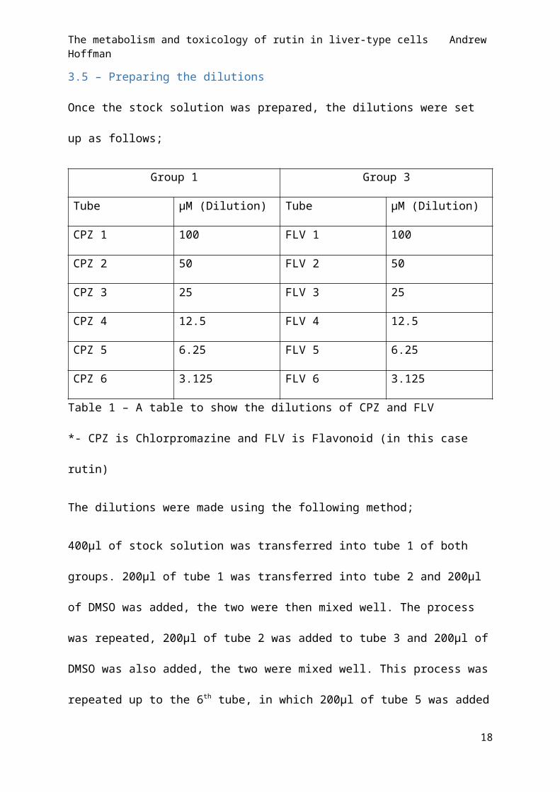
3.5 – Preparing the dilutions
Once the stock solution was prepared, the dilutions were set up as follows;
Group 1 Group 3
Tube µM (Dilution) Tube µM (Dilution)
CPZ 1 100 FLV 1 100
CPZ 2 50 FLV 2 50
CPZ 3 25 FLV 3 25
CPZ 4 12.5 FLV 4 12.5
CPZ 5 6.25 FLV 5 6.25
CPZ 6 3.125 FLV 6 3.125
Table 1 – A table to show the dilutions of CPZ and FLV
*- CPZ is Chlorpromazine and FLV is Flavonoid (in this case rutin)
The dilutions were made using the following method;
400µl of stock solution was transferred into tube 1 of both groups. 200µl of tube 1 was
transferred into tube 2 and 200µl of DMSO was added, the two were then mixed well. The
process was repeated, 200µl of tube 2 was added to tube 3 and 200µl of DMSO was also
added, the two were mixed well. This process was repeated up to the 6th tube, in which
200µl of tube 5 was added but only 100µl of DMSO was added this time. The compounds
were kept in the freezer for a period of 24 hours.
14

The metabolism and toxicology of rutin in liver-type cells Andrew Hoffman
Figure 3 - A drawing to represent the process of making up the dilutions.
a) 400µl of stock solution added to tube 1, b) 200µl taken forward from the previous tube
into the next tube, c) 200µl of DMSO added to the new tube, d) 100µl of DMSO added to
the new tube (only for the last tube).
The 12 previously made tubes were taken from the freezer and a new set of tubes was
made up by the following method;
From the first set of tubes, 20µl of group 1 CPZ 1 was transferred to the each of the new
groups, group 1 CPZ 1 and group 2 CPZ 1 + S9. 20µl of group 1 CPZ 2 was transferred to the
each of the new groups, group 1 CPZ 2 and group 2 CPZ 2 + S9. 20µl of group 1 CPZ 3 was
transferred to the each of the new groups, group 1 CPZ 3 and group 2 CPZ 3 + S9. 20µl of
group 1 CPZ 4 was transferred to the each of the new groups, group 1 CPZ 4 and group 2
CPZ 4 + S9. 20µl of group 1 CPZ 5 was transferred to the each of the new groups, group 1
CPZ 5 and group 2 CPZ 5 + S9. 20µl of group 1 CPZ 6 was transferred to the each of the new
groups, group 1 CPZ 6 and group 2 CPZ 6 + S9.
From the first set of tubes, 20µl of group 3 FLV 1 was transferred to the each of the new
groups, group 3 FLV 1 and group 4 FLV 1 + S9. 20µl of group 3 FLV 2 was transferred to the
each of the new groups, group 3 FLV 2 and group 4 FLV 2 + S9. 20µl of group 3 FLV 3 was
transferred to the each of the new groups, group 3 FLV 3 and group 4 FLV 3 + S9. 20µl of
15

The metabolism and toxicology of rutin in liver-type cells Andrew Hoffman
group 3 FLV 4 was transferred to the each of the new groups, group 3 FLV 4 and group 4 FLV
4 + S9. 20µl of group 3 FLV 5 was transferred to the each of the new groups, group 3 FLV 5
and group 4 FLV 5 + S9. 20µl of group 3 FLV 6 was transferred to the each of the new
groups, group 3 FLV 6 and group 4 FLV 6 + S9.
Once the new groups were made up, 380µl of fresh media was added to each of tubes in
Group 1 and Group 3. Groups 2 and 4 required S9 media, this was prepared by mixing
NADPH (6mg), 150µl of S9 and 6 ml of media. 380µl of this S9 media was added to each of
the tubes in groups 2 and 4.
Figure 4 – A drawing to show the how the dosing tubes are made up. This process is
repeated for each tube
*S9 media was made up using NADPH (6mg), S9 (150µl) and media (6ml).
16

The metabolism and toxicology of rutin in liver-type cells Andrew Hoffman
Group 1 Group 2 Group 3 Group 4Tube µM
(Dilution)Tube µM
(Dilution)Tube µM
(Dilution)Tube µM
(Dilution)CPZ 1 100 CPZ 1 +
S9100 FLV 1 100 FLV 1 +
S9100
CPZ 2 50 CPZ 2 + S9
50 FLV 2 50 FLV 2 + S9
50
CPZ 3 25 CPZ 3 + S9
25 FLV 3 25 FLV 3 + S9
25
CPZ 4 12.5 CPZ 4 + S9
12.5 FLV 4 12.5 FLV 4 + S9
12.5
CPZ 5 6.25 CPZ 5 + S9
6.25 FLV 5 6.25 FLV 5 + S9
6.25
CPZ 6 3.125 CPZ 6 + S9
3.125 FLV 6 3.125 FLV 6 + S9
3.125
Table 2 - A table to show the dilutions in each tube used for dosing the cells.
3.6 – Dosing the cells
Once the dilutions were complete, the cell plates could be dosed. Media, Media + S9, DMSO
and DMSO + S9 were used as controls in the experiment. The DMSO tubes were made up as
follows;
2 DMSO tubes were prepared, one with S9 and one without. The tube without S9 was made
up by mixing fresh media (570µl) with DMSO (30µl). The tube with DMSO with S9 was
prepared by mixing DMSO (30µl) with S9 media (570µl).
1 2 3 4 5 6 7 8 9 10 11 12A Media Media Media Media Media Media Media + S9 Media + S9 Media + S9 Media + S9 Media + S9 Media + S9B DMSO DMSO DMSO DMSO DMSO DMSO DMSO + S9 DMSO + S9 DMSO + S9 DMSO + S9 DMSO + S9 DMSO + S9C CPZ 1 CPZ 2 CPZ 3 CPZ 4 CPZ 5 CPZ 6 CPZ 1 CPZ 2 CPZ 3 CPZ 4 CPZ 5 CPZ 6D CPZ 1 + S9 CPZ 2 + S9 CPZ 3 + S9 CPZ 4 + S9 CPZ 5 + S9 CPZ 6 + S9 CPZ 1 + S9 CPZ 2 + S9 CPZ 3 + S9 CPZ 4 + S9 CPZ 5 + S9 CPZ 6 +S9E FLV 1 FLV 2 FLV 3 FLV 4 FLV 5 FLV 6 FLV 1 FLV 2 FLV 3 FLV 4 FLV 5 FLV 6F FLV 1 FLV 2 FLV 3 FLV 4 FLV 5 FLV 6 FLV 1 FLV 2 FLV 3 FLV 4 FLV 5 FLV 6G FLV 1 + S9 FLV 2 + S9 FLV 3 + S9 FLV 4 + S9 FLV 5 + S9 FLV 6 + S9 FLV 1 + S9 FLV 2 + S9 FLV 3 + S9 FLV 4 + S9 FLV 5 + S9 FLV 6 + S9H FLV 1 + S9 FLV 2 + S9 FLV 3 + S9 FLV 4 + S9 FLV 5 + S9 FLV 6 + S9 FLV 1 + S9 FLV 2 + S9 FLV 3 + S9 FLV 4 + S9 FLV 5 + S9 FLV 6 + S9
Table 3 - A table to show the layout of compounds in the plate wells
The wells in the cell plates were dosed with the compounds (as outlined in table 3), each
well was dosed with 25µl of said compound. Both plates were dosed at the same time, one
17
The metabolism and toxicology of rutin in liver-type cells Andrew Hoffman
plate had 4,000 cells per well and the second had 2,000 cells per well, and both plates were
incubated at 37.5oC.
3.7 – MTT Protocol - Running the MTT Assay
After dosing the cells, the plates were incubated at 37.5oC for different periods of time. The
plates which were seeded at 4,000 cells per well were only left to incubate for a period of 48
hours and this was used as a short term culture. The cell plate seeded at 2,000 cells per well
were left to incubate for a full week (168 hours) and was used as a long term culture.
The MTT assay was run by the following protocol;
MTT solution (50µl, 3mg/ml) was added to each well and was incubated for a further 3 – 4
hours at 37.5oC. After this time had passed, the media in the wells was discarded by hitting
the plate against a hard surface, to ensure that media held in the wells by water tension was
also discarded. The plates were left to dry for a further 1 – 2 hours. DMSO (100µl) was
added to each well and the plates were incubated at 37.5oC for 10 minutes. The plates were
then taken to the plate reader, which shook the plates well for approximately 15 seconds to
solubilize the formazan (a type of dye found in MTT) before reading the plates at a given
wavelength of 570nm.
18

The metabolism and toxicology of rutin in liver-type cells Andrew Hoffman
4 .Results
Below is an account of results obtained in the experiment, it should be known that all of the
graphs mentioned in the text can be found in the appendix section of the report.
4.1 - Short Term assay (HepG2 4000 cells per well [48 hours])
Graph 1 and graph 2 both show a great sigmoidal curve with regards to the cell viability.
They show that with an increased concentration of CPZ, the amount of cells which survived
decreased. This showed especially well in the S9 fraction as the viability was not over 100%,
whereas without S9 the cell viability was rather high at a low dose. Graph 3 and graph 4
show the cell viability of rutin on its own with no S9 added. Although there is no curve
shown, it is obvious that the cells have grown as the cell viability counts are at around 200%,
with some being even higher. Graph 5 and 6, like graphs 3 and 4, show the effect of rutin on
the cells but with the presence of S9. Compared to the previous graphs (3 and 4), it is again
shown that the cells grow at higher concentrations. With the presence of S9, at lower
concentrations, some of the cells have died but again, at higher concentrations it appears
that the cells have grown.
4.2 - Long Term Assay (HepG2 2000 cells per well [1 week (168 hours)])
Graph 7 and graph 8 show the cell viability for CPZ and CPZ with S9. CPZ on its own shows
another great sigmoidal curve showing that increased concentration of CPZ makes the cell
viability decrease. This shows true for CPZ with S9 also, as cell viability is below 70%, but the
results do not follow a significant pattern. The results for the long term flavonoid exposure
(graph 9 and graph 10) showed a negative correlation between cell viability and
concentration of the flavonoid. At lower concentrations the cell viability was higher than
100%, meaning cells had grown and/or replicated, but ultimately, as concentration
19

The metabolism and toxicology of rutin in liver-type cells Andrew Hoffman
increased, cell viability decreased below 100%. In the presence of rutin and the S9 fraction,
cell viability increased drastically as shown in graph 11 and graph 12. Here, it is shown that
at low concentrations (around 0.5 on the log scale) that rutin neither induces cell death or
cell growth. However, as concentrations of rutin increase, the cell viability climbs as high as
1000% in a very even pattern seemingly reaching a plateau at this point.
4.3 - Short Term Assay Repeat (HepG2 4000 cells per well [48 hours])
The cell viability for CPZ (graph 13) and CPZ with S9 (graph 14) in the repeated assays found
that viability was reduced as the concentration of CPZ increased. It should be noted that the
test of CPZ with S9 (graph 14) showed some growth at low concentrations. The results for
the cell viability in the presence of the flavonoid on its own (graph 15 and graph 16) show
that as concentrations of the flavonoid increase, the cell viability decreases. At first, cell
viability did increase slightly but increased concentrations show an overall decrease. When
the flavonoid and S9 were added together (graph 17 and graph 18), cell viability showed an
increase above 100%, but as the concentration of flavonoid and S9 increased, the cell
viability is negatively affected.
4.4 - Long Term Assay Repeat (HepG2 2000 cells per well [1 week (168
hours)])
The cell viability in the long term repeat for CPZ (graph 19) and CPZ with S9 (graph 20) again
show a negative correlation between cell viability and concentration of CPZ. In graph 20,
there is no showing of a curve however it is obvious from the plot that there is a negative
correlation. When the flavonoid was tested again, with no S9 added, results showed that
cell viability increased with concentrations of flavonoid but with no particular correlation.
Graph 21, the first flavonoid test in the repeat, shows that at a low concentration cell
20

The metabolism and toxicology of rutin in liver-type cells Andrew Hoffman
viability falls. This is likely to be an anomaly. Graphs 23 and 24 show the results for cell
viability when the flavonoid with S9 was added. The results, again, show that cell viability
increases when the flavonoid and S9 are added, however this time there is no particular
correlation between the two. All this shows is that there has been some growth, but does
not necessarily prove that the flavonoid and S9 are the cause of this.
21

The metabolism and toxicology of rutin in liver-type cells Andrew Hoffman
5. Discussion
Results obtained
The only results which follow a particular pattern of consistency are the tests which involve
CPZ. The results for CPZ across all 4 tests show a decrease in cell viability. The pattern is
shown in both the short and long term assays, the only slight difference in results are that of
lower concentrations having lower cell viability after long term exposure (whereas in the
short term the lower concentrations have slightly higher cell viability). The only obvious
outliers are shown in graph 1 and graph 20, both at the lowest concentration of CPZ. This is
likely due to some contamination as no other results go above 130%, whereas these results
show viability of around 180% (graph 20) and 300% (graph 1). The results show how CPZ is a
good positive control for this test. More specifically CPZ was used to induce cell death as it is
known to cause hepatic toxicity (MacAllister, et al., 2013). It has been previously reported
that CPZ could indirectly cause cell death by blocking Ca2+ channels, causing a build-up of
Ca2+ in the cytoplasm. An increase in the cytoplasm can increase in the nucleus and in other
compartments of the cell. If the nucleus has a build-up of Ca2+ then Ca2+ endonuclease is
activated and causes DNA fragmentation. This can lead to cell death via apoptosis or cell
death by necrosis. An increase in other compartments can activate the Ca2+ protease and
cause cell death by necrosis in this way (Ray, et al., 1993).
On a whole, in the tests where the flavonoid was added on its own, there was no strong
correlation between the results. The only possible conclusion from the results is that there
is some cell growth in both the short and long term assays. Conducting Mann-Whitney
statistical tests, it was shown that comparing the flavonoid to the control (CPZ) proved that
the flavonoid does not follow the controls pattern as p<0.05 at a 95% confidence level for
22

The metabolism and toxicology of rutin in liver-type cells Andrew Hoffman
each result (results can be found in the appendix). This shows that there is definite cell
growth over cell death. Graph’s 3 and 4 show the highest percentage of cell viability but
these results are likely to have been contaminated as the results all have similar amounts of
growth. It could be possible that rutin is not toxic enough to kill the cell, due to its high EC50
(shown throughout the tables of pharmacological dose response in the appendix), or it
could even show some possibility of cell growth inducing properties.
As it is possible for hydrolysis to occur in the liver, it could be possible that the molecule
rutin is broken down into its constituent parts, quercetin and rutinoside. Even if this is the
case, the results obtained go against other studies of flavonoids on cancerous cells.
Quercetin has previously been tested on hepatocellular carcinoma cells and had been
found to excite pathways involved with causing cell apoptosis (Granado-Serrano, et al.,
2006). It has also been shown that quercetin exerts pro-apoptotic effects on breast cancer
cells (Bulzomi, et al., 2012). Although this is a very unlikely pathway for rutin to take in the
liver, it is the only comparison to be made to the results not fitting the normal findings.
When rutin and S9 were added together, the results were conflicted. In the first run through of the
tests, all results showed a positive correlation between the dosage amount and the percentage of
cell viability. Although at low concentrations in the short term assay (graph 5 and graph 6), low
concentrations showed some cell death. The long term tests (graph 11 and graph 12) showed a
massive increase in cell viability compared to that of the short term test. From these results, it could
be concluded that rutin in fact causes cell growth rather than inhibit or reduce it. However, in the
repeated tests, it is harder to determine what happened. In the short term assay (graph 17 and 18),
results showed that as concentration of flavonoid and S9 increased, the cell viability decreased. But
on a whole the cells grew as percentage of cell viability was above 100% for all results. Finally, the
23

The metabolism and toxicology of rutin in liver-type cells Andrew Hoffman
long term assay on the repeated test showed no specific correlation between the results but again,
the cells had ultimately grown as cell viability was about that of 100% (graph 23 and graph 24).
Again, a Mann-Whitney statistical test was run and there are conflicting results in this test also. All
statistical results showed a P value which was greater than 0.05, except for the final test (Long term
assay repeat). This shows that the results follow the controls results and cell death occurs (except in
the final test in which p<0.05 meaning the results show cell growth). It could be argued that S9
reduces the anti-cancer properties of rutin, but the evidence from the Mann-Whitney tests would
disprove this theory.
In conclusion to the results obtained, I would say that more needs to be done to study the effect of
rutin on the HepG2 cell line. The results were inconclusive on a whole as the results were too varied.
Statistical tests proved that the results were too varied to come to an absolute conclusion. This could
be due to a number of reasons outlined below. If I were to conduct the test again, I would have liked
to have a short and long term test with both 2000 cells per well and 4000 cells per well to see if the
amount of cells present had an effect on the results.
Included below are some alternate approaches to study the metabolism and the toxicology of rutin
on liver-type cells.
Problems which may have occurred and changes which could be made
During the seeding phase, it is possible that the multi-channel pipettes used for dosing were
not calibrated accurately enough and/or the tips used were not connected correctly so this
could have altered the true amount of µm added. A problem could also arise in the fact that
the dosing channels can get easily clogged, so should be changed each time. It may be
possible to use a robot to seed the cell plates for accuracy, but, although they have great
accuracy there could be mechanical problems, so there would have to be someone present
to monitor the robot and keep it maintained.
24

The metabolism and toxicology of rutin in liver-type cells Andrew Hoffman
From the previous statement about possible contamination of the plates, it could be noted
that contamination could have occurred at points not controlled when conducting the
study. Plates are kept in communal incubators which are used by a multitude of staff and
students in the building. It may have been that the cells had been disturbed because of
another person catching the plate or moving it to retrieve another plate or flask being kept
in the incubator. Contamination could have also come from incubators not being cleaned
out properly or kept at an appropriate standard. To combat this problem, cell plates were
eventually kept sealed with tape so that it was hard to disturb the plate or cause
contamination by removing the lids by accident.
There is a chance that due to the cell line, the cells could be unhealthy and the flavonoid
could be acting differently than it would on a healthy cell. The HepG2 cells are living, which
means that the cell cycle is still progressing. This would suggest that the cells could be in
different phases (G1, G2, S or M) and this could mean that the MTT could act differently on
a different phase. It is impossible to tell which phase the cells are in unless the cells were
killed and suspended.
Alternative approaches to the methodology
MTT Assay
All the results come from how the flavonoid has acted on living cells. After incubating the
cells with the MTT, the wells of the plates were emptied. Dead cells no longer ‘stick’ to the
wells of the plate and are instantly disposed of in this step. However, just because the cells
are living, it is no evidence to support how healthy the cells may be. There are no means of
determining how healthy the cells are in the MTT assay, the cells would have to be analysed
before they are taken to be read in the photospectrometer. This could have been done by
25

The metabolism and toxicology of rutin in liver-type cells Andrew Hoffman
looking at the cells under a microscope or even by using a FACS assay (which is detailed later
on in this section). By analysing the cells under a microscope, it could have also shown
whether there was any true contamination in the wells. Hepatic carcinoma cells (HepG2 cell
line) should form clumps. If the cells are either not in clumps or moving rapidly (cells look
like they are vibrating) then there is definite contamination.
Figure 5 – An outline of the formation of the formazan product from the mitochondrial
reduction of MTT.
The MTT assay itself could give rise to problems due to formazan crystal production causing
cell membrane punctuation (Lu, et al., 2012).
XTT is great for use in a cell proliferation assay (an assay to measure the increase in the
number of cells as a result of cell growth and cell division). XTT could be used instead as it
has a higher sensitivity and higher dynamic range compared to the MTT test. Also, the XTT
reaction gives a formazan dye which is soluble, meaning there is no need for a solubilisation
step (like the addition of DMSO in MTT) and therefore meaning a reduced handling period
and less chance of human error (e.g. air bubbles being introduced into the solution). Finally,
readings can be taken immediately after XTT has been added as there is no incubation
period, compared to the 2-3 hour incubation period in the MTT assay.
26

The metabolism and toxicology of rutin in liver-type cells Andrew Hoffman
MTS, XTT and WTS are more recently developed tetrazolium reagents which generate an
already soluble formazan product when in the presence of viable cells. However, the
formazan species they produce are negatively charged and this can impair the permeability
(Scuderio, et al., 1988). To combat this, the cells are treated with PMS or PES which get
reduced in the cytoplasm and leave the cell. The reduced PMS or PES can convert the
negatively charged formazan product into the soluble formazan (Berridge, et al., 2005).
It could be said that the best type of assay to use is the FACS (Fluorescence activated cell
sorting) assay, commonly known as flow cytometry. Cells, living or dead, which are
suspended in a liquid medium, can be sent through the flow cytometer in single file. Each
cell can be analysed rapidly, quantitatively and under many different parameters (Sharrow,
S O, 2002). Through this, the cells can be analysed as to what stage of the cell cycle they are
in, which would have proved very beneficial in the investigation as the health of the cells
could have been determined.
S9 Fraction
S9 is a fairly weak resemblance of a true in vivo environment (Brandon, et al., 2003). The S9
is quite complex and not easily applicable but is very ethically acceptable. For an in vitro
model, better examples could be transgenic cell lines, primary hepatocytes, slices of liver
tissue or perfused liver models. After this, the tests may move onto in vivo models. Below is
the outline of what each of these models incur.
Transgenic Cell Lines:
Transgenic cell lines are an alternative to promoting the expression of phase I and phase II
enzymes. The HepG2 cell lines that were used in the experiment, have previously been
found to be able to be transfected (Caro & Cederbaum, 2001). All CYP’s that are involved in
27

The metabolism and toxicology of rutin in liver-type cells Andrew Hoffman
the drug biotransformation pathways have been made available for stable expression within
the HepG2 cell line. ( (Gasser, et al., 1999)and (Cavin, et al., 2001)). Transfected cell lines are
easy to culture, similarly to regular, non-transfected cell lines. They are readily available
from companies who specialise in making these highly efficient transfected cell lines, for
example gentest (www.gentest.com). It is possible to use a transgenic cell line for a single
enzyme or multiple enzyme reactions and could be especially useful for this type of
experiment as they can be used to make metabolites and study drug-drug interactions on a
metabolic level. A small problem encountered with transgenic cells is the fact that only a
few iso-enzymes can be expressed at one time. Iso-enzymes are a group of enzymes which
induce the same chemical reaction but each have different amino acid sequences. This is still
not a good enough reflection of a true in vivo environment.
Primary Hepatocytes:
Hepatocytes are cells found in parental tissue in the liver and make up around 70 – 85% of
the liver. There are two sub-types of hepatocytes that could be used; primary and cultured.
Primary hepatocytes are the second best in vitro model for an in vivo liver system before
moving on to the actual in vivo system and this is why it has been used extensively for drug
biotransformation research ( (Cross & Bayliss, 2000) and (Hengstler, et al., 2000)). They are
isolated for the liver by the use of a collagenase perfusion operation, first detailed in 1967
(Howard, et al., 1967) but has since been reduced to a simple two-stage step in recent years
(Lee, et al., 2013). The perfusion is mainly undergone when a person had a partial liver
resection. If this cannot be done at the time, the liver tissue the hepatocytes are taken from,
can be taken and stored at 4oC for around 2 days (48 hours) in a UW (University of
Wisconsin) solution and will show no signs of decreased viability in this period (Guyomard,
28

The metabolism and toxicology of rutin in liver-type cells Andrew Hoffman
et al., 1990).
Cultured hepatocytes (Chenery, et al., 1987)have been proved to be great models for in
vitro – in vivo correlations like primary hepatocytes, but it has been shown that over longer
periods of time, the hepatocytes can lose the liver-specific functions they originally had,
especially in the case of CYP expression (George, et al., 1997).
Advantageously, both primary and cultured hepatocytes can be cryopreserved. Through the
process of cryopreservation, it is possible to cut out the problem of liver function loss and
make the hepatocytes commercially available (Hengstler, et al., 2000). Disadvantages can
include the fact that hepatocytes only make up a maximum of 85% of liver cells, there are
other cells that may be involved in the metabolic pathway (i.e. cells which provide
cofactors). It is possible that damage can occur during the hepatocyte extraction phase.
Another problem can come from variation between organisms. Like a fingerprint, it is
possible for hepatocytes to be completely differentiated from person to person. However
this can be overpowered by using a cocktail of hepatocytes from many donors to create an
average.
Liver Slices:
The use of liver slices was first detailed in the 1920’s but only became available for
prolonged use in more recent years when more precise tissue slicers and better
suspensions, the UW solution, became available ( (Ekins, 1999) and (Olinga, et al., 1997)).
Compared to hepatocytes, the liver slices are a better model for drug metabolism as they
contain all cell types and perfect to see what the effect would be on a three dimensional
structure. The only problems that arise from the model is that it is very hard to handle due
to its delicate state and the limited viability period.
29

The metabolism and toxicology of rutin in liver-type cells Andrew Hoffman
Perfused liver:
Perfusion is the act of forcing blood and/or fluid through the vasculature of a certain tissue
or organ. If the liver can be isolated and perfused it could be considered as the closest in
vitro model for a representation of an in vivo model. However, there are many problems
that occur with perfused livers. The functional viability period for the liver is only around 3
hours (Wu et al, 1999) and it is not possible to induce a prolonged time period. It is also very
hard to come by a liver to use in this way, at least for a human model. It is fairly unethical so
models tend to come from animals like mice or rats which have a similar liver morphology.
The procedure is still new and there have yet been ways to suspend the viability and
cryopreservation has yet to be optimized for this (Brandon, et al., 2003).
The use of any of the above in vitro models would be great to use to determine a better
understanding of rutins action on liver-type cells. However, the cost of these models comes
much higher than that of the HepG2 cell line. The progression of in vitro models to an in vivo
model would follow a similar pattern to that which was outlined, starting with looking at the
effect of the flavonoid on liver enzymes and moving on through testing the flavonoid on
microsomes and cytosol, the S9 fraction, transgenic cell lines, primary hepatocytes, liver
slices and then finally a perfused liver. After this, testing may move onto in vivo animal
models before moving on to human trials (Brandon, et al., 2003).
Rutins fantastic qualities could also be its downfall. Rutin targets many different systems
and reactions in the body. Due to its size, there is no way of knowing if it is targeting any
other system in the cells we have used. If, for example, the target rutin is acting on has been
saturated, it could possibly start targeting other molecules. Keeping with saturation of
30

The metabolism and toxicology of rutin in liver-type cells Andrew Hoffman
targets, it could be possible that if there are no more target left for rutin to act on and the
cancerous cells continue to grow.
31

The metabolism and toxicology of rutin in liver-type cells Andrew Hoffman
6. Acknowledgments
A special thanks to Patricia and all the PhD students who helped with protocols and cell
splitting. Also, a special thanks to Cyprotex for the gifted HepG2 cells that we used to test
the flavonoids on.
32

The metabolism and toxicology of rutin in liver-type cells Andrew Hoffman
7. ReferencesAnnapurna, A., Reddy, C. S., Akondi, R. B. & Rao, S. R. C., 2009. Cardioprotective actions of two bioflavonoids, quercetin and rutin, in experimental myocardial infarction in both normal and streptozotocin-induced type I diabetic rats. Journal of Pharmacy and Pharmacology, Volume 61, pp. 1365-1374.
Arjumand, W., Seth, A. & Sultana, S., 2011. Rutin attenuates cisplatin induced renal inflammation and apoptosis by reducing NFkappaB, TNF-alpha and caspase-3 expression in Wistar rats. Food and Chemical Toxicology, Volume 49, pp. 2013-2021.
Berridge, M. V., Herst, P. M. & Tan, A. S., 2005. Tetrazolium dyes as tools in cell biology: New insights into their cellular reduction. Biotechnology Annual Reveiw, Volume 11, pp. 127-152.
Brandon, E. F. A. et al., 2003. An update on in vitro test methods in human hepatic drug biotransformation research: pros and cons. Toxicology and Applied Pharmacology, Volume 189, pp. 233-246.
Bulzomi, P. et al., 2012. The pro-apoptotic effect of quercetin in cancer cell lines requires ERβ-dependent signals. Cellular Physiology, Volume 227, pp. 1891-1898.
Cantero, G., Campanella, C., Mateos, S. & Cortes, F., 2006. Topisomerase II inhibition and high yield of endoreduplication induced by the flavonoids luteolin and quercetin. Mutagenisis, Volume 21, pp. 321-325.
Caro, A. A. & Cederbaum, A. I., 2001. Synergistic toxicity of iron and arachidonic acid in HepG2 cells overexpressing CYP2E1. Molecular Pharmacology, Volume 60, pp. 742-752.
Cavin, C., Mace, K., Offord, E. A. & Schilter, B., 2001. Protective effects of coffee diterpenes against aflatoxin B1-induced genotoxicity: mechanisms in rat and human cells. Food and Chemical Toxicology, Volume 39, pp. 549-556.
Chenery, R. J. et al., 1987. Diazepam metabolism in cultured hepatocytes from rat, rabbit, dog, guinea pig, and man. Drug Metabolism and Disposition, Volume 15, pp. 312-317.
Choi, T., Park, Y., Choi, H. & Lee, E. H., 2006. Anti-adipogenic activity of rutin in 3T3-L1 cells and mice fed with high-fat diet. Biofactors, Volume 26, pp. 273-281.
Chua, L. S., 2013. A review on plant-based rutin extraction methods and its pharmacological activities. Journal of Ethnopharmacology, 150(3), pp. 805 - 817.
Cross, D. M. & Bayliss, M. K., 2000. A commentary on the use of hepatocytes in drug metabolism studies during drug discovery and development. Drug Metabolism Reveiw, Volume 32, pp. 219-240.
Denizot, F. & Lang, R., 1986. Rapid colorimetric assay for cell growth and survival. Modifications to the tetrazolium dye procedure giving improved sensitivity and reliability. Journal of Immunological Methods, Volume 89, pp. 271-277.
Ekins, S., 1999. Past, present and future applications of precision-cut liver slices for in vitro xenobiotic metabolism. Drug Metabolism Review, Volume 28, pp. 591-623.
Fathiazad, F., Delazar, A., Amiri, R. & Sarker, S. D., 2006. Extraction of flavonoids and quantification of Rrutin from waste tobacco leaves. Iranian Journal of Pharmaceutical Research, Volume 3, pp. 222-227.
33

The metabolism and toxicology of rutin in liver-type cells Andrew Hoffman
Gasser, R. et al., 1999. Use of transgenic cell lines in tmechanistic studies of drug metabolism. Toxicology in Vitro, Volume 13, pp. 625-632.
George, J. et al., 1997. Time-dependent expression of cytochrome P450 genes in primary cultures of well-differentiated human hepatocytes. Journal of Laboratory and Clinical Medicine, Volume 129, pp. 638-648.
Granado-Serrano, A. B. et al., 2006. Quercetin Induces Apoptosis via Caspase Activation, Regulation of Bcl-2, and Inhibition of PI-3-Kinase/Akt and ERK Pathways in a Human Hepatoma Cell Line (HepG2). The Journal of Nutrition, Volume 136, pp. 2715-2721.
Guardia, T., Rotelli, A. E., Juarez, A. O. & Pelzer, L. E., 2001. Anti-inflammatory properties of plant flavonoids. Effects of rutin, quercetin and hesperidin on adjuvant arthirits in rats. Farmaco, Volume 56, pp. 683-687.
Guo, X. et al., 2012. The effect of administration of rutin on plasma levels of estrogen, prolactin, growth hormone and gene expression of their receptors in mammary glands in ovariectomized rats. Journal of Integrative Agriculutre, Volume 11, pp. 1700-1706.
Guyomard, C. et al., 1990. Primary culture of adult rat hepatocytes after 48-hour preservation of the liver with cold UW solution. Hepatology, Volume 12, pp. 1329-1336.
Hansen, M. B., Nielsen, S. E. & Berg, K., 1989. Re-examination and further development of a precise and rapid dye method for measuring cell growth/cell kill. Journal of Immunological Methods, Volume 119, pp. 203-210.
Hao, H. H. et al., 2012. Preventive effects of rutin on the development of experimental diabetic neuropathy in rats. Life Science, Volume 91, pp. 959-967.
Hengstler, J. G. et al., 2000. Cryopreserved primary hepatocytes as a constantly available in vitro model for the evaluation of human and animal drug metabolism and enzyme induction. Drug Metabolism Review, Volume 32, pp. 81-118.
Howard, R. B., Christensen, A. K., Gibbs, F. A. & Pesch, L. A., 1967. The enzymatic preparation of isolated intact parenchymal cells from rat liver. Journal of Cell Biology, Volume 35, pp. 675-684.
Jung, C. H., Lee, J. Y., Cho, C. H. & Kim, C. J., 2007. Anti-asthmatic action of quercetin and rutin in concious guinea-pigs challenged with aerosolized ovalbumin. Archives of Pharmacal Research, Volume 30, pp. 1599-1607.
Kamalakkannan, N. & Stanley Mainzen Prince, P., 2006. The influence of rutin on the extracellular matrix in streptozotocin-induced diabetic rat kidney. Journal of Pharmacy and Pharmacology, Volume 58, pp. 1091-1098.
Khan, M. M. et al., 2009. Rutin protects the neural damage induced by transient focal ischemia in rats. Brain Research, Volume 1292, pp. 123-135.
Klein, G. & Kullich, W., 2000. Short-term Treatment of Painful Osteoarthritis of the Knee With Oral Enzymes. Clinical Drug Investigation, 19(1), pp. 15-23.
Kopco, S., 2005. The use of rutin in a cat with idiopathic chylothorax. The Canadian Veterinary Journal, 46(8), pp. 729-731.
34

The metabolism and toxicology of rutin in liver-type cells Andrew Hoffman
Korkmaz, A. & Kolankaya, D., 2010. Protective effect of rutin on the ischemia/reperfusion in rat kidney. Journal of Surgical Research, Volume 164, pp. 309-315.
Kreft, S., Kreft, I. & Knapp, M., 1999. Extraction of rutin from buckwheat (Fagopyrum escukentum Moench) seeds and determination by capilary electrophoresis. Journal of agricultural food chemisty, 47(11), pp. 4649-4652.
Kuhnau, J., 1976. The flavonoids. A class of semi-essential food components: their role in human nutrition. World Review of Nutrition and Dietetics, Volume 24, pp. 117-191.
La Casa, C. et al., 2000. Evidence for protective and antioxidant properties of rutin, a natual flavone, against ethanol induced gastric lesions. Journal of Ethnopharmacology, Volume 71, pp. 45-53.
Lee, S. M. L. et al., 2013. Isolation of Human Hepatocytes by a Two-step Collagenase Perfusion Procedure. Journal of Visualised Experiments, Volume 79.
Lu, L. et al., 2012. Exocytosis of MTT formazan could exacerbate cell injury. Toxiology In Vitro, 26(4), pp. 636-644.
MacAllister, S. L. et al., 2013. Molecular ctotoxic mechanisms of chlorpromazine in isolated rat hepatocytes. Canadian Journal of Physiology & Pharmacology, Volume 91, pp. 56-63.
Mosmann, T., 1983. Rapid colorimetric assay for cellular growth and survival: application to proliferation and cytoxicity assays. Journal of Immunological Methods, Volume 65, pp. 55-62.
Oh, P., 2002. Londons Global University. [En ligne] Available at: https://www.ucl.ac.uk/anaesthesia/education/Pharmacology[Accès le 19 March 2015].
Olinga, P. et al., 1997. Influence of 48 hours of cold storage in University of Wisconsin organ preservation solution on metabolic capacity of ret hepatocytes. Journal of Hepatology, Volume 27, pp. 738-743.
Plant, N., 2004. Strategies for using in vitro screens in drug metabolism. Drug Discovery Today, 9(7), pp. 328-336.
Ray, S. D. et al., 1993. Ca2+ antagonists inhibit DNA fragmentation and toxic cell death induced by acetaminophen. FASEB Journal, 7(5), pp. 453-463.
Sadava, D., Hillis, D., Heller, C. & Berenbaum, M., 2011. Life: The Science of Biology. 9th éd. USA: Sinauer Associates.
Sharrow, S. O. 2002. Overview of Flow Cytometry. Current Protocols in Immunology. 50:5.1:5.1.1–5.1.8.
Scuderio, D. A. et al., 1988. Evaluation of a soluble tetrazolium/formazan assay for cell growth and drug sensitivity in culture using human and other tumour cell lines. Cancer Research, 48(17), pp. 4827-4833.
Shen, S. C. et al., 2002. In vitro and in vivo inhibitory activites of rutin, wogonin and quercetin on lipopolysaccharide induced nitic oxide and prostoglandin E2 production. European Journal of Pharmacology, Volume 446, pp. 187-194.
Tham, D. M., Gardener, C. D. & Haskel, W. L., 1998. Potential health benefits of dietary phytoestrogens: a reveiw of the clinical, epidemiological and mechanistic evidence. The Journal of Clinical Endocrinology & Metabolism, Volume 83, pp. 2223-2235.
35

The metabolism and toxicology of rutin in liver-type cells Andrew Hoffman
Tonjaroenbuangam, W. et al., 2011. Neuroprotective effects of quercetin, rutin and okra (Abelmoschus esculentus Linn.) in dexamethasone-treated mice. Neurochemistry International, Volume 59, pp. 677-685.
Umar, S. et al., 2012. Protective effect of rutin in attentuation of collagen-induced arthiritis in Wisar rat by inhibiting inflammation and oxidative stress. Indian Journal of Rheumatology, Volume 7, pp. 191-198.
Van der Logt, E. M., Roelofs, H. M., Nagengast, F. M. & Peters, W. H., 2003. Induction of rat hepatic and intestinal UDP glucuronosyltransferases by naturally occurring dietary anticarcinogens. Carsinogenesis, Volume 24, pp. 1651-1656.
Yang, J., Juan Guo, J. & Yuan, J., 2008. In vitro antioxidant properties of rutin. LWT- Food Science and Technology, Volume 41, pp. 1060-1066.
36

The metabolism and toxicology of rutin in liver-type cells Andrew Hoffman
8. AppendixHepG2 4000
Graph 1 – A graph to show the percentage of cell viability in regards to the concentration of CPZ (µm) on a logarithmic scale.
Table 4 – A table to show the data found when analysing the pharmacological dose response, most importantly the EC50, of the percentage cell viability in regards to the concentration of CPZ
37

The metabolism and toxicology of rutin in liver-type cells Andrew Hoffman
Graph 2 – A graph to show the percentage of cell viability in regards to the concentration of CPZ + S9 (µm) on a logarithmic scale.
Table 5 – A table to show the data found when analysing the pharmacological dose response, most importantly the EC50, of the percentage cell viability in regards to the concentration of CPZ + S9
38

The metabolism and toxicology of rutin in liver-type cells Andrew Hoffman
Graph 3 – A graph to show the percentage of cell viability in regards to the concentration of flavonoid (µm) on a logarithmic scale.
Table 6 – A table to show the data found when analysing the pharmacological dose response, most importantly the EC50, of the percentage cell viability in regards to the concentration of flavonoid
39

The metabolism and toxicology of rutin in liver-type cells Andrew Hoffman
Graph 4 – A graph to show the percentage of cell viability in a repeated test in regards to the concentration of flavonoid (µm) on a logarithmic scale.
Table 7 – A table to show the data found when analysing the pharmacological dose response, most importantly the EC50, of the percentage cell viability in regards to the concentration of flavonoid (repeat)
40

The metabolism and toxicology of rutin in liver-type cells Andrew Hoffman
Graph 5 – A graph to show the percentage of cell viability in regards to the concentration of Flavonoid + S9 (µm) on a logarithmic scale.
Table 8 – A table to show the data found when analysing the pharmacological dose response, most importantly the EC50, of the percentage cell viability in regards to the concentration of flavonoid and S9
41

The metabolism and toxicology of rutin in liver-type cells Andrew Hoffman
Graph 6 – A graph to show the percentage of cell viability in a repeated test in regards to the concentration of Flavonoid + S9 (µm) on a logarithmic scale.
Table 9 – A table to show the data found when analysing the pharmacological dose response, most importantly the EC50, of the percentage cell viability in regards to the concentration of flavonoid and S9
42

The metabolism and toxicology of rutin in liver-type cells Andrew Hoffman
HepG2 2000
Graph 7 – A graph to show the percentage of cell viability in regards to the concentration of CPZ (µm) on a logarithmic scale.
Table 10 – A table to show the data found when analysing the pharmacological dose response, most importantly the EC50, of the percentage cell viability in regards to the concentration of CPZ
43

The metabolism and toxicology of rutin in liver-type cells Andrew Hoffman
Graph 8 – A graph to show the percentage of cell viability in regards to the concentration of CPZ + S9 (µm) on a logarithmic scale.
*It should be noted that for graph 8, there is no value for the lowest CPZ concentration (which would be at around 0.4 on the logarithmic scale).*
Table 11 – A table to show the data found when analysing the pharmacological dose response, most importantly the EC50, of the percentage cell viability in regards to the concentration of CPZ and S9
44

The metabolism and toxicology of rutin in liver-type cells Andrew Hoffman
Graph 9 – A graph to show the percentage of cell viability in regards to the concentration of Flavonoid (µm) on a logarithmic scale.
Table 12 – A table to show the data found when analysing the pharmacological dose response, most importantly the EC50, of the percentage cell viability in regards to the concentration of flavonoid
45

The metabolism and toxicology of rutin in liver-type cells Andrew Hoffman
Graph 10 - A graph to show the percentage of cell viability in a repeated test in regards to the concentration of Flavonoid (µm) on a logarithmic scale.
Table 13 - A table to show the data found when analysing the pharmacological dose response, most importantly the EC50, of the percentage cell viability in regards to the concentration of flavonoid (repeat)
46

The metabolism and toxicology of rutin in liver-type cells Andrew Hoffman
Graph 11 – A graph to show the percentage of cell viability in regards to the concentration of Flavonoid (µm) on a logarithmic scale.
Table 14 - A table to show the data found when analysing the pharmacological dose response, most importantly the EC50, of the percentage cell viability in regards to the concentration of flavonoid and S9
47

The metabolism and toxicology of rutin in liver-type cells Andrew Hoffman
Graph 12 – A graph to show the percentage of cell viability in a repeated test in regards to the concentration of Flavonoid (µm) on a logarithmic scale.
Table 15 - A table to show the data found when analysing the pharmacological dose response, most importantly the EC50, of the percentage cell viability in regards to the concentration of flavonoid and S9 (repeat)
48

The metabolism and toxicology of rutin in liver-type cells Andrew Hoffman
HepG2 4000 (repeat)
Graph 13 – A graph to show the percentage of cell viability in regards to the concentration of CPZ (µm) on a logarithmic scale.
Table 16- A table to show the data found when analysing the pharmacological dose response, most importantly the EC50, of the percentage cell viability in regards to the concentration of CPZ
49

The metabolism and toxicology of rutin in liver-type cells Andrew Hoffman
Graph 14 – A graph to show the percentage of cell viability in regards to the concentration of CPZ + S9 (µm) on a logarithmic scale.
Table 17 - A table to show the data found when analysing the pharmacological dose response, most importantly the EC50, of the percentage cell viability in regards to the concentration of CPZ
50

The metabolism and toxicology of rutin in liver-type cells Andrew Hoffman
Graph 15 - A graph to show the percentage of cell viability in regards to the concentration of flavonoid (µm) on a logarithmic scale.
Table 18 - A table to show the data found when analysing the pharmacological dose response, most importantly the EC50, of the percentage cell viability in regards to the concentration of the flavonoid
51

The metabolism and toxicology of rutin in liver-type cells Andrew Hoffman
Graph 16 - A graph to show the percentage of cell viability in a repeated test in regards to the concentration of flavonoid (µm) on a logarithmic scale.
Table 19 - A table to show the data found when analysing the pharmacological dose response, most importantly the EC50, of the percentage cell viability in regards to the concentration of the flavonoid (Repeat)
52

The metabolism and toxicology of rutin in liver-type cells Andrew Hoffman
Graph 17 - A graph to show the percentage of cell viability in regards to the concentration of flavonoid + S9 (µm) on a logarithmic scale.
Table 20 - A table to show the data found when analysing the pharmacological dose response, most importantly the EC50, of the percentage cell viability in regards to the concentration of the flavonoid with S9
53

The metabolism and toxicology of rutin in liver-type cells Andrew Hoffman
Graph 18 - A graph to show the percentage of cell viability in a repeated test in regards to the concentration of flavonoid + S9 (µm) on a logarithmic scale.
Table 21 - A table to show the data found when analysing the pharmacological dose response, most importantly the EC50, of the percentage cell viability in regards to the concentration of the flavonoid with S9 (repeat)
54

The metabolism and toxicology of rutin in liver-type cells Andrew Hoffman
HepG2 2000
Graph 19 – A graph to show the percentage cell viability in regards to the concentration of CPZ (µm) on a logarithmic scale.
Table 22 - A table to show the data found when analysing the pharmacological dose response, most importantly the EC50, of the percentage cell viability in regards to the concentration of the CPZ
55

The metabolism and toxicology of rutin in liver-type cells Andrew Hoffman
Graph 20 – A graph to show the percentage cell viability in regards to the concentration of CPZ + S9 (µm) on a logarithmic scale.
Table 23 - A table to show the data found when analysing the pharmacological dose response, most importantly the EC50, of the percentage cell viability in regards to the concentration of the CPZ with S9
56

The metabolism and toxicology of rutin in liver-type cells Andrew Hoffman
Graph 21 – A graph to show the percentage of cell viability in regards to the concentration of flavonoid (µm) on a logarithmic scale.
Table 24 - A table to show the data found when analysing the pharmacological dose response, most importantly the EC50, of the percentage cell viability in regards to the concentration of the flavonoid
57

The metabolism and toxicology of rutin in liver-type cells Andrew Hoffman
Graph 22 – A graph to show the percentage of cell viability in a repeated test in regards to the concentration of flavonoid (µm) on a logarithmic scale.
Table 25 - A table to show the data found when analysing the pharmacological dose response, most importantly the EC50, of the percentage cell viability in regards to the concentration of the flavonoid (repeat)
58

The metabolism and toxicology of rutin in liver-type cells Andrew Hoffman
Graph 23 – A graph to show the percentage cell viability in regards to the concentration of flavonoid + S9 (µm) on a logarithmic scale.
Table 26 - A table to show the data found when analysing the pharmacological dose response, most importantly the EC50, of the percentage cell viability in regards to the concentration of the flavonoid with S9
59

The metabolism and toxicology of rutin in liver-type cells Andrew Hoffman
Graph 24– A graph to show the percentage cell viability in a repeat test in regards to the concentration of flavonoid + S9 (µm) on a logarithmic scale.
Table 27 - A table to show the data found when analysing the pharmacological dose response, most importantly the EC50, of the percentage cell viability in regards to the concentration of the flavonoid with S9 (repeat)
60

The metabolism and toxicology of rutin in liver-type cells Andrew Hoffman
raw data
1 2 3 4 5 6 7 8 9 10 11 12
A0.10
60.26
40.18
70.17
40.09
90.44
40.13
10.10
51.26
90.20
90.14
8 0.259
B0.10
80.34
90.26
70.22
60.28
20.44
40.34
60.31
70.31
10.26
3 0.38 0.323
C 0.080.06
8 0.10.21
20.09
9 0.25 0.10.08
10.10
30.47
50.65
1 1.284
D0.09
80.09
1 0.110.11
40.11
80.10
90.11
30.09
50.13
90.32
70.43
4 0.428
E0.57
20.51
40.25
90.42
80.17
60.29
91.42
20.72
30.68
20.73
20.85
6 0.795
F0.55
90.49
60.14
3 0.260.39
2 1.21.42
20.78
40.77
90.89
20.81
4 0.847
G0.56
80.43
20.11
60.14
6 0.120.10
1 1.420.28
10.30
80.31
40.29
8 0.266
H0.47
60.42
40.13
60.10
20.18
20.16
41.43
10.35
30.19
7 0.250.33
3 0.308Table 28 – A table to show raw data obtained from the MTT assay that is used to calculate cell viability in the short term assay (Highlighted results are marked as possible sites of contamination)
raw data
1 2 3 4 5 6 7 8 9 10 11 12
A2.26
81.64
50.78
51.18
80.96
4 1.160.17
60.15
50.15
80.16
20.16
9 0.216
B 0.811.50
21.52
61.45
91.38
91.42
9 0.330.14
20.38
90.14
20.15
8 0.161
C0.09
5 0.08 0.080.98
1 0.871.10
40.08
40.08
60.07
80.81
90.59
6 0.951
D0.10
70.08
80.09
20.14
40.14
80.16
40.14
50.09
40.08
50.15
80.14
7 4.823
E1.76
6 0.191.17
81.70
81.40
41.58
61.41
30.89
80.73
31.99
61.46
5 2.316
F1.69
11.84
61.39
71.48
31.59
11.66
71.35
41.62
40.89
61.99
31.74
8 2.204
G0.26
21.80
32.02
41.43
31.04
60.19
91.87
21.18
10.16
90.73
80.63
2 0.166
H2.21
42.25
22.73
32.75
92.13
40.24
52.24
91.09
11.09
30.18
30.17
9 0.222Table 29 - A table to show raw data obtained from the MTT assay that is used to calculate cell viability in the long term assay (Highlighted results are marked as possible sites of contamination)
raw
61

The metabolism and toxicology of rutin in liver-type cells Andrew Hoffman
data1 2 3 4 5 6 7 8 9 10 11 12
A0.48
60.58
2 0.630.65
20.65
30.67
2 0.440.39
90.43
10.40
20.41
4 0.418
B0.57
4 0.590.62
10.69
60.69
10.64
80.38
40.41
60.37
80.36
20.42
4 0.372
C0.07
30.28
30.58
70.56
90.72
20.61
20.08
2 0.410.57
50.63
60.61
7 0.618
D0.09
60.29
80.45
20.47
10.50
10.48
20.12
80.41
90.50
50.46
50.55
1 0.431
E0.63
90.61
40.68
20.74
3 0.80.76
50.71
80.77
40.74
50.82
9 0.83 0.976
F0.59
70.64
10.70
3 0.680.77
50.76
90.66
30.83
90.45
60.80
10.88
3 0.878
G0.34
90.40
20.42
60.48
80.48
5 0.580.54
70.51
40.51
70.52
30.53
9 0.577
H0.33
60.46
90.44
20.58
2 0.50.46
10.50
40.55
70.53
20.55
70.53
1 0.574Table 30 - A table to show raw data obtained from the MTT assay that is used to calculate cell viability in the short term assay (repeat)
raw data
1 2 3 4 5 6 7 8 9 10 11 12
A 0.108 1.114 1.284 1.279 1.229 1.262 0.295 0.251 0.236 0.248 0.165 0.159
B 0.692 1.145 1.211 1.254 1.249 1.297 0.341 0.226 0.22 0.217 0.195 0.179
C 0.096 0.295 1.175 0.931 1.265 1.538 0.168 0.379 1.173 0.959 1.143 0.615
D 0.123 0.121 0.269 0.285 0.208 0.429 0.178 0.114 0.286 0.266 0.256 0.324
E 0.971 1.387 1.346 1.354 1.385 1.411 1.411 1.382 1.242 1.358 1.315 0.515
F 1.097 1.421 1.277 1.408 1.386 1.7 1.444 1.407 1.366 1.354 1.331 0.762
G 0.344 0.321 0.261 0.354 0.353 0.439 0.425 0.329 0.297 0.321 0.278 0.295
H 0.299 0.282 0.361 0.419 0.391 0.485 0.383 0.394 0.389 0.34 0.22 0.322
Table 31 - A table to show raw data obtained from the MTT assay that is used to calculate cell viability in the long term assay (repeat) (Highlighted results are marked as possible contamination)
62

The metabolism and toxicology of rutin in liver-type cells Andrew Hoffman
Statistical Test Results
Cell Plate 4000Mann-Whitney Test and CI: Cpz, Flav
N MedianCpz 6 79.7Flav 6 201.7
Point estimate for ETA1-ETA2 is -139.095.5 Percent CI for ETA1-ETA2 is (-194.8,-34.1)W = 26.0Test of ETA1 = ETA2 vs ETA1 not = ETA2 is significant at 0.0453
Mann-Whitney Test and CI: Cpz, Flav_1
N MedianCpz 6 79.7Flav_1 6 222.5
Point estimate for ETA1-ETA2 is -171.995.5 Percent CI for ETA1-ETA2 is (-318.3,-42.1)W = 25.0Test of ETA1 = ETA2 vs ETA1 not = ETA2 is significant at 0.0306
Mann-Whitney Test and CI: Cpz + S9, Flav+S9
N MedianCpz + S9 6 61.8Flav+S9 6 68.4
Point estimate for ETA1-ETA2 is -22.295.5 Percent CI for ETA1-ETA2 is (-208.6,30.5)W = 33.0Test of ETA1 = ETA2 vs ETA1 not = ETA2 is significant at 0.3785
Mann-Whitney Test and CI: Cpz + S9, Flav+S9_1
N MedianCpz + S9 6 61.8Flav+S9_1 6 76.3
Point estimate for ETA1-ETA2 is -21.295.5 Percent CI for ETA1-ETA2 is (-196.1,25.8)W = 32.0Test of ETA1 = ETA2 vs ETA1 not = ETA2 is significant at 0.2980
63

The metabolism and toxicology of rutin in liver-type cells Andrew Hoffman
Cell plate 2000Mann-Whitney Test and CI: Cpz, Flav
N MedianCpz 6 30.41Flav 6 111.79
Point estimate for ETA1-ETA2 is -64.6695.5 Percent CI for ETA1-ETA2 is (-130.33,-16.43)W = 25.0Test of ETA1 = ETA2 vs ETA1 not = ETA2 is significant at 0.0306
Mann-Whitney Test and CI: Cpz, Flav_1
N MedianCpz 6 30.41Flav_1 6 125.86
Point estimate for ETA1-ETA2 is -78.3995.5 Percent CI for ETA1-ETA2 is (-122.36,-47.48)W = 21.0Test of ETA1 = ETA2 vs ETA1 not = ETA2 is significant at 0.0051
Mann-Whitney Test and CI: Cpz + S9, Flav+S9
N MedianCpz + S9 6 62.1Flav+S9 6 488.5
Point estimate for ETA1-ETA2 is -420.795.5 Percent CI for ETA1-ETA2 is (-457.5,454.7)W = 27.0Test of ETA1 = ETA2 vs ETA1 not = ETA2 is significant at 0.0656
Mann-Whitney Test and CI: Cpz + S9, Flav+S9_1
N MedianCpz + S9 6 62.1Flav+S9_1 6 713.1
Point estimate for ETA1-ETA2 is -605.595.5 Percent CI for ETA1-ETA2 is (-828.1,118.9)W = 27.0Test of ETA1 = ETA2 vs ETA1 not = ETA2 is significant at 0.0656
64

The metabolism and toxicology of rutin in liver-type cells Andrew Hoffman
Cell plate 4000 (repeat)Mann-Whitney Test and CI: Cpz, Flav
N MedianCpz 6 92.95Flav 6 117.76
Point estimate for ETA1-ETA2 is -31.8995.5 Percent CI for ETA1-ETA2 is (-94.40,-12.39)W = 21.0Test of ETA1 = ETA2 vs ETA1 not = ETA2 is significant at 0.0051
Mann-Whitney Test and CI: Cpz, Flav_1
N MedianCpz 6 92.95Flav_1 6 116.27
Point estimate for ETA1-ETA2 is -28.9095.5 Percent CI for ETA1-ETA2 is (-78.85,-2.33)W = 26.0Test of ETA1 = ETA2 vs ETA1 not = ETA2 is significant at 0.0453
Mann-Whitney Test and CI: Cpz + S9, Flav+S9
N MedianCpz + S9 6 118.73Flav+S9 6 125.47
Point estimate for ETA1-ETA2 is -11.9495.5 Percent CI for ETA1-ETA2 is (-86.28,5.25)W = 32.0Test of ETA1 = ETA2 vs ETA1 not = ETA2 is significant at 0.2980
Mann-Whitney Test and CI: Cpz + S9, Flav+S9_1
N MedianCpz + S9 6 118.73Flav+S9_1 6 132.08
Point estimate for ETA1-ETA2 is -13.6195.5 Percent CI for ETA1-ETA2 is (-79.11,3.33)W = 29.0Test of ETA1 = ETA2 vs ETA1 not = ETA2 is significant at 0.1282
65

The metabolism and toxicology of rutin in liver-type cells Andrew Hoffman
Cell Plate 2000 (Repeat)Mann-Whitney Test and CI: Cpz, Flav
N MedianCpz 6 88.56Flav 6 115.83
Point estimate for ETA1-ETA2 is -24.2295.5 Percent CI for ETA1-ETA2 is (-91.76,-1.58)W = 25.0Test of ETA1 = ETA2 vs ETA1 not = ETA2 is significant at 0.0306
Mann-Whitney Test and CI: Cpz, Flav_1
N MedianCpz 6 88.56Flav_1 6 117.41
Point estimate for ETA1-ETA2 is -27.6095.5 Percent CI for ETA1-ETA2 is (-96.30,-12.92)W = 21.0Test of ETA1 = ETA2 vs ETA1 not = ETA2 is significant at 0.0051
Mann-Whitney Test and CI: Cpz + S9, Flav+S9
N MedianCpz + S9 6 110.49Flav+S9 6 144.23
Point estimate for ETA1-ETA2 is -40.1795.5 Percent CI for ETA1-ETA2 is (-90.37,-0.68)W = 26.0Test of ETA1 = ETA2 vs ETA1 not = ETA2 is significant at 0.0453
Mann-Whitney Test and CI: Cpz + S9, Flav+S9_1
N MedianCpz + S9 6 110.49Flav+S9_1 6 155.88
Point estimate for ETA1-ETA2 is -46.8195.5 Percent CI for ETA1-ETA2 is (-97.74,-11.77)W = 25.0Test of ETA1 = ETA2 vs ETA1 not = ETA2 is significant at 0.0306
66

The metabolism and toxicology of rutin in liver-type cells Andrew Hoffman
Ethics Form
67