The Management of Community-Acquired Pneumonia … · IDSA GUIDELINES The Management of...
52
IDSA GUIDELINES The Management of Community-Acquired Pneumonia in Infants and Children Older Than 3 Months of Age: Clinical Practice Guidelines by the Pediatric Infectious Diseases Society and the Infectious Diseases Society of America John S. Bradley, 1, a Carrie L. Byington, 2, a Samir S. Shah, 3, a Brian Alverson, 4 Edward R. Carter, 5 Christopher Harrison, 6 Sheldon L. Kaplan, 7 Sharon E. Mace, 8 George H. McCracken Jr, 9 Matthew R. Moore, 10 Shawn D. St Peter, 11 Jana A. Stockwell, 12 and Jack T. Swanson 13 1 Department of Pediatrics, University of California San Diego School of Medicine and Rady Children's Hospital of San Diego, San Diego, California; 2 Department of Pediatrics, University of Utah School of Medicine, Salt Lake City, Utah; 3 Departments of Pediatrics, and Biostatistics and Epidemiology, University of Pennsylvania School of Medicine, and Division of Infectious Diseases, Children's Hospital of Philadelphia, Philadelphia, Pennsylvania; 4 Department of Pediatrics, Rhode Island Hospital, Providence, Rhode Island; 5 Pulmonary Division, Seattle Children's Hospital, Seattle Washington; 6 Department of Pediatrics, Children's Mercy Hospital, Kansas City, Missouri; 7 Department of Pediatrics, Baylor College of Medicine, Houston, Texas; 8 Department of Emergency Medicine, Cleveland Clinic, Cleveland, Ohio; 9 Department of Pediatrics, University of Texas Southwestern, Dallas, Texas; 10 Centers for Disease Control and Prevention, Atlanta, Georgia; 11 Department of Pediatrics, University of Missouri–Kansas City School of Medicine, Kansas City, Missouri; 12 Department of Pediatrics, Emory University School of Medicine, Atlanta, Georgia; and 13 Department of Pediatrics, McFarland Clinic, Ames, Iowa Evidenced-based guidelines for management of infants and children with community-acquired pneumonia (CAP) were prepared by an expert panel comprising clinicians and investigators representing community pediatrics, public health, and the pediatric specialties of critical care, emergency medicine, hospital medicine, infectious diseases, pulmonology, and surgery. These guidelines are intended for use by primary care and subspecialty providers responsible for the management of otherwise healthy infants and children with CAP in both outpatient and inpatient settings. Site-of-care management, diagnosis, antimicrobial and adjunctive surgical therapy, and prevention are discussed. Areas that warrant future investigations are also highlighted. EXECUTIVE SUMMARY Guidelines for the management of community-acquired pneumonia (CAP) in adults have been demonstrated to decrease morbidity and mortality rates [1, 2]. These guidelines were created to assist the clinician in the care of a child with CAP. They do not represent the only approach to diagnosis and therapy; there is considerable variation among children in the clinical course of pe- diatric CAP, even with infection caused by the same pathogen. The goal of these guidelines is to decrease morbidity and mortality rates for CAP in children by presenting recommendations for clinical management that can be applied in individual cases if deemed ap- propriate by the treating clinician. This document is designed to provide guidance in the care of otherwise healthy infants and children and ad- dresses practical questions of diagnosis and management of CAP evaluated in outpatient (offices, urgent care clinics, emergency departments) or inpatient settings in the United States. Management of neonates and young infants through the first 3 months, immunocompromised Received 1 July 2011; accepted 8 July 2011. a J. S. B., C. L. B., and S. S. S. contributed equally to this work. Correspondence: John S. Bradley, MD, Rady Children's Hospital San Diego/ UCSD, 3020 Children's Way, MC 5041, San Diego, CA 92123 ([email protected]). Clinical Infectious Diseases Ó The Author 2011. Published by Oxford University Press on behalf of the Infectious Diseases Society of America. All rights reserved. For Permissions, please e-mail: [email protected]. 1058-4838/2011/537-0024$14.00 DOI: 10.1093/cid/cir531 Pediatric Community Pneumonia Guidelines d CID d e1 Clinical Infectious Diseases Advance Access published August 30, 2011 Clinical Infectious Diseases Advance Access published August 31, 2011 at OGI School of Science and Engineering at OHSU on August 7, 2012 http://cid.oxfordjournals.org/ Downloaded from
Transcript of The Management of Community-Acquired Pneumonia … · IDSA GUIDELINES The Management of...

I D S A G U I D E L I N E S
The Management of Community-AcquiredPneumonia in Infants and Children Older Than3 Months of Age: Clinical Practice Guidelines bythe Pediatric Infectious Diseases Society and theInfectious Diseases Society of America
John S. Bradley,1,a Carrie L. Byington,2,a Samir S. Shah,3,a Brian Alverson,4 Edward R. Carter,5 Christopher Harrison,6
Sheldon L. Kaplan,7 Sharon E. Mace,8 George H. McCracken Jr,9 Matthew R. Moore,10 Shawn D. St Peter,11
Jana A. Stockwell,12 and Jack T. Swanson13
1Department of Pediatrics, University of California San Diego School of Medicine and Rady Children's Hospital of San Diego, San Diego, California;2Department of Pediatrics, University of Utah School of Medicine, Salt Lake City, Utah; 3Departments of Pediatrics, and Biostatistics and Epidemiology,University of Pennsylvania School of Medicine, and Division of Infectious Diseases, Children's Hospital of Philadelphia, Philadelphia, Pennsylvania;4Department of Pediatrics, Rhode Island Hospital, Providence, Rhode Island; 5Pulmonary Division, Seattle Children's Hospital, Seattle Washington;6Department of Pediatrics, Children's Mercy Hospital, Kansas City, Missouri; 7Department of Pediatrics, Baylor College of Medicine, Houston, Texas;8Department of Emergency Medicine, Cleveland Clinic, Cleveland, Ohio; 9Department of Pediatrics, University of Texas Southwestern, Dallas, Texas;10Centers for Disease Control and Prevention, Atlanta, Georgia; 11Department of Pediatrics, University of Missouri–Kansas City School of Medicine,Kansas City, Missouri; 12Department of Pediatrics, Emory University School of Medicine, Atlanta, Georgia; and 13Department of Pediatrics, McFarlandClinic, Ames, Iowa
Evidenced-based guidelines for management of infants and children with community-acquired pneumonia
(CAP) were prepared by an expert panel comprising clinicians and investigators representing community
pediatrics, public health, and the pediatric specialties of critical care, emergency medicine, hospital medicine,
infectious diseases, pulmonology, and surgery. These guidelines are intended for use by primary care and
subspecialty providers responsible for the management of otherwise healthy infants and children with CAP in
both outpatient and inpatient settings. Site-of-care management, diagnosis, antimicrobial and adjunctive
surgical therapy, and prevention are discussed. Areas that warrant future investigations are also highlighted.
EXECUTIVE SUMMARY
Guidelines for the management of community-acquired
pneumonia (CAP) in adults have been demonstrated to
decrease morbidity and mortality rates [1, 2]. These
guidelines were created to assist the clinician in the care
of a child with CAP. They do not represent the only
approach to diagnosis and therapy; there is considerable
variation among children in the clinical course of pe-
diatric CAP, even with infection caused by the same
pathogen. The goal of these guidelines is to decrease
morbidity and mortality rates for CAP in children by
presenting recommendations for clinical management
that can be applied in individual cases if deemed ap-
propriate by the treating clinician.
This document is designed to provide guidance in the
care of otherwise healthy infants and children and ad-
dresses practical questions of diagnosis and management
of CAP evaluated in outpatient (offices, urgent care
clinics, emergency departments) or inpatient settings in
the United States. Management of neonates and young
infants through the first 3 months, immunocompromised
Received 1 July 2011; accepted 8 July 2011.aJ. S. B., C. L. B., and S. S. S. contributed equally to this work.Correspondence: John S. Bradley, MD, Rady Children's Hospital San Diego/
UCSD, 3020 Children's Way, MC 5041, San Diego, CA 92123 ([email protected]).
Clinical Infectious Diseases� The Author 2011. Published by Oxford University Press on behalf of the InfectiousDiseases Society of America. All rights reserved. For Permissions, please e-mail:[email protected]/2011/537-0024$14.00DOI: 10.1093/cid/cir531
Pediatric Community Pneumonia Guidelines d CID d e1
Clinical Infectious Diseases Advance Access published August 30, 2011 Clinical Infectious Diseases Advance Access published August 31, 2011 at O
GI School of Science and E
ngineering at OH
SU on A
ugust 7, 2012http://cid.oxfordjournals.org/
Dow
nloaded from
children, children receiving home mechanical ventilation, and
children with chronic conditions or underlying lung disease, such
as cystic fibrosis, are beyond the scope of these guidelines and are
not discussed.
Summarized below are the recommendations made in the new
2011 pediatric CAP guidelines. The panel followed a process used
in the development of other Infectious Diseases Society of
America (IDSA) guidelines, which included a systematic weight-
ing of the quality of the evidence and the grade of the recom-
mendation [3] (Table 1). A detailed description of the methods,
background, and evidence summaries that support each of the
recommendations can be found in the full text of the guidelines.
SITE-OF-CARE MANAGEMENT DECISIONS
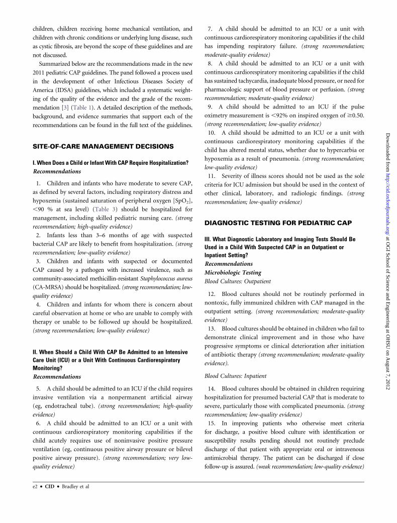
I. When Does a Child or Infant With CAP Require Hospitalization?Recommendations
1. Children and infants who have moderate to severe CAP,
as defined by several factors, including respiratory distress and
hypoxemia (sustained saturation of peripheral oxygen [SpO2],
,90 % at sea level) (Table 3) should be hospitalized for
management, including skilled pediatric nursing care. (strong
recommendation; high-quality evidence)
2. Infants less than 3–6 months of age with suspected
bacterial CAP are likely to benefit from hospitalization. (strong
recommendation; low-quality evidence)
3. Children and infants with suspected or documented
CAP caused by a pathogen with increased virulence, such as
community-associated methicillin-resistant Staphylococcus aureus
(CA-MRSA) should be hospitalized. (strong recommendation; low-
quality evidence)
4. Children and infants for whom there is concern about
careful observation at home or who are unable to comply with
therapy or unable to be followed up should be hospitalized.
(strong recommendation; low-quality evidence)
II. When Should a Child With CAP Be Admitted to an IntensiveCare Unit (ICU) or a Unit With Continuous CardiorespiratoryMonitoring?Recommendations
5. A child should be admitted to an ICU if the child requires
invasive ventilation via a nonpermanent artificial airway
(eg, endotracheal tube). (strong recommendation; high-quality
evidence)
6. A child should be admitted to an ICU or a unit with
continuous cardiorespiratory monitoring capabilities if the
child acutely requires use of noninvasive positive pressure
ventilation (eg, continuous positive airway pressure or bilevel
positive airway pressure). (strong recommendation; very low-
quality evidence)
7. A child should be admitted to an ICU or a unit with
continuous cardiorespiratory monitoring capabilities if the child
has impending respiratory failure. (strong recommendation;
moderate-quality evidence)
8. A child should be admitted to an ICU or a unit with
continuous cardiorespiratory monitoring capabilities if the child
has sustained tachycardia, inadequate blood pressure, or need for
pharmacologic support of blood pressure or perfusion. (strong
recommendation; moderate-quality evidence)
9. A child should be admitted to an ICU if the pulse
oximetry measurement is ,92% on inspired oxygen of $0.50.
(strong recommendation; low-quality evidence)
10. A child should be admitted to an ICU or a unit with
continuous cardiorespiratory monitoring capabilities if the
child has altered mental status, whether due to hypercarbia or
hypoxemia as a result of pneumonia. (strong recommendation;
low-quality evidence)
11. Severity of illness scores should not be used as the sole
criteria for ICU admission but should be used in the context of
other clinical, laboratory, and radiologic findings. (strong
recommendation; low-quality evidence)
DIAGNOSTIC TESTING FOR PEDIATRIC CAP
III. What Diagnostic Laboratory and Imaging Tests Should BeUsed in a Child With Suspected CAP in an Outpatient orInpatient Setting?Recommendations
Microbiologic Testing
Blood Cultures: Outpatient
12. Blood cultures should not be routinely performed in
nontoxic, fully immunized children with CAP managed in the
outpatient setting. (strong recommendation; moderate-quality
evidence)
13. Blood cultures should be obtained in children who fail to
demonstrate clinical improvement and in those who have
progressive symptoms or clinical deterioration after initiation
of antibiotic therapy (strong recommendation; moderate-quality
evidence).
Blood Cultures: Inpatient
14. Blood cultures should be obtained in children requiring
hospitalization for presumed bacterial CAP that is moderate to
severe, particularly those with complicated pneumonia. (strong
recommendation; low-quality evidence)
15. In improving patients who otherwise meet criteria
for discharge, a positive blood culture with identification or
susceptibility results pending should not routinely preclude
discharge of that patient with appropriate oral or intravenous
antimicrobial therapy. The patient can be discharged if close
follow-up is assured. (weak recommendation; low-quality evidence)
e2 d CID d Bradley et al
at OG
I School of Science and Engineering at O
HSU
on August 7, 2012
http://cid.oxfordjournals.org/D
ownloaded from
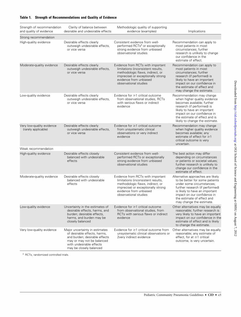
Table 1. Strength of Recommendations and Quality of Evidence
Strength of recommendation
and quality of evidence
Clarity of balance between
desirable and undesirable effects
Methodologic quality of supporting
evidence (examples) Implications
Strong recommendation
High-quality evidence Desirable effects clearlyoutweigh undesirable effects,or vice versa
Consistent evidence from well-performed RCTsa or exceptionallystrong evidence from unbiasedobservational studies
Recommendation can apply tomost patients in mostcircumstances; furtherresearch is unlikely to changeour confidence in theestimate of effect.
Moderate-quality evidence Desirable effects clearlyoutweigh undesirable effects,or vice versa
Evidence from RCTs with importantlimitations (inconsistent results,methodologic flaws, indirect, orimprecise) or exceptionally strongevidence from unbiasedobservational studies
Recommendation can apply tomost patients in mostcircumstances; furtherresearch (if performed) islikely to have an importantimpact on our confidence inthe estimate of effect andmay change the estimate.
Low-quality evidence Desirable effects clearlyoutweigh undesirable effects,or vice versa
Evidence for $1 critical outcomefrom observational studies, RCTswith serious flaws or indirectevidence
Recommendation may changewhen higher quality evidencebecomes available; furtherresearch (if performed) islikely to have an importantimpact on our confidence inthe estimate of effect and islikely to change the estimate.
Very low-quality evidence(rarely applicable)
Desirable effects clearlyoutweigh undesirable effects,or vice versa
Evidence for $1 critical outcomefrom unsystematic clinicalobservations or very indirectevidence
Recommendation may changewhen higher quality evidencebecomes available; anyestimate of effect for $1critical outcome is veryuncertain.
Weak recommendation
High-quality evidence Desirable effects closelybalanced with undesirableeffects
Consistent evidence from well-performed RCTs or exceptionallystrong evidence from unbiasedobservational studies
The best action may differdepending on circumstancesor patients or societal values;further research is unlikely tochange our confidence in theestimate of effect.
Moderate-quality evidence Desirable effects closelybalanced with undesirableeffects
Evidence from RCTs with importantlimitations (inconsistent results,methodologic flaws, indirect, orimprecise) or exceptionally strongevidence from unbiasedobservational studies
Alternative approaches are likelyto be better for some patientsunder some circumstances;further research (if performed)is likely to have an importantimpact on our confidence inthe estimate of effect andmay change the estimate.
Low-quality evidence Uncertainty in the estimates ofdesirable effects, harms, andburden; desirable effects,harms, and burden may beclosely balanced
Evidence for $1 critical outcomefrom observational studies, fromRCTs with serious flaws or indirectevidence
Other alternatives may be equallyreasonable; further research isvery likely to have an importantimpact on our confidence in theestimate of effect and is likelyto change the estimate.
Very low-quality evidence Major uncertainty in estimatesof desirable effects, harms,and burden; desirable effectsmay or may not be balancedwith undesirable effectsmay be closely balanced
Evidence for $1 critical outcome fromunsystematic clinical observations or2very indirect evidence
Other alternatives may be equallyreasonable; any estimate ofeffect, for at $1 criticaloutcome, is very uncertain.
a RCTs, randomized controlled trials.
Pediatric Community Pneumonia Guidelines d CID d e3
at OG
I School of Science and Engineering at O
HSU
on August 7, 2012
http://cid.oxfordjournals.org/D
ownloaded from
Follow-up Blood Cultures
16. Repeated blood cultures in children with clear clinical
improvement are not necessary to document resolution of
pneumococcal bacteremia. (weak recommendation; low-quality
evidence)
17. Repeated blood cultures to document resolution of
bacteremia should be obtained in children with bacteremia
caused by S. aureus, regardless of clinical status. (strong
recommendation; low-quality evidence)
Sputum Gram Stain and Culture
18. Sputum samples for culture and Gram stain should be
obtained in hospitalized children who can produce sputum.
(weak recommendation; low-quality evidence)
Urinary Antigen Detection Tests
19. Urinary antigen detection tests are not recommended
for the diagnosis of pneumococcal pneumonia in children;
false-positive tests are common. (strong recommendation; high-
quality evidence)
Testing For Viral Pathogens
20. Sensitive and specific tests for the rapid diagnosis of
influenza virus and other respiratory viruses should be used in
the evaluation of children with CAP. A positive influenza test
may decrease both the need for additional diagnostic studies
and antibiotic use, while guiding appropriate use of antiviral
agents in both outpatient and inpatient settings. (strong
recommendation; high-quality evidence)
21. Antibacterial therapy is not necessary for children, either
outpatients or inpatients, with a positive test for influenza virus
in the absence of clinical, laboratory, or radiographic findings
that suggest bacterial coinfection. (strong recommendation;
high-quality evidence).
22. Testing for respiratory viruses other than influenza virus
can modify clinical decision making in children with suspected
pneumonia, because antibacterial therapy will not routinely be
required for these children in the absence of clinical, laboratory,
or radiographic findings that suggest bacterial coinfection.
(weak recommendation; low-quality evidence)
Testing for Atypical Bacteria
23. Children with signs and symptoms suspicious for
Mycoplasma pneumoniae should be tested to help guide
antibiotic selection. (weak recommendation; moderate-quality
evidence)
24. Diagnostic testing for Chlamydophila pneumoniae is not
recommended as reliable and readily available diagnostic tests
do not currently exist. (strong recommendation; high-quality
evidence)
Ancillary Diagnostic Testing
Complete Blood Cell Count
25. Routine measurement of the complete blood cell count is
not necessary in all children with suspected CAP managed in the
outpatient setting, but in those with more serious disease it may
provide useful information for clinical management in the
context of the clinical examination and other laboratory and
imaging studies. (weak recommendation; low-quality evidence)
26. A complete blood cell count should be obtained for
patients with severe pneumonia, to be interpreted in the context
of the clinical examination and other laboratory and imaging
studies. (weak recommendation; low-quality evidence)
Acute-Phase Reactants
27. Acute-phase reactants, such as the erythrocyte sedimentation
rate (ESR), C-reactive protein (CRP) concentration, or serum
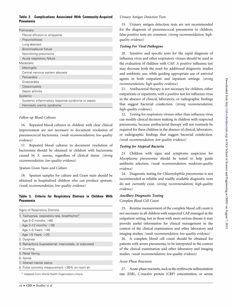
Table 3. Criteria for Respiratory Distress in Children WithPneumonia
Signs of Respiratory Distress
1. Tachypnea, respiratory rate, breaths/mina
Age 0–2 months: .60
Age 2–12 months: .50
Age 1–5 Years: .40
Age .5 Years: .20
2. Dyspnea
3. Retractions (suprasternal, intercostals, or subcostal)
4. Grunting
5. Nasal flaring
6. Apnea
7. Altered mental status
8. Pulse oximetry measurement ,90% on room air
a Adapted from World Health Organization criteria.
Table 2. Complications Associated With Community-AcquiredPneumonia
Pulmonary
Pleural effusion or empyema
Pneumothorax
Lung abscess
Bronchopleural fistula
Necrotizing pneumonia
Acute respiratory failure
Metastatic
Meningitis
Central nervous system abscess
Pericarditis
Endocarditis
Osteomyelitis
Septic arthritis
Systemic
Systemic inflammatory response syndrome or sepsis
Hemolytic uremic syndrome
e4 d CID d Bradley et al
at OG
I School of Science and Engineering at O
HSU
on August 7, 2012
http://cid.oxfordjournals.org/D
ownloaded from
procalcitonin concentration, cannot be used as the sole determinant
to distinguish between viral and bacterial causes of CAP. (strong
recommendation; high-quality evidence)
28. Acute-phase reactants need not be routinely
measured in fully immunized children with CAP who are
managed as outpatients, although for more serious disease,
acute-phase reactants may provide useful information for
clinical management. (strong recommendation; low-quality
evidence)
29. In patients with more serious disease, such as those
requiring hospitalization or those with pneumonia-associated
complications, acute-phase reactants may be used in
conjunction with clinical findings to assess response to
therapy. (weak recommendation; low-quality evidence)
Pulse Oximetry
30. Pulse oximetry should be performed in all children with
pneumonia and suspected hypoxemia. The presence of
hypoxemia should guide decisions regarding site of care and
further diagnostic testing. (strong recommendation; moderate-
quality evidence)
Chest Radiography
Initial Chest Radiographs: Outpatient
31. Routine chest radiographs are not necessary for the
confirmation of suspected CAP in patients well enough to be
treated in the outpatient setting (after evaluation in the
office, clinic, or emergency department setting). (strong
recommendation; high-quality evidence)
32. Chest radiographs, posteroanterior and lateral, should
be obtained in patients with suspected or documented
hypoxemia or significant respiratory distress (Table 3) and in
those with failed initial antibiotic therapy to verify the presence
or absence of complications of pneumonia, including
parapneumonic effusions, necrotizing pneumonia, and
pneumothorax. (strong recommendation; moderate-quality
evidence)
Initial Chest Radiographs: Inpatient
33. Chest radiographs (posteroanterior and lateral) should be
obtained in all patients hospitalized for management of CAP to
document the presence, size, and character of parenchymal
infiltrates and identify complications of pneumonia that may
lead to interventions beyond antimicrobial agents and supportive
medical therapy. (strong recommendation; moderate-quality
evidence)
Follow-up Chest Radiograph
34. Repeated chest radiographs are not routinely required in
children who recover uneventfully from an episode of CAP.
(strong recommendation; moderate-quality evidence)
35. Repeated chest radiographs should be obtained in
children who fail to demonstrate clinical improvement and
in those who have progressive symptoms or clinical
deterioration within 48–72 hours after initiation of
antibiotic therapy. (strong recommendation; moderate-quality
evidence)
36. Routine daily chest radiography is not recommended
in children with pneumonia complicated by parapneumonic
effusion after chest tube placement or after video-
assisted thoracoscopic surgery (VATS), if they remain
clinically stable. (strong recommendation; low-quality
evidence)
37. Follow-up chest radiographs should be obtained in
patients with complicated pneumonia with worsening
respiratory distress or clinical instability, or in those with
persistent fever that is not responding to therapy over 48-72
hours. (strong recommendation; low-quality evidence)
38. Repeated chest radiographs 4–6 weeks after the
diagnosis of CAP should be obtained in patients with
recurrent pneumonia involving the same lobe and in
patients with lobar collapse at initial chest radiography
with suspicion of an anatomic anomaly, chest mass, or
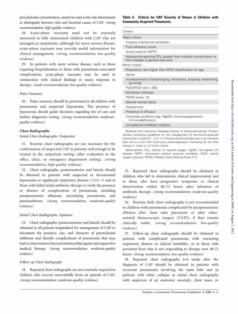
Table 4. Criteria for CAP Severity of Illness in Children withCommunity-Acquired Pneumonia
Criteria
Major criteria
Invasive mechanical ventilation
Fluid refractory shock
Acute need for NIPPV
Hypoxemia requiring FiO2 greater than inspired concentration orflow feasible in general care area
Minor criteria
Respiratory rate higher than WHO classification for age
Apnea
Increasedwork of breathing (eg, retractions, dyspnea, nasal flaring,grunting)
PaO2/FiO2 ratio ,250
Multilobar infiltrates
PEWS score .6
Altered mental status
Hypotension
Presence of effusion
Comorbid conditions (eg, HgbSS, immunosuppression,immunodeficiency)
Unexplained metabolic acidosis
Modified from Infectious Diseases Society of America/American Thoracic
Society consensus guidelines on the management of community-acquired
pneumonia in adults [27, table 4]. Clinician should consider care in an intensive
care unit or a unit with continuous cardiorespiratory monitoring for the child
having $1 major or $2 minor criteria.
Abbreviations: FiO2, fraction of inspired oxygen; HgbSS, Hemoglobin SS
disease; NIPPV, noninvasive positive pressure ventilation; PaO2, arterial
oxygen pressure; PEWS, Pediatric Early Warning Score [70].
Pediatric Community Pneumonia Guidelines d CID d e5
at OG
I School of Science and Engineering at O
HSU
on August 7, 2012
http://cid.oxfordjournals.org/D
ownloaded from

foreign body aspiration. (strong recommendation; moderate-
quality evidence)
IV. What Additional Diagnostic Tests Should Be Used in a ChildWith Severe or Life-Threatening CAP?Recommendations
39. The clinician should obtain tracheal aspirates for Gram
stain and culture, as well as clinically and epidemiologically
guided testing for viral pathogens, including influenza virus, at
the time of initial endotracheal tube placement in children
requiring mechanical ventilation. (strong recommendation; low-
quality evidence)
40. Bronchoscopic or blind protected specimen brush
sampling, bronchoalveolar lavage (BAL), percutaneous lung
aspiration, or open lung biopsy should be reserved for the
immunocompetent child with severe CAP if initial diagnostic
tests are not positive. (weak recommendation; low-quality
evidence)
ANTI-INFECTIVE TREATMENT
V. Which Anti-Infective Therapy Should Be Provided to a ChildWith Suspected CAP in Both Outpatient and Inpatient Settings?Recommendations
Outpatients
41. Antimicrobial therapy is not routinely required for
preschool-aged children with CAP, because viral pathogens are
responsible for the great majority of clinical disease. (strong
recommendation; high-quality evidence)
42. Amoxicillin should be used as first-line therapy for
previously healthy, appropriately immunized infants and
preschool children with mild to moderate CAP suspected to
be of bacterial origin. Amoxicillin provides appropriate
coverage for Streptococcus pneumoniae, the most prominent
invasive bacterial pathogen. Table 5 lists preferred agents and
alternative agents for children allergic to amoxicillin (strong
recommendation; moderate-quality evidence)
43. Amoxicillin should be used as first-line therapy for
previously healthy appropriately immunized school-aged
children and adolescents with mild to moderate CAP for
S. pneumoniae, the most prominent invasive bacterial
pathogen. Atypical bacterial pathogens (eg, M. pneumoniae),
and less common lower respiratory tract bacterial pathogens, as
discussed in the Evidence Summary, should also be considered in
management decisions. (strong recommendation; moderate-
quality evidence)
44. Macrolide antibiotics should be prescribed for treatment
of children (primarily school-aged children and adolescents)
evaluated in an outpatient setting with findings compatible
with CAP caused by atypical pathogens. Laboratory testing for
M. pneumoniae should be performed if available in a clinically
relevant time frame. Table 5 lists preferred and alternative agents
for atypical pathogens. (weak recommendation; moderate-quality
evidence)
45. Influenza antiviral therapy (Table 6) should be
administered as soon as possible to children with moderate
to severe CAP consistent with influenza virus infection during
widespread local circulation of influenza viruses, particularly
for those with clinically worsening disease documented at the
time of an outpatient visit. Because early antiviral treatment has
been shown to provide maximal benefit, treatment should not be
delayed until confirmation of positive influenza test results.
Negative results of influenza diagnostic tests, especially rapid
antigen tests, do not conclusively exclude influenza disease.
Treatment after 48 hours of symptomatic infection may still
provide clinical benefit to those with more severe disease. (strong
recommendation; moderate-quality evidence)
Inpatients
46. Ampicillin or penicillin G should be administered to the
fully immunized infant or school-aged child admitted to
a hospital ward with CAP when local epidemiologic data
document lack of substantial high-level penicillin resistance for
invasive S. pneumoniae. Other antimicrobial agents for empiric
therapy are provided in Table 7. (strong recommendation;
moderate-quality evidence)
47. Empiric therapy with a third-generation parenteral
cephalosporin (ceftriaxone or cefotaxime) should be
prescribed for hospitalized infants and children who are
not fully immunized, in regions where local epidemiology of
invasive pneumococcal strains documents high-level
penicillin resistance, or for infants and children with life-
threatening infection, including those with empyema
(Table 7). Non–b-lactam agents, such as vancomycin, have
not been shown to be more effective than third-generation
cephalosporins in the treatment of pneumococcal
pneumonia for the degree of resistance noted currently in
North America. (weak recommendation; moderate-quality
evidence)
48. Empiric combination therapy with a macrolide (oral or
parenteral), in addition to a b-lactam antibiotic, should be
prescribed for the hospitalized child for whom M. pneumoniae
and C. pneumoniae are significant considerations; diagnostic
testing should be performed if available in a clinically relevant
time frame (Table 7). (weak recommendation; moderate-quality
evidence)
49. Vancomycin or clindamycin (based on local susceptibility
data) should be provided in addition to b-lactam therapy if
clinical, laboratory, or imaging characteristics are consistent
with infection caused by S. aureus (Table 7). (strong
recommendation; low-quality evidence)
e6 d CID d Bradley et al
at OG
I School of Science and Engineering at O
HSU
on August 7, 2012
http://cid.oxfordjournals.org/D
ownloaded from
Table 5. Selection of Antimicrobial Therapy for Specific Pathogens
Pathogen Parenteral therapy
Oral therapy (step-down therapy
or mild infection)
Streptococcus pneumoniae withMICs for penicillin #2.0 lg/mL
Preferred: ampicillin (150–200 mg/kg/day every6 hours) or penicillin (200 000–250000 U/kg/dayevery 4–6 h);
Alternatives: ceftriaxone(50–100 mg/kg/day every 12–24 hours) (preferredfor parenteral outpatient therapy) or cefotaxime(150 mg/kg/day every 8 hours); may also beeffective: clindamycin (40 mg/kg/day every6–8 hours) or vancomycin (40–60 mg/kg/day every6–8 hours)
Preferred: amoxicillin (90 mg/kg/day in2 doses or 45 mg/kg/day in 3 doses);
Alternatives: second- or third-generationcephalosporin (cefpodoxime, cefuroxime,cefprozil); oral levofloxacin, if susceptible(16–20 mg/kg/day in 2 doses for children6 months to 5 years old and 8–10 mg/kg/dayonce daily for children 5 to 16 years old;maximum daily dose, 750 mg) or orallinezolid (30 mg/kg/day in 3 doses forchildren ,12 years old and 20 mg/kg/dayin 2 doses for children $12 years old)
S. pneumoniae resistant topenicillin, with MICs$4.0 lg/mL
Preferred: ceftriaxone (100 mg/kg/day every12–24 hours);
Alternatives: ampicillin(300–400 mg/kg/day every 6 hours), levofloxacin(16–20 mg/kg/day every 12 hours for children6 months to 5 years old and 8–10 mg/kg/dayonce daily for children 5–16 years old; maximumdaily dose, 750 mg), or linezolid (30 mg/kg/dayevery 8 hours for children ,12 years old and20 mg/kg/day every 12 hours for children $12 yearsold); may also be effective: clindamycina
(40 mg/kg/day every 6–8 hours) or vancomycin(40–60 mg/kg/day every 6–8 hours)
Preferred: oral levofloxacin (16–20 mg/kg/dayin 2 doses for children 6 months to 5 yearsand 8–10 mg/kg/day once daily for children5–16 years, maximum daily dose, 750 mg),if susceptible, or oral linezolid (30 mg/kg/dayin 3 doses for children ,12 years and20 mg/kg/day in 2 doses for children$12 years);
Alternative: oral clindamycina
(30–40 mg/kg/day in 3 doses)
Group A Streptococcus Preferred: intravenous penicillin (100000–250000U/kg/day every 4–6 hours) or ampicillin(200 mg/kg/day every 6 hours);
Alternatives: ceftriaxone (50–100 mg/kg/day every12–24 hours) or cefotaxime (150 mg/kg/day every8 hours); may also be effective: clindamycin, ifsusceptible (40 mg/kg/day every 6–8 hours) orvancomycinb (40–60 mg/kg/day every 6–8 hours)
Preferred: amoxicillin (50–75 mg/kg/day in2 doses), or penicillin V (50–75 mg/kg/day in3 or 4 doses);
Alternative: oral clindamycina
(40 mg/kg/day in 3 doses)
Stapyhylococcus aureus,methicillin susceptible(combination therapy notwell studied)
Preferred: cefazolin (150 mg/kg/day every 8 hours) orsemisynthetic penicillin, eg oxacillin(150–200 mg/kg/day every 6–8 hours);
Alternatives: clindamycina (40 mg/kg/day every6–8 hours) or >vancomycin (40–60 mg/kg/dayevery 6–8 hours)
Preferred: oral cephalexin (75–100 mg/kg/dayin 3 or 4 doses);
Alternative: oral clindamycina
(30–40 mg/kg/day in 3 or 4 doses)
S. aureus, methicillin resistant,susceptible to clindamycin(combination therapy notwell-studied)
Preferred: vancomycin (40–60 mg/kg/day every6–8 hours or dosing to achieve an AUC/MIC ratio of.400) or clindamycin (40 mg/kg/day every 6–8 hours);
Alternatives: linezolid (30 mg/kg/day every 8 hoursfor children ,12 years old and 20 mg/kg/day every12 hours for children $12 years old)
Preferred: oral clindamycin (30–40 mg/kg/dayin 3 or 4 doses);
Alternatives: oral linezolid(30 mg/kg/day in 3 doses for children,12 years and 20 mg/kg/day in 2 dosesfor children $12 years)
S. aureus, methicillin resistant,resistant to clindamycin(combination therapy notwell studied)
Preferred: vancomycin (40–60 mg/kg/day every6-8 hours or dosing to achieve an AUC/MIC ratio of.400);
Alternatives: linezolid (30 mg/kg/day every8 hours for children ,12 years old and 20 mg/kg/dayevery 12 hours for children $12 years old)
Preferred: oral linezolid (30 mg/kg/day in3 doses for children ,12 years and20 mg/kg/day in 2 doses for children$12 years old);
Alternatives: none; entire treatment course withparenteral therapy may be required
Pediatric Community Pneumonia Guidelines d CID d e7
at OG
I School of Science and Engineering at O
HSU
on August 7, 2012
http://cid.oxfordjournals.org/D
ownloaded from
VI. How Can Resistance to Antimicrobials Be Minimized?Recommendations
50. Antibiotic exposure selects for antibiotic resistance;
therefore, limiting exposure to any antibiotic, whenever
possible, is preferred. (strong recommendation; moderate-quality
evidence)
51. Limiting the spectrum of activity of antimicrobials to
that specifically required to treat the identified pathogen is
preferred. (strong recommendation; low-quality evidence)
52. Using the proper dosage of antimicrobial to be able to
achieve a minimal effective concentration at the site of infection
is important to decrease the development of resistance. (strong
recommendation; low-quality evidence)
53. Treatment for the shortest effective duration will
minimize exposure of both pathogens and normal microbiota
to antimicrobials and minimize the selection for resistance.
(strong recommendation; low-quality evidence)
VII. What Is the Appropriate Duration of Antimicrobial Therapyfor CAP?Recommendations
54. Treatment courses of 10 days have been best studied,
although shorter courses may be just as effective, particularly
for more mild disease managed on an outpatient basis. (strong
recommendation; moderate-quality evidence)
55. Infections caused by certain pathogens, notably CA-
MRSA, may require longer treatment than those caused by
S. pneumoniae. (strong recommendation; moderate-quality
evidence)
VIII. How Should the Clinician Follow the Child With CAP for theExpected Response to Therapy?Recommendation
56. Children on adequate therapy should demonstrate clinical
and laboratory signs of improvement within 48–72 hours. For
Table 5. (Continued)
Pathogen Parenteral therapy
Oral therapy (step-down therapy
or mild infection)
Haemophilus influenza, typeable(A-F) or nontypeable
Preferred: intravenous ampicillin (150-200 mg/kg/dayevery 6 hours) if b-lactamase negative, ceftriaxone(50–100 mg/kg/day every 12-24 hours) if b-lactamaseproducing, or cefotaxime (150 mg/kg/day every8 hours);
Alternatives: intravenous ciprofloxacin (30 mg/kg/dayevery 12 hours) or intravenous levofloxacin(16-20 mg/kg/day every 12 hours forchildren 6 months to 5 years oldand 8-10 mg/kg/day once daily for children 5 to16 years old; maximum daily dose, 750 mg)
Preferred: amoxicillin (75-100 mg/kg/day in3 doses) if b-lactamase negative) oramoxicillin clavulanate (amoxicillincomponent, 45 mg/kg/day in 3 doses or90 mg/kg/day in 2 doses) if b-lactamaseproducing;
Alternatives: cefdinir, cefixime,cefpodoxime, or ceftibuten
Mycoplasma pneumoniae Preferred: intravenous azithromycin(10 mg/kg on days 1 and 2 of therapy;transition to oral therapy if possible);
Alternatives: intravenous erythromycin lactobionate(20 mg/kg/day every 6 hours) or levofloxacin(16-20 mg/kg/day every 12 hours; maximum dailydose, 750 mg)
Preferred: azithromycin (10 mg/kg on day 1,followed by 5 mg/kg/day once daily ondays 2–5);
Alternatives: clarithromycin(15 mg/kg/day in 2 doses) or oralerythromycin (40 mg/kg/day in 4 doses);for children .7 years old, doxycycline(2–4 mg/kg/day in 2 doses; for adolescentswith skeletal maturity, levofloxacin(500 mg once daily) or moxifloxacin(400 mg once daily)
Chlamydia trachomatis orChlamydophila pneumoniae
Preferred: intravenous azithromycin(10 mg/kg on days 1 and 2 of therapy;transition to oral therapy if possible);
Alternatives: intravenous erythromycin lactobionate(20 mg/kg/day every 6 hours) or levofloxacin(16-20 mg/kg/day in 2 doses for children 6 monthsto 5 years old and 8-10 mg/kg/day once daily forchildren 5 to 16 years old; maximum daily dose,750 mg)
Preferred: azithromycin (10 mg/kg on day 1,followed by 5 mg/kg/day once dailydays 2–5);
Alternatives: clarithromycin(15 mg/kg/day in 2 doses) or oralerythromycin (40 mg/kg/day in 4 doses);for children .7 years old, doxycycline(2-4 mg/kg/day in 2 doses); for adolescentswith skeletal maturity, levofloxacin(500 mg once daily) or moxifloxacin(400 mg once daily)
Doses for oral therapy should not exceed adult doses.
Abbreviations: AUC, area under the time vs. serum concentration curve; MIC, minimum inhibitory concentration.a Clindamycin resistance appears to be increasing in certain geographic areas among S. pneumoniae and S. aureus infections.b For b-lactam–allergic children.
e8 d CID d Bradley et al
at OG
I School of Science and Engineering at O
HSU
on August 7, 2012
http://cid.oxfordjournals.org/D
ownloaded from
children whose condition deteriorates after admission and
initiation of antimicrobial therapy or who show no
improvement within 48–72 hours, further investigation should
be performed. (strong recommendation; moderate-quality evidence)
ADJUNCTIVE SURGICAL AND NON–
ANTI-INFECTIVE THERAPYFORPEDIATRICCAP
IX. How Should a Parapneumonic Effusion Be Identified?Recommendation
57. History and physical examination may be suggestive of
parapneumonic effusion in children suspected of having CAP,
but chest radiography should be used to confirm the presence of
pleural fluid. If the chest radiograph is not conclusive, then
further imaging with chest ultrasound or computed
tomography (CT) is recommended. (strong recommendation;
high-quality evidence)
X. What Factors Are Important in Determining Whether Drainageof the Parapneumonic Effusion Is Required?Recommendations
58. The size of the effusion is an important factor that
determines management (Table 8, Figure 1). (strong
recommendation; moderate-quality evidence)
Table 6. Influenza Antiviral Therapy
Drug [186187] Formulation
Dosing recommendations
Treatment Prophylaxisa
Children Adults Children Adults
Oseltamivir(Tamiflu)
75-mg capsule;60 mg/5 mLSuspension
$24 months old:�4 mg/kg/day in2 doses, for a5-day treatmentcourse
150 mg/day in2 doses for5 days
#15 kg: 30 mg/day; .15 to23 kg: 45 mg/day; .23 to40 kg: 60 mg/day; .40 kg:75 mg/day (once daily ineach group)
75 mg/dayonce daily
#15 kg: 60 mg/day;.15 to 23 kg: 90 mg/day;.23 to 40 kg: 120 mg/day;.40 kg: 150 mg/day(divided into 2 dosesfor each group)
9–23 months old:7 mg/kg/day in2 doses; 0–8 monthsold: 6 mg/kg/day in2 doses; prematureinfants: 2 mg/kg/dayin 2 doses
9–23 months old: 3.5 mg/kgonce daily; 3–8 months old:3 mg/kg once daily; notroutinely recommended forinfants ,3 months oldowing to limited data inthis age group
Zanamivir(Relenza)
5 mg per inhalation,using a Diskhaler
$7 years old: 2 inhalations(10 mg total per dose),twice daily for 5 days
2 inhalations(10 mg total perdose), twice dailyfor 5 days
$5 years old: 2 inhalations(10 mg total per dose),once daily for 10 days
2 inhalations(10 mg totalper dose),once dailyfor 10 days
Amantadine(Symmetrel)b
100-mg tablet;50 mg/5 mLsuspension
1–9 years old: 5–8 mg/kg/dayas single daily dose or in2 doses, not to exceed150 mg/day; 9–12 years old:200 mg/day in 2 doses (notstudied as single daily dose)
200 mg/day, assingle daily doseor in 2 doses
1–9 years old:same astreatment dose;9–12 years old:same astreatment dose
Same astreatmentdose
Rimantadine(Flumadine)b
100-mg tablet;50 mg/5 mLsuspension
Not FDA approved fortreatment in children, butpublished data exist on safetyand efficacy in children;suspension: 1–9 years old:6.6 mg/kg/day(maximum 150 mg/kg/day) in2 doses; $10 years old:200 mg/day, as single dailydose or in 2 doses
200 mg/day, eitheras a single dailydose, or dividedinto 2 doses
FDA approved forprophylaxis down to12 months of age.1–9 years old:5 mg/kg/dayonce daily, not to exceed150 mg; $10 years old:200 mg/day as single dailydose or in 2 doses
200 mg/day,as singledaily doseor in2 doses
NOTE. Check Centers for Disease Control and Prevention Website (http://www.flu.gov/) for current susceptibility data.a In children for whom prophylaxis is indicated, antiviral drugs should be continued for the duration of known influenza activity in the community because of the
potential for repeated and unknown exposures or until immunity can be achieved after immunization.b Amantadine and rimantadine should be used for treatment and prophylaxis only in winter seasons during which a majority of influenza A virus strains isolated
are adamantine susceptible; the adamantanes should not be used for primary therapy because of the rapid emergence of resistance. However, for patients requiring
adamantane therapy, a treatment course of �7 days is suggested, or until 24–48 hours after the disappearance of signs and symptoms.
Pediatric Community Pneumonia Guidelines d CID d e9
at OG
I School of Science and Engineering at O
HSU
on August 7, 2012
http://cid.oxfordjournals.org/D
ownloaded from
Table 7. Empiric Therapy for Pediatric Community-Acquired Pneumonia (CAP)
Empiric therapy
Site of care
Presumed bacterial
pneumonia
Presumed atypical
pneumonia
Presumed influenza
pneumoniaa
Outpatient
,5 years old (preschool) Amoxicillin, oral (90 mg/kg/dayin 2 dosesb)
Alternative:oral amoxicillin clavulanate(amoxicillin component,90 mg/kg/day in 2 dosesb)
Azithromycin oral (10 mg/kg onday 1, followed by 5 mg/kg/dayonce daily on days 2–5);
Alternatives: oral clarithromycin(15 mg/kg/day in 2 dosesfor 7-14 days) or oralerythromycin (40 mg/kg/dayin 4 doses)
Oseltamivir
$5 years old Oral amoxicillin (90 mg/kg/day in2 dosesb to a maximumof 4 g/dayc); for childrenwith presumed bacterialCAP who do not have clinical,laboratory, or radiographicevidence that distinguishesbacterial CAP fromatypical CAP, a macrolidecan be added to a b-lactamantibiotic for empiric therapy;alternative: oral amoxicillinclavulanate (amoxicillincomponent, 90 mg/kg/dayin 2 dosesb to a maximumdose of 4000 mg/day,eg, one 2000-mg tablettwice dailyb)
Oral azithromycin (10 mg/kg onday 1, followed by 5 mg/kg/dayonce daily on days 2–5 to amaximum of 500 mg on day 1,followed by 250 mg on days 2–5);alternatives: oral clarithromycin(15 mg/kg/day in 2 doses to amaximum of 1 g/day);erythromycin, doxycycline forchildren .7 years old
Oseltamivir or zanamivir(for children 7 yearsand older); alternatives:peramivir, oseltamivirand zanamivir(all intravenous) areunder clinicalinvestigation in children;intravenous zanamiviravailable forcompassionate use
Inpatient (all ages)d
Fully immunized withconjugate vaccines forHaemophilus influenzaetype b and Streptococcuspneumoniae; localpenicillin resistance ininvasive strains ofpneumococcus is minimal
Ampicillin or penicillin G;alternatives:ceftriaxone or cefotaxime;addition of vancomycin orclindamycin forsuspected CA-MRSA
Azithromycin (in addition tob-lactam, if diagnosis ofatypical pneumonia is indoubt); alternatives:clarithromycin orerythromycin;doxycycline for children.7 years old; levofloxacinfor children who havereached growth maturity,or who cannot toleratemacrolides
Oseltamivir or zanamivir(for children $7 years old;alternatives: peramivir,oseltamivir andzanamivir (all intravenous)are under clinicalinvestigationin children; intravenouszanamivir available forcompassionate use
Not fully immunized for H,influenzae type b andS. pneumoniae; localpenicillin resistance ininvasive strains ofpneumococcus issignificant
Ceftriaxone or cefotaxime; addition ofvancomycin or clindamycin forsuspected CA-MRSA; alternative:levofloxacin; addition of vancomycinor clindamycin for suspectedCA-MRSA
Azithromycin (in addition tob-lactam, if diagnosis indoubt); alternatives:clarithromycin or erythromycin;doxycycline for children .7 yearsold; levofloxacin for childrenwho have reached growthmaturity or who cannottolerate macrolides
As above
For children with drug allergy to recommended therapy, see Evidence Summary for Section V. Anti-Infective Therapy. For children with a history of possible,
nonserious allergic reactions to amoxicillin, treatment is not well defined and should be individualized. Options include a trial of amoxicillin under medical
observation; a trial of an oral cephalosporin that has substantial activity against S. pneumoniae, such as cefpodoxime, cefprozil, or cefuroxime, provided under
medical supervision; treatment with levofloxacin; treatment with linezolid; treatment with clindamycin (if susceptible); or treatment with a macrolide (if susceptible).
For children with bacteremic pneumococcal pneumonia, particular caution should be exercised in selecting alternatives to amoxicillin, given the potential for
secondary sites of infection, including meningitis.
Abbreviation: CA-MRSA, community-associated methicillin-resistant Staphylococcus aureus.a See Table 6 for dosages.b See text for discussion of dosage recommendations based on local susceptibility data. Twice daily dosing of amoxicillin or amoxicillin clavulanate may be
effective for pneumococci that are susceptible to penicillin.c Not evaluated prospectively for safety.d See Table 5 for dosages.
e10 d CID d Bradley et al
at OG
I School of Science and Engineering at O
HSU
on August 7, 2012
http://cid.oxfordjournals.org/D
ownloaded from
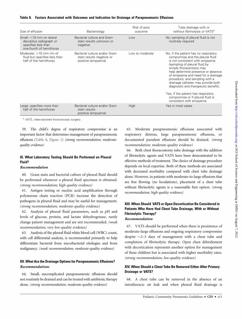
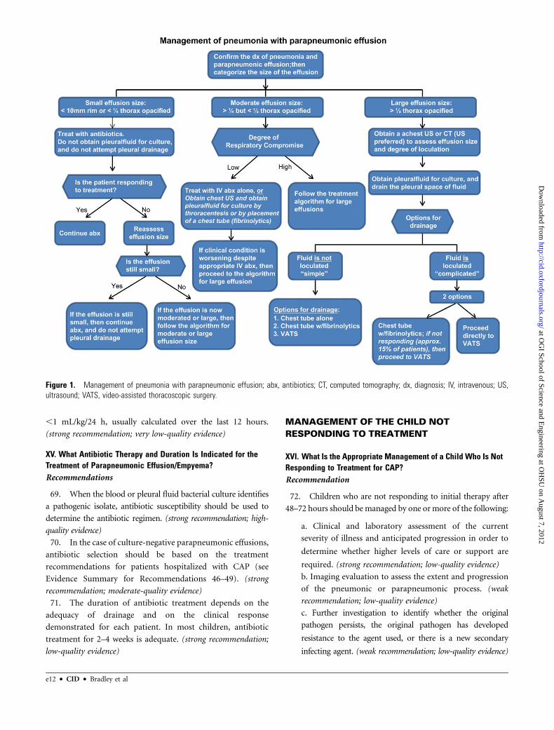
59. The child’s degree of respiratory compromise is an
important factor that determines management of parapneumonic
effusions (Table 8, Figure 1) (strong recommendation; moderate-
quality evidence)
XI. What Laboratory Testing Should Be Performed on PleuralFluid?Recommendation
60. Gram stain and bacterial culture of pleural fluid should
be performed whenever a pleural fluid specimen is obtained.
(strong recommendation; high-quality evidence)
61. Antigen testing or nucleic acid amplification through
polymerase chain reaction (PCR) increase the detection of
pathogens in pleural fluid and may be useful for management.
(strong recommendation; moderate-quality evidence)
62. Analysis of pleural fluid parameters, such as pH and
levels of glucose, protein, and lactate dehydrogenase, rarely
change patient management and are not recommended. (weak
recommendation; very low-quality evidence)
63. Analysis of the pleural fluid white blood cell (WBC) count,
with cell differential analysis, is recommended primarily to help
differentiate bacterial from mycobacterial etiologies and from
malignancy. (weak recommendation; moderate-quality evidence)
XII. What Are the Drainage Options for Parapneumonic Effusions?Recommendations
64. Small, uncomplicated parapneumonic effusions should
not routinely be drained and can be treatedwith antibiotic therapy
alone. (strong recommendation; moderate-quality evidence)
65. Moderate parapneumonic effusions associated with
respiratory distress, large parapneumonic effusions, or
documented purulent effusions should be drained. (strong
recommendation; moderate-quality evidence)
66. Both chest thoracostomy tube drainage with the addition
of fibrinolytic agents and VATS have been demonstrated to be
effective methods of treatment. The choice of drainage procedure
depends on local expertise. Both of these methods are associated
with decreased morbidity compared with chest tube drainage
alone. However, in patients with moderate-to-large effusions that
are free flowing (no loculations), placement of a chest tube
without fibrinolytic agents is a reasonable first option. (strong
recommendation; high-quality evidence)
XIII. When Should VATS or Open Decortication Be Considered inPatients Who Have Had Chest Tube Drainage, With or WithoutFibrinolytic Therapy?Recommendation
67. VATS should be performed when there is persistence of
moderate-large effusions and ongoing respiratory compromise
despite �2–3 days of management with a chest tube and
completion of fibrinolytic therapy. Open chest debridement
with decortication represents another option for management
of these children but is associated with higher morbidity rates.
(strong recommendation; low-quality evidence)
XIV. When Should a Chest Tube Be Removed Either After PrimaryDrainage or VATS?
68. A chest tube can be removed in the absence of an
intrathoracic air leak and when pleural fluid drainage is
Table 8. Factors Associated with Outcomes and Indication for Drainage of Parapneumonic Effusions
Size of effusion Bacteriology
Risk of poor
outcome
Tube drainage with or
without fibrinolysis or VATSa
Small: ,10 mm on lateraldecubitus radiograph oropacifies less thanone-fourth of hemithorax
Bacterial culture and Gramstain results unknown ornegative
Low No; sampling of pleural fluid is notroutinely required
Moderate: .10 mm rim offluid but opacifies less thanhalf of the hemithorax
Bacterial culture and/or Gramstain results negative orpositive (empyema)
Low to moderate No, if the patient has no respiratorycompromise and the pleural fluidis not consistent with empyema(sampling of pleural fluid bysimple thoracentesis mayhelp determine presence or absenceof empyema and need for a drainageprocedure, and sampling with adrainage catheter may provide bothdiagnostic and therapeutic benefit);
Yes, if the patient has respiratorycompromise or if pleural fluid isconsistent with empyema
Large: opacifies more thanhalf of the hemithorax
Bacterial culture and/or Gramstain resultspositive (empyema)
High Yes in most cases
a VATS, video-assisted thoracoscopic surgery.
Pediatric Community Pneumonia Guidelines d CID d e11
at OG
I School of Science and Engineering at O
HSU
on August 7, 2012
http://cid.oxfordjournals.org/D
ownloaded from
,1 mL/kg/24 h, usually calculated over the last 12 hours.
(strong recommendation; very low-quality evidence)
XV. What Antibiotic Therapy and Duration Is Indicated for theTreatment of Parapneumonic Effusion/Empyema?Recommendations
69. When the blood or pleural fluid bacterial culture identifies
a pathogenic isolate, antibiotic susceptibility should be used to
determine the antibiotic regimen. (strong recommendation; high-
quality evidence)
70. In the case of culture-negative parapneumonic effusions,
antibiotic selection should be based on the treatment
recommendations for patients hospitalized with CAP (see
Evidence Summary for Recommendations 46–49). (strong
recommendation; moderate-quality evidence)
71. The duration of antibiotic treatment depends on the
adequacy of drainage and on the clinical response
demonstrated for each patient. In most children, antibiotic
treatment for 2–4 weeks is adequate. (strong recommendation;
low-quality evidence)
MANAGEMENT OF THE CHILD NOT
RESPONDING TO TREATMENT
XVI. What Is the Appropriate Management of a Child Who Is NotResponding to Treatment for CAP?Recommendation
72. Children who are not responding to initial therapy after
48–72 hours should be managed by one ormore of the following:
a. Clinical and laboratory assessment of the current
severity of illness and anticipated progression in order to
determine whether higher levels of care or support are
required. (strong recommendation; low-quality evidence)
b. Imaging evaluation to assess the extent and progression
of the pneumonic or parapneumonic process. (weak
recommendation; low-quality evidence)
c. Further investigation to identify whether the original
pathogen persists, the original pathogen has developed
resistance to the agent used, or there is a new secondary
infecting agent. (weak recommendation; low-quality evidence)
Figure 1. Management of pneumonia with parapneumonic effusion; abx, antibiotics; CT, computed tomography; dx, diagnosis; IV, intravenous; US,ultrasound; VATS, video-assisted thoracoscopic surgery.
e12 d CID d Bradley et al
at OG
I School of Science and Engineering at O
HSU
on August 7, 2012
http://cid.oxfordjournals.org/D
ownloaded from

73. A BAL specimen should be obtained for Gram stain and
culture for the mechanically ventilated child. (strong
recommendation; moderate-quality evidence)
74. A percutaneous lung aspirate should be obtained for Gram
stain and culture in the persistently and seriously ill child for
whom previous investigations have not yielded a microbiologic
diagnosis. (weak recommendation; low-quality evidence)
75. An open lung biopsy for Gram stain and culture should
be obtained in the persistently and critically ill, mechanically
ventilated child in whom previous investigations have not
yielded a microbiologic diagnosis. (weak recommendation;
low-quality evidence)
XVII. How Should Nonresponders With Pulmonary Abscess orNecrotizing Pneumonia Be Managed?Recommendation
76. A pulmonary abscess or necrotizing pneumonia identified
in a nonresponding patient can be initially treated with
intravenous antibiotics. Well-defined peripheral abscesses
without connection to the bronchial tree may be drained under
imaging-guided procedures either by aspiration or with a drainage
catheter that remains in place, but most abscesses will drain
through the bronchial tree and heal without surgical or invasive
intervention. (weak recommendation; very low-quality evidence)
DISCHARGE CRITERIA
XVIII. When Can a Hospitalized Child With CAP Be SafelyDischarged?Recommendations
77. Patients are eligible for discharge when they have
documented overall clinical improvement, including level of
activity, appetite, and decreased fever for at least 12–24 hours.
(strong recommendation; very low-quality evidence)
78. Patients are eligible for discharge when they demonstrate
consistent pulse oximetry measurements .90% in room air
for at least 12–24 hours. (strong recommendation; moderate-
quality evidence)
79. Patients are eligible for discharge only if they demonstrate
stable and/or baseline mental status. (strong recommendation;
very low-quality evidence)
80. Patients are not eligible for discharge if they have
substantially increased work of breathing or sustained tachypnea
or tachycardia (strong recommendation; high-quality evidence)
81. Patients should have documentation that they can tolerate
their home anti-infective regimen, whether oral or intravenous,
and home oxygen regimen, if applicable, before hospital
discharge. (strong recommendation; low-quality evidence)
82. For infants or young children requiring outpatient oral
antibiotic therapy, clinicians should demonstrate that parents
are able to administer and children are able to comply
adequately with taking those antibiotics before discharge.
(weak recommendation; very low-quality evidence)
83. For children who have had a chest tube and meet the
requirements listed above, hospital discharge is appropriate
after the chest tube has been removed for 12–24 hours, either
if there is no clinical evidence of deterioration since removal or
if a chest radiograph, obtained for clinical concerns, shows
no significant reaccumulation of a parapneumonic effusion
or pneumothorax. (strong recommendation; very low-quality
evidence)
84. In infants and children with barriers to care, including
concern about careful observation at home, inability to comply
with therapy, or lack of availability for follow-up, these issues
should be identified and addressed before discharge. (weak
recommendation; very low-quality evidence)
XIX. When Is Parenteral Outpatient Therapy Indicated, InContrast to Oral Step-Down Therapy?Recommendations
85. Outpatient parenteral antibiotic therapy should be
offered to families of children no longer requiring skilled
nursing care in an acute care facility but with a demonstrated
need for ongoing parenteral therapy. (weak recommendation;
moderate-quality evidence)
86. Outpatient parenteral antibiotic therapy should be
offered through a skilled pediatric home nursing program
or through daily intramuscular injections at an appropriate
pediatric outpatient facility. (weak recommendation; low-quality
evidence)
87. Conversion to oral outpatient step-down therapy when
possible, is preferred to parenteral outpatient therapy. (strong
recommendation; low-quality evidence)
PREVENTION
XX. Can Pediatric CAP Be Prevented?Recommendations
88. Children should be immunized with vaccines for bacterial
pathogens, including S. pneumoniae, Haemophilus influenzae
type b, and pertussis to prevent CAP. (strong recommendation;
high-quality evidence)
89. All infants $6 months of age and all children and
adolescents should be immunized annually with vaccines for
influenza virus to prevent CAP. (strong recommendation; high-
quality evidence)
90. Parents and caretakers of infants ,6 months of age,
including pregnant adolescents, should be immunized with
vaccines for influenza virus and pertussis to protect the infants
from exposure. (strong recommendation; weak-quality evidence)
Pediatric Community Pneumonia Guidelines d CID d e13
at OG
I School of Science and Engineering at O
HSU
on August 7, 2012
http://cid.oxfordjournals.org/D
ownloaded from
91. Pneumococcal CAP after influenza virus infection is
decreased by immunization against influenza virus. (strong
recommendation; weak-quality evidence)
92. High-risk infants should be provided immune
prophylaxis with respiratory syncytial virus (RSV)–specific
monoclonal antibody to decrease the risk of severe pneumonia
and hospitalization caused by RSV. (strong recommendation;
high-quality evidence)
INTRODUCTION
Burden of DiseasePneumonia is the single greatest cause of death in children
worldwide [4]. Each year, .2 million children younger than
5 years die of pneumonia, representing �20% of all deaths in
children within this age group [5]. Although difficult to quan-
tify, it is believed that up to 155 million cases of pneumonia
occur in children every year worldwide [5].
In the developed world, the annual incidence of pneumonia is
�3–4 cases per 100 children ,5 years old [6, 7]. In the United
States, outpatient visit rates for CAP between 1994–1995 and
2002–2003 were defined using International Classification of
Diseases, Ninth Revision, Clinical Modification (ICD-9-CM) di-
agnosis codes and reported in the National Ambulatory Medical
Care Survey and the National Hospital Ambulatory Medical
Care Survey and identified rates ranging from 74 to 92 per 1000
children ,2 years old to 35–52 per 1000 children 3–6 years old
[8]. In 2006, the rate of hospitalization for CAP in children
through age 18 years, using data from the Healthcare Cost
Utilization Project’s Kids’ Inpatient Database, also based on
ICD-9-CM discharge diagnosis codes, was 201.1 per 100 000 [9].
Infants,1 year old had the highest rate of hospitalization (912.9
per 100 000) whereas children 13–18 years had the lowest rate
(62.8 per 100 000) [9]. Data from the Centers for Disease
Control and Prevention (CDC) document that in 2006,
525 infants and children,15 years old died in the United States
as a result of pneumonia and other lower respiratory tract in-
fections [10]. The reported incidence of pneumonia in children,
both pathogen specific and as a general diagnosis, varies across
published studies based on definitions used, tests performed,
and the goals of the investigators. CAP in children in the United
States, the focus of these guidelines, is defined simply as the
presence of signs and symptoms of pneumonia in a previously
healthy child caused by an infection that has been acquired
outside of the hospital [11, 12]. However, pneumonia defi-
nitions can also be designed to be very sensitive for epidemio-
logic considerations (eg, fever and cough) or very specific, as
defined by government regulatory agencies for approval of
antimicrobials to treat pneumonia (eg, clinical symptoms and
signs in combination with radiologic documentation or mi-
crobiologic confirmation) [13]. Pneumonia, broadly defined as
a lower respiratory tract infection (LRTI), may also be defined in
a way that is clinically oriented, to assist practitioners with di-
agnosis and management.
EtiologyMany pathogens are responsible for CAP in children, most
prominently viruses and bacteria [6, 7, 14–18]. Investigators
have used a variety of laboratory tests to establish a microbial
etiology of CAP. For example, diagnosis of pneumococcal
pneumonia has been based on positive cultures of blood, anti-
body responses, antigen detection, and nucleic acid detection.
Each test has different sensitivity, specificity, and positive and
negative predictive values that are dependent on the prevalence
of the pathogen at the time of testing. Therefore, comparing
etiologies of pneumonia between published studies is challeng-
ing. More recent investigations have used a variety of sensitive
molecular techniques including nucleic acid detection, particu-
larly for viral identification. In many children with LRTI, di-
agnostic testing may identify 2 or 3 pathogens, including
combinations of both viruses and bacteria, making it difficult to
determine the significance of any single pathogen [19–21].
Furthermore, unique to pediatrics, the developing immune
system and age-related exposures result in infection caused by
different bacterial and viral pathogens, requiring that the in-
cidence of CAP and potential pathogens be defined separately
for each age group [7].
The advent of polysaccharide-protein conjugate vaccines
for H. influenzae type b and 7 serotypes of S. pneumoniae
(7-valent pneumococcal conjugate vaccine [PCV7]) dramat-
ically decreased the incidence of infection, including CAP,
caused by these bacteria. Newer vaccines that protect against
a greater number of pneumococcal serotypes are in various
stages of clinical development, with a newly licensed 13-valent
pneumococcal conjugate vaccine (PCV13) available in the
United States. Reports of epidemiologic investigations on the
etiology of CAP before the widespread use of these vaccines
cited S. pneumoniae as the most common documented
bacterial pathogen, occurring in 4%–44% of all children
investigated [14–16, 18].
In some studies, viral etiologies of CAP have been docu-
mented inup to 80%of children younger than 2 years; in contrast,
investigations of older children, 10–16 years, who had both
clinical and radiographic evidence of pneumonia, documented
a much lower percentage of viral pathogens [15, 16, 18, 20].
Of viral pathogens, RSV is consistently the most frequently
detected, representing up to 40% of identified pathogens in those
younger than 2 years, but rarely identified in older children
with CAP. Less frequently detected are adenoviruses, bocavirus,
human metapneumovirus, influenza A and B viruses, para-
influenzaviruses, coronaviruses and rhinovirus [14, 16, 18, 22, 23].
Epidemiologic investigations of hospitalized children with CAP
e14 d CID d Bradley et al
at OG
I School of Science and Engineering at O
HSU
on August 7, 2012
http://cid.oxfordjournals.org/D
ownloaded from
document that 2%–33% are simultaneously infected by 2 or
more viruses [19, 20].
Epidemiologic studies that have assessed both viral and bac-
terial pathogens have reported bacterial pathogens isolated in
2%–50% of children with CAP; inpatient studies that enroll
more seriously ill children often document higher rates of bac-
terial infection compared with outpatient studies [16, 17, 20, 21].
Pathogens responsible for ‘‘atypical pneumonia’’ have been
identified in 3%–23% of children studied, with M. pneumoniae
most often identified in older children and C. pneumoniae in
infants [14–18]. Atypical pneumonia caused by Mycoplasma is
characteristically slowly progressing, with malaise, sore throat,
low-grade fever, and cough developing over 3–5 days. In con-
trast to adults with pneumonia, Legionella sp. has only rarely
been identified in children [24].
Although CAP caused by Mycobacterium tuberculosis and the
nontuberculous mycobacteria have been well-documented,
the incidence of these serious infections in the United States
is far less than that of viral or bacterial CAP and is often linked
to high-risk exposures [25]. Likewise, fungal pneumonia in
normal hosts caused by Histoplasma, Coccidioides, Blastomyces,
and Cryptococcus is uncommon, and in most epidemiologic
studies, children with fungal pneumonia are not identified.
Mycobacterial and fungal pneumonia are not addressed in these
guidelines.
Clinical Questions Addressed by the Expert PanelSite-of-Care Management Decisions
I. Whendoes a childor infantwithCAP require hospitalization?
II. When should a child with CAP be admitted to an intensive
care unit (ICU) or a unit with continuous cardiorespiratory
monitoring?
Diagnostic Testing for Pediatric CAP
III. What diagnostic laboratory and imaging tests should be
used in a child with suspected CAP in a clinic or hospital ward
setting?
IV. What additional diagnostic tests should be used in a child
with severe or life-threatening CAP?
Anti-Infective Treatment
V. Which anti-infective therapy should be provided to a child
with suspected CAP in both the outpatient and inpatient settings?
VI. How can resistance to antimicrobials be minimized?
VII. What is the appropriate duration of antimicrobial ther-
apy for CAP?
VIII. How should the clinician follow up the child with CAP
for the expected response to therapy?
Adjunctive Surgical and Non–Anti-infective Therapy for
Pediatric CAP
IX. How should a parapneumonic effusion be identified?
X. What factors are important in determining whether
drainage of the parapneumonic effusion is required?
XI. What laboratory testing should be performed on pleural
fluid?
XII. What are the drainage options for parapneumonic effu-
sions?
XIII. When should VATS or open surgical decortication be
considered in patients who have had chest tube drainage with or
without fibrinolytic therapy?
XIV. When should a chest tube be removed either after pri-
mary drainage or VATS?
XV. What antibiotic therapy and duration is indicated for the
treatment of parapneumonic effusion/empyema? (see also sec-
tion on Anti-infective Treatment)
Management in the Child Not Responding to Treatment
XVI. What is the appropriate management of a child who is
not responding to treatment for CAP?
XVII. How should the nonresponder with a pulmonary ab-
scess or necrotizing pneumonia be managed?
Discharge Criteria
XVIII. When can a hospitalized child with CAP be safely
discharged?
XIX. When is parenteral outpatient therapy indicated, in
contrast to oral step-down therapy?
Prevention
XX. Can pediatric CAP be prevented?
There aremany aspects to the clinical management of CAP and
its complications (Table 2). Clinical practice recommendations
regarding the daily management of children hospitalized with
CAP, including intravenous fluid management, techniques for
delivery of and monitoring oxygenation, and management of
respiratory tract secretions as well as important economic and
social issues were beyond the scope of this first edition of the
pediatric CAP guidelines and were not addressed by the panel.
METHODOLOGY
Practice GuidelinesPractice guidelines are ‘‘systematically developed statements to
assist practitioners and patients in making decisions about ap-
propriate health care for specific clinical circumstances’’ [26].
Attributes of good guidelines include validity, reliability, re-
producibility, clinical applicability, clinical flexibility, clarity,
multidisciplinary process, review of evidence, and documenta-
tion [26].
Panel CompositionThe Pediatric Infectious Diseases Society (PIDS) and the IDSA
Standards and Practice Guidelines Committee (SPGC) convened
experts in pediatric CAP from the fields of community pediat-
rics, public health, and the pediatric subspecialties of critical care
medicine, emergency medicine, hospital medicine, infectious
diseases, pulmonology, and surgery. Panel participants included
representatives from the following collaborating organizations:
Pediatric Community Pneumonia Guidelines d CID d e15
at OG
I School of Science and Engineering at O
HSU
on August 7, 2012
http://cid.oxfordjournals.org/D
ownloaded from

American Academy of Pediatrics (AAP), American College of
Emergency Physicians, American Thoracic Society–Pediatric
Section, Society for Hospital Medicine, the Society of Critical
Care Medicine, and the American Pediatric Surgical Association.
In addition, expert consultants in diagnostic microbiology
including virology, and interventional radiology were asked to
review and provide feedback on the draft guidelines.
Process OverviewAs with other clinical practice guidelines developed by IDSA,
a need for guidelines for pediatric CAP was demonstrated and
the goals for the guidelines were similar to those for CAP in
adults [27]. Clinical questions were developed by the writing
group and approved by the IDSA SPGC. Computerized litera-
ture searches of the National Library of Medicine PubMed da-
tabase were performed to identify data published through May
2010, although more recent articles with particular relevance to
these guidelines have been included. Relevant abstracts from
recent professional meetings and existing guidelines on pediatric
CAP were also identified, collected, and reviewed.
As with all IDSA clinical practice guidelines initiated after
1 October 2008, the expert panel employed the GRADE (Grades
of Recommendation, Assessment, Development, and Evalua-
tion) method of assigning strength of recommendation and
quality of the evidence to each recommendation (see Table 2)
[3]. As applied to these guidelines, the writing group believes
that in circumstances for which the quality of evidence is low or
very low, there are likely to be situations in which even strong
recommendations may not apply to specific subgroups within
a population that is intended for that recommendation. For
many conditions that lack moderate- or high-quality evidence,
clinical judgment still plays an important role in management.
Unfortunately, for many situations, current, prospectively col-
lected, high-quality evidence was not available, highlighting the
critical need for further investigation in order to establish a solid
basis for future recommendations.
Consensus Development Based on EvidenceThe expert panel met initially on 3 occasions via teleconference
to complete the organizational work of the guideline, and in
person at the 2009 Annual Meeting of the IDSA. Within the
panel, subgroups were formed for each clinical question.
Each subgroup reviewed the literature relevant to that clinical
question and was responsible for drafting the recom-
mendation(s) and evidence summaries for their assigned sec-
tion. The drafts were circulated within the panel for
commentary and discussed in additional conference calls and
during a face-to-face meeting held in conjunction with the 2010
Pediatric Academic Societies meeting. Further refinement of
the recommendations and evidence summaries occurred in
4 subsequent teleconference calls.
All members of the panel participated in the preparation and
review of the draft guidelines. Feedback was solicited from ex-
ternal peer reviewers and from the organizations represented on
the expert panel. These guidelines have been endorsed by the
AAP, the American College of Emergency Physicians, the
American Society of Microbiology, the American Thoracic So-
ciety, the Society for Hospital Medicine, and the Society of
Critical Care Medicine. The guidelines were reviewed and ap-
proved by the PIDS Clinical Affairs Committee, the IDSA SPGC,
the Council of the PIDS, and the Board of Directors of the IDSA
before dissemination.
Guidelines and Conflict of InterestAll members of the expert panel complied with the IDSA
policy on conflicts of interest that requires disclosure of any
financial or other interest that might be construed as consti-
tuting an actual, potential, or apparent conflict. They were
given the IDSA conflicts of interest disclosure statement
and were asked to identify ties to companies developing
products that might be affected by promulgation of the
guidelines. Information was requested regarding employ-
ment, consultancies, stock ownership, honoraria, research
funding, expert testimony, and membership on company
advisory committees. The panel made decisions on a case-
by-case basis as to whether an individual’s role should be
limited as a result of a conflict. Potential conflicts are listed in
the Acknowledgments section.
GUIDELINE RECOMMENDATIONS FOR
MANAGEMENT OF CAP IN INFANTS AND
CHILDREN
Site-of-Care Management DecisionsI. When Does a Child or Infant With CAP Require
Hospitalization?
Recommendations
1. Children and infants who have moderate to severe CAP as
defined by several factors, including respiratory distress and
hypoxemia (sustained SpO2, ,90 % at sea level) (Table 3)
should be hospitalized for management including skilled
pediatric nursing care. (strong recommendation; high-quality
evidence)
2. Infants ,3–6 months of age with suspected bacterial
CAP are likely to benefit from hospitalization. (strong
recommendation; low-quality evidence)
3. Children and infants with a suspicion or documentation
of CAP caused by a pathogen with increased virulence, such as
CA-MRSA, should be hospitalized. (strong recommendation;
low-quality evidence)
4. Children and infants for whom there is concern about
careful observation at home or who are unable to comply with
e16 d CID d Bradley et al
at OG
I School of Science and Engineering at O
HSU
on August 7, 2012
http://cid.oxfordjournals.org/D
ownloaded from

therapy or unable to be followed up should be hospitalized.
(strong recommendation; low-quality evidence)
Evidence Summary
These guidelines are primarily designed to address infants and
children living in the United States, with reasonable access to
healthcare. The history, presentation, and examination of the
child are the major determinants of the severity of the illness and
the appropriate level of care with respect to outpatient or in-
patient management. The physician’s overall assessment of the
child’s status, at the time of examination and the anticipated
clinical course should determine the site of care. However, the
guidelines writing group recognizes that data from chest radi-
ography, pulse oximetry, or laboratory studies are used variably
by practitioners to support medical decision making. For these
guidelines, we define ‘‘simple pneumonia’’ as either broncho-
pneumonia (primary involvement of airways and surrounding
interstitium), or lobar pneumonia involving a single lobe.
‘‘Complicated pneumonia’’ is defined as a pulmonary paren-
chymal infection complicated by parapneumonic effusions,
multilobar disease, abscesses or cavities, necrotizing pneumonia,
empyema, pneumothorax or bronchopleural fistula; or pneu-
monia that is a complication of bacteremic disease that includes
other sites of infection.
For resource-poor regions of the world, the World Health
Organization (WHO) defines pneumonia primarily as cough or
difficult breathing and age-adjusted tachypnea: (age 2–11 months,
$50/min; 1–5 years, $40/min; $5 years, .20 breaths/min)
[5]. Furthermore, severe pneumonia is defined as ‘‘cough or
difficulty breathing plus one of the following: lower chest in-
drawing, nasal flaring, or grunting.’’ Very severe pneumonia is
defined as ‘‘cough or difficulty breathing plus one of the fol-
lowing: cyanosis, severe respiratory distress, inability to drink
or vomiting everything, or lethargy/unconsciousness/con-
vulsions.’’ Such definitions of various levels of severity and
studies to validate interventions for each level of severity are not
well characterized for children living in resource-rich areas of
the world.
At the more severe end of the spectrum of clinical pre-
sentation, most experts and professional societies recommend
that any child or infant with respiratory distress (Table 3) should
be admitted to the hospital for management [28–31]. Com-
parative studies from the developed world, evaluating the out-
comes of children with various degrees of respiratory distress
who are managed as outpatients compared with those managed
as inpatients, have not been published. A ‘‘toxic appearance,’’
which is not well defined but is represented by the components
provided in Table 3, is universally accepted as an indication for
admission to the hospital [28, 29].
In the past few decades, many consensus guidelines and
clinical decision rules have been proposed for adults with CAP
[27, 32–38]. There are multiple adult studies that describe
scoring systems that have been demonstrated to be useful in
predicting both which adults should be hospitalized and which
adults require intensive care [27, 32–38]. Unfortunately, these
scoring systems have not been validated in children and do not
consider pediatric comorbid conditions, developmental stage, or
psychosocial factors that influence the treating clinician’s de-
cision on the site of treatment for pediatric patients with CAP
[39].
Validated scoring systems to predict which children with
pneumonia should be hospitalized do not exist. Scores to predict
mortality in critically ill children hospitalized in pediatric ICUs
have existed for 2 decades [40]. Severity of illness scores built
upon multiple logistic regression models, such as the Pediatric
Risk of Mortality score and the Pediatric Index of Mortality
predict the risk of death for children in ICU settings. These may
facilitate outcome prediction in the ICU but do not reliably help
the clinician to discriminate severity of illness in the less acutely
ill child, thereby limiting utility in level-of-care decision making
[41–44].
More directly relevant to evaluating severity of disease in CAP
is the simple measurement of oxygenation by pulse oximetry.
Hypoxemia is well established as a risk factor for poor outcome
in children and infants with any type of disease, especially re-
spiratory diseases. The use of pulse oximetry to detect hypox-
emia has confirmed this relationship such that guidelines and
clinical decision rules usually recommend pulse oximetry in any
patient with pneumonia. In the developing world, for pediatric
patients with nonsevere pneumonia (as defined by WHO),
a pulse oximetric SpO2 measurement of,90% at the initial visit
has been documented to be predictive of failure of outpatient
oral amoxicillin treatment [45]. In adults, hypoxemia is an in-
dicator for respiratory failure requiring ICU admission in pa-
tients with pneumonia [46, 47] and has also been independently
associated with short-term mortality [32, 48]. Widespread
agreement exists that admission is indicated in a previously
healthy child with CAP and an oxygen saturation in room air
(at sea level) of ,90%, although some would hospitalize chil-
dren who have oxygen saturations as high as 93% [49]. At higher
altitudes, lower oxygen saturations may be more appropriate to
define respiratory failure, as demonstrated in Bolivia [50].
Clinical surrogates exist for adequate oxygenation, or, con-
versely, for hypoxemia and severe pneumonia. The child or
infant’s overall general assessment and ability to be consoled
usually denote normal oxygenation [51]. ‘‘A moderate or severe
alteration of general status’’ was an independent risk factor for
death in children hospitalized in the developing world with an
acute LRTI [52]. Although cyanosis may sometimes be difficult
to detect, its presence denotes severe hypoxemia [52]. A sys-
tematic review of published studies, primarily in the developing
world, found that central cyanosis had a higher specificity for
predicting hypoxemia in children than other signs [53].
Pediatric Community Pneumonia Guidelines d CID d e17
at OG
I School of Science and Engineering at O
HSU
on August 7, 2012
http://cid.oxfordjournals.org/D
ownloaded from

Tachypnea is a nonspecific clinical sign, but may represent
a marker for respiratory distress and/or hypoxemia. ‘‘Rapid
breathing as perceived by the mother’’ was statistically associated
with hypoxemia in a study of children with pneumonia [50]. An
increase in the age-specific respiratory rate or tachypnea has
been linked to treatment failure in children with severe pneu-
monia in the developing world [54]. Although tachypnea in
infants with pneumonia may correlate with presence of hyp-
oxemia, tachypnea may also be caused by fever, dehydration, or
a concurrent metabolic acidosis [55]. In a study from a pediatric
emergency department in Boston of children ,5 years old un-
dergoing chest radiography for possible pneumonia, the re-
spiratory rates for those with documented pneumonia did not
differ significantly from those for children without pneumonia.
However, of children with WHO-defined tachypnea, 20% had
confirmed pneumonia, compared with 12% without tachypnea
[56].
Retractions and grunting have also been found to be in-
dicators of increased severity of LRTIs in children hospitalized in
Argentina [57]. Retractions, whether intercostal, suprasternal or
subcostal indicate a greater severity of pneumonia [29]. Nasal
flaring and ‘‘head bobbing’’ have also been statistically associ-
ated with hypoxemia [50].
Dehydration, vomiting, or inability to take oral medication
are additional considerations for hospitalization. Children in
whom oral outpatient antimicrobial therapy has been attempted
unsuccessfully and who demonstrate new and progressive re-
spiratory distress (Table 3) will most often require hospitaliza-
tion. Furthermore, those with psychosocial concerns, such as
noncompliance with therapy or lack of reliable follow-up for any
reason, may warrant admission [28, 29, 31]. Studies from both
the United States [58] and Canada [59] found that children and
infants with pneumonia were more likely to be hospitalized if
they were of lower socioeconomic status. This may be attributed,
in part, to nonmedical issues, including inaccessibility to ade-
quate outpatient services.
Children with pneumonia caused by CA-MRSA, as described
in case series, have a high incidence of necrotizing pneumonia
and frequently require ICU admission [60, 61]. In a retrospec-
tive study of both adults and children with Panton-Valentine
leukocidin–positive S. aureus CAP, 78% required mechanical
ventilation [43]. If there is high suspicion for or documentation
of CA-MRSA as a causative organism, the clinician should
hospitalize the child for treatment with parenteral antimicrobial
therapy and close observation, even if the respiratory symptoms
are not severe at the time of initial evaluation.
The presence of significant comorbid conditions is also a risk
factor for the development of pneumonia; the presence of
pneumonia often results in a worsening of the underlying
condition. In Dallas, Texas, 20% of children admitted with CAP
had comorbid conditions, including reactive airway disease,
genetic syndromes, and neurocognitive disorders [17]. Tan and
colleagues from 8 pediatric tertiary care centers found that 36%
of children hospitalized for pneumococcal pneumonia had un-
derlying comorbid conditions that also included immunologic
disorders and hematologic, cardiac, and chronic pulmonary
conditions [62]. Children with a comorbid condition and in-
fluenza infection are more likely to require hospitalization than
otherwise healthy children [23, 63, 64]. Although children who
have chronic conditions may be at greater risk of pneumonia,
these conditions are extremely diverse, so specific management
issues for comorbid conditions will not be addressed in these
guidelines [65, 66].
Young age is an additional risk factor for severity of pneu-
monia and need for hospitalization. The incidence of pneu-
monia and risk of severe pneumonia are greater in infants and
young children. The attack rates are �35–40 per 1000 infants
(age, ,12 months), 30–35 per 1000 preschool-aged children
(2–5 years), 15 per 1000 school-aged children (5–9 years), and
6–12 per 1000 children .9 years old [67]. Furthermore, infants
and young children tend to have more severe pneumonia with
a greater need for hospitalization and a higher risk of respiratory
failure. One independent risk factor for death in children hos-
pitalized for acute respiratory tract infections in the Central
African Republic was age between 2 and 11 months [52].
However, malnutrition may also contribute to severity of disease
in the developing world, tempering conclusions about mortality
in this age group from respiratory tract disease alone [68].
A clinical tool designed to predict which child with severe
pneumonia would have failure of oral antimicrobial therapy in
the developing world found that the age of the child was one of
the most important clinical predictors (highly significant for
those ,6 months of age) [54]. In the developed world, pro-
spectively collected data have not been published documenting
a cutoff age below which hospitalization is necessary for im-
proved outcomes. In the United States, very young infants (up
to 3 months of age) with CAP are generally admitted to the
hospital for initial management. Given the increased risk of
morbidity, the admission of infants up to 6 months of age with
suspected bacterial CAP is also prudent [29, 69].
II. When Should a Child with CAP Be Admitted to an IntensiveCare Unit (ICU) or a Unit With Continuous CardiorespiratoryMonitoring?Recommendations
5. A child should be admitted to an ICU if the child requires
invasive ventilation via a nonpermanent artificial airway (eg,
endotracheal tube). (strong recommendation; high-quality
evidence)
6. A child should be admitted to an ICU or a unit with
continuous cardiorespiratory monitoring capabilities if the
child acutely requires use of noninvasive positive pressure
e18 d CID d Bradley et al
at OG
I School of Science and Engineering at O
HSU
on August 7, 2012
http://cid.oxfordjournals.org/D
ownloaded from
ventilation (eg, continuous positive airway pressure or bilevel
positive airway pressure). (strong recommendation; very low-
quality evidence)
7. A child should be admitted to an ICU or a unit with
continuous cardiorespiratory monitoring capabilities if the child
has impending respiratory failure. (strong recommendation;
moderate-quality evidence)
8. A child should be admitted to an ICU or a unit with
continuous cardiorespiratory monitoring capabilities if the
child has sustained tachycardia, inadequate blood pressure, or
need for pharmacologic support of blood pressure or perfusion.
(strong recommendation; moderate-quality evidence)
9. A child should be admitted to an ICU if the pulse
oximetry measurement is #92% with inspired oxygen of
$0.50. (strong recommendation; low-quality evidence)
10. A child should be admitted to an ICU or a unit with
continuous cardiorespiratory monitoring capabilities if the child
has altered mental status, whether due to hypercarbia or due to
hypoxemia as a result of pneumonia. (strong recommendation;
low-quality evidence)
11. Severity of illness scores should not be used as the sole
criterion for ICU admission but should be used in the context
of other clinical, laboratory, and radiologic findings. (strong
recommendation; low-quality evidence)
Evidence Summary
When a child requires hospitalization for CAP, the clinician
needs to consider the capabilities of the accepting facility or unit.
Variations in the level of monitoring and in the skills of the
bedside providers (nurse, respiratory therapist, and physician)
will influence the decision on where to effectively monitor and
treat the ill child. Appropriate placement of the ill child with
increased work of breathing, tachypnea, or hypoxemia optimizes
the use of ICU and general care area resources. Consultation
with a specialist in pediatric critical care medicine is recom-
mended if there is any concern regarding appropriate patient
placement based on severity of disease (Table 4). ICU-level care is
not typically required for children with CAP. However, in a study
from Dallas, Texas, 6.5% of children hospitalized with CAP re-
quired mechanical ventilation [17], and 1.3% of children with
CAP died, although almost one-third had comorbid conditions.
A greater proportion of those with mixed bacterial and viral
infections required mechanical ventilation (8.3%); mortality was
5.6% in this subgroup of children hospitalized with CAP [17].
Hypoxemia is present in many children with CAP, and in
many cases low-flow supplemental oxygen provided by nasal
cannula or face mask will suffice to restore oxygenation satu-
ration for management on a hospital ward. Children requiring
a fraction of inspired oxygen (FiO2) of $0.50 to maintain sat-
uration .92% should be cared for in a unit capable of contin-
uous cardiorespiratory monitoring and rapid response should
the clinical situation worsen. Other signs of respiratory distress
and potential respiratory insufficiency include increased work of
breathing (as evidenced by retractions [suprasternal, subcostal,
or intercostals]), nasal flaring, and use of accessory muscles),
recurrent apnea, or grunting. Grunting, when present, is a sign
of severe disease and impending respiratory failure [71]. Oxygen
saturation by pulse oximetry is usually monitored continuously
for a child with increased work of breathing or significant dis-
tress, particularly if he or she has a decreased level of activity or
agitation [51].
The child’s overall clinical appearance and behavior may
predict as much about the severity of illness as any score avail-
able. The exclusive use of severity of illness scores at hospital
admission does not reliably provide the clinician with enough
data to determine the need for ICU-level care.
The arterial oxygen pressure PaO2/FiO2 ratio provides an
indication of the degree of respiratory insufficiency and im-
paired oxygen diffusion and, in conjunction with clinical ex-
amination, will enhance the determination of illness severity.
This test requires an arterial blood gas determination of the
PaO2, so its use is warranted only in evaluation of severe CAP
with interpretation of the PaO2/FiO2 ratio by a physician
experienced in treating children with respiratory failure.
The severity of pneumonia and need for ICU admission may
be defined in part by the etiology. In a retrospective review of
children admitted to a pediatric tertiary care center with invasive
pneumococcal infection, those with concurrently positive viral
studies (influenza, rhinovirus, adenovirus, RSV), were admitted
to pediatric ICU more frequently and found to have longer
pediatric ICU stays [72]. In 2 retrospective case series of pedi-
atric patients, CA-MRSA pneumonia has been shown to have
a high incidence of necrotizing pneumonia, a need for ICU care,
and high associated mortality [60, 61].
DIAGNOSTIC TESTING FOR PEDIATRIC CAP
III. What Diagnostic Laboratory and Imaging Tests Should BeUsed in a Child With Suspected CAP in an Outpatient orInpatient Setting?Recommendations
Microbiologic Testing
Blood Cultures: Outpatient
12. Blood cultures should not be routinely performed in
nontoxic, fully immunized children with CAP managed in the
outpatient setting. (strong recommendation; moderate-quality
evidence)
13. Blood cultures should be obtained in children who fail to
demonstrate clinical improvement and in those who have
progressive symptoms or clinical deterioration after initiation
of antibiotic therapy (strong recommendation; moderate-quality
evidence).
Pediatric Community Pneumonia Guidelines d CID d e19
at OG
I School of Science and Engineering at O
HSU
on August 7, 2012
http://cid.oxfordjournals.org/D
ownloaded from
Blood Cultures: Inpatient
14. Blood cultures should be obtained in children requiring
hospitalization for presumed bacterial CAP that is moderate to
severe, particularly those with complicated pneumonia. (strong
recommendation, low-quality evidence)
15. In improving patients who otherwise meet criteria for
discharge, a positive blood culture with identification or
susceptibility results pending should not routinely preclude
discharge of that patient with appropriate oral or intravenous
antimicrobial therapy. The patient can be discharged if close
follow-up is assured. (weak recommendation; low-quality
evidence)
Follow-up Blood Cultures
16. Repeat blood cultures in children with clear clinical
improvement are not necessary to document resolution of
pneumococcal bacteremia. (weak recommendation; low-quality
evidence)
17. Repeat blood cultures to document resolution of
bacteremia should be performed in children with bacteremia
caused by S. aureus, regardless of clinical status. (strong
recommendation; low-quality evidence)
Sputum Gram Stain and Culture
18. Sputum samples for culture and Gram stain should be
obtained in hospitalized children who can produce sputum.
(weak recommendation; low-quality evidence)
Urinary Antigen Detection Tests
19. Urinary antigen detection tests are not recommended
for the diagnosis of pneumococcal pneumonia in children;
false-positive results are common. (strong recommendation;
high-quality evidence)
Testing For Viral Pathogens
20. Sensitive and specific tests for the rapid diagnosis of
influenza virus and other respiratory viruses should be used in
the evaluation of children with CAP. A positive influenza test
result may both decrease the need for additional diagnostic
studies and decrease antibiotic use, while guiding appropriate
use of antiviral agents in both outpatient and inpatient settings.
(strong recommendation; high-quality evidence)
21. Antibacterial therapy is not necessary for children, either
outpatients, or inpatients, with a positive test result for
influenza virus in the absence of clinical, laboratory, or
radiographic findings that suggest bacterial coinfection.
(strong recommendation; high-quality evidence)
22. Testing for respiratory viruses other than influenza virus
can modify clinical decision making in children with suspected
pneumonia, because antibacterial therapy will not routinely be
required for these children in the absence of clinical, laboratory,
or radiographic findings that suggest bacterial coinfection.
(weak recommendation; low-quality evidence)
Testing for Atypical Bacteria
23. Children with signs and symptoms suspicious for
M. pneumoniae should be tested to help guide antibiotic
selection. (weak recommendation; moderate-quality evidence)
24. Diagnostic testing forC. pneumoniae is not recommended
as reliable, and readily available diagnostic tests do not currently
exist. (strong recommendation; high-quality evidence)
Ancillary Diagnostic Testing
Complete Blood Cell Count
25. Routine measurement of the complete blood cell count is
not necessary in all children with suspected CAP managed in the
outpatient setting, but for those with more serious disease it may
provide useful information for clinical management in the
context of the clinical examination and other laboratory and
imaging studies. (weak recommendation; low-quality evidence)
26. A complete blood cell count should be obtained for
patients with severe pneumonia, to be interpreted in the context
of the clinical examination and other laboratory and imaging
studies. (weak recommendation; low-quality evidence)
Acute-Phase Reactants
27. Acute-phase reactants such as the ESR, CRP, or serum
procalcitonin cannot be used as the sole determinant to
distinguish between viral and bacterial causes of CAP. (strong
recommendation; high-quality evidence)
28. Acute-phase reactants need not be routinely measured in
fully immunized children with CAP who are managed as
outpatients, although for more serious disease, they may
provide useful information for clinical management. (strong
recommendation; low-quality evidence)
29. In patients with more serious disease, such as those
requiring hospitalization or those with pneumonia-associated
complications, acute-phase reactants may be used in
conjunction with clinical findings to assess response to
therapy. (weak recommendation; low-quality evidence)
Pulse Oximetry
30. Pulse oximetry should be performed in all children with
pneumonia and suspected hypoxemia. The presence of hypoxia
should guide decisions regarding site of care and further
diagnostic testing. (strong recommendation; moderate-quality
evidence)
Chest Radiography
Initial Chest Radiographs: Outpatient
31. Routine chest radiographs are not necessary for the
confirmation of suspected CAP in patients well enough to be
e20 d CID d Bradley et al
at OG
I School of Science and Engineering at O
HSU
on August 7, 2012
http://cid.oxfordjournals.org/D
ownloaded from
treated in the outpatient setting (after evaluation in the
office, clinic, or emergency department setting). (strong
recommendation; high-quality evidence)
32. Chest radiographs, posteroanterior and lateral, should be
performed in patients with suspected or documented
hypoxemia or significant respiratory distress (Table 3) and in
patients with failed initial antibiotic therapy to verify the
presence or absence of complications of pneumonia, including
parapneumonic effusions, necrotizing pneumonia, and
pneumothorax. (strong recommendation; moderate-quality
evidence)
Initial Chest Radiographs: Inpatient
33. Chest radiographs (posteroanterior and lateral) should be
obtained in all patients hospitalized for management of CAP to
document the presence, size, and character of parenchymal
infiltrates and identify complications of pneumonia that may
lead to interventions beyond antimicrobial agents and supportive
medical therapy. (strong recommendation; moderate-quality
evidence)
Follow-up Chest Radiographs
34. Repeat chest radiographs are not routinely required in
children who recover uneventfully from an episode of CAP.
(strong recommendation; moderate-quality evidence)
35. A repeated chest radiograph should be obtained
in children who fail to demonstrate clinical improvement and
in those who have progressive symptoms or clinical
deterioration within 48–72 hours after initiation of antibiotic
therapy. (strong recommendation; moderate-quality evidence)
36. Routine daily chest radiography is not recommended in
children with pneumonia complicated by parapneumonic
effusion after chest tube placement or after VATS, if they
remain clinically stable. (strong recommendation; low-quality
evidence)
37. Follow-up chest radiographs should be obtained in
patients with complicated pneumonia with worsening
respiratory distress or clinical instability or in those with
persistent fever that is not responding to therapy over 48–72
hours. (strong recommendation; low-quality evidence)
38. Repeated chest radiographs 4–6 weeks after the diagnosis
of CAP should be obtained in patients with recurrent
pneumonia involving the same lobe and in patients with lobar
collapse on initial chest radiography with suspicion of an
anatomic anomaly, chest mass, or foreign body aspiration.
(strong recommendation; moderate-quality evidence)
Evidence Summary
An accurate and rapid diagnosis of the pathogen responsible for
CAP provides for informed decision making, resulting in
improved care with focused antimicrobial therapy, fewer
unnecessary tests and procedures, and, for those who are
hospitalized, potentially shorter inpatient stays. Unfortunately,
in the diagnosis of CAP, particularly bacterial CAP, there are no
single diagnostic tests that can be considered the reference
standard [73].
Microbiologic Testing
Microbiologic testing, when recommended, is intended to iden-
tify a pathogen so that narrow-spectrum antimicrobial therapy
directed at a specific bacterium or virus can be initiated. The
narrowest treatment possible is considered ideal, because it will
most often lead to less antimicrobial pressure for the selection of
resistance, fewer adverse drug reactions, and reduced costs.
Blood Cultures: Outpatient
Blood cultures, when positive, provide documentation of the
causative agent and important epidemiologic data; however,
most blood cultures obtained from fully immunized children
with nonsevere pneumonia are sterile. Furthermore, cultures of
blood fail to detect many important causes of childhood CAP,
including M. pneumoniae and all viral pathogens. Therefore,
blood cultures help define the etiology in only a small pro-
portion of children with CAP who are treated as outpatients.
Most current studies of blood cultures in the outpatient
evaluation of children with CAP were conducted after licensure
of the H. influenzae type b conjugate vaccine and before licen-
sure of PCV7. In these studies, blood cultures were positive for
pathogenic bacteria in ,2% of patients with pneumonia who
were well enough to be managed in the outpatient setting
[74–78]. In a randomized trial of PCV7, blood cultures were
positive in ,1% of vaccine recipients who developed pneu-
monia [79]. The rate of detection of ‘‘true-positive’’ cases of
bacteremia in children with CAP managed in the outpatient
setting is lower than the rate of ‘‘false-positive’’ blood cultures
reported in studies of childhood CAP (1.0%–8.2%) [74, 77, 80]
and in studies evaluating the role of blood cultures in the
emergency department evaluation of young children with fever
(1.2%–2.8%) [81–84]. It is not known to what extent this re-
lationship is attributable to the effect of preculture antibiotics,
inadequate blood culture technique, insufficient blood volume
for culture, or some combination of these factors [85–87]. Blood
volumes sampled for bacterial culture in infants and children are
less than those in adults. Most published series used FDA-
approved pediatric blood culture diagnostic tests, optimized for
2-3 ml of blood, but children were included in data analyses if
blood volumes were as low as 0.5 ml [75, 80-82].
Blood Cultures: Inpatient
In contrast to evaluation for outpatients, blood cultures are
more frequently positive for pathogenic bacteria in children
requiring hospitalization for CAP, with reported rates ranging
from 1.4% to 3.4% in most studies [74, 78, 80, 88]. However,
investigators in Utah, using stringent criteria for bacterial
CAP, reported that 11.4% of blood cultures were positive in
patients requiring hospitalization for CAP [89] with half of the
Pediatric Community Pneumonia Guidelines d CID d e21
at OG
I School of Science and Engineering at O
HSU
on August 7, 2012
http://cid.oxfordjournals.org/D
ownloaded from
organisms identified as S. pneumoniae, serotype 1, a serotype not
included in PCV7 but present in the 13-valent formulation
(PVC13). Blood cultures were not routinely performed in
all children hospitalized with CAP in prior studies [74, 78,
88, 89]. It is likely that blood cultures were performed dispro-
portionately in children with greater illness severity; thus, these
prior studies may overestimate the true rate of bacteremia in
children hospitalized with uncomplicated CAP. Among patients
with pneumonia complicated by parapneumonic effusion, rates
of bacteremia also vary, ranging from13.0%to 26.5% [80, 89–93].
The prevalence of bacteremia was 7.8% (95% confidence interval,
2.2%–18.9%) among children with any pneumonia-associated
complication, including sepsis and organ dysfunction [80].
Despite the low overall yield of blood cultures in patients who
require hospitalization, knowledge of the causative organism
provides information that allows the treating physician to target
antibiotic therapy to the causative agent. Culture-directed
antimicrobial selection may be associated with improved clinical
outcomes in only a minority of pediatric cases, as has been
shown in studies of adults with CAP [94–96]. In contrast to
adults with CAP, in whom positive blood cultures infrequently
affect clinical management [97], positive blood cultures did
result in narrowing or broadening of therapy in 5 of 6 patients
with positive cultures, among 291 children from whom blood
cultures were obtained [80]. However, the overall impact of
blood cultures on clinical management may be small because of
the low prevalence of bacteremia. In addition, it is worth noting
that epidemiologic data derived from blood culture results have
been essential in creating an evidence-based pneumococcal
vaccination policy in the United States [98, 99].
When blood cultures are positive because of contaminant
nonpathogenic bacteria (ie, false-positive cultures), results may
lead to unnecessary broadening of antibiotic therapy. It may be
difficult to determine whether broader therapy contributed to
a patient’s clinical improvement or led to a prolonged, in-
appropriate treatment course. The cost-effectiveness of obtaining
blood cultures in all children hospitalized with CAP is not known.
Sputum Gram Stain and Culture
Gram stain and culture of expectorated sputum are recom-
mended for adults hospitalized with CAP [27]. These tests are
infrequently performed in children with CAP, because children
cannot always provide adequate specimens for testing. Gram stain
and culture of expectorated sputum should be attempted in older
children and adolescents with more severe disease, including in-
patients, or in those in whom outpatient therapy has failed. Better
diagnostic tests are needed, particularly for children with non-
severe pneumonia, in whom the benefits of aggressive, invasive
diagnostic procedures may not be worth the risk to the child.
Urinary Antigen Detection Tests
Urinary antigen tests for the detection of S. pneumoniae
correlate well with sputum culture for S. pneumoniae [100, 101]
in adults, and therefore antigen detection is routinely used to
diagnose pneumococcal pneumonia in adults [27]. In children,
urine antigen tests were positive in 47 of 62 (76%) with lobar
pneumonia [102]; however, because the etiology of pneumonia
could not be confirmed, the relevance of this finding is not clear.
Of even greater concern, positive results occurred in 15% of
febrile children without evidence of invasive pneumococcal
disease; it was not clear whether these were false-positive results
attributable to pneumococcal nasopharyngeal colonization or
true-positive results in the context of early pulmonary disease
that did not produce characteristic radiographic findings or
whether they were associated with spontaneously resolved
pneumococcal infection [102]. Dowell et al found no significant
difference in the proportion of positive pneumococcal urinary
antigen results (�35%) in children with pneumonia compared
with children with dermatitis or diarrhea; however, a positive
result was strongly associated with pneumococcal colonization
[103]. Other studies also suggest that positive results can be
attributed to nasopharyngeal colonization with S. pneumoniae in
.15% of children [104, 105]. Positive results of pneumococcal
urinary antigen tests do not reliably distinguish children with
pneumococcal pneumonia from those who are merely colo-
nized. In the absence of a true reference standard, there is in-
sufficient information on the negative predictive value of this
test to recommend its use for excluding pneumococcal disease.
Testing For Viral Pathogens
There is substantial evidence that the risk of serious bacterial
infection is low in children with laboratory-confirmed viral in-
fection [106–112]. However, the diffuse lower respiratory tract
inflammation induced by viral respiratory tract infections pre-
disposes to bacterial superinfection, making it difficult to ex-
clude concurrent bacterial pneumonia with certainty in children
with laboratory-confirmed viral infections. Viral and bacterial
coinfections were detected in 23% of children with pneumonia
evaluated at a tertiary-care children’s hospital [17].
Randomized clinical trials [106, 107] and prospective studies
[108–110] of rapid influenza testing demonstrate significant
reductions in ancillary testing and antibiotic use among children
evaluated in the emergency department during influenza season.
Bonner et al enrolled 391 patients (aged 2 months to 21 years)
with fever and influenzalike illness [106]. Rapid influenza tests
were performed on nasopharyngeal specimens for all patients,
with 52% positive for influenza. Patients were then randomized
so that the treating physician was either provided or not pro-
vided with the results of influenza testing. Antibiotics were
prescribed to 7.3% of patients for whom the physician was aware
of a positive influenza test result, compared with 24.5% of pa-
tients for whom the physician was not aware of the results.
Similar reductions were noted in the performance of chest ra-
diography and other ancillary tests. No patient had lobar
pneumonia [106]. Esposito et al [107] randomized 957 children
e22 d CID d Bradley et al
at OG
I School of Science and Engineering at O
HSU
on August 7, 2012
http://cid.oxfordjournals.org/D
ownloaded from
who presented to their clinic with influenzalike illness to rapid
influenza testing or no testing, with 43 (8.9%) children testing
positive. Antibiotics were prescribed to 32.6% of influenza-
positive compared with 64.8% of influenza-negative patients; of
those who were randomized to no testing, 61.8% were given
antibiotics. No significant difference was noted in the perfor-
mance of chest radiographs between groups. In a retrospective
cohort study of hospitalized adults with laboratory-confirmed
influenza infection, a positive rapid influenza test result was
associated with 6-fold higher odds to discontinue or withhold
antibiotic therapy compared with influenza-positive patients
whose diagnosis was delayed because positive PCR results were
not readily available [112].
Doan et al conducted an open-label randomized controlled
trial in which children 3–36 months of age were randomized to
receive a multiviral rapid diagnostic test by direct immuno-
fluorescence assay (IFA) (n 5 90) or routine care (n 5 110)
[111]. At least one virus was detected in 66% of patients ran-
domized to viral testing. Differences in antibiotic prescribing or
in the performance of chest radiography or other ancillary tests
between virus-positive and virus-negative or untested patients
were not statistically significant. However, patients undergoing
viral testing were less likely to receive antibiotics when sub-
sequently seeing their primary care physician for the same illness
within 1 week of discharge from the emergency department. In
a retrospective review, Byington and colleagues documented
a significant decrease in antibiotic prescribing, with respect to
inpatient intravenous therapy and oral antibiotic therapy at
discharge, for hospitalized children who tested positive for RSV,
parainfluenza 1, 2, 3, or adenovirus, compared with those who
tested negative [113].
Although positive tests for viral pathogens are helpful, the
sensitivity and specificity of rapid viral tests are not 100%, and
false-negative and false-positive tests occur. For influenza, the
sensitivity of each type of test varies by both method and sam-
pling technique, and for the rapid tests, may also vary by the
strains of influenza circulating in any given year. For example,
the sensitivity of rapid influenza tests during 2009 pandemic
H1N1 was poor compared with the performance of tests for
seasonal influenza [114]. For children with influenzalike illness
in a community with documented influenza virus circulation,
a negative rapid influenza virus test in a child with CAP and
symptoms compatible with influenza may reflect inaccuracies of
the test, rather than reliably excluding influenza virus as a path-
ogen. For children with influenza, particularly those who require
mechanical ventilation, initial results of nasopharyngeal testing
for influenza may be negative, even with reverse-transcriptase
PCR techniques, because of many factors, including poor-quality
specimens, sampling of the upper rather than lower respiratory
tract, and prolonged duration from illness onset to specimen
collection. Multiple specimens on multiple days may be required
for diagnosis. Because early influenza antiviral therapy provides
the greatest benefit to the child, a clinician should not wait to
start empiric antiviral therapy until after obtaining respiratory
tract samples for diagnosis [115, 116].
Some children with viral LRTI may also have an associated
bacterial LRTI. In a study of 23 seriously ill, ventilated infants
with documented RSV CAP, Levin et al found that 39% had
specimens suggestive of concomitant bacterial infection based
on tracheal aspirate cultures. They concluded from their pa-
tients and a literature review that evidence of bacterial pneu-
monia in otherwise low-risk infants with RSV presenting with
respiratory failure is present in $20%, and the use of empiric
antibiotics for 24–48 hours pending culture results may be
justified until concomitant bacterial infection is excluded
[117]. However, for infants who do not have respiratory failure
or any other findings that suggest bacterial coinfection, care
process models have the potential to decrease inappropriate
antibiotic use when they discourage such use in children who
are documented to have a positive rapid test for a respiratory
virus.
Testing for Atypical Bacteria
The precise role of testing and treating forM. pneumoniae LRTI
in children is not well defined, because high-quality data on the
natural history of disease and impact of treatment are not
available. For younger children in particular, decisions regarding
testing are made more difficult by uncertainty regarding the
extent to which treatment of confirmed M. pneumoniae
infections improves clinical outcomes in this population (see
Evidence Summary for Recommendation 44). For treatment of
CAP in children, it is important to minimize unnecessary pre-
scribing of macrolide therapy, which may be inadequate for
treatment of S. pneumoniae, while offering the best care for
children with CAP caused by M. pneumoniae. Testing for
M. pneumoniaemay be most useful when the pretest probability
for M. pneumoniae infection is intermediate or high. The age at
which one should begin to strongly considerM. pneumoniae as the
cause of CAP is not well defined. M. pneumoniae is increasingly
being diagnosed serologically as a cause of LRTI in young children
[15, 17, 18, 118–122]. Testing may not be necessary in children
with a low likelihood of M. pneumoniae infection (eg, younger
children with symptoms more compatible with a primary viral
upper respiratory tract infection), in whom the positive predictive
value of a positive test may only be modest (ie, false-positive
results will occur). Testing may be most useful in guiding deci-
sions regarding empiric antibiotic therapy in school-aged children
and adolescents who have findings consistent with but not classic
for M. pneumoniae infection. In these situations, a positive test
result for M. pneumoniae may warrant treatment, whereas a neg-
ative result makes the diagnosis of M. pneumoniae pneumonia
unlikely. Epidemiologic aspects of M. pneumoniae infection and
commonly available tests are summarized below.
Pediatric Community Pneumonia Guidelines d CID d e23
at OG
I School of Science and Engineering at O
HSU
on August 7, 2012
http://cid.oxfordjournals.org/D
ownloaded from
A variety of tests exist for detection of M. pneumoniae in-
fections, including culture, cold agglutinating antibodies, se-
rology, and molecular-based methods such as PCR assays, each
with different performance characteristics (sensitivity, specific-
ity, positive and negative predictive values). The complex nu-
tritional requirements and slow growth of M. pneumoniae on
culture media make its identification impractical for most lab-
oratories; additionally, results from culture for M. pneumoniae
are not available in a clinically relevant time frame. The presence
of cold-reacting antibodies against red blood cells in the serum of
patients with primary atypical pneumonia is well known [123].
Cold agglutinin titers .1:64 are present at the time of acute
illness in�75% of adults with pneumonia due toM. pneumoniae.
Because the test is less well studied in children, its accuracy in
detecting respiratory infection due to M. pneumoniae is not
known. The specificity of a titer,1:64 is low because a variety of
other respiratory tract pathogens provoke modest increases in
cold agglutinins. Performance of the cold agglutinin test at the
bedside lacks the rigorous standards of high sensitivity and
specificity and reproducibility currently expected of medical
diagnostics and is not recommended in any setting.
Serologic methods include complement fixation, enzyme-
linked immunosorbent assays (ELISAs), and rapid enzyme im-
munoassay cards. Enzyme assays are less time consuming and
have thus largely replaced complement fixation tests in the
laboratory setting for detection of immunoglobulin (Ig) M.
Rapid serologic tests typically have results available within
10 min. The ImmunoCard rapid IgM test (Meridian Bioscience)
has been compared with other serology tests but not with PCR.
Alexander et al studied 896 specimens submitted to clinical
laboratories for M. pneumoniae serologic testing. When com-
pared with 2 M. pneumoniae IgM-specific assays (IFA and
ELISA) and a standard complement fixation procedure, the
ImmunoCard had sensitivities ranging from 74% (compared
with ELISA) to 96% (compared with complement fixation),
with inconsistent results resolved using IFA as the reference
standard [124]. ImmunoCard specificities ranged from 85%
(compared with IgM-specific ELISA) to 98% (compared with
IgM-specific IFA), with inconsistencies resolved using medical
record review [124]. Results were similar in a subsequent study
of 145 children referred for M. pneumoniae testing [125].
However, the specificity of IgM detection described during an
outbreak of M. pneumoniae pneumonia was only 43% for
children 10–18 years of age and 82% for those$19 years of age,
compared with a case definition reference standard for diagnosis
[126]. A combined IgG-IgM assay (Remel; Thermo Fisher Sci-
entific) assessed during this outbreak had a higher specificity in
children 10–18 years of age (74%) but a lower sensitivity (52%)
compared with IgM detection (89%) [126]. An IgM assay
(Platelia IgM capture; Sanofi Diagnostics) appears to be as
sensitive as PCR for detection of M. pneumoniae in CAP in
children [127]. Direct comparison of studies using PCR is dif-
ficult because specimens were obtained from different sites (eg,
nasal wash, nasopharyngeal swab, throat, sputum) using dif-
ferent primer sets and amplification techniques [128–132], but
PCR-based testing is neither readily available nor practical in
office, emergency department, or community hospital settings
using currently available test systems. In summary, we believe
that testing for Mycoplasma infection is important to optimize
use of macrolides in children. However, no single currently
available test offers the sensitivity and specificity desired in
a clinically relevant time frame. Therefore, the clinician should
be knowledgeable regarding the performance characteristics of
the tests offered by local laboratories.
None of the many diagnostic assays used worldwide to
identify C. pneumoniae has received approval by the US Food
and Drug Administration for clinical use. Recommendations for
standardized approaches to culture, PCR testing, serology, and
immunohistochemistry were published in 2001 by the CDC and
the Canadian Laboratory Centres for Disease Control (LCDC)
[133]. Serology has been the primary means of clinical di-
agnostic testing for C. pneumoniae because of its widespread
availability and relative simplicity. However, many of the
available assays, including complement fixation, whole inclusion
fluorescence, and various enzyme immunoassays, perform
poorly or have not been adequately validated; micro-
immunofluorescence testing remains the only endorsed ap-
proach [133]. During primary infection, IgM antibody appears
2–3 weeks after illness onset. IgG antibody may not reach a di-
agnostically high titer until 6–8 weeks after the onset of illness.
Therefore, confirmation of primary acute infection requires
documenting an IgM titer of 1:16 or greater or a 4-fold rise in
IgG titer between acute and convalescent serum specimens. In
case of reinfection, IgM antibody may not appear, and the level
of IgG antibody titer may rise quickly within 1–2 weeks of in-
fection [133]. IgG titers of 1:16 or greater are consistent with
previous exposure and are seen in approximately half of adults.
Therefore, a single elevated IgG titer does not confirm the di-
agnosis of C. pneumoniae infection. The organism or its DNA
can be directly identified by means of culture or PCR testing in
specimens from nasopharyngeal or throat swabs, sputum, blood,
or tissue. Few published PCR assays met the validation criteria
proposed by the CDC and LCDC [133]. In summary, no widely
available and timely test exists for the diagnosis of C. pneumoniae
infection.
Ancillary Diagnostic Testing
Complete Blood Cell Count
Results of a complete blood cell count with WBC differential
may influence therapy in ill children. In addition to evaluation
of WBCs, the presence of anemia or thrombocytopenia may
guide subsequent therapeutic interventions and raise concern
for hemolytic-uremic syndrome, a rare complication of
e24 d CID d Bradley et al
at OG
I School of Science and Engineering at O
HSU
on August 7, 2012
http://cid.oxfordjournals.org/D
ownloaded from

pneumococcal pneumonia [134–137]. The specificity of the
WBC count in making the diagnosis of bacterial pneumonia is
poor. Although the WBC count is elevated in many children
with bacterial pneumonia, the degree of elevation does not re-
liably distinguish bacterial from viral infection [138]. A radio-
graphic infiltrate has been detected in some children who
present for medical care only with fever and leukocytosis in the
absence of clinical signs of LRTI; the relevance of this finding of
‘‘occult’’ pneumonia is not clear [139–141]. Occult pneumonia
is addressed in the Evidence Summary for Recommendation 31.
Acute-Phase Reactants
Acute-phase reactants, including peripheral WBC count with
differential analysis, ESR, CRP, and procalcitonin do not reliably
distinguish bacterial from viral infections when used as the sole
diagnostic test. Korppi et al found that the WBC count, CRP,
and ESR were significantly higher in children with pneumo-
coccal pneumonia than in those with viral or atypical pneu-
monia [138]. However, the number of patients with
pneumococcal disease, diagnosed most often by serology, was
relatively small (n 5 29), there was considerable overlap in
values between the 2 groups, and the sensitivity and positive
predictive value for their specified WBC count criteria for
pneumonia were low. The sensitivities for a CRP .6.0 mg/dL
or an ESR .35 mm/h were 26% and 25%, respectively,
but increased when the 2 results were combined. The positive
predictive value for a CRP .6.0 mg/dL or an ESR .35 mm/h
was 43% and 38%, respectively [138]. Other Finnish inves-
tigators found wide variation in WBC count, CRP, and
ESR values between children with CAP attributable to bacteria
and viruses; the values did not differ significantly between
the 2 groups [142]. Procalcitonin, although promising as a sen-
sitive marker of serious bacterial infection, has limited value in
distinguishing nonserious bacterial from viral pneumonia
in children because the values are widely distributed. Elevated
procalcitonin concentrations in children with documented
viral infections raise the possibility that some children identified
as having viral pneumonia may actually have a viral-bacterial
coinfection [143–148]. However, low values may be helpful
in distinguishing viral pneumonia from bacterial pneumonia
associated with bacteremia [149]. Acute-phase reactants can
also be measured at baseline for patients requiring
hospitalization. Declining values of CRP or procalcitonin may
correlate with improvement in clinical symptoms and thus
have the potential to serve as objective measures of disease
resolution.
Pulse Oximetry
Hypoxemia is well established as a risk factor for poor out-
come in children and infants with systemic disease, especially
respiratory diseases. Criteria associated with hypoxemia include
those provided in Table 3. Oxygen saturation measurements
provide a simple, reliable, noninvasive estimate of arterial
oxygenation. Evidence supporting the routine use of pulse
oximetry measurements is discussed in the Evidence Summary
for Recommendation 1.
Chest Radiography
Initial Chest Radiographs
Chest radiographs cannot reliably distinguish viral from
bacterial CAP and do not reliably distinguish among the various
possible bacterial pathogens. Therefore, chest radiographs do
not have a substantial impact on clinical outcomes [150–152]. In
addition, it may be impractical to obtain chest radiographs,
especially in the office setting. Studies have documented that
chest radiographs performed in children with suspected acute
LRTI led to changes in the diagnosis or the use of antibiotics in
�25% of children evaluated in a clinic or emergency department
setting but rarely affected decisions regarding hospitalization
[153, 154]. Chest radiographs in these studies were least useful
when information from history and clinical examination were
consistent with the diagnosis of pneumonia, suggesting that
chest radiographs are not necessary in outpatients in whom the
diagnosis of CAP is strongly suspected on the basis of clinical
findings. In a study of the utility of chest radiographs, Alario and
colleagues studied 102 children between 1 month and 18 years of
age with fever or respiratory symptoms for whom a resident
ordered a chest radiograph in an outpatient setting [153]. Before
the chest radiograph was obtained, clinical assessments were
performed, and management plans were recorded by an expe-
rienced attending physician. For the experienced physician, the
chest radiographs supported the diagnosis of pneumonia in 11
of 12 patients (92%) with a preradiograph diagnosis of pneu-
monia [153]. The diagnosis of viral or bacterial pneumonia was
made in an another 6 of 40 patients (15%) with a preradiograph
diagnosis of ‘‘no LRTI’’ [153]. Data from a developing country
also suggest that the changes in management resulting from
chest radiographs in the outpatient setting are not typically
associated with improved clinical outcomes [151, 155].
Radiographic infiltrates have been reported in �5%–19% of
children with fever in the absence of tachypnea, hypoxemia,
respiratory distress, or signs of LRTI; this phenomenon has been
referred to as ‘‘occult’’ pneumonia [139–141]. The proportion
of children ,5 years old who had occult pneumonia decreased
from 15% before to 9% after recommendation for universal
vaccination with PCV7 [141]. Clinical features associated with
a higher likelihood of occult pneumonia included presence of
cough, fever for .5 days, fever .39�C, and leukocytosis (WBC
count .20 000/lL) [140]. Outcome data in the absence of
antibiotic therapy is not available for these patients, making the
relevance of the occult pneumonia diagnosis uncertain. Chest
radiography, though not routinely required, should be per-
formed in patients with prolonged fever and cough even in the
absence of tachypnea or respiratory distress.
Pediatric Community Pneumonia Guidelines d CID d e25
at OG
I School of Science and Engineering at O
HSU
on August 7, 2012
http://cid.oxfordjournals.org/D
ownloaded from
The rate of radiographically confirmed pneumonia among
children with wheezing is low (4.9% overall), particularly in the
absence of fever (2%) [156]. Among children with fever in the
context of wheezing, radiographic infiltrates were detected in
6.9% of those without hypoxemia and in 20.6% of those with
hypoxemia (defined as percutaneous oxygen saturation ,92%)
[156]. Therefore, chest radiography for the diagnosis of pneu-
monia is not routinely recommended in patients with wheezing
in the absence of fever or hypoxemia.
Abdominal pain is occasionally the predominant presenting
complaint in children with CAP, especially for those,5 years of
age. Although routine chest radiographs are not necessary in
children with abdominal pain, they should be considered in
those with unexplained abdominal pain, especially in the context
of tachypnea, cough, or fever [156, 157].
The evaluation of chest radiographs is subjective. As a conse-
quence, there is variation even among experts regarding the
presence or absence of radiographic features used for diagnosis of
CAP [158–163]. There is greater consensus regarding the pres-
ence of alveolar consolidation compared with other radiographic
features of pneumonia [159, 164, 165]. The WHO Vaccine Trial
Investigators’ RadiologyWorking Group has proposed standards
for interpretation of pediatric chest radiographs for the diagnosis
of pneumonia [166]. These standards include reporting of film
quality, classification of specific findings, and explicit statements
regarding radiograph interpretation. High agreement in identi-
fying radiographically confirmed pneumonia was achieved using
standardized definitions and training; the kappa index was .0.6
for 19 of 20 radiologists and clinicians in a review of 92 chest
radiographs [166]. Identification of an infiltrate on chest radio-
graph by attending clinicians compared with radiologists does
not consistently lead to meaningful differences in clinical
management or outcomes [167]. Widespread adoption of stan-
dard definitions may facilitate comparison of future studies of
childhood pneumonia.
Follow-up Chest Radiographs
Routine follow-up chest radiographs are not warranted in
children who recover uneventfully from an episode of CAP.
Radiologic abnormalities are known to lag behind clinical res-
olution. Follow-up chest radiographs obtained 3–6 weeks after
initial imaging reveal persistent or residual abnormalities in
10%–30% of children with radiographically confirmed CAP
[168–172]. Persistent abnormalities are more likely in patients
with signs or symptoms of pneumonia at the time of follow-up
radiography (up to 50% in one study) [168]; however, these
abnormalities rarely alter clinical management. Few studies have
systematically followed up children with radiographic abnor-
malities for extended periods of time. Radiographs performed
3–7 weeks after an episode of radiographically confirmed CAP
revealed residual or new changes in 59 (30%) of 196 children;
persistence of interstitial infiltrates and the interval development
of atelectasis were the most commonly noted findings [169].
Long-term follow-up 8–10 years later did not reveal any illnesses
attributable to the initial episode of CAP [169]. In a prospective
study of adults hospitalized with severe CAP, chest radiographs
were repeated 7 and 28 days after admission [173]. At day 7 and
day 28, 75% and 47% of patients, respectively, still had abnormal
findings. Delayed resolution of radiographic abnormalities was
associated with multilobar disease, dullness to percussion at
examination, higher CRP levels, and documented pneumococcal
infection. However, delayed resolution of radiographic abnor-
malities did not portend failure of antimicrobial therapy or
a worse clinical outcome. In summary, routine follow-up chest
radiographs do not provide additional clinical value but repre-
sent unnecessary radiation exposure to infants and children.
A subset of patients, such as those with lobar collapse or
recurrent pneumonia involving the same lobe, may benefit from
repeat chest radiography. Pathology affecting a single region can
be related to obstruction of the airway lumen to that region, to
extrinsic compression of the airway, to intrinsic narrowing of
the airway, or to an abnormality of the involved parenchyma,
such as a chest mass, that may appear to represent pneumonia at
imaging [174]. A detailed discussion of recurrent pneumonia is
beyond the scope of this document.
IV. What Additional Diagnostic Tests Should Be Used in a ChildWith Severe or Life-Threatening CAP?Recommendations
39. The clinician should obtain tracheal aspirates for Gram
stain and culture, as well as clinically and epidemiologically
guided testing for viral pathogens, including influenza virus, at
the time of initial endotracheal tube placement in children
requiring mechanical ventilation. (strong recommendation; low-
quality evidence)
40. Bronchoscopic or blind protected specimen brush
sampling, BAL, percutaneous lung aspiration, or open lung
biopsy should be reserved for the immunocompetent child with
severe CAP if initial diagnostic test results are not positive.
(weak recommendation; low-quality evidence)
Evidence Summary
The infectious etiologies of CAP are diverse. Multiple common
pathogens present with a spectrum of clinical disease from mild
to life threatening. The clinician, when faced with a child with
severe manifestations of CAP, should weigh the benefit of di-
agnostic modalities targeted to identify specific pathogens with
the impact on management decisions. The goal of performing
diagnostic testing is to obtain evidence of the causative pathogen
in order to avoid unnecessary use of antibiotics and to provide
optimal, pathogen-directed care for the child.
Tracheal aspirates are commonly obtained in the pediatric
ICU by suctioning a specimen from the endotracheal tube using
e26 d CID d Bradley et al
at OG
I School of Science and Engineering at O
HSU
on August 7, 2012
http://cid.oxfordjournals.org/D
ownloaded from
a standard suction catheter and specimen trap. Blind tracheal
aspirates may not adequately identify the pathogen of interest,
because the catheter may collect organisms from sources other
than infected lung during the procurement process, merely re-
flecting the organisms colonizing the endotracheal tube or tra-
chea. For children with influenza virus as the suspected etiology
of CAP, rapid PCR testing of pulmonary secretions obtained
through an endotracheal tube may yield positive results when
nasopharyngeal test results are negative [175].
BAL can provide quantitative data, but in the pediatric pop-
ulation the procedure is more complex because of patients’ small
airways, particularly for neonates. Flexible bronchoscopes with
the ability to obtain samples through a suction port can be used
for the child without intubation or the child with a preexisting
endotracheal tube , particularly with newer ultrathin broncho-
scopes. In general, flexible fiberoptic bronchoscopy and BAL,
which is directed to an area of concern, is well tolerated in me-
chanically ventilated children; the BAL procedure does not result
in significant reductions in PaO2/FiO2 ratios, nor an increase in
adverse effects on blood pH or PaCO2 arterial carbon dioxide
pressure [176]. In Bar-Zohar and Sivan’s series, 25 ventilated
children had blind tracheal aspirates and subsequent BAL.
The recovery of organisms by blind tracheal aspirate and BAL
were not concordant. Blind tracheal aspirates before BAL were
positive in 22 of 25 cases (88%), with negative BAL findings in 11
of these 22 aspirate-positive cases (50%). In only 4 patients (36%)
were the organisms isolated identical from both techniques [176].
These investigators found that 50% of children had antimicrobial
treatment changed as a consequence of the culture results from
bronchoscopy. In a study of BAL in children with a variety of
respiratory diagnoses, Tang and Chen found that a positive or
negative culture resulted in a treatment change in 23.4% of their
patients [177].
In a prospective study of 88 hospitalized adults with CAP,
routine sputum culture followed by bronchoscopy with a pro-
tected specimen brush technique and then by BAL within the
first 8 hours after admission revealed that BAL was the most
sensitive; 37.5% had a positive conventional sputum culture,
64.7% had a positive protected specimen brush culture, and
73.8% had a positive BAL culture [178]. The advantage of using
invasive techniques (bronchoscopic protected specimen brush
and BAL, with quantitative microbiologic studies) was docu-
mented by Fagon et al in a prospective, randomized study in-
cluding 413 ventilated adults. They demonstrated decreased
usage of antibiotics and lower mortality rates as enhanced cul-
ture data from invasive techniques allowed for more pathogen-
specific therapy [179]. Invasive techniques can decrease the
number of cases treated incorrectly because of tracheal coloni-
zation, thus allowing a more judicious use of antimicrobials.
Standard protected specimen brush technique through
a bronchoscope is not routinely used in ventilated children
because it requires a larger bronchoscope, and thus a larger en-
dotracheal tube [180]. However, blind protected BAL, can pro-
vide qualitative and quantitative cultures in children. Gauvin et al
reported that this technique in ventilated children was re-
producible and easy to perform at the bedside, yet complications
were frequent. The majority of complications were minor and
transitory, though 2 significant adverse events were reported
(pneumothorax requiring a chest tube, and increased intracranial
pressure) [181]. A similar technique, used in infants as young as
7 days, involves blindly inserting a double-sheathed protected
specimen brush, to a point just beyond the endotracheal tube tip.
The inner catheter is then extended 1 cm further and the pro-
tective plug expelled, allowing the brush to be advanced another
1-2 cm, and then retracted into the sheath and removed [182].
Other techniques for obtaining definitive cultures include
percutaneous needle aspiration of the affected lung area. Vuori-
Holopainen in a study of Finnish children with CAP used non–
CT-guided aspiration at the bedside of 34 patients. Analysis of the
aspirated samples provided a definitive diagnosis in 20 (59%) of
34 patients, with a total of 21 bacteria and 2 viruses identified. In
the 26 patients from whom a return of fluid (adequate sample)
was obtained, a pathogen was detected in 69%. Eighteen percent
of the children had evidence of a pneumothorax on the post-
procedure radiograph, although none required chest tube place-
ment and all showed resolution with 3 days [183]. Imaging (CT
or ultrasound)–guided percutaneous needle aspiration presents
another option for direct culture of infected lung tissue [184].
Finally, for children with life-threatening CAP of unknown
etiology, an open or thoracoscopic lung biopsy can be a useful,
though high-risk, diagnostic procedure. A retrospective review
of 31 children undergoing open lung biopsy demonstrated that
in 76% the open lung biopsy led to a pertinent change in clinical
management and in 80% of cases, open lung biopsy provided an
infectious diagnosis, when a preceding BAL was inadequate. Of
note, open lung biopsy was associated with a 45% complication
rate, thereby limiting its use to critical situations [185].
ANTI-INFECTIVE TREATMENT
V. Which Anti-Infective Therapy Should Be Provided to a ChildWith Suspected CAP in Both Outpatient and Inpatient Settings?Recommendations
Outpatients
41. Antimicrobial therapy is not routinely required for
preschool-aged children with CAP, because viral pathogens are
responsible for the great majority of clinical disease. (strong
recommendation; high-quality evidence)
42. Amoxicillin should be used as first-line therapy for
previously healthy, appropriately immunized infants and
preschool-aged children with mild to moderate CAP
suspected to be of bacterial origin. Amoxicillin provides
Pediatric Community Pneumonia Guidelines d CID d e27
at OG
I School of Science and Engineering at O
HSU
on August 7, 2012
http://cid.oxfordjournals.org/D
ownloaded from

appropriate coverage for S. pneumoniae, the most prominent
invasive bacterial pathogen. Table 5 lists preferred agents and
alternative agents for children allergic to amoxicillin (strong
recommendation; moderate-quality evidence)
43. Amoxicillin should be used as first-line therapy for
previously healthy, appropriately immunized school-aged
children and adolescents with mild to moderate CAP for
S. pneumoniae, the most prominent invasive bacterial pathogen.
Atypical bacterial pathogens (eg, M. pneumoniae) and less
common lower respiratory tract bacterial pathogens, as discussed
in the Evidence Summary, should also be considered in
management decisions. (strong recommendation; moderate-
quality evidence)
44. Macrolide antibiotics should be prescribed for treatment
of children (primarily school-aged children and adolescents)
evaluated in an outpatient setting with findings compatible
with CAP caused by atypical pathogens. Laboratory testing for
M. pneumoniae should be performed if available in a clinically
relevant time frame. Table 5 lists preferred and alternative
agents for atypical pathogens. (weak recommendation;
moderate-quality evidence)
45. Influenza antiviral therapy (Table 6) should be
administered as soon as possible to children with moderate
to severe CAP consistent with influenza virus infection during
widespread local circulation of influenza viruses, particularly
for those with clinically worsening disease documented at the
time of an outpatient visit. Because early antiviral treatment has
been shown to provide maximal benefit, treatment should not
be delayed for confirmation of positive influenza test results.
Negative influenza diagnostic tests, especially rapid antigen
tests, do not conclusively exclude influenza disease. Treatment
after 48 hours of symptomatic infection may still provide
clinical benefit to those with more severe disease. (strong
recommendation; moderate-quality evidence)
Inpatients
46. Ampicillin or penicillin G should be administered to the
fully immunized infant or school-aged child admitted to
a hospital ward with CAP when local epidemiologic data
document lack of substantial high-level penicillin-resistance for
invasive S. pneumoniae. Other antimicrobial agents for empiric
therapy are provided in Table 7. (strong recommendation;
moderate-quality evidence)
47. Empiric therapy with a third-generation parenteral
cephalosporin (ceftriaxone or cefotaxime) should be
prescribed for hospitalized infants and children who are not
fully immunized, in regions where local epidemiology
of invasive pneumococcal strains documents high-level
penicillin resistance, or for infants and children with life-
threatening infection, including empyema (Table 7). Non–
b-lactam agents such as vancomycin have not been shown to
be more effective than third-generation cephalosporins in the
treatment of pneumococcal pneumonia for the degree of
resistance noted currently in North America. (weak
recommendation; moderate-quality evidence)
48. Empiric combination therapy with a macrolide (oral or
parenteral), in addition to a b-lactam antibiotic, should be
prescribed for the hospitalized child for whom M. pneumoniae
and C. pneumoniae are significant considerations; diagnostic
testing should be performed if available in a clinically relevant
time frame (Table 7). (weak recommendation; moderate-quality
evidence)
49. Vancomycin or clindamycin (based on local susceptibility
data) should be provided in addition to b-lactam therapy if
clinical, laboratory, or imaging characteristics are consistent with
infection caused by S. aureus (Table 7). (strong recommendation;
low-quality evidence)
Evidence Summary
Most preschool-aged children with pediatric CAP, when
tested with sensitive PCR techniques for respiratory viruses
(such as rhinovirus, RSV, human metapneumovirus, para-
influenza viruses, influenza A and B viruses, adenovirus,
coronavirus, and human bocavirus), and bacteria (including
S. pneumoniae, H. influenzae, and M. pneumoniae) are found
to be positive for respiratory viruses more often than for
bacteria [20], although the sensitivity of molecular tests to
diagnose viral pathogens currently exceeds that of conven-
tional microbiologic tests for bacterial pathogens. In young
children with clinical characteristics compatible with upper
and lower respiratory tract viral infection, antibiotics are not
helpful, may cause drug toxicity, and will facilitate the
development of antimicrobial resistance.
Children with suspected bacterial CAP that is serious enough
to warrant hospitalization should routinely be treated with
parenteral antibiotics to provide reliable blood and tissue con-
centrations (Table 7). If the pathogen has been identified from
blood culture or culture of an appropriately collected respiratory
tract specimen, narrow-spectrum, safe, and effective therapy can
be chosen. Before the widespread use of pneumococcal conju-
gate vaccines, �1% of children with pneumococcal bacteremia
were documented to also have bacterial meningitis. The clinician
should be aware that the dosages of most antimicrobials, in-
cluding all b-lactam agents, are greater for the child with
meningitis than for the child with pneumonia.
Bacterial Pathogens in CAP
Empiric oral antimicrobial therapy for outpatients is designed to
provide effective treatment for the bacterial pathogens most
likely to cause LRTI, with particular emphasis on S. pneumoniae,
which is both the most common bacterial pathogen and one
that, when untreated or treated inadequately, may lead to seri-
ous sequelae [188]. Much less common lower respiratory tract
pathogens such as nontypeable strains of H. influenzae do not
e28 d CID d Bradley et al
at OG
I School of Science and Engineering at O
HSU
on August 7, 2012
http://cid.oxfordjournals.org/D
ownloaded from
routinely require empiric therapy. Of more concern are LRTIs
caused by S. aureus (including CA-MRSA), for which inpatient
management and initial parenteral therapy may minimize
morbidity and mortality rates.
Bacterial-viral coinfections have been well documented to
occur with influenza virus and S. pneumoniae, S. aureus (both
methicillin-susceptible and methicillin-resistant strains), and
group A streptococcus. For some children, empiric therapy may
require both antimicrobial and antiviral agents.
Streptococcus pneumoniae
For treatment of pneumococcal infections, penicillin G rep-
resents the most narrow-spectrum, effective antimicrobial agent.
The interpretation of in vitro tests of susceptibility to penicillin
has been revised recently, with the knowledge that higher doses
of parenterally administered penicillin can achieve tissue con-
centrations that will be effective against organisms with mini-
mum inhibitory concentrations (MICs) up to 2 lg/mL, may be
effective for strains with MICs of 4 lg/mL, and are not likely to
be effective for those with MICs of $8 lg/mL [189]. However,
with clear limitations on the ability to absorb orally adminis-
tered penicillin and its salts from the gastrointestinal tract, there
has been no change in interpretation of MIC values for pneu-
mococci treated with oral therapy: penicillin-susceptible strains
remain defined as having MICs , 0.06 lg/mL, intermediately
susceptible strains having MICs between 0.12 and 1.0 lg/mL,
and resistant strains as having MICs $2.0 lg/mL. Compared
with penicillin, amoxicillin displays more favorable pharmaco-
kinetics and tolerability with respect to oral therapy. The doses
of antimicrobials recommended for effective treatment are di-
rectly related to the susceptibility of the strains of S. pneumoniae
being treated.
At the time of initial pediatric registration trials for amoxi-
cillin in the early 1970s, the vast majority of isolates were highly
susceptible, and ‘‘standard’’ dosage therapy (40–45 mg/kg/day
divided into 3 equal doses) appeared to be uniformly successful.
With the development of widespread pneumococcal resistance
to penicillin in the 1990s, high-dosage amoxicillin (90 mg/kg/
day) was studied in an attempt to overcome resistance in
pneumococcus and found to be successful for acute otitis media
when given twice daily [190]. The half-life of amoxicillin in
middle ear fluid was documented to be 4–6 hours, compared
with 1 hour in serum, providing supporting evidence for twice-
daily dosing for acute otitis media. Similar prospective, com-
parative data supporting a recommendation for twice-daily
dosing have not been collected for documented pneumococcal
pneumonia in children [191]. To achieve the appropriate
amoxicillin exposure in lung infected by relatively resistant
pneumococci (MICs of 2.0 lg/mL), a high total daily dose
(90 mg/kg/day) in 3 equally divided portions is predicted to
achieve a clinical and microbiologic cure in about 90% of
children treated, compared with only 65% of children treated
with the same total daily dose divided into 2 equal doses [192].
However, for fully susceptible strains, a dosage of 90 mg/kg/day
in 2 divided portions, as indicated for otitis media, is likely to be
successful [193]. With the success of the 7-valent pneumococcal
vaccine in decreasing invasive pneumococcal infection,
a decrease in the degree of penicillin resistance in circulating
strains has been documented [194], suggesting that the appro-
priate dosage of amoxicillin may decrease to that recommended
in the prevaccine era. However, with the emergence of antibi-
otic-resistant serotype 19A strains of pneumococcus, most
experts believe that, when pneumococcal pneumonia is sus-
pected and oral therapy is appropriate, high-dosage amoxicillin
is still preferred. Although serotype 19A is included in the newer
13-valent pneumococcal vaccine, it is too soon to evaluate the
impact of this intervention on invasive disease or on the
emergence of antibiotic resistance to non-PCV13 serotypes.
Although amoxicillin has a broader spectrum of activity than
penicillin, it is recommended for oral therapy of pneumonia
caused by S. pneumoniae owing to better absorption from the
gastrointestinal tract yielding higher serum concentrations (with
the ability to treat less susceptible organisms), a longer serum
half-life that allows for less frequent dosing, and better taste and
tolerability for young children.
No oral cephalosporin at doses studied in children provides
activity at the site of infection that equals high-dose amoxicillin.
Most second- or third-generation oral cephalosporins provide
adequate activity against only 60%–70% of currently isolated
strains of pneumococcus. Clindamycin provides in vitro activity
against 60%–85% of pneumococci in certain geographic re-
gions, whereas oral levofloxacin or linezolid provide activity
against .95% of strains. Daily intramuscular injections of cef-
triaxone can be used for outpatient therapy, with in vitro activity
documented against .95% of pneumococci [195], with step-
down oral therapy after the child has demonstrated a clinical
response to parenteral therapy.
Significant macrolide resistance exists in currently isolated
strains of S. pneumoniae; therefore, currently available macrolides
(erythromycin, azithromycin, clarithromycin) are not recom-
mended as empiric therapy when pneumococcal CAP is suspected.
Furthermore, azithromycin, with a prolonged serum elimination
half-life and prolonged exposure to organisms on respiratory tract
mucosa, has been associated with the selection of resistant or-
ganisms onmucosal surfaces of treated patients and may represent
a source of resistant organisms to others in the community [196].
For those children with a history of nonserious allergic re-
actions to amoxicillin, treatment is not well defined and should
be individualized. Options include a trial of amoxicillin under
medical observation for the first dose or a trial of an oral
cephalosporin that has substantial activity against S. pneumo-
niae, such as cefpodoxime, cefprozil, or cefuroxime [197], also
under medical supervision. For more serious allergies, including
Pediatric Community Pneumonia Guidelines d CID d e29
at OG
I School of Science and Engineering at O
HSU
on August 7, 2012
http://cid.oxfordjournals.org/D
ownloaded from

a history of anaphylaxis, treatment options include a respiratory
fluoroquinolone, such as levofloxacin, linezolid, a macrolide
(recognizing that up to 40% of community isolates of S. pneu-
moniae may be resistant to this class of antibiotic), or clinda-
mycin (if susceptible). For those children with bacteremic
pneumococcal pneumonia, particular caution should be exer-
cised in selecting alternatives to amoxicillin, given the potential
for secondary sites of infection, including meningitis.
For parenteral therapy of inpatients, higher dosages of
b-lactams are also used in the treatment of nonsusceptible
strains of pneumococcus than those used for treatment of fully
susceptible strains. For regions in which high-level penicillin
resistance (MIC .8 lg/mL) among invasive strains is sub-
stantial (.25%), higher doses of penicillin G (up to 300 000 U/
kg/day given every 4 hours) or ampicillin (up to 400 mg/kg/day
given every 6 hours) may be used, similar to dosages demon-
strated to be safe for the treatment of meningitis. The required
dosing interval for penicillin G is more frequent than for am-
picillin or other b-lactam agents owing to a serum half-life in
infants as short as 30–40 min.
Alternatively, therapy can be provided with ceftriaxone or
cefotaxime in standard, nonmeningitis dosages, as has been
documented to be effective in adults with CAP caused by strains
previously considered resistant to ceftriaxone [198]. Ceftriaxone
and cefotaxime are substantially more active in vitro against
penicillin-resistant strains than penicillin G. Microbiologic
failures of ceftriaxone have not been reported in children for
pneumococcal pneumonia for organisms demonstrating a cef-
triaxone MIC,4.0 lg/mL; published data in adults support the
use of ceftriaxone for organisms with MICs for ceftriaxone of up
to 4 lg/mL [198]. Caution should be exercised when treating
resistant pneumococci with other parenteral cephalosporins, as
few prospectively collected data exist to document efficacy. Al-
though no prospective evaluations of intravenous ampicillin or
amoxicillin compared with ceftriaxone have been conducted in
children, limited data in adults suggest that intravenous amox-
icillin, as amoxicillin clavulanate (not available in the United
States), is as effective as ceftriaxone for strains demonstrating an
amoxicillin MIC of up to 2 lg/mL [199].
Costs for penicillin and ampicillin are less than for other antimi-
crobial agents, but utilization of hospital resources and overall costs
for administration of agents given every 4–6 hours may be greater
than those for agents given once or twice daily. The lower costs of
hospital care, however, need to be balanced by the increased
possibility of alteration of the normal microbiome and the emer-
gence of resistance that may occur with any agent that has greater
broad-spectrum activity than penicillin G. For children initially
treated with broad-spectrum antimicrobials but in whom adequate
cultures are either not obtained or are obtained after antimicrobial
treatment has begun and do not document a pathogen, transition
to oral therapy with amoxicillin is still appropriate.
With respect to the decision regarding the selection of paren-
teral versus oral empiric therapy of CAP, few prospective data
exist to specifically address this issue. Before widespread use of
the pneumococcal conjugate vaccine, a retrospective review of
children with pneumococcal bacteremia documented that among
61 children with pneumonia, those receiving initial parenteral
antibiotic therapy in the emergency department before discharge
were more likely to have clinical improvement, including lower
magnitude of fever, and less likely to require subsequent hospi-
talization than those receiving only oral antibiotic therapy [200].
The relevance of this finding to current management is unclear,
particularly with widespread use of pneumococcal conjugate
vaccines, but the finding suggests that parenteral therapy may be
associated with a more rapid response.
Haemophilus influenzae
Universal recommendation of H. influenzae type b conjugate
vaccine for the past 25 years has virtually eliminated this path-
ogen in children. Therefore, it is not considered a routine
pathogen in CAP. Nontypeable H. influenzae is not usually
considered a pathogen in pediatric pneumonia except in chronic
lung disease or if chronic obstruction develops. When it is iso-
lated and thought to be a true pathogen in pediatric CAP, oral
amoxicillin should be effective therapy for mild to moderate
infections caused by b-lactamase–negative strains. For b-lacta-
mase–producing organisms causing LRTI, amoxicillin clav-
ulanate or second-generation (cefuroxime), or third-generation
(cefdinir, cefixime, cefpodoxime, or ceftibuten) oral cepha-
losporins should all be effective therapy. Fluoroquinolones are
usually not needed for treatment of H. influenzae infection in
children, except in those who are severely allergic to all oral
b-lactam agents. For inpatient therapy, ampicillin is active
against virtually all b-lactamase–negative strains in North
America, whereas second-generation (cefuroxime) or third-
generation (ceftriaxone, cefotaxime) cephalosporins are active
against both b-lactamase–negative and –positive strains.
Group A Streptococcus
Streptococcus pyogenes (group A streptococcus) remains an
infrequent cause of pediatric CAP but may cause severe necro-
tizing pneumonia. Penicillin or cephalosporin resistance has not
been described in this pathogen. Clindamycin resistance is rare
and in most geographic areas is ,2%. For inpatients, penicillin
G may be used to treat disease at a dosage of 100 000–200 000 U/
kg/day in 4–6 divided doses, although dosages as high as
200 000–250 000 U/kg/day are well-tolerated and may be used
for more severe disease. Macrolides are not considered anti-
microbials of choice for treatment of streptococcal infections,
because compared with b-lactams, antibiotic resistance for all
macrolides is greater, and for erythromycin, tissue concen-
trations and tolerability are lower. For children with manifes-
tations of toxin-mediated disease (toxic shock–like syndrome),
some experts recommend combination therapy with a b-lactam
e30 d CID d Bradley et al
at OG
I School of Science and Engineering at O
HSU
on August 7, 2012
http://cid.oxfordjournals.org/D
ownloaded from
and clindamycin to decrease the severity of symptoms believed
to be caused by streptococcal toxins [201].
Staphylococcus aureus
Pneumonia suspected to be caused by S. aureus is most often
treated initially in the inpatient setting. Multiple strains or
clones of S. aureuswith differing susceptibility patterns currently
circulate in the United States. For inpatients with infection
caused by methicillin-susceptible S. aureus (MSSA), single drug
intravenous therapy with a b-lactamase–stable penicillin
(oxacillin or nafcillin) or a first-generation cephalosporin
(cefazolin) should be adequate. Combination therapy with an
aminoglycoside (gentamicin) is not well studied, although for
more severe infections, the combination is used by some experts,
particularly for the first days of therapy until a clinical response
is achieved. Similarly, combination therapy with a b-lactam and
rifampin, supported in part by in vitro data for many, but not
all, strains [202], may be used for more severe infections.
However, as with aminoglycosides, no prospectively collected,
well-controlled clinical data exist for combination therapy with
rifampin.
CA-MRSA is an increasing problem in many areas of the
United States and comprises.50%–70% of clinical isolates in
some regions [203, 204]. Virtually all strains of CA-MRSA
isolated from children are susceptible to vancomycin, a bac-
tericidal agent, which is considered the drug of choice for
serious infections. Intravenous clindamycin is an alternative
agent for both MSSA and MRSA for susceptible strains.
Knowledge of local resistance rates and laboratory docu-
mentation of susceptibility should guide therapy of serious
infections. Inducible resistance to clindamycin may be present
in staphylococci (ie, they may be D-test positive); for these
strains, clindamycin should not be used in high-inoculum
infections such as empyema, for which the risk of the presence
organisms that constitutively produce methylase is high.
Some experts use clindamycin or other ribosomal-targeted
antibiotics when manifestations of toxin-mediated infection
are present, but no data from prospective, controlled studies
have been collected to date [201]. Some experts use genta-
micin, clindamycin, or rifampin in combination with van-
comycin for treatment of life-threatening infections caused by
CA-MRSA, although no prospectively collected data exist to
support this practice. Virtually all strains of CA-MRSA are
also susceptible to linezolid, a bacteriostatic agent currently
considered second-line therapy. Linezolid may be advanta-
geous if the patient has preexisting renal dysfunction or is
receiving other nephrotoxic agents. Daptomycin is inactivated
by pulmonary surfactant and is not indicated for the treat-
ment of staphylococcal pneumonia, despite a laboratory re-
port that may document susceptibility.
Children with severe type 1 hypersensitivity to b-lactam drugs
who do not tolerate vancomycin or clindamycin can be treated
with linezolid, although this antibiotic has a relatively high ad-
verse effect profile. Platelet and neutrophil suppression, and
peripheral nerve injury occur more frequently than documented
for b-lactam antibiotics, although most adverse effects do not
occur until the end of the second week of therapy.
Atypical Pneumonia
Mycoplasma pneumoniae
Symptomatic LRTI has been associated with this pathogen, best
described in school-aged children and adults. However, no pro-
spective, randomized, blinded, placebo-controlled clinical trials
have been performed specifically for Mycoplasma LRTI in chil-
dren. Most studies of CAP in children that collected information
on Mycoplasma were retrospective or were prospective but en-
rolled insufficient numbers of children with infection docu-
mented to be caused by Mycoplasma to allow statistically valid
conclusions regarding the benefit of antibiotics [205]. The recent
emergence of macrolide-resistant M. pneumoniae in Japan pro-
vided an opportunity to compare clinical outcomes in children
treated with macrolides who were noted, retrospectively, to be
infected with either macrolide-susceptible (MS) strains (n 5 47;
mean age, 7.5 years) or macrolide-resistant (MR) strains (n5 22;
mean age 7.7 years) based on organisms that were cultured and
assessed for resistance by both microdilution techniques and PCR
identification of mutations previously linked to macrolide re-
sistance. In this retrospective analysis, comparing children in-
fected with MS versus MR strains, days of fever (1.5 vs 4.0 days)
and cough (7.0 vs 11.4 days) were both significantly decreased in
those infected with MS strains (P , .01) [206].
Data extrapolated from adult studies suggests a modest ben-
efit of therapy with tetracyclines and macrolides for illness of
mild to moderate severity [207, 208]. It is likely that children
with moderate to severe disease will benefit from treatment with
macrolides or tetracyclines (for children .7 years). Therapy
with the respiratory fluoroquinolones has demonstrated treat-
ment outcomes for adults that are not inferior to macrolides and
tetracyclines [27, 209]. Of note, for preschool children with
Mycoplasma LRTI documented serologically, amoxicillin clav-
ulanate demonstrated clinical outcomes equivalent to those for
levofloxacin when evaluated for the primary end point of clinical
cure at the test-of-cure visit, 10–17 days after the last dose of the
study drug. These results suggest a high rate of spontaneous
clinical resolution in Mycoplasma infection without need for
antimicrobial therapy in this younger age group, although lack
of benefit may have resulted from analysis at the test-of-cure
visit, without an additional analysis for benefit at much earlier
time points into therapy [210].
Chlamydia trachomatis and C. pneumoniae
Chlamydia trachomatis is most often identified as a cause of
afebrile LRTI in very young infants 2–12 weeks of age, born to
mothers with genital infection, whereas C. pneumoniae is be-
lieved to cause atypical pneumonia in school-aged children and
Pediatric Community Pneumonia Guidelines d CID d e31
at OG
I School of Science and Engineering at O
HSU
on August 7, 2012
http://cid.oxfordjournals.org/D
ownloaded from
adolescents [211]. Antimicrobial treatment effects in C.
pneumoniae LRTI in older children have been difficult to define.
Serologic methods of diagnosis have not been well standardized,
and a correlation between culture positivity and serologic pos-
itivity has been poor [212]. For children with cultures who are
treated with macrolides, eradication rates are 70%–80%, but
similar rates of clinical improvement occurred in those who
were persistently culture positive and in those demonstrating an
apparent microbiologic cure [118, 120].
Viral Pathogens in CAP
Influenza
Antiviral therapy exists for susceptible strains of influenza
A with adamantanes and neuraminidase inhibitors and for
susceptible strains of influenza B with neuraminidase inhibitors.
Because substantial genetic variation occurs in influenza from
year to year, resistance of influenza virus strains to either class of
antiviral agents may develop and spread quickly. The majority of
strains of influenza A virus isolated since the 2005–2006 season
have been adamantine resistant; resistance to adamantanes is
intrinsic to all influenza B strains. TheWHO and CDC track and
report resistance as strains are analyzed during the influenza
season. Dosages of antiviral agents that are currently recom-
mended for seasonal influenza were developed for fully sus-
ceptible strains and were evaluated in clinical trials that
mandated treatment within 48 hours of onset of symptoms.
Although earlier treatment will result in the most benefit [213],
patients with serious illness or those with ongoing clinical de-
terioration after 48 hours of symptoms are still likely to benefit
from therapy [214–217]. The degree of benefit has not been
defined in these situations.
The optimal dose of oseltamivir is not known for patients
with severe illness, those with highly pathogenic avian influenza
A (H5N1) infection, those receiving extracorporeal membrane
oxygenation [218] or for those with severe immune deficiency,
such as transplant recipients. Some experts have suggested
higher doses (eg, 150 mg administered orally every 12 hours for
persons .40 kg). However, no prospectively controlled data
exist on which to base higher dosages or a duration of therapy
beyond the standard 5-day treatment course. Investigational
neuroaminidase inhibitors that can be administered in-
travenously were available for treatment of documented H1N1
infections during the H1N1 pandemic, but adequate data on the
safety and efficacy of these antiviral agents, particularly for
children, are not yet available. More complete information on
antiviral therapy is available in the IDSA Guidelines for In-
fluenza [219].
As noted throughout these guidelines, bacterial-viral co-
infections have been well-documented to occur with influenza
virus, most often documented for S. pneumoniae, S. aureus (both
methicillin-susceptible and methicillin-resistant strains) and
group A streptococcus. Investigation of bacterial infection may
still be important in a child with a serious viral LRTI, and em-
piric therapy for bacterial agents may also be necessary.
Respiratory Syncytial Virus
Controversy exists regarding the efficacy of inhaled ribavirin
for treatment of RSV CAP in infants. Ribavirin has in vitro
activity against RSV, but use of this drug for RSV infection is not
routinely recommended in the management of lower respiratory
tract disease owing to considerations of cost, aerosol adminis-
tration, potential toxic effects among exposed healthcare pro-
viders, and efficacy. Effective prophylaxis for RSV infection is
available in palivizumab (Synagis), a humanized murine
monoclonal antibody administered intramuscularly. Recom-
mendations for target populations for prophylaxis, dosage, and
duration of prophylaxis are available through the AAP [220].
Investigational monoclonal antibodies against RSV that are
more potent than palivizumab have shown promise in the
prophylaxis of RSV infection [221].
Parainfluenza Virus, Adenovirus, Metapneumovirus, Rhinovi-
rus, Coronavirus, and Bocavirus
There no data from prospective, controlled studies for anti-
viral therapy against these viruses that are associated with
pediatric CAP.
VI. How Can Resistance to Antimicrobials Be Minimized?Recommendations
50. Antibiotic exposure selects for antibiotic resistance;
therefore, limiting exposure to any antibiotic, whenever
possible, is preferred. (strong recommendation; moderate-
quality evidence)
51. Limiting the spectrum of activity of antimicrobials to
that specifically required to treat the identified pathogen is
preferred. (strong recommendation; low-quality evidence)
52. Using the proper dosage of antimicrobial to be able to
achieve a minimal effective concentration at the site of infection
is important to decrease the development of resistance. (strong
recommendation; low-quality evidence)
53. Treatment for the shortest effective duration will
minimize exposure of both pathogens and normal microbiota
to antimicrobials, and minimize the selection for resistance.
(strong recommendation; low-quality evidence)
Evidence Summary
Evidence to support the impact of decreased antibiotic use on the
emergence of multidrug resistant organisms in hospitals is
available from a number of recent reviews of antimicrobial
stewardship programs [215–219]. Although many inpatient
programs have demonstrated that antibiotic use can be decreased,
few have shown that decreased use of a specific antibiotic can lead
to a decrease in the isolation of organisms resistant to that par-
ticular antibiotic in the institution. Furthermore, most of the
analyses have not assessed the reduction in the actual number of
patient infections caused by organisms resistant to a specific
e32 d CID d Bradley et al
at OG
I School of Science and Engineering at O
HSU
on August 7, 2012
http://cid.oxfordjournals.org/D
ownloaded from

antibiotic, after a reduction in the use of that specific antibiotic.
Even fewer high-quality data are available for pediatric infections
treated on an outpatient basis [222]. Evidence to support the use
of the lowest effective antimicrobial drug exposure (dose and
duration) required to prevent the emergence of resistance and
subsequent infection by antibiotic resistant organisms has not yet
been evaluated and published.
VII. What Is the Appropriate Duration of Antimicrobial Therapyfor CAP?Recommendations
54. Treatment courses of 10 days have been best studied,
although shorter courses may be just as effective, particularly
for more mild disease managed on an outpatient basis. (strong
recommendation; moderate-quality evidence)
55. Infections caused by certain pathogens, notably CA-MRSA,
may require longer treatment than those caused by S. pneumoniae.
(strong recommendation; moderate-quality evidence)
Evidence Summary
The duration of antimicrobial therapy in children in the de-
veloped world has primarily been studied in the context of an-
tibiotic registration trials, comparing newer agents with those
having a standard treatment course of 10 days (5 days for azi-
thromycin, which has distinctly different tissue-site pharmaco-
kinetics compared with b-lactam antibiotics) in these protocols
[13]. Short-course (3-day) therapy has been studied in the de-
veloping world, but the clinical, laboratory, and radiographic
description of these study patients is less definitive than in de-
veloped countries, and documentation of a bacterial pathogen is
infrequent [223]. Similar prospective, comparative studies of
short-course therapy with intensive investigation for etiology of
pneumonia have not been performed in the developed world.
Although the total course of therapy is often 10 days, transition to
oral therapy has been often used to allow discharge from an
inpatient setting, providing decreased risks from intravenous
administration of therapy and exposure to nosocomial pathogens.
Although no data from prospective, controlled studies are avail-
able, retrospective reviews suggest that this practice is effective.
Transition from intravenous to daily intramuscular therapy with
long-acting antibiotics, particularly b-lactams such as ceftriaxone
or ertapenem, may provide another option for hospital discharge
for the child who no longer requires supplemental oxygen and
skilled nursing care but may still require parenteral therapy. In
areas with skilled pediatric outpatient nursing resources, for
children who may require longer duration of parenteral therapy,
outpatient intravenous therapy through an indwelling central
catheter, with daily skilled pediatric home nursing visits, provides
another option for out-of-hospital care [224].
The duration of parenteral therapy before transition to oral
therapy is based on the severity of the initial presentation and
the rapidity of improvement. Improvement in fever, cough,
tachypnea, and supplemental oxygen dependency and in-
creased activity and appetite, supported by a reduction in
peripheral leukocyte counts and/or CRP or other acute-phase
reactants, are used by many clinicians to aid in decisions re-
garding transition to oral therapy. In the absence of bacter-
emia, or in children with bacteremia in whom secondary foci
of infection have not been found, transition to oral therapy
can take place as early as 2–3 days after the start of parenteral
therapy, although data to support this recommendation are
primarily retrospective [225].
In addition to amoxicillin, other oral antibiotic options are
available for the allergic child, depending on the antimicrobial
susceptibility of the pathogen. Certain antimicrobial drugs
demonstrate excellent absorption from the gastrointestinal tract
and, if deemed to be appropriate therapy, are likely to be as
effective by mouth as by parenteral route for the convalescing
child in a compliant family. Such antimicrobials include fluo-
roquinolones, linezolid, clindamycin, trimethoprim-sulfame-
thoxazole, and azithromycin.
Infections caused by certain pathogens, notably CA-MRSA,
may require longer treatment than those caused by S. pneu-
moniae [226]. Complicated infections that result in para-
pneumonic effusions, empyema, or lung abscess may also
require therapy for .10 days, but prospective, randomized
trials that define duration based on attributes of the pathogen
and the characteristics of the pneumonia have not been per-
formed. Some experts would treat an appropriately drained
effusion or empyema 7–10 days after resolution of fever,
whereas others recommend therapy for up to 4–6 weeks. Lung
abscesses are varied in size and microbial etiology; therapy
should be individualized based on the clinical, laboratory, and
imaging response to antimicrobial treatment.
VIII. How Should the Clinician Follow the Child With CAP for theExpected Response to Therapy?Recommendation
56. Children receiving adequate therapy should demonstrate
clinical and laboratory signs of improvement within 48–72 hours.
For children whose condition deteriorates after admission and
initiation of antimicrobial therapy or who show no improvement
within 48–72 hours, further investigation should be performed.
(strong recommendation; moderate-quality evidence)
Evidence Summary
Clinical signs and symptoms, including fever, respiratory rate, and
oxygenation (as measured by pulse oximetry and supplemental
oxygen requirement), should demonstrate improvement within
48–72 hours, based on data originally collected .50 years ago in
placebo-controlled trials when antimicrobial therapy first became
available [227]. Clinical findings of response to therapy may be
Pediatric Community Pneumonia Guidelines d CID d e33
at OG
I School of Science and Engineering at O
HSU
on August 7, 2012
http://cid.oxfordjournals.org/D
ownloaded from
supported by laboratory markers of inflammation (certain acute-
phase reactants such as procalcitonin or CRP), although these
laboratory tests have not been routinely incorporated into clinical
trial design and have not been adequately evaluated or validated
[228]. Further discussion on management of these children is
provided in the Evidence Summary for Recommendation 71.
ADJUNCTIVE SURGICAL AND NON–
ANTI-INFECTIVE THERAPYFORPEDIATRICCAP
IX. How Should a Parapneumonic Effusion Be Identified?Recommendation
57. History and physical examination findings may be
suggestive of parapneumonic effusion in children suspected
of having CAP, but chest radiography should be used to
confirm the presence of pleural fluid. If the chest radiograph
is not conclusive, then further imaging with chest ultrasound
or CT is recommended. (strong recommendation; high-quality
evidence)
Evidence Summary
Parapneumonic effusion is defined as a collection of fluid in the
pleural space associated with underlying pneumonia. In a large
study of patients with CAP in Canada, parapneumonic effusions
were observed in �9% of adult patients [229]. In children,
prospective studies of CAP in Europe and the Americas have
demonstrated parapneumonic effusions in 2%–12% [230–233].
Parapneumonic effusions may occur in up to 20% of pneu-
monias due toM. pneumoniae and may also be seen in�10% of
viral pneumonias [17, 234], but these effusions are rarely large
enough to require intervention. Parapneumonic effusions occur
most frequently with bacterial pneumonia, occurring in �50%
of cases due to typical bacteria including S. pneumoniae,
S. pyogenes, and S. aureus in countries where H. influenzae
type b vaccine is in general use [17].
Parapneumonic empyema is a collection of pus in the pleural
space associated with an underlying pneumonia. Pus may be
defined by gross appearance, WBC count (.50 000 WBCs/lL)
[235], or positive bacterial culture. Hospitalization rates for
parapneumonic empyema are increasing in the United States
[45, 89, 236–238].
In children with CAP, parapneumonic effusion may be sus-
pected based on history and physical examination. In children
with CAP, prolonged fever, chest pain, and abdominal pain have
all been associated with parapneumonic effusion [89, 239].
Physical examination may reveal signs of pleural fluid, including
dullness to percussion, diminished breath sounds, and a change
in the quality of breath sounds and transmitted speech over the
effusion. Chest radiography, including lateral decubitus views,
should be used to confirm the presence of pleural fluid in
children with CAP. If there is still a question of pleural fluid
versus parenchymal opacification, then further imaging with
chest ultrasound or CT scan is warranted. Chest ultrasound is
considered a safer imaging procedure than CT, owing to lack of
ionizing radiation.
X. What Factors Are Important in Determining Whether Drainageof the Parapneumonic Effusion Is Required?Recommendations
58. The size of the effusion is an important factor that
determines management (Table 8, Figure 1). (strong
recommendation; moderate-quality evidence).
59. The child’s degree of respiratory compromise is an
important factor that determines management of
parapneumonic effusions (Table 8, Figure 1) (strong
recommendation; moderate-quality evidence).
Evidence Summary Drainage of a parapneumonic effusion
may be required for several reasons. The most common cause of
parapneumonic effusion in children is infection. If there is
doubt about the infectious etiology of the effusion or if malig-
nancy is suspected, thoracentesis may be performed for cytologic
examination. When an empyema is present, drainage of puru-
lent material is usually required for adequate treatment of the
infection. Drainage of infected pleural fluid may result in de-
creased fever, inflammation, and hospital stay [240, 241]. Fi-
nally, the size of the effusion and the degree of respiratory
compromise are important factors to be considered when de-
termining the management plan. Small effusions (see Table 8)
often respond well to antibiotic therapy and usually require no
further intervention. Moderate to large effusions are more likely
to cause respiratory compromise, not resolve quickly and benefit
from drainage, as outlined in Figure 1.
Although the classification of effusion size is both qualitative
and arbitrary, the effusion size classification criteria used in this
guideline are similar to those recommended for adult patients
[242, 243], and they have also been used in at least 2 recent
investigations [244, 245]. Small effusions (,10 mm rim of fluid
on lateral decubitus or less than one-fourth of the hemithorax
opacified on an upright chest radiograph) are likely to resolve on
their own and usually do not require drainage. Reporting their
12-year, single-institution, retrospective experience with pneu-
monia and parapneumonic effusions, Carter et al noted that no
small pleural effusion required drainage; all patients with small
effusions recovered uneventfully with antibiotic therapy alone
[245]. In the absence of mediastinal shift, only 27% of patients
(23 of 84) with moderate pleural effusions required drainage.
Carter et al also noted that of 94 children with large effusions
(more than half of the hemithorax opacified), 62 (66%) ulti-
mately required pleural drainage. In adults, Ferguson et al noted
that simple aspiration and drainage were likely to fail in
e34 d CID d Bradley et al
at OG
I School of Science and Engineering at O
HSU
on August 7, 2012
http://cid.oxfordjournals.org/D
ownloaded from
effusions occupying .40% of the hemithorax [246]. This was
also the consensus in the British Thoracic Society guidelines for
children [241] and the adult-based American College of Chest
Physicians consensus statement [243]. There are few data con-
cerning moderate effusions, although Carter et al noted that the
majority of children with moderate effusions were successfully
managed without pleural drainage; only 1 child treated initially
with antibiotics alone required readmission for pleural fluid
drainage [245]. However, further prospective studies are needed
in this area.
In effusions large enough warrant consideration of drain-
age, the presence of loculated or ‘‘organizing’’ fluid influences
treatment decisions, because drainage by chest thoracostomy
tube alone may not be effective and adjunctive therapy may be
required. The choice of adjunctive therapy is discussed below.
The imaging study of choice to assess for pleural fluid
loculations is chest ultrasound, although lateral decubitus
radiography and CT of the chest can also be used. There is
some evidence that loculated parapneumonic effusions are
associated with more complicated hospital courses. Ramnath
et al found that children with loculated parapneumonic ef-
fusions treated with antibiotics alone, either with or without
chest tube placement, had longer lengths of stay and more
complicated courses than those with simple (nonloculated)
effusions that were treated similarly [247]. Himelman et al
noted that adults with loculated parapneumonic effusions had
larger effusions and longer hospital stays and underwent
thoracostomy procedures more frequently than those with
nonloculated effusions [248]. However, Carter et al found that
pleural fluid loculations were not associated with the need for
pleural drainage [245].
XI. What Laboratory Testing Should Be Performed on PleuralFluid?Recommendation
60. Gram stain and bacterial culture of pleural fluid should
be performed whenever a pleural fluid specimen is obtained.
(strong recommendation; high-quality evidence)
61. Antigen testing or nucleic acid amplification through
PCR increase the detection of pathogens in pleural fluid and
may be useful for management. (strong recommendation;
moderate-quality evidence)
62. Analysis of pleural fluid parameters, such as pH, glucose,
protein, and lactate dehydrogenase, rarely changes patient
management and is not recommended. (weak recommendation;
very weak-quality evidence)
63. Analysis of the pleural fluid WBC count, with cell
differential analysis, is recommended primarily to help
differentiate bacterial from mycobacterial, fungal, or malignancy
etiologies. (weak recommendation; moderate-quality evidence)
Evidence Summary
Gram stain and bacterial culture of pleural fluid are positive
in up to 49% of cases of pneumonia complicated by para-
pneumonic effusion, with most investigators reporting posi-
tive cultures in ,25% [89, 249–254]. When positive, pleural
fluid culture can direct antibiotic therapy. Unfortunately, the
majority of parapneumonic effusions, although thought to be
caused by pathogenic bacteria, are culture negative. Using
nucleic acid or antigen detection methods, several studies
in the United States and Europe have demonstrated
that culture-negative empyema is caused primarily by
S. pneumoniae, often nonvaccine serotypes that are susceptible
to penicillin [226, 249, 250, 255–258]. These methods
have greater sensitivity than traditional culture-based meth-
ods, identifying bacterial pathogens in 42%–80% of samples,
especially in patients pretreated with antibiotics [226, 259,
260] Antigen and nucleic acid assays for the diagnosis of
pathogens responsible for parapneumonic empyema, in-
cluding S. pneumoniae and S. aureus, are still in development,
but these tests may be helpful in centers where they are
available. Most of these tests are not currently FDA-approved,
but many developers may not wish to seek full FDA approval
for use in neonates, infants and children based on economic,
medical and ethical concerns.
Biochemical tests of pleural fluid are often performed in
adults to distinguish exudative from transudative effusions and
to help guide clinical management [235]. In children, the
overwhelming majority of parapneumonic effusions are due to
infection. Biochemical tests are rarely required to help establish
the etiology (eg, infection vs malignancy or other cause) of the
effusion and have rarely been associated with changes in patient
management. In adult patients, a meta-analysis demonstrated
that pH had the highest diagnostic accuracy for identifying
complicated parapneumonic effusions requiring pleural fluid
drainage with an area under the receiver operating characteristic
(ROC) curve of 0.92, compared with 0.84 for pleural fluid
glucose and 0.82 for lactate dehydrogenase. Even after exclusion
of pleural fluid that was grossly purulent, pH retained a high
diagnostic accuracy (area under the ROC curve, 0.89) with de-
cision thresholds at pH cutoff values between 7.21–7.29 [261].
In children, pH values ,7.2 have been associated with need for
pleural fluid drainage [262]. Biochemical testing of pleural fluid
in children with parapneumonic effusions associated with
pneumonia is not required. However, some experts believe that
measurement of pleural fluid pH, obtained at the time of initial
drainage, may help guide decisions regarding need for pleural
fluid drainage.
WBC counts with differential analysis have been conven-
tionally been performed in pleural fluid, but the value of this
analysis in demonstrating bacterial etiology is being replaced by
molecular analysis. The value of the pleural fluid cell count in
Pediatric Community Pneumonia Guidelines d CID d e35
at OG
I School of Science and Engineering at O
HSU
on August 7, 2012
http://cid.oxfordjournals.org/D
ownloaded from

predicting morbidity and outcome does not justify a strong
recommendation [263]. However, clues to the origin of pleural
fluid caused by less common etiologies, such as tuberculosis and
malignancy, may be found in the cell count, differential analysis,
and cytologic findings for the fluid [264, 265].
XII. What Are the Drainage Options for ParapneumonicEffusions?Recommendations
64. Small, uncomplicated parapneumonic effusions should
not routinely be drained and can be treated with antibiotic therapy
alone. (strong recommendation: moderate-quality evidence)
65. Moderate parapneumonic effusions associated with
respiratory distress, large parapneumonic effusions, or
documented purulent effusions should be drained. (strong
recommendation; moderate-quality evidence)
66. Both chest thoracostomy tube drainage with the addition
of fibrinolytic agents and VATS have been demonstrated to be
effective methods of treatment. The choice of drainage procedure
depends on local expertise. Both of these methods are associated
with decreased morbidity rates compared with chest tube
drainage alone. However, in patients with moderate to large
effusions that are free flowing (no loculations), placement of
a chest tube without fibrinolytic agents is a reasonable first
option. (strong recommendation; high-quality evidence)
Evidence Summary
The choice of drainage procedure depends on local or regional
expertise and experience. In patients with pleural fluid that is
not loculated, initial drainage with a chest tube alone is an
option, though proceeding directly to adjunctive therapy is
also reasonable. Loculated effusions cannot be drained with
a chest tube alone and thus require adjunctive therapy. Both
chest tube drainage with the instillation of fibrinolytic agents,
including urokinase or tissue plasminogen activator, and
VATS have been advocated as effective treatment measures
for pediatric parapneumonic effusions [92, 266–269]. Either
chest tube drainage with fibrinolysis or VATS is preferred
(over chest tube drainage alone) for complicated, loculated
effusions; currently available data are not adequate to de-
termine that one procedure is clearly preferred over the other.
See Table 9 for fibrinolytic regimens used with children
[92, 266, 267]. Both interventions have been reported to have
improved patient outcomes, including resolution of infection
and decreased length of hospital stay, when compared with
conservative treatment with chest tube drainage and anti-
biotics [92, 266–268, 270]. Two single-center randomized
controlled trials compared fibrinolytic therapy with VATS.
Although different strategies for fibrinolysis were used by Son-
nappa et al (30 patients treated with urokinase, 30 with VATS)
and St Peter et al (18 patients treated with alteplase, 18 treated
with VATS), both studies demonstrated similar patient out-
comes, including length of hospital stay, and both also demon-
strated a decreased cost for treatment of parapneumonic
empyema with fibrinolytic agents compared with VATS
[92, 267]. A randomized trial by Kurt et al (10 patients treated
with VATS, 8 treated with conventional tube thoracostomy with
reteplase fibrinolysis solely as rescue therapy) documented
shorter length of stay among patients undergoing initial VATS
[271]. Based on the currently available data, both chest tube with
fibrinolysis and VATS are considered acceptable initial drainage
strategies.
XIII. When Should VATS or Open Decortication Be Considered inPatients Who Have Had Chest Tube Drainage With or WithoutFibrinolytic Therapy?Recommendation
67. VATS should be performed when there is persistence
of moderate to large effusions and ongoing respiratory
compromise, despite �2–3 days of management with a chest
Table 9. Summary of Published Fibrinolysis Regimens
Published
source, year Fibrinolytic agent Concentration Regimen
Total no. of
doses
St Peter et al,2009 [92]
Tissue plasminogenactivator
4 mg mixed in 40 mL ofnormal saline
First dose at time of chest tube placementwith 1-hour dwell time,a after which chesttube is placed on continuous suction(–20 cm H2O); repeat every 24 hours
3
Hawkins et al,2004 [266]
Tissue plasminogenactivator
0.1 mg/kg with amaximum of 3 mgmixed in 10–30 mLof normal saline
First dose after placement of pigtail catheterwith 45–60-minute dwell time,a after whichchest tube is placed on continuous suction(–20 to –25 cm H2O); repeat every 8 hours
9
Sonnappa et al,2006 [267]
Urokinase 10 000 U in 10 mL ofnormal saline forchildren ,1 year old;40 000 U in 40 mL ofnormal saline forchildren .1 year old
First dose at placement of chest tube with4-hour dwell time,a after which chest tubeis placed on continuous suction(–10 to –20 cm H2O); repeat every 12 hours
6
a Chest drain remains clamped during ‘‘dwell time.’’
e36 d CID d Bradley et al
at OG
I School of Science and Engineering at O
HSU
on August 7, 2012
http://cid.oxfordjournals.org/D
ownloaded from
tube and completion of fibrinolytic therapy. Open chest
debridement with decortication represents another option
for management of these children but is associated with higher
morbidity rates. (strong recommendation; low-quality evidence)
Evidence Summary
Approximately 17% [92, 266, 267] of children with para-
pneumonic effusions treated with fibrinolytic agents via
a chest tube will require further intervention to complete
drainage of the pleural space. Children who require further
intervention with VATS or open thoracotomy or de-
cortication are those who have persistence of moderate or
large effusions and ongoing respiratory compromise despite
�2–3 days of management with a chest tube and fibrinolytic
agents. Persistence of fever alone is not an indication of
treatment failure. Chest CT should be performed to evaluate
the adequacy of pleural fluid drainage and to determine
whether loculated fluid collections or necrotizing parenchy-
mal disease are present. After failure of chest tube with
fibrinolytic agents, drainage of the pleural space is most often
accomplished by VATS; rarely, children will require open
decortication. It is not routinely necessary to obtain chest
radiographs serially after VATS or chest tube placement.
XIV. When Should a Chest Tube Be Removed After Either PrimaryDrainage or VATS?
68. A chest tube can be removed in the absence of an
intrathoracic air leak and when pleural fluid drainage is
,1 ml/kg/24 h, usually calculated over the last 12 hours.
(strong recommendation; very low-quality evidence)
Evidence Summary
Once a chest tube is placed, either as primary treatment or after
VATS, criteria for removal include the absence of an air leak and
,1 mL/kg/24 h of pleural fluid drainage, usually calculated over
the last 12 hours, or �25–60 mL total in a 24-hour period [92,
266, 267]. This can often be accomplished within 48–72 hours
after the operation or completion of fibrinolysis.
XV. What Antibiotic Therapy and Duration Is Indicated for theTreatment of Parapneumonic Effusion or Empyema?Recommendations
69. When the blood or pleural fluid bacterial culture identifies
a pathogenic isolate, antibiotic susceptibility should be used to
determine the antibiotic regimen. (strong recommendation; high-
quality evidence)
70. In the case of culture-negative parapneumonic effusions,
antibiotic selection should be based on the treatment
recommendations for patients hospitalized with CAP (see
Evidence Summary for Recommendations 46–49). (strong
recommendation; moderate-quality evidence)
71. The duration of antibiotic treatment depends on the
adequacy of drainage and on the clinical response
demonstrated for each patient. In most children, antibiotic
treatment for 2–4 weeks is adequate (strong recommendation;
low-quality evidence).
Evidence Summary
The antibiotic treatment of parapneumonic effusion or empy-
ema is similar to that for CAP without effusion. The bacterial
pathogens responsible for CAP and for parapneumonic effusion
or empyema are also similar, with S. pneumoniae the most
commonly isolated pathogen in most studies [236, 237, 242–
246], though S. aureus remains an important cause of empyema
but a less common cause of uncomplicated CAP. Whenever
possible, antibiotic therapy should be pathogen directed, based
on results of bacterial culture of either blood or pleural fluid.
Unfortunately, pleural fluid cultures are often negative owing to
the high frequency of antibiotic treatment initiated before fluid
is obtained for culture. In these circumstances, treatment is
based on regional epidemiology and selected to provide coverage
for the most common pathogens (see Evidence Summary for
Recommendations 46–49). When molecular testing is used,
culture-negative empyema is most often found to be due to
S. pneumoniae that had been partially treated before bacterial
cultures were obtained and has rarely been found to be due to
S. aureus [226, 250, 256–258]. Empyema caused by S. aureus,
especially CA-MRSA, results in positive bacterial culture more
often than empyema caused by pneumococcus. In a single-
center study of 63 pleural fluid specimens tested by bacterial
culture and PCR, all cases of CA-MRSA were demonstrated
by both culture and PCR. No cases of MRSA were demonstrated
by PCR in culture negative pleural fluid [226], although this
finding should be verified in settings with higher incidences of
CA-MRSA.
The optimal duration of antibiotic treatment for para-
pneumonic effusion or empyema is dependent on the adequacy
of the drainage procedure and may vary by pathogen, but it has
not been determined through randomized controlled trials.
Treatment for 2–4 weeks is commonly recommended; some
experts treat the infection for �10 days after resolution of fever.
MANAGEMENT OF THE CHILD NOT
RESPONDING TO TREATMENT
XVI. What Is the Appropriate Management of a Child Who Is NotResponding to Treatment for CAP?Recommendation
72. Children who are not responding to initial therapy after
48–72 hours should be managed by one ormore of the following:
a. Clinical and laboratory assessment to determine the current
severity of their illnesses and anticipated progression in order
Pediatric Community Pneumonia Guidelines d CID d e37
at OG
I School of Science and Engineering at O
HSU
on August 7, 2012
http://cid.oxfordjournals.org/D
ownloaded from
to determine whether higher levels of care or support are
required. (strong recommendation; low-quality evidence)
b. Imaging evaluation to assess the extent and progression of
the pneumonic or parapneumonic process. (weak recommen-
dation; low-quality evidence)
c. Further investigation to identify whether the original
pathogen persists, whether it has developed resistance to the
agent used, or whether there is a new secondary infecting agent.
(weak recommendation; low-quality evidence)
73. A BAL (BAL) specimen should be obtained for Gram
stain and culture for the mechanically ventilated child. (strong
recommendation; moderate-quality evidence)
74. A percutaneous lung aspirate should be obtained for Gram
stain and culture in the persistently and seriously ill child for
whom previous investigations have not yielded a microbiologic
diagnosis. (weak recommendation; low-quality evidence)
75. An open lung biopsy for Gram stain and culture should
be obtained in the persistently and critically ill, mechanically
ventilated child for whom previous investigations have not
yielded a microbiologic diagnosis. (weak recommendation; low-
quality evidence)
Evidence Summary
The decision to consider a patient as a nonresponder during
therapy for bacterial or viral CAP is based on the clinician’s in-
terpretation of the patient’s clinical course and, at times, labora-
tory data relative to the patient’s condition at the onset of therapy.
In general, the clinician should consider a patient a nonresponder
if there is a lack of improvement within 48–72 hours or significant
worsening at any time after initiation of therapy.
The frequency of nonresponse in pediatric pneumonia is
not well described but has been estimated overall at between
�5% and 15% in hospitalized children [272]. This is similar
to findings of a meta-analysis of prospective randomized trials
in adults investigating treatment failure, in which persistent fever
and deterioration of the patient’s condition requiring a change in
prescribed antibiotics was seen in 16% of patients [273].
Clinical judgment is paramount in defining nonresponse, but
the determination of nonresponse is also influenced by labora-
tory and/or imaging results. The relative weights of these factors
in the decision to consider a patient a nonresponder vary by age,
the setting (outpatient vs inpatient vs ICU), the severity of the
presentation, and finally the rate of clinical deterioration or
duration of the lack of improvement.
The following factors influence the decision to consider the
patient a nonresponder at 48–72 hours:
A. Vital signs and oxygen saturation [45]
1. Persistence or increase in the general fever pattern
2. Increased respiratory rate, grunting, chest retractions,
cyanosis
3. Persisting increased heart rate
4. Oxygen saturation ,90% with room air, need for
supplemental oxygen or ventilation
B. Systemic or focal symptoms or signs
1. Clinically defined ‘‘toxicity’’ based on clinical judgment or
change in mental status 2. Chest pain, splinting of the chest
3. Inability to maintain oral intake and hydration
4. Extent of abnormal or absent breath sounds at
auscultation or dullness in response to percussion
C. Laboratory and/or radiologic results
1. Peripheral WBC count, taking into account the total count
and percentage of immature forms of neutrophils
2. Levels of inflammatory markers (eg, procalcitonin, CRP)
3. Isolation of a pathogen by culture; nonresponsive pathogens
include either those with antimicrobial resistance to current
therapy or those susceptible to current therapy but with
inadequate drug exposure in infected tissues, inadequate drainage
of empyema or abscess, or inadequate duration of therapy.
4. Increased degree of parenchymal involvement, presence of
or increase in pleural fluid, or development of pulmonary
abscess or necrotizing pneumonia, as documented by imaging
with chest radiography, ultrasound, or CT.
Children with nonresponding CAP should have the clinical
severity of their condition repeatedly assessed to determine
whether they require higher levels of care, for example, ad-
mission to the hospital from the outpatient setting, skilled
transport from a community hospital to a tertiary pediatric
care center, or transfer to the ICU from a hospital ward. The
evaluation should include monitoring for the expected im-
provements in presenting findings that may include fever,
respiratory rate, respiratory distress (chest retractions,
grunting), and hypoxemia (with pulse oximetry). Children
should also be monitored for their global response in terms of
activity, appetite, and hydration status. Some outpatient
‘‘nonresponders’’ will require hospitalization (see Evidence
Summary for Recommendation 1) if they are unable to
maintain adequate oxygenation or hydration or show signs of
increased work of breathing or toxicity. Children treated
initially with oral antibiotic therapy for presumed bacterial or
atypical pneumonia as outpatients may actually be infected by
pathogens not susceptible to initial therapy, and may require
alternative or additional antibacterial or antiviral therapy.
Children with nonresponding CAP that is moderate to severe
should undergo radiographic imaging, particularly if clinical
evidence suggests increased respiratory effort, increased areas of
abnormal lung sounds, or dullness to percussion in areas where it
was not detected previously. For outpatients, the preferred im-
aging study is chest radiography including posteroanterior and
e38 d CID d Bradley et al
at OG
I School of Science and Engineering at O
HSU
on August 7, 2012
http://cid.oxfordjournals.org/D
ownloaded from
lateral views. If a moderate to large pleural effusion is suspected,
then a lateral decubitus chest radiograph or a chest ultrasound is
indicated (see Evidence Summary for Recommendation 57). If
a chest mass, pulmonary abscess, or necrotizing pneumonia is
suspected, chest CT should be performed.
Children with complications of pneumonia, including moder-
ate to large pleural effusions, require consultation with those
services in the institution that have expertise in obtaining pleural
fluid specimens andproviding drainage, fibrinolytic agents, and/or
VATS (see Evidence Summary for Recommendations 58 and 59).
Reassessment for bacterial pathogens may include sputum for
culture in children who can cough and expectorate. In children
with parapneumonic effusions who are not responding to anti-
microbial therapy alone, pleural fluid samples should be obtained
for culture, Gram stain, and, if available, either PCR [258, 259] or
antigen testing [274]; samples should also be evaluated for my-
cobacteria and fungi with appropriate stains and cultures, in the
context of a clinically relevant exposure and clinical presentation.
Children should also be considered for drainage or removal of the
effusion. In seriously ill children requiring mechanical ventilation,
cultures obtained by bronchoscopy using BAL, tracheal aspirate,
or bronchial brush may be helpful.
Although rare pathogens can present as CAP, CAP in children
is usually caused by the traditional respiratory tract pathogens (see
Etiology). When CAP is not responding to initial empiric anti-
microbial therapy, particularly if an attempt to discover a patho-
gen was initially not considered necessary, there should be a more
aggressive approach to pathogen identification. Furthermore, the
patient should be reassessed to consider whether more resistant
common bacterial or viral pathogens or unusual pathogens, in-
cluding fungal, mycobacterial, or parasitic organisms, may be
responsible for worsening signs and symptoms. Secondary bac-
terial infection from an airway obstructed from either intrinsic or
extrinsic mechanisms should also be considered.
Inpatients who fail to respond to initial therapy may require
expansion of antimicrobial therapy for pathogens that are not
included in the spectrum of the initial antibiotic choice or that
subsequently display resistance to the initial agent by means of
induction of resistance mechanisms, mutation, or selection of
a small subpopulation of the pathogen that is intrinsically re-
sistant to the agent but not detected on initial cultures. For
example, a patient initially treated with intravenous ampicillin
should have coverage broadened with either nafcillin-oxacillin
or cefazolin for MSSA or with clindamycin (moderately ill pa-
tients) or vancomycin (patients with severe or life-threatening
conditions) for MRSA. Another example is represented by pa-
tients receiving long-term treatment with vancomycin for
infection caused by CA-MRSA in whom selection for ‘‘hetero-
resistance’’ to vancomycin occurs, with increasing MICs that
require an increasing dosage of vancomycin to achieve cure
[275]. Patients who require significant intervention to maintain
adequate oxygenation or perfusion, such as mechanical venti-
lation, cardiovascular support, or extracorporeal membrane
oxygenation support, should be transferred to a unit capable of
providing intensive care.
When nonresponding CAP is suspected to be either viral in
origin or a result of coinfection with bacterial and viral patho-
gens, confirming a viral pathogen can be beneficial. Rapid an-
tigen testing and PCR have the advantage of rapid turnaround
times, but the availability and expense of PCR testing can be
a limiting factor. As the accessibility of molecular-based tech-
nologies such as PCR increases, and costs decrease, these tests
may replace many antigen-based tests, because they generally
have improved test performance characteristics and can identify
an increasing number of viral pathogens.
A nonresponding child with CAP may have influenza virus
infection alone that is resistant to empiric antiviral treatment with
oseltamivir. In such patients, testing for oseltamivir resistance
should be pursued through public health laboratories, and
treatment should be initiated with an alternative antiviral agent,
such as zanamivir, or an investigational antiviral agent that may
retain activity against the influenza strain. For children ,7 years
old, or for those who require intravenous antiviral therapy, in-
vestigational antiviral therapy may be required, usually through
the drug manufacturer. Children with worsening CAP and a viral
pathogen should receive antiviral treatment if available and
should undergo further testing aimed at identifying previously
undetected bacterial pathogens (see Evidence Summary for
Recommendations 28, 29, 39, and 40).
Children who present with initially confirmed viral CAP oc-
casionally develop secondary bacterial infection. Secondary
bacterial infection in infants and children with viral disease
occurs most frequently in hospitalized children, especially those
with influenza [276–278] or RSV infection requiring intensive
care [117, 279–282]. If secondary bacterial infection is suspected
with clinical deterioration supported by laboratory evidence of
increased systemic inflammation, then investigation for bacterial
pathogens is warranted, and antibacterial therapy should be
expanded to provide coverage for common bacterial pathogens,
keeping in mind the local resistance patterns. Occasionally, in
children $3–5 years old, testing for Mycoplasma or C. pneu-
moniae is warranted, particularly if pulmonary infiltrates are
perihilar and bilateral and wheezing is present. If test results
require several days, clinicians should start empiric therapy with
the addition of a macrolide, tetracycline, or fluoroquinolone
(see Evidence Summary for Recommendations 44 and 48).
XVII. How Should Nonresponders With Pulmonary Abscess orNecrotizing Pneumonia Be Managed?Recommendation
76. A pulmonary abscess or necrotizing pneumonia identified
in a nonresponding patient can be initially treated with
Pediatric Community Pneumonia Guidelines d CID d e39
at OG
I School of Science and Engineering at O
HSU
on August 7, 2012
http://cid.oxfordjournals.org/D
ownloaded from
intravenous antibiotics. Well-defined peripheral abscesses
without connection to the bronchial tree may be drained
under imaging-guided procedures either by aspiration or with
a drainage catheter that remains in place, but most will drain
through the bronchial tree and heal without surgical or invasive
intervention. (weak recommendation; very low-quality evidence)
Evidence Summary
Most pulmonary abscesses arise in previously normal lung as
a result of an initial pneumonia. The abscess and/or lung
necrosis may lead to a lack of clinical response. The non-
responding patient who has a lesion on chest radiograph
suggestive of abscess or necrotizing pneumonia should
undergo CT of the chest with contrast medium enhancement
to help confirm or rule out these processes. In general, surgical
intervention should be avoided, because most abscesses re-
solve with antibiotics alone [283, 284]. However, if the abscess
is peripheral and not associated with airway connection, then
CT-guided drainage or catheter placement is a reasonable
option [285–287]. Retrospective data suggest that drainage
shortens hospital stays and facilitates earlier recovery [288].
Specimens obtained at drainage should be methodically
investigated for potential pathogens.
Patients with a secondary abscess due to an underlying pul-
monary anomaly or lesion (eg, congenital cystic adenomatoid
malformation, pulmonary sequestration) require surgical con-
sultation for evaluation of long-term management of the lesion,
and to determine whether surgical resection is required. Nec-
rotizing pneumonia should be treatedmedically because surgical
intervention and/or placement of chest tubes via trocar may
increase the risk for bronchopleural fistula [286].
Discharge Criteria
XVIII. When Can a Hospitalized Child With CAP Be SafelyDischarged?Recommendations
77. Patients are eligible for discharge when they have
documented overall clinical improvement, including level of
activity, appetite, and decreased fever for at least 12–24 hours.
(strong recommendation; very low-quality evidence)
78. Patients are eligible for discharge when they demonstrate
consistent pulse oximetry measurements .90% in room air for
at least 12–24 hours. (strong recommendation; moderate-quality
evidence)
79. Patients are eligible for discharge only if they
demonstrate stable and/or baseline mental status. (strong
recommendation; very low-quality evidence)
80. Patients are not eligible for discharge if they have
substantially increased work of breathing or sustained tachypnea
or tachycardia (strong recommendation; high-quality evidence)
81. Patients should have documentation that they can
tolerate their home anti-infective regimen, whether oral or
intravenous, and home oxygen regimen, if applicable, before
hospital discharge. (strong recommendation; low-quality
evidence)
82. For infants or young children requiring outpatient oral
antibiotic therapy, clinicians should demonstrate that parents
are able to administer and children are able to adequately
comply with taking those antibiotics before discharge. (weak
recommendation, very low-quality evidence)
83. For children who have had a chest tube and meet the
requirements listed above, hospital discharge is appropriate
after the chest tube has been removed for 12–24 hours, with
either no clinical evidence of deterioration since removal,
or if a chest radiograph was obtained for clinical concerns,
radiographic evidence of no significant reaccumulation of
a parapneumonic effusion or pneumothorax. (strong
recommendation; very low-quality evidence)
84. In infants and children with barriers to care, including
concern about careful observation at home, inability to
comply with therapy, or inability to be followed up, these
issues should be identified and addressed before discharge.
(weak recommendation; very low-quality evidence)
Evidence Summary
There are no studies that clearly determine the best criteria for
hospital discharge. However, the following criteria are commonly
used: (1) the child has decreasing fever, (2) no supplemental
oxygen is required, (3) the child has been taking foods and liquids
adequately for at least 12–24 hours, and (4) if a chest tube was
placed, the child is free of intrathoracic air leak for at least 12–24
hours after the tube is removed. In adults, improvement of
pneumonia has been primarily determined by improved fever
course, resolution of tachycardia and tachypnea, improved sys-
tolic blood pressure, and resolution of a need for supplemental
oxygen as assessed by pulse oximetry [289]. In children, criteria
for stability in the course of treatment of pneumonia are far less
well defined.
Fever is extremely common in pneumonia, and may persist
for several days despite adequate therapy, particularly for
children with complicated pneumonia [290, 291]. In a study
of adults, lowering of a threshold of what is considered
a ‘‘stable’’ temperature does not alter time to discharge from
the hospital, implying that, at least in that group, temperature
stability is not the prime consideration for discharge [289].
Because resolution of fever is a sign of adequate therapy for
bacterial pneumonia, an improving fever curve can be used
to document the adequacy of therapy in the absence of a de-
finitive organism and sensitivities.
There is wide variability in practice among physicians as to
what is considered a safe pulse oximetry level for discharged
e40 d CID d Bradley et al
at OG
I School of Science and Engineering at O
HSU
on August 7, 2012
http://cid.oxfordjournals.org/D
ownloaded from

patients with pneumonia [49]. However, the use of 90% as
a cutoff for oxygen supplementation is recommended for
viral respiratory illness [292]. As pulse oximetry measurements
fall below 90% (acid-base status, temperature, and other con-
siderations notwithstanding), further decreases in oxygenation
result in a faster decline in saturation rates, as determined by the
oxygen-dissociation curve of hemoglobin.
Infants and children given unpleasant-tasting antibiotics
are more likely to spit out their dose [293, 294]. It has been
suggested that for infants and children taking liquid medi-
cations, taste has more of an impact on adherence with
a prescribed therapy than interval or duration of dosing [295].
A trial of oral antimicrobial therapy before discharge is im-
portant, particularly for agents such as liquid clindamycin,
which is known to have an unpalatable taste. Ways to improve
the palatability of certain antibiotic suspensions exist, in-
cluding both flavorings available in the home and flavorings
that can be added at the time the antibiotic is reconstituted in
a pharmacy. Close follow-up with the primary care practi-
tioner is important to make sure that the child continues to
tolerate oral antimicrobial therapy.
Children with complicated pneumonia often have surgical
procedures to drain accumulation of pleural fluid. Up to
a third of patients who have primary chest tube placement
may require a second surgical procedure for further fluid
drainage [270]. Length of stay and likelihood of re-
accumulation of fluid will be significantly reduced, but not
eliminated, by VATS or fibrinolytic therapy via chest tube
[270, 298] (see Evidence Summary for Recommendations 64
and 65). However, care must be taken that patients do not
have ongoing accumulation of pleural fluid before discharge,
which may necessitate a more conservative approach to
discharge criteria. For patients who do not receive fibrinolytic
therapy or VATS, a longer period of observation for accu-
mulation may be warranted.
It is prudent to take into consideration both economic and
social conditions that will impact compliance with care and
safety of discharge in these patients. Although the effect of
cost of outpatient medication on adherence has not been
studied in pediatric pneumonia, low-income parents are less
likely to comply with prescribed medicines for a variety of
medical illnesses [297, 298]. For children with pneumonia
who are being discharged, it is reasonable to verify that a pa-
tient’s prescribed regimen as well as follow-up outpatient
services and care will not incur a cost that will reduce the
likelihood of compliance.
In one large Canadian study, children with pneumonia were
more likely to be hospitalized simply because they were of lower
socioeconomic status, presumably because of poor timely access
to adequate outpatient services [59]. In another study in the
United States, children hospitalized with CAP who came from
families with incomes below the federal poverty threshhold
represented 11% of children whose hospitalizations were con-
sidered avoidable [58].
XIX. When Is Parenteral Outpatient Therapy Indicated, inContrast to Oral Step-Down Therapy?Recommendations
85. Outpatient parenteral antibiotic therapy should be
offered to families of children no longer requiring skilled
nursing care in an acute care facility but having a demonstrated
need for ongoing parenteral therapy. (weak recommendation;
moderate-quality evidence)
86. Outpatient parenteral antibiotic therapy should be
offered through a skilled pediatric home nursing program or
through daily intramuscular injections at an appropriate
pediatric outpatient facility. (weak recommendation; low-
quality evidence)
87. Conversion to oral outpatient step-down therapy, when
possible, is preferred to parenteral outpatient therapy. (strong
recommendation; low-quality evidence)
Evidence Summary
Outpatient parenteral antimicrobial therapy has been used suc-
cessfully for.2 decades in both children and adults for treatment
of a wide variety of infections, including pneumonia, leading to
the creation of IDSA practice guidelines for outpatient parenteral
antimicrobial therapy [224, 299]. With use of a set of clinical
parameters that document no further need for skilled nursing care
and with the creation of an outpatient management team—
consisting of a pediatrician, skilled pediatric nurse, and pediatric
pharmacist—outpatient parenteral therapy for CAP can be suc-
cessful with a variety of antimicrobial agents [224]. Examples of
infants and children who may require ongoing parenteral therapy
include those who may have ongoing disease requiring a high
serum antibiotic concentration in order to achieve sufficient an-
tibiotic exposure in infected tissues, including those with exten-
sive parenchymal disease, parapneumonic effusions, or lung
abscess. Specific criteria to identify children with a need for
prolonged parenteral therapy have not been well defined.
No randomized trials have examined the appropriateness of
oral compared with parenteral outpatient antibiotic therapy in
children with CAP. Selection of oral antimicrobial therapy that
is well tolerated and well absorbed, achieving the required an-
timicrobial exposure at the site of infection, is essential for on-
going outpatient treatment in a compliant family. The risks of
adverse events from oral therapy are less than those of in-
travenous therapy [300].
Early retrospective studies documented the efficacy of oral
step-down therapy in children, including children with CAP
[225]. More recent studies of oral step-down therapy of osteo-
myelitis, with some prospectively collected data on treated
Pediatric Community Pneumonia Guidelines d CID d e41
at OG
I School of Science and Engineering at O
HSU
on August 7, 2012
http://cid.oxfordjournals.org/D
ownloaded from

children, have demonstrated the safety and effectiveness of oral
outpatient therapy for serious bacterial infections [301, 302].
Studies have also highlighted the relatively high frequency of
complications of peripherally inserted central venous catheters
[300], suggesting that parenteral outpatient therapy should be
reserved for children who are unable to tolerate (either unable to
take or unable to absorb) appropriate oral antibiotics and those
with infections caused by resistant bacteria for which appro-
priate oral antibiotics are unavailable.
PREVENTION
XX. Can Pediatric CAP Be Prevented?Recommendations
88. Children should be immunized with vaccines for
bacterial pathogens including S. pneumoniae, H. influenzae
type b and pertussis to prevent CAP. (strong recommendation;
high-quality evidence)
89. All children and adolescents $6 months of age should
be immunized annually with vaccines for influenza virus to
prevent CAP. (strong recommendation; high-quality evidence)
90. Parents and caretakers of infants ,6 months of age,
including pregnant adolescents, should be immunized with
vaccines for influenza virus and pertussis to protect the
infants from exposure. (strong recommendation; weak-quality
evidence)
91. Pneumococcal CAP after influenza virus infection is
decreased by immunization against influenza virus. (strong
recommendation; weak-quality evidence)
92. High-risk infants should be provided immune
prophylaxis with RSV-specific monoclonal antibody to decrease
the risk of severe pneumonia and hospitalization caused by RSV.
(strong recommendation; high-quality evidence)
Evidence Summary
Infections with S. pneumoniae and H. influenzae type b are
among the most common causes of pediatric CAP worldwide
[303, 304]. These 2 pathogens account for approximately half
of pneumonia deaths globally in children ,5 years old [305].
Infection with both of these pathogens is preventable through
immunization. In the United States, pneumococcal conjugate
and H. influenzae type b conjugate vaccines have been rec-
ommended for infants and children as part of the routine
infant immunization schedule and have reduced rates of
morbidity and mortality from pneumococcal and H. influ-
enzae type b pneumonia [306–308]. In 2010, the US Food and
Drug Administration approved the 13-valent pneumococcal
conjugate vaccine, and the CDC Advisory Committee on
Immunization Practices has issued guidelines for the use of
this immunization in children [98, 309]. The 13-valent vaccine
(PCV13) contains antigen for the 7 serotypes in the PCV7 vac-
cine (serotypes 4, 6B, 9V, 14, 18C, 19F, and 23F) and for 6
additional serotypes (1, 3, 5, 6A, 7F, and 19A). Some of these
additional serotypes have been reported in North America,
Table 10. Areas for Future Research in Pediatric Community-Acquired Pneumonia (CAP)
1. Define the epidemiology of community acquired pneumoniacaused by specific bacteria, viruses, atypical bacteria, anddisease caused by combinations of $1 virus and bacteria forall pediatric age groups, in countries with universal use ofprotein-conjugate vaccines for Streptococcus pneumoniae andHaemophilus influenzae type b
2. Define risk factors (epidemiologic, clinical and laboratory) forrespiratory failure and hospitalization in the developed world
3. Define mild, moderate, and severe pneumonia for children inthe developed world using clinical, laboratory, and oximetryparameters that will enable reliable assessment of the outcomeof interventions for each set of children
4. Develop diagnostic tests (on respiratory tract secretions, blood, orrespiratory tract tissue) that are noninvasive yet sensitive andspecific in documenting clinical disease caused by singlepathogens or combinations of pathogens
5. Develop and validate for universal use interpretive criteria for chestradiographs in the diagnosis of pediatric CAP
6. Enhance the ability to track antimicrobial resistance on local,regional, and national levels and communicate these data inways that can affect local decisions on selecting the mostappropriate antimicrobial at the most appropriate dosage
7. Develop diagnostic tests, such as acute-phase reactants, that canvalidate a clinical impression of severity of disease and can beused to assess appropriate response to therapy
8. Collect and publish data on the expected response of CAP, bypathogen, to appropriately active antimicrobial agents
9. Conduct more studies on the impact of viral testing on patientoutcomes and antibiotic prescribing behavior to potentially limitthe use of inappropriate antibiotic treatment
10. Assess the role of antimicrobial therapy for atypical bacterialpathogens in pediatrics, particularly for children ,5 years of age
11. Develop clinical trial designs that can provide information on thelowest effective antimicrobial dose for the shortest duration oftherapy to decrease the development of antimicrobial resistanceand the risk of antimicrobial toxicity
12. Develop clinical trial designs that assess the value of combinationantimicrobial therapy for severe pneumonia, includingcombinations that are designed to decrease toxin production incertain pathogens while also inhibiting growth
13. Analyze the cost-effectiveness of each diagnostic and therapeuticintervention for children in the developed world
14. Determine the best imaging techniques for parapneumoniceffusions that provide high-quality diagnostic information withminimal radiation exposure
15. Determine which children with parapneumonic effusions requiredrainage procedures andwhich procedures are most appropriatefor children with complicated effusions
16. Standardize management of thoracostomy catheters withcreation of standard criteria for removal of catheters
17. Determine appropriate duration of antimicrobial therapy inchildren with complicated parapneumonic effusions
18. Determine the criteria required for hospital discharge for childrenwho continue to need antibiotics administered intravenously,intramuscularly, or orally
19. Identify and address barriers to medical care for children withCAP
e42 d CID d Bradley et al
at OG
I School of Science and Engineering at O
HSU
on August 7, 2012
http://cid.oxfordjournals.org/D
ownloaded from
South America, and Europe and are often implicated in pneu-
monia, especially pneumonia complicated by empyema or ne-
crosis [89, 236, 250, 258, 310–316]. The licensure of PCV13 may
decrease complicated pediatric pneumonia and empyema.
Influenza virus LRTIs in children may be associated with
bacterial pneumonia, with or without empyema [260, 317–320].
Immunization with the inactivated trivalent vaccines provides
an average vaccine efficacy of �86% (95% confidence interval,
29%–97%) [321], and live, cold-adapted, attenuated vaccine,
provides even greater efficacy in young children 6–59 months of
age [322], compared with inactivated trivalent vaccine. The
highest rates of protection were documented for years in which
the vaccines strains were well matched for circulating strains of
influenza in the community, particularly for the inactivated
trivalent vaccines.
In children, bacterial pneumonia, particularly pneumococcal
pneumonia and, more recently, CA-MRSA pneumonia, has been
associated with preceding seasonal influenza virus infection [277,
323, 324]. Complicated pneumonia and empyema have also been
associated with historical influenza pandemics [63, 325–327] and
the 2009 H1N1 pandemic [259]. The CDC Advisory Committee
on Immunization Practices and the AAP currently recommend
universal annual influenza immunization for infants and children
aged $6 months [328]. Universal influenza immunization can
decrease pediatric CAP in the United States.
Respiratory syncytial virus is the most common viral etiology
of hospitalization for CAP in infants [329]. Studies have docu-
mented the ability of palivizumab (Synagis) to decrease the risk
of hospitalization due to RSV disease in otherwise healthy,
premature young infants and those with medical conditions that
place them at greater risk of hospitalization from infection, in-
cluding chronic lung disease of prematurity, congenital abnor-
malities of the airway, and neuromuscular disease [330].
Guidelines for the use of palivizumab have been published by
the AAP and focus on those most likely to benefit from pro-
phylaxis during the RSV season: the most premature infants and
those with comorbid conditions, including underlying lung
pathology or congenital abnormalities of the airways, hemody-
namically significant congenital heart disease, and neuromus-
cular diseases [220].
AREAS FOR FUTURE RESEARCH
Throughout these guidelines, it has been noted that high-quality
evidence to support recommendations is often lacking. Areas
that have been specifically highlighted in the guidelines are
summarized in Table 10.
Objective Outcome MeasuresObjective outcome measures are needed to guide decisions
surrounding initial site of care for patients evaluated in the
ambulatory setting and to guide the admission, management,
and discharge decisions for hospitalized patients. Outcomes that
can be standardized, measured, and compared will allow us to
establish benchmarks for the care of children with CAP, with an
understanding of the variability in the clinical course between
pathogens (bacterial, viral, fungal, tuberculosis, and co-
infections), between age groups, between socioeconomic groups,
and between those with genetic differences in immune re-
sponse). In addition, defined outcome measures with current
standards of care will enable subsequent documentation of the
benefits of new therapeutic interventions.
Relevant outcomes to be considered in the evaluation of
children hospitalized with pneumonia include time to resolution
of observed clinical and vital sign abnormalities (including fever,
work of breathing, respiratory rate, tachycardia, need for paren-
teral fluid administration, need for surgical intervention, de-
velopment of pneumonia-associated local, metastatic, or systemic
complications, and mortality). Additional outcomes that can
be measured to assess the effectiveness of interventions include
the requirement for hospitalization, length of hospitalization,
readmission after discharge, and costs of care. Few of these out-
comes have been considered in studies of childhood CAP. Sev-
eral, such as the requirement for hospitalization and length of
hospitalization, are subjective and may be related to important
nonclinical factors, including psychosocial or behavioral consid-
erations, socioeconomic considerations, potential for non-
adherence to prescribed therapy, and barriers to follow-up
medical care. Others, such as persistence of clinical symptoms,
may be related to nonbacterial causes of pneumonia.
Many randomized trials of adults hospitalized with CAP use
mortality as the primary outcome measure. Among children,
mortality attributable to CAP has decreased by 97% over the
past 50 years to ,5% of children hospitalized with CAP [331].
In a large cohort of children hospitalized with CAP at 38 tertiary
care children’s hospitals, only 156 of 20,703 children (0.75%)
hospitalized with CAP died [332]. Mortality rates should be
examined in all studies of childhood pneumonia, though the
infrequency of deaths precludes the use of mortality as a primary
outcome measure in the United States and other developed
countries.
Directly related to the issue of outcome measures for child-
hood CAP is the selection of the initial site of care, whether
outpatient or in the hospital. This decision is important, because
it directly affects the intensity of subsequent testing and therapy.
The wide variation in CAP-related admission rates between
neighboring geographic regions [333] suggests that physicians
do not use consistent criteria to make site-of-care decisions.
Unnecessary hospitalization has disadvantages, including nos-
ocomial infection, exposure to ionizing radiation, and increased
healthcare costs. However, outpatient management of high-risk
patients may increase CAP-associated morbidity rates. As is the
Pediatric Community Pneumonia Guidelines d CID d e43
at OG
I School of Science and Engineering at O
HSU
on August 7, 2012
http://cid.oxfordjournals.org/D
ownloaded from
case for CAP in adults [32–35, 38], triage decisions might be
facilitated by the creation of clinical prediction rules that identify
patients at high or low risk of clinical deterioration and pneu-
monia-associated complications.
Cost AnalysisThe medical costs of caring for a child with CAP are $1464 per
episode (in 1997 dollars) [334]. The mean costs for the subset
of patients requiring hospitalization are �$12 000 per episode
[335]. Contributing to the family burden are parental days of
work loss, ranging from 2 days for CAP treated in the am-
bulatory setting to 4 days for CAP requiring hospitalization,
and family stress, leading to repercussions for parents’ health
and family morale [336]. Cost is not considered a primary
outcome for childhood pneumonia. However, cost may be in
important factor in choosing among therapies with similar
efficacy. Therefore, studies examining the comparative effec-
tiveness of different treatment strategies for uncomplicated
pneumonia and severe pneumonia complicated by para-
pneumonic effusions, empyema, abscesses or necrosis should
examine cost as a secondary outcome measure. Cost analyses
may also include nonmedical costs, such as lost parental in-
come.
Long-Term DisabilityFew studies have examined long-term outcomes of children with
pneumonia. Several longitudinal studies suggest that children
with LRTIs in childhood are at higher risk of subsequently de-
veloping obstructive lung disease; most of these studies, however,
did not confirm the diagnosis of pneumonia with chest radi-
ography, and whether the respiratory tract infection was the
cause or consequence of airway hyperreactivity is unclear.
Among children with pneumonia complicated by para-
pneumonic effusion or empyema, scoliosis, though uncommon,
may occur but is usually transient. Abnormalities in lung func-
tion are common, but no consistent pattern of abnormalities
exists, and the sample sizes are too small to enable meaningful
comparisons between drainage procedure and lung function
abnormalities. Furthermore, because these children were not
evaluated for lung function before the diagnosis of pneumonia, it
also possible that premorbid conditions involving lung function
existed before pneumonia but were assumed by investigators to
be the result of pneumonia. Among 36 children with compli-
cated pneumonia evaluated by Kohn et al [337], 19% had mild
restrictive lung disease and 16% had mild obstructive lung dis-
ease. Among 10 patients studied by McLaughlin et al [338], 26
years ago, 5 patients had a total lung capacity $1 standard de-
viation below the mean for age; 1 of these patients was consid-
ered to have mild restrictive lung disease (defined as a total lung
capacity $2 standard deviations below the mean for age). In
contrast, 7 of the 15 patients studied by Redding et al [339] 20
years ago had evidence of only mild obstructive lung disease,
whereas no lung function abnormalities were reported among 13
patients studied by Satish et al [340] just 7 years ago. The impact
of the improved quality of care provided by pediatric hospital
medicine specialists and pediatric critical care specialists during
the past 3 decades is likely to be substantial but remains poorly
defined.
Notes
Acknowledgments. The members of the panel wish to express their
gratitude to Drs Joseph St Geme, Richard L. Hodinka, Michael Light, and
Karen L. McGowan for their thoughtful review of earlier drafts of the
manuscript. In addition, the panel is greatly indebted to Jennifer Padberg,
MPH (IDSA), and Christy Phillips, MSA (PIDS), for exceptional organiza-
tional skills in coordinating meetings, conference calls and several drafts of
the guidelines manuscript conforming to the new GRADE (Grades of Rec-
ommendation, Assessment, Development, and Evaluation) method of as-
signing a strength to the recommendations and the quality of the evidence.
The recommendations in this report do not represent an official doc-
ument of the Centers for Disease Control and Prevention. It is important
to realize that guidelines cannot always account for individual variation
among patients. They are not intended to supplant physician judgment
with respect to particular patients or special clinical situations. IDSA
considers adherence to the guidelines listed below to be voluntary, with the
ultimate determination regarding their application to be made by the
physician in the light of each patient’s individual circumstances.
Financial support. This work was supported by the IDSA.
Potential conflicts of interest. J. S. B. has received no pharmaceutical
funding or support during the past 36 months for management of pedi-
atric CAP. C. L. B. served as principal investigator on Wyeth/Pfizer
clinical trials of PCV13; the funding was to her employer, the University of
Utah. C. H. has received honoraria from Sanofi Pasteur, and his employer
has received grant funds for research performed by C. H. from Johnson &
Johnson Pharmaceuticals, Cubist, Merck, Sanofi Pasteur, Astellas, and
GlaxoSmithKline. S. L. K. has served as a consultant for Pfizer, Glax-
oSmithKline, and Novartis. S. E. M. has served as principal investigator on
a Gebauer clinical trial for vapocoolant and a clinical site investigator for
a multicenter Baxter Hylenex clinical trial, the funding for both trials was
to her employer, the Cleveland Clinic; she has also served as consultant for
Baxter Health Care, Halozyme Therapeutics, Pricara (Ortho-McNeil-
Janssen), Rox-888, and Venasite. J. A. S. has given expert testimony for
Finley, Alt, Smith, and Schamberg. S. S. S. receives research support from
the National Institutes of Health and the Robert Wood Johnson Foun-
dation. He received past research support from Wyeth Pharmaceuticals
(completed September 2009); the funding was to his employer. All other
authors:No reported conflicts.
All authors have submitted the ICMJE Form for Disclosure of Potential
Conflicts of Interest. Conflicts that the editors consider relevant to the
content of the manuscript have been disclosed.
References
1. Dean NC, Bateman KA, Donnelly SM, et al. Improved clinical out-
comes with utilization of a community-acquired pneumonia guide-
line. Chest 2006; 130:794–9.
2. McCabe C, Kirchner C, Zhang H, et al. Guideline-concordant therapy
and reduced mortality and length of stay in adults with community-
acquired pneumonia: playing by the rules. Arch Intern Med 2009;
169:1525–31.
3. Guyatt GH, Oxman AD, Vist GE, et al. GRADE: an emerging con-
sensus on rating quality of evidence and strength of recommenda-
tions. BMJ 2008; 336:924–6.
e44 d CID d Bradley et al
at OG
I School of Science and Engineering at O
HSU
on August 7, 2012
http://cid.oxfordjournals.org/D
ownloaded from
4. Wardlaw T, Salama P, Johansson EW, et al. Pneumonia: the leading
killer of children. Lancet 2006; 368:1048–50.
5. World Health Organization. Pneumonia. Fact sheet No. 331. 2009.
Available at: http://www.who.int/mediacentre/factsheets/fs331/en/index.
html. Accessed 7 September 2010.
6. McCracken GH Jr. Etiology and treatment of pneumonia. Pediatr
Infect Dis J 2000; 19:373–7.
7. McIntosh K. Community-acquired pneumonia in children. N Engl
J Med 2002; 346:429–37.
8. Grijalva CG, Poehling KA, Nuorti JP, et al. National impact of uni-
versal childhood immunization with pneumococcal conjugate vaccine
on outpatient medical care visits in the United States. Pediatrics 2006;
118:865–73.
9. Lee GE, Lorch SA, Sheffler-Collins S, et al. National hospitalization
trends for pediatric pneumonia and associated complications. Pedi-
atrics 2010; 126:204–13.
10. Heron M, Hoyert DL, Murphy SL, et al. Deaths: final data for 2006.
Natl Vital Stat Rep 2009; 57:1–134.
11. British Thoracic Society Standards of Care Committee. British Tho-
racic Society guidelines for the management of community acquired
pneumonia in childhood. Thorax 2002; 57:i1–24.
12. Lee PI, Chiu CH, Chen PY, et al. Guidelines for the management of
community-acquired pneumonia in children. Acta Paediatr Taiwan
2007; 48:167–80.
13. US Department of Health and Human Services Food and Drug Ad-
ministration, Center for Drug Evaluation and Research. Guidance for
industry. Community-acquired bacterial pneumonia: developing
drugs for treatment. 2009. Available at: http://www.fda.gov/down-
loads/Drugs/GuidanceComplianceRegulatoryInformation/Gui-
dances/ucm123686.pdf. Accessed 7 September 2010.
14. Drummond P, Clark J, Wheeler J, et al. Community acquired pneu-
monia: a prospective UK study. Arch Dis Child 2000; 83:408–12.
15. Heiskanen-Kosma T, Korppi M, Jokinen C, et al. Etiology of child-
hood pneumonia: serologic results of a prospective, population-based
study. Pediatr Infect Dis J 1998; 17:986–91.
16. Juven T, Mertsola J, Waris M, et al. Etiology of community-acquired
pneumonia in 254 hospitalized children. Pediatr Infect Dis J 2000;
19:293–8.
17. Michelow IC, Olsen K, Lozano J, et al. Epidemiology and clinical
characteristics of community-acquired pneumonia in hospitalized
children. Pediatrics 2004; 113:701–7.
18. Wubbel L, Muniz L, Ahmed A, et al. Etiology and treatment of
community-acquired pneumonia in ambulatory children. Pediatr
Infect Dis J 1999; 18:98–104.
19. Bonzel L, Tenenbaum T, Schroten H, et al. Frequent detection of viral
coinfection in children hospitalized with acute respiratory tract in-
fection using a real-time polymerase chain reaction. Pediatr Infect Dis
J 2008; 27:589–94.
20. Hamano-Hasegawa K, Morozumi M, Nakayama E, et al. Compre-
hensive detection of causative pathogens using real-time PCR to di-
agnose pediatric community-acquired pneumonia. J Infect Chemother
2008; 14:424–32.
21. Techasaensiri C, Messina AF, Katz K, et al. Epidemiology and evo-
lution of invasive pneumococcal disease caused by multidrug resistant
serotypes of 19A in the 8 years after implementation of pneumococcal
conjugate vaccine immunization in Dallas, Texas. Pediatr Infect Dis
J 2010; 29:294–300.
22. Brieu N, Guyon G, Rodiere M, et al. Human bocavirus infection in
children with respiratory tract disease. Pediatr Infect Dis J 2008;
27:969–73.
23. Dawood FS, Fiore A, Kamimoto L, et al. Influenza-associated pneu-
monia in children hospitalized with laboratory-confirmed influenza,
2003–2008. Pediatr Infect Dis J 2010; 29:585–90.
24. Ng V, Tang P, Jamieson F, et al. Laboratory-based evaluation of le-
gionellosis epidemiology in Ontario, Canada, 1978 to 2006. BMC
Infect Dis 2009; 9:68.
25. Tuberculosis. Red Book 2009; 2009:680–701.
26. Field MJ, Lohr KN. Institute of Medicine Committee to Advise the
Public Health Service on Clinical Practice Guidelines. Clinical practice
guidelines: directions for a new program. Washington, DC: National
Academies Press, 1990; 8.
27. Mandell LA, Wunderink RG, Anzueto A, et al. Infectious Diseases
Society of America/American Thoracic Society consensus guidelines
on the management of community-acquired pneumonia in adults.
Clin Infect Dis 2007; 44(Suppl 2):S27–72.
28. Mace S. Pneumonia. In: Ahrens WR, Schafermeyer RW, eds. Pediatric
emergency medicine. Vol. Chapter 142. New York: McGraw-Hill, 2010.
29. Jadavji T, Law B, Lebel MH, et al. A practical guide for the
diagnosis and treatment of pediatric pneumonia. CMAJ 1997;
156:S703–11.
30. Sandora TJ, Harper MB. Pneumonia in hospitalized children. Pediatr
Clin North Am 2005; 52:1059–81, viii.
31. Kirelik S. Pneumonia. In: Marx JA, Hockenberger R, Walls RM et al.
eds. Rosen’s emergency medicine concepts and clinical practice.
Philadelphia, PA: Mosby Elsevier, 2008; 2554–64.
32. Fine MJ, Auble TE, Yealy DM, et al. A prediction rule to identify low-
risk patients with community-acquired pneumonia. N Engl J Med
1997; 336:243–50.
33. Lim WS, van der Eerden MM, Laing R, et al. Defining community
acquired pneumonia severity on presentation to hospital: an in-
ternational derivation and validation study. Thorax 2003; 58:377–82.
34. Capelastegui A, Espana PP, Quintana JM, et al. Validation of a pre-
dictive rule for the management of community-acquired pneumonia.
Eur Respir J 2006; 27:151–7.
35. Aujesky D, Auble TE, Yealy DM, et al. Prospective comparison of
three validated prediction rules for prognosis in community-acquired
pneumonia. Am J Med 2005; 118:384–92.
36. Fine MJ, Stone RA, Singer DE, et al. Processes and outcomes of care
for patients with community-acquired pneumonia: results from the
Pneumonia Patient Outcomes Research Team (PORT) cohort study.
Arch Intern Med 1999; 159:970–80.
37. Mortensen EM, Coley CM, Singer DE, et al. Causes of death for
patients with community-acquired pneumonia: results from the
Pneumonia Patient Outcomes Research Team cohort study. Arch
Intern Med 2002; 162:1059–64.
38. Auble TE, Yealy DM, Fine MJ. Assessing prognosis and selecting an
initial site of care for adults with community-acquired pneumonia.
Infect Dis Clin North Am 1998; 12:741–59, x.
39. Nazarian DJ, Eddy OL, Lukens TW, et al. Clinical policy: critical issues
in the management of adult patients presenting to the emergency
department with community-acquired pneumonia. Ann Emerg Med
2009; 54:704–31.
40. Ruttimann UE, Pollack MM. Objective assessment of changing
mortality risks in pediatric intensive care unit patients. Crit Care Med
1991; 19:474–83.
41. Peters MJ, Tasker RC, Kiff KM, et al. Acute hypoxemic respiratory
failure in children: case mix and the utility of respiratory severity
indices. Intensive Care Med 1998; 24:699–705.
42. Delport SD, Brisley T. Aetiology and outcome of severe community-
acquired pneumonia in children admitted to a paediatric intensive
care unit. S Afr Med J 2002; 92:907–11.
43. Gillet Y, Vanhems P, Lina G, et al. Factors predicting mortality in
necrotizing community-acquired pneumonia caused by Staphylococ-
cus aureus containing Panton-Valentine leukocidin. Clin Infect Dis
2007; 45:315–21.
44. Mayordomo-Colunga J, Medina A, Rey C, et al. Predictive factors of
non invasive ventilation failure in critically ill children: a pro-
spective epidemiological study. Intensive Care Med 2009; 35:
527–36.
45. Fu LY, Ruthazer R, Wilson I, et al. Brief hospitalization and pulse
oximetry for predicting amoxicillin treatment failure in children with
severe pneumonia. Pediatrics 2006; 118:e1822–30.
Pediatric Community Pneumonia Guidelines d CID d e45
at OG
I School of Science and Engineering at O
HSU
on August 7, 2012
http://cid.oxfordjournals.org/D
ownloaded from
46. Niederman MS, Bass JB Jr., Campbell GD, et al. Guidelines for the
initial management of adults with community-acquired pneumonia:
diagnosis, assessment of severity, and initial antimicrobial therapy.
American Thoracic Society. Medical Section of the American Lung
Association. Am Rev Respir Dis 1993; 148:1418–26.
47. Bartlett JG, Mundy LM. Community-acquired pneumonia. N Engl
J Med 1995; 333:1618–24.
48. Fine MJ, Hanusa BH, Lave JR, et al. Comparison of a disease-specific
and a generic severity of illness measure for patients with community-
acquired pneumonia. J Gen Intern Med 1995; 10:359–68.
49. Brown L, Dannenberg B. Pulse oximetry in discharge decision-
making: a survey of emergency physicians. CJEM 2002; 4:388–93.
50. Lozano JM, Steinhoff M, Ruiz JG, et al. Clinical predictors of acute
radiological pneumonia and hypoxaemia at high altitude. Arch Dis
Child 1994; 71:323–7.
51. Margolis PA, Ferkol TW, Marsocci S, et al. Accuracy of the clinical
examination in detecting hypoxemia in infants with respiratory ill-
ness. J Pediatr 1994; 124:552–60.
52. Demers AM, Morency P, Mberyo-Yaah F, et al. Risk factors for
mortality among children hospitalized because of acute respiratory
infections in Bangui, Central African Republic. Pediatr Infect Dis J
2000; 19:424–32.
53. Ayieko P, English M. In children aged 2-59 months with pneumonia,
which clinical signs best predict hypoxaemia? J Trop Pediatr 2006;
52:307–10.
54. Mamtani M, Patel A, Hibberd PL, et al. A clinical tool to predict failed
response to therapy in children with severe pneumonia. Pediatr
Pulmonol 2009; 44:379–86.
55. Smyth A, Carty H, Hart CA. Clinical predictors of hypoxaemia in
children with pneumonia. Ann Trop Paediatr 1998; 18:31–40.
56. Shah S, Bachur R, Kim D, et al. Lack of predictive value of tachypnea
in the diagnosis of pneumonia in children. Pediatr Infect Dis J 2010;
29:406–9.
57. Murtagh P, Cerqueiro C, Halac A, et al. Acute lower respiratory in-
fection in Argentinian children: a 40 month clinical and epidemio-
logical study. Pediatr Pulmonol 1993; 16:1–8.
58. Flores G, Abreu M, Chaisson CE, et al. Keeping children out of
hospitals: parents’ and physicians’ perspectives on how pediatric
hospitalizations for ambulatory care-sensitive conditions can be
avoided. Pediatrics 2003; 112:1021–30.
59. Agha MM, Glazier RH, Guttmann A. Relationship between social
inequalities and ambulatory care-sensitive hospitalizations persists for
up to 9 years among children born in a major Canadian urban center.
Ambul Pediatr 2007; 7:258–62.
60. Castaldo ET, Yang EY. Severe sepsis attributable to community-
associated methicillin-resistant Staphylococcus aureus: an emerging
fatal problem. Am Surg 2007; 73:684–7discussion 87–8.
61. Gonzalez BE, Martinez-Aguilar G, Hulten KG, et al. Severe staphylo-
coccal sepsis in adolescents in the era of community-acquired
methicillin-resistant Staphylococcus aureus. Pediatrics 2005; 115:642–8.
62. Tan TQ, Mason EO Jr., Barson WJ, et al. Clinical characteristics and
outcome of children with pneumonia attributable to penicillin-
susceptible and penicillin-nonsusceptible Streptococcus pneumoniae.
Pediatrics 1998; 102:1369–75.
63. Bender JM, Ampofo K, Gesteland P, et al. Development and valida-
tion of a risk score for predicting hospitalization in children with
influenza virus infection. Pediatr Emerg Care 2009; 25:369–75.
64. Louie JK, Schechter R, Honarmand S, et al. Severe pediatric influenza
in California, 2003–2005: implications for immunization recom-
mendations. Pediatrics 2006; 117:e610–8.
65. von Renesse A, Schildgen O, Klinkenberg D, et al. Respiratory syn-
cytial virus infection in children admitted to hospital but ventilated
mechanically for other reasons. J Med Virol 2009; 81:160–6.
66. Graf JM, Montagnino BA, Hueckel R, et al. Pediatric tracheostomies:
a recent experience from one academic center. Pediatr Crit Care Med
2008; 9:96–100.
67. US Bureau of the Census. Statistical abstract of the United States.
127th ed. Washington, DC: US Government Printing Office, 2008;
159.
68. Shann F, Barker J, Poore P. Clinical signs that predict death in chil-
dren with severe pneumonia. Pediatr Infect Dis J 1989; 8:852–5.
69. Sebastian R, Skowronski DM, Chong M, et al. Age-related trends in
the timeliness and prediction of medical visits, hospitalizations and
deaths due to pneumonia and influenza, British Columbia, Canada,
1998–2004. Vaccine 2008; 26:1397–403.
70. Duncan H, Hutchison J, Parshuram CS. The Pediatric Early Warning
System score: a severity of illness score to predict urgent medical need
in hospitalized children. J Crit Care 2006; 21:271–8.
71. Campbell H, Byass P, Lamont AC, et al. Assessment of clinical criteria
for identification of severe acute lower respiratory tract infections in
children. Lancet 1989; 1:297–9.
72. Techasaensiri B, Techasaensiri C, Mejias A, et al. Viral coinfections
in children with invasive pneumococcal disease. Pediatr Infect Dis
J 2009; 29:519–23.
73. Lynch T, Bialy L, Kellner JD, et al. A systematic review on the di-
agnosis of pediatric bacterial pneumonia: when gold is bronze. PLoS
One 2010; 5:e11989.
74. Hickey RW, Bowman MJ, Smith GA. Utility of blood cultures in
pediatric patients found to have pneumonia in the emergency de-
partment. Ann Emerg Med 1996; 27:721–5.
75. Shah SS, Alpern ER, Zwerling L, et al. Risk of bacteremia in young
children with pneumonia treated as outpatients. Arch Pediatr Adolesc
Med 2003; 157:389–92.
76. Claesson BA, Trollfors B, Brolin I, et al. Etiology of community-
acquired pneumonia in children based on antibody responses to
bacterial and viral antigens. Pediatr Infect Dis J 1989; 8:856–62.
77. Tsarouhas N, Shaw KN, Hodinka RL, et al. Effectiveness of in-
tramuscular penicillin versus oral amoxicillin in the early treatment
of outpatient pediatric pneumonia. Pediatr Emerg Care 1998;
14:338–41.
78. Bonadio WA. Bacteremia in febrile children with lobar pneumonia
and leukocytosis. Pediatr Emerg Care 1988; 4:241–2.
79. Black SB, Shinefield HR, Ling S, et al. Effectiveness of heptavalent
pneumococcal conjugate vaccine in children younger than five years
of age for prevention of pneumonia. Pediatr Infect Dis J 2002;
21:810–5.
80. Shah SS, Dugan MH, Bell LM, et al. Blood cultures in the emergency
department evaluation of childhood pneumonia. Pediatr Infect Dis
J 2011; 30:475–79.
81. Alpern ER, Alessandrini EA, Bell LM, et al. Occult bacteremia from
a pediatric emergency department: current prevalence, time to de-
tection, and outcome. Pediatrics 2000; 106:505–11.
82. Stoll ML, Rubin LG. Incidence of occult bacteremia among highly
febrile young children in the era of the pneumococcal conjugate
vaccine: a study from a Children’s Hospital Emergency De-
partment and Urgent Care Center. Arch Pediatr Adolesc Med 2004;
158:671–5.
83. Herz AM, Greenhow TL, Alcantara J, et al. Changing epidemiology of
outpatient bacteremia in 3- to 36-month-old children after the in-
troduction of the heptavalent-conjugated pneumococcal vaccine.
Pediatr Infect Dis J 2006; 25:293–300.
84. Sard B, Bailey MC, Vinci R. An analysis of pediatric blood cultures in
the postpneumococcal conjugate vaccine era in a community hospital
emergency department. Pediatr Emerg Care 2006; 22:295–300.
85. Mtunthama N, Gordon SB, Kusimbwe T, et al. Blood culture col-
lection technique and pneumococcal surveillance in Malawi during
the four year period 2003–2006: an observational study. BMC Infect
Dis 2008; 8:137.
86. Petti CA, Woods CW, Reller LB. Streptococcus pneumoniae antigen
test using positive blood culture bottles as an alternative method
to diagnose pneumococcal bacteremia. J Clin Microbiol 2005; 43:
2510–2.
e46 d CID d Bradley et al
at OG
I School of Science and Engineering at O
HSU
on August 7, 2012
http://cid.oxfordjournals.org/D
ownloaded from
87. Resti M, Micheli A, Moriondo M, et al. Comparison of the effect of
antibiotic treatment on the possibility of diagnosing invasive pneu-
mococcal disease by culture or molecular methods: a prospective,
observational study of children and adolescents with proven pneu-
mococcal infection. Clin Ther 2009; 31:1266–73.
88. Sandora TJ, Desai R, Miko BA, et al. Assessing quality indicators for
pediatric community-acquired pneumonia. Am J Med Qual 2009;
24:419–27.
89. Byington CL, Spencer LY, Johnson TA, et al. An epidemiological
investigation of a sustained high rate of pediatric parapneumonic
empyema: risk factors and microbiological associations. Clin Infect
Dis 2002; 34:434–40.
90. Freij BJ, Kusmiesz H, Nelson JD, et al. Parapneumonic effusions and
empyema in hospitalized children: a retrospective review of 227 cases.
Pediatr Infect Dis 1984; 3:578–91.
91. Hoff SJ, Neblett WW, Edwards KM, et al. Parapneumonic empyema
in children: decortication hastens recovery in patients with severe
pleural infections. Pediatr Infect Dis J 1991; 10:194–9.
92. St Peter SD, Tsao K, Spilde TL, et al. Thoracoscopic decortication vs
tube thoracostomy with fibrinolysis for empyema in children:
a prospective, randomized trial. J Pediatr Surg 2009; 44:106–11;
discussion 11.
93. Buckingham SC, King MD, Miller ML. Incidence and etiologies of
complicated parapneumonic effusions in children, 1996 to 2001.
Pediatr Infect Dis J 2003; 22:499–504.
94. Campbell SG, Marrie TJ, Anstey R, et al. The contribution of blood
cultures to the clinical management of adult patients admitted to the
hospital with community-acquired pneumonia: a prospective obser-
vational study. Chest 2003; 123:1142–50.
95. Corbo J, Friedman B, Bijur P, et al. Limited usefulness of initial blood
cultures in community acquired pneumonia. Emerg Med J 2004;
21:446–8.
96. Kennedy M, Bates DW, Wright SB, et al. Do emergency department
blood cultures change practice in patients with pneumonia? Ann
Emerg Med 2005; 46:393–400.
97. Afshar N, Tabas J, Afshar K, et al. Blood cultures for community-
acquired pneumonia: are they worthy of two quality measures?
A systematic review. J Hosp Med 2009; 4:112–23.
98. Centers for Disease Control and Prevention. Licensure of a 13-valent
pneumococcal conjugate vaccine (PCV13) and recommendations for
use among children: Advisory Committee on Immunization Practices
(ACIP), 2010. MMWR Morb Mortal Wkly Rep 2010; 59:258–61.
99. Centers for Disease Control and Prevention. Invasive pneumococcal
disease in young children before licensure of 13-valent pneumococcal
conjugate vaccine: United States, 2007. MMWR Morb Mortal Wkly
Rep 2010; 59:253–7.
100. Ishida T, Hashimoto T, Arita M, et al. A 3-year prospective study of
a urinary antigen-detection test for Streptococcus pneumoniae in
community-acquired pneumonia: utility and clinical impact on the
reported etiology. J Infect Chemother 2004; 10:359–63.
101. Roson B, Fernandez-Sabe N, Carratala J, et al. Contribution of a uri-
nary antigen assay (Binax NOW) to the early diagnosis of pneumo-
coccal pneumonia. Clin Infect Dis 2004; 38:222–6.
102. NeumanMI, Harper MB. Evaluation of a rapid urine antigen assay for
the detection of invasive pneumococcal disease in children. Pediatrics
2003; 112:1279–82.
103. Dowell SF, Garman RL, Liu G, et al. Evaluation of Binax NOW,
an assay for the detection of pneumococcal antigen in urine
samples, performed among pediatric patients. Clin Infect Dis 2001;
32:824–5.
104. Esposito S, Bosis S, Colombo R, et al. Evaluation of rapid assay for
detection of Streptococcus pneumoniae urinary antigen among infants
and young children with possible invasive pneumococcal disease.
Pediatr Infect Dis J 2004; 23:365–7.
105. Charkaluk ML, Kalach N, Mvogo H, et al. Assessment of a rapid
urinary antigen detection by an immunochromatographic test for
diagnosis of pneumococcal infection in children. Diagn Microbiol
Infect Dis 2006; 55:89–94.
106. Bonner AB, Monroe KW, Talley LI, et al. Impact of the rapid diagnosis
of influenza on physician decision-making and patient management
in the pediatric emergency department: results of a randomized,
prospective, controlled trial. Pediatrics 2003; 112:363–7.
107. Esposito S, Marchisio P, Morelli P, et al. Effect of a rapid influenza
diagnosis. Arch Dis Child 2003; 88:525–6.
108. Iyer SB, Gerber MA, Pomerantz WJ, et al. Effect of point-of-care
influenza testing on management of febrile children. Acad Emerg Med
2006; 13:1259–68.
109. Benito-Fernandez J, Vazquez-RoncoMA,Morteruel-Aizkuren E, et al.
Impact of rapid viral testing for influenza A and B viruses on man-
agement of febrile infants without signs of focal infection. Pediatr
Infect Dis J 2006; 25:1153–7.
110. Abanses JC, Dowd MD, Simon SD, et al. Impact of rapid influenza
testing at triage on management of febrile infants and young children.
Pediatr Emerg Care 2006; 22:145–9.
111. Doan QH, Kissoon N, Dobson S, et al. A randomized, controlled trial
of the impact of early and rapid diagnosis of viral infections in chil-
dren brought to an emergency department with febrile respiratory
tract illnesses. J Pediatr 2009; 154:91–5.
112. Falsey AR, Murata Y, Walsh EE. Impact of rapid diagnosis on man-
agement of adults hospitalized with influenza. Arch Intern Med 2007;
167:354–60.
113. Byington CL, Castillo H, Gerber K, et al. The effect of rapid re-
spiratory viral diagnostic testing on antibiotic use in a children’s
hospital. Arch Pediatr Adolesc Med 2002; 156:1230–4.
114. Louie JK, Guevara H, Boston E, et al. Rapid influenza antigen test for
diagnosis of pandemic (H1N1) 2009. Emerg Infect Dis 2010; 16:824–6.
115. Bhat N, Wright JG, Broder KR, et al. Influenza-associated deaths
among children in the United States, 2003–2004. N Engl J Med 2005;
353:2559–67.
116. Centers for Disease Control and Prevention. Surveillance for pediatric
deaths associated with 2009 pandemic influenza A (H1N1) virus in-
fection: United States, April–August 2009. MMWR Morb Mortal
Wkly Rep 2009; 58:941–7.
117. Levin D, TribuzioM, Green-Wrzesinki T, et al. Empiric antibiotics are
justified for infants with respiratory syncytial virus lower respiratory
tract infection presenting with respiratory failure: a prospective study
and evidence review. Pediatr Crit Care Med 2010; 11:390–5.
118. Block S, Hedrick J, Hammerschlag MR, et al.Mycoplasma pneumoniae
and Chlamydia pneumoniae in pediatric community-acquired pneu-
monia: comparative efficacy and safety of clarithromycin vs. eryth-
romycin ethylsuccinate. Pediatr Infect Dis J 1995; 14:471–7.
119. Gendrel D. Antibiotic treatment of Mycoplasma pneumoniae in-
fections. Pediatr Pulmonol Suppl 1997; 16:46–7.
120. Harris JA, Kolokathis A, Campbell M, et al. Safety and efficacy of
azithromycin in the treatment of community-acquired pneumonia in
children. Pediatr Infect Dis J 1998; 17:865–71.
121. Korppi M, Heiskanen-Kosma T, Kleemola M. Incidence of community-
acquired pneumonia in children caused by Mycoplasma pneumoniae:
serological results of a prospective, population-based study in primary
health care. Respirology 2004; 9:109–14.
122. Glezen WP, Loda FA, Clyde WA Jr, et al. Epidemiologic patterns of
acute lower respiratory disease of children in a pediatric group
practice. J Pediatr 1971; 78:397–406.
123. Marmion BP. Eaton agent–science and scientific acceptance: a his-
torical commentary. Rev Infect Dis 1990; 12:338–53.
124. Alexander TS, Gray LD, Kraft JA, et al. Performance of Meridian
ImmunoCard Mycoplasma test in a multicenter clinical trial. J Clin
Microbiol 1996; 34:1180–3.
125. Dunn JJ, Malan AK, Evans J, et al. Rapid detection of Mycoplasma
pneumoniae IgM antibodies in pediatric patients using ImmunoCard
Mycoplasma compared to conventional enzyme immunoassays. Eur
J Clin Microbiol Infect Dis 2004; 23:412–4.
Pediatric Community Pneumonia Guidelines d CID d e47
at OG
I School of Science and Engineering at O
HSU
on August 7, 2012
http://cid.oxfordjournals.org/D
ownloaded from
126. Thurman KA, Walter ND, Schwartz SB, et al. Comparison of labo-
ratory diagnostic procedures for detection ofMycoplasma pneumoniae
in community outbreaks. Clin Infect Dis 2009; 48:1244–9.
127. Waris ME, Toikka P, Saarinen T, et al. Diagnosis of Mycoplasma
pneumoniae pneumonia in children. J Clin Microbiol 1998; 36:3155–9.
128. Bernet C, Garret M, de Barbeyrac B, et al. Detection of Mycoplasma
pneumoniae by using the polymerase chain reaction. J Clin Microbiol
1989; 27:2492–6.
129. Hardegger D, Nadal D, Bossart W, et al. Rapid detection of Myco-
plasma pneumoniae in clinical samples by real-time PCR. J Microbiol
Methods 2000; 41:45–51.
130. Jensen JS, Sondergard-Andersen J, Uldum SA, et al. Detection of
Mycoplasma pneumoniae in simulated clinical samples by polymerase
chain reaction. Brief report. APMIS 1989; 97:1046–8.
131. Luneberg E, Jensen JS, Frosch M. Detection of Mycoplasma pneu-
moniae by polymerase chain reaction and nonradioactive hybridiza-
tion in microtiter plates. J Clin Microbiol 1993; 31:1088–94.
132. Nadala D, Bossart W, Zucol F, et al. Community-acquired pneumonia
in children due to Mycoplasma pneumoniae: diagnostic performance of
a seminested 16S rDNA-PCR. DiagnMicrobiol Infect Dis 2001; 39:15–9.
133. Dowell SF, Peeling RW, Boman J, et al. Standardizing Chlamydia
pneumoniae assays: recommendations from the Centers for Disease
Control and Prevention (USA) and the Laboratory Centre for Disease
Control (Canada). Clin Infect Dis 2001; 33:492–503.
134. Copelovitch L, Kaplan BS. Streptococcus pneumoniae–associated he-
molytic uremic syndrome: classification and the emergence of sero-
type 19A. Pediatrics 2010; 125:e174–82.
135. Waters AM, Kerecuk L, Luk D, et al. Hemolytic uremic syndrome
associated with invasive pneumococcal disease: the United Kingdom
experience. J Pediatr 2007; 151:140–4.
136. Brandt J, Wong C, Mihm S, et al. Invasive pneumococcal disease and
hemolytic uremic syndrome. Pediatrics 2002; 110:371–6.
137. Bender JM, Ampofo K, Byington CL, et al. Epidemiology of Strepto-
coccus pneumoniae-induced hemolytic uremic syndrome in Utah
children. Pediatr Infect Dis J 2010; 29:712–6.
138. Korppi M, Heiskanen-Kosma T, Leinonen M. White blood cells,
C-reactive protein and erythrocyte sedimentation rate in pneumo-
coccal pneumonia in children. Eur Respir J 1997; 10:1125–9.
139. Bachur R, Perry H, Harper MB. Occult pneumonias: empiric chest
radiographs in febrile children with leukocytosis. Ann Emerg Med
1999; 33:166–73.
140. Murphy CG, van de Pol AC, Harper MB, et al. Clinical predictors of
occult pneumonia in the febrile child. Acad Emerg Med 2007;
14:243–9.
141. Rutman MS, Bachur R, Harper MB. Radiographic pneumonia in
young, highly febrile children with leukocytosis before and after
universal conjugate pneumococcal vaccination. Pediatr Emerg Care
2009; 25:1–7.
142. Nohynek H, Valkeila E, Leinonen M, et al. Erythrocyte sedimentation
rate, white blood cell count and serum C-reactive protein in assessing
etiologic diagnosis of acute lower respiratory infections in children.
Pediatr Infect Dis J 1995; 14:484–90.
143. Korppi M, Remes S. Serum procalcitonin in pneumococcal pneu-
monia in children. Eur Respir J 2001; 17:623–7.
144. Korppi M, Remes S, Heiskanen-Kosma T. Serum procalcitonin
concentrations in bacterial pneumonia in children: a negative result in
primary healthcare settings. Pediatr Pulmonol 2003; 35:56–61.
145. Toikka P, Irjala K, Juven T, et al. Serum procalcitonin, C-reactive
protein and interleukin-6 for distinguishing bacterial and viral
pneumonia in children. Pediatr Infect Dis J 2000; 19:598–602.
146. Moulin F, Raymond J, Lorrot M, et al. Procalcitonin in children
admitted to hospital with community acquired pneumonia. Arch Dis
Child 2001; 84:332–6.
147. Khan DA, Rahman A, Khan FA. Is procalcitonin better than C-
reactive protein for early diagnosis of bacterial pneumonia in chil-
dren? J Clin Lab Anal 2010; 24:1–5.
148. Prat C, Dominguez J, Rodrigo C, et al. Procalcitonin, C-reactive
protein and leukocyte count in children with lower respiratory tract
infection. Pediatr Infect Dis J 2003; 22:963–8.
149. Nascimento-Carvalho CM, Cardoso MR, Barral A, et al. Procalcitonin
is useful in identifying bacteraemia among children with pneumonia.
Scand J Infect Dis 2010; 42:644–9.
150. Swingler GH, Zwarenstein M. Chest radiograph in acute respiratory
infections. Cochrane Database Syst Rev 2008; 23:CD001268.
151. Swingler GH, Hussey GD, Zwarenstein M. Randomised controlled
trial of clinical outcome after chest radiograph in ambulatory acute
lower-respiratory infection in children. Lancet 1998; 351:404–8.
152. Novack V, Avnon LS, Smolyakov A, et al. Disagreement in the in-
terpretation of chest radiographs among specialists and clinical out-
comes of patients hospitalized with suspected pneumonia. Eur
J Intern Med 2006; 17:43–7.
153. Alario AJ, McCarthy PL, Markowitz R, et al. Usefulness of chest ra-
diographs in children with acute lower respiratory tract disease.
J Pediatr 1987; 111:187–93.
154. Grossman LK, Caplan SE. Clinical, laboratory, and radiological in-
formation in the diagnosis of pneumonia in children. Ann EmergMed
1988; 17:43–6.
155. Bushyhead JB, Wood RW, Tompkins RK, et al. The effect of chest
radiographs on the management and clinical course of patients with
acute cough. Med Care 1983; 21:661–73.
156. Mathews B, Shah S, Cleveland RH, et al. Clinical predictors of
pneumonia among children with wheezing. Pediatrics 2009; 124:
e29–36.
157. Homier V, Bellavance C, Xhignesse M. Prevalence of pneumonia in
children under 12 years of age who undergo abdominal radiography
in the emergency department. CJEM 2007; 9:347–51.
158. Bloomfield FH, Teele RL, Voss M, et al. Inter- and intra-observer
variability in the assessment of atelectasis and consolidation in neo-
natal chest radiographs. Pediatr Radiol 1999; 29:459–62.
159. AlbaumMN, Hill LC, Murphy M, et al. Interobserver reliability of the
chest radiograph in community-acquired pneumonia. PORT Inves-
tigators. Chest 1996; 110:343–50.
160. Johnson J, Kline JA. Intraobserver and interobserver agreement of the
interpretation of pediatric chest radiographs. Emerg Radiol 2010;
17:285–90.
161. Hopstaken RM, Witbraad T, van Engelshoven JM, et al. Inter-
observer variation in the interpretation of chest radiographs for
pneumonia in community-acquired lower respiratory tract infections.
Clin Radiol 2004; 59:743–52.
162. Tudor GR, Finlay D, Taub N. An assessment of inter-observer
agreement and accuracy when reporting plain radiographs. Clin Ra-
diol 1997; 52:235–8.
163. Stickler GB, Hoffman AD, Taylor WF. Problems in the clinical and
roentgenographic diagnosis of pneumonia in young children. Clin
Pediatr (Phila) 1984; 23:398–9.
164. Davies HD, Wang EE, Manson D, et al. Reliability of the chest ra-
diograph in the diagnosis of lower respiratory infections in young
children. Pediatr Infect Dis J 1996; 15:600–4.
165. Young M, Marrie TJ. Interobserver variability in the interpretation of
chest roentgenograms of patients with possible pneumonia. Arch
Intern Med 1994; 154:2729–32.
166. Cherian T, Mulholland EK, Carlin JB, et al. Standardized in-
terpretation of paediatric chest radiographs for the diagnosis of
pneumonia in epidemiological studies. Bull World Health Organ
2005; 83:353–9.
167. Gatt ME, Spectre G, Paltiel O, et al. Chest radiographs in the
emergency department: is the radiologist really necessary? Postgrad
Med J 2003; 79:214–7.
168. Gibson NA, Hollman AS, Paton JY. Value of radiological follow up of
childhood pneumonia. BMJ 1993; 307:1117.
169. Virkki R, Juven T, Mertsola J, et al. Radiographic follow-up of
pneumonia in children. Pediatr Pulmonol 2005; 40:223–7.
e48 d CID d Bradley et al
at OG
I School of Science and Engineering at O
HSU
on August 7, 2012
http://cid.oxfordjournals.org/D
ownloaded from
170. Grossman LK, Wald ER, Nair P, et al. Roentgenographic follow-up of
acute pneumonia in children. Pediatrics 1979; 63:30–1.
171. Wacogne I, Negrine RJ. Are follow up chest x ray examinations helpful
in the management of children recovering from pneumonia? Arch Dis
Child 2003; 88:457–8.
172. Heaton P, Arthur K. The utility of chest radiography in the follow-up
of pneumonia. N Z Med J 1998; 111:315–7.
173. Bruns AH, Oosterheert JJ, Prokop M, et al. Patterns of resolution
of chest radiograph abnormalities in adults hospitalized with
severe community-acquired pneumonia. Clin Infect Dis 2007; 45:
983–91.
174. Fiorino EK, Panitch HB. Recurrent pneumonia. In: Shah SS, ed. Pe-
diatric practice: infectious diseases. New York: McGraw-Hill Medical,
2009: 321–31.
175. Uyeki T. Diagnostic testing for 2009 pandemic influenza A (H1N1)
virus infection in hospitalized patients. New Engl J Med 2009;
361:e114.
176. Bar-Zohar D, Sivan Y. The yield of flexible fiberoptic bronchoscopy in
pediatric intensive care patients. Chest 2004; 126:1353–9.
177. Tang LF, Chen ZM. Fiberoptic bronchoscopy in neonatal and pedi-
atric intensive care units: a 5-year experience. Med Princ Pract 2009;
18:305–9.
178. Manali E, Papadopoulos A, Tsiodras S, et al. The impact on com-
munity acquired pneumonia empirical therapy of diagnostic bron-
choscopic techniques. Scand J Infect Dis 2008; 40:286–92.
179. Fagon JY, Chastre J, Wolff M, et al. Invasive and noninvasive
strategies for management of suspected ventilator-associated pneu-
monia. A randomized trial. Ann Intern Med 2000; 132:621–30.
180. Chastre J, Fagon JY. Ventilator-associated pneumonia. Am J Respir
Crit Care Med 2002; 165:867–903.
181. Gauvin F, Lacroix J, Guertin MC, et al. Reproducibility of blind
protected bronchoalveolar lavage in mechanically ventilated children.
Am J Respir Crit Care Med 2002; 165:1618–23.
182. Labenne M, Poyart C, Rambaud C, et al. Blind protected specimen
brush and bronchoalveolar lavage in ventilated children. Crit Care
Med 1999; 27:2537–43.
183. Vuori-Holopainen E, Salo E, Saxen H, et al. Etiological diagnosis of
childhood pneumonia by use of transthoracic needle aspiration
and modern microbiological methods. Clin Infect Dis 2002;
34:583–90.
184. Manhire A, Charig M, Clelland C, et al. Guidelines for radiologically
guided lung biopsy. Thorax 2003; 58:920–36.
185. Kornecki A, Shemie SD. Open lung biopsy in children with re-
spiratory failure. Crit Care Med 2001; 29:1247–50.
186. Acosta EP, Kimberlin DW. Determination of appropriate dosing of
influenza drugs in pediatric patients. Clin Pharmacol Ther 2010.
187. American Academy of Pediatrics. Committee on Infectious Diseases.
Policy statement: recommendations for prevention and control of
influenza in children, 2010–2011. Pediatrics 2010; 126:816–26.
188. Klein JO. Bacterial pneumonias. In: Cherry J, Kaplan S, Demmler-
Harrison G eds. Feigin & Cherry’s textbook of pediatric infectious dis-
eases, 6th ed. Vol 1. Philadelphia, PA: Saunders/Elsevier, 2009; 302–14.
189. Weinstein MP, Klugman KP, Jones RN. Rationale for revised peni-
cillin susceptibility breakpoints versus Streptococcus pneumoniae:
coping with antimicrobial susceptibility in an era of resistance. Clin
Infect Dis 2009; 48:1596–600.
190. Dagan R, Hoberman A, Johnson C, et al. Bacteriologic and clinical
efficacy of high dose amoxicillin/clavulanate in children with acute
otitis media. Pediatr Infect Dis J 2001; 20:829–37.
191. Hazir T, Qazi SA, Bin Nisar Y, et al. Comparison of standard versus
double dose of amoxicillin in the treatment of non-severe pneu-
monia in children aged 2–59 months: a multi-centre, double blind,
randomised controlled trial in Pakistan. Arch Dis Child 2007;
92:291–7.
192. Bradley JS, Garonzik SM, Forrest A, et al. Pharmacokinetics, phar-
macodynamics, and Monte Carlo simulation: selecting the best anti-
microbial dose to treat an infection. Pediatr Infect Dis J 2010;
29:1043–6.
193. Bradley JS, Garonzik SM, Forrest A, et al. Pharmacokinetics, phar-
macodynamics, and Monte Carlo simulation: selecting the best anti-
microbial dose to treat an infection. Pediatr Infect Dis J 2010;
29:1043–46.
194. Kyaw MH, Lynfield R, Schaffner W, et al. Effect of introduction of the
pneumococcal conjugate vaccine on drug-resistant Streptococcus
pneumoniae. N Engl J Med 2006; 354:1455–63.
195. Harrison CJ, Woods C, Stout G, et al. Susceptibilities of Haemo-
philus influenzae, Streptococcus pneumoniae, including serotype
19A, and Moraxella catarrhalis paediatric isolates from 2005 to
2007 to commonly used antibiotics. J Antimicrob Chemother 2009;
63:511–9.
196. Morita JY, Kahn E, Thompson T, et al. Impact of azithromycin on
oropharyngeal carriage of group A Streptococcus and nasopharyngeal
carriage of macrolide-resistant Streptococcus pneumoniae. Pediatr
Infect Dis J 2000; 19:41–6.
197. Sader HS, Jacobs MR, Fritsche TR. Review of the spectrum and po-
tency of orally administered cephalosporins and amoxicillin/
clavulanate. Diagn Microbiol Infect Dis 2007; 57:5S–12S.
198. Pallares R, Capdevila O, Linares J, et al. The effect of cephalosporin
resistance on mortality in adult patients with nonmeningeal systemic
pneumococcal infections. Am J Med 2002; 113:120–6.
199. Roson B, Carratala J, Tubau F, et al. Usefulness of betalactam therapy
for community-acquired pneumonia in the era of drug-resistant
Streptococcus pneumoniae: a randomized study of amoxicillin-
clavulanate and ceftriaxone. Microb Drug Resist 2001; 7:85–96.
200. Chumpa A, Bachur RG, Harper MB. Bacteremia-associated pneu-
mococcal pneumonia and the benefit of initial parenteral anti-
microbial therapy. Pediatr Infect Dis J 1999; 18:1081–5.
201. Lappin E, Ferguson AJ. Gram-positive toxic shock syndromes. Lancet
Infect Dis 2009; 9:281–90.
202. Forrest GN, Tamura K. Rifampin combination therapy for non-
mycobacterial infections. Clin Microbiol Rev 2010; 23:14–34.
203. Como-Sabetti K, Harriman KH, Buck JM, et al. Community-associated
methicillin-resistant Staphylococcus aureus: trends in case and isolate
characteristics from six years of prospective surveillance. Public Health
Rep 2009; 124:427–35.
204. Miller LG, Kaplan SL. Staphylococcus aureus: a community pathogen.
Infect Dis Clin North Am 2009; 23:35–52.
205. Mulholland S, Gavranich JB, Chang AB. Antibiotics for community-
acquired lower respiratory tract infections secondary to Mycoplasma
pneumoniae in children. Cochrane Database Syst Rev 2010;
7:CD004875.
206. Matsubara K, Morozumi M, Okada T, et al. A comparative clinical study
of macrolide-sensitive and macrolide-resistant Mycoplasma pneumoniae
infections in pediatric patients. J Infect Chemother 2009; 15:380–3.
207. Kingston JR, Chanock RM, Mufson MA, et al. Eaton agent pneu-
monia. JAMA 1961; 176:118–23.
208. Rasch JR, Mogabgab WJ. Therapeutic effect of erythromycin on
Mycoplasma pneumoniae pneumonia. Antimicrob Agents Chemother
(Bethesda) 1965; 5:693–9.
209. Plouffe JF. Importance of atypical pathogens of community-acquired
pneumonia. Clin Infect Dis 2000; 31(Suppl 2):S35–9.
210. Bradley JS, Arguedas A, Blumer JL, et al. Comparative study of
levofloxacin in the treatment of children with community-acquired
pneumonia. Pediatr Infect Dis J 2007; 26:868–78.
211. Burillo A, Bouza E. Chlamydophila pneumoniae. Infect Dis Clin North
Am 2010; 24:61–71.
212. Hammerschlag MR. Pneumonia due to Chlamydia pneumoniae in
children: epidemiology, diagnosis, and treatment. Pediatr Pulmonol
2003; 36:384–90.
213. Heinonen S, Silvennoinen H, Lehtinen P, et al. Early oseltamivir
treatment of influenza in children 1-3 years of age: a randomized
controlled trial. Clin Infect Dis 2010; 51:887–94.
Pediatric Community Pneumonia Guidelines d CID d e49
at OG
I School of Science and Engineering at O
HSU
on August 7, 2012
http://cid.oxfordjournals.org/D
ownloaded from
214. McGeer A, Green KA, Plevneshi A, et al. Antiviral therapy and out-
comes of influenza requiring hospitalization in Ontario, Canada. Clin
Infect Dis 2007; 45:1568–75.
215. Siston AM, Rasmussen SA, Honein MA, et al. Pandemic 2009 in-
fluenza A(H1N1) virus illness among pregnant women in the United
States. JAMA 2010; 303:1517–25.
216. Farias JA, Fernandez A, Monteverde E, et al. Critically ill infants and
children with influenza A (H1N1) in pediatric intensive care units in
Argentina. Intensive Care Med 2010; 36:1015–22.
217. Lee N, Choi KW, Chan PK, et al. Outcomes of adults hospitalised with
severe influenza. Thorax 2010; 65:510–5.
218. Wildschut ED, de Hoog M, Ahsman MJ, et al. Plasma concentrations
of oseltamivir and oseltamivir carboxylate in critically ill children on
extracorporeal membrane oxygenation support. PLoS One 2010;
5:e10938.
219. Harper SA, Bradley JS, Englund JA, et al. Seasonal influenza in adults
and children: diagnosis, treatment, chemoprophylaxis, and in-
stitutional outbreak management: clinical practice guidelines of the
Infectious Diseases Society of America. Clin Infect Dis 2009;
48:1003–32.
220. Committee on Infectious Diseases. From the American Academy of
Pediatrics: policy statements—modified recommendations for use of
palivizumab for prevention of respiratory syncytial virus infections.
Pediatrics 2009; 124:1694–701.
221. Carbonell-Estrany X, Simoes EA, Dagan R, et al. Motavizumab for
prophylaxis of respiratory syncytial virus in high-risk children:
a noninferiority trial. Pediatrics 2010; 125:e35–51.
222. Patel SJ, Larson EL, Kubin CJ, et al. A review of antimicrobial control
strategies in hospitalized and ambulatory pediatric populations. Pe-
diatr Infect Dis J 2007; 26:531–7.
223. Haider BA, Saeed MA, Bhutta ZA. Short-course versus long-course
antibiotic therapy for non-severe community-acquired pneumonia in
children aged 2 months to 59 months. Cochrane Database Syst Rev
2008; 16:CD005976.
224. Tice AD, Rehm SJ, Dalovisio JR, et al. Practice guidelines for out-
patient parenteral antimicrobial therapy. IDSA guidelines. Clin Infect
Dis 2004; 38:1651–72.
225. Bradley JS, Ching DK, Hart CL. Invasive bacterial disease in child-
hood: efficacy of oral antibiotic therapy following short course par-
enteral therapy in non-central nervous system infections. Pediatr
Infect Dis J 1987; 6:821–5.
226. Blaschke AJ, Heyrend C, Byington CL, et al. Molecular analysis im-
proves pathogen identification and epidemiologic study of pediatric
parapneumonic empyema. Pediatr Infect Dis J 2011; 30:289–94.
227. Spellberg B, Talbot GH, Brass EP, et al. Position paper: recommended
design features of future clinical trials of antibacterial agents for
community-acquired pneumonia. Clin Infect Dis 2008; 47(Suppl 3):
S249–65.
228. Bradley JS, McCracken GH. Unique considerations in the evaluation
of antibacterials in clinical trials for pediatric community-acquired
pneumonia. Clin Infect Dis 2008; 47(Suppl 3):S241–8.
229. Hasley PB, Albaum MN, Li YH, et al. Do pulmonary radiographic
findings at presentation predict mortality in patients with
community-acquired pneumonia? Arch Intern Med 1996;
156:2206–12.
230. Senstad AC, Suren P, Brauteset L, et al. Community-acquired pneu-
monia (CAP) in children in Oslo, Norway. Acta Paediatr 2009; 98:332–6.
231. Clark JE, Hammal D, Spencer D, et al. Children with pneumonia: how
do they present and how are they managed? Arch Dis Child 2007;
92:394–8.
232. Bueno Campana M, Agundez Reigosa B, Jimeno Ruiz S, et al. Is the
incidence of parapneumonic pleural effusion increasing? Pediatr
(Barc) 2008; 68:92–8.
233. Weigl JA, Puppe W, Belke O, et al. Population-based incidence of
severe pneumonia in children in Kiel, Germany. Klin Padiatr 2005;
217:211–9.
234. Fine NL, Smith LR, Sheedy PF. Frequency of pleural effusions in
mycoplasma and viral pneumonias. N Engl J Med 1970; 283:790–3.
235. Light RW. Pleural diseases. Dis Mon 1992; 38:261–331.
236. Byington CL, Korgenski K, Daly J, et al. Impact of the pneumococcal
conjugate vaccine on pneumococcal parapneumonic empyema. Pe-
diatr Infect Dis J 2006; 25:250–4.
237. Byington CL, Samore MH, Stoddard GJ, et al. Temporal trends of
invasive disease due to Streptococcus pneumoniae among children in
the intermountain west: emergence of nonvaccine serogroups. Clin
Infect Dis 2005; 41:21–9.
238. Hendrickson DJ, Blumberg DA, Joad JP, et al. Five-fold increase in
pediatric parapneumonic empyema since introduction of pneu-
mococcal conjugate vaccine. Pediatr Infect Dis J 2008; 27:
1030–2.
239. Lahti E, Peltola V, Virkki R, et al. Development of parapneumonic
empyema in children. Acta Paediatr 2007; 96:1686–92.
240. Chan W, Keyser-Gauvin E, Davis GM, et al. Empyema thoracis in
children: a 26-year review of the Montreal Children’s Hospital ex-
perience. J Pediatr Surg 1997; 32:870–2.
241. Balfour-Lynn IM, Abrahamson E, Cohen G, et al. BTS guidelines for
the management of pleural infection in children. Thorax 2005;
60(Suppl 1):i1–21.
242. Light RW. Parapneumonic effusions and empyema. Proc Am Thorac
Soc 2006; 3:75–80.
243. Colice GL, Curtis A, Deslauriers J, et al. Medical and surgical treat-
ment of parapneumonic effusions: an evidence-based guideline. Chest
2000; 118:1158–71.
244. Prais D, Kuzmenko E, Amir J, et al. Association of hypoalbuminemia
with the presence and size of pleural effusion in children with
pneumonia. Pediatrics 2008; 121:e533–8.
245. Carter E, Waldhausen J, ZhangW, et al. Management of children with
empyema: pleural drainage is not always necessary. Pediatr Pulmonol
2010; 45:475–80.
246. Ferguson AD, Prescott RJ, Selkon JB, et al. The clinical course and
management of thoracic empyema. QJM 1996; 89:285–9.
247. Ramnath RR, Heller RM, Ben-Ami T, et al. Implications of early
sonographic evaluation of parapneumonic effusions in children with
pneumonia. Pediatrics 1998; 101:68–71.
248. Himelman RB, Callen PW. The prognostic value of loculations in
parapneumonic pleural effusions. Chest 1986; 90:852–6.
249. Casado Flores J, Nieto Moro M, Berron S, et al. Usefulness of pneu-
mococcal antigen detection in pleural effusion for the rapid diagnosis
of infection by Streptococcus pneumoniae. Eur J Pediatr 2009.
250. Obando I, Munoz-Almagro C, Arroyo LA, et al. Pediatric para-
pneumonic empyema, Spain. Emerg Infect Dis 2008; 14:1390–7.
251. Goldbart AD, Leibovitz E, Porat N, et al. Complicated community
acquired pneumonia in children prior to the introduction of the
pneumococcal conjugated vaccine. Scand J Infect Dis 2009;
41:182–7.
252. Kunyoshi V, Cataneo DC, Cataneo AJ. Complicated pneumonias with
empyema and/or pneumatocele in children. Pediatr Surg Int 2006;
22:186–90.
253. Schultz KD, Fan LL, Pinsky J, et al. The changing face of pleural
empyemas in children: epidemiology and management. Pediatrics
2004; 113:1735–40.
254. Hernandez-Bou S, Garcia-Garcia JJ, Esteva C, et al. Pediatric
parapneumonic pleural effusion: epidemiology, clinical character-
istics, and microbiological diagnosis. Pediatr Pulmonol 2009; 44:
1192–200.
255. Ani A, Okpe S, Akambi M, et al. Comparison of a DNA based PCR
method with conventional methods for the detection of M. tuber-
culosis in Jos, Nigeria. J Infect Dev Ctries 2009; 3:470–5.
256. Le Monnier A, Carbonnelle E, Zahar JR, et al. Microbiological di-
agnosis of empyema in children: comparative evaluations by culture,
polymerase chain reaction, and pneumococcal antigen detection in
pleural fluids. Clin Infect Dis 2006; 42:1135–40.
e50 d CID d Bradley et al
at OG
I School of Science and Engineering at O
HSU
on August 7, 2012
http://cid.oxfordjournals.org/D
ownloaded from
257. Ploton C, Freydiere AM, Benito Y, et al. Streptococcus pneumoniae
thoracic empyema in children: rapid diagnosis by using the Binax
NOW immunochromatographic membrane test in pleural fluids.
Pathol Biol (Paris) 2006; 54:498–501.
258. Tarrago D, Fenoll A, Sanchez-Tatay D, et al. Identification of pneu-
mococcal serotypes from culture-negative clinical specimens by novel
real-time PCR. Clin Microbiol Infect 2008; 14:828–34.
259. Ampofo K, Herbener A, Blaschke AJ, et al. Association of 2009 pan-
demic influenza A (H1N1) infection and increased hospitalization
with parapneumonic empyema in children in Utah. Pediatr Infect Dis
J 2010; 29:905–9.
260. Lahti E, Mertsola J, Kontiokari T, et al. Pneumolysin polymerase
chain reaction for diagnosis of pneumococcal pneumonia and em-
pyema in children. Eur J Clin Microbiol Infect Dis 2006; 25:783–9.
261. Heffner JE, Brown LK, Barbieri C, et al. Pleural fluid chemical analysis
in parapneumonic effusions. A meta-analysis. Am J Respir Crit Care
Med 1995; 151:1700–8.
262. Mitri RK, Brown SD, Zurakowski D, et al. Outcomes of primary
image-guided drainage of parapneumonic effusions in children. Pe-
diatrics 2002; 110:e37.
263. Picard E, Joseph L, Goldberg S, et al. Predictive factors of morbidity in
childhood parapneumonic effusion-associated pneumonia: a retro-
spective study. Pediatr Infect Dis J 2010; 29:840–3.
264. Burgess LJ, Maritz FJ, Le Roux I, et al. Combined use of pleural
adenosine deaminase with lymphocyte/neutrophil ratio. Increased
specificity for the diagnosis of tuberculous pleuritis. Chest 1996;
109:414–9.
265. Chiu CY, Wu JH, Wong KS. Clinical spectrum of tuberculous pleural
effusion in children. Pediatr Int 2007; 49:359–62.
266. Hawkins JA, Scaife ES, Hillman ND, et al. Current treatment of pe-
diatric empyema. Semin Thorac Cardiovasc Surg 2004; 16:196–200.
267. Sonnappa S, Cohen G, Owens CM, et al. Comparison of urokinase
and video-assisted thoracoscopic surgery for treatment of childhood
empyema. Am J Respir Crit Care Med 2006; 174:221–7.
268. Grewal H, Jackson RJ, Wagner CW, et al. Early video-assisted thoracic
surgery in the management of empyema. Pediatrics 1999; 103:e63.
269. Kokoska ER, Chen MK. Position paper on video-assisted thoraco-
scopic surgery as treatment of pediatric empyema. J Pediatr Surg
2009; 44:289–93.
270. Shah SS, DiCristina CM, Bell LM, et al. Primary early thoracoscopy
and reduction in length of hospital stay and additional procedures
among children with complicated pneumonia: results of a multicenter
retrospective cohort study. Arch Pediatr Adolesc Med 2008; 162:
675–81.
271. Kurt BA, Winterhalter KM, Connors RH, et al. Therapy of para-
pneumonic effusions in children: video-assisted thoracoscopic sur-
gery versus conventional thoracostomy drainage. Pediatrics 2006;
118:e547–53.
272. Menendez R, Torres A. Treatment failure in community-acquired
pneumonia. Chest 2007; 132:1348–55.
273. Genne D, Kaiser L, Kinge TN, et al. Community-acquired pneumonia:
causes of treatment failure in patients enrolled in clinical trials. Clin
Microbiol Infect 2003; 9:949–54.
274. Casado Flores J, Nieto Moro M, Berron S, et al. Usefulness of pneu-
mococcal antigen detection in pleural effusion for the rapid diagnosis
of infection by Streptococcus pneumoniae. Eur J Pediatr 2010;
169:581–4.
275. Deresinski S. Vancomycin heteroresistance and methicillin-resistant
Staphylococcus aureus. J Infect Dis 2009; 199:605–9.
276. Centers for Disease Control and Prevention. Bacterial coinfections in
lung tissue specimens from fatal cases of 2009 pandemic influenza
A (H1N1)—United States, May–August 2009. MMWR Morb Mortal
Wkly Rep 2009; 58:1071–4.
277. Ampofo K, Bender J, Sheng X, et al. Seasonal invasive pneumococcal
disease in children: role of preceding respiratory viral infection. Pe-
diatrics 2008; 122:229–37.
278. Jansen AG, Sanders EA, van der Ende A, et al. Invasive pneumo-
coccal and meningococcal disease: association with influenza virus
and respiratory syncytial virus activity? Epidemiol Infect 2008;
136:1448–54.
279. Duttweiler L, Nadal D, Frey B. Pulmonary and systemic bacterial co-
infections in severe RSV bronchiolitis. Arch Dis Child 2004;
89:1155–7.
280. Randolph AG, Reder L, Englund JA. Risk of bacterial infection in
previously healthy respiratory syncytial virus-infected young children
admitted to the intensive care unit. Pediatr Infect Dis J 2004; 23:990–4.
281. Kneyber MC, Blusse van Oud-Alblas H, van Vliet M, et al. Concurrent
bacterial infection and prolonged mechanical ventilation in infants
with respiratory syncytial virus lower respiratory tract disease. In-
tensive Care Med 2005; 31:680–5.
282. Thorburn K, Harigopal S, Reddy V, et al. High incidence of pulmo-
nary bacterial co-infection in children with severe respiratory syncy-
tial virus (RSV) bronchiolitis. Thorax 2006; 61:611–5.
283. Chidi CC, Mendelsohn HJ. Lung abscess. A study of the results of
treatment based on 90 consecutive cases. J Thorac Cardiovasc Surg
1974; 68:168–72.
284. Estrera AS, Platt MR, Mills LJ, et al. Primary lung abscess. J Thorac
Cardiovasc Surg 1980; 79:275–82.
285. Ball WS Jr., Bisset GS 3rd, Towbin RB. Percutaneous drainage of chest
abscesses in children. Radiology 1989; 171:431–4.
286. Hoffer FA, Bloom DA, Colin AA, et al. Lung abscess versus necro-
tizing pneumonia: implications for interventional therapy. Pediatr
Radiol 1999; 29:87–91.
287. Lorenzo RL, Bradford BF, Black J, et al. Lung abscesses in children:
diagnostic and therapeutic needle aspiration. Radiology 1985;
157:79–80.
288. Patradoon-Ho P, Fitzgerald DA. Lung abscess in children. Paediatr
Respir Rev 2007; 8:77–84.
289. Halm EA, Fine MJ, Marrie TJ, et al. Time to clinical stability in pa-
tients hospitalized with community-acquired pneumonia: implica-
tions for practice guidelines. JAMA 1998; 279:1452–7.
290. Wexler ID, Knoll S, Picard E, et al. Clinical characteristics and out-
come of complicated pneumococcal pneumonia in a pediatric pop-
ulation. Pediatr Pulmonol 2006; 41:726–34.
291. Lynch T, Platt R, Gouin S, et al. Can we predict which children with
clinically suspected pneumonia will have the presence of focal in-
filtrates on chest radiographs? Pediatrics 2004; 113:e186–9.
292. American Academy of Pediatrics Subcommittee on Diagnosis and
Management of Bronchiolitis. Diagnosis and management of bron-
chiolitis. Pediatrics 2006; 118:1774–93.
293. Mennella JA, Beauchamp GK. Optimizing oral medications for chil-
dren. Clin Ther 2008; 30:2120–32.
294. Cohen R, de La Rocque F, Lecuyer A, et al. Study of the acceptability
of antibiotic syrups, suspensions, and oral solutions prescribed to
pediatric outpatients. Eur J Pediatr 2009; 168:851–7.
295. Ramgoolam A, Steele R. Formulations of antibiotics for children in
primary care: effects on compliance and efficacy. Paediatr Drugs 2002;
4:323–33.
296. Kobr J, Pizingerova K, Sasek L, et al. Treatment of encapsulated
pleural effusions in children: a prospective trial. Pediatr Int 2010;
52:453–8.
297. Bender BG, Bender SE. Patient-identified barriers to asthma treat-
ment adherence: responses to interviews, focus groups, and ques-
tionnaires. Immunol Allergy Clin North Am 2005; 25:107–30.
298. Snodgrass SR, Vedanarayanan VV, Parker CC, et al. Pediatric patients
with undetectable anticonvulsant blood levels: comparison with
compliant patients. J Child Neurol 2001; 16:164–8.
299. Bradley JS, Ching DK, Phillips SE. Outpatient therapy of serious pe-
diatric infections with ceftriaxone. Pediatr Infect Dis J 1988; 7:160–4.
300. Ruebner R, Keren R, Coffin S, et al. Complications of central venous
catheters used for the treatment of acute hematogenous osteomyelitis.
Pediatrics 2006; 117:1210–5.
Pediatric Community Pneumonia Guidelines d CID d e51
at OG
I School of Science and Engineering at O
HSU
on August 7, 2012
http://cid.oxfordjournals.org/D
ownloaded from

301. Peltola H, Unkila-Kallio L, Kallio MJ. Simplified treatment of acute
staphylococcal osteomyelitis of childhood. The Finnish Study Group.
Pediatrics 1997; 99:846–50.
302. Zaoutis T, Localio AR, Leckerman K, et al. Prolonged intravenous
therapy versus early transition to oral antimicrobial therapy for acute
osteomyelitis in children. Pediatrics 2009; 123:636–42.
303. Watt JP, Wolfson LJ, O’Brien KL, et al. Burden of disease caused by
Haemophilus influenzae type b in children younger than 5 years: global
estimates. Lancet 2009; 374:903–11.
304. O’Brien KL, Wolfson LJ, Watt JP, et al. Burden of disease caused by
Streptococcus pneumoniae in children younger than 5 years: global
estimates. Lancet 2009; 374:893–902.
305. World pneumonia day: November 2, 2009. MMWR 2009; 58:1184.
306. Whitney CG, Farley MM, Hadler J, et al. Decline in invasive pneu-
mococcal disease after the introduction of protein-polysaccharide
conjugate vaccine. N Engl J Med 2003; 348:1737–46.
307. Adams WG, Deaver KA, Cochi SL, et al. Decline of childhood Hae-
mophilus influenzae type b (Hib) disease in the Hib vaccine era. JAMA
1993; 269:221–6.
308. Pilishvili T, Lexau C, Farley MM, et al. Sustained reductions in in-
vasive pneumococcal disease in the era of conjugate vaccine. J Infect
Dis 2010; 201:32–41.
309. Centers for Disease Control and Prevention. ACIP provisional
recommendations for use of 13-valent pneumococcal conjugate vac-
cien (PCV13) among infants and children. Available at: http://
www.cdc.gov/mmwr/pdf/wk/mm5909.pdf. Accessed 14 August 2011.
310. Bender JM, Ampofo K, Korgenski K, et al. Pneumococcal necrotizing
pneumonia in Utah: does serotype matter? Clin Infect Dis 2008;
46:1346–52.
311. Hortal M, Sehabiague G, Camou T, et al. Pneumococcal pneumonia
in hospitalized Uruguayan children and potential prevention with
different vaccine formulations. J Pediatr 2008; 152:850–3.
312. Tan TQ, Mason EO Jr., Wald ER, et al. Clinical characteristics of
children with complicated pneumonia caused by Streptococcus pneu-
moniae. Pediatrics 2002; 110:1–6.
313. Langley JM, Kellner JD, Solomon N, et al. Empyema associated with
community-acquired pneumonia: a Pediatric Investigator’s Collabo-
rative Network on Infections in Canada (PICNIC) study. BMC Infect
Dis 2008; 8:129.
314. Ramphul N, Eastham KM, Freeman R, et al. Cavitatory lung disease
complicating empyema in children. Pediatr Pulmonol 2006; 41:
750–3.
315. Eastham KM, Freeman R, Kearns AM, et al. Clinical features, aetiol-
ogy and outcome of empyema in children in the north east of Eng-
land. Thorax 2004; 59:522–5.
316. EltringhamG, Kearns A, Freeman R, et al. Culture-negative childhood
empyema is usually due to penicillin-sensitive Streptococcus pneu-
moniae capsular serotype 1. J Clin Microbiol 2003; 41:521–2.
317. Neuzil KM, Mellen BG, Wright PF, et al. The effect of influenza on
hospitalizations, outpatient visits, and courses of antibiotics in chil-
dren. N Engl J Med 2000; 342:225–31.
318. Ampofo K, Gesteland PH, Bender J, et al. Epidemiology, complica-
tions, and cost of hospitalization in children with laboratory-
confirmed influenza infection. Pediatrics 2006; 118:2409–17.
319. Izurieta HS, ThompsonWW, Kramarz P, et al. Influenza and the rates
of hospitalization for respiratory disease among infants and young
children. N Engl J Med 2000; 342:232–9.
320. Poehling KA, Edwards KM, Weinberg GA, et al. The underrecog-
nized burden of influenza in young children. N Engl J Med 2006;
355:31–40.
321. Joshi AY, Iyer VN, St Sauver JL, et al. Effectiveness of inactivated
influenza vaccine in children less than 5 years of age over multiple
influenza seasons: a case-control study. Vaccine 2009; 27:4457–61.
322. Belshe RB, Edwards KM, Vesikari T, et al. Live attenuated versus
inactivated influenza vaccine in infants and young children. N Engl
J Med 2007; 356:685–96.
323. O’Brien KL, Walters MI, Sellman J, et al. Severe pneumococcal
pneumonia in previously healthy children: the role of preceding in-
fluenza infection. Clin Infect Dis 2000; 30:784–9.
324. Finelli L, Fiore A, Dhara R, et al. Influenza-associated pediatric
mortality in the United States: increase of Staphylococcus aureus
coinfection. Pediatrics. 2008 Oct; 122(4):805–11.
325. Chien YW, Klugman KP, Morens DM. Bacterial pathogens and
death during the 1918 influenza pandemic. N Engl J Med 2009;
361:2582–3.
326. Klugman KP, Chien YW, Madhi SA. Pneumococcal pneumonia and
influenza: a deadly combination. Vaccine 2009; 27(Suppl 3):C9–C14.
327. Klugman KP, Astley CM, Lipsitch M. Time from illness onset to
death, 1918 influenza and pneumococcal pneumonia. Emerg Infect
Dis 2009; 15:346–7.
328. Centers for Disease Control and Prevention. ACIP provisional recom-
mendations for the use of influenza vaccines. 2010. Available at: http://
www.cdc.gov/mmwr/pdf/rr/rr59e0729.pdf. Accessed 14 August 2011.
329. Hall CB, Weinberg GA, Iwane MK, et al. The burden of respiratory
syncytial virus infection in young children. N Engl J Med 2009;
360:588–98.
330. Meissner HC, Bocchini JA Jr, Brady MT, et al. The role of im-
munoprophylaxis in the reduction of disease attributable to re-
spiratory syncytial virus. Pediatrics 2009; 124:1676–9.
331. Dowell SF, Kupronis BA, Zell ER, et al. Mortality from pneumonia in
children in the United States, 1939 through 1996. N Engl J Med 2000;
342:1399–407.
332. Weiss AK, Hall M, Lee GE, et al. Adjunct corticosteroids in children
hospitalized with community-acquired pneumonia. Pediatrics 2011;
127:e255–63.
333. Gorton CP, Jones JL. Wide geographic variation between Pennsyl-
vania counties in the population rates of hospital admissions for
pneumonia among children with and without comorbid chronic
conditions. Pediatrics 2006; 117:176–80.
334. Lieu TA, Ray GT, Black SB, et al. Projected cost-effectiveness of
pneumococcal conjugate vaccination of healthy infants and young
children. JAMA 2000; 283:1460–8.
335. Paladino JA, Adelman MH, Schentag JJ, et al. Direct costs in patients
hospitalised with community-acquired pneumonia after non-
response to outpatient treatment with macrolide antibacterials in the
US. Pharmacoeconomics 2007; 25:677–83.
336. Shoham Y, Dagan R, Givon-Lavi N, et al. Community-acquired
pneumonia in children: quantifying the burden on patients and their
families including decrease in quality of life. Pediatrics 2005;
115:1213–9.
337. Kohn GL, Walston C, Feldstein J, et al. Persistent abnormal lung
function after childhood empyema. Am J Respir Med 2002; 1:441–5.
338. McLaughlin FJ, Goldmann DA, Rosenbaum DM, et al. Empyema in
children: clinical course and long-term follow-up. Pediatrics 1984;
73:587–93.
339. Redding GJ, Walund L, Walund D, et al. Lung function in children
following empyema. Am J Dis Child 1990; 144:1337–42.
340. Satish B, Bunker M, Seddon P. Management of thoracic empyema in
childhood: does the pleural thickening matter? Arch Dis Child 2003;
88:918–21.
e52 d CID d Bradley et al
at OG
I School of Science and Engineering at O
HSU
on August 7, 2012
http://cid.oxfordjournals.org/D
ownloaded from