
The Magazine for Molecular Imaging Innovation - Siemens · The Magazine for Molecular Imaging...
52
The Magazine for Molecular Imaging Innovation Imaging Life Issue Number 02/June 2011 SNM-Edition | June 4–8, 2011 02 Cover Story Expanding the Footprint of Molecular Imaging: Innovation Leadership Makes It a Mainstay Page 08 Outcomes Pioneers in PET: Clínica Villas Boas Expands PET/CT Throughout Brazil Page 14 Science Amyloid-Related Disease: Detection and Treatment Page 32 Clinical Case Studies 18 F Tyrosine PET/CT Guided Radiotherapy for a Patient with Recurrent Brain Tumor Page 36 Customer Care Biograph World Summit Page 46
Transcript of The Magazine for Molecular Imaging Innovation - Siemens · The Magazine for Molecular Imaging...

The Magazine for Molecular Imaging Innovation
Imaging Life
Issue Number 02/June 2011 SNM-Edition | June 4–8, 2011
02
Cover Story Expanding the Footprint of Molecular Imaging: Innovation Leadership Makes It a MainstayPage 08
Outcomes Pioneers in PET: Clínica Villas Boas Expands PET/CT Throughout BrazilPage 14
ScienceAmyloid-Related Disease: Detection and TreatmentPage 32
Clinical Case Studies 18F Tyrosine PET/CT Guided Radiotherapy for a Patient with Recurrent Brain TumorPage 36
Customer CareBiograph World SummitPage 46
IL_June2011.indb 1 5/12/11 5:23 PM

2 Imaging Life · June 2011 · www.siemens.com/imaginglife
“The availability of our cutting-edge innovations has led to phenomenal growth and widespread acceptance of molecular imaging over the last few years.”
Editorial
Alexander R. Zimmermann, Vice President Marketing and Sales, Molecular Imaging, Siemens Healthcare
Cover Page: All images were generated on Symbia™ SPECT•CT, Biograph™ PET•CT and Biograph mMR scanners.
IL_June2011.indb 2 5/12/11 5:24 PM
Imaging Life · June 2011 · www.siemens.com/imaginglife 3
Editorial
Dear Reader,
By visualizing the biological processes of life, innovations from Siemens contrib-uted significantly to the transformation of nuclear medicine from a research-driven imaging test into clinical practice. Regardless of whether we talk about the early diagnoses of neurological disor-ders, cardiovascular disease or cancer, Siemens’ molecular imaging technology helps physicians to reveal the complete picture of disease.
The availability of our cutting-edge inno-vations has led to phenomenal growth and widespread acceptance of molecular imaging over the last few years. For instance, in oncology in nearly four out of 10* cases, physicians changed their intended patient management due to molecular imaging. This additional diag-nostic information has made molecular imaging integral to diagnosis, staging and treatment monitoring of cancer. Moreover, molecular imaging has taken on a similar essential role in cardiology and neurology.
We have come a long way since the incep-tion of molecular imaging, making it one of the fastest growing imaging procedures worldwide. To meet this growing demand, Siemens’ research and development has focused on expanding the availability of molecular imaging in three directions: in
routine clinical utilization, patient access and new clinical applications.
Two of the most challenging issues phy-sicians face when expanding their clini-cal utilization of molecular imaging scanners are the dual mandates to improve patient safety and increase pro-ductivity. Siemens Molecular Imaging is meeting both of these challenges through our commitment to “Minimum Dose. Maximum Speed.” We offer scan-ners so innovative that the lowest dose can be used, while still scanning patients faster than ever before. By reducing dose and increasing speed, greater clinical utilization is achieved.
Innovation is not only about expanding the clinical utilization of ground-breaking technology, but also about finding ways to bring these advanced patient-focused technologies to more people. Siemens’ long-standing belief in enabling earlier disease detection is at the heart of its lat-est innovation. Expanding patient access to better healthcare is a guiding principle for Siemens. With the introduction of Biograph mCT 20 Excel, Siemens contin-ues to lead molecular imaging innovations by offering affordable imaging perfor-mance. The world’s first molecular CT is now available to a greater variety of insti-tutions, and ultimately to more patients.
Expanding the availability of molecular imaging also entails continuously enabling new insights into disease pro-gression. Biograph mMR revolutionizes diagnostic imaging by enabling our cus-tomers to expand the clinical applications of molecular imaging. For the first time, advanced molecular imaging PET and state-of-the-art 3T MR are fully integrated as one. By simultaneously acquiring mor-phological, functional and metabolic information, we are once again expand-ing the realm of molecular imaging.
In growing molecular imaging across these three directions, Siemens innova-tions are redefining the benchmark for diagnostic imaging. I invite you to read in this issue of Imaging Life to see how Siemens Molecular Imaging’s latest developments will help you expand your “Return on Innovation.”
Enjoy reading,
Alexander R. Zimmermann
Alexander R. Zimmermann Vice President
Marketing and Sales Molecular Imaging
Siemens Healthcare
* Source: NOPR study - investigating the impact of more than 40,000 PET/CT studies on physicians’ intended management for patients with known cancer.
IL_June2011.indb 3 5/12/11 5:24 PM

4 Imaging Life · June 2011 · www.siemens.com/imaginglife
Contents
Cover Story
Contents
8 Molecular imaging is one of the fastest growing imaging procedures. Until recently though, it has been categorized as a specialized technology, mainly reserved for academic medical centers, used on a limited basis. Siemens Healthcare, however, has demonstrated the innovative leadership, vision and commitment to change that paradigm. The company’s innovations significantly contribute to meet the growing demand to expand molecular imaging across the globe.
18 PET/CT: The Right Solution for Routine and Research
14 Pioneers in PET: Clínica Villas Boas Expands PET/CT Throughout Brazil
The Magazine for Molecular Imaging Innovation
Imaging Life
Issue Number 02/June 2011 SNM-Edition | June 4–8, 2011
02
Cover Story Expanding the Footprint of Molecular Imaging: Innovation Leadership Makes It a MainstayPage 08
Outcomes Pioneers in PET: Clínica Villas Boas Expands PET/CT Throughout BrazilPage 14
ScienceAmyloid-Related Disease: Detection and TreatmentPage 32
Clinical Case Studies 18F Tyrosine PET/CT Guided Radiotherapy for a Patient with Recurrent Brain TumorPage 10
Customer CareBiograph World SummitPage 46
Covers-Front_IL_June2011.indd 1 5/12/11 11:54 AM
Cover Story
8 Expanding the Footprint of Molecular Imaging: Innovation Leadership Makes It a Mainstay
News
6 Siemens PETNET Solutions Receives FDA Approval for Fludeoxyglucose
F 18 Injection, USP 7 IQ•SPECTTechnologyfromSiemens
Is Recognized with Frost & Sullivan Product Differentiation Excellence Award, Nuclear Cardiology, North America 2011
IL_June2011.indb 4 5/12/11 5:24 PM

Imaging Life · June 2011 · www.siemens.com/imaginglife 5
Contents
18 PET/CT: The Right Solution for Routine and Research
32 Amyloid-Related Disease: Detection and Treatment
44 Particle Acceleration Reaches the 9th Floor: High Marks from UPMC in Cyclotron Uggrade
Outcomes
14 Pioneers in PET: Clínica Villas Boas Expands PET/CT Throughout Brazil
18 PET/CT: The Right Solution for Routine and Research
22 Sentinel Node Detection with SymbiaSPECT•CT
26 Biograph mCT 20 Excel: The Ideal Choice to Expand Access to Better Imaging in Chile
27 Expanding Image Access in a Land of Extremes: Remote Reading from 612 km Away in Canada
28 The Secret to High-Volume PET/CT: Expanding Clinical Utilization in Sydney
30 Repiratory Motion No Challenge for HD•Chest:FullHDLesionDetectionfor Every Patient, Every Day
Science
32 Amyloid-Related Disease: Detection and Treatment
Clinical Case Studies
36 18F Tyrosine PET/CT Guided Radiotherapy for a Patient with Recurrent Brain Tumor
38 Neuroblastoma: Post-surgical 123I MIBG SPECT/CT Study Demonstrating Residual Tumor
40 18F FDG PET/CT-Based Identification of Sarcoidosis in a Patient with Suspected Thoracic Malignancy
42 Lymphoma: Characterization of Cervical Lymph Node Lesions with Simultaneous PET and MR
Customer Care
43 Frequently Asked Question: SPECT/CT
44 Particle Acceleration Reaches the 9th Floor: High Marks from UPMC in Cyclotron Upgrade
46 2011 Preclinical Image of the Year46 Biograph World Summit Invitation
47 Subscription
48 Imprint
IL_June2011.indb 5 5/12/11 5:24 PM

6 Imaging Life · June 2011 · www.siemens.com/imaginglife
News
Siemens PETNET Solutions Receives FDA Approval for Fludeoxyglucose F 18 Injection, USPSiemens Demonstrates Industry Leadership Well in Advance of Regulatory Requirements.
By Claudette Yasell, MBA, Molecular Imaging Business Unit, Siemens Healthcare, Hoffman Estates, Ill., USA
In February 2011, Siemens wholly-owned subsidiary, PETNET Solutions, received an approval from the U.S. Food and Drug Administration (FDA) on an Abbreviated New Drug Application (ANDA) for Fludeoxyglucose F 18 Injec-tion, USP (18F FDG). PETNET Solutions’ submission of an ANDA, well in advance of regulatory requirements for the PET radiopharmaceutical industry, reinforces its industry leadership in manufacturing and distribution of 18F FDG. Siemens PETNET Solutions is the first commercial manufacturer and distributor to achieve approval of an ANDA for 18F FDG.“PETNET Solutions continues to set the standard in manufacturing and delivery of molecular imaging biomarkers,” said Ian Turner, CEO, Siemens PETNET Solu-tions. “U.S. Food and Drug Administra-
tion approval ensures our customers that they are receiving product that meets all regulatory and pharmacopeial require-ments for safety, quality and purity for 18F FDG. This achievement is unique in the commercial PET radiopharmaceutical marketplace for 18F FDG. We are commit-ted to the highest level of quality manu-facturing throughout our global network of production facilities. With the FDA approval of our ANDA for 18F FDG, we continue to demonstrate an enduring commitment to leadership in the U.S. market for PET radiopharmaceuticals.”This milestone comes in parallel with PETNET Solutions’ significant invest-ments to increase its radiopharmaceuti-cal manufacturing and distribution capa-bilities worldwide, and lays the foundation for quality manufacturing of new molecular imaging biomarkers as they are commercialized. PETNET Solu-tions operates the largest network of PET radiopharmaceutical production facilities with 54 production and distri-bution centers worldwide.
IndicationsFludeoxyglucose F 18 Injection is indi-cated for positron emission tomography (PET) imaging in the following settings:•Oncology: For assessment of abnormal
glucose metabolism to assist in the eval-uation of malignancy in patients with known or suspected abnormalities found by other testing modalities, or in patients with an existing diagnosis of cancer.
•Cardiology: For the identification of left ventricular myocardium with resid-
ual glucose metabolism and reversible loss of systolic function in patients with coronary artery disease and left ventricular dysfunction, when used together with myocardial perfusion imaging.
•Neurology: For the identification of regions of abnormal glucose metabo-lism associated with foci of epileptic seizures.
Important Safety InformationRadiation Risks: Radiation-emitting products, including Fludeoxyglucose F 18 Injection, may increase the risk for can-cer, especially in pediatric patients. Use the smallest dose necessary for imaging and ensure safe handling to protect the patient and health care worker.Blood Glucose Abnormalities: In the oncology and neurology setting, subop-timal imaging may occur in patients with inadequately regulated blood glu-cose levels. In these patients, consider medical therapy and laboratory testing to assure at least two days of normogly-cemia prior to Fludeoxyglucose F 18 Injection administration.Adverse Reactions: Hypersensitivity reactions with pruritus, edema and rash have been reported; have emergency resuscitation equipment and personnel immediately available.
Ian Turner, CEO, Siemens PETNET Solutions
The full prescribing information for Flu-deoxyglucose F 18 Injection can be found on pages 49-51.
IL_June2011.indb 6 5/12/11 5:24 PM

Imaging Life · June 2011 · www.siemens.com/imaginglife 7
Siemens IQ•SPECT Wins All Categories• UniqueFeatures/Functionality
• Quality/Complexity
• MatchedtoTarget Market‘s Needs
• BrandPerception
David Frigstad (center), Chairman of Frost & Sullivan presents Doug Darrow (left), Vice President, Molecular Imag-ing USA, and Hans Vija, MD (right), Se-nior Manager, Science and Technology with the Product Differentiation Excel-lence Award, Nuclear Cardiology, North America, 2011.
News
IQ•SPECT Technology from Siemens Is Recognized with Frost & Sullivan Product Differentiation Excellence Award, Nuclear Cardiology, North America 2011By Claudette Yasell, MBA, Molecular Imaging Business Unit, Siemens Healthcare, Hoffman Estates, Ill., USA
In recognition of its market-differentiat-ing features, Siemens Healthcare recently received the Product Differentiation Excellence Award, Nuclear Cardiology, North America, 2011 from industry ana-lyst group Frost & Sullivan. “Despite the benefits to clinical decision-making conferred by the modality, the cardiac SPECT market [in the U.S.] has been faced with challenging circum-stances that have led to a significant shift in the traditional provider setting,” said Sangeetha Prabakar, Industry Ana-lyst,Frost&Sullivan.“WithIQ•SPECT,Siemens has delivered a solution that addresses the cost sensibilities engen-dered by decreased reimbursement and the relative scarcity of the radiotracer. Using a field-upgradeable approach to providing high-quality imaging in almost a quarter of the time of conventional
SPECT, Siemens has clearly demon-strated its leadership in this space.”“Nuclear cardiology professionals are looking for ways to become more effi-cientandIQ•SPECTprovidesoptimiza-tions for every aspect of the workflow,” said Doug Darrow, Vice President, Molecular Imaging USA, Siemens Healthcare. “We also understand the constraints on the profession as a result of the Technetium shortage. Siemens’ dose-conscious approach provides an opportunity to make more with less.”IQ•SPECTbringsenhancedvaluetopatients, medical facilities, and cardiolo-gists and is an ideal fit for hospital-based cardiology imaging needs. It has been shown to reduce radiotracer doses by up to 50 percent, thus translating into lower amounts of radiation for the patient, and potential increased cost
savings and access to radiotracer supply for the medical facility. IQ•SPECTutilizesSMARTZOOMcollima-tors, which focus on the heart, collect-ing up to four times more counts than conventional, large-bore, parallel-hole collimators; cardio-centric orbit acquisi-tion, which ensures that the heart is always in the SMARTZOOM collimator’s “sweetspot,” or magnification zone, as opposed to the gantry’s mechanical cen-ter; and, lastly, a proprietary 3D recon-struction algorithm that models the position of each of the 48,000 collima-tor holes on each detector, allowing state-of-the-art distant-dependent iso-tropic (3D) resolution recover, CT-based attenuation correction (when using SPECT/CT), and energy window-based scatter correction.
IL_June2011.indb 7 5/12/11 5:24 PM
8 Imaging Life · June 2011 · www.siemens.com/imaginglife
Cover Story
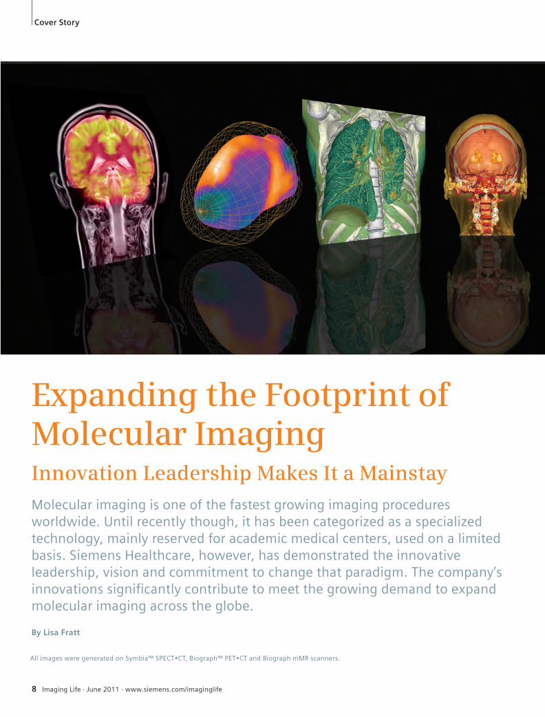
Expanding the Footprint of Molecular ImagingInnovation Leadership Makes It a Mainstay
Molecular imaging is one of the fastest growing imaging procedures worldwide. Until recently though, it has been categorized as a specialized technology, mainly reserved for academic medical centers, used on a limited basis. Siemens Healthcare, however, has demonstrated the innovative leadership, vision and commitment to change that paradigm. The company’s innovations significantly contribute to meet the growing demand to expand molecular imaging across the globe.
By Lisa Fratt
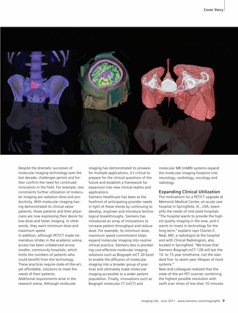
AllimagesweregeneratedonSymbia™SPECT•CT,Biograph™PET•CTandBiographmMRscanners.
IL_June2011.indb 8 5/12/11 5:24 PM
Imaging Life · June 2011 · www.siemens.com/imaginglife 9
Cover Story
Despite the dramatic successes of molecular imaging technology over the last decade, challenges persist and fur-ther confirm the need for continued innovation in the field. For example, two constraints further utilization of molecu-lar imaging are radiation dose and pro-ductivity. With molecular imaging hav-ing demonstrated its clinical value patients, these patients and their physi-cians are now expressing their desire for low-dose and faster imaging. In other words, they want minimum dose and maximum speed. In addition, although PET/CT made tre-mendous strides in the academic arena, access has been unbalanced across smaller, community hospitals, which limits the numbers of patients who could benefit from the technology. These practices require state-of-the-art, yet affordable, solutions to meet the needs of their patients. Additional requirements arise in the research arena. Although molecular
imaging has demonstrated its prowess for multiple applications, it’s critical to prepare for the clinical questions of the future and establish a framework for expansion into new clinical realms and applications. Siemens Healthcare has been at the forefront of anticipating provider needs in light of these trends by continuing to develop, engineer and introduce techno-logical breakthroughs. Siemens has introduced an array of innovations to increase patient throughput and reduce dose. For example, its minimum dose, maximum speed commitment helps expand molecular imaging into routine clinical practice. Siemens also is pioneer-ing cost-effective molecular imaging solutions such as Biograph mCT 20 Excel to enable the diffusion of molecular imaging into a broader group of prac-tices and ultimately make molecular imaging accessible to a wider patient population. Finally, innovations such as Biograph molecular CT (mCT) and
molecular MR (mMR) systems expand the molecular imaging footprint into neurology, cardiology, oncology and radiology.
Expanding Clinical UtilizationThe motivations for a PET/CT upgrade at Memorial Medical Center, an acute care hospital in Springfield, Ill., USA, exem-plify the needs of mid-sized hospitals. “The hospital wants to provide the high-est quality imaging in the area, and it wants to invest in technology for the long-term,” explains says Charles E. Neal, MD, a radiologist at the hospital and with Clinical Radiologists, also located in Springfield. “We know that Siemens Biograph mCT 128 will last the 10- to 15-year timeframe, not the stan-dard five- to seven-year lifespan of most systems.”Neal and colleagues realized that the state-of-the-art PET scanner combining the highest possible resolution with swift scan times of less than 10 minutes
IL_June2011.indb 9 5/12/11 5:24 PM

10 Imaging Life · June 2011 · www.siemens.com/imaginglife
Cover Story
Memorial Medical Center in Springfield, Ill., USA, is a good example of a mid-sized healthcare provider making excellent use of the flexibility of both the PET/CT and dedicated CT capabilities of the Biograph mCT 128-slice scanner, keeping up with increasing volumes and providing excellent scan quality.
could provide an opportunity to re-invent the business model. “Our biggest issue is throughput,” he explains.Although the center’s PET/CT volume has steadily nudged up since the June 2009 deployment of Biograph mCT, at five to six studies daily, PET/CT volume does not justify a dedicated PET/CT system. Bio-graph mCT, with a 128-slice CT scanner, can double as a dedicated CT scanner. With the new system, Memorial Medical Center also increased its patient
throughput, completing about 15 to 20 CT patient scans daily—an uptick that benefits patients, physicians and the bottom line.Prior to adding the Biograph mCT, some physicians referred patients to other sites for CT and to Memorial Medical Center for PET on the previous 16-slice PET/CT system. Physicians had to corre-late both datasets, and the model required patients to visit two centers. “Now, referring physicians and oncolo-
gists realize we have the best PET scan-ner and the best CT in town, so many are referring patients to us for both the PET and CT studies,” shares Neal.
Reducing DoseAnother critical component to increase the clinical utilization is to reduce any risk factor for the patients, it is dose reduction. In the Netherlands, the impe-tus for low-dose PET/CT imaging is strong and straightforward. As in other
“Now, referring physicians and oncologists realize we have the best PET scanner and the best CT in town, so many are referring patients to us for both the PET and CT studies.”Charles E. Neal, MD, Radiologist, Memorial Medical Center, Springfield, Ill., USA
IL_June2011.indb 10 5/12/11 5:24 PM

Imaging Life · June 2011 · www.siemens.com/imaginglife 11
Cover Story
European countries and around the globe, physicians and the public are very aware of radiation dose. Consequently, when Free University in Amsterdam, dis-seminated its RFP for a new PET/CT sys-tem early in 2009, it emphasized the need for a system that could support low-dose PET/CT imaging. Ultimately, Gerrit W. Sloof, MD, of the departments of nuclear medicine and cardiology, and colleagues opted to install one of the first Siemens Biograph mCT systems in Europe in October 2009. Biograph mCT not only delivered the low-dose capabilities the university required, it also enabled cost-effective molecular imaging.“We knew from the specifications that the Biograph mCT is very sensitive. When we calculated the optimal dose for our patients and compared it with other systems and estimated the total cost of ownership, we realized that we could save a great deal over the 10-year lifes-pan of the system, while providing high-quality imaging at the lowest dose,” Sloof explains.Although dose took center stage in the PET/CT decision-making process, Free University also focused on image quality. In fact, in the last 18 months, referring physicians have been able to immedi-ately recognize when patient studies are performed on the Biograph mCT, says Sloof, because of its high image quality. At Memorial Medical Center, Neal says improved resolution across the field of view has increased diagnostic confi-dence and therefore improved clinical utilization. In most PET/CT scanners, res-olution is highest at the center of the images and drops as the focus moves away the center. Biograph mCT provides equally high-resolution across the field of view, resulting in clearer scans.For example, with our previous scanner, a lesion on the axilla might have been indeterminate, says Neal. “With Biograph mCT, we can definitively tell it’s a spe-cific lymph node, and we can more pre-cisely isolate the area of activity. The datasets better correlate, so we can be more confident about these types of findings.”
Moreover, reconstruction speed is quite fast, improving productivity. Which means in addition that physicians can check image quality nearly immediately. “If the patient moves his head, we can do an additional scan of the head. The whole configuration is much more flexi-ble than in the early days,” Sloof explains.
Minimizing Dose Across the Molecular Imaging SpectrumMany of the same principles that impact PET/CT utilization also apply to SPECT.
SPECT is, for instance, the gold standard as a powerful nuclear technique for assessing cardiac perfusion and function to diagnose coronary artery disease, assessing risk and guiding management decisions; it continues to play a valuable role in cardiac imaging. However, pro-viders are pressed to deliver low-dose scans for increased patient safety with high image quality combined and higher throughput.SiemensIQ•SPECTtechnol-ogy was developed to reduce the acqui-sition time for myocardial perfusion imaging and focus counts on the heart
Expanding PET/CT into New Applications Siemens Biograph mCT is firmly rooted in oncology, cardiology and neurology applications. The primary application for PET/CT is still oncology, including lung and colorectal cancers and lymphoma. However, its role continues to expand not only in the already important PET/CT fields of cardiology and neurology.Physicians at Free University in Amsterdam, the Netherlands, are leveraging the PET/CT system for treatment monitoring and recurrence, especially in lung cancer and lymphoma. In addition, Gerrit W. Sloof, MD, of the departments of nuclear medicine and cardiology, says the oncology team finds the system
helpful for measuring the control of radiofre-quency ablation on liver metastases.The university hospital also has employed the system in infection imaging. And while con-tinued innovation in molecular imaging tech-nology is critical, effective molecular imaging hinges on several inputs, with radiotracers playing a key role. In the U.S., one of the major developments in molecular imaging is Medicare reimbursement for 18F sodium fluo-ride (NaF) PET for sites participating in the National Oncologic PET Registry (NOPR). Although referring physicians are still learning about indications, experts anticipate that NaF PET imaging will supplant conventional bone scanning in some cases, once again indicating the power and value of molecular imaging. Tracer development is proceeding on other fronts as well and allowing sites that have invested in state-of-the-art PET/CT technology to capitalize on their investment. For example,
Memorial Medical Center in Springfield, Ill., will use Siemens Biograph mCT 128 as the cornerstone of its participation in an American College of Radiology Imaging Network (ACRIN) trial evaluating a new prostate cancer tracer.These applications, while impressive, merely scratch the surface of the poten-tial of molecular imaging. Researchers at leading medical centers around the globe continue to drive the expansion of molecular imaging.
Lung cancer with medistinal metastases. (Image courtesy of University of Minnesota, Minneapolis, Minnesota, USA.
IL_June2011.indb 11 5/12/11 5:24 PM

12 Imaging Life · June 2011 · www.siemens.com/imaginglife
Cover Story
by using a cardio-centric acquisition. WithIQ•SPECT,afull-countSPECTscantakes only four minutes and can be achieved using half the traditional dose. WithinnovationssuchasIQ•SPECT,Sie-mens is taking the forefront in embrac-ing the responsibility of minimizing radi-ation dose for the purposes of patient safety, while simultaneously preserving excellent image quality and increasing productivity significantly. The risks of radiation exposure need to be balanced with the benefits of the imaging study, emphasizes Manuel Cer-queira, MD, chairman of nuclear medi-cine in the Imaging Institute and a staff cardiologist in the Sydell and Arnold Miller Family Heart & Vascular Institute
at Cleveland Clinic in Cleveland, Ohio, USA. “For many of our patients, there are a lot of benefits to the information that’s obtained from the imaging study,” he says. In cardiology, specific benefits include differentiating between the presence and absence of coronary dis-ease in symptomatic patients and in optimizing management and outcomes in patients with known coronary artery disease.“If radiation exposure can be avoided, that is the best path,” Cerqueira says. “Our approach is to look at published appropriateness criteria. Only if it is an appropriate indication, we do the study. But then [we always image] at the low-est dose possible while maintaining the best image quality to assure accuracy of imaging results.” IQ•SPECTsupportsthisstrategy,offeringimaging solutions where the lowest dose can be used while still scanning patients faster than ever before.In addition to the utilization of the latest technology, Cerqueira and colleagues advocate a multi-pronged approach to reducing radiation exposure. For exam-ple, the Clinic no longer uses thallium for SPECT studies as its longer half-life translates into higher radiation expo-sure. A second strategy is stress-only imaging for the diagnosis of coronary disease. “When a stress study is normal, we don’t need to perform a rest study because the normal result indicates that the patient does not have disease,” explains Cerqueira. Expanding Patient Access While academic medical centers are well-outfitted with cutting edge molecular imaging systems for clinical and research applications and mid-sized organizations have been able to carve a niche with the introduction of robust systems geared to their needs such as the Biograph mCT, small, community-based hospitals remain the first option for millions of patients in the U.S. These sites need an affordable, state-of-the-art molecular imaging solu-tion to provide access for their patients. Siemens Biograph mCT 20 Excel answers their needs.
Siemens designed Biograph mCT 20 Excel to meet the needs of community hospitals, enabling expanded access to molecular imaging technology to more patients by delivering an affordable investment for providers.“This new configuration of Biograph mCT gives us the highest resolution available thanks to its ultra-high resolu-tion based on the most recent techno-logical addition of image processing knownasultraHD•PET.Todayitispossi-ble to achieve spatial resolutions close to 2 mm,” explains Horacio Amaral, MD, director Centro de Medicina Nuclear y PET/CT in Santiago, Chile. The center recently deployed Biograph mCT 20 Excel to meet increasing local demand and is using the investment as a springboard to expand access to molecu-lar imaging technology. (See more in story on page 26)
Today’s Research— Tomorrow’s Clinical RealityWith the recent introduction of Biograph mMR, Siemens helps to expand molecu-lar imaging in one more direction, enabling new clinical applications. The Athinoula A. Martinos Center for Biomed-ical Imaging at Massachusetts General Hospital (MGH) in Boston, recently took delivery of the first Biograph mMR system in the U.S. All signs point to a smooth deployment process, shares Ciprian Cat-ana, MD, PhD, instructor in radiology at Harvard Medical School and assistant in neuroscience at MGH. The integrated whole-body molecular MR system has already been installed at the center.The system represents a completely new generation in molecular imaging tech-nology and builds on previous projects spearheaded by Catana and his col-leagues. For the last three years, the researchers have used Siemens BrainPET technology, a prototype PET/MR system comprised of MR-compatible PET detec-tors inserted inside an MR scanner. BrainPET set the stage for Biograph mMR, allowing the researchers to explore the concept of simultaneous data acquisition and prepare for whole-body PET/MR.
“If radiation exposure can be avoided, that is the best path.”Manuel D. Cerqueira, MD, FACC FAHA FASNC, Professor of Radiology and Medicine, Cleveland Clinic Lerner College of Medicine of Case Western Reserve University, Chairman, Department of Nuclear Medicine, Imaging Institute, Cleveland, Ohio, USA
IL_June2011.indb 12 5/12/11 5:24 PM

Imaging Life · June 2011 · www.siemens.com/imaginglife 13
Cover Story
Early PET/MR applications included neu-rology, as well as feasibility studies in healthy volunteers. However, two barri-ers prevented more widespread use of PET/MR. First, the Martinos Center lacked an on-site cyclotron to produce radioisotopes, and second, BrainPET was limited to brain imaging. Both barriers have been addressed. The center invested in an on-site cyclotron, and it has deployed Biograph mMR, which enables whole-body PET/MR.“We’re looking forward to exploring new opportunities,” shares Catana. With hun-dreds of potential collaborators, Harvard Medical School and MGH provide the ideal environment for the new system.One of the early focuses will be on neu-ropsychiatry; the center has already lured some researchers and helped them successfully obtain National Institutes of Health (NIH) grant funding. The first studies will launch later this year and include diverse topics such as Parkin-son’s disease, multiple sclerosis, chronic pain and nicotine dependence, to name just a few.At the same time, Catana expects to build on earlier research projects. One of his primary research interests applied Brain-PET to delve into the mechanism of action of antiangiogenic treatment on glioblastomas. Specifically, they have used the standard 18F-FDG for studying changes in tumor glucose uptake while simultaneously assessing changes in per-meability using F-MISO to investigate hypoxia in glioblastoma and plan to use FLT to study tumor proliferation. The researchers also are leveraging the new cyclotron for producing novel radioiso-topes that are not commercially available. However, Catana admits that the early efforts merely scratched the surface of what’s possible. “There are a lot of potentially very interesting research applications that can be performed on the combined scanner. For example, PET and MR measurements of cerebral per-fusion could be cross-calibrated and improved. Similarly, brain activation studies could be cross-validated using simultaneously acquired fMRI and PET data. Some of these studies could not be
completed on the BrainPET [e.g. those requiring visual stimuli] because of the restricted space inside the scanner. Also, we can start to explore research applica-tions at other sites in the body.”The design of the Siemens scanner, which provides simultaneous acquisition of PET and MR data, offers workflow advantages in addition to opening the door to new clinical applications, shares Catana. With sequential scanning, the technologist has to very carefully focus on protocols or the acquisition time may be doubled. “By acquiring data simulta-neously, Biograph mMR solves that issue,” he confirms.Virtually all of the neurological patients referred for a PET study also undergo an MR examination. “It will save the patient from an extra exam,” says Catana. One example of how the center’s early work with BrainPET will translate into Bio-graph mMR is centered on improving the PET data quantification by including the simultaneously acquired MR informa-tion. For example, motion is a major issue in PET because dynamic scans can be up to 90 minutes long, explains Cat-ana. “There’s no way for patients to remain still, which degrades the quality of the data.” The magnitude of the prob-lem is amplified in the case of high spa-tial resolution scanners, such as the BrainPET. Consequently, even a very small amount of motion will degrade the images. However, the researchers have developed a method to use MR to track the patient’s head motion (while acquir-ing the anatomical or functional MR data of interest) and apply the correction to the PET data.Catana envisions a significant down-stream impact, with MR-assisted PET motion correction benefiting all neuro-imaging applications that involve lengthy studies or in the case of patients who find it difficult to remain still. The process can be migrated to whole-body applications with Biograph mMR both for respiratory and cardiac motion that cannot be avoided, not even during shorter length studies.In fact, the design of Siemens Biograph mMR, which employs simultaneous
acquisition of PET and MR data, bypasses some of the motion correction chal-lenges. The alternate configuration—sequential acquisition of PET and MR datasets—can be problematic because the MR data are not going to match the actual motion during the PET acquisi-tion, explains Catana. “With Biograph mMR, we are hoping to use MR data to detect the motion of the heart, thorax or abdomen and use those data to correct the PET images,” he shares.“Biograph mMR has great potential,” states Catana, explaining, “The excellent soft-tissue contrast provided by MR will allow a better characterization of suspi-cious lesions in oncology applications. Additionally, the near perfect spatial co-registration between the PET and MR volumes will help studies focusing on the head and neck, breast and liver.”
Maximizing Return on InnovationMolecular imaging is the future of radi-ology. Over the past several years, the growth in PET/CT procedures has out-paced other modalities. And now PET/MR is dawning. The future is bright. Molecular imaging is rapidly evolving across multiple arenas, opening the door to more diverse providers and applica-tions and fueling clinical research to pro-vide answers to today’s pressing medical questions. One of the cornerstones of success is partnering with a leader in the field with a demonstrated commitment to innova-tion. This type of partnership can help a molecular imaging provider establish a sound foundation in multiple key areas: dose reduction, productivity and clinical
The Biograph mMR system requires 510(k) review by the FDA and is not commercially available in all countries. Due to regulatory reasons, its future availability in any country cannot be guaranteed.
Some of the biomarkers referenced herein are not currently recognized by the U.S. FDA as being safe and effective, and Siemens does not make any claims regarding its use. Please contact your local Siemens organization for further details.
Siemens BrainPET scanner is a first-generation research unit and not commercially available.
Lisa Fratt is an award-winning medical and technology writer based in Wisconsin, USA.
IL_June2011.indb 13 5/12/11 5:24 PM

14 Imaging Life · June 2011 · www.siemens.com/imaginglife
● Manaus
● Teresina
BrasÍlia
● Belem
● São LuízFortaleza ●
Natal ●
Recife ●
● Cuiabá
● Campo Grande
Goiânia ●
Curitiba ●
Salvador ●
Rio de Janeiro ●São Paulo ●
IL_June2011.indb 14 5/12/11 5:24 PM

Imaging Life · June 2011 · www.siemens.com/imaginglife 15
Mundim credits his father with educat-ing him about the benefits that PET/CT could provide to oncology patients. “The project was envisioned by my father, Tito Livio Mundim, MD, who is the head of the company. He was the one who taught me about the technology,” says the younger Dr. Mundim. “Clínica Villas Boas has always been a pioneer in the acquisition of new tech-nologies,” explains Mundim. The clinic determined that PET technology, which is essential in modern oncology practice, would be no exception. In 2005, PET/CT services were not avail-able in Brasília, due in part to the high cost of these systems, and partly because production of FDG was restricted to the Brazilian government. And thus began Mundim’s journey as a PET pioneer. In a little more than five years, Mundim has not only brought PET/CT services to Brazil’s capital city of roughly 2.5 million people, he and his team also have created a separate busi-ness generating an FDG production stream for other PET/CT providers that is expanding throughout the country. In the process, Clínica Villas Boas
became the first private institution in Brazil to produce FDG. Development of a third source of FDG, behind two govern-ment facilities, meant more healthcare organizations could acquire PET/CT scan-ners and have easy access to vital FDG. The business currently provides FDG on a routine basis to 12 radiology facilities. “Back in 2005, we [began the process of acquiring] a PET/CT scanner to be installed in our 37-year-old imaging cen-ter in Brasília, Districo Federal. It would be the third city in Brazil to provide this kind of procedure,” Mundim recalls. “At that time, we were worried about FDG supply because there were only two government-operated facilities that pro-duced FDG in the entire country: one in Rio de Janeiro—and the other one in São Paulo. Until that year, the production of short-half-life radioisotopes was still a government monopoly.” Undeterred, Mundim purchased the PET/CT equipment and began using it to diagnose patients the following year. However, because both of the country’s government FDG providers were located far from his facility, “we started to have some issues concerning the FDG supply.
Pioneers in PETClínica Villas Boas Expands PET/CT Throughout Brazil
As a radiologist and director of Clínica Villas Boas, an imaging facility in Brasília, Brazil, Tiago Mundim, MD, might not have planned on becoming a PET pioneer in Brazil, but says he could clearly see the potential of PET/CT and the benefits that this advanced imaging technique could bring to his patients. The year was 2005, and he wanted to be among the very first to bring PET/CT, a Siemens Biograph™ mCT 128, to Brasília.
By Mary Stevens
Outcomes
“Because of our cyclotron facility, 12 PET centers have emerged on the North, Northeast and Midwest of Brazil.”Tiago Mundim, MD, Radiologist, Clínica Villas Boas, Brasília, Brazil
IL_June2011.indb 15 5/12/11 5:24 PM

16 Imaging Life · June 2011 · www.siemens.com/imaginglife
Outcomes
The number of PET Centers in Brazil was rising and the FDG demand was rising as well,” he says. “Because of that, we started to receive fewer doses than we ordered and also began to experience some cancellations.”
A Supply to Meet Demand It became clear that a more reliable source for Clínica Villas Boas’ radiophar-maceuticals had to be found. The ideal situation would be to produce FDG on-site or close by the facility. Mundim and his colleagues started to realize that investing in their own cyclotron facility would not only allow them to reliably provide the FDG necessary to ensure the right dose was available for each patient, but also would foster growth of PET/CT both in the city of Brasília and in the rest of the country. The restrictions on FDG production, however, remained an obstacle. While Clínica Villas Boas was investigat-ing its particle accelerator options under the Brazilian government’s FDG controls, “we had a pleasant surprise. In 2006, under pressure from the Brazilian medical community, the government ended its monopoly on the production of short-lived radioisotopes such as FDG,” says Mundim. “It was just what we needed to install a cyclotron.” Clínica Villas Boas subsequently bought the Siemens Eclipse HP 11 MeV particle accelerator. As the FDG business developed, Clínica Villas Boas established Villas Boas Radio-pharmaceuticals Brazil, which was the first private company in Brazil to pro-duce FDG. The results have been remarkable: In less than two and a half years, the availability of an additional FDG supply has helped bring advanced imaging to new areas of the country. “Because of our cyclotron facility, 12 PET centers have emerged on the North, Northeast and Midwest of Brazil,” Mun-dim asserts. Villas Boas Radiopharma-ceuticals Brazil delivers FDG to a widen-ing area from Manaus, located 1,936 kilometers (1,200 miles) northwest from Brasília—to Curitiba, which is 1,073 kilo-meters (670 miles) to the south of the capital.
The cyclotron also has powered business at the clinic where it all began. Clínica Villas Boas now performs approximately 100,000 imaging procedures each year and employs 170 people, according to Mundim. Its on-site molecular imaging technologies include: › one Siemens Biograph mCT 128
PET•CTscanner;› oneSiemensSymbia16SPECT•CT
scanner;› one multidetector CT;With state-of-the-art imaging, electronic medical records and RIS/PACS integra-tion readily available, patients can view images and consult with Clínica Villas Boas physicians online. The Villas Boas Radiopharmaceuticals Brazil FDG cyclotron and business is a separate entity from the imaging center, but is integral to its success, according to Mundim. “From our point of view, the FDG business is becoming very impor-tant” and in fact, could reach the same level of profitability as the imaging cen-ter next year, he says. Although Mundim declined to say how much the business made last year, he allows that Villas Boas Radiopharmaceuticals Brazil “surely had an increase” in sales. Mundim’s entrepreneurship is creating a PET market boom in Brazil, a country of close to 191 million people, where vast distances separate cities and towns and where the terrain can challenge trans-portation of almost every kind. It is help-ing to accelerate installation of PET capabilities in parts of the country where advanced imaging might other-wise have taken years to implement. For example, in the northeastern region of the country, PET is now available in cit-ies including Fortaleza, Teresina, Natal, Recife, Salvador and São Luiz. Villas Boas Radiopharmaceuticals Brazil also supplies FDG and enables PET in Manaus and Belem in the North, as well as in Cuiabá, Campo Grande and Goiânia in the Midwest part, Mundim says. “Half of these cities already have at least one PET center and the other half are in the process of acquisition [of a system],” he adds. The only provider to Manaus—shipping by air—is Villas Boas Radio-
pharmaceuticals Brazil. Perhaps equally telling is that in areas where organizations have a choice of FDG suppliers, such as Goiânia, many facilities still prefer to use Villas Boas. “Goiânia could buy [FDG] from São Paulo, but prefers Villas Boas because of the proximity and readiness,” Mundim says. This means shorter journeys and less waiting for patients, in addition to being a boost for business. Despite the presence of the govern-ment’s FDG manufacturing facilities in Rio de Janeiro (IEN) and in São Paulo (IPEN), Villas Boas supplies the biophar-maceutical to facilities in Rio de Janeiro “depending on the reliability of their local supplier,” he adds.
FDG on DemandFlexibility is one advantage of Villas Boas Radiopharmaceuticals Brazil’s approach to FDG sales. The company doesn’t sign binding contracts with hos-pitals and other facilities to supply the radioisotope—instead, clients buy FDG doses according to demand and are free to buy from other producers as well, according to Mundim. Geography and market forces aren’t the only challenges to developing and oper-ating PET/CT centers, he says. The installation of a PET unit demands a large financial investment, and requires trained professionals to operate. In addi-tion, “to obtain government licenses to operate these facilities in Brazil is hard due to bureaucracy,” says Mundim. “Technician and physician training are crucial for good outcomes. We keep track of the news and trends in this busi-ness, attending many medical conven-tions and meetings worldwide” to stay up to date, he says.
The FDG Business While a variety of other tracer molecules can be used in the PET/CT process, Villas Boas is dedicated to providing FDG. The organization has 12 people working to manufacture FDG daily. 18F-FDG radio-iso-tope synthesis is fully automated, allowing production of as many as four rounds without human intervention.
IL_June2011.indb 16 5/12/11 5:24 PM

Imaging Life · June 2011 · www.siemens.com/imaginglife 17
Outcomes
“We produce an average of two batches per day because some of our clients are located far from our facility, and require higher output,” Mundim explains. Hav-ing far-flung customers can become an issue because FDG’s radioactive strength is cut by 50 percent every 110 minutes, making timely delivery imperative. This might be one reason Mundim describes delivery distances in terms of air travel time, not kilometers. The process begins early in the day. The facility’s engineers start by accelerating the cyclotron, spinning protons to create the radioactive part of the radiopharma-ceutical. Their work is followed by the pharmacists, who complete the work on the FDG in the radiopharmacy and pre-pare it for travel and use. Currently, Villas Boas Radiopharmaceuti-cals Brazil is the only FDG producer in Brasília, according to Mundim. Worldwide, too, the demand is growing for PET/CT scanning. Sales of PET scan-ners are projected to increase 66 per-cent worldwide from 2009 to 2017,
according to a November 2010 study by Bio-Tech Systems. Likewise, sales of FDG are expected to increase 29 percent dur-ing the same period. Demand in Brazil is no exception. As Mundim points out, PET/CT is mainly a diagnostic tool for oncology patients. “It provides high specificity and sensitivity for imaging several tumor types,” he adds. The primary diagnostic use for PET/CT in Brazil is in oncology (more than 90 per-cent). Due to its high degree of accuracy in lesion evaluation, PET/CT reduces unnecessary treatments and can lead to better patient outcomes in such cancers as solitary pulmonary nodules, lym-phoma, colorectal cancer, breast cancer, melanoma, esophagus cancer and many others, Mundim says.
A Bright FutureLooking toward the future, he envisions PET/CT as a diagnostic tool that’s avail-able almost everywhere and is used as a standard clinical technology throughout the country and beyond. “In oncology,
PET/CT will become the standard clinical technology, replacing stand-alone CT,” he predicts, because the combination can provide more detailed images, and thus more accurate diagnoses, during one patient visit.The expansion of PET and advanced diagnostics will depend on a reliable supply of FDG. Mundim is planning for that future: two additional cyclotrons are in the works for Villas Boas Radio-pharmaceuticals Brazil, and the com-pany intends to start producing ammo-nia-based radiopharmaceuticals as well as FDG, he says. As Villas Boas Radio-pharmaceuticals Brazil expands its FDG operations, Mundim is working to lead the way toward ubiquitous PET/CT diag-nostics, while making it easier for others to follow.
SiemensBiographmCT128-slicePET•CTscannerinstalledatClínicaVillasBoas.
Mary Stevens is a medical and technology journalist with extensive writing experience in magazines, websites and newsletters for physicians and administrators. She is based in Massachusetts, USA.
IL_June2011.indb 17 5/12/11 5:24 PM

18 Imaging Life · June 2011 · www.siemens.com/imaginglife
Outcomes
PET/CT: The Right Solution for Routine and ResearchLocated just a few short minutes from the Brandenburg Gate in Berlin, the Diagnostic Therapeutic Center is attracting more than casual tourists. Physicians here are using the Siemens hybrid imaging Biograph™ TruePoint PET•CTscannertoprovideeffectivecancerstagingandalsotoimageanextremely rare pediatric disorder. They also have a vision for the future of cancer care, closely joining their current excellence in diagnosis with a new therapy center next door that will open next year. Their mission is a first of its kind, a patient-focused, one-stop shop for cancer patients. By Mary C. Tierney, MS
IL_June2011.indb 18 5/12/11 5:24 PM

Imaging Life · June 2011 · www.siemens.com/imaginglife 19
PET/CT for Cancer StagingThe Diagnostic Therapeutic Center is an outpatient imaging facility that per-forms approximately 60,000 studies per year. About 1,500 of those procedures are performed on the Biograph 64. Since installing their first PET scanner, they have seen annual imaging volume triple. The only challenge to increasing the uti-lization for PET imaging has been a diffi-cult reimbursement climate in Germany. “Our utilization has grown dramatically since we first adopted the technology,” says Prof. Dr. Wolfgang Mohnike, MD, co-owner of the Diagnostic Therapeutic Center and member of the Medical Council Berlin.“The problem is that reimbursement is not clear in Germany,” he says. “If you want a PET/CT in a private practice, you need a system that will give you the best return on investment. We started with
very few investigations and then went very rapidly to about 500 imaging stud-ies per year and we have seen that num-ber triple. We will increase the amount of annual procedures in the future because we have contracts with insur-ance companies that pay for our investi-gations. The system was an economical purchase and it has justified its cost.”“Insurance companies pay for these advanced imaging examinations because they know they will save money in the long run in the course of taking care of patients,” Dr. Mohnike says. As a council member, he meets with the insurers to share successes in patient outcomes and why PET/CT is vital to proper patient management, namely in oncology.Part of the increased utilization for PET/CT at the Diagnostic Therapeutic Center is increasing confidence in the
modality’s ability to influence the course of treatment. “Over the years, we have seen significant changes in how we manage and stage cancer,” says Dr. Mohnike. “The first disease we started with was staging for lung cancer. At the time, surgeons accepted our imaging results, but PET/CT imaging was not a high priority for them. Now the PET/CT images are routine prior to surgical intervention. The same goes for breast cancer where we have seen PET/CT used to define the results of first-line chemo-therapy. We have seen PET/CT imaging significantly change the management of women with breast cancer, as it can bet-ter define the effects of chemotherapy. Similarly, we see PET/CT making huge strides in the management of malignant lymphoma and we are often consulted to determine the efficacy of the chemo-therapy.
Outcomes
Prof. Dr. Wolfgang Mohnike reviews brain PET/CT images on a workstation in the control room for the Biograph TruePoint scanner on-site at the Diagnostic Therapeutic Center in Berlin, Germany.
IL_June2011.indb 19 5/12/11 5:24 PM

20 Imaging Life · June 2011 · www.siemens.com/imaginglife
1 18F F-DOPA PET/CT in an infant with congenital hyper-insulinemia demonstrates a focal area of insulin overpro-duction in the pancreatic tail, which is amenable to partial resection.
2 18F F-DOPA PET/CT in an infant demonstrates a focal area of insulin overproduc-tion in the pancreatic head.
Outcomes
“We specialize in the diagnosis, staging and management of all cancers,” Dr. Mohnike notes. “Today, most often we are helping with breast, colorectal and lung cancer as well as lymphoma. Pros-tate cancer is the newest for us.“
Research on Rare Pediatric Disorder Aided by PET/CTFamilies make visits to the Diagnostic Therapeutic Center from all over the world in order to find answers about a potentially devastating and very rare genetic disorder that affects their chil-dren. They come from England, Scot-land, Israel, India, Argentina, central Europe, the United States and from the Far East. Those born with persistent hyperinsulinaemic hypoglycemic of infancy (PHHI) can have severely low blood sugar levels and seizures. These children also are at high risk of brain damage if their blood sugar levels are not maintained. This also is a family affair as Mohnike’s brother, a pediatri-cian, was the person who first appealed to him to look more deeply into this genetic disorder via PET/CT images. PET/CT uniquely offers excellent visualization of a newborn pancreas that measures just half the size of an adult’s pinky fin-ger. “Very, very tiny,” he says, demon-strating the size by pinching his finger and thumb from nail to knuckle on the other hand.
This inherited genetic disease is clini-cally detected by the presence of high insulin levels in conjunction with low glucose levels. Dr. Mohnike and col-leagues have published research in the Journal of Clinical Endocrinology and Metabolism on the effectiveness of PET/CT in imaging this rare disorder. Once diagnosed, the treatment options vary. If they can find the focal area of the pancreas, it can be removed and the child will live a normal life.PET imaging has been used frequently in research as one of the most reliable imaging techniques in characterizing the disorder. Dr. Mohnike and colleagues have found this always to be true. Due to the nature of this disease, there is an increased level of F-DOPA uptake and the information gained has been found to provide insight for research physi-cians on whether the entire pancreas is involved, or just a focal area of over pro-duction. They also then determine the necessity and extent of pancreatic resec-tion. In some cases as much as 95 per-cent of the pancreas is removed.The challenge to physicians is that the pancreas in an infant has a diameter of between 3 mm to 4 mm. So having some of the most advanced PET scan-ning available makes a difference in the management of these patients. “We were not able to image the pancreas with our older PET technology because
the spatial resolution was insufficient,” says Dr. Mohnike. “With our Biograph 64, we have improved the spatial resolu-tion to 2 mm to 3 mm, the sensitivity, and the count rate so now we can do this type of imaging. We get very good quality imaging and this has helped us establish our center as an expert site for imaging this rare disorder.”
PET/CT BenefitsAll the features available from hybrid PET/CT imaging give Dr. Mohnike and colleagues the ability to image this rare disorder. “We have investigated approxi-mately 170 infants with PHHI from all over the world,” says Dr. Mohnike. “It is so rare that the number of newly detected instances per year is only about 20 to 25 in Germany. With such a low number, having a diagnostic modality like the Biograph 64 allows us to play an important role in confirming the diagno-sis so treatment can begin quickly.”“The leading scientists in our center discussed the purchasing decision sev-eral times and the Biograph 64 clearly demonstrated itself to be the best fit for our practice—both from perspec-tives of cost and of its clinical capabili-ties,” says Dr. Mohnike.
Siemens Helps New VisionVisionary discussions with Siemens have helped the Diagnostic Therapeutic Cen-
1 2
IL_June2011.indb 20 5/12/11 5:24 PM

Imaging Life · June 2011 · www.siemens.com/imaginglife 21
ter formulate its next undertaking, the new therapy center located next door that they broke ground on recently and will open in 2012. Seven physicians will be a part of the facility, imagers as well as oncologists who will help to diag-nose, stage, monitor and treat cancer and other disease—all in one state-of-the-art facility. While traditionally strictly divided, this center with three floors that looks to treat more than 200 patients per day, will bring together diagnosis and treatment. “It will be a great connection of physicians in nuclear medicine, radiotherapy and radi-ology,” Dr. Mohnike says. This mission breaks with the model of
most facilities in Germany, but it meets the physicians’ shared and progressive goal of working closely together and putting patients at the center of care. They are creating a facility in which patients will arrive for a scan and, in the same day, receive a diagnosis and even a therapy appointment, if needed. And Siemens technology will be there as well, from imaging and diagnosis with PET/CT and MRI and eventually PET/MR, through exacting cancer therapy plan-ning and execution via linear accelera-tors. And even in between, Siemens IT products such as syngo.via and syngo.plaza will play a role in merging caregiv-ers and care plans together. Partnership
among physicians, as well as vendors, makes medicine stronger. “We seek to make Siemens a partner in this new facility, like we have in our current [facil-ity],” he says. “We see care of our patients [stretching] from primary diag-nosis through therapy planning and fol-lowup. That is what is best for our patients. We can do all of this with the help of Siemens.”
Outcomes
Some of the biomarkers in this publication are not currently recognized by the U.S. Food and Drug Administration (FDA) or other regulatory agencies as being safe and effective, and Siemens does not make any claims regarding their use.
Mary C. Tierney, MS, is a medical and healthcare editor with two decades of experience in print, digital and online media. She is based in Rhode Island, USA.
Just next door to its current location in Berlin, the team at the Diagnostic Therapeutic Center is building a new three-floor therapy center. Siemens imaging and therapy equipment will assist the facility’s mission of patient-focused cancer care, from diagnosis through staging, therapy and disease monitoring. It is set to open next year.
IL_June2011.indb 21 5/12/11 5:24 PM
22 Imaging Life · June 2011 · www.siemens.com/imaginglife
Outcomes
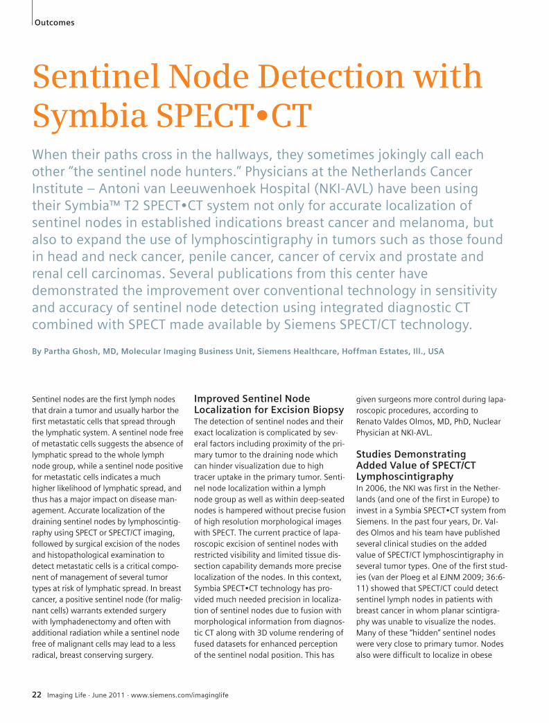
Sentinel nodes are the first lymph nodes that drain a tumor and usually harbor the first metastatic cells that spread through the lymphatic system. A sentinel node free of metastatic cells suggests the absence of lymphatic spread to the whole lymph node group, while a sentinel node positive for metastatic cells indicates a much higher likelihood of lymphatic spread, and thus has a major impact on disease man-agement. Accurate localization of the draining sentinel nodes by lymphoscintig-raphy using SPECT or SPECT/CT imaging, followed by surgical excision of the nodes and histopathological examination to detect metastatic cells is a critical compo-nent of management of several tumor types at risk of lymphatic spread. In breast cancer, a positive sentinel node (for malig-nant cells) warrants extended surgery with lymphadenectomy and often with additional radiation while a sentinel node free of malignant cells may lead to a less radical, breast conserving surgery.
Improved Sentinel Node Localization for Excision BiopsyThe detection of sentinel nodes and their exact localization is complicated by sev-eral factors including proximity of the pri-mary tumor to the draining node which can hinder visualization due to high tracer uptake in the primary tumor. Senti-nel node localization within a lymph node group as well as within deep-seated nodes is hampered without precise fusion of high resolution morphological images with SPECT. The current practice of lapa-roscopic excision of sentinel nodes with restricted visibility and limited tissue dis-section capability demands more precise localization of the nodes. In this context, SymbiaSPECT•CTtechnologyhaspro-vided much needed precision in localiza-tion of sentinel nodes due to fusion with morphological information from diagnos-tic CT along with 3D volume rendering of fused datasets for enhanced perception of the sentinel nodal position. This has
given surgeons more control during lapa-roscopic procedures, according to Renato Valdes Olmos, MD, PhD, Nuclear Physician at NKI-AVL.
Studies Demonstrating Added Value of SPECT/CT Lymphoscintigraphy In 2006, the NKI was first in the Nether-lands (and one of the first in Europe) to investinaSymbiaSPECT•CTsystemfromSiemens. In the past four years, Dr. Val-des Olmos and his team have published several clinical studies on the added value of SPECT/CT lymphoscintigraphy in several tumor types. One of the first stud-ies (van der Ploeg et al EJNM 2009; 36:6-11) showed that SPECT/CT could detect sentinel lymph nodes in patients with breast cancer in whom planar scintigra-phy was unable to visualize the nodes. Many of these “hidden” sentinel nodes were very close to primary tumor. Nodes also were difficult to localize in obese
Sentinel Node Detection with Symbia SPECT•CTWhen their paths cross in the hallways, they sometimes jokingly call each other “the sentinel node hunters.” Physicians at the Netherlands Cancer Institute – Antoni van Leeuwenhoek Hospital (NKI-AVL) have been using theirSymbia™T2SPECT•CTsystemnotonlyforaccuratelocalizationofsentinel nodes in established indications breast cancer and melanoma, but also to expand the use of lymphoscintigraphy in tumors such as those found in head and neck cancer, penile cancer, cancer of cervix and prostate and renal cell carcinomas. Several publications from this center have demonstrated the improvement over conventional technology in sensitivity and accuracy of sentinel node detection using integrated diagnostic CT combined with SPECT made available by Siemens SPECT/CT technology.
By Partha Ghosh, MD, Molecular Imaging Business Unit, Siemens Healthcare, Hoffman Estates, Ill., USA
IL_June2011.indb 22 5/12/11 5:24 PM

Imaging Life · June 2011 · www.siemens.com/imaginglife 23
Outcomes
Renato Valdes Olmos, MD, PhD, Nuclear Physician
Netherlands Cancer Institute – Antoni van Leeuwenhoek Hospital
1 Images show SPECT/CT lymphoscintigraphy in three patients with cancer of the oral cavity. Tracer-avid sentinel nodes are localized in the deep cervical nodal group. Thin-slice diagnostic CT helps to de-fine small nodes. One patient demonstrates sentinel nodes in the supraclavicular region as well.
2 Images show SPECT/CT lymphscintigraphy in a patient with pros-tate cancer and demonstrate sentinel nodes in the left iliac and pre-sacral nodal groups. Localization is assisted by integrated diagnostic CT, able to differentiate small nodes from surrounding vascular structures and mesenteric fat.
1 2
IL_June2011.indb 23 5/12/11 5:24 PM

24 Imaging Life · June 2011 · www.siemens.com/imaginglife
Outcomes
patients. SPECT/CT also revealed addi-tional sentinel nodes compared to planar scanning which then had to be separately sampled. In one study by the same group (van der Ploeg JNM 2007; 48: 1756-1760), out of 31 patients with breast cancer or melanoma who underwent both planar and SPECT/CT lymphoscintig-raphy, six additional sentinel nodes in four patients were demonstrated using SPECT/CT imaging. Two of these nodes detected with SPECT/CT were tumor-posi-tive and significantly changed patient management. Detection of sentinel nodes in internal mammary nodal groups in breast carcinoma and atypically located nodes in melanoma demands modification in approach prior to surgery or laparoscopy, especially related to inci-sion and soft tissue dissection.
In several cancers, especially involving head and neck tumors, the lymphatic drainage pattern is often unpredictable. Using SPECT/CT to image head and neck tumor malignancies has proven to be highly accurate in depicting atypically located sentinel nodes, as well as defining the relationship of the sentinel nodes with vital vascular structures like the carotid vessels. In a study involving 33 patients (Vermeeren et al Journal of Oncology 2009) with head and neck cancer, SPECT/CT identified additional affected sentinel nodes in 18 percent of patients and accu-rately characterized false positive lesions on planar scintigraphy.
SPECT/CT and Prostate CancerDr. Lenka Vermeeren, a research physi-cian at the NKI, has been studying the
potential of using SPECT/CT lymphoscin-tigraphy in tumors with deep nodal drain-age such as prostate cancer. Lymphade-nectomy for sentinel nodes in prostate cancer routinely involves dissection of the obturator fossa and the region around the external iliac vein. However, extended pelvic lymphadenectomy can be performed in the area between the external iliac arteries, the pelvic wall and the area around the common iliac and internal iliac arteries, if there is clear evi-dence of sentinel nodes in these areas. SPECT/CT imaging was instrumental in the localization of sentinel nodes in the region of extended pelvic lymphadenec-tomy in 35 percent of patients in a study involving 46 patients with prostate can-cer (Lenka Vermeeren et al JNM 2009; 50:865-870). SPECT/CT imaging demon-strated sentinel nodes in 98 percent of patients compared to planar (91 per-cent). In four out of 46 patients the nodes detected only on SPECT/CT were the only positive nodes and SPECT/CT dramatically changed the stage of tumor and eventual disease management in these patients. Such findings, according to Dr. Valdes Olmos, justify the use of SPECT/CT for all prostatic tumors.
Additional ApplicationsExpanding the role of SPECT/CT into sev-eral other tumor types with deep-seated abdominal and para-aortic lymph node drainage, the NKI group also studied penile carcinoma and renal cell carcinoma. It is well known that sentinel node biopsy is not 100 percent accurate for the prediction
3 A patient with breast carcinoma imaged with planar lymphoscintigraphy showed no sentinel nodes with high uptake in primary tumor injection site. However, SPECT/CT defined axillary as well as internal mammary nodes. Accurate localization of both nodes was due to the high quality CT co-registration.
4 SPECT/CT lymphoscintigraphy was performed in a patient with prostate carcinoma without clinically detectable metastases. Sentinel nodes are visualized in the right inguinal, external ili-ac and common iliac nodes at the aortic bifurcation, as well as in the left inguinal lymph nodal group. Integrated, diagnostic CT helps to accurately localize small pelvic and abdominal sentinel lymph nodes, thereby helping to improve surgical planning for excisional biopsy.
3
4
IL_June2011.indb 24 5/12/11 5:24 PM

Imaging Life · June 2011 · www.siemens.com/imaginglife 25
of subsequent lymph node metastases, and that nodal metastases have been reported in patients with negative sentinel node biopsy results. There is a strong belief that in several tumors, the principal senti-nel node may often remain undetected due to tumor infiltration, and the lymph may be diverted to secondary nodes, which are then identified as sentinel nodes but which may be free of tumor cells and therefore, may be misleading. This assumption was tested in a study (Leiijte et al JNM, 50(3): 364-367) of 17 patients with penile cancer with unilaterally enlarged and palpable inguinal lymph node metastases, who were evaluated with SPECT/CT lymphoscintigraphy. SPECT/CT revealed an uptake of radioactivity in only four of 17 palpable nodal metastases, thus demonstrating the extent of the lack of sentinel node detection due to tumor involvement. Rerouting of the lymph to a neo-sentinel node was visualized in 10 of 17 (59 percent) patients. These findings suggest that lymph nodes with tumor infil-tration which may not be palpable may influence sentinel node detection and thus diagnostic CT integrated with SPECT offers a major advantage in such situations since borderline enlarged nodes without tracer uptake may be identified by CT. SPECT/CT also was used by Dr. Vermeeren and her colleagues for preoperative iden-tification of sentinel lymph nodes in renal cell carcinoma. SPECT/CT enabled the detection of sentinel nodes in 14 out of 20 patients (70 percent) including four patients with undetected sentinel nodes on planar imaging. A majority of the sen-tinel nodes were localized to the para-aortic region, but several aberrant nodes were detected including celiac and inter-nal mammary nodes. SPECT/CT lymphos-cintigraphy also has been used to further expand tumor indications such as testicu-lar and cervical carcinoma.
Navigation SystemNKI uses SPECT/CT images not only for the planning of excision biopsy of sen-tinel nodes, but also during the surgical procedure to ensure proper removal of the exact node. The surgery for sentinel node excision is assisted by a gamma
probe which emits a signal when in the vicinity of a radioactive sentinel node. The physicians at NKI have introduced the use of the intraoperative portable gamma camera to facilitate excision biopsy in urological malignancies like prostate, renal cell and testicular carci-nomas. Following intratumoral injection of 99mTc colloid, SPECT/CT imaging was performed with evaluation of sentinel nodal locations prior to the surgical procedure, which was carried out within hours. Before laparoscopy, a 125I seed was fixed on the laparoscopic gamma probe as a pointer of N seeking. The portable gamma camera was set to display the 99mTc signal for sentinel node localization and the 125I signal for seeking. Matching of these signals on screen indicated exact sentinel node localization, and conse-quential removal. The physician also viewed the preoperative SPECT/CT images and 3D reconstructions on another screen during surgery that served as
reference images. In 20 patients, a total of 59 sentinel nodes were removed and the portable gamma camera enabled real time sentinel node display and identifica-tion in 90 percent of patients. Success of this approach has led to development of a futuristic program of integrating the preoperative and intraoperative imaging procedures and is expected to be com-pleted in a few years time.
Great Expansion AnticipatedThe clinical publications from Dr. Valdes Olmos and his team have created signifi-cant awareness of the value of SPECT/CT with integrated diagnostic CT. SPECT/CT technology from Siemens has been acquired by several leading centers in Europe and there is an increasing volume of publications using this technique.
Outcomes
5
5 Lymphoscintigraphy in a patient with penile carcinomas. Planar images in early and delayed phases, showed lymph vessels draining to the right, inguinal region with an enlarged metastat-ic node, which did not show tracer uptake. Delayed images also showed crossing over of lym-phatics from the right side to the left with visualization of a neo-sentinel node on the left side. SPECT/CT with diagnostic CT clearly defined the enlarged right inguinal, metastatic nodes with-out tracer uptake as well as multiple, small, left inguinal sentinel nodes.
Some of the biomarkers in this article are not cur-
rently recognized by the U.S. Food and Drug Admin-
istration as being safe and effective, and Siemens
does not make any claims regarding their use.
IL_June2011.indb 25 5/12/11 5:24 PM

26 Imaging Life · June 2011 · www.siemens.com/imaginglife
Because of its technical features and the financing options provided, in 2005, FALP opted for the purchase of a Bio-graph™ 6 with Pico-3D and HI-REZ tech-nology. Since then, they performed more than 9,000 PET/CT studies and educated the medical community to rec-ognize the major indications for PET/CT. At the same time, they demonstrated the importance of molecular imaging in the healthcare community.“Our success has motivated other institu-tions to incorporate PET/CT into their equipment portfolios; making this modal-ity a must in a large number of diseases,” says Dr. Horacio Amaral, Director Centro de Medicina Nuclear y PET/CT. “With the launch of Biograph mCT 20 Excel, as well as the significant increase in local demand for PET/CT, we recognized the opportunity to expand access to molecular imaging technology in Chile and we began to eval-uate an ambitious project that would underscore our leadership position in our specialty as top providers of quality care. To fulfill our mission of being a national center of the highest quality, focused on patients, it was decided to construct a new three-story facility to house a cyclo-tron, production laboratories and quality control with GMP certification for the pro-duction of PET radiopharmaceuticals, a Symbia™T2SPECT•CTsystemandaBio-graph mCT 20 Excel,“ Amaral adds.
Why Biograph mCT 20 Excel?For a center devoted almost exclusively to the care of oncology patients, it is essential to provide high-quality PET/CT imaging for the benefit of staging and follow-up. “This new configuration of Biograph mCT gives us the highest resolution available thanks to its ultra high resolution based on the most recent technological addition of image processing known as ultraHD•PET.Todayitispossibletoachievespatial resolutions close to 2 mm, which is tremendously important for oncology exams. Additionally, it allows the incorpo-ration of respiratory gating using HD•Chesttoachievefurtherenhance-ments in thorax imaging," says Amaral.Biograph mCT 20 Excel was designed specifically to meet the needs of institu-tions like FALP; to ensure PET/CT imaging
is available to medical institutions throughout the world at an affordable investment for providers, and to expand the access of molecular imaging technol-ogy to more patients, including those in community hospitals, smaller imaging centers and emerging markets exploring hybrid imaging for the first time. "In my opinion Biograph mCT 20 Excel, with the aforementioned characteristics should be the platform of choice for can-cer centers with high demand for PET/CT," says Amaral. The newly constructed center in Chile with the addition of Biograph mCT 20 Excel has further expanded its molecular imaging capabilities and has been trans-formed into the best-equipped imaging center in the country, according to Ama-ral, and the first in the world to have a Biograph mCT 20 Excel.
(Left) Dr. Horacio Amaral is the Director Centro de Medicina Nuclear y PET/CT of the Arturo López Pérez Foundation (right), in Santiago, Chile—the first institution to install PET/CT in the country.
Outcomes
Biograph mCT 20 ExcelThe Ideal Choice to Expand Access to Better Imaging in ChileWith a fair amount of uncertainty and a balance between optimism and risk, in 2005 leaders at the Arturo López Pérez Foundation (FALP) in Chile decided to install the first PET/CT in the country. This institution, founded in Santiago 55 years ago, is devoted exclusively to the fight against cancer and it seemed the natural place to first incorporate this imaging modality into Chile.By Claudette Yasell, MBA, Molecular Imaging Business Unit, Siemens Healthcare, Hoffman Estates, Ill., USA
IL_June2011.indb 26 5/12/11 5:24 PM

Imaging Life · June 2011 · www.siemens.com/imaginglife 27
Outcomes
Dr. Pavlosky, could you describe your imaging needs, geographic challenges and how you use Symbia.net?Pavlosky: I work half the time at Timmins and District Hospital and the other half from my home office in London, Ontario, 613 km (381 miles) to the south. What is great about Symbia.net is that regardless of where I am, I can log into the nuclear medicine server at Timmins using my laptop and review cases, just as if I were sitting in the department. I use Symbia.net to view and analyze all nuclear medi-cine imaging, whether I am on-site or working remotely. The workload varies but consists of approximately 10 to 15 cases a day with the department offering a full spectrum of nuclear medicine investigations.
How does installation and access work?Pavlosky: There is no significant installa-tion at my end at my remote home office in London. Whether I’m reading from there or from the reporting area in Timmins, the look and functionality of the system is identical. I just access the Internet to link into the hospital’s server. Essentially, it’s as if I am sitting at Tim-mins, right next to the technologist. I’m linked in and manipulating data and images on the hospital’s system, no mat-
ter where I am. There is no difference in speed as long as my Internet bandwidth is appropriate. And I can do that anywhere in the world. If I’m travelling with my lap-top, all I have to do is just log right into the hospital’s server, with full capability.
How was the process of transitioning to Symbia.net?Pavlosky: In essence, there was no tran-sition. As I mentioned, there was no sig-nificant installation required, and other-wise the displays are identical. The biggest difference with Symbia.net is speed. And it’s fast because I am manip-ulating the data on the hospital’s com-puter. I don’t have to download any applications or datasets as I did previ-ously, and because of this, my ability to interpret the images and view all of the datasets is vastly improved.That is really the beauty of the system—as opposed to having to only look at JPEG images or data that I can’t manipulate, all the data are manipulated over the hospi-tal server. Before Symbia.net, I was lim-ited to what the technologists gave me. So now, if I’m not happy with the recon-structions or if I want more detail or reso-lution, I can re-analyze any of the data myself, whether for a complex cardiac study or any other type of study.
What are the principal advantages to Symbia.net?Pavlosky: The biggest benefits are speed and seamlessness.
Expanding Image Access in a Land of ExtremesRemote Reading from 612 km Away in CanadaAs a leader in innovative and cutting-edge imaging technologies, Siemens unveiled Symbia.net™, its advanced client-server solution that brings the processing and reading capabilities of conventional workstations to physicians anywhere with Internet access. Imaging Life recently talked with William Pavlosky, MD, Director of Nuclear Medicine at Timmins and District Hospital, Timmins, Ontario, about how he uses Symbia.net for remote reading in Canada. By Clint vanSonnenberg
William Pavlosky, MD, is Director of Nuclear Medicine at Timmins and District Hospital, Timmins, Ontario, and an Associate Professor in the Department of Medical Imaging at the Schulich School of Medicine and Dentistry, University of Western Ontario, London, Ontario.
Clint vonSonnenberg is a medical and technology journalist based in Rhode Island, USA.
IL_June2011.indb 27 5/12/11 5:24 PM

28 Imaging Life · June 2011 · www.siemens.com/imaginglife
Outcomes
Defining a PartnershipRoyal Prince Alfred (RPA) Hospital’s col-laboration with Siemens began in 1992 with the installation of a Siemens ECAT 951R whole-body (PET-only) scanner—one of the only two PET scanners in Aus-tralia at the time then. In 2003, RPA retired the 951R from clinical scanning when it installed the Siemens Biograph Duo(2-slice)PET•CTscanner.“Whenwe
decommissioned the ECAT 951R in 2003, it had carried out 13,212 patient studies,” reports Michael Fulham, MD, Director, PET and Nuclear Medicine, Royal Prince Alfred Hospital, and Professor, University of Sydney, Sydney, Australia. “The installation of the Biograph Duo, with lutetium oxyorthosilicate (LSO) detector technology, in mid-2003 and the commissioning of our own cyclotron
allowed us to markedly increase our patient throughput,” says Fulham. RPA retired the Biograph Duo in late 2009 after it had performed 23,788 patient studies, replacing it with a 128-slice Biograph mCT with time of flight (TOF) technology. Now all patient stud-ies are carried out with this technology. “At each stage of the change in technol-ogy, the improvement in image quality
The Secret to High-Volume PET/CT: Expanding Clinical Utilization in SydneyIn November 2010, Royal Prince Alfred Hospital in Sydney, Australia, reached a significant milestone: the facility scanned its 50,000th patient with Siemens Biograph™PETandPET•CTtechnologysincePETimagingbegantherein1992. Along the way, PET and PET/CT have helped physicians at the hospital provide highly personalized treatment as well as enabling them to make key changes to patient management strategies when needed.By Manjula Puthenedam, PhD
Royal Prince Alfred Hospital in Sydney, Australia.
IL_June2011.indb 28 5/12/11 5:24 PM

Imaging Life · June 2011 · www.siemens.com/imaginglife 29
Outcomes
was obvious; with each iteration, my confidence in identifying sites of disease and response/lack of response to ther-apy improved,” Fulham says.
Pushing High VolumeThe number of PET/CT devices in the Syd-ney region expanded from a single site (RPA) in 2003 to eight other sites in 2011. Yet, RPA’s patient volume has con-tinued to increase each year. “We cur-rently scan an average of 26 patients a day,” shares Fulham. How do they do it? There are many components to the answer, among them are teamwork, a can-do attitude, a high speed scanner that offers minimal radiation dose to the patient, excellent efficiency and provid-ing high-quality, personalized care for the patients who come from near and far.
It Starts with Teamwork“We were not able to go from 9 patients a day to 26 patients overnight. It was a gradual process as we refined the work-flow and distributed the work across the administration, technologist, nursing, scientific and medical staff. Our admin-istration staff contacts the patients prior to the scan to ensure that they under-stand the scan preparation, confirms the appointment time and gives the patients directions. Nurses explain the procedure to the patients on arrival and insert the
venous lines; technologists inject the isotope, position the patients on the bed and run the scanners. Junior medical staff take a short patient history and the senior staff report the scan. The scien-tific staff operate the cyclotron which allows us to inject the first patient at 07:30 hours,” says Fulham.
Minimum Dose, Maximum SpeedBiograph mCT allows Fulham to use a minimum dose of 10mCi at a scanning speed of about 10 minutes. “We have a very good relationship with Siemens and in our opinion the equipment is the best in the market and meets our needs as a clinical and research site. The Sie-mens approach to development and innovation in their instrumentation, the software to reduce the dose, the recon-struction algorithms and the outstand-ing image quality exemplifies the 'can do' approach,” says Fulham.
Driving EfficiencyRPA’s data indicate that for patients who had a scan request marked “urgent,” 97.5 percent of patients were scanned within 3 days, according to Fulham. RPA uses voice recognition technology for reporting. The median turn-around-time (TAT) from the completion of the study to the delivery of the report to the refer-ring doctor’s office by fax and email is 72 minutes based on data from 2010, shares Fulham. This means that, gener-ally, the referring doctor has the scan report and images before the patient has returned home! But more importantly, RPA has allowed referring doctors to refine their own workflow. “For example, clinicians at RPA see patients from throughout the state of New South Wales and so we will scan patients who travel to Sydney from the country in the morning before they see their specialist surgeon or physician later in the morning. This enables the patient to spend the minimum amount of time away from home and allows the clinicians to better plan the patient’s management because they have the results from arguably the most impor-
tant investigation before they see the patient. This is more efficient than the patient being assessed and then having to wait for a scan and then revisit the cli-nician to discuss the results and the next steps,” says Fulham. Personalized TreatmentSiemensBiographPET•CTalsohelpsintailoring the treatment to suit the spe-cific needs of the patient. You can design and personalize therapy for the patient only if you can provide the most accurate information about the stage of the disease, says Fulham. Currently, 64.1 percent of RPA’s referrals come from physicians, 27.4 percent from sur-geons, 7.5 percent from radiation oncol-ogists and 1.1 percent come from other referrers.In 2010, RPA scanned 6,327 patients (see table) and the bulk of the work related to cancer followed by neurologi-cal disease. Staging lung cancer used to be our biggest group, but now it is lym-phomas—because the doctors use the unique capability of PET/CT to accurately stage and restage and assess response to therapy, says Fulham. The profile reflects, in part, the role of PET/CT in clinical medicine as well as the funding status in Australia where fewer indica-tions are reimbursed by the Federal Gov-ernment when compared to the U.S., he adds. However, “in spite of the limited number of funded indications, the direct impact of scanning such patient num-bers is that revenue from our PET/CT program has been re-invested to allow us to self-fund all our major equipment purchases including the scanners, the cyclotron and the associated laborato-ries,” Fulham says. RPA has shown the world that with Sie-mensBiographPET•CTyoucanhaveitall—increased productivity as well as meeting the needs of each patient with quicker, high-quality scans and reduced radiation dose. Highly detailed, speedy reports for referring physicians also help keep referrals high.
2010 PET/CT Exam Mix: Royal Prince Alfred HospitalStudy Scans %
Cardiac 27 0.4%
Neuro 412 6.5%
Whole Body 5,888 93.1%
Total Patients: 6,327
Source: Royal Prince Alfred Hospital
Manjula Puthenedam, PhD, is a medical researcher turned medical writer. She writes a daily global news service on molecular imaging from Connecticut, USA.
IL_June2011.indb 29 5/12/11 5:24 PM

30 Imaging Life · June 2011 · www.siemens.com/imaginglife
The visualization and quantification of thoracic lesions by PET is complicated by respiratory motion, which often causes lesion blurring. This can lead to an over-estimation of lesion volume, as well as under-estimation of radionuclide uptake by the lesion (expressed as the PET stan-dardized uptake value or SUV). Motion-related blurring is commonly seen in lung nodules, as well as in liver and pan-creatic tumors. Lung tissue, especially in the lower lung, moves mainly in the superior-inferior direction. Respiratory motion can be as large as 3 cm for lesions in lung bases. One study (1) reported that the range of tumor motion varied from 8 mm to 25 mm among 5 lung cancer patients. In addition to the advancements in PET technology like improved spatial resolution, point-spread function techniques and Time of Flight,HD•ChestfromSiemenshascon-siderably improved the ability of small lesions to be visualized by eliminating the impact of motion related blurring on visualization and quantification. Conventional phase-based respiratory gating of PET acquisition decreases motion artifacts and partial volume effects and delivers accurate estimation of tumor motion and more precise mea-surement of the maximum SUV (SUV-max). However, traditional gated PET techniques are associated with consider-ably increased acquisition and post-pro-cessing time as well as high noise levels in individual gate images. Due to complex patient setup and the needed increase in acquisition time, conventional phase-based respiratory gating during PET acqui-sition has not yet become routine. Lower count levels in individual gates necessi-
tate longer scan times in order to achieve adequate count statistics. A respiratory-gated acquisition for a lung lesion in a sin-gle PET bed position my require 10 min-utes, while standard non-gated PET images can be acquired in two to three minutes. In patients with irregular pat-terns of respiration, gated PET can lead to errors because of incorrect timing of data acquisition in the phase bins. Amplitude-based PET gating, which is a novel approach for gated PET acquisition, is a good method for respiratory motion reduction without the penalty of longer acquisition times and high image noise as seen in phase-based respiratory gating.
Siemens Amplitude-Based Gating: HD•ChestSiemens amplitude-based gating, HD•Chest,emphasizesthatthisapproachdelivers a high-definition PET image of the chest even though the patient
breathes freely during the acquisition. HD•Chestintegratesamplitude-basedgating as an integral part of the whole-body PET acquisition and shortens the overall acquisition time compared to phase-based gating. This ensures images with minimum motion can be obtained for the relevant PET bed positions as part of the routine PET acquisition without undue increase in acquisition time as well as image noise as in 4D gated PET. HD•Chest—Initial ResultsEarlyworkusingHD•Chesthasbeenper-formed at the MAASTRO clinic in Maas-tricht, Netherlands. In 26 patients with lung tumors, non-gated PET, amplitude-based(HD•Chest)and4DgatedPETwascompared. In general, SUVmax values cal-culatedwithHD•ChestandPET-4Dweresignificantly higher than values calculated with non-gated PET. Average SUVmax val-ues for 26 primary tumors were 12.9 ± 5.6
Outcomes
Respiratory Motion No Challenge for HD•ChestFull HD Lesion Detection for Every Patient, Every Day By Partha Ghosh, MD, Molecular Imaging Business Unit, Siemens Healthcare, Hoffman Estates, Ill., USA
1 Thiscomparisonofnon-gatedPET(bottom)andHD•Chest(top)inalargelungtumoratthehilar level shows significant increase in SUVmax and almost 50 percent reduction in tumor vol-umewithHD•Chestduetoeliminationofmotionrelatedblurring.Suchimprovedquantificationespecially of tumor volume has implications in radiation therapy planning and can potentially help decrease GTV in radiation planning similar to what has been demonstration by 4D gated PET. (Data courtesy of National Jewish Hospital, Denver, CO, USA)
1 SUVmax
54.2, volume 2.05 cc
SUVmax
33.6, volume 4.09 cc
IL_June2011.indb 30 5/12/11 5:24 PM
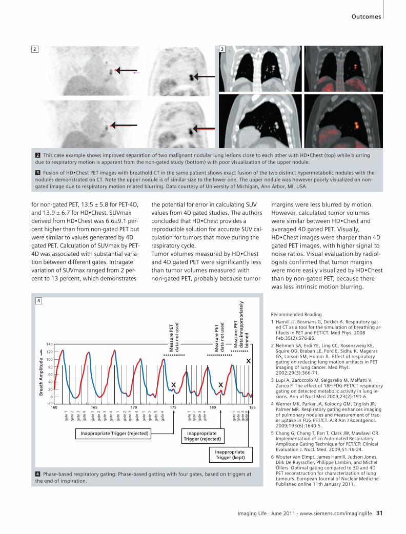
Imaging Life · June 2011 · www.siemens.com/imaginglife 31
Outcomes
for non-gated PET, 13.5 ± 5.8 for PET-4D, and13.9±6.7forHD•Chest.SUVmaxderivedfromHD•Chestwas6.6±9.1per-cent higher than from non-gated PET but were similar to values generated by 4D gated PET. Calculation of SUVmax by PET-4D was associated with substantial varia-tion between different gates. Intragate variation of SUVmax ranged from 2 per-cent to 13 percent, which demonstrates
the potential for error in calculating SUV values from 4D gated studies. The authors concludedthatHD•Chestprovidesareproducible solution for accurate SUV cal-culation for tumors that move during the respiratory cycle.TumorvolumesmeasuredbyHD•Chestand 4D gated PET were significantly less than tumor volumes measured with non-gated PET, probably because tumor
margins were less blurred by motion. However, calculated tumor volumes weresimilarbetweenHD•Chestandaveraged 4D gated PET. Visually, HD•Chestimagesweresharperthan4Dgated PET images, with higher signal to noise ratios. Visual evaluation by radiol-ogists confirmed that tumor margins weremoreeasilyvisualizedbyHD•Chestthan by non-gated PET, because there was less intrinsic motion blurring.
2 ThiscaseexampleshowsimprovedseparationoftwomalignantnodularlunglesionsclosetoeachotherwithHD•Chest(top)whileblurringdue to respiratory motion is apparent from the non-gated study (bottom) with poor visualization of the upper nodule.
3 FusionofHD•ChestPETimageswithbreatholdCTinthesamepatientshowsexactfusionofthetwodistincthypermetabolicnoduleswiththenodules demonstrated on CT. Note the upper nodule is of similar size to the lower one. The upper nodule was however poorly visualized on non-gated image due to respiratory motion related blurring. Data courtesy of University of Michigan, Ann Arbor, MI, USA.
2 3
Recommended Reading
1 Hamill JJ, Bosmans G, Dekker A. Respiratory gat-ed CT as a tool for the simulation of breathing ar-tifacts in PET and PET/CT. Med Phys. 2008 Feb;35(2):576-85.
2 Nehmeh SA, Erdi YE, Ling CC, Rosenzweig KE, Squire OD, Braban LE, Ford E, Sidhu K, Mageras GS, Larson SM, Humm JL. Effect of respiratory gating on reducing lung motion artifacts in PET imaging of lung cancer. Med Phys. 2002;29(3):366-71.
3 Lupi A, Zaroccolo M, Salgarello M, Malfatti V, Zanco P. The effect of 18F-FDG-PET/CT respiratory gating on detected metabolic activity in lung le-sions. Ann of Nucl Med 2009;23(2):191-6.
4 Werner MK, Parker JA, Kolodny GM, English JR, Palmer MR. Respiratory gating enhances imaging of pulmonary nodules and measurement of trac-er uptake in FDG PET/CT. AJR Am J Roentgenol. 2009;193(6):1640-5.
5 Chang G, Chang T, Pan T, Clark JW, Mawlawi OR. Implementation of an Automated Respiratory Amplitude Gating Technique for PET/CT: Clinical Evaluation J. Nucl. Med. 2009;51:16-24.
6 Wouter van Elmpt, James Hamill, Judson Jones, Dirk De Ruysscher, Philippe Lambin, and Michel Öllers Optimal gating compared to 3D and 4D PET reconstruction for characterization of lung tumours. European Journal of Nuclear Medicine Published online 11th January 2011.
4 Phase-based respiratory gating: Phase-based gatting with four gates, based on triggers at the end of inspiration.
4
IL_June2011.indb 31 5/12/11 5:24 PM

32 Imaging Life · June 2011 · www.siemens.com/imaginglife
Science
There are some two dozen different amyloid-related diseases. “Another amy-loid polypeptide is associated with type 2 diabetes,” says Alan Solomon, MD, Pro-fessor of Medicine at the University of Tennessee Graduate School of Medicine, in Knoxville, Tenn., USA. “Men over the age of 80 develop amyloid in the semi-nal vesicles, the significance of which is not clear,” he added. And yet another type of amyloid affects the heart, dis-rupting contraction and leading to heart failure and death. Amyloid peptides also can cause joint damage as well as affect the function of the nerves, kidneys, liver and spleen. Unfortunately, in all amy-loid-related disease, the body does not recognize amyloid as a foreign sub-stance. Consequently, it does not attack the amyloid as it accumulates. In instances other than within the brain, amyloid-related disease is found, gener-ally by selective biopsy, often after it has spread throughout the body.While research into Alzheimer’s disease-related amyloid is nearing final approv-als, which could lead to routine clinical screenings. Many any other forms of amyloidosis, unrelated to the brain, are still being researched to find viable treatment options. “If there is any hope
in improving the outcomes in patients with amyloidosis, there has to be a bet-ter mechanism for earlier detection of these diseases,” says Jonathan Wall, PhD, Director of the Amyloid and Preclin-ical Molecular Imaging Laboratory at the University of Tennessee Graduate School of Medicine in Knoxville. The two most common types of amyloid disease that do not affect the brain are light chain (AL) and secondary (AA) amyloidosis. AL results from the aggre-gation of certain free immunoglobulin light chain proteins, which may deposit in any organ or tissue, leading to dys-function and invariably death. AA also is the most widespread form of amyloido-sis, and arises secondary to chronic inflammatory disorders, such as rheu-matoid arthritis. In the U.K., it is possible for patients with amyloidosis to receive a serum amyloid P scintigraphy scan, with radio-labeled 124I. The whole-body scan identi-fies all forms of amyloid deposits outside of the brain. The technique to isolate the serum amyloid P component involves a mild viral inactivation step—if it were too strong, the protein would be destroyed. In the U.S., however, the Food and Drug Administration (FDA)
considers the viral inactivation step not strong enough, therefore leaving a risk of viral contamination, albeit small given the 20-plus years the technique has been approved in the U.K. and other European countries. Consequently, the FDA has not approved the method for use in the U.S. yet. “In the U.S., patients typically get biop-sied, which will reveal if they have amy-loidosis, but selective biopsy will not reveal the extent of the disease. Know-ing the extent could help determine the most effective treatment, as well as identifying those whose disease is too extensive and would not benefit from treatment,” Wall says. Current therapies include various chemo regimens, as well as stem cell transplants. “The clinical symptoms of the disease vary for each patient because of the dif-ferent organs that can be infiltrated by amyloid. The organ predominantly involved is the kidney,” Solomon says. “The filter gets clogged up with amyloid, typically gets swollen, and fails. The patient will need dialysis or a trans-plant.” If the heart is the target, symp-toms of heart failure are common. The liver’s involvement causes it to swell and not function properly. The patient might
Amyloid-Related Disease: Detection and TreatmentAlthough rare, amyloidosis is fatal. Characterized by the aggregation of normally soluble proteins and peptides, amyloid deposits form well-ordered fibrils in tissues and organs. The accumulation of these fibril leads to mechanical dysfunction of the tissues and organs in question. The most well-known form of amyloid-related disease is Alzheimer’s disease, in which patients suffer from aggregation of the Aß peptide in the brain, leading to neuronal loss and death.
By C.P. Kaiser
IL_June2011.indb 32 5/12/11 5:24 PM

Imaging Life · June 2011 · www.siemens.com/imaginglife 33
Science
1 Small animal SPECT/CT image demonstrates the amyloid-imaging capability of a radiolabeled fibril-reactive monoclonal antibody. Accumulation within the localized amyloidoma on the back of the mouse is readily visible.
1
get jaundice. Postural hypotension is a potential symptom when the nerves are affected by amyloid deposits. “Given the severity of this disease, patients with a lot of involvement can have three to six months to live. They have a terrible prognosis,” Solomon says.
Antibody BreakthroughDuring their research, Wall and col-leagues discovered a monoclonal anti-body (mAb) designated as 11-1F4 that
bound to many forms of AL amyloid fibrils in ex vivo and in vitro studies. Interestingly, this antibody specifically binds with the pathologic fibrils and does not interact with the soluble, non-fibrillar form of light chains. “It’s important to note that patients with AL amyloidosis also have significantly high concentra-tions of amyloid precursor protein in their circulation, which could interfere with a therapeutic or diagnostic reagent if that reagent was not specific for the fibrillar
form of the protein,” Wall says. Since there are no naturally occurring animal models of AL amyloidosis, Wall and colleagues used human AL amyloid material, isolated from patient autopsy samples, for their experimental studies. They injected the AL amyloid material subcutaneously into healthy and immu-nodeficient mice. They hypothesized that the 11-1F4 mAB would bind to the “amyloidoma,” or amyloid mass, but they had no way of quantifying it until
IL_June2011.indb 33 5/12/11 5:24 PM

34 Imaging Life · June 2011 · www.siemens.com/imaginglife
Science
they acquired the first generation microCAT+SPECT preclinical imaging scanner from Siemens. The researchers, including Stephan Ken-nel, PhD, a Radio-biochemist in the Pre-clinical and Diagnostic Molecular Imag-ing Laboratory at the University of Tennessee Graduate School of Medicine, tested various tracer compounds to determine which would be best to radio-label the antibody. “We had access to a Siemens micro-SPECT/CT scanner, so we chose to use 125I-labeled 11-1F4 mAb in mice with human AL amyloidomas because that tracer is relatively inexpensive and is amenable to autoradiography,” says Ken-nel. He says that these studies were purely to establish the equilibrium distri-bution of the radiotracer, and as such the vast majority of the imaging was performed on mice post-mortem. The imaging showed that the antibody co-localized with the amyloid deposits. More recent studies performed on the next-generation Inveon™ trimodal pre-clinical imaging system (PET/SPECT/CT) allowed the group to confirm their origi-nal findings with higher clarity and quantitative accuracy.“Having an antibody that binds to amy-loid fibrils theoretically could ‘trick’ the body into mounting an immune defense against the amyloid. And that is exactly what we found happened in mice that were not immuno-compro-
mised,” Solomon says. Micro-autoradiography revealed that the mAb was uniformly distributed through-out the amyloid mass and did not bind to any healthy tissues. In contrast, an isotope-matched control mAb was seen only in the blood pool and at very low density in the interstitial fluid and blood within the amyloidoma. “These studies demonstrated unequivocally that the 11-1F4 mAb bound to human AL amy-loid in vivo and, equally importantly, that this reagent did not bind to healthy tissues and cells,” Kennel says.The researchers took their success to the National Cancer Institute (NCI), which, although impressed with the animal results, asked them to reproduce the same results in humans. “The NCI wanted us to show that the antibody will stick to amyloid fibrils in humans, because that is the mode of therapeutic action,” Wall says.
Treatment PossibilitiesThe FDA approved an investigational new drug (IND) application to test a human-grade mouse antibody in patients with AL amyloidosis. To move to this next step, however, researchers had to rethink their imaging protocols. They first considered an alternative to the use of 125I, which cannot be detected by clinical scanners because of the low photon energy. “To ensure consistent chemistry and
maintain the immunoreactivity of the mAb, we naturally considered using either 123I or 124I for SPECT or PET clinical imaging, respectively,” Wall says. “How-ever, it soon became clear that the low resolution of clinical gamma imaging—we would likely have had to use planar scintigraphy rather than SPECT in order to survey the entire body—and the short half life of 123I were not compatible with the imaging protocol we had developed.”In contrast, 124I has a long half-life (4.2 days), PET/CT imaging is a quantitative modality and the resolution of PET scan-ners is clearly greater than planar scinti-graphic images, Wall says. However, 124I is generated in a cyclotron, is expensive and the dosimetry can be high. “That said, we chose PET/CT imaging via Bio-graph™ 16 and Biograph mCT, as the path forward using 124I and identified an FDA-approved source of the nuclide that could be used for human studies under our exploratory IND.” Patients are assessed for eligibility; nota-bly, they should have chemically-diag-nosed AL and be negative for human anti-mouse antibodies, because the 11-1F4 mAb used in the study is murine in origin. The patients travel to Knoxville from all over North America and receive 2 mCi of 124I-11-1F4 mAb (< 1 mg pro-tein) infused in 30 ml over 10 minutes. The patients then return two and five days later for a whole-body PET/CT. On Biograph mCT, this involves seven bed
2 The 124I-labeled monoclonal antibody 11-1F4 was shown to bind specifically to known sites of AL amyloid deposition in patients by PET/CT imag-ing using the BiographTM mCT from Siemens. Axial images of this patient revealed uptake of the antibody in amyloid deposits in the liver, spleen and bone marrow (intense red color indicates the presence of the radioactive 11-1F4 antibody bound to amyloid). The patient was administered only 2 mCi of 124I and these images were acquired two days post injection.
2
IL_June2011.indb 34 5/12/11 5:24 PM

Imaging Life · June 2011 · www.siemens.com/imaginglife 35
positions, five minutes per bed with TrueX reconstruction and prompt gamma correction.
Promising Clinical ResultsWall and colleagues demonstrated the safety of the 124I-labeled antibody in the first three patients and received permis-sion to perform the test in more. They have completed the experiment in 37 patients to date and in about 60 percent of them, “the imaging is spectacular,” says Solomon. “We have shown that the antibody localizes to amyloid in more than half of the patients. We have dem-onstrated the potential therapeutic abil-ity of this antibody.”However, Wall says, “It is important to reiterate that, at present, amyloid depos-its cannot be imaged clinically or pre-clinically using standard nuclear medi-cine tracers, ultrasound or MRI. The pathological sequelae can be visualized, such as increases in organ size, or reduced cardiac muscle contractibility and function, on CT, MR and ultrasound imaging, but the amyloid load and distri-bution is not accessible information.”
In that regard, the promise of the radio-labeled antibody to diagnose, stage and monitor treatment and the promise of the therapeutic antibody to activate the immune system to destroy the amyloid is tangible. After their first 18 patients, Wall and colleagues concluded, “Given these results, we posit that 124I 11-1F4 radioimmuno-imaging could be used to predict which AL patients would be can-didates for passive immunotherapy using the chimeric version of this mAb” (Blood 2010;116(13):2241-2244).
Amyloid Binding Agents for the BodyWall and colleagues have since developed small peptides as imaging agents for AL amyloidosis. A peptide might gain FDA approval easier because it is not an anti-body, Kennel says. The peptides are labeled with 124I for use with PET/CT scan-ning. “Ideally, the best imaging modality would be quantitative and allow rapid, whole-body, high-resolution imaging. That is the definition of PET,” Wall says. Unlike the 11-1F4 antibody, which binds to amyloid fibrils, the peptide binds to
heparin sulfate proteoglycan, a different component of the amyloid. “It’s been known that amyloid contains these mate-rials, but no one had found a reagent that could specifically target it until now,” says Wall, adding that he and his col-leagues serendipitously identified the peptide. Luck or not, the peptide binds to the amyloids, can produce diagnostic images one hour post-injection and stays bound for 24 hours. The research around diagnosis and ther-apy amyloidosis in the body has advanced considerably in the last decade. The group from Tennessee has shown that a radiolabeled antibody and peptide attaches to the amyloid, while bypassing healthy tissue. Therefore, amyloidosis could be imaged and found earlier and, thus, more appropriate treatments could be prescribed as a result of the whole-body imaging data. In addition, the antibody can then be used therapeutically as passive immuno-therapy.
Science
“We have shown that the antibody localizes to amyloid in more than half of the patients. We have demonstrated the potential therapeutic ability of this antibody.”Jonathan Wall, PhD, Director of the Amyloid and Preclinical Molecular Imaging Laboratory at the University of Tennessee Graduate School of Medicine in Knoxville, Tenn., USA
C.P. Kaiser is a medical journalist focused on imaging and cardiology. He is based in Pennsylvania, USA.
IL_June2011.indb 35 5/12/11 5:25 PM

36 Imaging Life · June 2011 · www.siemens.com/imaginglife
Clinical Case Studies
Case 1 18F Tyrosine PET/CT Guided Radiotherapy for a Patient with Recurrent Brain TumorBy Wolfgang Mohnike, MD, and Matthias Lampe, MD
Case study data courtesy of: Diagnostic Therapeutic Center, Berlin, Germany
1 18F Tyrosine PET/CT in a patient with operated oligodendroglioma show focal uptake of tracer in the resection margin, suggestive of tumor recurrence.
1
HISTORY
A 62-year-old male with a 20-year his-tory of brain tumor presented with pro-gressive fatigue. The patient was diag-nosed with oligodendroglioma in the left frontal lobe in 1991, which was sub-sequently resected. Three years follow-ing initial surgery, the patient had a local tumor recurrence detected by CT and MRI. The recurrent tumor was fur-
demonstrated by the higher histopatho-logical grading, adjuvant radiation ther-apy was delivered. Whole-brain irradia-tion with 15 fractions of 2 Gy at each fraction and a 30 Gy Boost was per-formed. The patient tolerated radiation very well and without significant side effects.
ther resected and was proven as grade III oligodendroglioma by histopathology. Due to the unfavorable location of the recurrent tumor, complete resection could not be achieved and a small amount of residual tumor was left behind. In view of the tumor-positive resection margin and the increased malignant potential of the tumor as
IL_June2011.indb 36 5/12/11 5:25 PM

Imaging Life · June 2011 · www.siemens.com/imaginglife 37
Clinical Case Studies
DIAGNOSIS
Due to the symptoms of progressive fatigue, the patient was referred for a follow-up MRI, which was performed in August 2010. A mass at the tumor resec-tion site with variegated uptake of MR contrast was visualized. Due to infiltra-tion of the corpus callosum by the recur-rent tumor, only partial surgical removal was possible. A subsequent MRI showed a zone of contrast enhancement in the resected tumor bed just to the right of the cingulate gyrus, which was suspi-cious of residual tumor. In view of the residual tumor, further adjuvant irradia-tion was deemed necessary even though the patient had been treated with up to 60 Gy in the same area 15 years earlier.The patient was referred for PET/CT for evaluation of metabolically active resid-ual tumor for radiation therapy plan-ning. An 18F Fluoroethyl Tyrosine PET/CT study was performed.The 18F FET PET/CT study demonstrated high tracer uptake at the inferior and posterior aspect of the resection area (arrows), partially corresponding to the region of contrast enhancement in MRI, suggesting functionally active recurrent tumor. There was, however, an absence of tracer uptake in the rest of the region of altered signal on MRI suggesting it was related to radiation necrosis. The CT, PET and MRI studies were com-
bined for treatment planning. A stereo-tactic helical therapy was performed in fractions of 1.8 Gy with a total dose of 59.4 Gy. The therapy was performed without any side effects.There was no evidence of local relapse at the time of the most recent follow-up. Since the initial radiation to the tumor involved a small volume, dose escalation to the site of the recurrent tumor was achieved without severe side effects. Although MRI demonstrated contrast enhancement in the resected tumor bed, it could not differentiate between the extent of residual tumor and amount of post radiation fibrosis. Thus metabolic imaging with PET/CT was essential for delineation of the true extent of viable tumor for effective therapy planning.
COMMENTS
The most commonly used PET tracer 18F-Fluorodeoxyglucose (FDG), has very limited utility for brain tumors due to the high physiological uptake in normal cerebral parenchyma. To delineate meta-bolically-active tumor and differentiate from post-radiation changes, an 18F tyro-sine-PET/CT exam was performed. 18F Tyrosine (FET), an amino acid analog, accumulates in malignant cells, probably due to increased expression of amino
acid transporters. The labeled ligand is not metabolized in the cell but contin-ues to accumulate, leading to high tumor uptake compared to background. Disruption of blood brain also increases influx of labeled amino acid within tumor tissue.
EXAMINATION PROTOCOL
Three days prior to the examination, the patient followed a vegetarian diet in order to avoid excess dietary protein. Since the availability of Fluor-18-tyrosine on the market is very limited, the tracer was synthesized on-site. The patient was injected with 289 MBq of 18F FET and after 30 minutes, PET/CT was performed on a Biograph™ 64 PET•CTsystem.The initial CT was acquired at 120 kV, 300mAs with 1 mm thick slices. 1mm with 120kV, 300mAs following intravenous injection of Ultravist 370 (100ml), a low osmolar, non-ionic contrast agent. The PET study was acquired in a single bed position with transverse slice thickness of 3 mm. Attenuation correction of PET data was performed using the CT data.
18F Tyrosine referenced herein is (are) not currently
recognized by the U.S. FDA as being safe and effec-
tive, and Siemens does not make any claims regard-
ing its use.
IL_June2011.indb 37 5/12/11 5:25 PM

38 Imaging Life · June 2011 · www.siemens.com/imaginglife
Clinical Case Studies
Case 2 Neuroblastoma: Post-surgical 123I MIBG SPECT/CT Study Demonstrating Residual Tumor By Jerry Froelich, MD
Case study data provided by University of Minnesota, Minneapolis, MN, USA
HISTORY
A 4-year-old boy presented with respira-tory distress. Routine chest X-ray showed mass in left upper lung. A CT scan was performed to evaluate the lung mass.
FINDINGS
The CT study showed a large mass in the left upper lobe of lung with multiple foci of intratumoral calcification. There was no other lesion suggestive of metasta-ses. Histopathology suggested primary neuroblastoma. Patient was subjected to surgery for removal of primary tumor. Within 2 days of surgery, the patient was referred for 123I
MIBG SPECT/CT study for detection of resid-ual tumor and functioning metastases. Patient was injected with 4.2 mCi 123I MIBG. After 24 hours, whole-body pla-nar and SPECT/CT study was performed on a Symbia™ T6 system. Diagnostic CT including thorax, abdomen and pelvis was performed prior to SPECT/CT study as an integrated procedure.
1
1 Contrast CT in a 4-year-old boy with neuroblastoma shows large mass in left upper lobe of lung with focal calcification.
IL_June2011.indb 38 5/12/11 5:25 PM

Imaging Life · June 2011 · www.siemens.com/imaginglife 39
Clinical Case Studies
2 SPECT/CT study demonstrates foci of increased tracer uptake in the soft-tissue mass at the left paratracheal region at the thoracic inlet as well as a small foci of uptake in the left supraclavicular region (white ar-row). These foci represent residual tumor. Post-operative changes relat-ed to recent resection of the lung mass are evident in the CT images.
3 A smaller foci of increased uptake also is visualized in the left para-tracheal region at the level of the aortic arch at the resected tumor bed which also represents residual tumor. No other well-defined functioning metastases visualized. Bilateral adrenal glands showed high tracer up-take probably reflecting adrenal hyperplasia. Liver and spleen show nor-mal uptake of 123I MIBG.
2
3
COMMENTS
Diagnostic CT integrated with 123I MIBG SPECT was critical in this patient for accurate localization and characteriza-tion of residual tumor tissue, exclusion of metastatic foci and planning of fur-ther therapy. Presence of residual tumor tissue related to partial resection of pri-mary tumor would require radiation therapy including 131I MIBG therapy. Exact determination of the extent of residual tumor tissue and its relationship to surrounding structures (in this case the trachea and great vessels at the tho-racic inlet and mediastinum) helps plan and follow up after MIBG therapy.
IL_June2011.indb 39 5/12/11 5:25 PM

40 Imaging Life · June 2011 · www.siemens.com/imaginglife
Clinical Case Studies
1 18F FDG PET/CT image shows markedly glucose-avid foci in both upper lobes of the lung.
2 18F FDG PET/CT image shows subdiaphragmatic glucose avid lymph nodes at the porta hepatis in addition to the bilateral lung and mediastinal lymph node lesions.
Case 3 18F FDG PET/CT-Based Identification of Sarcoidosis in a Patient with Suspected Thoracic MalignancyBy Partha Ghosh, MD, Molecular Imaging Business Unit, Siemens Healthcare, Hoffman Estates, Ill., USA
Case study data provided by Royal Prince Alfred Hospital, Sydney, Australia
1 2
HISTORY
A 45-year-old male smoker (20 pack years) presented with right-sided pleu-ritic chest pain. A chest CT revealed bilateral lung infiltrates as well as right hilar lymph node enlargement. A fine-needle tissue biopsy (FNAB) of the left upper lobe lesion, performed at a
regional hospital, revealed adenocarci-noma. FNAB on two attempts of the right upper lobe lesion was not success-ful. He was referred for staging with PET/CT. 18F FDG PET/CT was performed on a Biograph™ mCT.
IL_June2011.indb 40 5/12/11 5:25 PM

Imaging Life · June 2011 · www.siemens.com/imaginglife 41
Clinical Case Studies
3 18F FDG PET/CT image shows the extensive involvement of subcarinal, bilateral hilar and lower para-aortic and para-tracheal nodes.
4 Axial CT and PET images at the level of the thoracic inlet shows focal increase in FDG uptake in a 7 mm right scalene lymph node.
3
DIAGNOSIS
The PET/CT study showed markedly glu-cose-avid foci in both upper lobes of the lung (Fig 1). SUVmax of the left lobar mass was 18.2; SUVmax of right lobar mass was 25.5. There were additional markedly glucose-avid foci in the hilar and mediastinal lymph nodes on both sides (SUVmax 32.5). The CT showed enlarged hilar nodes as well as lobar masses corresponding to the regions of FDG uptake.
COMMENTS
On PET/CT, the pattern of uptake in the hilar, mediastinal and right scalene lymph nodes was consistent with sar-coidosis. Pulmonary infiltrates demon-strated on CT also are commonly associ-ated with sarcoidosis. The diagnostic dilemma was the previous FNAB of the left lobe lesion had suggested adenocar-cinoma. The PET/CT findings were sug-gestive of sarcoidosis, with the possibil-ity of a synchronous primary lung tumor in view of the FNAB findings. However, the PET/CT findings in isolation were quite typical of sarcoidosis given the clinical history of the patient. In view of the previous FNAB result, suggestive of adenocarcinoma and the PET/CT findings favoring sarcoidosis, it was decided to confirm the diagnosis by surgical exci-sion of the lung lesion. At surgery, the left upper lobe lesion was removed and histopathology revealed necrotizing non-caseating granulomas consistent with sarcoidosis, which confirmed the PET/CT findings. Improved lesion con-trastusingultraHD•PETonBiographmCT provided exquisite images of the thoracic, mediastinal and portal lymph node lesions, including a 7 mm right scalene node.
4
IL_June2011.indb 41 5/12/11 5:25 PM
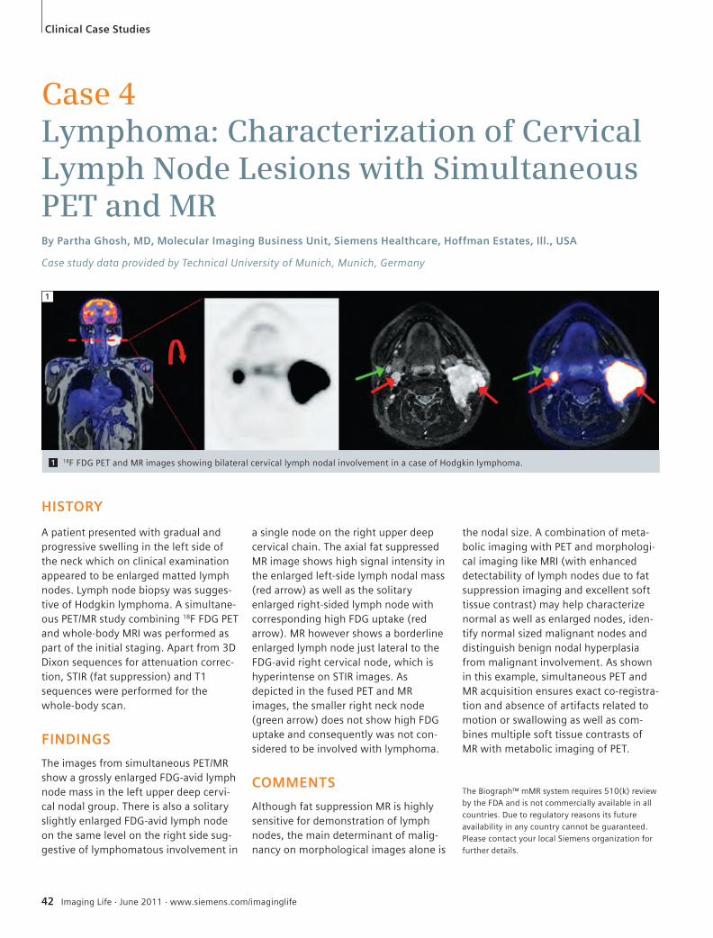
42 Imaging Life · June 2011 · www.siemens.com/imaginglife
Clinical Case Studies
Case 4 Lymphoma: Characterization of Cervical Lymph Node Lesions with Simultaneous PET and MRBy Partha Ghosh, MD, Molecular Imaging Business Unit, Siemens Healthcare, Hoffman Estates, Ill., USA
Case study data provided by Technical University of Munich, Munich, Germany
HISTORY
A patient presented with gradual and progressive swelling in the left side of the neck which on clinical examination appeared to be enlarged matted lymph nodes. Lymph node biopsy was sugges-tive of Hodgkin lymphoma. A simultane-ous PET/MR study combining 18F FDG PET and whole-body MRI was performed as part of the initial staging. Apart from 3D Dixon sequences for attenuation correc-tion, STIR (fat suppression) and T1 sequences were performed for the whole-body scan.
FINDINGS
The images from simultaneous PET/MR show a grossly enlarged FDG-avid lymph node mass in the left upper deep cervi-cal nodal group. There is also a solitary slightly enlarged FDG-avid lymph node on the same level on the right side sug-gestive of lymphomatous involvement in
a single node on the right upper deep cervical chain. The axial fat suppressed MR image shows high signal intensity in the enlarged left-side lymph nodal mass (red arrow) as well as the solitary enlarged right-sided lymph node with corresponding high FDG uptake (red arrow). MR however shows a borderline enlarged lymph node just lateral to the FDG-avid right cervical node, which is hyperintense on STIR images. As depicted in the fused PET and MR images, the smaller right neck node (green arrow) does not show high FDG uptake and consequently was not con-sidered to be involved with lymphoma.
COMMENTS
Although fat suppression MR is highly sensitive for demonstration of lymph nodes, the main determinant of malig-nancy on morphological images alone is
the nodal size. A combination of meta-bolic imaging with PET and morphologi-cal imaging like MRI (with enhanced detectability of lymph nodes due to fat suppression imaging and excellent soft tissue contrast) may help characterize normal as well as enlarged nodes, iden-tify normal sized malignant nodes and distinguish benign nodal hyperplasia from malignant involvement. As shown in this example, simultaneous PET and MR acquisition ensures exact co-registra-tion and absence of artifacts related to motion or swallowing as well as com-bines multiple soft tissue contrasts of MR with metabolic imaging of PET.
1 18F FDG PET and MR images showing bilateral cervical lymph nodal involvement in a case of Hodgkin lymphoma.
1
The Biograph™ mMR system requires 510(k) review
by the FDA and is not commercially available in all
countries. Due to regulatory reasons its future
availability in any country cannot be guaranteed.
Please contact your local Siemens organization for
further details.
IL_June2011.indb 42 5/12/11 5:25 PM
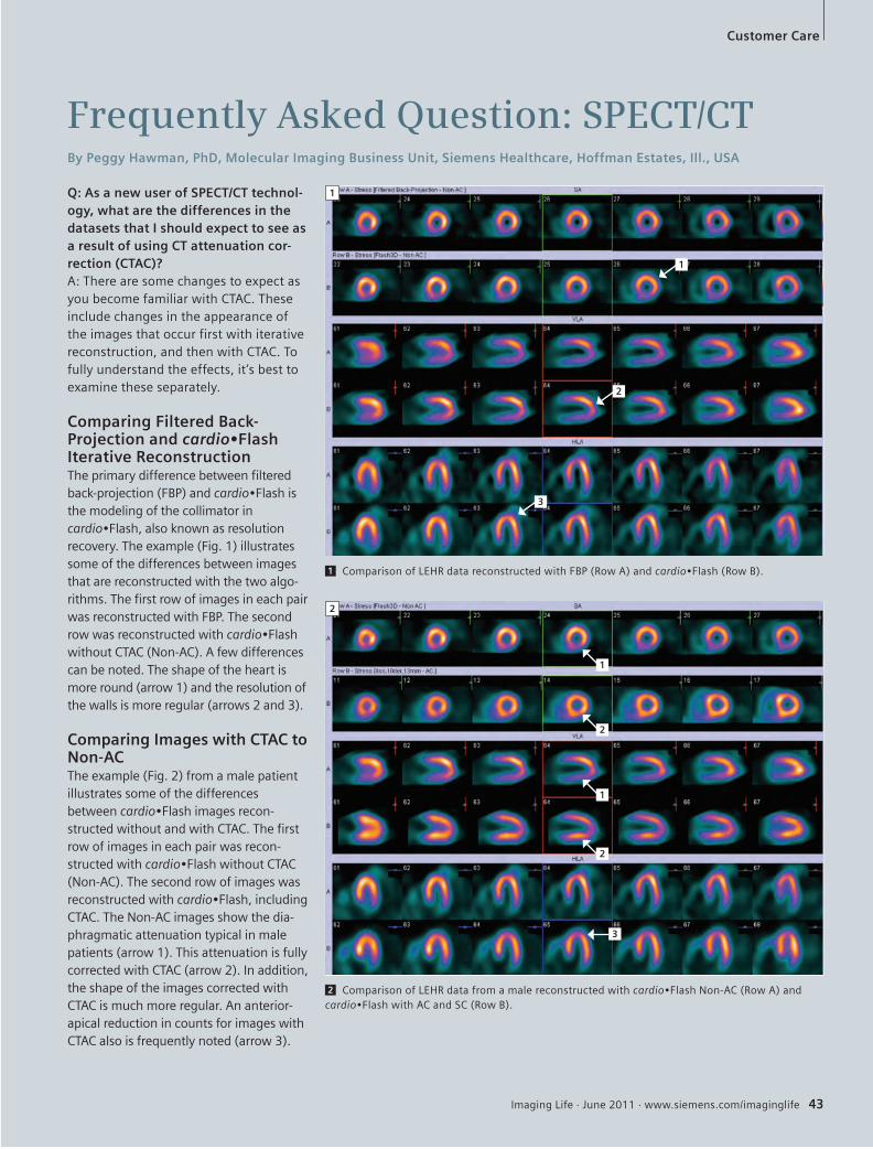
Imaging Life · June 2011 · www.siemens.com/imaginglife 43
Customer Care
Q: As a new user of SPECT/CT technol-ogy, what are the differences in the datasets that I should expect to see as a result of using CT attenuation cor-rection (CTAC)?A: There are some changes to expect as you become familiar with CTAC. These include changes in the appearance of the images that occur first with iterative reconstruction, and then with CTAC. To fully understand the effects, it’s best to examine these separately.
Comparing Filtered Back-Projection and cardio•Flash Iterative ReconstructionThe primary difference between filtered back-projection (FBP) and cardio•Flashisthe modeling of the collimator in cardio•Flash,alsoknownasresolutionrecovery. The example (Fig. 1) illustrates some of the differences between images that are reconstructed with the two algo-rithms. The first row of images in each pair was reconstructed with FBP. The second row was reconstructed with cardio•Flashwithout CTAC (Non-AC). A few differences can be noted. The shape of the heart is more round (arrow 1) and the resolution of the walls is more regular (arrows 2 and 3).
Comparing Images with CTAC to Non-ACThe example (Fig. 2) from a male patient illustrates some of the differences between cardio•Flashimagesrecon-structed without and with CTAC. The first row of images in each pair was recon-structed with cardio•FlashwithoutCTAC(Non-AC). The second row of images was reconstructed with cardio•Flash,includingCTAC. The Non-AC images show the dia-phragmatic attenuation typical in male patients (arrow 1). This attenuation is fully corrected with CTAC (arrow 2). In addition, the shape of the images corrected with CTAC is much more regular. An anterior-apical reduction in counts for images with CTAC also is frequently noted (arrow 3).
Frequently Asked Question: SPECT/CTBy Peggy Hawman, PhD, Molecular Imaging Business Unit, Siemens Healthcare, Hoffman Estates, Ill., USA
1 Comparison of LEHR data reconstructed with FBP (Row A) and cardio•Flash(RowB).
1
2 Comparison of LEHR data from a male reconstructed with cardio•FlashNon-AC(RowA)andcardio•FlashwithACandSC(RowB).
2
1
2
3
1
2
1
2
3
IL_June2011.indb 43 5/12/11 5:25 PM

44 Imaging Life · June 2011 · www.siemens.com/imaginglife
Customer Care
Particle Acceleration Reaches the 9th Floor High Marks from UPMC in Cyclotron Upgrade Over the course of the mid- to late-1990s, a team of researchers at the University of Pittsburgh Medical Center (UPMC) in the United States developed hundreds of compounds in an attempt to visually depict Alzheimer’s disease and other neurological disorders in the brain. But they needed to do more. In light of the growing need for targets, UPMC opted to retire a particle accelerator in 2010 with the help of Siemens Customer Care program, and literally “elevate” their new system. The Eclipse HP Cyclotron from Siemens that took its place was an engineering feat that took place nine floors above ground.
By Clint vanSonnenberg
The UPMC PET Research Center is a mecca of nuclear imaging innovation, with more than 25 different compounds in the works, all of which are dedicated to research. Chester Mathis, PhD, Direc-tor of the PET Research Center, describes the Research Center faculty’s decision to move to a higher-capacity cyclotron in Siemens’ Eclipse HP.
What Goes Up Must Come Down The PET Research Center at UPMC has differentiated its cyclotron research and defied skeptics since its founding. Strapped for space in 1991, UPMC decided to locate the PET Facility on the ninth floor of UPMC Presbyterian Hospi-tal, a one and a half-floor space wedged
between the MR Research Center and the hospital cafeteria. The installation of a cyclotron was viewed as impossible by many experts. “No cyclotron was or is engineered to have anything underneath it—to my knowledge nobody else puts a cyclotron up this high,” Mathis explains. This fact is especially relevant when what’s below the magnetic system are one 1.5 T and three 3.0 T MRI scanners, within just 20 feet of the cyclotron. To the disbelief of many, the PET facility’s cyclotron and hot cells worked not only problem-free, but with cutting-edge success. By 2010, with Mathis and the rest of the PET research-ers hungry for more radioisotopes and the expansion of the facility’s research program, Mathis opted to stay with Sie-
View from the ground: A special crane —only one of the two in existence in the United States—hoists a major piece of the cyclotron shielding onto the 9th floor.
IL_June2011.indb 44 5/12/11 5:25 PM
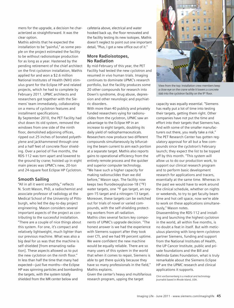
Imaging Life · June 2011 · www.siemens.com/imaginglife 45
mens for the upgrade, a decision he char-acterized as straightforward. It was the clear option.Mathis admits that he expected the installation to be “painful,” as some peo-ple on the project estimated the facility to be without radioisotope production for as long as a year. Hastened by the pending retirement of the chief architect on the first cyclotron installation, Mathis applied for and won a $2.6 million National Institutes of Health (NIH) stim-ulus grant for the Eclipse HP and related projects, which he had to complete by February 2011. UPMC architects and researchers got together with the Sie-mens’ team immediately, collaborating on a menu of cyclotron features and installment specifications.By September 2010, the PET Facility had shut down its old system, removed the windows from one side of the ninth floor, demolished adjoining offices, ripped out 25 inches of borated polyeth-ylene and jackhammered through one and a half feet of concrete floor shield-ing. Over a period of five months, the RDS-112 was torn apart and lowered to the ground by crane; hoisted up in eight main pieces was UPMC’s new, 20-ton and 24-square foot Eclipse HP Cyclotron.
Smooth Sailing“All in all it went smoothly,” reflects N. Scott Mason, PhD, a radiochemist and associate professor of radiology at the Medical School of the University of Pitts-burgh, who led the day-to-day project engineering. Mason considers several important aspects of the project as con-tributing to the successful installation. “There are a couple of nice things about this system. For one, it’s compact and relatively lightweight, much lighter than our previous machine. What was also a big deal for us was that the machine is self-shielded [from emanating radia-tion]. These aspects allowed us to put the new cyclotron on the ninth floor.” In less than half the time that many had expected—just five months—the Eclipse HP was spinning particles and bombarding the targets, with the system totally shielded from the MR center below and
cafeteria above, electrical and water hooked back up, the floor renovated and the facility testing its new isotopes. Mathis doesn’t neglect to point out one important detail, “Plus, I got a new office out of it.”
More Radioisotopes, No RadiationBy mid-February of this year, the PET Facility had tested the new cyclotron and resumed in vivo human trials. Imaging continues to dominate UPMC’s research portfolio, but the facility produces some 20 other compounds for research into Down’s syndrome, drug abuse, depres-sion and other neurologic and psychiat-ric disorders. With more than 40 publicly and privately funded researchers vying for radionu-clides from the cyclotron, UPMC saw an advantage to the Eclipse HP in an increase to eight targets, doubling its daily yield of radiopharmaceuticals. Researchers now produce two different compounds simultaneously by bifurcat-ing the beam current to aim each portion at a separate target. Mathis also reports gains to operational efficiency from the entirely remote process and the quicker and superior computer technology. “We have such a higher capacity for making radionuclides than we did before,” Mason says. The facility now keeps two flurodeoxyglucose-18 (18F) water targets, one 18F gas target, an oxy-gen-15 target and a nitrogen-13 target. Moreover, these targets can be switched out for trials of novel or varied com-pounds, with the self-shielding protect-ing workers from all radiation.Mathis cites several factors key compo-nents of UPMC’s upgrade decision. “The honest answer is we had the experience with Siemens support after they took over CTI, and we had 99 percent uptime. We were confident the new machine would be equally reliable. There are so many users of this system in the world that when it comes to repair, Siemens is able to get there quickly because they have so many professionals in the field,” Mathis explains.Given the center’s heavy and multifarious research program, upping the target
capacity was equally essential. “Siemens has really put a lot of time into testing their targets, getting them right. Other companies have not put the time and effort into their targets that Siemens has. And with some of the smaller manufac-turers out there, you really take a risk.”The PET Research Center has gotten reg-ulatory approval for all but a few com-pounds since the cyclotron’s February launch; they expect the list to be topped off by this month. “This system will allow us to do our production work, to support our clinical research program and to perform basic development research for applications and tracers, essentially at the same time. Whereas in the past we would have to work around the clinical schedule, whether on nights or weekends, to try to get faculty beam time and hot cell space, now we’re able to work on these applications simultane-ously,” Mason notes. Disassembling the RDS-112 and install-ing and launching the highest cyclotron in the world, all within five months, is no doubt a feat in itself. But with metic-ulous planning with long-term cyclotron partner Siemens, funding and support from the National Institutes of Health, the UP Cancer Institute, public and pri-vate foundations and the Bill and Melinda Gates Foundation, what is truly remarkable about the Siemens Eclipse HP are the UPMC research and clinical applications it supports.
Customer Care
Clint vonSonnenberg is a medical and technology journalist based in Rhode Island, USA.
View from the top: Installation crew members keep a close eye on the crane while it lowers a concrete slab into the cyclotron facility on the 9th floor.
IL_June2011.indb 45 5/12/11 5:25 PM

46 Imaging Life · June 2011 · www.siemens.com/imaginglife
Customer Care
2011 Image of the Year Siemens Preclinical Image of the Year Competition Celebrating 7 Years of Excellence
The 2011 Image of the Year event is being held for the seventh consecutive year to recognize accomplishments in preclinical imaging. As part of the event, owners and users of Siemens preclinical scanners from around the world are encouraged to submit images that are representative of their institute’s preclin-ical research. Submissions will be evaluated on the basis of image quality, scientific merit and information gleaned along the fol-lowing categories: Inveon™ Image of the Year – demon-strate Inveon capabilities for any single or multimodality configuration of PET, SPECT or CT Multimodality Image of the Year – demonstrate Siemens scanner capabili-ties for any configuration of PET, SPECT
or CT, including images acquired in com-bination with other modalities, such as
MR or ultrasound, using Siemens Health-care productsTranslational Image of the Year – span the “mouse to man” spectrum with excit-ing images showing the translational capabilities of PET, SPECT and/or CTBest Presented Image of the Year – stretch the limits of image acquisition and post-processing skills for optimum image representation.Please see the image submission guide-lines posted online (www.siemens.com/mi-preclinical-ioy). Three image submis-sions are permitted per scanner. Submis-sions will be accepted until Friday, August 5, 2011. Winners will be announced at the con-clusion of the Siemens Preclinical User’s Meeting in San Diego, California (USA), on Tuesday, September 6th, 2011.
Uptake of 64Cu-Oxo-Trastuzumab in HER-2 positive (right mouse) and negative (left mouse) breast cancer tumors at 24 hours.
Join Us for The First Biograph World Summit July 21–23, 2011 in Munich, Germany
The Biograph World Summit is a unique opportunity to meet with peers and to exchange valuable information. Hear from the world‘s leading experts on clinical trends, share best practices and engage in discussions on trend-setting developments in molecular imaging. Discover innovative solutions and techniques for Biograph, and leave with invaluable information on how to maximize its full potential.
Summit discussions include:• Trends in PET/CT Imaging • Answering Today’s Clinical and Economic Challenges • The Clinical Impact of New Biomarkers • New Advancements in Oncology • New Advancements in Cardiology and Neurology • Expanding the Footprint of Molecular Imaging
Register for the event and view recorded presentations following the meeting at www.siemens.com/mi-summit.
IL_June2011.indb 46 5/12/11 5:25 PM

Imaging Life · June 2011 · www.siemens.com/imaginglife 47
Subscription
Siemens Healthcare – Customer MagazinesOur customer magazine family offers the latest information and background for every healthcare field. From the hospital director to the radiological assistant – here, you can quickly find information relevant to your needs.
For current and older issues and to order the magazines, please visit www.siemens.com/healthcare-magazine
Imaging LifeEverything from the world of molecular imaging innovations. This semi-annual magazine presents clinical case reports and applications, customer experiences and product news and is primarily designed for physicians, hospital management and researchers.
AXIOM InnovationsEverything from the worlds of interventional radiology, cardiology, fluoroscopy and radiography. This semi-annual magazine is primar-ily designed for physicians, physicists, researchers and medical technical personnel.
MAGNETOM FlashEverything from the world of magnetic resonance imaging. The magazine presents case reports, technology, product news and how-to articles. It is primarily designed for physicians, physicists and medical technical personnel.
SOMATOM SessionsEverything from the world of computed tomography. With its innovations, clinical applications and visions, this semiannual magazine is primarily designed for physicians, physicists, researchers and medical technical personnel.
Medical SolutionsInnovations and trends in healthcare. The magazine is designed especially for members of hospital man-agement, administration personnel and heads of medical departments.
There‘s an Imaging Life App!Visit Apple‘s App Store and search ‘Imaging Life‘ to download your free app today.
The Magazine for Molecular Imaging Innovation
Imaging Life
Issue Number 02/June 2011 SNM-Edition | June 4–8, 2011
02
Cover Story Expanding the Footprint of Molecular Imaging: Innovation Leadership Makes It a MainstayPage 08
Outcomes Pioneers in PET: Clínica Villas Boas Expands PET/CT Throughout BrazilPage 14
ScienceAmyloid-Related Disease: Detection and TreatmentPage 32
Clinical Case Studies 18F Tyrosine PET/CT Guided Radiotherapy for a Patient with Recurrent Brain TumorPage 10
Customer CareBiograph World SummitPage 46
Covers-Front_IL_June2011.indd 1 5/12/11 11:54 AM
IL_June2011.indb 47 5/12/11 5:25 PM

48 Imaging Life · June 2011 · www.siemens.com/imaginglife
Imprint
Imaging Life © 2011 by Siemens Medical Solutions USA, Inc. All rights reserved.
Publisher: Siemens Medical Solutions USA, Inc. Healthcare Sector Molecular Imaging 2501 N. Barrington Road Hoffman Estates, IL 60192 USA Telephone: + 1 847 304-7700 www.siemens.com/mi
Chief Editor: Claudette Yasell [email protected]
Design and Editorial Consulting: TriMed Media Group, Rhode Island, USA
Printer: Lane Press, Vermont, USA
Photo Credits:
Claudette Yasell: Martin Ho
Ian Turner: Jez Coulson, insight-visual
Xavier Battle: Charles Garvey
Berlin: Frank Johannes, hl-studios
Netherlands Cancer Institute Antoni van Leeuwenhoek Hospital (NKI-AVL)
Note in accordance with § 33 Para.1 of the German Federal Data Protection Law: Dis-patch is made using an address file which is maintained with the aid of an automated data processing system.
Siemens Molecular Imaging reserves the right to modify the design and specifications con-tained herein without prior notice. Trade-marks and service marks used in this material are property and service names may be trade-marks or registered trademarks of their re-spective holders.
We remind our readers that when printed, X-ray films never disclose all the information content of the original. Artifacts in CT, MR, SPECT, SPECT/CT, PET, PET/CT and PET/MR im-ages are recognizable by their typical features and are generally distinguishable from exist-ing pathology. As referenced below, health-care practioners are expected to utilize their own learning, training and expertise in evalu-ating images.
Please contact your local Siemens sales repre-sentative for the most current information.
Note: Original images always lose a certain amount of detail when reproduced. All com-parative claims derived from competitive data at the time of printing. Data on file.
Partial reproduction in printed form of indi-vidual contributions is permitted, provided the customary bibliographical data, such as author’s name and title of the contribution as well as date and pages of Imaging Life, are named. The editor requests that two copies be sent to her attention. The consent of the authors and editors is required for the com-plete reprint of an article. Suggestions, pro-posals and information are always welcome; they are carefully examined and submitted to the editorial board for attention. Imaging Life is not responsible for loss, damage or any other injury to unsolicited manuscripts or ma-terials.
We welcome your questions and comments about the editorial content of Imaging Life. Please contact us at [email protected].
Imaging Life is available on the internet: www.siemens.com/imaginglife
Imaging Life also has a free iPad and iPhone App available. From your iPad or iPhone, go to the App Store and search “Imaging Life,” then download.
The Biograph mMR system requires 510(k) re-view by the FDA and is not commercially available in all countries. Due to regulatory reasons, its future availability in any country cannot be guaranteed. Please contact your lo-cal Siemens organization for further details.
Some of the biomarkers in this publication are not currently recognized by the U.S. Food and Drug Administration (FDA) or other regu-latory agencies as being safe and effective, and Siemens does not make any claims re-garding their use.
Siemens BrainPET scanner is a first-genera-tion research unit and not commercially avail-able.
DISCLAIMERS: Imaging Life: “The information presented in this magazine is for illustration only and is not intended to be relied upon by the reader for instruction as to the practice of medicine. Healthcare practitioners reading this information are reminded that they must use their own learning, training and expertise in dealing with their individual patients. This material does not substitute for that duty and is not in-tended by Siemens Healthcare to be used for any purpose in that regard.” Contrast Agents: “The drugs and doses mentioned herein are con-sistent with the approved labeling for uses and/or indications of the drug. The treating physician bears the sole responsibility for the diagno-sis and treatment of patients, including drugs and doses prescribed in connection with such use. The Operating Instructions must always be strictly followed when operating your Siemens system. The source for the technical data is the corresponding data sheets.” Trademarks: “All trademarks mentioned in this document are property of their respective owners.” Results: “The outcomes achieved by the Siemens custom-ers described herein were achieved in the customer’s unique setting. Since there is no “typical” hospital and many variables exist (e.g., hospi-tal size, case mix, level of IT adoption), there can be no guarantee that others will achieve the same results.”
IL_June2011.indb 48 5/12/11 5:25 PM
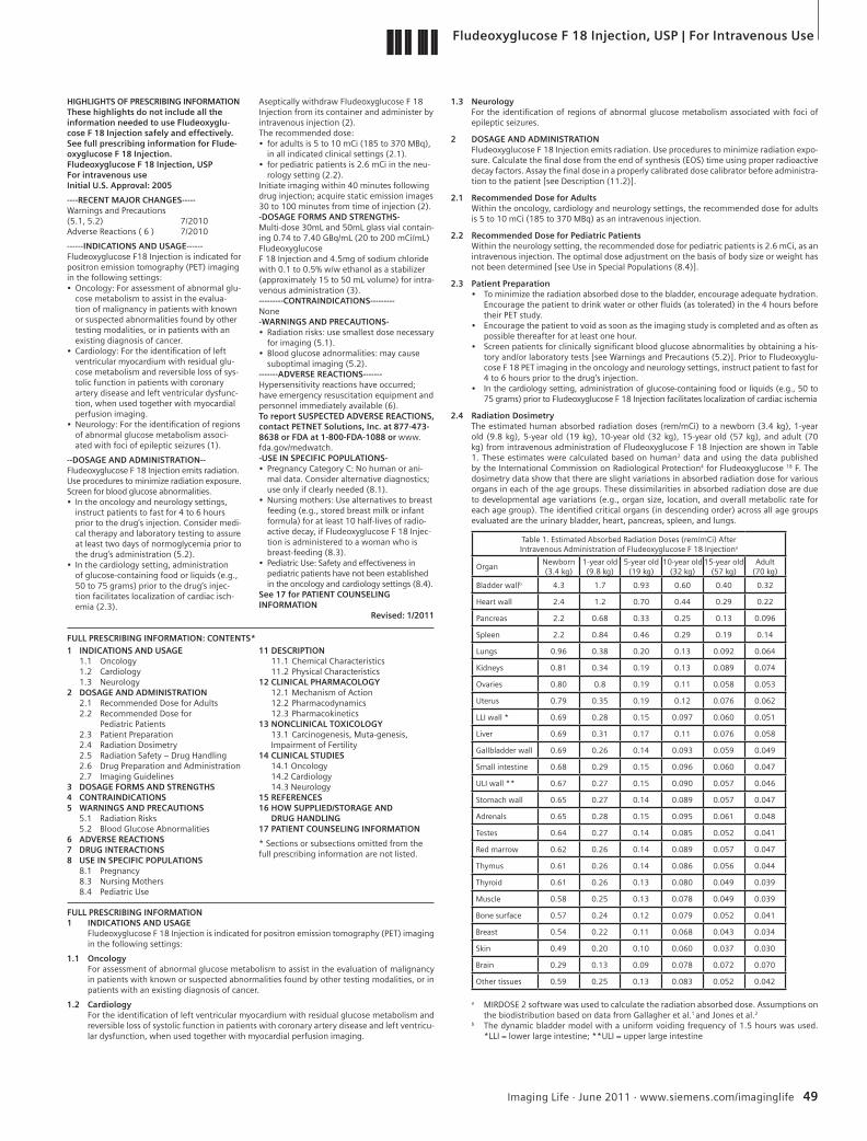
Imaging Life · June 2011 · www.siemens.com/imaginglife 49
Fludeoxyglucose F 18 Injection, USP | For Intravenous Use
23
56
91011
1314
Hashmark guide for AMERISOURCEAMERISOURCE.
1. Bars 1-3 are always white2. Start bar is bar #43. Code is numbered item followed by letter of alphabet. Ex: 01N4. Use Hashmark Bar Code Guide to create bar
Code 93R - McKESSON
LIGHT DARK1 1 12 1 23 1 34 2 15 2 26 2 37 3 18 3 29 3 30 1 4
A=01B=12C=23D=34E=45F=56G=67H=78I=89J=00K=11L=22M=33
N=44O=55P=66Q=77R=88S=99T=10U=21V=32W=43X=54Y=65Z=76
HASHMARK BAR CODE GUIDE
HIGHLIGHTS OF PRESCRIBING INFORMATIONThese highlights do not include all the information needed to use Fludeoxyglu-cose F 18 Injection safely and effectively. See full prescribing information for Flude-oxyglucose F 18 Injection.Fludeoxyglucose F 18 Injection, USPFor intravenous use Initial U.S. Approval: 2005
----RECENT MAJOR CHANGES-----Warnings and Precautions (5.1, 5.2) 7/2010Adverse Reactions ( 6 ) 7/2010
------INDICATIONS AND USAGE------Fludeoxyglucose F18 Injection is indicated for positron emission tomography (PET) imaging in the following settings: • Oncology: For assessment of abnormal glu-
cose metabolism to assist in the evalua-tion of malignancy in patients with known or suspected abnormalities found by other testing modalities, or in patients with an existing diagnosis of cancer.
• Cardiology: For the identification of left ventricular myocardium with residual glu-cose metabolism and reversible loss of sys-tolic function in patients with coronary artery disease and left ventricular dysfunc-tion, when used together with myocardial perfusion imaging.
• Neurology: For the identification of regions of abnormal glucose metabolism associ-ated with foci of epileptic seizures (1).
--DOSAGE AND ADMINISTRATION--Fludeoxyglucose F 18 Injection emits radiation. Use procedures to minimize radiation exposure. Screen for blood glucose abnormalities. • In the oncology and neurology settings,
instruct patients to fast for 4 to 6 hours prior to the drug’s injection. Consider medi-cal therapy and laboratory testing to assure at least two days of normoglycemia prior to the drug’s administration (5.2).
• In the cardiology setting, administration of glucose-containing food or liquids (e.g., 50 to 75 grams) prior to the drug’s injec-tion facilitates localization of cardiac isch-emia (2.3).
Aseptically withdraw Fludeoxyglucose F 18 Injection from its container and administer by intravenous injection (2). The recommended dose: • for adults is 5 to 10 mCi (185 to 370 MBq),
in all indicated clinical settings (2.1). • for pediatric patients is 2.6 mCi in the neu-
rology setting (2.2).Initiate imaging within 40 minutes following drug injection; acquire static emission images 30 to 100 minutes from time of injection (2).-DOSAGE FORMS AND STRENGTHS-Multi-dose 30mL and 50mL glass vial contain-ing 0.74 to 7.40 GBq/mL (20 to 200 mCi/mL) FludeoxyglucoseF 18 Injection and 4.5mg of sodium chloride with 0.1 to 0.5% w/w ethanol as a stabilizer (approximately 15 to 50 mL volume) for intra-venous administration (3).---------CONTRAINDICATIONS---------None-WARNINGS AND PRECAUTIONS-• Radiation risks: use smallest dose necessary
for imaging (5.1). • Blood glucose adnormalities: may cause
suboptimal imaging (5.2).-------ADVERSE REACTIONS-------Hypersensitivity reactions have occurred; have emergency resuscitation equipment and personnel immediately available (6). To report SUSPECTED ADVERSE REACTIONS, contact PETNET Solutions, Inc. at 877-473-8638 or FDA at 1-800-FDA-1088 or www.fda.gov/medwatch.-USE IN SPECIFIC POPULATIONS-• Pregnancy Category C: No human or ani-
mal data. Consider alternative diagnostics; use only if clearly needed (8.1).
• Nursing mothers: Use alternatives to breast feeding (e.g., stored breast milk or infant formula) for at least 10 half-lives of radio-active decay, if Fludeoxyglucose F 18 Injec-tion is administered to a woman who is breast-feeding (8.3).
• Pediatric Use: Safety and effectiveness in pediatric patients have not been established in the oncology and cardiology settings (8.4).
See 17 for PATIENT COUNSELING INFORMATION Revised: 1/2011
FULL PRESCRIBING INFORMATION: CONTENTS*1 INDICATIONS AND USAGE 1.1 Oncology 1.2 Cardiology 1.3 Neurology2 DOSAGE AND ADMINISTRATION 2.1 Recommended Dose for Adults 2.2 Recommended Dose for Pediatric Patients 2.3 Patient Preparation 2.4 Radiation Dosimetry 2.5 Radiation Safety – Drug Handling 2.6 Drug Preparation and Administration 2.7 Imaging Guidelines3 DOSAGE FORMS AND STRENGTHS4 CONTRAINDICATIONS5 WARNINGS AND PRECAUTIONS 5.1 Radiation Risks 5.2 Blood Glucose Abnormalities6 ADVERSE REACTIONS7 DRUG INTERACTIONS8 USE IN SPECIFIC POPULATIONS 8.1 Pregnancy 8.3 Nursing Mothers 8.4 Pediatric Use
11 DESCRIPTION 11.1 Chemical Characteristics 11.2 Physical Characteristics12 CLINICAL PHARMACOLOGY 12.1 Mechanism of Action 12.2 Pharmacodynamics 12.3 Pharmacokinetics13 NONCLINICAL TOXICOLOGY 13.1 Carcinogenesis, Muta-genesis, Impairment of Fertility14 CLINICAL STUDIES 14.1 Oncology 14.2 Cardiology 14.3 Neurology15 REFERENCES16 HOW SUPPLIED/STORAGE AND DRUG HANDLING17 PATIENT COUNSELING INFORMATION
* Sections or subsections omitted from the full prescribing information are not listed.
FULL PRESCRIBING INFORMATION1 INDICATIONS AND USAGE
Fludeoxyglucose F 18 Injection is indicated for positron emission tomography (PET) imaging in the following settings:
1.1 Oncology For assessment of abnormal glucose metabolism to assist in the evaluation of malignancy in patients with known or suspected abnormalities found by other testing modalities, or in patients with an existing diagnosis of cancer.
1.2 Cardiology For the identification of left ventricular myocardium with residual glucose metabolism and reversible loss of systolic function in patients with coronary artery disease and left ventricu-lar dysfunction, when used together with myocardial perfusion imaging.
1.3 Neurology For the identification of regions of abnormal glucose metabolism associated with foci of epileptic seizures.
2 DOSAGE AND ADMINISTRATION Fludeoxyglucose F 18 Injection emits radiation. Use procedures to minimize radiation expo-sure. Calculate the final dose from the end of synthesis (EOS) time using proper radioactive decay factors. Assay the final dose in a properly calibrated dose calibrator before administra-tion to the patient [see Description (11.2)].
2.1 Recommended Dose for Adults Within the oncology, cardiology and neurology settings, the recommended dose for adults is 5 to 10 mCi (185 to 370 MBq) as an intravenous injection.
2.2 Recommended Dose for Pediatric PatientsWithin the neurology setting, the recommended dose for pediatric patients is 2.6 mCi, as an intravenous injection. The optimal dose adjustment on the basis of body size or weight has not been determined [see Use in Special Populations (8.4)].
2.3 Patient Preparation• Tominimizetheradiationabsorbeddosetothebladder,encourageadequatehydration.
Encourage the patient to drink water or other fluids (as tolerated) in the 4 hours before their PET study.
• Encourage the patient to void as soon as the imaging study is completed and as often as possible thereafter for at least one hour.
• Screen patients for clinically significant blood glucose abnormalities by obtaining a his-tory and/or laboratory tests [see Warnings and Precautions (5.2)]. Prior to Fludeoxyglu-cose F 18 PET imaging in the oncology and neurology settings, instruct patient to fast for 4 to 6 hours prior to the drug’s injection.
• In the cardiology setting, administration of glucose-containing food or liquids (e.g., 50 to 75 grams) prior to Fludeoxyglucose F 18 Injection facilitates localization of cardiac ischemia
2.4 Radiation DosimetryThe estimated human absorbed radiation doses (rem/mCi) to a newborn (3.4 kg), 1-year old (9.8 kg), 5-year old (19 kg), 10-year old (32 kg), 15-year old (57 kg), and adult (70 kg) from intravenous administration of Fludeoxyglucose F 18 Injection are shown in Table 1. These estimates were calculated based on human2 data and using the data published by the International Commission on Radiological Protection4 for Fludeoxyglucose 18 F. The dosimetry data show that there are slight variations in absorbed radiation dose for various organs in each of the age groups. These dissimilarities in absorbed radiation dose are due to developmental age variations (e.g., organ size, location, and overall metabolic rate for each age group). The identified critical organs (in descending order) across all age groups evaluated are the urinary bladder, heart, pancreas, spleen, and lungs.
Table 1. Estimated Absorbed Radiation Doses (rem/mCi) After Intravenous Administration of Fludeoxyglucose F 18 Injectiona
OrganNewborn (3.4 kg)
1-year old (9.8 kg)
5-year old (19 kg)
10-year old (32 kg)
15-year old (57 kg)
Adult (70 kg)
Bladder wallb 4.3 1.7 0.93 0.60 0.40 0.32
Heart wall 2.4 1.2 0.70 0.44 0.29 0.22
Pancreas 2.2 0.68 0.33 0.25 0.13 0.096
Spleen 2.2 0.84 0.46 0.29 0.19 0.14
Lungs 0.96 0.38 0.20 0.13 0.092 0.064
Kidneys 0.81 0.34 0.19 0.13 0.089 0.074
Ovaries 0.80 0.8 0.19 0.11 0.058 0.053
Uterus 0.79 0.35 0.19 0.12 0.076 0.062
LLI wall * 0.69 0.28 0.15 0.097 0.060 0.051
Liver 0.69 0.31 0.17 0.11 0.076 0.058
Gallbladder wall 0.69 0.26 0.14 0.093 0.059 0.049
Small intestine 0.68 0.29 0.15 0.096 0.060 0.047
ULI wall ** 0.67 0.27 0.15 0.090 0.057 0.046
Stomach wall 0.65 0.27 0.14 0.089 0.057 0.047
Adrenals 0.65 0.28 0.15 0.095 0.061 0.048
Testes 0.64 0.27 0.14 0.085 0.052 0.041
Red marrow 0.62 0.26 0.14 0.089 0.057 0.047
Thymus 0.61 0.26 0.14 0.086 0.056 0.044
Thyroid 0.61 0.26 0.13 0.080 0.049 0.039
Muscle 0.58 0.25 0.13 0.078 0.049 0.039
Bone surface 0.57 0.24 0.12 0.079 0.052 0.041
Breast 0.54 0.22 0.11 0.068 0.043 0.034
Skin 0.49 0.20 0.10 0.060 0.037 0.030
Brain 0.29 0.13 0.09 0.078 0.072 0.070
Other tissues 0.59 0.25 0.13 0.083 0.052 0.042
a MIRDOSE 2 software was used to calculate the radiation absorbed dose. Assumptions on the biodistribution based on data from Gallagher et al.1 and Jones et al.2
b The dynamic bladder model with a uniform voiding frequency of 1.5 hours was used. *LLI = lower large intestine; **ULI = upper large intestine
IL_June2011.indb 49 5/12/11 5:25 PM
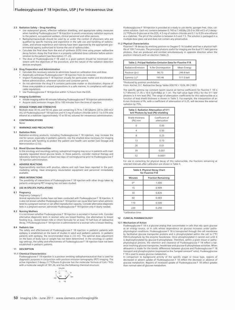
50 Imaging Life · June 2011 · www.siemens.com/imaginglife
Fludeoxyglucose F 18 Injection, USP | For Intravenous Use
2.5 Radiation Safety – Drug Handling• Use waterproof gloves, effective radiation shielding, and appropriate safety measures
when handling Fludeoxyglucose F 18 Injection to avoid unnecessary radiation exposure to the patient, occupational workers, clinical personnel and other persons.
• Radiopharmaceuticals should be used by or under the control of physicians who are qualified by specific training and experience in the safe use and handling of radionu-clides, and whose experience and training have been approved by the appropriate gov-ernmental agency authorized to license the use of radionuclides.
• Calculate the final dose from the end of synthesis (EOS) time using proper radioactive decay factors. Assay the final dose in a properly calibrated dose calibrator before admin-istration to the patient [see Description (11.2)].
• The dose of Fludeoxyglucose F 18 used in a given patient should be minimized con-sistent with the objectives of the procedure, and the nature of the radiation detection devices employed.
2.6 Drug Preparation and Administration• Calculate the necessary volume to administer based on calibration time and dose. • Aseptically withdraw Fludeoxyglucose F 18 Injection from its container. • Inspect Fludeoxyglucose F 18 Injection visually for particulate matter and discoloration
before administration, whenever solution and container permit. • Do not administer the drug if it contains particulate matter or discoloration; dispose of
these unacceptable or unused preparations in a safe manner, in compliance with appli-cable regulations.
• Use Fludeoxyglucose F 18 Injection within 12 hours from the EOS.
2.7 Imaging Guidelines• Initiate imaging within 40 minutes following Fludeoxyglucose F 18 Injection administration. • Acquire static emission images 30 to 100 minutes from the time of injection.
3 DOSAGE FORMS AND STRENGTHS Multiple-dose 30 mL and 50 mL glass vial containing 0.74 to 7.40 GBq/mL (20 to 200 mCi/mL) of Fludeoxyglucose F 18 Injection and 4.5 mg of sodium chloride with 0.1 to 0.5% w/w ethanol as a stabilizer (approximately 15 to 50 mL volume) for intravenous administration.
4 CONTRAINDICATIONS None
5 WARNINGS AND PRECAUTIONS
5.1 Radiation Risks Radiation-emitting products, including Fludeoxyglucose F 18 Injection, may increase the risk for cancer, especially in pediatric patients. Use the smallest dose necessary for imaging and ensure safe handling to protect the patient and health care worker [see Dosage and Administration (2.5)].
5.2 Blood Glucose Abnormalities In the oncology and neurology setting, suboptimal imaging may occur in patients with inad-equately regulated blood glucose levels. In these patients, consider medical therapy and laboratory testing to assure at least two days of normoglycemia prior to Fludeoxyglucose F 18 Injection administration.
6 ADVERSE REACTIONS Hypersensitivity reactions with pruritus, edema and rash have been reported in the post-marketing setting. Have emergency resuscitation equipment and personnel immediately available.
7 DRUG INTERACTIONS The possibility of interactions of Fludeoxyglucose F 18 Injection with other drugs taken by patients undergoing PET imaging has not been studied.
8 USE IN SPECIFIC POPULATIONS
8.1 Pregnancy Pregnancy Category C Animal reproduction studies have not been conducted with Fludeoxyglucose F 18 Injection. It is also not known whether Fludeoxyglucose F 18 Injection can cause fetal harm when adminis-tered to a pregnant woman or can affect reproduction capacity. Consider alternative diagnostic tests in a pregnant woman; administer Fludeoxyglucose F 18 Injection only if clearly needed.
8.3 Nursing MothersIt is not known whether Fludeoxyglucose F 18 Injection is excreted in human milk. Consider alternative diagnostic tests in women who are breast-feeding. Use alternatives to breast feeding (e.g., stored breast milk or infant formula) for at least 10 half-lives of radioactive decay, if Fludeoxyglucose F 18 Injection is administered to a woman who is breast-feeding.
8.4 Pediatric Use The safety and effectiveness of Fludeoxyglucose F 18 Injection in pediatric patients with epilepsy is established on the basis of studies in adult and pediatric patients. In pediatric patients with epilepsy, the recommended dose is 2.6 mCi. The optimal dose adjustment on the basis of body size or weight has not been determined. In the oncology or cardiol-ogy settings, the safety and effectiveness of Fludeoxyglucose F 18 Injection have not been established in pediatric patients.
11 DESCRIPTION
11.1 Chemical Characteristics Fludeoxyglucose F 18 Injection is a positron emitting radiopharmaceutical that is used for diagnostic purposes in conjunction with positron emission tomography (PET) imaging. The active ingredient 2-deoxy-2-[18F]fluoro-D-glucose has the molecular formula of C6H1118FO5 with a molecular weight of 181.26, and has the following chemical structure:
Fludeoxyglucose F 18 Injection is provided as a ready to use sterile, pyrogen free, clear, col-orless solution. Each mL contains between 0.740 to 7.40GBq (20.0 to 200 mCi) of 2-deoxy-2-[18F]fluoro-D-glucose at the EOS, 4.5 mg of sodium chloride and 0.1 to 0.5% w/w ethanol as a stabilizer. The pH of the solution is between 4.5 and 7.5. The solution is packaged in a multiple-dose glass vial and does not contain any preservative.
11.2 Physical Characteristics Fluorine F 18 decays by emitting positron to Oxygen O 16 (stable) and has a physical half-life of 109.7 minutes. The principal photons useful for imaging are the dual 511 keV gamma photons, that are produced and emitted simultaneously in opposite direction when the positron interacts with an electron (Table 2).
*Produced by positron annihilation From: Kocher, D.C. Radioactive Decay Tables DOE/TIC-I 1026, 89 (1981)
The specific gamma ray constant (point source air kerma coefficient) for fluorine F 18 is 5.7 R/hr/mCi (1.35 x 10⁻⁶ Gy/hr/kBq) at 1 cm. The half-value layer (HVL) for the 511 keV photons is 4 mm lead (Pb). The range of attenuation coefficients for this radionuclide as a function of lead shield thickness is shown in Table 3. For example, the interposition of an 8 mm thickness of Pb, with a coefficient of attenuation of 0.25, will decrease the external radiation by 75%.
For use in correcting for physical decay of this radionuclide, the fractions remaining at selected intervals after calibration are shown in Table 4.
*calibration time
12 CLINICAL PHARMACOLOGY
12.1 Mechanism of ActionFludeoxyglucose F 18 is a glucose analog that concentrates in cells that rely upon glucose as an energy source, or in cells whose dependence on glucose increases under patho-physiological conditions. Fludeoxyglucose F 18 is transported through the cell membrane by facilitative glucose transporter proteins and is phosphorylated within the cell to [18F] FDG-6-phosphate by the enzyme hexokinase. Once phosphorylated it cannot exit until it is dephosphorylated by glucose-6-phosphatase. Therefore, within a given tissue or patho-physiological process, the retention and clearance of Fludeoxyglucose F 18 reflect a bal-ance involving glucose transporter, hexokinase and glucose-6-phosphatase activities. When allowance is made for the kinetic differences between glucose and Fludeoxyglucose F 18 transport and phosphorylation (expressed as the ‚‘lumped constant‘‘ ratio), Fludeoxyglucose F 18 is used to assess glucose metabolism. In comparison to background activity of the specific organ or tissue type, regions of decreased or absent uptake of Fludeoxyglucose F 18 reflect the decrease or absence of glucose metabolism. Regions of increased uptake of Fludeoxyglucose F 18 reflect greater than normal rates of glucose metabolism.
Table 2. Pricipal Radiation Emission Data for Fluorine F18
Radiation/Emission % Per Disintegration Mean Energy
Positron (b+) 96.73 249.8 keV
Gamma (±)* 193.46 511.0 keV
Table 3. Radiation Attenuation of 511keV Photons by lead (Pb) shielding
Shield thickness(Pb) mm
Coefficient ofattenuation
0 0.00
4 0.50
8 0.25
13 0.10
26 0.01
39 0.001
52 0.0001
Table 4. Physical Decay Chartfor Fluorine F18
Minutes Fraction Remaining
0* 1.000
15 0.909
30 0.826
60 0.683
110 0.500
220 0.250
IL_June2011.indb 50 5/12/11 5:25 PM

Fludeoxyglucose F 18 Injection, USP | For Intravenous Use
Imaging Life · June 2011 · www.siemens.com/imaginglife 51
12.2 Pharmacodynamics Fludeoxyglucose F 18 Injection is rapidly distributed to all organs of the body after intrave-nous administration. After background clearance of Fludeoxyglucose F 18 Injection, optimal PET imaging is generally achieved between 30 to 40 minutes after administration. In cancer, the cells are generally characterized by enhanced glucose metabolism partially due to (1) an increase in activity of glucose transporters, (2) an increased rate of phosphorylation activity, (3) a reduction of phosphatase activity or, (4) a dynamic alteration in the balance among all these processes. However, glucose metabolism of cancer as reflected by Fludeoxy-glucose F 18 accumulation shows considerable variability. Depending on tumor type, stage, and location, Fludeoxyglucose F 18 accumulation may be increased, normal, or decreased. Also, inflammatory cells can have the same variability of uptake of Fludeoxyglucose F 18.In the heart, under normal aerobic conditions, the myocardium meets the bulk of its energy requirements by oxidizing free fatty acids. Most of the exogenous glucose taken up by the myocyte is converted into glycogen. However, under ischemic conditions, the oxidation of free fatty acids decreases, exogenous glucose becomes the preferred myocardial substrate, glycolysis is stimulated, and glucose taken up by the myocyte is metabolized immediately instead of being converted into glycogen. Under these conditions, phosphorylated Flude-oxyglucose F 18 accumulates in the myocyte and can be detected with PET imaging. In the brain, cells normally rely on aerobic metabolism. In epilepsy, the glucose metabolism varies. Generally, during a seizure, glucose metabolism increases. Interictally, the seizure focus tends to be hypometabolic.
12.3 Pharmacokinetics Distribution: In four healthy male volunteers, receiving an intravenous administration of 30 seconds in duration, the arterial blood level profile for Fludeoxyglucose F 18 decayed triexponentially. The effective half-life ranges of the three phases were 0.2 to 0.3 minutes, 10 to 13 minutes with a mean and standard deviation (STD) of 11.6 (±) 1.1 min, and 80 to 95 minutes with a mean and STD of 88 (±) 4 min.
Plasma protein binding of Fludeoxyglucose F 18 has not been studied.
Metabolism: Fludeoxyglucose F 18 is transported into cells and phosphorylated to [18F]-FDG-6- phosphate at a rate proportional to the rate of glucose utilization within that tissue. [F18]-FDG-6-phosphate presumably is metabolized to 2-deoxy-2-[F18]fluoro-6-phospho-D-mannose([F 18]FDM-6-phosphate).
Fludeoxyglucose F 18 Injection may contain several impurities (e.g., 2-deoxy-2-chloro-D-glucose (ClDG)). Biodistribution and metabolism of ClDG are presumed to be similar to Fludeoxyglucose F 18 and would be expected to result in intracellular formation of 2-deoxy-2-chloro-6-phospho-D-glucose (ClDG-6-phosphate) and 2-deoxy-2-chloro-6-phospho-D-mannose (ClDM-6-phosphate). The phosphorylated deoxyglucose compounds are dephos-phorylated and the resulting compounds (FDG, FDM, ClDG, and ClDM) presumably leave cells by passive diffusion. Fludeoxyglucose F 18 and related compounds are cleared from non-cardiac tissues within 3 to 24 hours after administration. Clearance from the cardiac tissue may require more than 96 hours. Fludeoxyglucose F 18 that is not involved in glucose metabolism in any tissue is then excreted in the urine.
Elimination: Fludeoxyglucose F 18 is cleared from most tissues within 24 hours and can be eliminated from the body unchanged in the urine. Three elimination phases have been identified in the reviewed literature. Within 33 minutes, a mean of 3.9% of the adminis-trated radioactive dose was measured in the urine. The amount of radiation exposure of the urinary bladder at two hours post-administration suggests that 20.6% (mean) of the radioactive dose was present in the bladder.
Special Populations: The pharmacokinetics of Fludeoxyglucose F 18 Injection have not been studied in renally-impaired, hepatically impaired or pediatric patients. Fludeoxyglucose F 18 is eliminated through the renal system. Avoid excessive radiation exposure to this organ system and adjacent tissues.
The effects of fasting, varying blood sugar levels, conditions of glucose intolerance, and dia-betes mellitus on Fludeoxyglucose F 18 distribution in humans have not been ascertained [see Warnings and Precautions (5.2)].
13 NONCLINICAL TOXICOLOGY
13.1 Carcinogenesis, Mutagenesis, Impairment of Fertility Animal studies have not been performed to evaluate the Fludeoxyglucose F 18 Injection carcinogenic potential, mutagenic potential or effects on fertility.
14 CLINICAL STUDIES
14.1 Oncology The efficacy of Fludeoxyglucose F 18 Injection in positron emission tomography cancer imaging was demonstrated in 16 independent studies. These studies prospectively evalu-ated the use of Fludeoxyglucose F 18 in patients with suspected or known malignancies, including non-small cell lung cancer, colo-rectal, pancreatic, breast, thyroid, melanoma, Hodgkin‘s and non-Hodgkin‘s lymphoma, and various types of metastatic cancers to lung, liver, bone, and axillary nodes. All these studies had at least 50 patients and used pathology as a standard of truth. The Fludeoxyglucose F 18 Injection doses in the studies ranged from 200 MBq to 740 MBq with a median and mean dose of 370 MBq.
In the studies, the diagnostic performance of Fludeoxyglucose F 18 Injection varied with the type of cancer, size of cancer, and other clinical conditions. False negative and false positive scans were observed. Negative Fludeoxyglucose F 18 Injection PET scans do not exclude the diagnosis of cancer. Positive Fludeoxyglucose F 18 Injection PET scans can not replace pathology to establish a diagnosis of cancer. Non-malignant conditions such as fungal infections, inflammatory processes and benign tumors have patterns of increased glucose metabolism that may give rise to false-positive scans. The efficacy of Fludeoxyglucose F 18 Injection PET imaging in cancer screening was not studied.
14.2 Cardiology The efficacy of Fludeoxyglucose F 18 Injection for cardiac use was demonstrated in ten independent, prospective studies of patients with coronary artery disease and chronic left ventricular systolic dysfunction who were scheduled to undergo coronary revascularization.
Before revascularization, patients underwent PET imaging with Fludeoxyglucose F 18 Injec-tion (74 to 370 MBq, 2 to 10 mCi) and perfusion imaging with other diagnostic radiophar-maceuticals. Doses of Fludeoxyglucose F 18 Injection ranged from 74 to 370 MBq (2 to 10 mCi). Segmental, left ventricular, wall-motion assessments of asynergic areas made before revascularization were compared in a blinded manner to assessments made after successful revascularization to identify myocardial segments with functional recovery.
Left ventricular myocardial segments were predicted to have reversible loss of systolic function if they showed Fludeoxyglucose F 18 accumulation and reduced perfusion (i.e., flow-metabolism mismatch). Conversely, myocardial segments were predicted to have irre-versible loss of systolic function if they showed reductions in both Fludeoxyglucose F 18 accumulation and perfusion (i.e., matched defects).
Findings of flow-metabolism mismatch in a myocardial segment may suggest that success-ful revascularization will restore myocardial function in that segment. However, false-positive tests occur regularly, and the decision to have a patient undergo revascularization should not be based on PET findings alone. Similarly, findings of a matched defect in a myocardial segment may suggest that myocardial function will not recover in that segment, even if it is successfully revascularized. However, false-negative tests occur regularly, and the decision to recommend against coronary revascularization, or to recommend a cardiac transplant, should not be based on PET findings alone. The reversibility of segmental dysfunction as predicted with Fludeoxyglucose F 18 PET imaging depends on successful coronary revascularization. Therefore, in patients with a low likelihood of successful revascularization, the diagnostic use-fulness of PET imaging with Fludeoxyglucose F 18 Injection is more limited.
14.3 Neurology In a prospective, open label trial, Fludeoxyglucose F 18 Injection was evaluated in 86 patients with epilepsy. Each patient received a dose of Fludeoxyglucose F 18 Injection in the range of 185 to 370 MBq (5 to 10 mCi). The mean age was 16.4 years (range: 4 months to 58 years; of these, 42 patients were less than 12 years and 16 patients were less than 2 years old). Patients had a known diagnosis of complex partial epilepsy and were under evaluation for surgical treatment of their seizure disorder. Seizure foci had been previously identified on ictal EEGs and sphenoidal EEGs. Fludeoxyglucose F 18 Injection PET imaging confirmed previous diagnostic findings in 16% (14/87) of the patients; in 34% (30/87) of the patients, Fludeoxyglucose F 18 Injection PET images provided new findings. In 32% (27/87), imaging with Fludeoxyglucose F 18 Injection was inconclusive. The impact of these imaging findings on clinical outcomes is not known.
Several other studies comparing imaging with Fludeoxyglucose F 18 Injection results to subsphenoidal EEG, MRI and/or surgical findings supported the concept that the degree of hypometabolism corresponds to areas of confirmed epileptogenic foci. The safety and effec-tiveness of Fludeoxyglucose F 18 Injection to distinguish idiopathic epileptogenic foci from tumors or other brain lesions that may cause seizures have not been established.
15 REFERENCES1. Gallagher B.M., Ansari A., Atkins H., Casella V., Christman D.R., Fowler J.S., Ido T., Mac-
Gregor R.R., Som P., Wan C.N., Wolf A.P., Kuhl D.E., and Reivich M. “Radiopharmaceuti-cals XXVII. 18F-labeled 2-deoxy-2-fluoro-d-glucose as a radiopharmaceutical for mea-suring regional myocardial glucose metabolism in vivo: tissue distribution and imaging studies in animals,” J Nucl Med, 1977; 18, 990-6.
2. Jones S.C., Alavi, A., Christman D., Montanez, I., Wolf, A.P., and Reivich M. “The radiation dosimetry of 2 [F-18] fluoro-2-deoxy-D-glucose in man,” J Nucl Med, 1982; 23, 613-617.
3. Kocher, D.C. “Radioactive Decay Tables: A handbook of decay data for application to radiation dosimetry and radiological assessments,” 1981, DOE/TIC-I 1026, 89.
4. ICRP Publication 53, Volume 18, No. l-4,1987, pages 75-76.
16 HOW SUPPLIED/STORAGE AND DRUG HANDLING Fludeoxyglucose F 18 Injection is supplied in a multi-dose, capped 30 mL and 50 mL glass vial containing between 0.740 to 7.40 GBq/mL (20 to 200 mCi/mL), of no carrier added 2-deoxy-2-[F 18] fluoro-D-glucose, at end of synthesis, in approximately 15 to 50 mL. The contents of each vial are sterile, pyrogen-free and preservative-free.
NDC 40028-511-30; 40028-511-50
Receipt, transfer, handling, possession, or use of this product is subject to the radioactive material regulations and licensing requirements of the U.S. Nuclear Regulatory Commis-sion, Agreement States or Licensing States as appropriate.
Store the Fludeoxyglucose F 18 Injection vial upright in a lead shielded container at 25°C (77°F); excursions permitted to 15-30°C (59-86°F).
Store and dispose of Fludeoxyglucose F 18 Injection in accordance with the regulations and a general license, or its equivalent, of an Agreement State or a Licensing State.
The expiration date and time are provided on the container label. Use Fludeoxyglucose F 18 Injection within 12 hours from the EOS time.
17 PATIENT COUNSELING INFORMATIONInstruct patients in procedures that increase renal clearance of radioactivity. Encourage patients to: • drink water or other fluids (as tolerated) in the 4 hours before their PET study. • void as soon as the imaging study is completed and as often as possible thereafter for
at least one hour.
Manufactured by: PETNET Solutions Inc.810 Innovation DriveKnoxville, TN 37932
Distributed by: PETNET Solutions Inc.810 Innovation DriveKnoxville, TN 37932
PN0002262 Rev. AMarch 1, 2011
IL_June2011.indb 51 5/12/11 5:25 PM

On account of certain regional limitations of sales rights and service availability, we cannot guarantee that all products included in this brochure are available through the Siemens sales organization worldwide. Availability and packaging may vary by country and is subject to change without prior notice. Some/All of the features and products described herein may not be available in the United States.
The information in this document contains general technical descriptions of specifications and options as well as standard and optional features which do not always have to be present in individual cases.
Siemens reserves the right to modify the design, packaging, specifications and options described herein without prior notice.
Please contact your local Siemens sales representative for the most current information.
Note: Any technical data contained in this document may vary within defined tolerances. Original images always lose a certain amount of detail when reproduced.
www.siemens.com/healthcare-magazine
Global Business Unit
Siemens Medical Solutions USA, Inc.Molecular Imaging2501 N. Barrington RoadHoffman Estates, IL 60192USATelephone: + 1 847 304-7700www.siemens.com/mi
Local Contact Information
Asia/Pacific:Siemens Medical SolutionsAsia Pacific HeadquartersThe Siemens Center60 MacPherson RoadSingapore 348615Phone: +65 9622 - 2026 www.siemens.com/healthcare
Canada:Siemens Canada LimitedMedical Solutions2185 Derry Road WestMississauga ON L5N 7A6CanadaPhone: +1 905 819 - 5800 www.siemens.com/healthcare
Europe/Africa/Middle East:Siemens AGMedical SolutionsHenkestraße 127D-91052 ErlangenGermanyPhone: +49 9131 84 - 0 www.siemens.com/healthcare
Latin America:Siemens S.A.Medical SolutionsAvenida de Pte. Julio A. Roca No 516, Piso 7C1067ABN Buenos Aires ArgentinaPhone: +54 11 4340 - 8400 www.siemens.com/healthcare
USA:Siemens Medical Solutions USA, Inc.51 Valley Stream ParkwayMalvern, PA 19355-1406USAPhone: +1-888-826 - 9702 www.siemens.com/healthcare
Global SiemensHealthcare Headquarters
Siemens AGHealthcare SectorHenkestraße 12791052 ErlangenGermanyPhone: +49 9131 84 - 0www.siemens.com/healthcare
Global Siemens Headquarters
Siemens AGWittelsbacherplatz 280333 MuenchenGermany
Order No. A91MI 10354 1M 7600 | Printed in USA | MI-003.CY.TM.TM.10M | © 06.2011, Siemens AG
IL_June2011.indb 52 5/12/11 5:25 PM