THE LINEAGE DECISIONS OF HELPER T CELLS
12
© 2002 Nature Publishing Group After engagement of the T-cell receptor (TCR) by the appropriate peptide–MHC complex, which triggers clonal expansion, helper T (T H ) cells rapidly undergo programmed differentiation (reviewed in REFS 1,2). This differentiation process can result in highly polarized immune responses in the case of chronic infections, such as parasitic infections. A more heterogeneous response is often shown by analyses of acute immune responses. Naive T H cells can differentiate to at least two func- tional classes of cell during an immune response 3 — T H 1 cells, which secrete interferon-γ (IFN-γ), and T H 2 cells, which secrete interleukin-4 (IL-4) (reviewed in REFS 1,2). Hereafter, we refer to IFN-γ and IL-4 as effector cytokines. T H 1 cells are responsible for cell-mediated immunity, whereas T H 2 cells are responsible for extra- cellular immunity. In terms of the control of parasites, T H 1 immunity provides protection against intracellular protozoa, such as Leishmania species and Toxoplasma gondii, whereas T H 2 immunity is associated with protec- tion against intestinal helminths. As well as their protec- tive roles in host defence, both subsets of T H cell have been implicated in pathological responses. T H 1 cells can mediate organ-specific autoimmunity and T H 2 cells have been implicated in the pathogenesis of asthma and allergy. The final composition of the T H -cell response to antigen can, therefore, determine whether the outcome of infectious, inflammatory and autoimmune responses is favourable or unfavourable. The process by which an uncommitted T H cell devel- ops into a mature T H 1 or T H 2 cell is a useful model of developmentally regulated gene expression (FIG. 1). There is good evidence to indicate that this differentiation process is highly plastic. Many factors influence the deci- sion to become a T H 1 or T H 2 cell. The cytokines IL-12 and IL-4, acting through signal transducer and activator of transcription 4 (STAT4) and STAT6, respectively, are key determinants of the outcome (reviewed in REFS 1,2). It has been proposed also that antigen dose, co-stimulators, genetic modifiers and other non-cytokine factors have crucial roles in determining the dominance of a T H -cell THE LINEAGE DECISIONS OF HELPER T CELLS Kenneth M. Murphy* and Steven L. Reiner ‡ After encountering antigen, helper T (T H ) cells undergo differentiation to effector cells, which can secrete high levels of interferon-γ, interleukin-4 (IL-4), IL-10 and other immunomodulators. How T H cells acquire, and remember, new patterns of gene expression is an area of intensive investigation. The process is remarkably plastic, with cytokines being key regulators. Extrinsic signals seem to be integrated into cell-intrinsic programming, in what is becoming an intriguing story of regulated development. We summarize the latest insights into mechanisms that govern the lineage choices that are made during T H -cell responses to foreign pathogens. NATURE REVIEWS | IMMUNOLOGY VOLUME 2 | DECEMBER 2002 | 933 *Howard Hughes Medical Institute and Department of Pathology and Immunology, Washington University School of Medicine, St Louis, Missouri 63110, USA. ‡ Abramson Family Cancer Research Institute and Department of Medicine, University of Pennsylvania, Philadelphia 19104, USA. Correspondence to K.M.M. e-mail: [email protected] doi:10.1038/nri954 REVIEWS T-bet GATA3 IL-2 IFN-γ IL-4 T H 1 T H 2 IL-12 IL-4 Naive Figure 1 | The original instructive model of helper T-cell differentiation. An uncommitted, naive helper T (T H )-cell precursor can become either a T H 1 or T H 2 cell under the instructive influence of interleukin-12 (IL-12) or IL-4, respectively. T H 1 cells express T-bet and secrete interferon-γ (IFN-γ). T H 2 cells express GATA3 and secrete IL-4. DECISION MAKING IN THE IMMUNE SYSTEM
Transcript of THE LINEAGE DECISIONS OF HELPER T CELLS

© 2002 Nature Publishing Group
After engagement of the T-cell receptor (TCR) by theappropriate peptide–MHC complex, which triggersclonal expansion, helper T (T
H) cells rapidly undergo
programmed differentiation (reviewed in REFS 1,2). Thisdifferentiation process can result in highly polarizedimmune responses in the case of chronic infections, suchas parasitic infections.A more heterogeneous response isoften shown by analyses of acute immune responses.
Naive TH
cells can differentiate to at least two func-tional classes of cell during an immune response3 —T
H1 cells, which secrete interferon-γ (IFN-γ), and T
H2
cells, which secrete interleukin-4 (IL-4) (reviewed inREFS 1,2). Hereafter, we refer to IFN-γ and IL-4 as effectorcytokines. T
H1 cells are responsible for cell-mediated
immunity, whereas TH2 cells are responsible for extra-
cellular immunity. In terms of the control of parasites,T
H1 immunity provides protection against intracellular
protozoa, such as Leishmania species and Toxoplasmagondii, whereas T
H2 immunity is associated with protec-
tion against intestinal helminths. As well as their protec-tive roles in host defence, both subsets of T
Hcell have
been implicated in pathological responses. TH1 cells can
mediate organ-specific autoimmunity and TH
2 cellshave been implicated in the pathogenesis of asthma andallergy. The final composition of the T
H-cell response to
antigen can, therefore, determine whether the outcomeof infectious, inflammatory and autoimmune responsesis favourable or unfavourable.
The process by which an uncommitted TH
cell devel-ops into a mature T
H1 or T
H2 cell is a useful model of
developmentally regulated gene expression (FIG. 1). Thereis good evidence to indicate that this differentiationprocess is highly plastic. Many factors influence the deci-sion to become a T
H1 or T
H2 cell. The cytokines IL-12
and IL-4, acting through signal transducer and activatorof transcription 4 (STAT4) and STAT6, respectively, arekey determinants of the outcome (reviewed in REFS 1,2). Ithas been proposed also that antigen dose, co-stimulators,genetic modifiers and other non-cytokine factors havecrucial roles in determining the dominance of a T
H-cell
THE LINEAGE DECISIONS OF HELPER T CELLSKenneth M. Murphy* and Steven L. Reiner‡
After encountering antigen, helper T (TH) cells undergo differentiation to effector cells, which can secrete high levels of interferon-γ, interleukin-4 (IL-4), IL-10 and other immunomodulators.How TH cells acquire, and remember, new patterns of gene expression is an area of intensiveinvestigation. The process is remarkably plastic, with cytokines being key regulators. Extrinsicsignals seem to be integrated into cell-intrinsic programming, in what is becoming an intriguingstory of regulated development. We summarize the latest insights into mechanisms that governthe lineage choices that are made during TH-cell responses to foreign pathogens.
NATURE REVIEWS | IMMUNOLOGY VOLUME 2 | DECEMBER 2002 | 933
*Howard Hughes MedicalInstitute and Department ofPathology and Immunology,Washington UniversitySchool of Medicine, St Louis,Missouri 63110, USA.‡Abramson Family CancerResearch Institute andDepartment of Medicine,University of Pennsylvania,Philadelphia 19104, USA.Correspondence to K.M.M.e-mail:[email protected]:10.1038/nri954
R E V I E W S
T-bet
GATA3IL-2
IFN-γ
IL-4
TH1
TH2
IL-12
IL-4
Naive
Figure 1 | The original instructive model of helper T-celldifferentiation. An uncommitted, naive helper T (TH)-cellprecursor can become either a TH1 or TH2 cell under theinstructive influence of interleukin-12 (IL-12) or IL-4, respectively.TH1 cells express T-bet and secrete interferon-γ (IFN-γ). TH2 cellsexpress GATA3 and secrete IL-4.
D E C I S I O N M A K I N G I N T H E I M M U N E S Y S T E M
© 2002 Nature Publishing Group
EPIGENETIC
Refers to the heritable, butpotentially reversible, states ofgene activity that are imposed bythe structure of chromatin orcovalent modifications of DNAand histones.
934 | DECEMBER 2002 | VOLUME 2 www.nature.com/reviews/immunol
R E V I E W S
In this article, we highlight some recent advances inour understanding of this model of developmentallyregulated alterations in gene activity. We do not attemptto be encyclopaedic, but we try primarily to discuss andunite the transcriptional and EPIGENETIC mechanisms thatact during T
H1- and T
H2-cell development, as well as
mentioning a few topical cytokine-related issues.
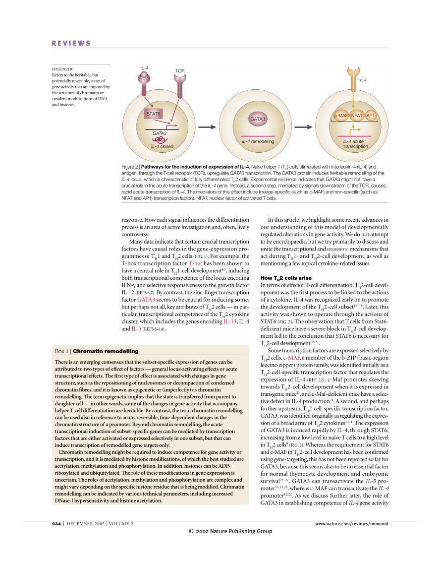
How TH2 cells ariseIn terms of effector T-cell differentiation, T
H2-cell devel-
opment was the first process to be linked to the actionsof a cytokine. IL-4 was recognized early on to promotethe development of the T
H2-cell subset15–18. Later, this
activity was shown to operate through the actions ofSTAT6 (FIG. 2). The observation that T cells from Stat6-deficient mice have a severe block in T
H2-cell develop-
ment led to the conclusion that STAT6 is necessary forT
H2-cell development19–21.Some transcription factors are expressed selectively by
TH2 cells. c-MAF, a member of the b-ZIP (basic-region
leucine-zipper) protein family, was identified initially as aT
H2-cell-specific transcription factor that regulates the
expression of IL-4 (REF. 22). c-Maf promotes skewingtowards T
H2-cell development when it is expressed in
transgenic mice23, and c-Maf-deficient mice have a selec-tive defect in IL-4 production24. A second, and perhapsfurther upstream, T
H2-cell-specific transcription factor,
GATA3, was identified originally as regulating the expres-sion of a broad array of T
H2 cytokines10,11. The expression
of GATA3 is induced rapidly by IL-4, through STAT6,increasing from a low level in naive T cells to a high levelin T
H2 cells9 (FIG. 2). Whereas the requirement for STAT6
and c-MAF in TH2-cell development has been confirmed
using gene-targeting, this has not been reported so far forGATA3, because this seems also to be an essential factorfor normal thymocyte development and embryonic survival25–27. GATA3 can transactivate the IL-5 pro-moter11,12,28, whereas c-MAF can transactivate the IL-4promoter12,22. As we discuss further later, the role ofGATA3 in establishing competence of IL-4 gene activity
response. How each signal influences the differentiationprocess is an area of active investigation and, often, livelycontroversy.
Many data indicate that certain crucial transcriptionfactors have causal roles in the gene-expression pro-grammes of T
H1 and T
H2 cells (FIG. 1). For example, the
T-box transcription factor T-bet has been shown tohave a central role in T
H1-cell development4,5, inducing
both transcriptional competence of the locus encodingIFN-γ and selective responsiveness to the growth factorIL-12 (REFS 6,7). By contrast, the zinc-finger transcriptionfactor GATA3 seems to be crucial for inducing some,but perhaps not all, key attributes of T
H2 cells — in par-
ticular, transcriptional competence of the TH2 cytokine
cluster, which includes the genes encoding IL-13, IL-4and IL-5 (REFS 8–14).
GATA3
GATA3
c-MAF NFAT
IL-4 remodellingIL-4 closed
IL-4 acutetranscription
AP1
TCR
TCR
IL-4
STAT6
Figure 2 | Pathways for the induction of expression of IL-4. Naive helper T (TH) cells stimulated with interleukin-4 (IL-4) andantigen, through the T-cell receptor (TCR), upregulate GATA3 transcription. The GATA3 protein induces heritable remodelling of theIL-4 locus, which is characteristic of fully differentiated TH2 cells. Experimental evidence indicates that GATA3 might not have acrucial role in the acute transcription of the IL-4 gene. Instead, a second step, mediated by signals downstream of the TCR, causesrapid acute transcription of IL-4. The mediators of this effect include lineage-specific (such as c-MAF) and non-specific (such asNFAT and AP1) transcription factors. NFAT, nuclear factor of activated T cells.
Box 1 | Chromatin remodelling
There is an emerging consensus that the subset-specific expression of genes can beattributed to two types of effect of factors — general locus-activating effects or acutetranscriptional effects. The first type of effect is associated with changes in genestructure, such as the repositioning of nucleosomes or decompaction of condensedchromatin fibres, and it is known as epigenetic or (imperfectly) as chromatinremodelling. The term epigenetic implies that the state is transferred from parent todaughter cell — in other words, some of the changes in gene activity that accompanyhelper T-cell differentiation are heritable. By contrast, the term chromatin remodellingcan be used also in reference to acute, reversible, time-dependent changes in thechromatin structure of a promoter. Beyond chromatin remodelling, the acutetranscriptional induction of subset-specific genes can be mediated by transcriptionfactors that are either activated or expressed selectively in one subset, but that caninduce transcription of remodelled gene targets only.
Chromatin remodelling might be required to induce competence for gene activity ortranscription, and it is mediated by histone modifications, of which the best studied areacetylation, methylation and phosphorylation. In addition, histones can be ADP-ribosylated and ubiquitylated. The role of these modifications in gene expression isuncertain. The roles of acetylation, methylation and phosphorylation are complex andmight vary depending on the specific histone residue that is being modified. Chromatinremodelling can be indicated by various technical parameters, including increasedDNase-I hypersensitivity and histone acetylation.

© 2002 Nature Publishing GroupNATURE REVIEWS | IMMUNOLOGY VOLUME 2 | DECEMBER 2002 | 935
R E V I E W S
repressed TH
2-cell development induced by ectopicGATA3 in STAT6−/− T cells and decreased the efficiencyof GATA3 transcriptional autoactivation34,35. ROG wasisolated as a GATA3-interacting protein; its overexpres-sion decreases GATA3 activity and the production ofT
H2 cytokines36.
How TH1 cells ariseT-bet was identified recently as a T
H1-cell-specific factor
that can induce the production of IFN-γ by developingT
H2 cells5. A member of the T-box family of transcrip-
tion factors, T-bet seems to be expressed in developingand committed T
H1 cells. In addition to its role in
inducing the expression of IFN-γ, T-bet seems to beinvolved in chromatin remodelling of the gene thatencodes IFN-γ6 (FIG. 3), induction of expression of theIL-12 receptor β2-subunit (IL-12Rβ2)6,37 and stabilizingits own expression, either through an intrinsic auto-catalytic loop or the autocrine effects of IFN-γsignalling6,37. Although both CD4+ and CD8+ T cells, aswell as natural killer (NK) cells, express T-bet, thereseems to be less dependence on T-bet for high-levelexpression of IFN-γ in CD8+ T cells than in CD4+ T cellsor NK cells4. Nevertheless, the crucial importance ofT-bet for the development of T
H1 responses in vivo is
underscored by the susceptibility of T-bet-knockoutmice to challenge with Leishmania major4 and theirpredisposition to allergic airway disease38.
How T-bet induces expression of IFN-γ is still anarea of active investigation. Recently, a genetic interac-tion was reported between T-bet and HLX (H2.0-likehomeobox 1)7. The HLX gene seems to be expressed indeveloping T
H1 cells, although its expression is induced
at a slower rate than for T-bet. Transcription of HLXseems to be downstream of T-bet activity, and HLXprotein seems to interact with T-bet protein, whichmediates the synergistic induction of IFN-γ expression7.
seems to be related to its ability to induce the remodellingof CHROMATIN8,12,29 (BOX 1 and FIG. 2).
GATA3 has been shown to promote TH2-cell devel-
opment strongly, when expressed by a transgene10 or by aretrovirus9,28. Furthermore, GATA3 induces T
H2-cell
development of STAT6−−/−− T cells8, including the produc-tion of T
H2-cell-specific cytokines and expression of
c-MAF, which depend normally on STAT6. A putativedominant-negative mutant of GATA3 decreases T
H2-
cell-mediated pulmonary allergic responses whenexpressed by a transgene in mice30. In addition, antisenseoligonucleotide treatment directed against GATA3 canrepress T
H2-cell responses in vivo, which further implies
a requirement for GATA3 for TH2-cell development31.
Several non-TH2-cell-specific transcription factors
contribute to the regulation of expression of TH
2cytokines, particularly members of the nuclear factor ofactivated T cells (NFAT) family (reviewed in REF. 2).Interactions between the different NFAT-family mem-bers on T
H2-cytokine genes are complex, and the tran-
scription factors can have both positive and negativeeffects. In general, the role of these factors in T
H2-
cytokine gene expression seems to be in mediating theacute transcription of inducible cytokines after trigger-ing of a differentiated T
H2 cell through the TCR (FIG. 2).
As well as STAT6, MEL18, a Polycomb-group pro-tein, also seems to regulate the expression of GATA3(REF. 32). Although Polycomb-group proteins are associ-ated generally with the heritable silencing of loci33,MEL18 seems to act as a positive regulator of GATA3transcription. So, in the absence of MEL18, the levels ofGATA3 and IL-4 are reduced, and reintroduction ofGATA3 using a retroviral vector can correct the defect inIL-4 expression partially32. There are at least two poten-tial post-transcriptional regulators of GATA3 — thezinc-finger proteins FOG1 (friend of GATA1) and ROG (repressor of GATA). Overexpression of FOG1
CHROMATIN
Composed of nucleosomes,which are the basic repeatingunits of eukaryotic genomes.Nucleosomes consist of 146 basepairs of DNA wound around anoctamer of histone proteins.
T-betNFAT NF-κB
TCR
IL-12IL-18
IFN-γ
IFN- remodellingγ IFN- acutetranscription
γ
T-bet
TCR
STAT1
IFN- closedγ
STAT4
Figure 3 | Pathways for the induction of expression of IFN-γ. Naive helper T (TH) cells activated under TH1-inducing conditionsare exposed to interferon-γ (IFN-γ) signalling during T-cell receptor (TCR) engagement, leading to the activation of signal transducerand activator of transcription 1 (STAT1). Downstream of STAT1, the expression of T-bet is induced, and T-bet acts in this model toinduce remodelling of the repressed IFN-γ locus (shown) and induce expression of the interleukin-12 receptor β2-subunit (IL-12Rβ2)(not shown). Subsequently, the committed TH1 cell, which expresses receptors for IL-12 and IL-18, has at least two pathwaysavailable to induce the acute transcription of IFN-γ — the TCR signalling pathway or a cytokine signalling pathway that can includecombined IL-12 and IL-18 signalling. TCR-induced transcription of the gene encoding IFN-γ is distinguishable pharmacologicallyfrom cytokine-induced transcription — the former is sensitive to cyclosporin A (CsA), whereas the latter is CsA resistant. Differentnuclear factors might mediate each pathway. NFAT, nuclear factor of activated T cells; NF-κB, nuclear factor-κB.

© 2002 Nature Publishing Group936 | DECEMBER 2002 | VOLUME 2 www.nature.com/reviews/immunol
R E V I E W S
that there is an exocrine mechanism. Because only earlytime points in development have been examined in eachof these studies, it is possible that a shift from a STAT-dependent pathway to an autoactivation pathway couldoccur at some later time. Finally, as STAT1-deficient cellscan be driven to T
H1-cell polarization, it is possible either
that expression of a low level of T-bet might be indepen-dent of IFN-γ signalling, allowing T
H1-cell development,
or that the requirement for T-bet for TH1-cell develop-
ment is conditional. The latter possibility is raised by theobservation that T-bet-deficient CD4+ T cells from theautoimmune Lpr mouse background produce abundantIFN-γ 41, which indicates that there is some flexibility inthe requirement for T-bet. So, it is uncertain whetherSTAT1-independent T
H1-cell development arises from an
alternative extrinsic signal or an autonomous property ofthe activated T cell.
Acute transcription of the gene encoding IFN-γγIn terminally differentiated T
H1 cells, reiteration of IFN-γ
expression can occur through two experimentally distinctpathways — TCR ligation or cytokine (IL-12 and IL-18)stimulation. IL-18 augments the production of IFN-γ bydifferentiated T
H1 cells, despite its inability to drive the
development of TH1 cells on its own42. The combination
of IL-12 and IL-18 can induce the production of IFN-γ bydifferentiated T cells in the absence of signalling throughthe TCR42,43 (FIG. 3). The IFN-γ production that is inducedis more prolonged and more resistant to treatment withcyclosporin A (CsA) (an inhibitor of NF-AT activation)than that induced by TCR crosslinking43, which indi-cates that cytokine-driven IFN-γ production is not cou-pled to the NFAT pathway. In vivo, it seems that IL-12and IL-18 act synergistically to induce maximum IFN-γproduction44,45.
IL-12- and IL-18-induced IFN-γ production corre-lates with induction of expression of the GADD-FAMILY
PROTEINS GADD45β and GADD45γ. Overexpression ofGADD45β augments IFN-γ production46, and absenceof GADD45γ decreases IFN-γ production, therebyinhibiting T
H1-cell development47. The transcriptional
details of IL-12- and IL-18-induced IFN-γ production areuncertain still. It is clear that IL-12- and IL-18-inducedIFN-γ production depends strongly on STAT4, whereasTCR signalling can induce IFN-γ production, although ata reduced level, in STAT4−/− T
H1 cells37. The other factors
that mediate cytokine-induced IFN-γ production — incontrast to CsA-sensitive, anti-CD3 antibody-inducedIFN-γ production — are uncertain still, although someevidence indicates that these might be p38 mitogen-activated protein kinase (p38 MAPK)-sensitive fac-tors46,48, which is consistent with previous suggestions(reviewed in REF. 49).
The secondary, cytokine-dependent pathway of IFN-γexpression that has been described for CD4+ T
H1 cells
might operate in other cell types. IL-12 and IL-18 have anactive role in inducing the production of IFN-γ by CD8+
T cells and NK cells in response to bacterial pathogens50.Similarly, there is some indirect evidence that such abystander pathway might operate in CD4+ T cells in vivo.Even after a T
H1 response has been initiated, it has been
The role of HLX in vivo has not been examined fully yetbecause knockout mice die during embryogenesis39.
The expression of T-bet seems to be induced readilyin naive T cells by IFN-γ signalling, mediated by STAT1(REFS 37,40) (FIG. 3). Although STAT1 seems to be a crucialfactor for the induction of expression of T-bet, type IIFNs, which activate STAT1 also, do not seem to be ableto induce T-bet transcription40. Whether T-bet proteincan autoactivate transcription of the T-bet gene is lessclear. Endogenous expression of T-bet was shown to beinduced by ectopic T-bet, which implies that autoactiva-tion does occur6,7, but a reduction in the level of endoge-nous T-bet transcription in the absence of autocrineIFN-γ signalling37,40 was observed also, which indicates
GADD-FAMILY PROTEINS
(Growth-arrest and DNA-damage inducible proteins). Inresponse to environmentalstresses, these proteins mediateactivation of the p38 mitogen-activated protein kinase pathwayand are involved in theregulation of growth andapoptosis.
GATA3
NF-κBSTAT6 NFAT
Integration for initial GATA3 induction
GATA3
TH2 cytokines
B7H
ICOS
CD28
CD80/86
CTLA4
LFA1
ICAM
IL-4receptor
MHC
TCR–CD3
Antigen
T cell
Dendritic cell
Figure 4 | Redundant pathways of TH2-cell development — integration at the level ofGATA3 transcription. The observation that T helper 2 (TH2) cells can develop in the absence ofsignal transducer and activator of transcription 6 (STAT6), both in vivo and in vitro (see text), mightindicate that there are alternative pathways for the induction of expression of GATA3. As GATA3expression undergoes positive feedback by transcriptional autoactivation, signalling through nuclearfactor-κB (NF-κB) by CD28, and perhaps other receptors, might reach the threshold that isnecessary for sustainable GATA3 autoactivation and TH2-cell commitment. Strong co-stimulation (in the absence of inhibition by IL-12, IFN-γ or LFA1) might, therefore, provide a STAT6-independentpathway of TH2-cell development. CTLA4, cytotoxic T-lymphocyte antigen 4; ICAM1, intercellularadhesion molecule 1; ICOS, inducible co-stimulator; IFN-γ, interferon-γ; IL-12, interleukin-12; LFA1,leukocyte function-associated antigen 1; NFAT, nuclear factor of activated T cells.

© 2002 Nature Publishing GroupNATURE REVIEWS | IMMUNOLOGY VOLUME 2 | DECEMBER 2002 | 937
R E V I E W S
deletion of the gene encoding IL-18 results in reducedresistance to some pathogens44,54, despite the non-essential role of this cytokine in T
H1-cell development42.
So, the extent to which the TCR and cytokine-drivenpathways act to induce IFN-γ production in vivo duringthe effector phase of T
H1 responses will require additional
investigation.
Recently discovered TH1-cell-promoting factorsThe recently discovered cytokine IL-23 is composed ofthe p40 subunit of IL-12 paired with a unique chain, p19(IL-23α), that is related distantly to IL-12 p35 (REF. 55).IL-23 binds IL-12Rβ1, but not IL-12Rβ2; it interactsinstead with a unique receptor subunit, IL-23R56. IL-23 isreported to activate STAT4 (REF. 55), and it might act dur-ing both the induction of T
H1-cell development and the
acute reiteration of IFN-γ production induced bycytokines, in cooperation with IL-18 (as described forIL-12 and IL-18; REF. 46). However, a role for IL-23 ininitiating early T
H1-cell development has not been
established yet. In addition to its effects on T cells, IL-23might influence dendritic cells (DCs), by enhancing thestimulatory capacity of these antigen-presenting cells(APCs)57.
Another heterodimeric cytokine, IL-27 (REF. 58), wasreported recently, which is composed of EBI3 (Epstein–Barr virus-induced gene 3)59 and p28, a subunit related toIL-12 p35. IL-27 is produced by APCs; it induces theproliferation of naive T cells selectively, acts with IL-12to promote IFN-γ production and is the ligand forTCCR (T-cell cytokine receptor, also known as WSX1),a receptor on T cells that is involved in early T
H1-cell
development60,61. The precise mechanisms by which IL-27 and TCCR act in early T
H1-cell development are
unclear, but IL-27 seems to interact synergistically withIL-12 at an early checkpoint in T
H1-cell commitment62.
Instruction, selection and parental controlsMost previous studies have been interpreted to supportan instructive model of T
H-cell differentiation (FIG. 1). In
this model, IL-4 carries instructive signals to the T-cellnucleus through a series of intermediate molecules.First, the IL-4 receptor activates STAT6 in the naive T cell. Then, phosphorylated, dimeric STAT6, in the con-text of a fully activated T cell, efficiently increases theexpression of GATA3 from a low to high level. At somepoint, a feedback of transcriptional autoactivationoccurs, which stabilizes GATA3 expression8,12. GATA3acts directly on certain cytokine promoters, as in the caseof IL-5 (REFS 11,12), or indirectly through important ciselements, as for the IL-4- and IL-13-encoding cytokineloci29, to transactivate the promoter or mediate the rever-sal of chromatin-based repression. In this model, the IL-4 signalling pathway does not provide signals for cellgrowth or proliferation, but simply instructs the cell to‘open’ the T
H2-cytokine loci for expression.
Cytokines might select rather than instruct. Severalquestions have, however, arisen concerning models thatindicate that the lineage-defining transcription factorsT-bet and GATA3 are absent in naive cells, and that the
shown that continuous IL-12 production is required forprotection in some experimental infections, includingwith T. gondii51 and L. major52,53. It will be of interest,therefore, to determine whether continuous productionof IL-12 is required to stabilize the T
H1-cell phenotype or
the magnitude of the effector response — that is, IFN-γproduction. Perhaps consistent with the latter possibility,
a Positive selection
GATA3
T-bet
IL-12
IL-12
IL-12
TGF-β
TGF-β
Naive
Naive
b Negative selection
IL-4
IL-4
IL-4
TH1
TH2
TH1
TH2
GATA3
T-bet
Figure 5 | Models of secondary roles for cytokines in differentiation. a | Positive selection.After activation, the expression of T-bet and GATA3 is induced and T cells divide, acquiring amixed T helper 1 (TH1)- and TH2-lineage profile (indicated as burgandy shading). With furtherdivision, lineage commitment progresses, including the selective responsiveness to growth-factor signals. The presence of interleukin-12 (IL-12) will favour the growth of cells that expressthe IL-12 receptor β2-subunit (IL-12Rβ2) (committed TH1 cells; blue), whereas the presence of IL-4 will favour the growth of cells that express GATA3 (committed TH2 cells; red), which seems tosynergize with growth-factor-independent 1 (GFI1), a downstream mediator of signal transducerand activator of transcription 6 (STAT6) signals. b | Negative instruction. As cells acquirecompetence for growth-factor signal transduction, IL-12 silences GATA3, IL-4 silences T-bet, and transforming growth factor-β (TGF-β) works to repress both transactivators. Thisinstructive effect of cytokines helps to ‘lock-in’ patterns of differentiation, but it still relies on someprior degree of cytokine-independent lineage commitment. It is speculated that T-bet and GATA3are active during the initial cell division(s), because their silencing by cytokines might requirepassage through the cell cycle.

© 2002 Nature Publishing Group938 | DECEMBER 2002 | VOLUME 2 www.nature.com/reviews/immunol
R E V I E W S
development of TH2 cells in Stat6−/− mice could be evi-
dence of an intrinsic mechanism that generates a diver-sity of committed T
H-cell fates in the progeny of a single
cell without requiring instructive signals. There could,however, be additional instructive signals, other than IL-4 and STAT6, that give rise to the T
H2-cell fate.
Redundant signalling pathways, for example, mightinduce the transcription of GATA3 directly, indepen-dently of STAT6 (FIG. 4). CD28 co-stimulation has beenreported to augment the expression of GATA3 (REF. 70),and a role for nuclear factor-κB (NF-κB) has been pro-posed also71. Although these studies did not examinethe efficiency of these effects in the absence of STAT6, itis conceivable that strong CD28 co-stimulation, whichcan activate NF-κB72, could be a physiological, STAT6-independent pathway to promote T
H2-cell develop-
ment. In fact biasing TH
1- or TH
2-cell development,caused by some DC subsets73, has been proposed to becytokine independent in some cases (reviewed in REF. 74).
As GATA3 has a transcriptional autoactivating prop-erty — which leads to the massive upregulation of GATA3transcription by GATA3 protein8,12 — it is possible thatthis pathway could be effective as an initial source of IL-4(FIG. 4). It can be speculated that inducible co-stimulator(ICOS), another CD28-family co-stimulatory receptorpresent on T cells, could regulate the expression ofGATA3, because ICOS signalling might overlap partiallywith CD28 signalling and is known to promote T
H2
responses (reviewed in REF. 75). Finally, the LFA1 (leuko-cyte function-associated antigen 1)–ICAM1 (intercellularadhesion molecule 1) pathway has been reported to regu-late the T
H1–T
H2 balance, such that LFA1 signalling
favours TH1-cell development, although this has not been
examined in the setting of STAT6 deficiency76–79.Regulation of the expression of ICAM1 by DCs couldmodify the strength of LFA1 ligation, thereby tuning theT
H1–T
H2 balance (FIG. 4). It is uncertain how LFA1 sig-
nalling is integrated with the other TH1 signalling path-
ways , but it might act in a STAT6-independent manner.
induction of their expression is caused by exposure to IL-12 and IL-4, respectively (FIG. 1). One problem lies inthe observation that an effector T-cell lineage might havethe unique ability to respond to a specific cytokine.Indeed, there is evidence that T
H2 cells transduce signals
more efficiently through the IL-4 receptor63 and that TH1
cells selectively express a signalling chain of the IL-12receptor64. So, the ability of a cytokine to stimulate anincreased level of transactivator messenger RNA in a T-cell population65 might be an indirect effect of favour-ing the growth of T cells that express such mRNA.
Recently, expression of growth-factor independent 1(GFI1) was described to be induced selectively by STAT6signalling in T cells and to mediate the clonal expansionof T
H2 cells66. This result provides a potential explanation
for a selective, rather than instructive, component toT
H2-cell development. Previously, GATA3 was found to
influence mainly TH2-cytokine gene expression rather
than cell growth67, which indicates that there is aninstructive effect. However, GFI1 seems to drive clonalexpansion of only those T
Hcells that co-express GATA3,
which indicates a cooperative model that incorporatesboth instructive and selective components. It is uncertainprecisely how the interaction between GATA3 and GFI1occurs in T
H2-cell development. Despite the known
requirement of STAT6 for TH2-cell development, Stat6−/−
TH2 cells have been observed in several systems8,68,69. In
two systems of pathogen-induced responses, reduced,but detectable, T
H2 responses were seen in Stat6−/− back-
grounds68,69. In an in vitro system, the ectopic expressionof GATA3, introduced by a retrovirus, in Stat6−/− T cellsinduced endogenous expression of GATA3 and triggeredfull T
H2-cell development8. However, in none of these
systems has the expression of GFI1 been examined.
Instructions from signals other than cytokines. As theprevious discussion indicates, it is not entirely clearwhether instructive or selective models can explainT
H2-cell development. The interesting, albeit reduced,
IL-4 silent IL-4 expression induced Heritably active IL-4
CNS1
Silent chromatin Remodelled chromatin
Me
Me
CNS2CNS1 CNS2
CNS1
CNS2
MeCP1GATA3
GATA3
GA
TA3 GATA3 GATA3
AcAc
Ac
Ac
MBD2 +NURD
Hyper-acetylatedhistone tails Methyl-CpG Methylated
histone
Figure 6 | Model of orderly derepression of the IL-4 gene. The interleukin-4 (IL-4) locus is actively repressed in naive CD4+
T cells by molecules that link methylated DNA (MBD2) to repressive chromatin (NURD). Together, MBD2 (methyl-CpG-bindingdomain protein 2) and NURD (nucleosome remodelling and histone deacetylase) form the MeCP1 complex. The sites of action ofthis repression probably overlap with crucial cis-acting elements in the locus, such as the IL-4–IL-13 intergenic region (CNS1), thesecond intron of IL-4, and the 3′ enhancer (CNS2). Activation of the locus occurs in a stepwise fashion. First, GATA3 mediatesdisplacement of MBD2, leading to chromatin changes such as histone acetylation, an effect that is concurrent with the induction ofIL-4 transcription. Later, the acquisition of additional epigenetic changes, such as stable CpG demethylation, occurs along thecytokine gene. CNS, conserved non-coding sequence.

© 2002 Nature Publishing GroupNATURE REVIEWS | IMMUNOLOGY VOLUME 2 | DECEMBER 2002 | 939
R E V I E W S
IL-12 signalling in developing TH2 cells cannot induce
transcriptional competence of the gene encodingIFN-γ86,87. Consistent with the notion of positive selec-tion by cytokines (FIG. 5a), STAT4 and STAT6 help medi-ate cell division and the migration of lymph-nodeeffector cells to peripheral tissues6,88, perhaps by antag-onizing proliferative arrest89 and counter-regulatinglymph-node homing molecules90,91.
Consistent with the concept of negative instruction(FIG. 5b), the cytokines IL-12 and IL-4 have a prominentrole in the silencing of GATA3 and T-bet, respec-tively6,8,9,82. Transforming growth factor-β (TGF-β)seems to be able to silence transcription of both GATA3and T-bet82,92–95. It could be speculated that the groundstate (or default) for transcription of T-bet and GATA3loci is one of activity (FIG. 5) in naive T cells. Evidence forthis comes from the finding that silencing of T-bet orGATA3 loci by prohibitive cytokine signals seems torequire passage through S-phase of the cell cycle82, andthat cells that do not divide remain multi-potent82,96.One potential benefit of such a system of gene regulationwould be to provide a default state of diversification atthe outset of T
H-cell differentiation.
The requirement for cell-cycle progression81–83 forgene silencing, and its corollary, the ability of clonallyrelated T
Hcells to adopt many fates, might a provide a
basis for the various extracellular cues that have beendescribed to shape highly pleiotropic outcomes.Indeed, the existence of STAT6-independent T
H2-cell
commitment8,68,69 and STAT4-independent TH
1-cellcommitment6,37,40, the non-selective transcription ofcytokines96,97 and transactivators82, and the acetylationof histones98,99 are all consistent with a degree of intrin-sic commitment that might precede cytokine-drivenselection or instruction.
In summary, there are a few candidate proteins thatcould regulate the expression of GATA3 directly andpossibly trigger transcriptional autoactivation ofGATA3, independently of STAT6. These proteins wouldalso be considered to be instructive signals (FIG. 4).
Extrinsic meets intrinsic during proliferation. Despitethe existence of these potentially instructive mecha-nisms, it is still possible that a programmed diversifica-tion mechanism operates during early T-cell priming(FIG. 5). One unresolved issue concerning the nature ofthe T
H-cell-fate decision is whether naive T cells them-
selves adopt new fates or whether they proliferate andproduce progeny that acquire new fates80–85. The impor-tance of this distinction lies in whether an antigen-specific clonotype must make an exclusive decision tobecome only T
H1 cells or only T
H2 cells. If differentia-
tion occurs before proliferation, then naive parental T cells might be forced to make exclusive, binarychoices (FIG. 1). By contrast, differentiation during orafter proliferation would be consistent with an ability ofthe naive parental T cell to have many progeny, eachwith potentially distinct, or multi-lineage, fates (FIG. 5).In this case, achieving polarized differentiation mightdepend more on the selective growth and survival ofparticular cell fates (FIG. 5a; positive selection) or theselective repression of particular transcription factors(FIG. 5b; negative instruction) than on signals that giverise to the new cell fate (FIG. 1; conventional, positiveinstruction).
Just as there seem to be STAT6-independent TH
2cells8,68,69, STAT4-independent T
H1-cell commitment
has been described6,37,40. A further problem in invokingSTAT4 as the most upstream factor of T
H1-cell com-
mitment arises from the surprising result that ectopic
Lineage commitment Terminal maturation
Open chromatin CpG demethylation
Induction Maintenance(memory)
Naive
Lymph node Peripheral tissue
T-bet
IL-12Rβ2
T-bet
HLX
↑IL-12↑IFN-γ
T-bet + HLX T-bet
Progressive induction of the IFN- geneγ
Figure 7 | Model of stages of TH1-cell induction and maturation. Hypothetical schema in which the intrinsic regulation of T helper 1 (TH1)-cell development is triggered by T-bet. T-bet induces expression of the interleukin-12 receptor β2-subunit (IL-12Rβ2)and chromatin remodelling of the locus encoding interferon-γ (IFN-γ). Later, T-bet induces expression of HLX (H2.0-like homeobox1), which cooperates synergistically with T-bet to mediate high-level induction of transcription of the gene encoding IFN-γ. After this stage, the gene encoding IFN-γ undergoes demethylation, probably corresponding to the point at which its transcriptionalcompetence no longer depends strictly on T-bet activity. This might be considered the memory of a differentiated effector cell. It isspeculated that the initial cell divisions occur in the lymph nodes, whereas the later stages occur in sites of tissue inflammation. Cell-extrinsic input is provided by IFN-γ signalling, which helps induce expression of T-bet, and IL-12 signalling, which helps TH1 cellsto divide and augments transcription of the gene encoding IFN-γ.

© 2002 Nature Publishing Group940 | DECEMBER 2002 | VOLUME 2 www.nature.com/reviews/immunol
R E V I E W S
loci in naive T cells. However, this ground state ofincomplete gene silencing might indicate simply thatthere are many levels of chromatin-based repression inthe regulation of these lineage-restricted genes.
How loci are derepressedFunctional evidence that silencing elements in naive T cells are rate-limiting for the induction of cytokinegene expression was obtained using small-moleculeinhibitors of DNA methylation and histone deacety-lases81. Genetic evidence for silencing effects wasobtained recently in mice with a conditional deletion ofthe maintenance methyltransferase Dnmt1 in T cells112.In addition, mice with a deletion of Mbd2, whichencodes a methyl-CpG-binding protein that recruitsrepressive complexes, also have ectopic expression ofeffector-cytokine genes100 (FIG. 6). MBD2 links genesilencing to DNA methylation and seems to set thethreshold of GATA3 function. Absence of MBD2markedly increases the efficiency of GATA3-dependentand -independent induction of expression of IL-4 (REF. 100). Indeed, one role of GATA3 as an inducer ofexpression of IL-4 might be to displace MBD2 fromchromatin at the IL-4 locus, bringing the two opposingregulators — GATA3 the activator and MBD2 thesilencer — into genetic and biochemical competition.So, chromatin structure and organization are emergingas important regulatory principles, but it is still contro-versial how T
Hcells develop heritable states of gene
activity or silencing.Several crucial regulatory cis elements have been dis-
covered in the IL-4 locus. The conserved non-codingsequence (CNS) that lies between the IL-4 and IL-13 loci,CNS1, has been deleted from transgenic mice harbour-ing a human T
H2-cytokine gene cluster113, and the
endogenous sequence has been deleted also from themouse loci114. Both situations result in a reduction in thelevel of expression of IL-4. The hypersensitive sites at the3′ end of the IL-4 gene, known as V/VA, have beendeleted also, leading to a defect in the expression of IL-4by T cells and mast cells115. By contrast, deletion of CNS1perturbs T-cell production of IL-4 only114. So, the chro-matin organization of a single locus might be cell-typespecific. It seems that the elements contained in CNS1and the second intron of IL-4 are sufficient for tissue-specific, chromatin-dependent, GATA3-dependent geneinduction13. Sites at the 3′ end of the IL-4 gene might alsohave a redundant role in promoting T
H2-cell-specific
activation13. Binding of MBD2 has been detected atCNS1 and the second intron of the IL-4 locus100.
GATA3 induces changes in chromatin structure of theIL-4 gene8,12 that have been associated previously withT
H2-cell-specific expression101. In addition to altering
chromatin structure, GATA3 has been placed geneticallyupstream of the selective pattern of histone acetylationthat is acquired at the IL-4 locus in T
H2 cells99. Also,
GATA3 seems to influence chromatin activation of the IL-4 locus by displacing MBD2 (REF. 100) (FIG. 6). Thisseems to occur at the time of transcriptional inductionand before demethylation. Later during differentiation,it seems that the IL-4 locus undergoes progressive
Epigenetic effects — silence is goldenIn the past few years, several lines of evidence have indi-cated that chromatin structure has an integral role in T
H-
cell differentiation. The induction of competence foreffector-cytokine gene expression seems to involve thederepression of silent chromatin contexts81,100–102. Inmammals, silencing might involve the concerted actionsof epigenetic mechanisms (including DNA methyla-tion), condensation of chromatin and transposition ofloci near to heterochromatic nuclear subdomains103.Some of these mechanisms are highly interdependent.Proteins that bind methylated CpG, for example, havebeen shown to recruit histone deacetylases and repressivechromatin-remodelling complexes104–106. Conversely,some repressive chromatin- and histone-modifying pro-teins have been shown recently to influence patterns ofDNA methylation107–109. The HISTONE CODE has received agreat deal of attention as a determinant of transcriptionalcompetence110. This is owing to the association of geneactivity with specific post-translational modifications ofhistone tails that differ from the modifications that areassociated with gene silencing. How chromatin states(active or silent) are established, maintained, altered andinherited are now fertile areas of investigation.
It is accepted generally that there are various chro-matin modifications, lacking in naive T cells, thataccompany the acquisition of effector-cytokine geneactivity81,101. The IL-4 locus acquires DNASE-I HYPERSENSITIVITY
(BOX 1) in TH
2 cells, but not in naive TH
or TH
1 cells101.This acquisition of DNase-I hypersensitivity is geneti-cally downstream of GATA3 expression8. Also, the IL-4locus becomes demethylated in certain regulatoryregions (such as the second intron enhancer)81, but thishas not been shown yet to be a direct effect of GATA3(REF. 100). Similarly, the gene encoding IFN-γ acquiresnuclease hypersensitivity in some of its introns in T
H1
cells, but not TH2 cells6,7,101. The formation of hypersen-
sitivity site I is genetically downstream of the expressionof T-bet6,7. Later in differentiation, this region of thegene undergoes demethylation7. It is not clear, however,what mediates this effect. Moreover, it has been shownrecently that heritably permissive cytokine loci aremarked by acetylated histones98,99, which are a feature ofdecondensed chromatin.
The role of gene silencing in TH
cells is complicatedfurther by the recent observation that the inactivecytokine loci of naive T cells might undergo a deeperstate of silencing in lineages that finally ‘forbid’ theirexpression. Using highly sensitive techniques, rapidtranscriptional activation of the effector-cytokine genesand localization of these genes to non-centromeric sub-domains of the nucleus can be detected in naive CD4+
T cells96. In differentiated TH
1 and TH
2 cells, however,the ‘forbidden’ cytokine loci seem to become reposi-tioned to centromeres96 and they can also show evidenceof de novo CpG methylation111. The studies showingnon-centromeric location of effector-cytokine loci dur-ing their early activity96,97 might seem to be at odds withthe absence of DNase-I hypersensitivity sites6,7,101,102,hyperacetylated histones98,99 or demethylation of CpGdinucleotides81,111, which characterize effector-cytokine
HISTONE CODE
Refers to various post-translational modifications ofhistones that might imposestates of gene activity or silence.
DNASE-I HYPERSENSITIVITY
Refers to sites of nucleasesensitivity when nuclei fromcells are exposed to limitingconcentrations of DNase I. Thedigested regions of DNAcorrespond to sites of openDNA, which might be factor-binding sites or areas of alterednucleosome conformation.

© 2002 Nature Publishing GroupNATURE REVIEWS | IMMUNOLOGY VOLUME 2 | DECEMBER 2002 | 941
R E V I E W S
genes during liver differentiation116. The essential role ofGATA3 in the induction of IL-4 gene expression con-trasts with its non-essential role as a conventional tran-scription factor for the IL-4 promoter, but a strongtransactivator of the proximal IL-5 promoter12,117,118. Inaddition to the lack of evidence of a role for GATA3 as a
demethylation in TH2 cells81,111, although this does not
seem to be a proximate effect mediated by GATA3(REF. 100).
These described actions of GATA3 are reminiscent ofthe roles of GATA4 and HNF3β (hepatocyte nuclearfactor-3β), transcription factors that decompact target
NK Macrophage/DC
NaiveCD4 TH1 TH1
Innate immunity
Adaptive immunity
TLRs
? TLRs
Type 1pathogen
Antigen
TCCRIL-27 IL-23
T-betautoactivation
STAT1STAT4
T-bet
TCR TCR
IFN-γ
IFN-γ IL-12 IL-12IL-18
IL-4
IL-12RIL-12R
IL-12R
IL-18R
a
NKT
NaiveCD4 TH2 TH2
Innate immunity
Adaptive immunity
Type 2pathogen
Antigen Antigen
GATA3autoactivation
AllergicresponseAlernative TH2 triggers:
ICOS/CD28, IL-6
STAT6TCR TCR
? IL-4
Antigen
IL-4
IL-12R
b
?
Macrophage/DC
β1 β1 β1
β1 β2β1
Figure 8 | Relationships between pathogens, the innate immune system and TH-cell development. a | T helper 1 (TH1)-celldevelopment is augmented by signals from innate immune responses. The first commitment step results from interferon-γ (IFN-γ) actingthrough signal transducer and activator of transcription 1 (STAT1), together with T-cell receptor (TCR) signalling, to increase markedly theexpression of T-bet by naive T cells. This results in remodelling of the gene encoding IFN-γ to an active status and induces expression ofthe interleukin-12 receptor β2-subunit (IL-12Rβ2). Next, IL-12 signalling can amplify TH1 responses in two ways. IL-12 can augment theproduction of IFN-γ directly and can promote expression of the IL-18 receptor (IL-18R), opening up an alternative pathway for IFN-γproduction, which might operate for as long as the inflammatory cytokines are produced. DC, dendritic cell; NK cell, natural killer cell. b | TH2-cell development could occur in response to an extrinsic source of IL-4 or as a default pathway in the absence of inhibition byinnate immune signals. In naive T cells, basal expression of GATA3 is low, but might be sufficient for low-level production of IL-4 by somenaive T cells, leading to a cascade of TH2-cell development if no inhibition is encountered. Furthermore, the expression of GATA3 mightbe under positive regulation by CD28 and inducible co-stimulator (ICOS) signalling, and repression through engagement of leukocytefunction-associated antigen 1 (LFA1). So, the priming dendritic cell (DC) might influence TH2-cell commitment through expression ofCD80/CD86, PD1 and intercellular adhesion molecule 1 (ICAM1). NKT cell, natural killer T cell. TLR, Toll-like receptor.

© 2002 Nature Publishing Group942 | DECEMBER 2002 | VOLUME 2 www.nature.com/reviews/immunol
R E V I E W S
that govern gene expression controlled by tissue- orlineage-specific factors in general. Previous debates aboutinstructive versus selective models have tended to dissi-pate as examples of both processes in T
H2-cell develop-
ment have been shown to be valid. The original modelshown in FIG. 1 has now been modified and extended toinclude many points of control. FIGURE 8 presents newermodels of T
H1- and T
H2-cell development that incorpo-
rate much of the recent transcriptional and signallingadvances.
For TH1-cell development, we continue to emphasize
the important contribution of the innate immune sys-tem, which was recognized initially in 1993 (REF. 120).However, now we understand that both IL-12 and IFN-γprovide important signals for T
H1 responses. IFN-γ,
derived perhaps from pathogen-activated NK cells, canstrongly augment the expression of T-bet during pri-mary T-cell activation through STAT1 signalling, andincreased expression of T-bet acts in primary T
H1-cell
commitment by remodelling at the IFN-γ locus and bydriving expression of the IL-12 receptor. IL-12, derivedperhaps from pathogen-activated macrophages (orfrom appropriately activated DCs) amplifies the produc-tion of IFN-γ by T cells and drives increased expressionof the IL-18 receptor, which opens the door to an alter-native pathway for inducing IFN-γ production. Issuesthat are not completely resolved yet include the mecha-nisms of action of IL-27 and TCCR in early T
H1-cell
priming, whether expression of T-bet involves transcrip-tional autoactivation in a similar manner to thatascribed to GATA3, and the role of IL-23 in pathogen-mediated versus autoimmune responses. In summary,T
H1 responses seem to be driven primarily by active sig-
nals derived from pathogen-activated innate immunesources, and a strong T
H1 response might be maintained
only in the presence of such signals. In this manner, whenthe pathogen has been cleared, signals driving the high-level production of IFN-γ (and its potentially dangerousconsequences) should abate.
For TH
2-cell development, it is still uncertainwhether any active innate signals drive this process.Although certain possible sources, such as NKT cellsand basophils, have been considered (reviewed in REF. 121), these do not seem to be necessary. Potentially, itmight be that the absence of activation of the innateimmune system removes inhibition of T
H2-cell develop-
ment by IFN-γ and IL-12, which allows TH2-cell devel-
opment to be driven by positive feedback through IL-4and GATA3. As T
H2-lineage commitment in some cells
can occur independently of STAT6 activation, it is possi-ble that some alternative signal, such as IL-6 (IFN-β2) orB7H expressed by DCs, might trigger the initial produc-tion of IL-4. However, the identity of such alternativestimuli remains an unresolved issue. Importantly,GATA3 transcription itself seems to be responsive toGATA3 protein, providing an intrinsic signal that main-tains T
H2-cell commitment at the cellular level. After
TH2-cell commitment, GATA3 has downstream effects
on several genes, and it is probable that in the nearfuture we will make progress in understanding preciselyhow these effects occur.
classical transactivator of the IL-4 gene, some evidenceindicates that T-bet might also be a remodeller of con-densed chromatin structure at the gene encoding IFN-γ,rather than a crucial transactivator of the proximal pro-moter of this gene. A dominant-negative form of T-betreduced T
H1-cell commitment of naive T cells, but had
little effect on the production of IFN-γ by fully differenti-ated T
H1 cells7. This indicates that T-bet might not be a
classical transcription factor for the IFN-γ promoter, afinding that is in line with the non-essential role of T-betin the expression of IFN-γ by CD8+ T cells4. This findingindicates also that a key inducer of competence to expressa gene might be different from both the factors that main-tain heritable competence of gene expression (FIG. 7) andthose that mediate acute transcription from that gene(FIG. 3).
Remembrance of things past DNA methylation seems to have a pleiotropic role inregulation of the loci encoding IL-4 and IFN-γ. T cellsfrom Dnmt1-knockout mice produce excessive amountsof effector cytokines112. DNA methylation might have animportant role at the start of T
H-cell differentiation as a
direct inhibitor of factor binding or as a scaffold forsilencing complexes100. The remodelling of chromatin atthe IL-4 locus and activation of transcription seem toprecede the demethylation of this gene100,111 (FIG. 6). Thisis true also for the locus encoding IFN-γ7 (FIG. 7).Together, these data imply that the methyl-mediatedrepression of gene expression can be separated from themethylation modification itself.
Demethylation seems to correlate with maturationof transcriptional output from a locus111. Whetherdemethylation causes the elevated transcriptional out-put or is, instead, a modification that ‘locks-in’ this stateis an area of active controversy119. The programmedelimination of methyl groups, as arises from interfer-ence with maintenance methylation, might simply be aconvenient way to imprint the activity of a locus inlymphocytes. The ability to clonally inherit competencefor activity of the genes encoding IFN-γ and IL-4,which characterizes terminally differentiated T
H1 and
TH
2 cells, respectively, might be owing to the loss ofCpG methylation81. Indeed, transcriptional maturationof the gene encoding IFN-γ, which is mediated by thecombined actions of T-bet and HLX, seems to precedethe onset of demethylation7 (FIG. 7). Instead, demethyla-tion of the locus encoding IFN-γ seems to correlatemost closely with the stage of differentiation at which adominant-negative form of T-bet no longer antago-nizes transcription and chromatin remodelling of thegene encoding IFN-γ7. It is uncertain still which factorsare responsible ultimately for demethylation of thecytokine loci.
Where are we now?Much detail has been added to our picture of T
H1- and
TH2-cell commitment. To a large extent, the transcrip-
tional and epigenetic changes in cytokine genes thatare associated with their selective activation or repres-sion during T-cell activation are the same processes
© 2002 Nature Publishing GroupNATURE REVIEWS | IMMUNOLOGY VOLUME 2 | DECEMBER 2002 | 943
R E V I E W S
1. Abbas, A. K., Murphy, K. M. & Sher, A. Functional diversityof helper T lymphocytes. Nature 383, 787–793 (1996).
2. Ho, I. C. & Glimcher, L. H. Transcription: tantalizing times forT cells. Cell 109, S109–S120 (2002).
3. Mosmann, T. R., Cherwinski, H., Bond, M. W., Giedlin, M. A.& Coffman, R. L. Two types of murine helper T-cell clone. I.Definition according to profiles of lymphokine activities andsecreted proteins. J. Immunol. 136, 2348–2357 (1986).
4. Szabo, S. J. et al. Distinct effects of T-bet in TH1-lineagecommitment and IFN-γ production in CD4 and CD8 T cells.Science 295, 338–342 (2002).
5. Szabo, S. J. et al. A novel transcription factor, T-bet, directsTH1-lineage commitment. Cell 100, 655–669 (2000).This study reported the cloning and characterizationof T-bet, which is a key regulator of T helper 1 (TH1)-lineage commitment.
6. Mullen, A. C. et al. Role of T-bet in commitment of TH1 cellsbefore IL-12-dependent selection. Science 292, 1907–1910(2001).
7. Mullen, A. C. et al. Hlx is induced by and genetically interactswith T-bet to promote heritable TH1 gene induction. NatureImmunol. 3, 652–658 (2002).
8. Ouyang, W. et al. Stat6-independent GATA-3 autoactivationdirects IL-4-independent TH2 development andcommitment. Immunity 12, 27–37 (2000).This study showed that GATA3 might actindependently of STAT6 and that GATA3 induceschromatin remodelling of the IL-4 locus.
9. Ouyang, W. et al. Inhibition of TH1 development mediated byGATA-3 through an IL-4-independent mechanism. Immunity9, 745–755 (1998).
10. Zheng, W. & Flavell, R. A. The transcription factor GATA-3 isnecessary and sufficient for TH2 cytokine gene expression inCD4 T cells. Cell 89, 587–596 (1997).
11. Zhang, D. H., Cohn, L., Ray, P., Bottomly, K. & Ray, A.Transcription factor GATA-3 is differentially expressed inmurine TH1 and TH2 cells and controls TH2-specificexpression of the interleukin-5 gene. J. Biol. Chem. 272,21597–21603 (1997).References 10 and 11 were the first studies toimplicate GATA3 as a TH2-cell-specific transcriptionfactor.
12. Lee, H. J. et al. GATA-3 induces T helper cell type 2 (TH2)cytokine expression and chromatin remodeling incommitted TH1 cells. J. Exp. Med. 192, 105–115 (2000).
13. Lee, G. R., Fields, P. E. & Flavell, R. A. Regulation of IL-4gene expression by distal regulatory elements and GATA-3at the chromatin level. Immunity 14, 447–459 (2001).This study defined the regulatory elements of the IL-4locus that mediate GATA3-dependent chromatininduction.
14. Ferber, I. A. et al. GATA-3 significantly downregulates IFN-γproduction from developing TH1 cells in addition to inducingIL-4 and IL-5 levels. Clin. Immunol. 91, 134–144 (1999).
15. Hsieh, C. S., Heimberger, A. B., Gold, J. S., O’Garra, A. &Murphy, K. M. Differential regulation of T helper phenotypedevelopment by interleukins 4 and 10 in an αβT-cell-receptor transgenic system. Proc. Natl Acad. Sci.USA 89, 6065–6069 (1992).
16. Seder, R. A., Paul, W. E., Davis, M. M. & Fazekas de StGroth, B. The presence of interleukin-4 during in vitropriming determines the lymphokine-producing potential ofCD4+ T cells from T-cell-receptor-transgenic mice. J. Exp.Med. 176, 1091–1098 (1992).
17. Le Gros, G., Ben-Sasson, S. Z., Seder, R., Finkelman, F. D.& Paul, W. E. Generation of interleukin-4 (IL-4)-producingcells in vivo and in vitro: IL-2 and IL-4 are required for in vitrogeneration of IL-4-producing cells. J. Exp. Med. 172,921–929 (1990).
18. Swain, S. L., Weinberg, A. D., English, M. & Huston, G. IL-4 directs the development of TH2-like helper effectors. J. Immunol. 145, 3796–3806 (1990).
19. Kaplan, M. H., Schindler, U., Smiley, S. T. & Grusby, M. J.Stat6 is required for mediating responses to IL-4 and fordevelopment of TH2 cells. Immunity 4, 313–319 (1996).
20. Shimoda, K. et al. Lack of IL-4-induced TH2 response andIgE class switching in mice with disrupted Stat6 gene.Nature 380, 630–633 (1996).
21. Takeda, K. et al. Essential role of Stat6 in IL-4 signalling.Nature 380, 627–630 (1996).
22. Ho, I. C., Hodge, M. R., Rooney, J. W. & Glimcher, L. H. The proto-oncogene c-maf is responsible for tissue-specificexpression of interleukin-4. Cell 85, 973–983 (1996).This study was the first to identify c-MAF as a TH2-cell-specific transcription factor.
23. Ho, I. C., Lo, D. & Glimcher, L. H. c-maf promotes T helpercell type 2 (TH2) and attenuates TH1 differentiation by bothinterleukin-4-dependent and -independent mechanisms. J. Exp. Med. 188, 1859–1866 (1998).
24. Kim, J. I., Ho, I. C., Grusby, M. J. & Glimcher, L. H. Thetranscription factor c-Maf controls the production ofinterleukin-4 but not other TH2 cytokines. Immunity 10,745–751 (1999).
25. Hendriks, R. W. et al. Expression of the transcription factorGATA-3 is required for the development of the earliest T-cellprogenitors and correlates with stages of cellularproliferation in the thymus. Eur. J. Immunol. 29, 1912–1918(1999).
26. Pandolfi, P. P. et al. Targeted disruption of the GATA3 genecauses severe abnormalities in the nervous system and infetal-liver haematopoiesis. Nature Genet. 11, 40–44(1995).
27. Ting, C. N., Olson, M. C., Barton, K. P. & Leiden, J. M.Transcription factor GATA-3 is required for development ofthe T-cell lineage. Nature 384, 474–478 (1996).
28. Ranganath, S. et al. GATA-3-dependent enhancer activity inIL-4 gene regulation. J. Immunol. 161, 3822–3826 (1998).
29. Takemoto, N. et al. Cutting edge: chromatin remodeling atthe IL-4/IL-13 intergenic regulatory region for TH2-specificcytokine gene cluster. J. Immunol. 165, 6687–6691 (2000).
30. Zhang, D. H. et al. Inhibition of allergic inflammation in amurine model of asthma by expression of a dominant-negative mutant of GATA-3. Immunity 11, 473–482 (1999).
31. Finotto, S. et al. Treatment of allergic airway inflammationand hyperresponsiveness by antisense-induced localblockade of GATA-3 expression. J. Exp. Med. 193,1247–1260 (2001).
32. Kimura, M. et al. Regulation of TH2-cell differentiation by mel-18, a mammalian polycomb-group gene. Immunity 15,275–287 (2001).
33. Francis, N. J. & Kingston, R. E. Mechanisms oftranscriptional memory. Nature Rev. Mol. Cell. Biol.2, 409–421 (2001).
34. Zhou, M. et al. Friend of GATA-1 represses GATA-3-dependent activity in CD4+ T cells. J. Exp. Med. 194,1461–1471 (2001).
35. Kurata, H. et al. Friend of GATA is expressed in naive TH cellsand functions as a repressor of GATA-3-mediated TH2-celldevelopment. J. Immunol. 168, 4538–4545 (2002).
36. Miaw, S. C., Choi, A., Yu, E., Kishikawa, H. & Ho, I. C. ROG,repressor of GATA, regulates the expression of cytokinegenes. Immunity 12, 323–333 (2000).
37. Afkarian, M. et al. T-bet is a STAT1-induced regulator of IL-12R expression in naive CD4+ T cells. Nature Immunol.3, 549–557 (2002).
38. Finotto, S. et al. Development of spontaneous airwaychanges consistent with human asthma in mice lacking T-bet. Science 295, 336–338 (2002).
39. Hentsch, B. et al. Hlx homeobox gene is essential for aninductive tissue interaction that drives expansion ofembryonic liver and gut. Genes Dev. 10, 70–79 (1996).
40. Lighvani, A. A. et al. T-bet is rapidly induced by interferon-γin lymphoid and myeloid cells. Proc. Natl Acad. Sci. USA 98,15137–15142 (2001).
41. Peng, S. L., Gerth, A. J., Ranger, A. M. & Glimcher, L. H.NFATc1 and NFATc2 together control both T- and B-cellactivation and differentiation. Immunity 14, 13–20 (2001).
42. Robinson, D. et al. IGIF does not drive TH1 development butsynergizes with IL-12 for interferon-γ production andactivates IRAK and NF-κB. Immunity 7, 571–581 (1997).
43. Yang, J., Murphy, T. L., Ouyang, W. & Murphy, K. M.Induction of interferon-γ production in TH1 CD4+ T cells:evidence for two distinct pathways for promoter activation.Eur. J. Immunol. 29, 548–555 (1999).
44. Neighbors, M. et al. A critical role for interleukin-18 inprimary and memory effector responses to Listeriamonocytogenes that extends beyond its effects oninterferon-γ production. J. Exp. Med. 194, 343–354 (2001).
45. Takeda, K. et al. Defective NK-cell activity and TH1 responsein IL-18-deficient mice. Immunity 8, 383–390 (1998).
46. Yang, J., Zhu, H., Murphy, T. L., Ouyang, W. & Murphy, K. M.IL-18-stimulated GADD45β required in cytokine-induced,but not TCR-induced, IFN-γ production. Nature Immunol. 2,157–164 (2001).
47. Lu, B. et al. GADD45γ mediates the activation of the p38and JNK MAP kinase pathways and cytokine production ineffector TH1 cells. Immunity 14, 583–590 (2001).
48. Rincon, M. et al. Interferon-γ expression by TH1 effector Tcells mediated by the p38 MAP kinase signaling pathway.EMBO J. 17, 2817–2829 (1998).
49. Dong, C., Davis, R. J. & Flavell, R. A. MAP kinases in theimmune response. Annu. Rev. Immunol. 20, 55–72 (2002).
50. Lertmemongkolchai, G., Cai, G., Hunter, C. A. & Bancroft,G. J. Bystander activation of CD8+ T cells contributes to therapid production of IFN-γ in response to bacterialpathogens. J. Immunol. 166, 1097–1105 (2001).
51. Yap, G., Pesin, M. & Sher, A. Cutting edge: IL-12 is requiredfor the maintenance of IFN-γ production in T cells mediatingchronic resistance to the intracellular pathogen, Toxoplasmagondii. J. Immunol. 165, 628–631 (2000).
52. Park, A. Y., Hondowicz, B. D. & Scott, P. IL-12 is required tomaintain a TH1 response during Leishmania major infection.J. Immunol. 165, 896–902 (2000).
53. Stobie, L. et al. The role of antigen and IL-12 in sustainingTH1 memory cells in vivo: IL-12 is required to maintainmemory/effector TH1 cells sufficient to mediate protection toan infectious parasite challenge. Proc. Natl Acad. Sci. USA97, 8427–8432 (2000).
54. Kinjo, Y. et al. Contribution of IL-18 to TH1 response andhost defense against infection by Mycobacteriumtuberculosis: a comparative study with IL-12 p40. J. Immunol. 169, 323–329 (2002).
55. Oppmann, B. et al. Novel p19 protein engages IL-12 p40 toform a cytokine, IL-23, with biological activities similar aswell as distinct from IL-12. Immunity 13, 715–725 (2000).
56. Parham, C. et al. A receptor for the heterodimeric cytokine IL-23 is composed of IL-12Rβ1 and a novel cytokine receptorsubunit, IL-23R. J. Immunol. 168, 5699–5708 (2002).
57. Belladonna, M. L. et al. IL-23 and IL-12 have overlapping,but distinct, effects on murine dendritic cells. J. Immunol.168, 5448–5454 (2002).
58. Pflanz, S. et al. IL-27, a heterodimeric cytokine composed ofEBI3 and p28 protein, induces proliferation of naive CD4+ Tcells. Immunity 16, 779–790 (2002).
59. Devergne, O. et al. A novel interleukin-12 p40-relatedprotein induced by latent Epstein–Barr virus infection in B lymphocytes. J. Virol. 70, 1143–1153 (1996).
need to analyse TH1- and T
H2-cell commitment in vivo.
The first reagents for such studies have now beenreported in the form of cytokine-reporter-knockoutmice, in which cytokine gene expression is not per-turbed88,122. Although in vitro analyses have providedinformation about the signals and factors that act inlineage commitment, we might soon know more aboutthe physiological context — when and where varioussteps in commitment take place in response to pathogens,and from which APCs T cells receive instructive or selective signals.
Concluding remarksThe study of T
H-cell differentiation in vitro is antici-
pated to continue to yield insights into the problem ofhow cells give rise to alternative fates. This in vitro sys-tem provides robust conditions to drive polarized differentiation and exciting tools to analyse the devel-opmental genetics and biochemical foundations of her-itable states of gene expression. However, we shouldkeep in mind that ultimately these T
H-cell subsets pro-
vide protection against pathogens, which require differ-ent types of effector mechanism. Future studies will
© 2002 Nature Publishing Group944 | DECEMBER 2002 | VOLUME 2 www.nature.com/reviews/immunol
R E V I E W S
60. Chen, Q. et al. Development of TH1-type immune responsesrequires the type I cytokine receptor TCCR. Nature 407,916–920 (2000).
61. Yoshida, H. et al. WSX-1 is required for the initiation of TH1responses and resistance to L. major infection. Immunity 15,569–578 (2001).
62. Robinson, D. S. & O’Garra, A. Further checkpoints in TH1development. Immunity 16, 755–758 (2002).
63. Huang, H. & Paul, W. E. Impaired interleukin-4 signaling in Thelper type 1 cells. J. Exp. Med. 187, 1305–1313 (1998).
64. Szabo, S. J., Dighe, A. S., Gubler, U. & Murphy, K. M.Regulation of the interleukin (IL)-12R β2-subunit expressionin developing T helper 1 (TH1) and TH2 cells. J. Exp. Med.185, 817–824 (1997).
65. Kurata, H., Lee, H. J., O’Garra, A. & Arai, N. Ectopicexpression of activated Stat6 induces the expression of TH2-specific cytokines and transcription factors in developingTH1 cells. Immunity 11, 677–688 (1999).
66. Zhu, J. et al. Growth factor independent-1 induced by IL-4regulates TH2-cell proliferation. Immunity 16, 733–744 (2002).This study identified GFI1 as a downstream mediatorof STAT6 signal transduction. It provides a basis forhow IL-4 can induce the proliferation of TH2 cellsselectively.
67. Farrar, J. D. et al. An instructive component in T helper celltype 2 (TH2) development mediated by GATA-3. J. Exp.Med. 193, 643–650 (2001).
68. Finkelman, F. D. et al. Stat6 regulation of in vivo IL-4responses. J. Immunol. 164, 2303–2310 (2000).
69. Jankovic, D. et al. Single-cell analysis reveals that IL-4receptor/Stat6 signaling is not required for the in vivo or invitro development of CD4+ lymphocytes with a TH2 cytokineprofile. J. Immunol. 164, 3047–3055 (2000).
70. Rodriguez-Palmero, M., Hara, T., Thumbs, A. & Hunig, T.Triggering of T-cell proliferation through CD28 inducesGATA-3 and promotes T helper type 2 differentiation in vitroand in vivo. Eur. J. Immunol. 29, 3914–3924 (1999).
71. Das, J. et al. A critical role for NF-κB in GATA3 expressionand TH2 differentiation in allergic airway inflammation. NatureImmunol. 2, 45–50 (2001).
72. Ghosh, P., Tan, T. H., Rice, N. R., Sica, A. & Young, H. A.The interleukin-2 CD28-responsive complex contains atleast three members of the NF-κB family: c-Rel, p50 andp65. Proc. Natl Acad. Sci. USA 90, 1696–1700 (1993).
73. Rissoan, M. C. et al. Reciprocal control of T-helper-cell anddendritic-cell differentiation. Science 283, 1183–1186(1999).
74. Liu, Y. J., Kanzler, H., Soumelis, V. & Gilliet, M. Dendritic-celllineage, plasticity and cross-regulation. Nature Immunol. 2,585–589 (2001).
75. Greenwald, R. J., Latchman, Y. E. & Sharpe, A. H. Negativeco-receptors on lymphocytes. Curr. Opin. Immunol. 14,391–396 (2002).
76. Salomon, B. & Bluestone, J. A. LFA-1 interaction with ICAM-1 and ICAM-2 regulates TH2 cytokine production. J.Immunol. 161, 5138–5142 (1998).
77. Camacho, S. A. et al. A key role for ICAM-1 in generatingeffector cells mediating inflammatory responses. NatureImmunol. 2, 523–529 (2001).
78. Luksch, C. R. et al. Intercellular adhesion molecule-1 inhibitsinterleukin-4 production by naive T cells. Proc. Natl Acad.Sci. USA 96, 3023–3028 (1999).
79. Smits, H. H. et al. Intercellular adhesion molecule-1/LFA-1ligation favors human TH1 development. J. Immunol. 168,1710–1716 (2002).
80. Ben-Sasson, S. Z., Gerstel, R., Hu-Li, J. & Paul, W. E. Celldivision is not a ‘clock’ measuring acquisition ofcompetence to produce IFN-γ or IL-4. J. Immunol. 166,112–120 (2001).
81. Bird, J. J. et al. Helper T-cell differentiation is controlled bythe cell cycle. Immunity 9, 229–237 (1998).This study was one of the first to propose a role forcell-cycle and epigenetic effects in TH-celldifferentiation.
82. Mullen, A. C. et al. Cell cycle controlling the silencing andfunctioning of mammalian activators. Curr. Biol. 11,1695–1699 (2001).
83. Richter, A., Lohning, M. & Radbruch, A. Instruction forcytokine expression in T helper lymphocytes in relation toproliferation and cell-cycle progression. J. Exp. Med. 190,1439–1450 (1999).
84. Laouar, Y. & Crispe, I. N. Functional flexibility in T cells:independent regulation of CD4+ T-cell proliferation andeffector function in vivo. Immunity 13, 291–301 (2000).
85. Sad, S. & Mosmann, T. R. Single IL-2-secreting precursorCD4 T cell can develop into either TH1 or TH2 cytokinesecretion phenotype. J. Immunol. 153, 3514–3522 (1994).
86. Heath, V. L. et al. Cutting edge: ectopic expression of the IL-12 receptor-β2 in developing and committed TH2 cells doesnot affect the production of IL-4 or induce the production ofIFN-γ. J. Immunol. 164, 2861–2865 (2000).
87. Nishikomori, R. et al. Activated STAT4 has an essential rolein TH1 differentiation and proliferation that is independent ofits role in the maintenance of IL-12Rβ2 chain expression andsignaling. J. Immunol. 169, 4388–4398 (2002).
88. Mohrs, M., Shinkai, K., Mohrs, K. & Locksley, R. M. Analysisof type-2 immunity in vivo with a bicistronic IL-4 reporter.Immunity 15, 303–311 (2001).
89. Doyle, A. M. et al. Induction of cytotoxic T lymphocyteantigen 4 (CTLA-4) restricts clonal expansion of helper Tcells. J. Exp. Med. 194, 893–902 (2001).
90. Langenkamp, A., Messi, M., Lanzavecchia, A. & Sallusto, F.Kinetics of dendritic-cell activation: impact on priming ofTH1, TH2 and nonpolarized T cells. Nature Immunol. 1,311–316 (2000).
91. Sallusto, F., Lenig, D., Forster, R., Lipp, M. & Lanzavecchia,A. Two subsets of memory T lymphocytes with distincthoming potentials and effector functions. Nature 401,708–712 (1999).
92. Gorelik, L., Constant, S. & Flavell, R. A. Mechanism oftransforming growth factor β-induced inhibition of T helpertype 1 differentiation. J. Exp. Med. 195, 1499–1505 (2002).
93. Gorelik, L., Fields, P. E. & Flavell, R. A. Cutting edge: TGF-βinhibits TH-type 2 development through inhibition of GATA-3expression. J. Immunol. 165, 4773–4777 (2000).
94. Heath, V. L., Murphy, E. E., Crain, C., Tomlinson, M. G. &O’Garra, A. TGF-β1 down-regulates TH2 development andresults in decreased IL-4-induced STAT6 activation andGATA-3 expression. Eur. J. Immunol. 30, 2639–2649 (2000).
95. Neurath, M. F. et al. The transcription factor T-bet regulatesmucosal T-cell activation in experimental colitis and Crohn’sdisease. J. Exp. Med. 195, 1129–1143 (2002).
96. Grogan, J. L. et al. Early transcription and silencing ofcytokine genes underlie polarization of T-helper cell subsets.Immunity 14, 205–215 (2001).This study was one of the first to show that genesilencing mediated by trans-postioning of loci toheterochromatin might ‘lock-in’ states of TH-celldifferentiation.
97. Kamogawa, Y., Minasi, L. A., Carding, S. R., Bottomly, K. &Flavell, R. A. The relationship of IL-4- and IFN-γ-producing Tcells studied by lineage ablation of IL-4-producing cells. Cell75, 985–995 (1993).
98. Avni, O. et al. TH-cell differentiation is accompanied bydynamic changes in histone acetylation of cytokine genes.Nature Immunol. 3, 643–651 (2002).
99. Fields, P. E., Kim, S. T. & Flavell, R. A. Cutting edge: changesin histone acetylation at the IL-4 and IFN-γ loci accompanyTH1/TH2 differentiation. J. Immunol. 169, 647–650 (2002).
100. Hutchins, A. S. et al. Gene silencing quantitatively controlsthe function of a developmental trans-activator. Mol. Cell 10,81–91 (2002).
101. Agarwal, S. & Rao, A. Modulation of chromatin structureregulates cytokine gene expression during T-celldifferentiation. Immunity 9, 765–775 (1998).
102. Agarwal, S., Avni, O. & Rao, A. Cell-type-restricted bindingof the transcription factor NFAT to a distal IL-4 enhancer invivo. Immunity 12, 643–652 (2000).
103. Richards, E. J. & Elgin, S. C. R. Epigenetic codes forheterochromatin formation and silencing: rounding up theusual suspects. Cell 108, 489–500 (2002).
104. Ng, H. H. et al. MBD2 is a transcriptional repressorbelonging to the MeCP1 histone deacetylase complex.Nature Genet. 23, 58–61 (1999).
105. Nan, X. et al. Transcriptional repression by the methyl-CpG-binding protein MeCP2 involves a histone deacetylasecomplex. Nature 393, 386–389 (1998).
106. Jones, P. L. et al. Methylated DNA and MeCP2 recruithistone deacetylase to repress transcription. Nature Genet.19, 187–191 (1998).
107. Jackson, J. P., Lindroth, A. M., Cao, X. & Jacobsen, S. E.Control of CpNpG DNA methylation by the KRYPTONITEhistone H3 methyltransferase. Nature 416, 556–560 (2002).
108. Jeddeloh, J. A., Stokes, T. L. & Richards, E. J. Maintenanceof genomic methylation requires a SWI2/SNF2-like protein.Nature Genet. 22, 94–97 (1999).
109. Dennis, K., Fan, T., Geiman, T., Yan, Q. & Muegge, K. Lsh, amember of the SNF2 family, is required for genome-widemethylation. Genes Dev. 15, 2940–2944 (2001).
110. Jenuwein, T. & Allis, C. D. Translating the histone code.Science 293, 1074–1080 (2001).
111. Lee, D. U., Agarwal, S. & Rao, A. TH2-lineage commitmentand efficient IL-4 production involves extendeddemethylation of the IL-4 gene. Immunity 16, 649–660(2002).
112. Lee, P. P. et al. A critical role for Dnmt1 and DNA methylationin T-cell development, function and survival. Immunity 15,763–774 (2001).
113. Loots, G. G. et al. Identification of a coordinate regulator ofinterleukins 4, 13 and 5 by cross-species sequencecomparisons. Science 288, 136–140 (2000).
114. Mohrs, M. et al. Deletion of a coordinate regulator of type-2cytokine expression in mice. Nature Immunol. 2, 842–847(2001).
115. Solymar, D. C., Agarwal, S., Bassing, C. H., Alt, F. W. & Rao,A. A 3′ enhancer in the IL-4 gene regulates cytokineproduction by TH2 cells and mast cells. Immunity 17, 41–50(2002).
116. Cirillo, L. A. et al. Opening of compacted chromatin by earlydevelopmental transcription factors HNF3 (FoxA) and GATA-4. Mol. Cell 9, 279–289 (2002).
117. Zhang, D. H., Yang, L. & Ray, A. Differential responsivenessof the IL-5 and IL-4 genes to transcription factor GATA-3. J. Immunol. 161, 3817–3821 (1998).
118. Takemoto, N., Arai, K. & Miyatake, S. Cutting edge: thedifferential involvement of the N-finger of GATA-3 inchromatin remodeling and transactivation during TH2development. J. Immunol. 169, 4103–4107 (2002).
119. Bird, A. DNA methylation patterns and epigenetic memory.Genes Dev. 16, 6–21 (2002).
120. Hsieh, C. S. et al. Development of TH1 CD4+ T cells throughIL-12 produced by Listeria-induced macrophages. Science260, 547–549 (1993).
121. Murphy, K. M. et al. Signaling and transcription in T-helperdevelopment. Annu. Rev. Immunol. 18, 451–494 (2000).
122. Stetson, D. B., Mohrs, M., Mallet-Designe, V., Teyton, L. &Locksley, R. M. Rapid expansion and IL-4 expression byLeishmania-specific naive helper T cells in vivo. Immunity 17,191–200 (2002).
Online links
DATABASESThe following terms in this article are linked online to:LocusLink: http://www.ncbi.nlm.nih.gov/LocusLink/B7H | CD28 | c-MAF | DNase I | Dnmt1 | EBI3 | FOG1 | GADD45β |GADD45γ | GATA3 | GATA4 | GFI1 | HNF3β | HLX | ICAM1 | ICOS |IFN-γ | IL-4 | IL-5 | IL-6 | IL-12 | IL-12Rβ1 | IL-12Rβ2 | IL-13 | IL-18 |IL-23α | LFA1 | Mbd2 | MEL18 | NFAT | p28 | p38 MAPK | ROG |STAT1 | STAT4 | STAT6 | T-bet | TGF-β | type I IFNs | WSX1Access to this interactive links box is free online.