The latest version is at ... · PDF fileCYCLIN DEPENDENT KINASE 5 (CDK5) REGULATES...
20
CYCLIN DEPENDENT KINASE 5 (CDK5) REGULATES ENDOTHELIAL CELL MIGRATION AND ANGIOGENESIS Johanna Liebl 1 , Sabine B. Weitensteiner 1 , György Vereb 2 , Lili Takács 3 , Robert Fürst 1 , Angelika M. Vollmar 1 , Stefan Zahler 1 1 Center for Drug Research, Pharmaceutical Biology, Ludwig-Maximilians-University, 81377 Munich, Germany. 2 Medical and Health Science Center, Department of Biophysics and Cell Biology, University of Debrecen, H-4032 Debrecen, Hungary. 3 Medical and Health Science Center, Department of Ophthalmology, University of Debrecen, H-4032 Debrecen, Hungary Running head: Cdk5 regulates endothelial cell migration and angiogenesis Address correspondence to: Stefan Zahler, Ph.D.; Center for Drug Research, Pharmaceutical Biology, Ludwig-Maximilians-University, Butenandtstr. 5-13, 81377 Munich, Germany; Phone: 0049 89 2180 77196, Fax: 0049 89 2180 77170, email: [email protected] Angiogenesis contributes to various pathological conditions. Due to the resistance against existing antiangiogenic therapy an urgent need exists to understand the molecular basis of vessel growth and to identify new targets for antiangiogenic therapy. Here we show that cyclin dependent kinase 5 (Cdk5), an important modulator of neuronal processes, regulates endothelial cell migration and angiogenesis, suggesting Cdk5 as a novel target for antiangiogenic therapy. Inhibition or knockdown of Cdk5 reduce endothelial cell motility and block angiogenesis in vitro and in vivo. We elucidate a specific signalling of Cdk5 in the endothelium: in contrast to neuronal cells, the motile defects upon inhibition of Cdk5 are not caused by an impaired function of focal adhesions or microtubules, but by the reduced formation of lamellipodia. Inhibition or downregulation of Cdk5 decrease the activity of the small GTPase Rac1 and result in a disorganized actin cytoskeleton. Constitutive active Rac1 compensates for the inhibiting effects of Cdk5 knockdown on migration, suggesting that Cdk5 exerts its effects in endothelial cell migration via Rac1. Our work highlights Cdk5 as a pivotal new regulator of endothelial cell migration and angiogenesis. It suggests Cdk5 as a novel, pharmacologically accessible target for antiangiogenic therapy and provides the basis for a new therapeutic application of Cdk5 inhibitors as antiangiogenic agents. Angiogenesis is involved in various pathological conditions, including arthritis, psoriasis, diabetic retinopathy, macula degeneration, and cancer (1). During recent years, the search for antiangiogenic compounds and their molecular targets has been intensified. Due to its key role in angiogenesis, research initially focused on vascular endothelial growth factor (VEGF). VEGF receptor inhibitors such as the monoclonal antibody bevacizumab (Avastin ® ) as well as VEGF tyrosine kinase inhibitors such as sunitinib (Sutent ® ) or sorafenib (Nexavar ® ) have been approved for cancer therapy. Unfortunately, the benefits of these therapeutics are at best transitory and mostly followed by a restoration of tumor growth and progression (2). This resistance to anti- angiogenic therapy causes a great need for new targets to inhibit vessel growth, interfering with steps in the angiogenic cascade different from the response to a single growth factor. Cyclin dependent kinase 5 (Cdk5) is a small serine/threonine kinase belonging to the family of Cdks. In contrast to the cell cycle-related Cdks (e.g. Cdk1, 2, 4 or 6), Cdk5 is not implicated in cell cycle control (3). Instead, it is an important regulator of neuronal development and controls various processes in postmitotic neurons (4). Although it is expressed ubiquitously, so far, only few reports indicate a function of Cdk5 beyond the nervous system. Scarcely anything is known about a potential function of Cdk5 in the vasculature and its exact functions and signalling mechanisms in the endothelium remain unknown (5-8). Our aim was to close this gap of knowledge. This is the first study that focuses on the function of Cdk5 in the vascular system. It demonstrates that Cdk5 regulates endothelial cell migration and angiogenesis and provides first information concerning the underlying signalling. 1 http://www.jbc.org/cgi/doi/10.1074/jbc.M110.126177 The latest version is at JBC Papers in Press. Published on September 7, 2010 as Manuscript M110.126177 Copyright 2010 by The American Society for Biochemistry and Molecular Biology, Inc. by guest on May 4, 2018 http://www.jbc.org/ Downloaded from
Transcript of The latest version is at ... · PDF fileCYCLIN DEPENDENT KINASE 5 (CDK5) REGULATES...

CYCLIN DEPENDENT KINASE 5 (CDK5) REGULATES ENDOTHELIAL CELL
MIGRATION AND ANGIOGENESIS
Johanna Liebl1, Sabine B. Weitensteiner
1, György Vereb
2, Lili Takács
3, Robert Fürst
1, Angelika
M. Vollmar1, Stefan Zahler
1
1Center for Drug Research, Pharmaceutical Biology, Ludwig-Maximilians-University, 81377 Munich,
Germany. 2Medical and Health Science Center, Department of Biophysics and Cell Biology,
University of Debrecen, H-4032 Debrecen, Hungary. 3Medical and Health Science Center, Department
of Ophthalmology, University of Debrecen, H-4032 Debrecen, Hungary
Running head: Cdk5 regulates endothelial cell migration and angiogenesis
Address correspondence to: Stefan Zahler, Ph.D.; Center for Drug Research, Pharmaceutical Biology,
Ludwig-Maximilians-University, Butenandtstr. 5-13, 81377 Munich, Germany; Phone: 0049 89 2180
77196, Fax: 0049 89 2180 77170, email: [email protected]
Angiogenesis contributes to various
pathological conditions. Due to the
resistance against existing antiangiogenic
therapy an urgent need exists to understand
the molecular basis of vessel growth and to
identify new targets for antiangiogenic
therapy. Here we show that cyclin
dependent kinase 5 (Cdk5), an important
modulator of neuronal processes, regulates
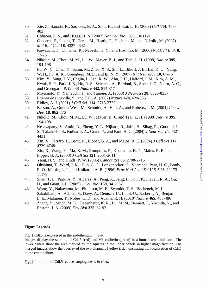
endothelial cell migration and angiogenesis,
suggesting Cdk5 as a novel target for
antiangiogenic therapy.
Inhibition or knockdown of Cdk5 reduce
endothelial cell motility and block
angiogenesis in vitro and in vivo. We
elucidate a specific signalling of Cdk5 in the
endothelium: in contrast to neuronal cells,
the motile defects upon inhibition of Cdk5
are not caused by an impaired function of
focal adhesions or microtubules, but by the
reduced formation of lamellipodia.
Inhibition or downregulation of Cdk5
decrease the activity of the small GTPase
Rac1 and result in a disorganized actin
cytoskeleton. Constitutive active Rac1
compensates for the inhibiting effects of
Cdk5 knockdown on migration, suggesting
that Cdk5 exerts its effects in endothelial
cell migration via Rac1.
Our work highlights Cdk5 as a pivotal new
regulator of endothelial cell migration and
angiogenesis. It suggests Cdk5 as a novel,
pharmacologically accessible target for
antiangiogenic therapy and provides the
basis for a new therapeutic application of
Cdk5 inhibitors as antiangiogenic agents.
Angiogenesis is involved in various
pathological conditions, including arthritis,
psoriasis, diabetic retinopathy, macula
degeneration, and cancer (1). During recent
years, the search for antiangiogenic
compounds and their molecular targets has
been intensified. Due to its key role in
angiogenesis, research initially focused on
vascular endothelial growth factor (VEGF).
VEGF receptor inhibitors such as the
monoclonal antibody bevacizumab (Avastin®)
as well as VEGF tyrosine kinase inhibitors
such as sunitinib (Sutent®) or sorafenib
(Nexavar®) have been approved for cancer
therapy. Unfortunately, the benefits of these
therapeutics are at best transitory and mostly
followed by a restoration of tumor growth and
progression (2). This resistance to anti-
angiogenic therapy causes a great need for new
targets to inhibit vessel growth, interfering
with steps in the angiogenic cascade different
from the response to a single growth factor.
Cyclin dependent kinase 5 (Cdk5) is a small
serine/threonine kinase belonging to the family
of Cdks. In contrast to the cell cycle-related
Cdks (e.g. Cdk1, 2, 4 or 6), Cdk5 is not
implicated in cell cycle control (3). Instead, it
is an important regulator of neuronal
development and controls various processes in
postmitotic neurons (4). Although it is
expressed ubiquitously, so far, only few reports
indicate a function of Cdk5 beyond the
nervous system. Scarcely anything is known
about a potential function of Cdk5 in the
vasculature and its exact functions and
signalling mechanisms in the endothelium
remain unknown (5-8).
Our aim was to close this gap of knowledge.
This is the first study that focuses on the
function of Cdk5 in the vascular system. It
demonstrates that Cdk5 regulates endothelial
cell migration and angiogenesis and provides
first information concerning the underlying
signalling.
1
http://www.jbc.org/cgi/doi/10.1074/jbc.M110.126177The latest version is at JBC Papers in Press. Published on September 7, 2010 as Manuscript M110.126177
Copyright 2010 by The American Society for Biochemistry and Molecular Biology, Inc.
by guest on May 4, 2018
http://ww
w.jbc.org/
Dow
nloaded from

Experimental Procedures
Cell culture- HUVECs were prepared by
digestion of umbilical veins with collagenase
A as described previously and cultured in
endothelial cell growth medium (ECGM,
Provitro, Berlin, Germany) (9). Umbilical
cords were collected from local hospitals in
accordance to the declaration of Helsinki.
Roscovitine was from Sigma-Aldrich.
Migration assay- Confluent HUVECs were
scratched with a pipette tip and treated as
indicated. After 16 h, cells were fixed with 3%
formaldehyde and images were taken using the
TILLvisON system (Lochham, Germany)
connected to an Axiovert 200 microscope
(Zeiss, Germany). Evaluation of pictures was
made by S.CO LifeScience (Garching,
Germany). Migration was quantified as the
ratio of the area covered with cells and the area
of the cell free wound. Experiments with the
proliferation inhibitor 5-hydroxyurea were
performed in order to exclude an influence of
antiproliferative effects in the scratch assay in
our setting.
Chemotaxis assay- Cells were seeded into µ-
slides chemotaxis (ibidi GmbH, Martinsried,
Germany). After 4 h, a FCS gradient from 0%
FCS to 10% FCS was generated, according to
the manufacturer’s protocol. Images were
obtained with a Zeiss LSM 510 META
confocal microscope and the appropriate LSM
software. The used objective was a Ph1-
NEOFLUAR 10x/0.30. A heating stage from
EMBLem (Heidelberg, Germany) was used to
keep cells at 37°C and 5% CO2. Images of
cells have been obtained for 20 h.
Tube formation assay- 1x10⁴
HUVECs in
ECGM containing roscovitine were seeded
onto Matrigel®-coated (Schubert & Weiss-
OMNILAB, Munich, Germany) ibidi
angiogenesis-slides (ibidi GmbH, Martinsried,
Germany). After 16 h, images were taken
using the TILLvisON system. Evaluation of
pictures was performed by S.CO LifeScience.
Tube length (displayed in red) was analyzed.
Chorioallantoic membrane (CAM) assay-
Fertilized White Leghorn chicken eggs
(Lohmann Tierzucht, Cuxhaven, Germany)
were incubated at 37°C for 72 h with constant
humidity. Eggs were transferred into dishes (ex
ovo), incubated for further 72 h and stimulated
with VEGF (1 ng per disk) alone or VEGF and
roscovitine (45 µg per disk) using small
cellulose disks. The next day, CAMs were
photographed using a stereomicroscope
(Olympus, Munich, Germany).
Mouse aortic ring assay- Mouse aortic rings
were embedded into Matrigel®. Once
endothelial cell sprouting occurred, rings were
either treated with ECGM or ECGM
containing roscovitine. After 72 h, images
were taken using the TILLvisON system.
Cornea micropocket assay- Both eyes of
C57BL/6J 8-week old female mice were
implanted with pellets containing bFGF
(80ng/pellet) as described previously (10-11).
Starting at the time of surgery, mice (20-21 g
body weight) were injected daily with either
DMSO (control) or with roscovitine for 5 days.
For each application, 200 µl of the solutions
were injected intraperitoneally and mice
received 100 mg per kg body weight
roscovitine each time. On postoperative day 6,
vascularization was quantified. Vessel length
(VL) and clock hours (CH) were measured and
the vascularized area (VA) was calculated as
1/2π*VL*0.4CH. The study complies with the
Guide for the Care and Use of Laboratory
Animals published by the US National
Institutes of Health (NIH Publication No. 85-
23, revised 1996), and with local regulations at
the University of Debrecen.
Transfection
Cdk2 and Cdk5 siRNA- 2x10⁶ HUVECs were
transfected with 3.0 µg ON-TARGETplus
Cdk2 siRNA (1.5 µg J-003236-1,
5’-PUAUUAGGAUGGUUAAGCUCUU-3’
and 1.5 µg J-003236-12,
5’-PUCUCCCGUCAACUUGUUUCUU-3’;
Dharmacon, Lafayette, CO, USA), or 3.0 µg
ON-TARGETplus Cdk5 siRNA (1.5 µg
J-003239-09,
5’-PACAUCGGAUAGGGCUUAUAUU-3’
and 1.5 µg J-003239-10,
5’-PGAUCUCAUGAGUCUCCCGGUU-3’),
respectively, by electroporation with the
NucleofectorTM
II (Amaxa, Cologne, Germany)
according to the manufacturer’s protocol. For
transfection control, HUVECs were transfected
with 3.0µg ON-TARGETplus Non-targeting
siRNA (nt siRNA) (D-001810-01,
5’-UGGUUUACAUGUCGACUAA-3’).
2
by guest on May 4, 2018
http://ww
w.jbc.org/
Dow
nloaded from

Silencing of Cdk2 and Cdk5 was examined by
Western blot analysis.
Cdk5 shRNA- Adenoviral transduction of
HUVECs with Cdk5 shRNA and non targeting
(nt) shRNA was performed by SIRION
BIOTECH GmbH (Martinsried, Germany).
Knockdown of Cdk5 was examined by
Western Blot analysis.
Rac V12- CoshRNA treated HUVECs or Cdk5
shRNA treated HUVECs were transfected with
2 µg of Rac V12 (A. Görlach, Munich,
Germany) or pcDNA3 (Molecular
Probes/Invitrogen, Karlsruhe, Germany). 12h
after transfection, scratch assays were
performed. Transfection efficiency was
examined by Western blot analysis.
Cdk5 overexpression- HUVECs were
transfected with 3 µg of wildtype Cdk5 (Cdk5-
wt) or a dominant negative Cdk5 mutant Cdk5-
D145N (Cdk5-dn, addgene, Cambridge, MA;
No 1871, 1873, S. v. d. Heuvel). pCMV-neo-
Bam (3 µg, addgene, No 16440, B. Vogelstein)
was used as a control. 24 h after transfection,
scratch assays ware performed. Transfection
efficiency was examined by Western blot
analysis.
CellTiter-Blue® cell viability assay-
Transfected cells were seeded into 96 well
plates (2x10⁴ cells per well) for 24 h. CellTiter-
Blue® assay was performed according to the
manufacturer’s protocol (Promega
Corporation, Madison, WI). Fluorescence was
measured at 560nm using a TECAN
SPECTRAFluor Plus Fluorescence,
Absorbance and Luminescence Reader (MTX
Lab Systems Inc, Crailsheim, Germany).
Western blot analysis- Western blot analysis
was performed as described previously (12).
Antibodies against Cdk5 were from Molecular
Probes/Invitrogen, Karlsruhe, Germany,
antibodies against FAK and FAK phospho-
Tyr397 were from Santa Cruz Biotechnology
(Santa Cruz, CA), antibodies against p27kip1
and FAK phospho-Ser732 were from
Biosource (Camarillo, CA), the antibody
against myc-tag was from Cell Signaling
(Danvers, MA, USA). Alexa Fluor 680 goat
anti mouse and Alexa Fluor 800 goat anti
rabbit were used as secondary antibodies
(Molecular Probes/Invitrogen, Karlsruhe,
Germany). For detection, an Odyssey infrared
imaging system (Li-Cor Biosciences, Lincoln,
NE) was used.
Adhesion assay- HUVECs were trypsinized,
suspended in ECGM or ECGM containing
roscovitine (30µM), and 1x10⁶ cells per well
were seeded in 24 well plates which were
either left uncoated or coated with fibronectin
(25 µg per ml), collagen (0.001% in PBS) or
Matrigel® (10% in serum free medium) After
30 min, cells were stained with crystal violet
and absorption was measured using the
TECAN SPECTRAFluor Plus Fluorescence,
Absorbance and Luminescence Reader.
Confocal laser scanning microscopy- Primary
antibodies against Rac1 (Millipore, Billerica,
MA) and cortactin (Cell Signaling Technology,
Denver, MA) were diluted 1:100 in PBS
containing 0.2% BSA. F-actin was stained with
rhodamine/phalloidin (1:400, R 415, Molecular
Probes/Invitrogen, Karlsruhe, Germany).
For life-cell imaging, cells were transfected
with the indicated plasmids and experiments
were started 24 h (eGFP-Cdk5) or 48 h (YFP-
Rac1, eGFP-CLIP-170, eYFP-vinculin) after
transfection, respectively. eGFP-CLIP-170 was
kindly provided by N. Galjart (Rotterdam,
Netherlands), eYFP-vinculin by A. Bershadsky
(Rehovot, Israel), eYFP-Rac1 was from
ATCC/Promochem (Wesel, Germany), and
eGFP-Cdk5 was from addgene (Cambridge,
MA; No 1346, L-H. Tsai).
Images were obtained with a Zeiss LSM 510
META confocal microscope and the
appropriate LSM software.
Tubulin fractionation- Cells were treated as
indicated for 4 h and lyzed using lysis-buffer
containing Pipes (100 mM), glycerol (2 M),
Triton X-100 (0.5%), MgCl2 (2 mM), EGTA
(2 mM), taxol (5 µM), GTP (guanosine
triphosphate, 1 mM), PMSF
(phenylmethylsulfonyl fluoride, 1 mM), and
Complete® (4%). Lysates were centrifuged
(45min, 47000rpm), and the supernatant was
mixed with 3x Laemmli sample buffer and
boiled for 5 min. The pellet was incubated with
40 µl of a buffer containing TRIS/HCl (100
mM), MgCl2 (1 mM), and CaCl2 (10 mM) at
4°C for 60 min, mixed with 3x Laemmli
samplebuffer and boiled for 5min. Western
blot analysis was performed using anti β-
tubulin antibody (Santa Cruz Biotechnology,
Santa Cruz, CA).
Pull down assay- Cells were treated as
indicated and plated for 30 min. Pull down
assays were performed according to the
3
by guest on May 4, 2018
http://ww
w.jbc.org/
Dow
nloaded from

manufacturer’s protocol (Rho Activation
Assay Kit 17-294 and Rac1 Activation Assay
Kit 17-441, both from Millipore, Billerica,
MA).
Statistical analysis- The number of
independently performed experiments and the
statistical tests used are stated in the respective
figure legend. Graph data represent means ±
SEM. Statistical analysis was performed with
the SigmaStat software Version 3.1 (Point
Richmond, CA, USA). Statistical significance
is assumed if p≤0.05.
Results
Cdk5 is expressed in the endothelium.
To demonstrate the presence of Cdk5 in the
endothelium in situ, human umbilical cords
were stained for Cdk5 (red) together with VE-
cadherin (green) which was used as endothelial
marker (Fig. 1). As expected, Cdk5, which is
expressed ubiquitously (13), localizes to all
tissues in the human umbilical cords. Merged
images clearly show the expression of Cdk5 in
the endothelium (overlay of Cdk5 and VE-
cadherin, yellow).
Inhibition of Cdk5 with roscovitine disrupts
angiogenesis.
The impact of inhibition of Cdk5 on
angiogenesis was examined by performing
various functional angiogenesis assays in vitro
and in vivo using the Cdk5 inhibitor
roscovitine.
Inhibition of Cdk5 by roscovitine significantly
reduced endothelial cell migration by 20% (10
µM) and 67% (30 µM), respectively (Fig. 2A,
upper panel). Possible false positive results due
to anti-proliferative effects have been excluded
by using 5-hydroxyurea (5-HU), an inhibitor of
proliferation. Treatment with 5-HU alone did
not influence wound closure, and the
migration-inhibiting effect of roscovitine was
not altered in the presence of 5-HU (Fig. 2A,
lower panel). Chemotaxis assays revealed that
untreated cells moved along a FCS gradient
(0% - 10%, Fig, 2B, upper left panel). Cells
treated with 10 µM roscovitine still moved
(indicated by an intact cumulative distance),
but did not follow the FCS gradient, suggesting
a loss of orientation (indicated by a reduced
euclidean distance). Roscovitine at a
concentration of 30 µM reduced both
cumulative and euclidean distance, reflecting a
completely defective motility (Fig. 2B, lower
panels). Applying tube formation assays,
untreated cells formed three-dimensional
structures. In contrast, tube formation of
roscovitine-treated cells was reduced
significantly after 16 h. Again, the
antiangiogenic effects of roscovitine are not
caused by an inhibition of proliferation. The
treatment with 5-HU (16 h) had no effect on
tube formation and roscovitine significantly
reduced tube length already after 4 h treatment
(Fig. 2C). Supplementary video 1 shows cells
seeded on Matrigel® for 16 h. Cells organize
themselves into tube-like structures by
changing their shape and establishing contacts
to neighboring cells, but they do not proliferate
significantly during tube formation.
Inhibition of Cdk5 impaired endothelial cell
sprouting out of aortic rings (Fig. 3A). By
applying chorioallantoic membrane (CAM)
assays, we found a strong induction of vessel
formation by vascular endothelial growth
factor (VEGF), which was completely blocked
upon inhibition of Cdk5 (Fig. 3B). The mouse
cornea micropocket assay demonstrated a
pronounced reduction of bFGF-induced
neovascularization in vivo (Fig. 3C) upon
intraperitoneal administration of roscovitine
(100 mg/kg/d).
Cdk5 is required for endothelial cell
migration.
In order to verify the function of Cdk5 in
angiogenesis, Cdk5 was specifically
downregulated using RNAi. Two different
approaches were used: Silencing of Cdk5 with
siRNA (nucleofection) and downregulation of
Cdk5 using shRNA (adenoviral transfer). In
scratch assays, Cdk5 siRNA reduced the
migration of HUVECs by 40%. In contrast, the
silencing of Cdk2, another prominent target of
roscovitine, showed no significant effect (Fig.
4A) (14). Successful downregulation of the
proteins was determined 24 h after transfection
and no compensatory reaction was observed
(Fig. 4B). The viability of cells transfected
with Cdk5 siRNA was not changed (CellTiter-
Blue® assay, Fig. 4C). Downregulation of
Cdk5 by shRNA resulted in a very similar
effect: performing scratch assays, cells treated
with Cdk5 shRNA showed a reduced migration
by 49% (Fig. 4D). Cdk5 shRNA also
significantly reduced the euclidean distance of
HUVECs in a chemotactic gradient (Fig. 4E)
suggesting a defective chemokinesis (loss of
4
by guest on May 4, 2018
http://ww
w.jbc.org/
Dow
nloaded from

orientation). Fig. 4F shows successful
knockdown of Cdk5 with shRNA.
To find out whether Cdk5 kinase activity is
required for endothelial cell migration, we
examined migration of HUVECs
overexpressing wild-type Cdk5 (Cdk5-wt) or a
kinase dead mutant (dominant-negative Cdk5-
D145N, Cdk5-dn). HUVECs overexpressing
the empty vector (pCMV-neo-Bam) served as
control. Overexpression of Cdk5dn reduced
migration by 46 %, an extent similar to RNAi
experiments. The overexpression of the
respective Cdk5 mutants was checked by
Western blot analysis (Fig. 4G).
Cdk5 does not influence cell adhesion and
microtubules.
Inhibition and silencing of Cdk5 reduced the
phosphorylation of focal adhesion kinase
(FAK) at Ser732. The autophosphorylation site
of FAK at tyrosine 397 was not affected (Fig.
5A, B). The functional impact of the inhibition
of Cdk5 on focal adhesion dynamics and
adhesion as well as microtubule organization
of HUVECs was analyzed. Focal adhesions
were visualized by overexpressing vinculin-
eYFP. Inhibition of Cdk5 neither changed
focal adhesion structure (maturation) nor
dynamics during cell spreading (Fig. 5C).
Furthermore, cell adhesion on various
substrates was not affected (Fig. 5D).
Moreover, no effect of Cdk5 inhibition on the
structure of the tubulin cytoskeleton in
migrating HUVECs was found (Fig. 5E). To
analyze microtubule dynamics, we
overexpressed CLIP-170-eGFP. Treatment
with roscovitine neither changed microtubule
dynamics (Fig. 5F) nor had any influence on
the polymerization of tubulin in contrast to
classical tubulin targeting compounds like
taxol, which stabilizes microtubules, or
vinblastine, which causes fragmentation of
microtubules (Fig. 5G).
Cdk5 influences the actin cytoskeleton.
The influence of Cdk5 on the actin
cytoskeleton in endothelial cells was examined
by analyzing F-actin distribution in migrating
cells. Untreated cells and cells treated with nt
siRNA formed lamellipodia with a densely
packed actin seam at the leading edge (Fig. 6A,
upper panels). Cells treated with roscovitine or
Cdk5 siRNA showed a reduced formation of
lamellipodia (Fig. 6A, lower panels). In
addition, the inhibition of Cdk5 reduced the
localization of cortactin, a lamellipodial
marker, to the leading edge of migrating cells
(Fig. 6B) (15).
Overexpression of Cdk5-eGFP in HUVECs
demonstrated the localization of Cdk5 to
lamellipodia during cell spreading (Fig. 6C,
left panel; supplementary video 2) and cell
migration (Fig. 6C, right panel; supplementary
video 3), suggesting a role of Cdk5 in the
formation of lamellipodia.
Cdk5 regulates the activity of RhoA and Rac1.
In pull down assays, inhibition or
downregulation of Cdk5 increased levels of
GTP-bound active RhoA (Fig. 7A, B),
accompanied by a decrease of p27kip1
protein
expression (Fig. 7C, D). Performing scratch
assays, pre-incubation with the Rho-associated
protein kinase (ROCK) inhibitor Y27632 (Y)
could not significantly compensate for the
inhibition of migration upon treatment with
roscovitine (Fig. 7E).
The activity of Rac1 was dramatically
decreased upon inhibition and knockdown of
Cdk5 (Fig. 8A, B). Furthermore, inhibition or
downregulation of Cdk5 abolished the
localization of Rac1 and its effector cortactin
to the leading edge of migrating cells (Fig. 8C,
D). Performing life-cell imaging with cells
overexpressing Rac1-eYFP, untreated cells
showed a regular cell shape with Rac1
localized to the cell membrane (Fig. 8E, upper
panel; supplementary video 4). In cells treated
with roscovitine, Rac1 was distributed over the
whole cell. This was accompanied by an
irregular cell shape and an irregular formation
of ruffles (Fig. 8E, lower panel; supplementary
video 5). Overexpression of a constitutively
active Rac mutant (Rac V12) compensated for
the inhibition of HUVEC migration upon
downregulation of Cdk5 by shRNA (Fig. 8F).
Discussion
In the present study, we demonstrate a crucial
role of Cdk5 in the regulation of endothelial
cell migration and angiogenesis. We identify
the Cdk5 inhibitor roscovitine as
antiangiogenic compound and propose Cdk5 as
the target of roscovitine responsible for its
antiangiogenic effects. We provide
informations concerning the signalling of
endothelial Cdk5 suggesting that Cdk5
regulates endothelial cell migration via the
small GTPase Rac1.
5
by guest on May 4, 2018
http://ww
w.jbc.org/
Dow
nloaded from
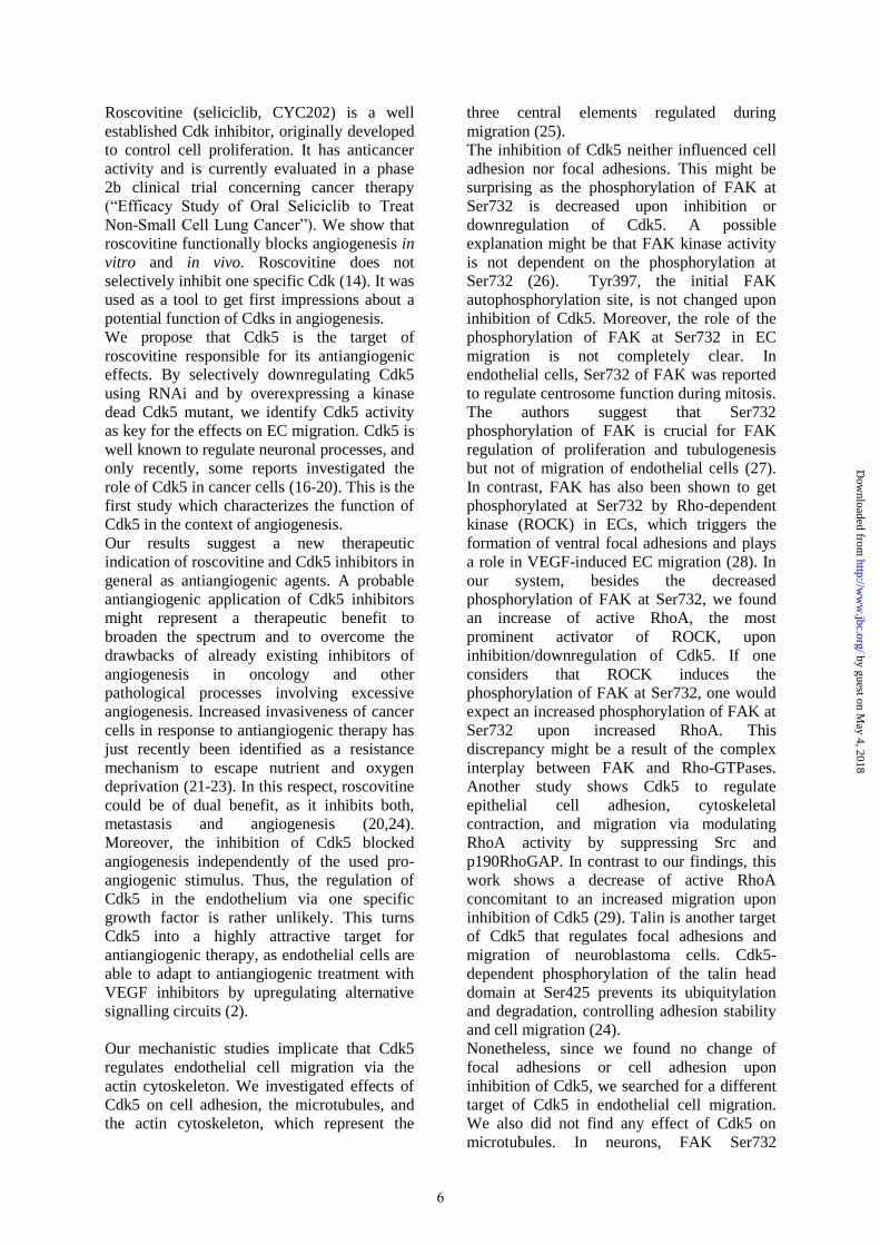
Roscovitine (seliciclib, CYC202) is a well
established Cdk inhibitor, originally developed
to control cell proliferation. It has anticancer
activity and is currently evaluated in a phase
2b clinical trial concerning cancer therapy
(“Efficacy Study of Oral Seliciclib to Treat
Non-Small Cell Lung Cancer”). We show that
roscovitine functionally blocks angiogenesis in
vitro and in vivo. Roscovitine does not
selectively inhibit one specific Cdk (14). It was
used as a tool to get first impressions about a
potential function of Cdks in angiogenesis.
We propose that Cdk5 is the target of
roscovitine responsible for its antiangiogenic
effects. By selectively downregulating Cdk5
using RNAi and by overexpressing a kinase
dead Cdk5 mutant, we identify Cdk5 activity
as key for the effects on EC migration. Cdk5 is
well known to regulate neuronal processes, and
only recently, some reports investigated the
role of Cdk5 in cancer cells (16-20). This is the
first study which characterizes the function of
Cdk5 in the context of angiogenesis.
Our results suggest a new therapeutic
indication of roscovitine and Cdk5 inhibitors in
general as antiangiogenic agents. A probable
antiangiogenic application of Cdk5 inhibitors
might represent a therapeutic benefit to
broaden the spectrum and to overcome the
drawbacks of already existing inhibitors of
angiogenesis in oncology and other
pathological processes involving excessive
angiogenesis. Increased invasiveness of cancer
cells in response to antiangiogenic therapy has
just recently been identified as a resistance
mechanism to escape nutrient and oxygen
deprivation (21-23). In this respect, roscovitine
could be of dual benefit, as it inhibits both,
metastasis and angiogenesis (20,24).
Moreover, the inhibition of Cdk5 blocked
angiogenesis independently of the used pro-
angiogenic stimulus. Thus, the regulation of
Cdk5 in the endothelium via one specific
growth factor is rather unlikely. This turns
Cdk5 into a highly attractive target for
antiangiogenic therapy, as endothelial cells are
able to adapt to antiangiogenic treatment with
VEGF inhibitors by upregulating alternative
signalling circuits (2).
Our mechanistic studies implicate that Cdk5
regulates endothelial cell migration via the
actin cytoskeleton. We investigated effects of
Cdk5 on cell adhesion, the microtubules, and
the actin cytoskeleton, which represent the
three central elements regulated during
migration (25).
The inhibition of Cdk5 neither influenced cell
adhesion nor focal adhesions. This might be
surprising as the phosphorylation of FAK at
Ser732 is decreased upon inhibition or
downregulation of Cdk5. A possible
explanation might be that FAK kinase activity
is not dependent on the phosphorylation at
Ser732 (26). Tyr397, the initial FAK
autophosphorylation site, is not changed upon
inhibition of Cdk5. Moreover, the role of the
phosphorylation of FAK at Ser732 in EC
migration is not completely clear. In
endothelial cells, Ser732 of FAK was reported
to regulate centrosome function during mitosis.
The authors suggest that Ser732
phosphorylation of FAK is crucial for FAK
regulation of proliferation and tubulogenesis
but not of migration of endothelial cells (27).
In contrast, FAK has also been shown to get
phosphorylated at Ser732 by Rho-dependent
kinase (ROCK) in ECs, which triggers the
formation of ventral focal adhesions and plays
a role in VEGF-induced EC migration (28). In
our system, besides the decreased
phosphorylation of FAK at Ser732, we found
an increase of active RhoA, the most
prominent activator of ROCK, upon
inhibition/downregulation of Cdk5. If one
considers that ROCK induces the
phosphorylation of FAK at Ser732, one would
expect an increased phosphorylation of FAK at
Ser732 upon increased RhoA. This
discrepancy might be a result of the complex
interplay between FAK and Rho-GTPases.
Another study shows Cdk5 to regulate
epithelial cell adhesion, cytoskeletal
contraction, and migration via modulating
RhoA activity by suppressing Src and
p190RhoGAP. In contrast to our findings, this
work shows a decrease of active RhoA
concomitant to an increased migration upon
inhibition of Cdk5 (29). Talin is another target
of Cdk5 that regulates focal adhesions and
migration of neuroblastoma cells. Cdk5-
dependent phosphorylation of the talin head
domain at Ser425 prevents its ubiquitylation
and degradation, controlling adhesion stability
and cell migration (24).
Nonetheless, since we found no change of
focal adhesions or cell adhesion upon
inhibition of Cdk5, we searched for a different
target of Cdk5 in endothelial cell migration.
We also did not find any effect of Cdk5 on
microtubules. In neurons, FAK Ser732
6
by guest on May 4, 2018
http://ww
w.jbc.org/
Dow
nloaded from
phosphorylation has previously been shown to
influence microtubule organization, nuclear
movement and neuronal migration (30). Thus,
we propose a specific signalling of Cdk5 in the
vasculature.
We identify the actin cytoskeleton to be the
relevant target of Cdk5 in endothelial cell
migration. Endothelial Cdk5 regulates the
formation of lamellipodia, actin-based
structures that are essential for cell migration
(31). Our findings are in line with reports
concerning the function of Cdk5 in neurons.
Neuronal Cdk5 affects the actin cytoskeleton
by phosphorylating p27kip
, PAK1, as well as
Neurabin-I (32-34), it controls dendritic spine
morphology via phosphorylation of the RhoA
guanine-nucleotide exchange factor (GEF)
ephexin1 (35), and it phosphorylates the actin-
binding proteins WAVE1 and WAVE2 (36-
37).
The small Rho GTPases RhoA and Rac1
represent the most prominent regulators of the
actin cytoskeleton (38-39). We found that
inhibition of Cdk5 increased RhoA activity,
which was concomitant to a decrease of p27kip1
protein. Also neuronal Cdk5 has been shown to
phosphorylate and stabilize p27kip1
, leading to a
reduction of active RhoA. Subsequently, an
increase of activated cofilin was shown,
promoting actin turnover and cell migration
(33). The migration defect of p27kip1
–null
fibroblasts is rescued by the inhibition of
ROCK, the most prominent downstream
effector of RhoA (40). In endothelial cells, in
contrast, blockade of ROCK could not
compensate for the anti-migratory effect of
roscovitine. Thus, although Cdk5 indeed
modulates RhoA activity, it seems not to
regulate endothelial cell migration primarily
via the p27kip1
– RhoA – ROCK pathway. In
the context of epithelial cell migration, Cdk5
was described as a regulator of RhoA activity
and cytoskeletal contraction. In contrast to our
findings, in epithelial cells, inhibition of Cdk5
blocks RhoA-ROCK signalling and increases
migration (29). The discrepancies concerning
the different effects of Cdk5 on the activity of
RhoA and the distinct functional consequences
might be due to the diverse cell types and
suggest a specific signalling of Cdk5 in the
endothelium.
Our data indicate Rac1 to be the most likely
link between Cdk5 function and endothelial
cell migration. We show that inhibition or
downregulation of Cdk5 dramatically reduces
Rac1 activity and impairs the localization of
Rac1 to the cell membrane. Constitutively
active Rac1 compensates for the migration-
inhibiting effect of the knockdown of Cdk5.
Thus, we propose that Cdk5 exerts its effects
in endothelial cell migration via Rac1. This can
be interpreted contrariwise to the report of
Nikolic et al., where neuronal Cdk5 has been
elucidated as a downstream effector of
Rac1.(41) A different and much more
interesting explanation might be a feedback
loop between Cdk5 and Rac1. As a kinase,
Cdk5 might regulate Rac1 by the
phosphorylation of a guanine nucleotide
exchange factor (GEF), a GTPase-activating
protein (GAP), or a GDP-dissociation-inhibitor
(GDI) for Rac1. Neuronal Cdk5 was shown to
phosphorylate various regulators of Rac1.
Cdk5-mediated phosphorylation of RasGRF2
downregulates the activity of Rac1, the
regulation of Trio and Kalirin by Cdk5
activates Rac1 (42-44). By phosphorylating
ezrin, Cdk5 was shown to modulate RhoGDI,
inhibiting Rac1(45). The fact that Cdk5 seems
to be able both to increase and to decrease the
activity of Rac1 and, in contrary, to be
regulated by Rac1 suggests that the function of
Cdk5 strongly depends on the cellular system
and/or on the functional context, respectively.
Our work highlights a vascular function of
Cdk5. Consequently, one would expect a
vascular phenotype of Cdk5 knockout mice.
Unfortunately, vascular or cardiac defects of
these mice have not been investigated in the
original publication. According to the crucial
function of Cdk5 in the nervous system, Cdk5
knockout mice show abnormal corticogenesis,
neuronal defects, and die perinatally (46). Mice
exhibiting vascular phenotypes often die
during embryogenesis (47-48) or show
neonatal lethality (49), depending on the
severity of the phenotype. Thus, it might well
be that the lethality in the Cdk5 knockout mice
is partly due to vascular defects. To make use
of endothelial specific Cdk5 knockout mice
might provide the opportunity to study the
function of Cdk5 in the endothelium in vivo.
In conclusion, this study for the first time
presents a novel and crucial function of Cdk5
in endothelial cell migration, it elucidates a
specific signalling of endothelial Cdk5 and it
highlights Cdk5 as a promising target for
antiangiogenic therapy.
7
by guest on May 4, 2018
http://ww
w.jbc.org/
Dow
nloaded from

References
1. Tonini, T., Rossi, F., and Claudio, P. P. (2003) Oncogene 22, 6549-6556
2. Bergers, G., and Hanahan, D. (2008) Nat.Rev.Cancer 8, 592-603
3. Vermeulen, K., Van Bockstaele, D. R., and Berneman, Z. N. (2003) Cell Prolif. 36,
131-149
4. Dhavan, R., and Tsai, L. H. (2001) Nat.Rev.Mol.Cell Biol. 2, 749-759
5. Maggiorella, L., Aubel, C., Haton, C., Milliat, F., Connault, E., Opolon, P., Deutsch,
E., and Bourhis, J. (2009) Cell Prolif 42, 38-48
6. Mitsios, N., Pennucci, R., Krupinski, J., Sanfeliu, C., Gaffney, J., Kumar, P., Kumar,
S., Juan-Babot, O., and Slevin, M. (2007) Brain Pathol 17, 11-23
7. Sharma, P., Sharma, M., Amin, N. D., Albers, R. W., and Pant, H. C. (1999) Proc Natl
Acad Sci U S A 96, 11156-11160
8. Slevin, M., and Krupinski, J. (2009) Curr Opin Pharmacol 9, 119-124
9. Kiemer, A. K., Weber, N. C., Furst, R., Bildner, N., Kulhanek-Heinze, S., and
Vollmar, A. M. (2002) Circ.Res. 90, 874-881
10. Kenyon, B. M., Voest, E. E., Chen, C. C., Flynn, E., Folkman, J., and D'Amato, R. J.
(1996) Invest Ophthalmol.Vis.Sci. 37, 1625-1632
11. Licht, T., Tsirulnikov, L., Reuveni, H., Yarnitzky, T., and Ben-Sasson, S. A. (2003)
Blood 102, 2099-2107
12. Koltermann, A., Hartkorn, A., Koch, E., Furst, R., Vollmar, A. M., and Zahler, S.
(2007) Cell Mol.Life Sci. 64, 1715-1722
13. Dhavan, R., and Tsai, L. H. (2001) Nat Rev Mol Cell Biol 2, 749-759
14. Meijer, L., Borgne, A., Mulner, O., Chong, J. P., Blow, J. J., Inagaki, N., Inagaki, M.,
Delcros, J. G., and Moulinoux, J. P. (1997) Eur J Biochem 243, 527-536
15. Weed, S. A., and Parsons, J. T. (2001) Oncogene 20, 6418-6434
16. Goodyear, S., and Sharma, M. C. (2007) Exp Mol Pathol 82, 25-32
17. Lin, H., Chen, M. C., Chiu, C. Y., Song, Y. M., and Lin, S. Y. (2007) J Biol Chem
282, 2776-2784
18. Liu, R., Tian, B., Gearing, M., Hunter, S., Ye, K., and Mao, Z. (2008) Proc Natl Acad
Sci U S A 105, 7570-7575
19. Sandal, T., Stapnes, C., Kleivdal, H., Hedin, L., and Doskeland, S. O. (2002) J Biol
Chem 277, 20783-20793
20. Strock, C. J., Park, J. I., Nakakura, E. K., Bova, G. S., Isaacs, J. T., Ball, D. W., and
Nelkin, B. D. (2006) Cancer Res 66, 7509-7515
21. Ebos, J. M., Lee, C. R., Cruz-Munoz, W., Bjarnason, G. A., Christensen, J. G., and
Kerbel, R. S. (2009) Cancer Cell 15, 232-239
22. Loges, S., Mazzone, M., Hohensinner, P., and Carmeliet, P. (2009) Cancer Cell 15,
167-170
23. Paez-Ribes, M., Allen, E., Hudock, J., Takeda, T., Okuyama, H., Vinals, F., Inoue, M.,
Bergers, G., Hanahan, D., and Casanovas, O. (2009) Cancer Cell 15, 220-231
24. Huang, C., Rajfur, Z., Yousefi, N., Chen, Z., Jacobson, K., and Ginsberg, M. H.
(2009) Nat Cell Biol 11, 624-630
25. Wehrle-Haller, B., and Imhof, B. A. (2003) Int J Biochem Cell Biol 35, 39-50
26. Xie, Z., Sanada, K., Samuels, B. A., Shih, H., and Tsai, L. H. (2003) Cell 114, 469-
482
27. Park, A. Y., Shen, T. L., Chien, S., and Guan, J. L. (2009) J Biol Chem 284, 9418-
9425
28. Le, B. F., Houle, F., Sussman, M., and Huot, J. (2006) Mol.Biol.Cell 17, 3508-3520
29. Tripathi, B. K., and Zelenka, P. S. (2009) Mol Cell Biol 29, 6488-6499
8
by guest on May 4, 2018
http://ww
w.jbc.org/
Dow
nloaded from
30. Xie, Z., Sanada, K., Samuels, B. A., Shih, H., and Tsai, L. H. (2003) Cell 114, 469-
482
31. Chhabra, E. S., and Higgs, H. N. (2007) Nat.Cell Biol. 9, 1110-1121
32. Causeret, F., Jacobs, T., Terao, M., Heath, O., Hoshino, M., and Nikolic, M. (2007)
Mol.Biol.Cell 18, 4327-4342
33. Kawauchi, T., Chihama, K., Nabeshima, Y., and Hoshino, M. (2006) Nat.Cell Biol. 8,
17-26
34. Nikolic, M., Chou, M. M., Lu, W., Mayer, B. J., and Tsai, L. H. (1998) Nature 395,
194-198
35. Fu, W. Y., Chen, Y., Sahin, M., Zhao, X. S., Shi, L., Bikoff, J. B., Lai, K. O., Yung,
W. H., Fu, A. K., Greenberg, M. E., and Ip, N. Y. (2007) Nat.Neurosci. 10, 67-76
36. Kim, Y., Sung, J. Y., Ceglia, I., Lee, K. W., Ahn, J. H., Halford, J. M., Kim, A. M.,
Kwak, S. P., Park, J. B., Ho, R. S., Schenck, A., Bardoni, B., Scott, J. D., Nairn, A. C.,
and Greengard, P. (2006) Nature 442, 814-817
37. Miyamoto, Y., Yamauchi, J., and Tanoue, A. (2008) J Neurosci 28, 8326-8337
38. Etienne-Manneville, S., and Hall, A. (2002) Nature 420, 629-635
39. Ridley, A. J. (2001) J.Cell Sci. 114, 2713-2722
40. Besson, A., Gurian-West, M., Schmidt, A., Hall, A., and Roberts, J. M. (2004) Genes
Dev. 18, 862-876
41. Nikolic, M., Chou, M. M., Lu, W., Mayer, B. J., and Tsai, L. H. (1998) Nature 395,
194-198
42. Kesavapany, S., Amin, N., Zheng, Y. L., Nijhara, R., Jaffe, H., Sihag, R., Gutkind, J.
S., Takahashi, S., Kulkarni, A., Grant, P., and Pant, H. C. (2004) J Neurosci 24, 4421-
4431
43. Xin, X., Ferraro, F., Back, N., Eipper, B. A., and Mains, R. E. (2004) J Cell Sci 117,
4739-4748
44. Xin, X., Wang, Y., Ma, X. M., Rompolas, P., Keutmann, H. T., Mains, R. E., and
Eipper, B. A. (2008) J Cell Sci 121, 2601-2611
45. Yang, H. S., and Hinds, P. W. (2006) Cancer Res 66, 2708-2715
46. Ohshima, T., Ward, J. M., Huh, C. G., Longenecker, G., Veeranna, Pant, H. C., Brady,
R. O., Martin, L. J., and Kulkarni, A. B. (1996) Proc Natl Acad Sci U S A 93, 11173-
11178
47. Shen, T. L., Park, A. Y., Alcaraz, A., Peng, X., Jang, I., Koni, P., Flavell, R. A., Gu,
H., and Guan, J. L. (2005) J Cell Biol 169, 941-952
48. Wang, Y., Nakayama, M., Pitulescu, M. E., Schmidt, T. S., Bochenek, M. L.,
Sakakibara, A., Adams, S., Davy, A., Deutsch, U., Luthi, U., Barberis, A., Benjamin,
L. E., Makinen, T., Nobes, C. D., and Adams, R. H. (2010) Nature 465, 483-486
49. Zhang, Y., Singh, M. K., Degenhardt, K. R., Lu, M. M., Bennett, J., Yoshida, Y., and
Epstein, J. A. (2009) Dev Biol 325, 82-93
Figure Legends
Fig. 1 Cdk5 is expressed in the endothelium in vivo.
Images display the staining of Cdk5 (red) and VE-cadherin (green) in a human umbilical cord. The
lower panels show the area marked by the squares in the upper panels in higher magnification. The
merged images show the overlay of the two channels (yellow), demonstrating the localization of Cdk5
to the endothelium.
Fig. 2 Inhibition of Cdk5 reduces angiogenesis in vitro.
9
by guest on May 4, 2018
http://ww
w.jbc.org/
Dow
nloaded from

(A) Inhibition of Cdk5 by roscovitine (rosc) decreases endothelial cell migration (Kruskal-Wallis One
Way ANOVA on Ranks, * p < 0.05, n = 4; upper panels). Treatment with 5-hydroxyurea (5-HU, 2
mM) alone had no effect on HUVEC migration and did not alter the migration-inhibiting effect of
roscovitine (t-test, * p < 0.001; n = 5; lower panel). (B) Inhibition of Cdk5 disturbs chemotaxis of
HUVECs. Untreated cells (co) move along the FCS gradient (0% - 10%, upper left panel). Roscovitine
(rosc) at 10 µM inhibits cell orientation (upper middle panel) and reduces cell motility at 30 µM
(upper right panel), respectively. Cells migrating in direction of the FCS are shown in black, cells
migrating in other directions in red (One Way ANOVA/Dunnett, * p < 0.05, n = 3). (C) Inhibition of
Cdk5 reduces endothelial tube formation. The images in the upper panel show tube formation of cells
with or without roscovitine (rosc, 16 h). Tube structures identified by the software are displayed in
red, quantitative evaluations are displayed in the lower panels. Roscovitine (rosc, 16 h) reduces tube
length, treatment with 5-hydroxyurea (5-HU, 2 mM, 16 h) had no effect. Tube formation is reduced
upon inhibition of Cdk5 after 4 h, excluding effects of roscovitine on cell proliferation. (roscovitine,
16 h: One Way ANOVA/Dunnett, * p < 0.05, n = 3; 5-HU, 16 h: Kruskal-Wallis One Way ANOVA
on Ranks, * p < 0.05, n = 4, roscovitine 4 h: One Way ANOVA/Dunnett, * p < 0.05, n = 4).
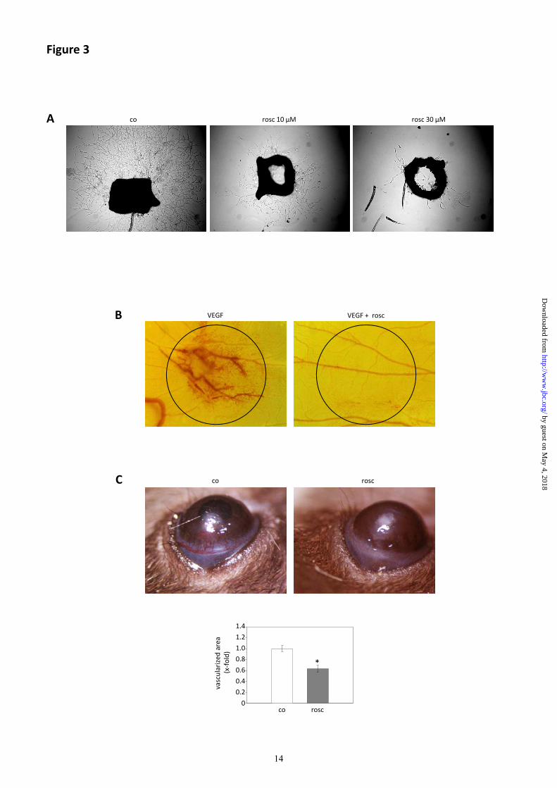
Fig. 3 Inhibition of Cdk5 reduces angiogenesis ex vivo and in vivo.
(A) Inhibition of Cdk5 with roscovitine (rosc) inhibits endothelial cell sprouting from mouse aortic
rings. (B) Inhibition of Cdk5 abolishes VEGF-induced vessel formation in the CAM assay. Circles
represent localization of cellulose disks containing VEGF (1 ng/disk) or VEGF combined with
roscovitine (rosc, 45 µg/disk) (n=3). (C) Inhibition of Cdk5 reduces bFGF-induced neovascularization
in the mouse cornea micropocket assay. The growth of blood vessels into the pellet containing bFGF
in mice treated with solvent (DMSO, co) is shown in the upper left panel. The upper right panel
indicates one eye of a mouse injected intraperitoneally with roscovitine (rosc, 100 mg/kg/d). The
graph (lower panel) represents the quantitative evaluation of the vascularized area. (Student’s t-test
corrected for unequal variances, p = 0.00018, n = 10 for control, n = 16 for roscovitine).
Fig. 4 Cdk5 is implicated in the regulation of endothelial cell migration.
(A) Silencing of Cdk5 with siRNA (Cdk5 siRNA) reduces endothelial cell migration, Cdk2 siRNA has
no influence, non-targeting siRNA (nt siRNA) serves as control (One Way ANOVA/Dunnett, * p <
0.05, n = 5). (B) Cdk2 and Cdk5 are successfully downregulated 24 h after treatment with siRNA, nt
siRNA serves as control, β-actin indicates equal loading. (C) Cdk5 siRNA does not influence cell
viability (t-test, p = 0,974, n = 4). (D) Endothelial cell migration is reduced upon downregulation of
Cdk5 with shRNA; non-targeting shRNA (nt shRNA) serves as control (t-test, * p < 0,001, n = 5) (E)
Knockdown of Cdk5 inhibits endothelial cell chemokinesis. Cells treated with nt shRNA move along
the FCS gradient (0% - 10%, left panel). Cdk5 shRNA inhibits cell orientation (right panel). Cells
migrating in direction of FCS are shown in black, cells migrating in other directions in red. The
quantitative evaluation of cumulative and euclidean distances is displayed in the lower panels (t-test, *
p < 0.05, n = 4). (F) The Western blot indicates successful knockdown of Cdk5 by shRNA, nt siRNA
serves as control, β-actin indicates equal loading. (G) Kinase activity of Cdk5 is required for
endothelial cell migration. Scratch assays of HUVECs overexpressing wild-type Cdk5 (Cdk5-wt) or
dominant-negative Cdk5 (Cdk5-dn) are shown. Cells transfected with pCMV-neo-Bam (empty vector)
were used as control. Dominant-negative Cdk5 significantly decreases HUVEC migration (One Way
ANOVA/Holm-Sidak, * p < 0.05, n=4).
Fig. 5 Cdk5 does not influence cell adhesion and microtubules.
(A) Roscovitine decreases the phosphorylation of FAK at Ser732, the phosphorylation of FAK at
Tyr397 is not influenced. (B) Cdk5 siRNA reduces the phosphorylation of FAK specifically at Ser732.
(C) Cdk5 inhibition does not influence the size, localization, and dynamics of focal adhesions. (D)
Roscovitine (rosc, 30 µM) does not influence the adhesion of cells on differentially coated surfaces (t-
test, n = 3). (E) Microtubule structure is not changed by Cdk5 inhibition. (F) Cdk5 inhibition does not
influence the dynamics of microtubules. Representative images show HUVECs expressing CLIP170-
eGFP during migration in the absence (co) or presence of roscovitine (rosc, 30 µM) (n = 2). (G)
roscovitine (rosc, 30 µM) does not influence the polymerization of tubulin. Taxol (T) and vinblastine
(VB) show the expected stabilization or fragmentation of microtubules, respectively. Fractions of
polymerized tubulin are shown (n = 3).
10
by guest on May 4, 2018
http://ww
w.jbc.org/
Dow
nloaded from
Fig. 6 Cdk5 regulates the formation of lamellipodia.
(A, B) Inhibition of Cdk5 using roscovitine (rosc, 30 µM) or Cdk5 siRNA reduces the formation of
lamellipodia during migration. (A) Migrating HUVECs stained for f-actin (n = 3). (B) Migrating cells
stained with anti-cortactin antibodies (n = 3). (C) Cdk5 localizes to lamellipodia. Images represent
HUVECs overexpressing Cdk5-eGFP during spreading (left panel) and migration (right panel),
respectively (each n = 3).
Fig. 7 Cdk5 influences the p27kip
/RhoA pathway.
(A) Inhibition of Cdk5 increases the amount of active GTP-bound RhoA (RhoA-GTP). Total RhoA
serves as loading control (n = 3). (B) Knockdown of Cdk5 with shRNA (Cdk5 shRNA) increases the
amount of RhoA-GTP in comparison to cells treated with non-targeting shRNA (nt shRNA) Total
RhoA serves as loading control (n = 3). (C) Inhibition of Cdk5 decrease p27kip1
protein expression.
Cells were either left untreated or were treated with roscovitine (rosc, 30µM, n = 3) (D) p27kip1
expression of HUVECs transfected with Cdk5 siRNA or with nt siRNA, respectively, is shown (n =
3). (E) Preincubation with Y27632 (Y, 10 µM) does not significantly abolish the effect of roscovitine
(rosc, 10 µM and 30 µM) on HUVEC migration. Representative images show migrating cells treated
as indicated. The bar graph shows the quantitative evaluation (One Way ANOVA/Holm-Sidak, n = 3).
Fig. 8 Cdk5 influences Rac1.
(A) Inhibition of Cdk5 with roscovitine (rosc, 30 µM) reduces the amount of active GTP-bound Rac1.
Total Rac1 serves as loading control (n = 3). (B) Knockdown of Cdk5 with shRNA (Cdk5 shRNA)
decreases the amount of Rac1-GTP in comparison to cells treated with non-targeting shRNA (nt
shRNA). Total Rac1 serves as loading control (n = 3). (C, D) Roscovitine (rosc, 30 µM) as well as
Cdk5 siRNA inhibit localization of Rac1 (green) and cortactin (red) to lamellipodia of migrating
endothelial cells. Untreated cells (co) or nt siRNA treated cells serve as the respective controls (n = 3).
(E) During cell spreading, inhibition of Cdk5 abrogates the localization of Rac1 to the cell periphery.
Representative images display cells overexpressing Rac1-eYFP during spreading untreated or treated
with roscovitine (rosc, 30 µM), respectively (n = 3). (F) Overexpression of constitutively active Rac1
(Rac V12) compensates for the migration-inhibiting effect of Cdk5 knockdown. Upper panels: in
scratch assays, knockdown of Cdk5 with shRNA significantly reduces migration of cells treated with
the empty vector (pcDNA3). In HUVECs overexpressing constitutive active Rac1 (Rac V12),
treatment with Cdk5 shRNA does not reduce migration. The bar graphs display the quantitative
evaluation (pcDNA3: Rank Sum Test, * p < 0.05, n = 5; Rac V12: Rank Sum Test; n = 5). Lower
panels: the Western blots show the knockdown of Cdk5 by shRNA and overexpression of myc-tagged
Rac V12.
11
by guest on May 4, 2018
http://ww
w.jbc.org/
Dow
nloaded from
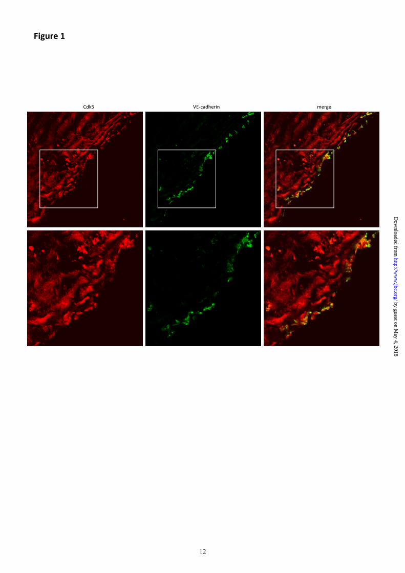
Figure 1
Cdk5 VE‐cadherin merge
12
by guest on May 4, 2018
http://ww
w.jbc.org/
Dow
nloaded from

*
*
tion(x‐fold)
0.6
0.8
1.0
1.2A rosc 30 µMrosc 10 µMco
Figure 2
fold)
1.0
1.2
*
migrat
co rosc10 µM
rosc30 µM
0
0.2
0.4
5‐HU + rosc5‐HU
*
migratio
n(x‐f
5‐HU 5‐HU + rosc
0
0.2
0.4
0.6
0.8
B
FCS
10%
0
100
200
‐100
‐200
Y‐axis [u
nits]
rosc 30 µM
0
100
200
‐100
‐200
Y‐axis [u
nits]
rosc 10 µM
0
100
200
‐100
‐200
Y‐axis [u
nits]
control
mulativedistance
(x‐fold) *
0.4
0.6
0.8
1.0
1.2
**
ideandistance
(x‐fold)
0.4
0.6
0.8
1.0
1.2
0% ‐200 ‐100 0 100
X‐axis [units]
200‐200 ‐100 0 100
X‐axis [units]
200‐200 ‐100 0 100
X‐axis [units]
200
cum
co rosc10 µM
rosc30 µM
0
0.2 eucl
co rosc10 µM
rosc30 µM
0
0.2
C rosc 30 µMrosc 10 µMco
d) 1 2
1.4
d) 1 2
1.4
d) 1 2
1.44 h16 h16 h
tube
length
(x‐fold
n.s.
0
0.2
0.4
0.6
0.8
1.0
1.2
co 5‐HU
tube
length
(x‐fold
*
0
0.2
0.4
0.6
0.8
1.0
1.2
co rosc10 µM
rosc30 µM
tube
length
(x‐fold
*
0
0.2
0.4
0.6
0.8
1.0
1.2
co rosc10 µM
rosc30 µM
13
by guest on May 4, 2018
http://ww
w.jbc.org/
Dow
nloaded from
A rosc 30 µMrosc 10 µMco
Figure 3
A rosc 30 µMrosc 10 µMco
VEGF VEGF + roscB
co roscC
rizedarea
‐fold)
0 6
0.8
1.0
1.2
1.4
*
vascula (x‐
0
0.2
0.4
0.6
co rosc
14
by guest on May 4, 2018
http://ww
w.jbc.org/
Dow
nloaded from

A Cdk5 siRNACdk2 siRNAnt siRNA
Figure 4
0.6
0.8
1.0
1.2
*
ion(x‐fold)
Cdk5
Cdk2
B
0 6
0.8
1.0
1.2
1.4
ability
dcontrol)
C0
0.2
0.4
migrat
ntsiRNA
Cdk2siRNA
Cdk5siRNA
Cdk2
β‐actin
nt siRNA
Cdk2siRNA
Cdk5siRNA
D1.0
ol)
1.2
0
0.2
0.4
0.6
via
(x‐fol
nt siRNA
Cdk5siRNA
Cdk5 shRNAnt shRNA
200nt shRNA Cdk5 shRNA
20010%
0
0.2
0.4
0.6
0.8
*migratio
n(x‐foldcontr o
ntshRNA
Cdk5shRNAE
200
0
100
‐100
‐200‐200 ‐100 0 100
Y‐axis [u
nits]
X‐axis [units]
0
100
‐100
‐200‐200 ‐100 0 100 200
Y‐axis[units]
X‐axis [units]
FCS
0% Cdk5
β actin
F
0
0.2
0.4
0.6
0.8
1.0
1.2
*
Euclidean distance
(x‐fold control)
nt Cdk50
0.2
0.4
0.6
0.8
1.0
1.2
Cumulative distance
(x‐fold control)
nt Cdk5
ntshRNA
Cdk5 shRNA
β‐actin
shRNA shRNAshRNA shRNA
Cdk5‐dnCdk5‐wtcoG
**
0.60.81.0
igratio
n(x‐fold)
1.21.4
1.81.6
co Cdk5‐wt
Cdk5
β‐actin
Cdk5‐dn
co Cdk5‐wt
Cdk5‐dn
00.20.4m
i
15
by guest on May 4, 2018
http://ww
w.jbc.org/
Dow
nloaded from

FAK pSer732
BAFAK pSer732
Figure 5
FAK
FAK pTyr397
ntsiRNA
Cdk5siRNA
0 5 15 30 45 60 rosc [min]
FAK
FAK pTyr397
vinculin‐eYFPC
1.2
1.4ld)
Dcorosc
1.6
co0
0.2
0.4
0.6
0.8
1.0
adhe
sion
(x‐fol
uncoated fibronectin collagen matrigel
rosc
β‐tubulinE F Clip170‐eGFP
co
migratio
n
co
migratio
n
T VB co rosc
β‐tubulin
β‐actin
G
rosc rosc
16
by guest on May 4, 2018
http://ww
w.jbc.org/
Dow
nloaded from

A F‐actin B cortactin
Figure 6
co
migratio
n
nt siRNA
migratio
n
co nt siRNA
rosc Cdk5 siRNA rosc Cdk5 siRNA
C spreading migrating
Cdk5‐eGFP
migratio
n
17
by guest on May 4, 2018
http://ww
w.jbc.org/
Dow
nloaded from

ARhoA‐GTP RhoA‐GTP
B
Figure 7
co rosc
Total RhoA
ntshRNA
Cdk5shRNA
Total RhoA
co rosc
C
ntsiRNA
Cdk5siRNA
p27kip1
β‐actin
p27kip1
β‐actin
D
E co Y
rosc10 µM
0.2
0.4
0.6
0.8
1.0
1.2
1.4
migratio
n(x‐fold)
n.s.
n.s.
rosc30 µM
0co Y
rosc 10µM rosc 30µM
co Y co Y
18
by guest on May 4, 2018
http://ww
w.jbc.org/
Dow
nloaded from

A CRac1 cortactin merge
T t l R 1
Rac1‐GTP
Figure 8
co roscco co co
migratio
n
B
Total Rac1
Total Rac1
Rac1‐GTP
EeYFP Rac1
rosc rosc rosc
DRac1 cortactin merge
ntshRNA
Cdk5 shRNA
Total Rac1
co
migratio
n
nt siRNA nt siRNA nt siRNA
roscCdk5 siRNA Cdk5 siRNA Cdk5 siRNA
nt shRNA Cdk5 shRNA
pcDNA3Fnt shRNA Cdk5 shRNA
Rac V12
1.5
2.0
old) 1.5
2.0
n.s.
old)
0
0.5
1.0
1.5
0
0.5
1.0
migratio
n(x‐fo 1.5
ntshRNA
Cdk5shRNA
ntshRNA
Cdk5shRNA
*
migratio
n(x‐fo
ntshRNA
Cdk5 shRNA
Cdk5
β‐actin
myc
pcDNA3
RacV12
β‐actin
nt shRNA Cdk5 shRNA
pcDNA3
RacV12
19
by guest on May 4, 2018
http://ww
w.jbc.org/
Dow
nloaded from

Angelika M. Vollmar and Stefan ZahlerJohanna Liebl, Sabine B. Weitensteiner, György Vereb, Lili Takács, Robert Füerst,
angiogenesisCyclin dependent kinase 5 (Cdk5) regulates endothelial cell migration and
published online September 7, 2010J. Biol. Chem.
10.1074/jbc.M110.126177Access the most updated version of this article at doi:
Alerts:
When a correction for this article is posted•
When this article is cited•
to choose from all of JBC's e-mail alertsClick here
Supplemental material:
http://www.jbc.org/content/suppl/2010/09/07/M110.126177.DC1
by guest on May 4, 2018
http://ww
w.jbc.org/
Dow
nloaded from