The Kruppel-like Factor KLF2 inhibits PPARγ … Kruppel-like Factor KLF2 inhibits PPARγ expression...
23
The Kruppel-like Factor KLF2 inhibits PPARγ expression and adipogenesis. Sucharita Sen(Banerjee)*, Mark W. Feinberg*, Masafumi Watanabe*, Susan Gray*, Richard L. Haspel*, Diane J. Denkinger † , Rodney Kawahara † , Hans Hauner ¶, Mukesh K. Jain* § *Cardiovascular Division, Brigham and Women’s Hospital, 75 Francis St., Boston, MA 02115; † University of Nebraska Medical Center Omaha, Nebraska 68198-6260; ¶ Deutsches Diabetes-Forschungsinstitut Auf’m Hennekamp 65, 40255 Dusseldorf, Germany. § Correspondence should be addressed to Mukesh K. Jain MD, Cardiovascular Division, Brigham and Women’s Hospital, Thorn Building Room 1123, 20 Shattuck Street, Boston, MA 02115. Email: [email protected] Tel: (617) 278-0142, Fax: (617) 732-5132. Running title: KLF2 inhibits adipogenesis by guest on June 8, 2018 http://www.jbc.org/ Downloaded from
-
Upload
trinhtuyen -
Category
Documents
-
view
218 -
download
2
Transcript of The Kruppel-like Factor KLF2 inhibits PPARγ … Kruppel-like Factor KLF2 inhibits PPARγ expression...
The Kruppel-like Factor KLF2 inhibits PPARγ expression and adipogenesis.
Sucharita Sen(Banerjee)*, Mark W. Feinberg*, Masafumi Watanabe*, Susan Gray*,
Richard L. Haspel*, Diane J. Denkinger†, Rodney Kawahara†, Hans Hauner ¶,
Mukesh K. Jain*§
*Cardiovascular Division, Brigham and Women’s Hospital, 75 Francis St., Boston, MA
02115;† University of Nebraska Medical Center Omaha, Nebraska 68198-6260; ¶
Deutsches Diabetes-Forschungsinstitut Auf’m Hennekamp 65, 40255 Dusseldorf,
Germany.
§Correspondence should be addressed to Mukesh K. Jain MD, Cardiovascular Division,
Brigham and Women’s Hospital, Thorn Building Room 1123, 20 Shattuck Street, Boston,
MA 02115. Email: [email protected] Tel: (617) 278-0142, Fax: (617) 732-5132.
Running title: KLF2 inhibits adipogenesis
by guest on June 8, 2018http://w
ww
.jbc.org/D
ownloaded from
Summary:
Obesity is an important public health problem associated with a number of disease states such as
diabetes and arteriosclerosis. As such, an understanding of the mechanisms governing adipose
tissue differentiation and function are of considerable importance. We recently reported that the
Kruppel-like zinc-finger transcription factor KLF15 can induce adipocyte maturation and
GLUT4 expression. In this study we identify that a second family member – KLF2/LKLF – as a
negative regulator of adipocyte differentiation. KLF2 is highly expressed in adipose tissue and
studies in cell lines and primary cells demonstrate that KLF2 is expressed in preadipocytes but
not mature adipocytes. Constitutive overexpression of KLF2 (but not KLF15) potently inhibits
PPARγ expression with no effect on the upstream regulators C/EBPβ and C/EBPδ. However,
expression of C/EBPα and SREBP1c/ADD1, two factors which feedback in a positive manner to
enhance PPARγ function, were also markedly reduced. In addition, transient transfection studies
show that KLF2 directly inhibits PPARγ2 promoter activity (70% inhibition; p<0.001). Using a
combination of promoter mutational analysis and gel mobility shift assays we have identified a
binding site within the PPARγ2 promoter which mediates this inhibitory effect. These data
identify a novel role for KLF2 as a negative regulator of adipogenesis.
by guest on June 8, 2018http://w
ww
.jbc.org/D
ownloaded from
Introduction:
Obesity is recognized as a worldwide public health problem which contributes to a wide range of
disease conditions (1) (2). Accumulating evidence derived from both clinical and experimental
observations highlight the association of obesity with a number of chronic diseases such as type
II diabetes, atherosclerosis, restrictive lung disease, cancer, and degenerative arthritis.
Furthermore, there is growing recognition that a greater understanding of mechanisms regulating
fat cell differentiation and the modulation of specific genes in the adipocyte may allow for novel
strategies to limit obesity and its attendant consequences (1) (2).
Current models of the transcriptional basis for adipocyte differentiation highlight an interplay
between members of several major families — the CCAAT/enhancer binding protein (C/EBP),
the peroxisome proliferator activated receptor family (PPAR), and the basic-helix-loop-helix
protein ADD1/SREBP1c (adipocyte determination and differentiation factor-1/sterol regulatory
element binding protein-1; reviewed in (3,4)). Studies to-date suggests that C/EBPβ and C/EBPδ
induce PPARγ which, in turn, initiates the adipogenic program(3,4). Both gain and loss of
function experiments argue strongly that PPARγ is necessary and sufficient to promote fat cell
differentiation (5) (6). C/EBPα has also been shown to play a critical role in certain aspects of
adipogenesis (7) (8). Recent studies using C/EBPα null fibroblasts suggest that the critical role
of this factor is to induce and maintain PPARγ levels as well as to confer insulin sensitivity to
adipocytes(7). Finally, ADD1/SREBP1c can also promote adipogenesis. This likely occurs
through several mechanisms including direct stimulation of PPARγ expression as well as
production of the endogenous ligand that activates PPARγ (9) (10) (11).
by guest on June 8, 2018http://w
ww
.jbc.org/D
ownloaded from
Kruppel-like factors are zinc –finger proteins that constitute an important class of transcriptional
regulators. Members of this family are characterized by multiple zinc-fingers containing regions
with conserved sequences CX2CX3FX5LX2HX3H (X is any amino acid; underlined cysteine
and histidine residues coordinate zinc) (12). The zinc fingers are usually found at the C-terminus
of the protein and bind to the consensus sequence 5’CNCCC’. The N-terminus is involved in
transcriptional activation and/or repression as well as protein-protein interaction(12). Previous
studies show that Kruppel-like proteins typically regulate critical aspects of cellular
differentiation and function in diverse cell types (13,14) (15) (16). A role for this gene family in
adipocyte biology was recently demonstrated by studies from our laboratory showing that
KLF15 is highly expressed in adipose tissue and can induce GLUT4 expression(17). Upon
further investigation we found that KLF2/LKLF was also highly expressed in white and brown
adipose tissue in mice. However, in contrast to KLF15, KLF2/LKLF is expressed in
preadipocytes but not mature adipocytes. In this report we provide evidence that KLF2 can
function as a negative regulator of adipogenesis via inhibition of PPARγ.
by guest on June 8, 2018http://w
ww
.jbc.org/D
ownloaded from
Materials and Methods:
Cell Culture: Adipocyte differentiation using 3T3L1 cells was performed as described (18).
Cells were cultured in 10% FBS/DMEM until two days post confluence (day 0). Medium was
changed to 10%FBS/DMEM supplemented with 0.5mM MIX (3-isobutyl-1-methyl-xanthine,
1µM dexamethasone and 1.7 µM insulin). After 48 hours (day 2) medium was changed to 10%
FBS/DMEM supplemented with 1/4 insulin concentration used at day 0. Media of the cultured
cells were changed every 48 hours during the experiment. Insulin was removed from the
maintenance medium on day 4.
For retroviral studies, KLF2 cDNA was cloned into the retroviral vector GFP – RV (gift of K.
Murphy) and retrovirus was generated as described (19). For infection of target cells, retroviral
supernatant and 10% FBS/DMEM culture medium was mixed at 1:1 ratio and added to cells
along with 4µg/ml polybrene. Within 24-48 hours nearly 100% infectivity was noted by
assessment for green fluorescent protein (GFP). Differentiation of cells was then performed as
described above.
Oil Red-O Staining: Oil red O staining of intracellular lipid droplets was performed on plates
overexpressing EV, KLF2 and KLF15 as previously described (8).
RT-PCR in human preadipocytes and adipocytes: Human preadipocyte and adipocyte mRNA
were kindly provided by Dr. H. Hauner, Germany and RT-PCR performed as previously
described (20) (21). The primer sequences used for KLF2 were sense
by guest on June 8, 2018http://w
ww
.jbc.org/D
ownloaded from
5’CACCGACGACGACCTCAACA 3’ and antisense 5’CGCACAGAT GGCACTGGAAT 3’(
Invitrogen, USA); for PPARγ were sense 5’CCAGCATTTCTACTC CACATTA 3’ and
antisense 5’ TCGTTCAAGTCAAGATTTACAA 3’ (Invitrogen, USA); for Glut 4 sense
5’CGCAGAGAGCCACCCCAGGAAA3’ and antisense
5’GGGTGACGGGGTGGACGGAGAG3’ (Invitrogen,USA); for Sp1 sense 5’GTT CAG AGC
ATC AGA CCC CTC-3’ and antisense 5’ GAG AGT GGC TCA CAG CCT GTC-3’(Invitrogen,
USA) (21).
Generation of promoter and site-directed mutagenesis constructs: The PPARγ2 promoter
was kindly provided by J.K. Reddy (NorthWestern University,Chicago). This plasmid was used
as a template for the generation of the –250bp-Luc construct using the following primers
sense 5’-CGGGGTACCTTGTAATGTACCAAGTCTTG-3’ and
antisense 5’-GGAAGATCTCATAACAGCATAAAACAGAG-3’
The PCR products were digested with Kpn1 and BglII and ligated into the KpnI and BglII site of
the luciferase expression vector pGL2-basic (Promega). The KLF binding site of PPARγ2-
250bp-Luc was mutated using the Quikchange Mutagenesis Kit according to manufacturer’s
recommendations (Strategene). This mutation changed the wild-type sequence 5’-
CCCACCTCTCCCA-3’ to 5’-GTGACCTCTGTGA-3’.
Transient transfection and luciferase assay: For the transfection experiment, 3T3-L1 cells
were plated in 6 well dishes at a density of 1x10⁶ cells per well and transient transfections were
performed the following day using Fugene Transfection Reagent (Roche) according to
manufacturer’s recommendations. The total amount of DNA used for the transfection assay per
by guest on June 8, 2018http://w
ww
.jbc.org/D
ownloaded from

well was always held constant to one microgram. The KLF2 expression plasmid was kindly
provided by J.Leiden (Abbott Laboratories,Chicago). Luciferase and beta-galactosidase assays
were carried out as previously described (16). Promoter activity of each construct was expressed
as the ratio of luciferase /βgalactoside activity. All transfections were performed in triplicate
from 3 independent experiments.
Electrophoretic mobility shift assays: Gel mobility shift assays using the GST-KLF2 fusion
protein were performed as previously described (22). Modifications of this procedure indlluced a
reduction in the amount of PMSF to 200ug/ml. The radioactive wild-type probe was
approximately 10,000 cpm and each reaction tube contained 1ul of rabbit preimmune serum
containing dextran (for enhanced binding). Competition studies were performed using 10X,
100X, and 1000X wild-type and mutant competitor. The sequence for the mutant competitor is
the same as that used for the mutated primers above. For supershift studies the anti-KLF2
antibody was kindly provided by J. Leiden (Abbott Laboratories, Chicago).
by guest on June 8, 2018http://w
ww
.jbc.org/D
ownloaded from

Results:
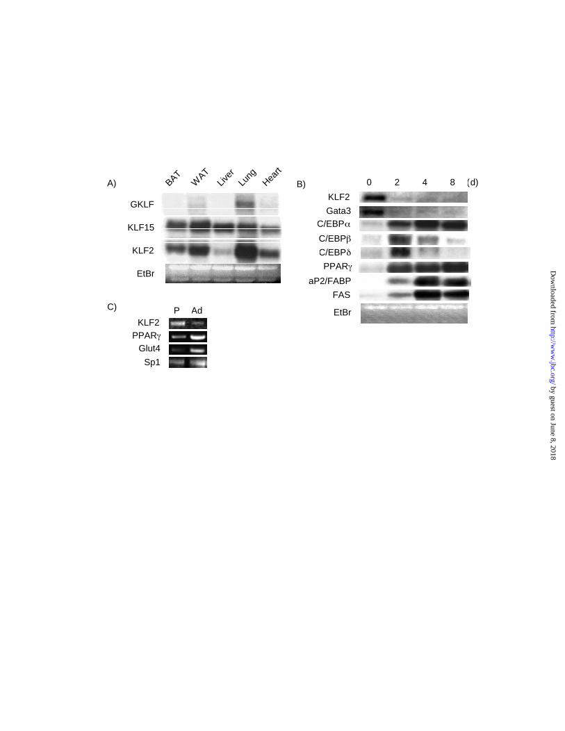
Expression of KLF2 in adipose tissue —We recently reported that a member of the KLF family
termed KLF15 is expressed in mature adipocytes and regulates GLUT4(17). In the course of
these studies we assessed the expression of several other KLFs in adipose tissue. KLF1(EKLF)
and KLF4 (GKLF) mRNA were barely detected in mouse adipose tissue (data not shown and
Figure 1A). However, as shown in Figure 1A, KLF2 mRNA was strongly expressed in both
white and brown adipose tissue. Given the availability of cell lines that faithfully recapitulate
adipogenesis in vitro, we assessed the expression of KLF2 during 3T3-L1 differentiation(23).
Stimulation of these cells with an empirically derived prodifferentiation mix leads to a
characteristic pattern of induction for various transcription factors and target genes involved in
adipogenesis and lipogenesis. We observed that KLF2 mRNA was expressed in 3T3-L1
preadipocytes and markedly diminished upon adipocyte differentiation (Fig 1B). As expected,
other factors such as PPARγ, members of the C/EBP family, and aP2/FABP were induced with
3T3-L1 differentiation. The expression pattern of KLF2 was most similar to two other
transcription factors which have been shown to inhibit adipogenesis termed GATA3 and 2 (Fig
1B and data not shown) (24). To verify this expression pattern in primary cells, we assessed the
expression of KLF2 in primary human preadipocytes and adipocytes by RT-PCR(21). Similar to
the KLF2 expression pattern in 3T3-L1 cells, KLF2 is highly expressed in human preadipocytes
and is markedly reduced in mature adipocytes (Figure 1C). Consistent with previous studies,
Glut4 and PPARγ expression are induced with differentiation whereas Sp1 levels are minimally
affected (Fig 1C).
by guest on June 8, 2018http://w
ww
.jbc.org/D
ownloaded from
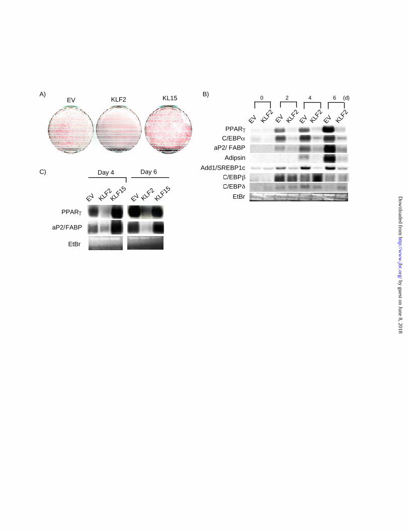
Overexpression of KLF2 inhibits adipogenesis in vitro— The reduced expression of KLF2
mRNA expression during 3T3-L1 differentiation suggested a potentially inhibitory role in
adipogenesis. To test this possibility directly, we retrovirally overexpressed KLF2 and empty
virus (EV) as a control in 3T3-L1 cells and induced differentiation. In parallel, we also
overexpressed KLF15 which has been shown to promote 3T3-L1 differentiation. As shown in
Figure 2A, compared to EV infected cells, KLF2 overexpressing cells showed a decrease in
intracellular lipid accumulation as evidenced by reduced Oil-red-O staining (Fig 2A). In contrast,
KLF15 infected cells exhibited increased lipid accumulation.
To determine the mechanism by which KLF2 may inhibit 3T3-L1 differentiation, we considered
several possibilities. In theory, inhibition of adipocyte differentiation may occur via inhibition of
factors that promote differentiation or induction of factors which inhibit adipogenesis. With
respect to the latter, we did not observe any effect of KLF2 overexpression on known negative
regulators of adipogenesis such as GATA3(24), β-catenin(25), Smad proteins(26), CHOP(27), or
pref-1(32) (data not shown). Furthermore, overexpression of KLF2 did not significantly alter
mRNA levels of C/EBPβ or C/EBPδ, two upstream factors which promote adipogenesis (Fig
2B). In contrast, KLF2 overexpression potently inhibited PPARγ expression at all time points
tested (Fig 2B). In addition, a strong inhibitory effect is seen on the expression of
ADD1/SREBP1c and C/EBPα - two positive regulators of PPARγ expression and adipogenesis.
Finally the expression of downstream genes characteristic of a mature adipocyte such as
aP2/FABP, adipsin, and FAS were also inhibited (Figure 2B and data not shown). The inhibitory
effect of KLF2 was specific as the other family member expressed in adipose tissue - KLF15 -
did not inhibit PPARγ expression (Fig 2C).
by guest on June 8, 2018http://w
ww
.jbc.org/D
ownloaded from
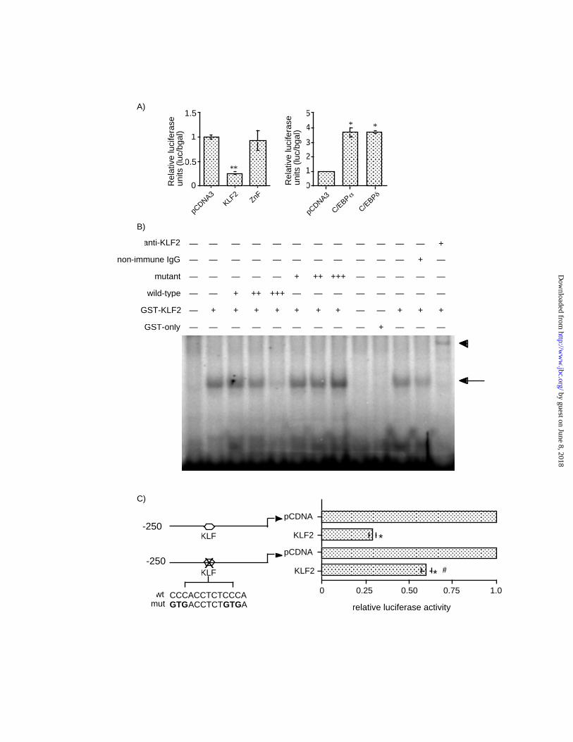
KLF2 inhibits the PPARγ promoter — Recent studies support a central role for PPARγ in
adipogenesis. Given the potent inhibitory effect of KLF2 overexpression on PPARγ expression
we sought to determine the mechanistic basis for this observation. We considered several
possibilities such as (a) direct inhibition of the PPARγ promoter or (b) inhibition of regulatory
factor(s) which can induce PPARγ expression. With respect to the second possibility, we did
observe a marked inhibition of two factors that have been demonstrated to positively regulate
PPARγ function — C/EBPα and ADD1/SREBP1c (Figure 2B). To assess the former possibility,
we tested whether KLF2 may directly inhibit the promoter of PPARγ2, the major isoform in
adipose tissue. Co-transfection of KLF2 the –250bp-Luc PPARγ2 promoter resulted in a ~70%
inhibition of promoter activity (Fig 3A left panel). This inhibition was not seen with a mutant
KLF2 construct consisting of only the zinc-finger DNA-binding domain. Consistent with
previous reports, members of the C/EBP family are able to transactivate the PPARγ2 promoter
(Fig 3A right panel) (28). These data suggest that KLF2 inhibition of PPARγ2 may be mediated
at the level of the promoter within the –250bp proximal promoter region. Examination of this
250 bp region revealed the presence of two tandem Kruppel binding site 5’-
CCCACCTCTCCCA-3’ (base pairs -82->-93). To determine if KLF2 was able to bind this
sequence, electrophoretic mobility gel-shift studies were performed. As shown in Figure 3B,
KLF2 is able to bind to this site (Figure 3B; arrow) and the specificity of this interaction was
confirmed by competition with identical but not a mutated oligomer (5’-GTGACCTCTGTGA-
3’). Furthermore, this DNA-protein complex was supershifted in the presence of an anti-KLF2
antibody (Figure 3B; arrowhead) but not IgG. To verify that this site was important in KLF2
mediated repression we mutated the KLF binding site within the context of the PPARγ2-250bp-
by guest on June 8, 2018http://w
ww
.jbc.org/D
ownloaded from

Luc promoter. As shown in Figure 3C, mutation of this site significantly attenuated KLF2 ’s
inhibitory effect, suggesting that this site is important for KLF2 mediated inhibition of the
PPARγ2 promoter.
by guest on June 8, 2018http://w
ww
.jbc.org/D
ownloaded from
DISCUSSION:
Members of the Kruppel-like family of transcription factors have been shown to play important
roles in cellular differentiation and growth (12). KLF2 was initially identified through a
homology screening strategy using the zinc finger domain of KLF1 as a probe(29). Studies
targeting KLF2 in mice have shown an essential role in embryonic development as these animals
die during mid-gestation due to abnormal blood vessel development (30). In addition, T cell
maturation (15) as well as lung development(31) are impaired. However, despite the importance
of KLF2 very few direct targets have been identified (22). In this regard, the observations from
this study may bear important implications not only for adipocyte biology but also for the role of
KLF2 in other tissues.
In this report we provide evidence that the Kruppel-like factor KLF2 is a negative regulator of
adipogenesis. Consistent with an inhibitory role, KLF2 is expressed in preadipocytes and
expression is markedly reduced in mature adipocytes (Fig 1). In addition, overexpression of
KLF2 markedly inhibits lipid accumulation in 3T3-L1 cells (Figure 2). This ability is specific to
KLF2 as another member of this family – KLF15 – augments lipid accumulation. To understand
the basis for KLF2’s inhibitory effect, we considered several possibilities such as (a) inhibition
of factors which have been shown to positively regulate adipocyte differentiation or (b) induction
of factors identified as negative regulators of adipocyte differentiation. With respect to the latter
possibility, we did not observe any effect on expression of several previously identified negative
regulators such as GATA3 (24), β-catenin(32), Smad proteins(26), CHOP(27), or pref-1(33).
With respect to factors which promote adipogenesis, KLF2 did not significantly effect
expression of C/EBPβ and C/EBPδ, two upstream regulators of adipogenesis. In contrast, KLF2
by guest on June 8, 2018http://w
ww
.jbc.org/D
ownloaded from
potently inhibits expression of PPARγ — the central regulator of adipogenesis. Given that
PPARγ expression is essential for adipocyte development, it is likely that this inhibition is
critical for KLF2 mediated inhibition of adipocyte differentiation. In addition to PPARγ, KLF2
also inhibits expression of both C/EBPα and ADD1/SREBP1c, two factors which play important
roles in adipocyte differentiation. Recent studies suggest that the principal role of C/EBPα is to
induce and maintain PPARγ expression(8). As such, inhibition of C/EBPα expression may
contribute to the reduced levels of PPARγ seen in KLF2 overexpressing cells thereby leading to
inhibition of adipocyte differentiation. Furthermore, ADDI/SREBP1c has been shown to
augment adipogenesis via direct induction of PPARγ expression as well as through production of
an endogenous PPARγ ligand (9) (10) (11). The inhibition of ADD1/SREBP1c expression by
KLF2 may therefore lead to both a reduction in PPARγ expression and activation. Thus, KLF2
may arrest the adipogenic program through inhibition of PPARγ expression and inhibition of
factors which positively regulate PPARγ expression and function.
The mechanism by which KLF2 is able to inhibit PPARγ expression has yet to be fully
elucidated. As suggested above, part of the mechanism may be via inhibition of factors such as
C/EBPα and ADD1/SREBP 1c which positively regulate PPARγ expression and function.
However, our data also suggests that KLF2 can inhibit PPARγ2 promoter activity thus
supporting a direct mechanism. As shown in Figure 3C, KLF2 inhibited PPARγ2 promoter
activity by approximately 70%. Examination of the PPARγ2 promoter revealed the presence of a
single consensus Kruppel-binding site. Gel-shift studies demonstrate that KLF2 can bind to this
site (Figure 3B) and promoter mutation analyses support a role for this site in mediating the
by guest on June 8, 2018http://w
ww
.jbc.org/D
ownloaded from
inhibitory effect of KLF2 (Figure 3C). Importantly, however, mutation of this site alone was not
sufficient to completely abrogate KLF2 mediated inhibition of promoter activity suggesting that
non – DNA binding dependent mechanisms are likely involved for the full inhibitory effect. It is
also noteworthy that the KLF binding site lies in between the two binding sites identified by
Tong et. al. as essential for GATA mediated inhibition of the PPARγ2 promoter activity(24). The
proximity of these two sites raises the possibility of a cooperative interaction between these two
factors and is the subject of future investigations.
Our current understanding of the transcriptional basis for adipogenesis highlights the function of
several major positive and negative regulatory families such as the C/EBPs, PPARs, and GATA
factors. We recently reported a role for the Kruppel-like factor KLF15 in adipocyte biology by
virtue of its ability to induce the insulin-sensitive glucose transporter GLUT4. In this report we
provide a novel function for another KLF family member - KLF2 - as a negative regulator of
adipogenesis. Taken together these studies support a role for the Kruppel-like factors as potential
regulators of adipogenesis. Future studies evaluating these factors in vivo will help elucidate
more precisely the role of Kruppel-like factors in adipocyte biology.
Acknowledgements: This work was supported by grants from NHLBI (K08HL03747-MKJ;
HL69477- MKJ) and American Heart Association (0060159T-MKJ; 0250030N-MKJ).
by guest on June 8, 2018http://w
ww
.jbc.org/D
ownloaded from

FIGURE LEGENDS:
Figure 1. Expression of KLF2 in adipose tissue and adipocytes. A) Northern analysis of KLF2,
KLF4 and KLF15 expression in selected mouse tissues. B) KLF2expression during 3T3-L1
differentiation. 3T3-L1 cells were induced to differentiate using hormonal agents as described
under " Materials and methods". Cells were harvested at the indicated number of days of post
induction and total RNA was isolated and subjected to Northern analysis using indicated probes.
C) KLF2 expression in human preadipocytes and adipocytes. Semi-quantative RT-PCR was
performed as described in the “Materials and Methods” section.
Figure 2. Constituitive expression of KLF2 inhibits adipocyte differentiation. A) Effect of KLF2
overexpression on lipid accumulation in 3T3-L1 cells. 3T3-L1 cells were retrovirally infected
with EV, KLF2 and KLF15 and subjected to Oil-Red-O staining to assess for lipid accumulation
at day 4 differentiation. B) Effect of KLF2 overexpression on adipocyte gene expression. 3T3-L1
cells were retrovirally infected with EV and KLF2 and differentiated as described in “Materials
and Methods.” Total RNA was harvested at the indicated number of days post induction of
differentiation and subjected to Northern analysis using the indicated probes. C) Effect of KLF2
and KLF15 on PPARγ expression. 3T3-L1 cells were retrovirally infected with EV, KLF2 and
KLF 15 and differentiated. Cells were harvested at the indicated time points and Northern
analysis performed using the indicated probes.
by guest on June 8, 2018http://w
ww
.jbc.org/D
ownloaded from

Figure 3. KLF2 represses PPARγ2 promoter activity. A) Effect of KLF2 on PPARγ2 promoter
activity. 3T3-L1 cells were transfected with PPARγ2 promoter (-250bp-Luc) and either KLF2 or
ZnF expression construct (left panel). 3T3-L1 cells were transfected with PPARγ2 promoter and
C/EBPα and C/EBPδ expression constructs (right panel). Luciferase and β-galactosidase assays
were performed as described in Materials and Methods. Results are expressed as fold repression
compared to vector (pCDNA3.1) alone (n= 6-10 per group) * p < 0.001 compared to the empty
vector. B) KLF2 binds to a CACCC element. Electrophoretic mobility shift assays were
performed as described in “Materials and Methods.” The GST-KLF2 fusion protein was
incubated with a 32P labeled wild-type oligomer extending from base pairs –82->-93 of the
PPARγ2 promoter. A single dominant DNA-protein complex was seen (arrow). Competition and
supershift studies (arrowhead) were performed to verify specificity. C) Effect of mutation of the
CACCC element on KLF2 mediated inhibition of the PPARγ2 promoter. 3T3-L1 cells were
transfected with KLF2 and the PPARγ2 promoter (-250bp-Luc and mutated –250bp-Luc). (n=9
per group; * p < 0.001; # p<0.001 statistically significant by comparison to KLF2 on –250Luc
promoter).
by guest on June 8, 2018http://w
ww
.jbc.org/D
ownloaded from
REFERENCES:
1.Spiegelman, B. M., and Flier, J. S. (2001) Cell 104(4), 531-43
2.Kahn, B. B., and Flier, J. S. (2000) Journal of Clinical Investigation 106(4), 473-81
3.Rosen, E. D., Walkey, C. J., Puigserver, P., and Spiegelman, B. M. (2000) Genes &
Development 14(11), 1293-307
4.Rosen, E. D., and Spiegelman, B. M. (2000) Annual Review of Cell & Developmental Biology
16, 145-71
5.Barak, Y., Nelson, M. C., Ong, E. S., Jones, Y. Z., Ruiz-Lozano, P., Chien, K. R., Koder, A.,
and Evans, R. M. (1999) Molecular Cell 4(4), 585-95
6.Rosen, E. D., Sarraf, P., Troy, A. E., Bradwin, G., Moore, K., Milstone, D. S., Spiegelman, B.
M., and Mortensen, R. M. (1999) Molecular Cell 4(4), 611-7
7.Wu, Z., Rosen, E. D., Brun, R., Hauser, S., Adelmant, G., Troy, A. E., McKeon, C.,
Darlington, G. J., and Spiegelman, B. M. (1999) Molecular Cell 3(2), 151-8
8.Rosen, E. D., Hsu, C., Wang, X., Sakai, S., Freeman, M. W., Gonzalez, F. J., and Spiegelman,
B. (2002) Genes&Development 16, 22-26
9.Kim, J. B., Wright, H. M., Wright, M., and Spiegelman, B. M. (1998) Proceedings of the
National Academy of Sciences of the United States of America 95(8), 4333-7
10.Kim, J. B., and Spiegelman, B. M. (1996) Genes & Development 10(9), 1096-107
11.Fajas, L., Schoonjans, K., Gelman, L., Kim, J. B., Najib, J., Martin, G., Fruchart, J. C.,
Briggs, M., Spiegelman, B. M., and Auwerx, J. (1999) Molecular & Cellular Biology 19(8),
5495-503
12.Bieker, J. J. (2001) Journal of Biological Chemistry 276(37), 34355-8
by guest on June 8, 2018http://w
ww
.jbc.org/D
ownloaded from
13.Nuez, B., Michalovich, D., Bygrave, A., Ploemacher, R., and Grosveld, F. (1995) Nature
375(6529), 316-8
14.Perkins, A. C., Sharpe, A. H., and Orkin, S. H. (1995) Nature 375(6529), 318-22
15.Kuo, C. T., Veselits, M. L., and Leiden, J. M. (1997) Science 277(5334), 1986-90
16.Segre, J. A., Bauer, C., and Fuchs, E. (1999) Nature Genetics 22(4), 356-60
17.Gray, S., Feinberg, M. W., Hull, S., Kuo, C. T., Watanabe, M., Sen(Banerjee), S., Depina, A.,
Haspel, R., and Jain, M. K. (2002) Journal of Biological Chemistry 277(37), 34322-34328
18.Morrison, R. F., and Farmer, S. R. (1999) Journal of Biological Chemistry 274(24), 17088-97
19.Ranganath, S., Ouyang, W., Bhattarcharya, D., Sha, W. C., Grupe, A., Peltz, G., and Murphy,
K. M. (1998) Journal of Immunology 161(8), 3822-6
20.Hauner, H., Skurk, T., and Wabitsch, M. (2001) Methods in Molecular Biology 155, 239-47
21.Hube, F., and Hauner, H. (2000) Endocrinology 141(7), 2582-8.
22.Denkinger, D. J., Cushman-Vokoun, A. M., and Kawahara, R. S. (2001) Gene 281(1-2), 133-
42
23.Green, H., and Kehinde, O. (1975) Cell 5(1), 19-27
24.Tong, Q., Dalgin, G., Xu, H., Ting, C., Leiden, J. M., and Hotamisligil, G. S. (2000) Science
290, 134-8.
25.Ross, S. E., Hemati, N., Longo, K. A., Bennett, C. N., Lucas, P. C., Erickson, R. L., and
MacDougald, O. A. (2000) Science 289(5481), 950-3
26.Choy, L., Skillington, J., and Derynck, R. (2000) Journal of Cell Biology 149(3), 667-82
27.Ron, D., and Habener, J. F. (1992) Genes & Development 6(3), 439-53
28.Zhu, Y., Qi, C., Korenberg, J. R., Chen, X. N., Noya, D., Rao, M. S., and Reddy, J. K. (1995)
Proceedings of the National Academy of Sciences of the United States of America 92(17), 7921-5
by guest on June 8, 2018http://w
ww
.jbc.org/D
ownloaded from
29.Anderson, K. P., Kern, C. B., Crable, S. C., and Lingrel, J. B. (1995) Molecular & Cellular
Biology 15(11), 5957-65
30.Kuo, C. T., Veselits, M. L., Barton, K. P., Lu, M. M., Clendenin, C., and Leiden, J. M. (1997)
Genes & Development 11(22), 2996-3006
31.Wani, M. A., Wert, S. E., and Lingrel, J. B. (1999) Journal of Biological Chemistry 274(30),
21180-5
32.Ross, S. E., Hemati, N., Longo, K. A., Bennett, C. N., Lucas, P. C., Ericksson, R. L., and
MaDougald, O. A. (2000) Science 289, 950-3.
33.Smas, C. M., and Sul, H. S. (1993) Cell 73(4), 725-34
by guest on June 8, 2018http://w
ww
.jbc.org/D
ownloaded from
GKLF
KLF15
KLF2
BAT
EtBr
WAT
Liver
Lung
Heart
A)
C)
(d)0 2 4 8
C/EBPα
KLF2
PPARγ
C/EBPβ
C/EBPδ
Gata3
FAS
EtBr
aP2/FABP
B)
A)
P Ad
PPARγ
Glut4
KLF2
Sp1
by guest on June 8, 2018http://w
ww
.jbc.org/D
ownloaded from
Ad
EV KLF2 KL15
EV
Day 4 Day 6
PPARγ
aP2/FABP
EtBr
KLF2KLF
15
EV KLF2
KLF15
A) B)
C)
(d)
EtBr
C/EBPα
aP2/ FABP
Adipsin
PPARγ
C/EBPβ
EV KLF2
EV
EV EVKLF2
KLF2
KLF2
0 2 4 6
Add1/SREBP1c
C/EBPδ
by guest on June 8, 2018http://w
ww
.jbc.org/D
ownloaded from
0
1
2
3
4
5
Rel
ativ
e lu
cife
rase
un
its (
luc/
bgal
)
pCDNA3
C/EBPα
C/EBPδ
* *
1.5
0
0.5
1
pCDNA3
KLF2
ZnF
**R
elat
ive
luci
fera
se
units
(lu
c/bg
al)
A)
B)
GST-only — — — — — — — — — + — — —
GST-KLF2 — + + + + + + + — — + + +
wild-type — — + ++ +++ — — — — — — — —
mutant — — — — — — — — — —
non-immune IgG — — — — — — — — — — — + —
anti-KLF2 — — — — — — — — — — — — +
+ ++ +++
C)
relative luciferase activity
KLF2
pCDNA
KLF2
pCDNA
0 0.25 0.50 0.75 1.0
*
* #
KLF-250
X-250
mut
KLF
wt CCCACCTCTCCCAGTGACCTCTGTGA
by guest on June 8, 2018http://w
ww
.jbc.org/D
ownloaded from

Haspel, Diane J. Denkinger, Rodney Kawahara, Hans Hauner and Mukesh K. JainSucharita Sen(Banerjee), Mark W. Feinberg, Masafumi Watanabe, Susan Gray, Richard L.
expression and adipogenesisγThe Kruppel-like factor KLF2 inhibits PPAR
published online November 7, 2002J. Biol. Chem.
10.1074/jbc.M210859200Access the most updated version of this article at doi:
Alerts:
When a correction for this article is posted•
When this article is cited•
to choose from all of JBC's e-mail alertsClick here
by guest on June 8, 2018http://w
ww
.jbc.org/D
ownloaded from