The Knee Joint. Muscles that cross the Hip and Knee Joints Sartorius: –Hip flexionKnee flexion...
23
The Knee Joint
-
date post
19-Dec-2015 -
Category
Documents
-
view
224 -
download
0
Transcript of The Knee Joint. Muscles that cross the Hip and Knee Joints Sartorius: –Hip flexionKnee flexion...
The Knee Joint
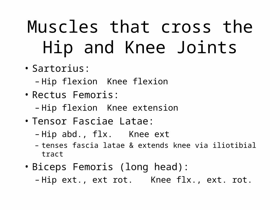
Muscles that cross theHip and Knee Joints
• Sartorius: – Hip flexion Knee flexion
• Rectus Femoris: – Hip flexion Knee extension
• Tensor Fasciae Latae:– Hip abd., flx. Knee ext– tenses fascia latae & extends knee via iliotibial tract
• Biceps Femoris (long head): – Hip ext., ext rot. Knee flx., ext. rot.
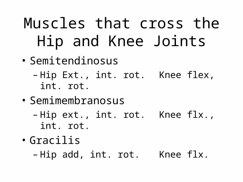
Muscles that cross theHip and Knee Joints
• Semitendinosus– Hip Ext., int. rot. Knee flex, int. rot.
• Semimembranosus– Hip ext., int. rot. Knee flx., int. rot.
• Gracilis– Hip add, int. rot. Knee flx.
The knee joint
• Complex structure because of the knee functions:– allow mobility (flexion/extension)– must have some inherent stability– weight bearing joint
The knee joint
• Largest joint in the body.
• Classified as a ginglymus (hinge) joint
• Allows for primarily flexion and extension
• There is, however, some rotation allowed about the knee joint.
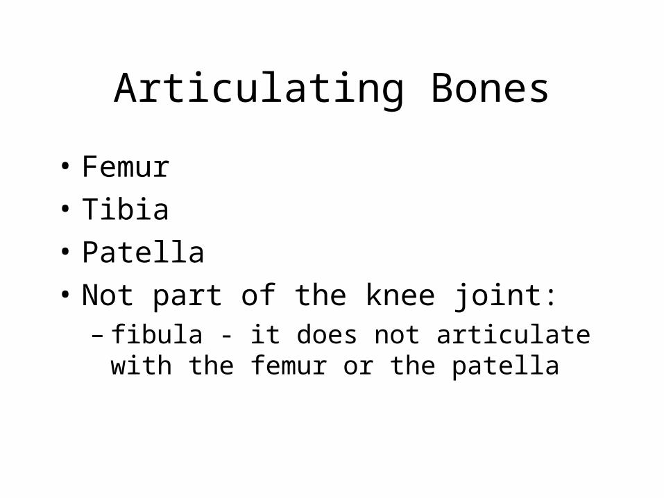
Articulating Bones
• Femur
• Tibia
• Patella
• Not part of the knee joint:– fibula - it does not articulate with the femur or
the patella
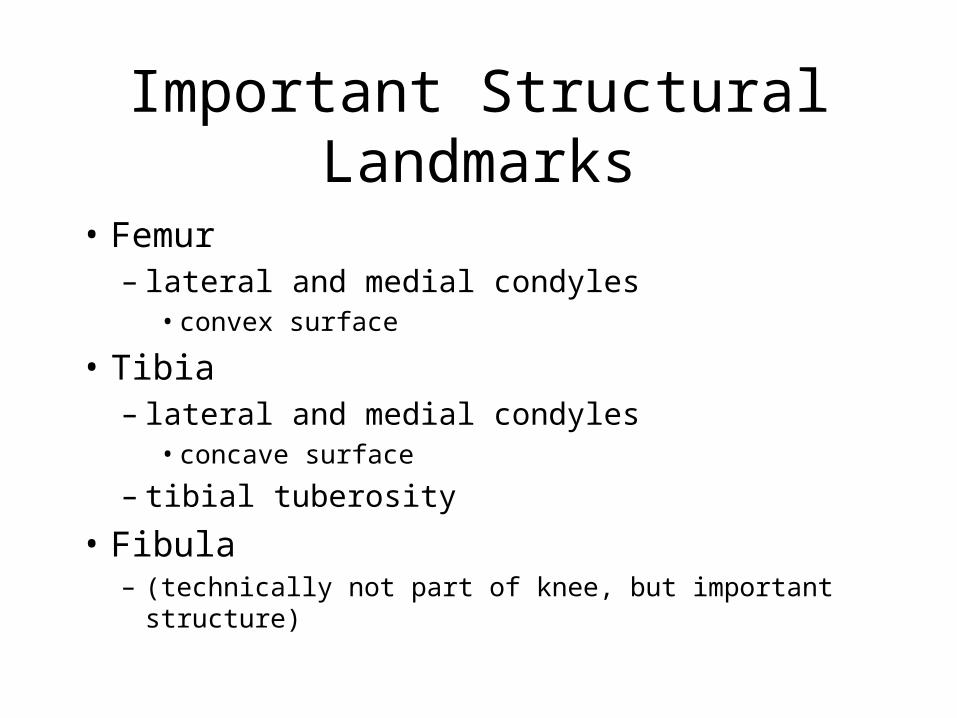
Important Structural Landmarks
• Femur– lateral and medial condyles
• convex surface
• Tibia– lateral and medial condyles
• concave surface
– tibial tuberosity
• Fibula – (technically not part of knee, but important structure)
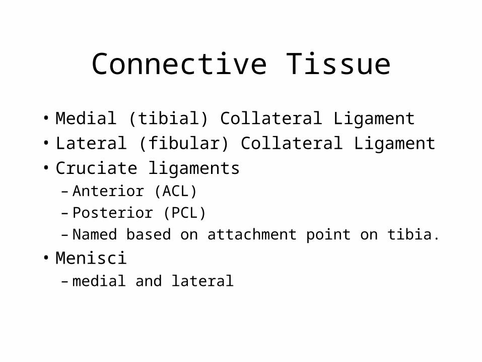
Connective Tissue
• Medial (tibial) Collateral Ligament• Lateral (fibular) Collateral Ligament• Cruciate ligaments
– Anterior (ACL)– Posterior (PCL)– Named based on attachment point on tibia.
• Menisci– medial and lateral
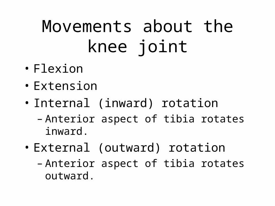
Movements about the knee joint
• Flexion
• Extension
• Internal (inward) rotation– Anterior aspect of tibia rotates inward.
• External (outward) rotation– Anterior aspect of tibia rotates outward.
Muscles
• Knee Extensors– Rectus femoris (two joint muscle)– Vastus medialis– Vastus intermedius– Vastus lateralis
Muscles
• Knee flexors– Biceps femoris (long *, short)– Semimembranosus *– Semitendinosus *– Sartorius *– Gracilis *– Popliteus– Gastrocnemius *
• (* = crosses two joints)
Muscles
• Internal rotation about the knee– popliteus– semimembranosus– semitendinosus
• External rotation about the knee– biceps femoris
Popliteus (p104)
• Origin– posterior surface of lateral epicondyle of the
femur
• Insertion– Popliteal surface of the tibia
• Action– Knee flexion– Internal rotation about the knee
Hamstring Muscles
• Semimembranosus (medial muscle)
• Semitendinosis (medial muscle)
• Biceps Femoris (lateral muscle)
• Actions– Knee flexion– Hip extension– thigh and leg rotation
• internal (ST, SM)
• external (BF)
Biceps Femoris Muscle (p105)
• Origin– Long head: ischial tuberosity– Short head: posterior aspect (linea aspera) of femur
• Insertion– Lateral condyle of the tibia and head of the fibula
• Action– Hip extension– Knee flexion– External rotation of the hip and knee
Semimembranosus Muscle (p106)
• Origin– Ischial tuberosity
• Insertion– Postero-superior surface of the medial tibial condyle
• Action– Hip extension– Knee flexion– Internal rotation about the hip and knee
Semitendinosus Muscle (p107)
• Origin– Ischial tuberosity
• Insertion– Upper anterior medial surface of the tibia
• Action– Hip extension– Knee flexion– Internal rotation about the hip and knee
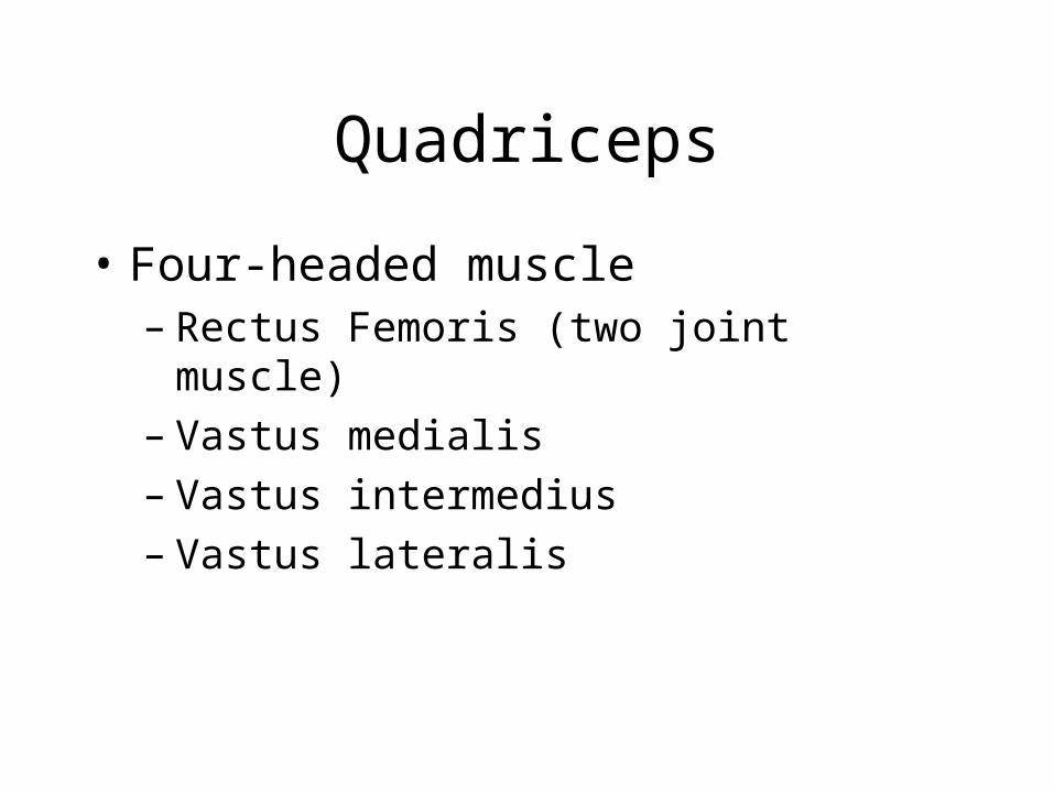
Quadriceps
• Four-headed muscle– Rectus Femoris (two joint muscle)– Vastus medialis– Vastus intermedius– Vastus lateralis
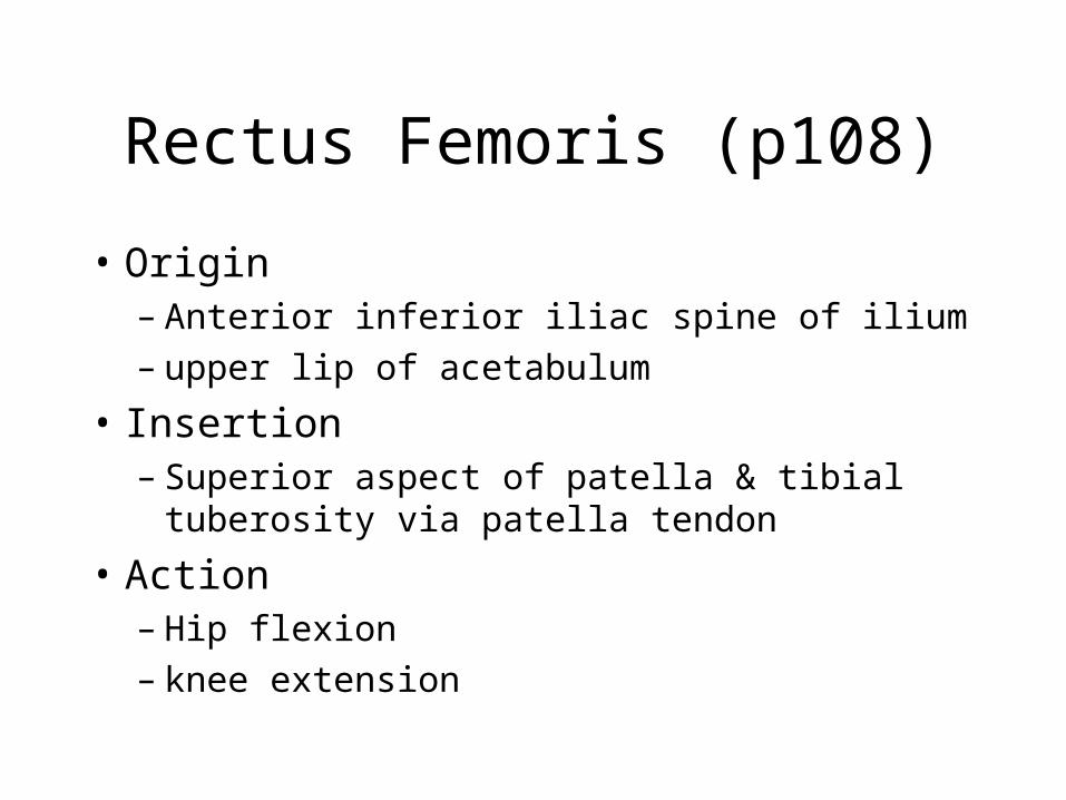
Rectus Femoris (p108)
• Origin– Anterior inferior iliac spine of ilium– upper lip of acetabulum
• Insertion– Superior aspect of patella & tibial tuberosity via
patella tendon
• Action– Hip flexion– knee extension
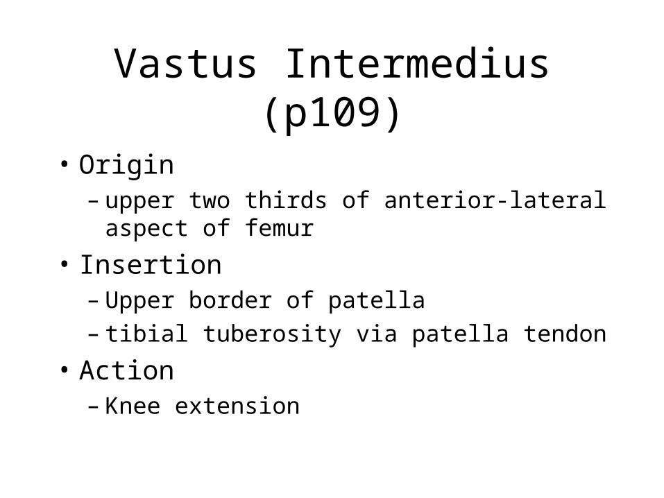
Vastus Intermedius (p109)
• Origin– upper two thirds of anterior-lateral aspect of
femur
• Insertion– Upper border of patella– tibial tuberosity via patella tendon
• Action– Knee extension
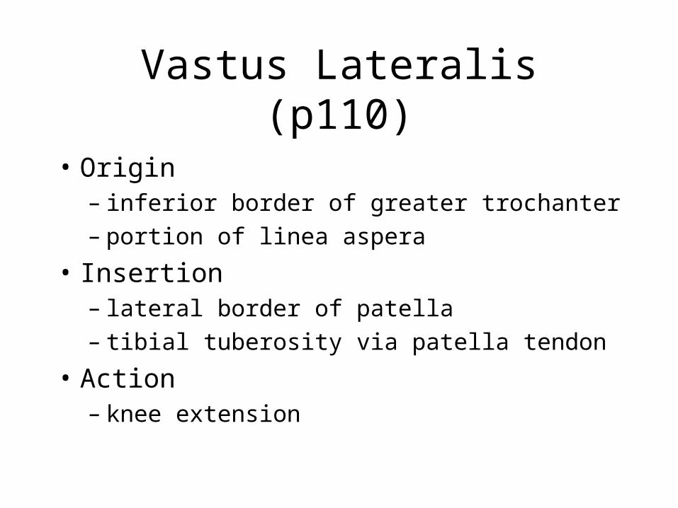
Vastus Lateralis (p110)
• Origin– inferior border of greater trochanter– portion of linea aspera
• Insertion– lateral border of patella– tibial tuberosity via patella tendon
• Action– knee extension
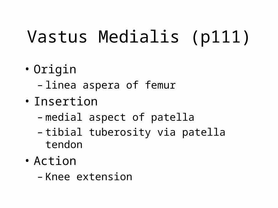
Vastus Medialis (p111)
• Origin– linea aspera of femur
• Insertion– medial aspect of patella– tibial tuberosity via patella tendon
• Action– Knee extension
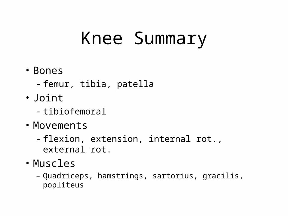
Knee Summary
• Bones– femur, tibia, patella
• Joint– tibiofemoral
• Movements– flexion, extension, internal rot., external rot.
• Muscles– Quadriceps, hamstrings, sartorius, gracilis, popliteus