
THE JOURNAL OF CHEMISTRY 10, 11808-11815, in Purification ... · THE JOURNAL OF BIOLOGICAL...
8
THE JOURNAL OF BIOLOGICAL CHEMISTRY Printed in U.S. A. Vol. 257, No. 19, Issue of October 10, pp. 11808-11815, 1982 Purification and Structural Determination of Nontoxic Lipid A Obtained from the Lipopolysaccharide of Salmonella ep?zimurium* (Received for publication, April 16, 1982) Nilofer Qureshi and Kuni Takayama From the Mycobacteriology Research Laboratory, William S. Middleton Memorial Veterans Hospital, Madison, Wisconsin 53705 and The Institute for Enzyme Research, University of Wisconsin, Madison, Wisconsin 53706 Edgar Ribi From Ribi ImmunoChem Research, Inc., Hamilton, Montana 64644 Endotoxin extracted from the heptose-less mutant of Salmonella typhimurium was hydrolyzed in 0.1 N HCl in methanol/water (l:l, v/v) at 100 "C to yield lipid A, which was then fractionated on a Sephadex LH-20 col- umn to yield a major monophosphoryl lipid A fraction. The monophosphoryl lipid A was further fractionated by preparative thin layerchromatography. This proc- ess yielded three major bands ("LC-1, -3, and -5) and two minor bands (TLC-7 and -9). The purity of these fractions was established by ion exchange and reverse phase high performance liquid chromatography. The thin layer fractions were analyzed by fast atom bombardment mass spectrometry. TLC-1 and -3 gave molecular ions (M-H)- at m/e 1730 and 1716, respec- tively. Both of these fractions contained P-hydroxy- myristic, lauric, and 3-myristoxymyristic acids in 0- acyl linkages. The molecular formula and M, of TLC-1 are CgSH179022N2P and 1731.16; those of TLC-3 are Cg4H1T7022NZP and 1717.15. "LC-1 was a methyl homo- log of TLC-3. The major component of TLC-5 (CsoHlslO22NzP and M, = 1506.99) gave a molecular ion at m/e 1506 and contained two 8-hydroxymyristic acids and a lauric acid in the 0-acyl linkages. The major component of TLC-7 (CsaH126019N2P and M, = 1280.83) and the single component of TLC-9 gave molecular ions at m/e 1280 and 1098, respectively. "LC-7 contained lauric and 8-hydroxymyristic acids in the 0-acyl link- ages. TLC-9 ( C S ~ H ~ O ~ O ~ ~ N Z P and M, = 1098.69)contained a single 0-acylated 8-hydroxymyristate group. TLC-1 and -3 were nontoxic in the chick embryo lethality test and regressed established tumors in the syngeneic guinea pigs. Ribi et al. (1) showed that when the crude endotoxin from the heptose-less mutant of Salmonella typhimurium was com- bined with trehalose dimycolate from mycobacteria in oil droplets and injected directly into established tumors (line 10 hepatocellular carcinoma) in syngeneic guinea pigs, over 90% of the animals were cured. Recently, Takayama et al. (2) isolated a nontoxic lipid A fraction that also has antitumor activity. These studies indicated that the next step to take is * This work was supported in part by the Medical Service of the Veterans Administration. A preliminary report of some of this work was presented at the Annual Meeting of the American Society of Biological Chemists in New Orleans on April 18-21, 1982. The costs of publication of this article were defrayed in part by the payment of page charges. This article must therefore be hereby marked "aduer- tisement" in accordance with 18 U.S.C. Section 1734 solely to indicate this fact. to determine the relationship of the structure of lipid A to both toxicity and tumor regression activity. However, struc- tural determination requires highly purified samples and lipid A is acknowledged to be a complex mixture. A review of the literature suggested that the task of fractionating lipid A is extremely difficult (3). Lipid A is a glucosamine disaccharide with a (P)1 --* 6 linkage to which are attached two phosphate groups at posi- tions 1 and 4' (4, 5) (Structure 1). Thereare two N-acyl linkages at positions 2 and 2' with /I-hydroxymyristoyl resi- dues (R,), and three 0-acyl linkages at positions 3,4, and 6' to which 3-myristoxymyristoyl, P-hydroxymyristoyl, lauroyl, myristoyl, or palmitoyl residues (R2) can be linked. An ex- amination of this structure shows that thevariable factors in the biological activity-structure relationshipmight be: (a) the presence or absence of the two phosphate groups, (b) the number and kinds of 0-acyl fattyacids, and (c) the presence or absence of 4-amino arabinose or pyrophosphorylethano- lamine. Our previous studies had already shown that the removal of the acid-labile sugar 1-phosphate group reduces the toxicity of lipid A (2, 6). This problem called for the development of methods to completely separate lipid A and to analyze its purified fractions. In our study, we were able to separate many structural analogs of lipid A from acid-hydrolyzed endotoxin to virtual homogeneity and to establish their exact sizes by FAB' mass spectrometry. Two of these well characterized fractions were found to be nontoxic and retained tumor regression activity. EXPERIMENTAL PROCEDURES Materials-Acetonitrile (distilled in glass), THF, chloroform, and methanol were purchased from Burdick and Jackson Laboratories, Muskegon, MI. Chemicals were purchased from the following com- panies: [l-I4C]acetate, Amersham; TEA and myristoyl chloride, Ald- rich Chemical Co.; tetrabutylammonium phosphate (Pic A), Eastman Kodak Co. and Waters Associates, Inc., Milford, MA; Silica Gel H thin layer plates, Analabs, North Haven, CT; and pPorasil (10-pm silica gel), Waters Associates. Growth ofBacteria-Cells of the heptose-less mutant of S. typhi- murium G30/C21 were grown in a New Brunswick 28-liter fermentor at 37 "C for 12-16 h in a modified ammonium medium of Anderson (2, 7). Radiolabeled cells of S. typhimurium were grown in 100-ml cultures containing 1 mCi of [l-14C]acetate (60 pCi/pmol). These cells were the source of I4C-endotoxin, which contained the label in the fatty acids to the extent of 99.7%. fying lipid A from S. typhimurium G30/C21 is shown in Fig. 1. The Isolation of the Endotoxin-The scheme for preparing and puri- endotoxin was extracted from 1-kg cells by the method of Galanos et ' The abbreviations used are: FAB, fast atom bombardment; HPLC, high performance liquid chromatography; THF, tetrahydro- furan; TEA, triethylamine. 11808
Transcript of THE JOURNAL OF CHEMISTRY 10, 11808-11815, in Purification ... · THE JOURNAL OF BIOLOGICAL...

THE JOURNAL OF BIOLOGICAL CHEMISTRY
Printed in U.S. A . Vol. 257, No. 19, Issue of October 10, pp. 11808-11815, 1982
Purification and Structural Determination of Nontoxic Lipid A Obtained from the Lipopolysaccharide of Salmonella ep?zimurium*
(Received for publication, April 16, 1982)
Nilofer Qureshi and Kuni Takayama From the Mycobacteriology Research Laboratory, William S. Middleton Memorial Veterans Hospital, Madison, Wisconsin 53705 and The Institute for Enzyme Research, University of Wisconsin, Madison, Wisconsin 53706
Edgar Ribi From Ribi ImmunoChem Research, Inc., Hamilton, Montana 64644
Endotoxin extracted from the heptose-less mutant of Salmonella typhimurium was hydrolyzed in 0.1 N HCl in methanol/water (l:l, v/v) at 100 "C to yield lipid A, which was then fractionated on a Sephadex LH-20 col- umn to yield a major monophosphoryl lipid A fraction. The monophosphoryl lipid A was further fractionated by preparative thin layer chromatography. This proc- ess yielded three major bands ("LC-1, -3, and -5) and two minor bands (TLC-7 and -9). The purity of these fractions was established by ion exchange and reverse phase high performance liquid chromatography.
The thin layer fractions were analyzed by fast atom bombardment mass spectrometry. TLC-1 and -3 gave molecular ions (M-H)- at m / e 1730 and 1716, respec- tively. Both of these fractions contained P-hydroxy- myristic, lauric, and 3-myristoxymyristic acids in 0- acyl linkages. The molecular formula and M, of TLC-1 are CgSH179022N2P and 1731.16; those of TLC-3 are Cg4H1T7022NZP and 1717.15. "LC-1 was a methyl homo- log of TLC-3. The major component of TLC-5 (CsoHlslO22NzP and M, = 1506.99) gave a molecular ion at m/e 1506 and contained two 8-hydroxymyristic acids and a lauric acid in the 0-acyl linkages. The major component of TLC-7 (CsaH126019N2P and M, = 1280.83) and the single component of TLC-9 gave molecular ions at m/e 1280 and 1098, respectively. "LC-7 contained lauric and 8-hydroxymyristic acids in the 0-acyl link- ages. TLC-9 ( C S ~ H ~ O ~ O ~ ~ N Z P and M, = 1098.69) contained a single 0-acylated 8-hydroxymyristate group. TLC-1 and -3 were nontoxic in the chick embryo lethality test and regressed established tumors in the syngeneic guinea pigs.
Ribi et al. (1) showed that when the crude endotoxin from the heptose-less mutant of Salmonella typhimurium was com- bined with trehalose dimycolate from mycobacteria in oil droplets and injected directly into established tumors (line 10 hepatocellular carcinoma) in syngeneic guinea pigs, over 90% of the animals were cured. Recently, Takayama et al. (2) isolated a nontoxic lipid A fraction that also has antitumor activity. These studies indicated that the next step to take is
* This work was supported in part by the Medical Service of the Veterans Administration. A preliminary report of some of this work was presented at the Annual Meeting of the American Society of Biological Chemists in New Orleans on April 18-21, 1982. The costs of publication of this article were defrayed in part by the payment of page charges. This article must therefore be hereby marked "aduer- tisement" in accordance with 18 U.S.C. Section 1734 solely to indicate this fact.
to determine the relationship of the structure of lipid A to both toxicity and tumor regression activity. However, struc- tural determination requires highly purified samples and lipid A is acknowledged to be a complex mixture. A review of the literature suggested that the task of fractionating lipid A is extremely difficult (3).
Lipid A is a glucosamine disaccharide with a (P)1 --* 6 linkage to which are attached two phosphate groups at posi- tions 1 and 4' (4, 5) (Structure 1). There are two N-acyl linkages at positions 2 and 2' with /I-hydroxymyristoyl resi- dues (R , ) , and three 0-acyl linkages at positions 3,4, and 6' to which 3-myristoxymyristoyl, P-hydroxymyristoyl, lauroyl, myristoyl, or palmitoyl residues (R2) can be linked. An ex- amination of this structure shows that the variable factors in the biological activity-structure relationship might be: (a ) the presence or absence of the two phosphate groups, ( b ) the number and kinds of 0-acyl fatty acids, and ( c ) the presence or absence of 4-amino arabinose or pyrophosphorylethano- lamine. Our previous studies had already shown that the removal of the acid-labile sugar 1-phosphate group reduces the toxicity of lipid A (2, 6). This problem called for the development of methods to completely separate lipid A and to analyze its purified fractions.
In our study, we were able to separate many structural analogs of lipid A from acid-hydrolyzed endotoxin to virtual homogeneity and to establish their exact sizes by FAB' mass spectrometry. Two of these well characterized fractions were found to be nontoxic and retained tumor regression activity.
EXPERIMENTAL PROCEDURES
Materials-Acetonitrile (distilled in glass), THF, chloroform, and methanol were purchased from Burdick and Jackson Laboratories, Muskegon, MI. Chemicals were purchased from the following com- panies: [l-I4C]acetate, Amersham; TEA and myristoyl chloride, Ald- rich Chemical Co.; tetrabutylammonium phosphate (Pic A), Eastman Kodak Co. and Waters Associates, Inc., Milford, MA; Silica Gel H thin layer plates, Analabs, North Haven, CT; and pPorasil (10-pm silica gel), Waters Associates.
Growth ofBacteria-Cells of the heptose-less mutant of S. typhi- murium G30/C21 were grown in a New Brunswick 28-liter fermentor at 37 "C for 12-16 h in a modified ammonium medium of Anderson (2, 7). Radiolabeled cells of S. typhimurium were grown in 100-ml cultures containing 1 mCi of [l-14C]acetate (60 pCi/pmol). These cells were the source of I4C-endotoxin, which contained the label in the fatty acids to the extent of 99.7%.
fying lipid A from S. typhimurium G30/C21 is shown in Fig. 1. The Isolation of the Endotoxin-The scheme for preparing and puri-
endotoxin was extracted from 1-kg cells by the method of Galanos et
' The abbreviations used are: FAB, fast atom bombardment; HPLC, high performance liquid chromatography; THF, tetrahydro- furan; TEA, triethylamine.
11808

Structure of Nontoxic Lipid A 11809
, RI I OH
Structure I
S tnhimurium t?Y)/CtZI
-TY CWOTOXIN
J"*atW,
CRUDE LIPID A
y"Amm - TLC-I A TLt-J
U u ' n - m m
LIPIDA y~ UOWOPHOSPMCUTL FATTY
LIPID A -
1- PURIFIED YONOPHOSPHORYL LIPID A
FIG. 1. Scheme for preparing and purifying monophos- phoryl lipid A from S. typhimurium G30/C21.
al. (8) with one modification: because the water precipitation step to recover the endotoxin from the phenol solution is incomplete, it was omitted. Instead, we added 6 volumes of diethyl ether/acetone (k5, v/v) to 1 volume of the phenol solution. The resulting precipitate was filtered through a sintered glass funnel and air-dried. The yield was 6.7 g of endotoxin/204 g of cells, dry weight.
Preparation of Monophosphoryl Lipid A-The endotoxin (4.9 g) was suspended in 700 ml of 0.1 N HCI in methanol/water (l:l, v/v), refluxed for 45 min, and centrifuged at 10,000 X g for 5 min. The residue was suspended in 120 ml of chloroform/methanol (21, v/v), washed with 48 ml of water, filtered, and dried. The yield of crude lipid A was 3.4 g. The lipid A (680 mg) was dissolved in 3 ml of chloroform/methanol (4:1, v/v) by warming, applied to a Sephadex LH-20 column (2 X 144 cm), and eluted with chloroform/methanol (4:1, v/v). Mass peaks were located by total phosphorus analysis of the column fractions or by charring aliquots on a Silica Gel G plate. Results of a representative fractionation which included 4 X lo7 cpm of I4C-lipid A are shown in Fig. 2. A total of 3.4 g of acid-hydrolyzed endotoxin was fractionated on the Sephadex LH-20 column to yield 1.66 g of monophosphoryl lipid A.
The purified monophosphoryl lipid A was applied to Silica Gel H plates (500 pm) (20 X 20 cm) as a streak at 6-7 mg/plate and developed in chloroform/methanol/water-concentrated ammonium hydroxide (50:25:4:2, v/v) (9). The sample separated into three major (TLC-1, -3, and -5) and two minor (TLC-7 and -9) bands, as shown in Fig. 3A.2
HPLC Fractionation-HPLC was performed with two Waters 6000A solvent delivery systems, a Waters 660 solvent programmer, a Waters U6K universal liquid chromatograph injector, a variable wave- length detector (Model LC-55, Perkin-Elmer Corp., Analytical Instru- ments), and a radial compression module (Model RCM-100, Waters Associates). A Radial Pak A cartridge (8 mm X 10 cm) (C-18-bonded silica, Waters Associates) was used at a flow rate of 3 ml/min. For the fractionation of labeled monophosphoryl lipid A, a linear gradient of 80% acetonitrile in water to 80% THF in water was used over a period of 1 h. Both solvent systems contained 5 mM Pic A. I4C-Lipid A was also fractionated on a stainless steel DEAE-pPorasil column (3.9 X 30 cm) prepared according to the method of Kundu and Roy (10). The column was eluted with a linear gradient of 0-0.1 M ammonium acetate in methanol/water (95:5, v/v) over a period of 80 min at a flow rate of 1 ml/min. Fractions (30 s) were collected in counting vials and their radioactivity was determined.
TLC-9 was obtained in greater amounts when the endotoxin was hydrolyzed with aqueous 0.1 N HCl at 100 "C for 40 min.
Analytical Procedures-Total phosphorus and 2-keto-3-deoxy- octonate content were determined by the methods of Bartlett (11) and Osborn (12), respectively. Samples for the glucosamine assay were hydrolyzed in 3 N HCl for 4 h at 95 "C and analyzed by the method of Enghofer and Kress (13).
Fatty Acid Analy~es-~~C-labeled TLC-1, -3, and -5 (80,000 cpm each) were hydrolyzed in 0.5 ml of 4 N HC1 at 100 "C for 5 h. P- Hydroxymyristic, lauric, myristic, and palmitic acids (350 pg) were added as carriers and the reaction mixture was extracted with 4 ml of petroleum ether three times. The extract was dried, methylated with diazomethane, and subjected to HPLC. A Radial Pak A cartridge (8 mm X 10 cm) was used with a solvent system of acetonitrile/water (173, v/v) at a flow rate of 3 ml/min. The fatty acid content was quantitated by determining the radioactivity of the separated fatty acid esters. The recovery of radioactivity from the cartridge was quantitative.
TEA Treatment-Purified labeled TLC-3 (20 pg and 20,000 cpm) was suspended in 300 pI of water and I0 pl of TEA and heated at 100 "C for 0-10 min. One of the samples of TLC-3 (60,000 cpm) was suspended in water and heated to 100 "C. TEA was then added and the mixture was allowed to stand at 22 "C for 4 days. Samples were lyophilized and standard oleic and P-hydroxymyristic acids (10 pg each) were added. Samples were applied to Silica Gel H plates (250 Fm) and developed in chloroform/methanol/water/concentrated am- monium hydroxide (50:254:2, v/v). Bands were visualized by either exposing the plates to iodine vapor or by spraying with dichromate- sulfuric acid reagent and charring very lightly. The visualized bands were scraped into vials and counted in a scintillation spectrometer. The reaction mixture, incubated at 22 "C, was analyzed by both TLC and reverse phase HPLC.
Chemical Synthesis of Myristoxymyristic Acid-P-Hydroxymyris- tic acid (26.8 mg) and myristoyl chloride (50 pl) were added to 2 ml of anhydrous toluene and heated at 100 "C for 30 min. Water (2 ml) was added to the reaction mixture. The mixture was heated for 15 min to hydrolyze excess acyl chloride and extracted three times with 3-ml portions of chloroform/methanol (21, v/v), and the extract was dried with a stream of Nz. The synthesized 3-myristoxymyristic acid was purified by TLC using Silica Gel H (250 pm) plates and the solvent system of chloroform/methanol/water/concentrated ammonium hy- droxide (5025:4:2, v/v). The following RF values were obtained: 0.61 for 3-mvristoxvmyristic acid, 0.50 for oleic acid, and 0.41 for the p - hydroxymyristic acid.
" -
FIG. 2. Sephadex LH-20 column chromatography of acid-hy- drolyzed 14C-endotoxin (crude lipid A). Sample (220 mg, 4 x lo7 cpm) was dissolved in 3 ml of chloroform/methanol (4:1, v/v) and applied to a Sephadex LH-20 column (2 X 144 cm). Fractions of 2.5 ml were collected and aliquots were analyzed for both radioactivity (50 p1) and total phosphorus (10 p l ) (absorbance at 830 nm). The second peak eluting from the column at 148 ml contained the mono- phosphoryl lipid A.

11810 Structure of Nontoxic Lipid A
TLC-I-
-2 3-
8 -4
5-
?- -6
-0 9-
ORIGIN-
A B C D
FIG. 3. Silica gel TLC of purified monophosphoryl lipid A. The solvent system was chloroform/methanol/water/concentrated ammonium hydroxide (50:25:4:2, v/v). Spots were visualized by spray- ing the plate with dichromate/sulfuric acid reagents and charring. Identification of samples (left lo rig/G): A, monophosphoryl lipid A; B, TLC-l; C, TLC-3; D, TLC-S.
Isolation and Purification of Myristoxymyristir Acid from Lipid A-Monophosphoryl lipid A (30 mg) was suspended in 6 ml of water and 400 ~1 of TEA and heated at 100 “C for 1 h. The mixture was dried and the free fatty acids were extracted with 3-ml aliquots of acetone three times, filtered, and dried to yield 15 mg. The fatty acids were purified by TLC as previously described.
Biological Assays-The endotoxicity of the purified monophos- phoryl lipid A fractions was measured as described by Milner and Finkelstein (14). The data are described as the chick embryo lethal dose. Tumor regression assay was carried out in the tumor model developed at the National Cancer Institute (15). The carcinogen- induced line 10 hepatocellular carcinoma was maintained in the ascites form and transplanted intradermally into 400-500 g of synge- neic strain 2 guinea pigs. Testing with lipid A fractions was carried out as described previously (2).
Mass Spectral Analyses-FAB mass spectrometry was performed on a MS-50 ma&v spectrometer (AEI/Kratos, Manchester, England), utilizing a neutral beam of xenon atoms with a translational energy of 8 kV. Negative ion mass spectra were obtained and the scans were recorded manually. Measurements were based on a calibration stan- dard of glycerol/potassium iodide (1:1, mol/mol) or phosphazine (R = pertluoroalkenes). Samples (100 c(g) were dissolved in 50 PI of chloroform/methanol (l:l, v/v) and an equal volume of TEA was added prior to analyzing the sample. Samples were introduced at the inlet with a probe at ambient temperature.
Electron impact mass spectrometry of methyl 3-myristoxymyris- tate was performed on an AEI Model MS 902 mass spectrometer using an ionization potential of 70 eV and source temperature of lOO- 140 OC. Samples were introduced at the inlet with a probe.
RESULTS
Put ification of Monophosphoryl Lipid A-The scheme for the preparation of highly purified monophosphoryl lipid A from endotoxin of S. typhimurium G30/C21 is presented in Fig. 1. Endotoxin extracted from S. typhimurium by the method of Galanos et al. (8) was hydrolyzed in acid to yield the crude lipid A. Crude lipid A was fractionated on a Seph- adex LH-20 column to yield partially purified monophos- phoryl lipid A (Fig. 2). The first peak to elute from the column contained the unhydrolyzed endotoxin and diphosphoryl lipid A. The second peak (a major fraction) contained the mono- phosphoryl lipid A. The third peak (which includes the radio- active shoulder) was identified by TLC to contain monophos- phoryl lipid A and some fatty acids. The last radioactive peak was devoid of phosphorus and contained mostly fatty acids which were probably released from the endotoxin during the acid treatment.”
The major peak containing monophosphoryl lipid A was fractionated by TLC to yield five bands: three major, TLC-l, -3, and -5; and two minor, TLC-7 and -9 (Fig. 3). It is notable that the hydrolysis was carried out in 0.1 N HCl in methanol/ water (l:l, v/v). However, when the endotoxin was hydrolyzed in aqueous 0.1 N HCl, the amount of TLC-l produced was small (or nil), whereas TLC-3, -5, -7, and -9 were present.
TLC-l through -7 were analyzed for purity by reverse phase HPLC (Fig. 4). TLC-l and -3 resolved into one major peak which represented about 85’% of the total radioactivity’ (Fig. 4, A and B). Both of these fractions had an identical elution time of 41 min, indicating similarity of molecular weight.
TLC-5 showed one major (72%) and one minor (18%‘) peaks with elution times of 35 and 38 min, respectively (Fig. 4C). Most of the minor component in TLC-5 was present in TLC- 4, an intermediate TLC area that contained a mixture of TLC- 3 and -5. TLC-4 resolved into four major peaks which eluted between 28 and 45 min (data not presented). TLC-7 resolved into one major (48%) and two minor (21 and 16%) peaks, eluting at 25,28, and 35 min, respectively (Fig. 4L)). The peak eluting at 35 min was thought to be an overlapping contami- nant from TLC-5. Based on the retention time values on reverse phase HPLC, the size differences could be predicted as follows (major components only): TLC-l = TLC-3 > TLC- 5 > TLC-7. Although TLC-l and -3 could not be separated by reverse phase HPLC, these two fractions were separated by ion exchange HPLC using a DEAE-PPorasil column. As shown in Fig. 5, the TLC-l peak eluted at 45 min, while the TLC-3 peak eluted at 52 min. This suggested that TLC-l has a lower net negative charge than TLC-3.
Chemical Analyses-The chemical compositions of various TLC fractions of monophosphoryl lipid A are shown in Table I. The glucosamine/phosphate ratio of these purified samples ranged from 1.98-2.15. These purified samples were devoid of 2-keto-3-deoxyoctonate. Fatty acid analysis was carried out on %-monophosphoryl lipid A using reverse phase HPLC (Fig. 6). Both TLC-l and -3 showed a 4:l:l ratio of [j-hydrox- ymyristic acid/lauric acid/myristic acid. This ratio was 4:l:O for TLC-5. Only qualitative fatty acid analysis was done for TLC-7 and -9. The results were consistent with the gas-liquid chromatographic analysis performed as previously described (2) (data not presented).
Identification of Myristoxymyristic Acid in Monophos- phoryl Lipid A-The 3-myristoxymyristic acid was isolated
” Acid-catalyzed hydrolysis of O-acyl esters can occur by the A,,,2 mechanism (16).
’ The shoulder fractions representing about 10% of the total radio- activity in these samples are thought to be the higher homologues of the major components.

Structure of Nontoxic Lipid A 11811
from TEA-treated monophosphoryl lipid ?A and purified by TLC. The chemically synthesized 3-myristoxymyristic acid and the fatty acid isolated from the lipid A were methylated and subjected to electron impact mass spectrometry (Fig. 7).
Molecular ion (M) at m/e 468 and M-31 at m/e 437 were present. The cleavage of the internal ester bond yielded prom- inent fragments at m/e 211,240, and 257. This fragmentation pattern was consistent with the structure of the methyl ester of 3-myristoxymyristic acid.
TEA Hydrolysis of Purified Monophosphog4 Lipid A-
; 40- c .m- D 4 8 4
f
A 3or El- r”
r at
I--- ,1’ -100 .,1’
__’ ,_,’
MINUTES
FIG. 4. Reverse phase HPLC of purified ‘T-monophos- Dhorvl liaid A. A, TLC-l; B. TLC-3; C, TLC-5 D. TLC-7. Radial Pak A ca&idge was used and the mobile phase was a linear gradient of 80% acetonitrile in water to 80% THF in water over a period of 60 min at a flow rat,e of 3 ml/min. Both solvents contained 5 mM Pit A. Fractions were collected at 30-s intervals and assayed for radioactiv- ity.
20------- A
15-
1 7 0 IO- ; 2
5- /’
,/’ ,,,’
,/’
oJ on 20 ’
I’ #’
,’ ,I
:
60 60 100
1
Incubation of %-TLC-3 in aqueous TEA for 1, 2, 5, and 10 min at 100 “C followed by the TLC analysis of the hydrolysis products showed that several new deacylated products were formed (Fig. 8). In this experiment, oleic and P-hydroxymy- ristic acid standards were added to the samples before TLC was performed. All of the visualized bands were scraped into counting vials and assayed for radioactivity. The time course of degradation of %-TLC-3 by TEA treatment was then determined (Fig. 9). Fig. 9A shows a rapid release of hydroxy fatty acid and a corresponding appearance of the first product, designated Prod-l (Fig. 9B). The rates of release of lauric and 3-myristoxymyristic acids were similar, corresponding to the rise in the second and third products (Prod-2 and Prod-3).
TEA hydrolysis carried out at 22 “C for 4 days showed the formation of only Prod-l (Fig. 8) and the accumulation of only 3-[%]hydroxymyristic acid. A portion of this reaction mixture was analyzed by reverse phase HPLC (Fig. 10). TLC-3, which has an elution time of 42 min, was degraded to yield a product with an elution time of 35 min. This deacylated TLC-3 was shown to give a molecular ion (M-H)) at m/e 1490 by FAB mass spectrometry (Table II). This corresponded to a loss of a ,8-hydroxymyristic acid from TLC-3 (m/e 1716 - hydroxy- myristic acid + HzO). This suggests that one of three O-acyl positions (position 3,4, or 6’) on the glucosamine disaccharide is very labile to hydrolysis and that this position is occupied by a P-hydroxymyristic acid.
FAB Mass Spectral Analysis-The results of the FAB mass spectral analysis of TLC-l through -9 are summarized in Table II. Representative spectra of TLC-l and -3 are shown in Fig. 11.
TABLE I Chemical analyses of the structural series ofpurified
monophosphoryl lipid A content TLC-1 TLC-3 TLC-5 TLC-7
Phosphate (pmollmg) 0.54 0.52 0.58 0.61 Glucosamine (pmol/mg) 1.07 1.12 1.24 1.24 2-Keto-3-deoxyoctonate 0.0 0.0 0.0 0.0
@mol/mg) Glucosamine/phosphate 1.98 2.15 2.14 2.03
(molar ratio) Fatty acid” (cpm)
n-Fatty acids 8,276 7,208 5,107 Hydroxy fatty acids 16,282 14,972 22,119
Hydroxy/normal fatty 1.97 2.08 4.33 acids (molar ratio)
cL Analyzed by reverse phase HPLC of 14C-labeled samples.
/,’ ,,*’
A 1 I 20 40 60
I 80
, 100
B
I---- ,’
,:’ /”
-0
-0
-0
FIG. 5. DEAE-silica HPLC of pu- rified I%-monophosphoryl lipid A. A, TLC-I; B, TLC-3. The mobile phase was a linear gradient of O-O.1 M ammo- nium acetate in methanol/water (95:5, v/v) in 80 min at a flow rate of 1 ml/min. Fractions were collected at 30-s intervals and assayed for radioactivity.
MINUTES

11812 Structure of Nontoxic Lipid A
TLC-1 gave a major molecular ion (M-H)- at m/e 1730 which was identified as a monophosphoryl lipid A containing P-hydroxymyristic, lauric, and 3-myristoxymyristic acids in 0- acyl linkages. The molecule also contained a methyl group. The assignment was consistent with chemical and chroma-
m
MINUTE
FIG. 6. Reverse phase HPLC of methylated "C-fatty acids obtained by acid hydrolysis of radioactive TLC-3. Radial Pak A cartridge was used and the mobile phase was acetonitrile/water (173, v/v) a t a flow rate of 3 ml/min. Fractions were collected at 30-s intervals and assayed for radioactivity.
00
7 0
60
5 0
4 0
30
f ::
7 0
LO
ao
40
30
20
10
0
FIG. 7. Electron impact mass spectra of methyl ester of (A) chemically synthesized 3-myristoxymyristic acid and (B) 3- myristoxymyristic acid isolated from monophosphoryl lipid A. Inset shows the fragmentation pattern of methyl 3-myristoxymy- ristate ester.
FIG. 8. Silica gel TLC of the products obtained after TEA hydrolysis of "C-TLC-3. Samples were hydrolyzed at 100 "C for: A, 1 min; B, 2 min; C, 5 min; D, 10 min. Sample in lane E was hydrolyzed for 4 days a t 22 "C. The normal fatty (nfa) and hydroxy fatty (OH-C,,) acids added as carriers were oleic and P-hydroxymy- ristic acids, respectively. The darker band above nfa in lane D is 3- myristoxymyristic acid released by the TEA treatment.
3, '7 B
n
0 a a
Minutos M i n m
FIG. 9. Time course of hydrolysis of purified W-monophos- phoryl lipid A by TEA treatment. The treated sample was radio- active TLC-3. A, the rate of release of I4C-fatty acids: A, hydroxy fatty acid; 0, normal fatty acid 0, myristoxymyristic acid (the plot is counts/min + 2). B, the rate of degradation of I4C-TLC-3 and the formation of deacylated products: X, TLC-3; A, 0, and 0 were Prod- 1, -2, and -3, respectively.
tographic analysis as well as degradation studies performed on the purified sample. Since there were two N-acyl-P-hy- droxymyristates and one phosphate group in the glucosamine disaccharide, the molecular formula is C9RH119022N2P and the M, = 1731.16 for the free acid. The minor ions observed in this fraction were at m/e 1716, identified as m / e 1730 - methanol + H20; at m / e 1520, m / e 1730 - myristic acid + H20; and at m / e 1294, m/e 1730 - 3-myristoxymyristic acid + H20.5 We do not know whether these minor ions were products of degradation or fragmentation.
TLC-3 gave a major molecular ion at m / e 1716, identified as m / e 1730 - methanol + H20. The molecular formula is C94H17i022H2P and the M , = 1717.15 for the free acid. It contains P-hydroxymyristic, lauric, and P-myristoxymyristic
' Alternatively, it could be: -myristic and P-hydroxymyristic acids + 2H20. However, this is less likely since the normal fatty acid presumably attached directly to the sugar in our preparation is predominantly lauric acid.

Structure of Nontoxic Lipid A 11813
acids in the 0-acyl linkages. A minor ion appeared at m/e 1744, similar to one discussed above (m/e 1730), except that it contains a myristic acid in place of a lauric acid. Other minor ions were noted at m/e 1520 and 1294 which appeared to belong to the TLC-1 series. Minor ions were also observed at m/e 1506 (m/e 1716 - myristic acid + H20) and m/e 1280 (m/e 1716 - 3-myristoxymyristic acid + H20).
TLC-3 (treated with TEA) gave a major molecular ion at m/e 1490 which represented m/e 1716 - P-hydroxymyristic acid + H20. The minor ions observed at m/e 1638 and 1462 were difficult to interpret. The ion at m / e 1262 might represent the dehydrated derivative of the m/e 1280 species. The ion at m/e 1054 represented m/e 1716 - both P-hydroxymyristic and 3-myristoxymyristic acids + 2H20.
TLC-5 gave a molecular ion at m/e 1506 which was identi- tied to contain two P-hydroxymyristic and one lauric acids in 0-acyl linkages (or m/e 1716 - myristic acid + H20). The molecular formula and M, of the major component are CmH151022N2P and 1506.99, respectively. Other ions were ob- served at m/e 1534 (which might be m / e 1744 - myristic acid + H20), 1520, and 1280.
TLC-7 gave a major molecular ion at m/e 1280, identified
MINUTES
FIG. 10. Reverse phase HPLC of I4C-labeled “LC-3 hydro- lyzed in TEA for 4 days at 22 “C. The conditions of the chrorna- tography were similar to those described in Fig. 4.
TABLE I1 FAB mass spectral analysis of the structural series ofpurified
monophosphoryl lipid A .~ ”
(M-H) TLC fraction
Other ions (minor) lar ion W e
TLC-1 1730 1716, 1520, 1294 TLC-3 1716 1520, 1506, 1294, 1280 TLC-3 (TEA-treated) 1490 1638, 1462, 1262, 1054
1506 1534, 1520, 1280 TLC-5 TLC-7 1280 1324, 1308, 1294 TLC-9 1098 None -
TABLE 111 Toxicity and tumor regression activity of endotoxin and purified
derivatives from S. typhimurium G30/C21 Tumor regressionh
Material tested Lethality N’?;nogfiF- for chick cured/to- Animals
tal no. of cured ~ ~.
animals treated
w % Endotoxin“ 0.008 5/8 63 ~ 0 . 0 1 Diphosphoryl lipid Ad 0.055 NT’ Monophosphoryl lipid A
7/8 88 <0.001 110 6/8 75 <0.005
0.199 8/8 100 tO.OO1
TLC- 1 r 1 0 TLC-3 TLC-5
ACP 0 NS’ Oil/Tween/saline 0/8 o’8 0
“The test material was solubilized in pyrogen-free 0.15 M NaCl containing 0.5% TEA and diluted appropriately before intravenous injection into 11-day-old chick embryo.
Each guinea pig received a single intratumor injection of 150 pg of the test material combined with 50 pg of trehalose dimycolate and 150 pg of ACP (a nontoxic acetone/chloroform precipitate side frac- tion of endotoxin that contains an ingredient needed for tumor regression in guinea pigs) in 0.4 ml of oil/Tween/O.SR NaCI. All fractions, when combined with trehalose dimycolate without ACP, were inactive in regressing tumors. All control animals given ACP plus trehalose dimycolate died within 60-90 days.
As determined by x’ contingency table analysis for difference with guinea pigs given oil/Tween/saline.
Data from Takayama et al. (2). e NT, not tested. ’NS, not significant.
B 1 7 0 6
FIG. 11. FAB mass spectra of pu- rified monophosphoryl lipid A. The samples analyzed were: A, TLC-3; B, TLC-1 and phosphazine standard (m/e 1706).

11814 Structure of Nontoxic Lipid A
to contain one lauric and one P-hydroxymyristic acid in 0- acyl linkages (or m/e 1716 - 3-myristoxymyristic acid + Hz()). The molecular formula and M, of the major component are C S ~ H I ~ ~ O I ~ N ~ P and 1280.83, respectively. The minor ions a t m/e 1294 might represent m/e 1744 - 3-myristoxymyristic acid + H20, and at m/e 1308, m/e 1716 - P-hydroxymyristic and lauric acids + 2Hz0. We could not determine the origin of the ion at m/e 1324.
TLC-9 gave a single molecular ion a t m/e 1098, identified to contain a single 0-acylated P-hydroxymyristic acid residue. The molecular formula is C%HIo30~8N2P and the M, = 1098.69.
Biological Activity-The biological tests carried out on the purified monophosphoryl lipid A are shown in Table 111. The chick embryo lethality test showed that both TLC-1 and -3 were nontoxic, whereas TLC-5 exhibited some toxicity. All three of the purified monophosphoryl lipid A fractions were active in the tumor regression assay.
DISCUSSION
We have extracted the endotoxin from the rough mutant of S. typhimurium by the method of Galanos et al. (8) and isolated monophosphoryl lipid A from acid-hydrolyzed endo- toxin. The monophosphoryl lipid A was found to be amenable to fractionation by silica gel TLC, ion exchange HPLC, and reverse phase HPLC. These separations were achieved ac- cording to the degree of polarity and 0-acylation. The purified samples were not only analyzed by the conventional chemical means but also by a new technique, FAB mass spectrometry, which yielded precise molecular ion (M-H)- values.
We were able to sort out the complex mixture of the structural analogs present in our lipid A preparation. The distribution showed a wide range of different monophosphoryl lipid A(s) with varying number and kinds of 0-acyl groups (summarized in Table IV). Our results were compared with the calculated 0-acyl contents of isolated endotoxins reported by other investigators. TLC-1 and -3 represented the lipid As with the highest degree of 0-acylation. It is notable that our preparations do not contain a significant amount of palmitic acid presumably attached directly to the sugar residue. The myristic acid in our preparation was predominantly associated with 3-myristoxymyristic acid.
Smaller lipid A(s) were present in our preparation. These included TLC-5, -7, and -9 with M, = 1507, 1281, and 1099, respectively. We suggest that the 3-myristoxymyristic acid in TLC-5 is absent, and in its place, there is a P-hydroxymyristic acid. We also suggest the absence of 3-myristoxymyristic acid in the lower analogs of monophosphoryl lipid A. It is presently difficult to determine whether the 0-acyl contents of the
lower analogs were naturally occurring or degradation prod- ucts of TLC-1, -3, and -5. The 0-acyl content of TLC-9 was similar to that of the lipid A precursor reported in the 2-keto- 3-deoxyoctonate-defective mutant of S. typhimurium by Rick et al. (18).
We have developed a new reverse phase HPLC system for the fractionation of monophosphoryl lipid A. This fractiona- tion on a C-18-bonded silica cartridge required the addition of tetrabutylammonium phosphate to the solvent system to serve as a paired-ion. High resolution was achieved based on the total chain length of the fatty acid residues. This system easily separated the lipid A(s) differing by only one fatty acid unit. Partial separation was indicated for lipid A(s) differing by only 28 atomic mass units (C-2). However, we could not follow the separation by changes in absorbance at 210 nm since highly purified T H F with low ultraviolet absorbance was needed. To our knowledge, this is the fist report which describes the successful HPLC fractionation of a large and amphipathic molecule such as lipid A.
The presence of 3-myristoxymyristic acid in lipid A pre- pared from the endotoxin of Re mutant of S. typhimurium was confirmed by isolating the acid after TEA hydrolysis at 100 "C and comparing its mass spectrum to that of the chemically synthesized acid. Myristoxymyristic acid was ini- tially shown to be present in Salmonella minnesota R595 by Rietschel et al. (17).
TLC-1 and -3 had identical fatty acid composition. How- ever, the two compounds differed by 14 atomic mass units, suggesting the presence of an additional methyl group in TLC- 1. Since no methoxy fatty acid was detected on fatty acid analysis, the methyl group could not be on the fatty acid. Because of the large difference in the elution time of TLC-1 and TLC-3 on DEAE-pPorasil column chromatography (also on TLC), suggesting a difference in charge, the methyl group might be on the phosphate residue. We do not know whether a TLC-1-type structure occurs naturally or i s a product of the acidic methanol/water hydrolysis of endotoxin.
Since monophosphoryl lipid A(s) are nonvolatile, they are not amenable to analysis by the usual electron impact mass spectrometry. We showed that a newly developed technique of FAB mass spectrometry (21, 22) is eminently suited for lipid A analysis. By this method, we were able to obtain the molecular ion (M-H)- values with an accuracy of k1.0 atomic mass units. The presence of the phosphate group appeared to contribute to the strong signals received for the molecular ion in the negative mode of operation.
When TLC-3 was treated with TEA at 22 "C, P-hydroxy- myristic acid was selectively released, probably from the 6'
TABLE IV A comparison of the 0-acyl fatty acid content of the purified monophosphoryl lipid A fractions and the various endotoxin preparations as
reported in the literature 0-Acyl fatty acids"
Source of lipid A/endotoxin OH-C-14 C-12 C-14 C-16 MM
M , Reference
S. typhimurium G30/C21 TLC-1 1 1 I 1731 Present study TLC-3 1 1 1 1717 Present study TLC-3 (TEA) 1 1 1491 Present study TLC-5 2 1 1507 Present study TLC-7 1 1 1281 Present study TLC-9 1 1099 Present study
S. minnesota R595 1 1 1 1729' Rietschel et al. (17) S. typhimurium PRXZO 1 1099'. Rick et al. (18) E . coli K12 1 1 1265b Boman and Monner (19) E. coli K12 1 1 1 1491' Rosner et al. (20)
* Calculated for the monophosphoryl derivative of lipid A. ' I OH-C,,, P-hydroxymyristic acid MM, 3-myristoxymyristic acid.
The diphosphoryl lipid A is believed to be a precursor of lipid A.
Structure of Nontoxic Lipid A 11815
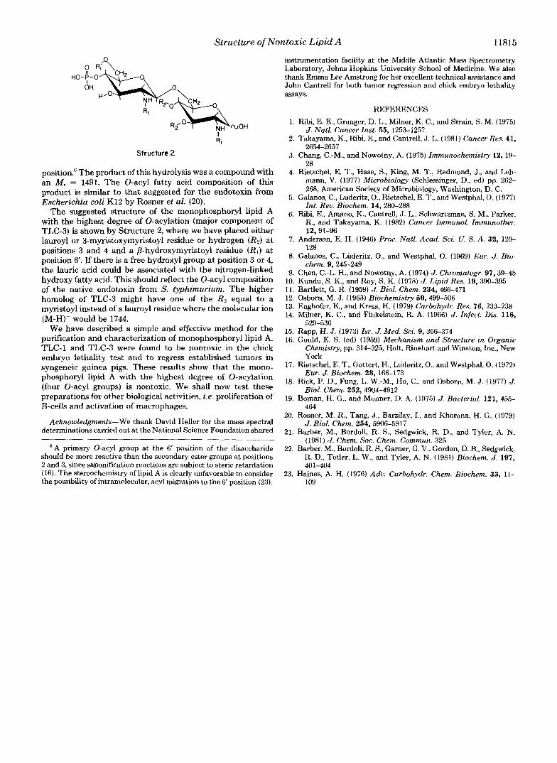
Structure 2
position." The product of this hydrolysis was a compound with an M, = 1491. The 0-acyl fatty acid composition of this product is similar to that suggested for the endotoxin from Escherichia coli K12 by Rosner et al. (20).
The suggested structure of the monophosphoryl lipid A with the highest degree of 0-acylation (major component of TLC-3) is shown by Structure 2, where we have placed either lauroyl or 3-myristoxymyristoyl residue or hydrogen (R2) at positions 3 and 4 and a ,L?-hydroxymyristoyl residue (RI) at position 6'. If there is a free hydroxyl group at position 3 or 4, the lauric acid could be associated with the nitrogen-linked hydroxy fatty acid. This should reflect the 0-acyl composition of the native endotoxin from S. typhimurium. The higher homolog of TLC-3 might have one of the RZ equal to a myristoyl instead of a lauroyl residue where the molecular ion (M-H)- would be 1744.
We have described a simple and effective method for the purification and characterization of monophosphoryl lipid A. TLC-1 and TLC-3 were found to be nontoxic in the chick embryo lethality test and to regress established tumors in syngeneic guinea pigs. These results show that the mono- phosphoryl lipid A with the highest degree of 0-acylation (four 0-acyl groups) is nontoxic. We shall now test these preparations for other biological activities, i e . proliferation of B-cells and activation of macrophages.
Acknowledgments-We thank David Heller for the mass spectral determinations carried out at the National Science Foundation shared
'A primary 0-acyl group at the 6' position of the disaccharide should be more reactive than the secondary ester groups at positions 2 and 3, since saponification reactions are subject to steric retardation (16). The stereochemistry of lipid A is clearly unfavorable to consider the possibility of intramolecular, acyl migration to the 6' position (23).
~~
instrumentation facility at the Middle Atlantic Mass Spectrometry Laboratory, Johns Hopkins University School of Medicine. We also thank Emma Lee Amstrong for her excellent technical assistance and John Cantrell for both tumor regression and chick embryo lethality assays.
REFERENCES 1. Ribi, E. E., Granger, D. L., Milner, K. C., and Strain, S. M. (1975)
2. Takayama, K., Ribi, E., and Cantrell, J. L. (1981) Cancer Res. 41, 2654-2657
3. Chang, C.-M., and Nowotny, A. (1975) Immunochemistry 12.19- 28
4. Rietschel, E. T., Hase, S., King, M. T., Redmond, J., and Leh- mann, V. (1977) Microbiology (Schlessinger, D., ed) pp. 262- 268, American Society of Microbiology, Washington, D. C.
5. Galanos, C., Luderitz, O., Rietschel, E. T., and Westphal, 0. (1977) Int. Rev. Biochem. 14, 280-280
6. Ribi, E., Amano, K., Cantrell, J . L., Schwartzman, S. M., Parker, R., and Takayama, K. (1982) Cancer Immunol. Immunother.
7. Anderson, E. H. (1946) Proc. Natl. Acad. Sci. U. S. A . 32, 120-
8. Galanos, C., Luderitz, O., and Westphal, 0. (1969) Eur. J. Bio-
9. Chen, C.-L. H., and Nowotny, A. (1974) J. Chromatogr. 97.39-45
J. Natl. Cancer Inst. 55, 1253-1257
12,91-96
128
chem. 9,245-249
10. Kundu, S. K., and Roy, S. K. (1978) J. Lipid Res. 19, 390-395 11. Bartlett, G. R. (1959) J. Biol. Chem. 234, 466-471 12. Osborn, M. J. (1963) Biochemistry 50,499-506 13. Enghofer, E., and Kress, H. (1979) Curbohydr. Res. 76, 233-238 14. Milner, K. C., and Finkelstein, R. A. (1966) J. Infect. Dis. 116,
15. Rapp, H . J. (1973) Zsr. J. Med. Sci. 9, 366-374 16. Gould, E. S. (ed) (1959) Mechanism and Structure in Organic
Chemistry, pp. 314-325, Holt, Rinehart and Winston, Inc., New York
17. Rietschel, E. T., Gottert, H., Lideritz, O., and Westphal, 0. (1972) Eur. J. Biochem. 28,166-173
18. Rick, P. D., Fung, L. W.-M., Ho, C., and Osborn, M. J. (1977) J. Biol. Chem. 252, 4904-4912
19. Boman, H. G., and Monner, D. A. (1975) J. Bacteriol. 121, 455- 464
20. Rosner, M . R., Tang, J., Barzilay, I., and Khorana, H. G. (1979) J. Biol. Chem. 254, 5906-5917
21. Barber, M., Bordoli, R. S., Sedgwick, R. D., and Tyler, A. N. (1981) J. Chem. SOC. Chem. Commun. 325
22. Barber, M., Bordoli, R. S., Garner, G. V., Gordon, D. B., Sedgwick, R. D., Tetler, L. W., and Tyler, A. N. (1981) Biochem. J. 197,
23. Haines, A. H. (1976) Adu. Carbohydr. Chem. Biochem. 33, 11-
529-536
401-404
109


![LRA 11815 Optimal Dose of Hyperbaric Bupivacaine 0 5 for Unilateral s 082510[1]](https://static.fdocuments.in/doc/165x107/577ce4ef1a28abf1038f6f46/lra-11815-optimal-dose-of-hyperbaric-bupivacaine-0-5-for-unilateral-s-0825101.jpg)


