The Journal of Basic & Applied Zoology
8
Comparative histological studies on the intestinal wall between the prenatal, the postnatal and the adult of the two species of Egyptian bats. Frugivorous Rousettus aegyptiacus and insectivorous Taphozous nudiventris Atteyat Selim * , Eman El Nahas Zoology Department, Faculty of Science, Tanta University, Tanta, Egypt Received 25 February 2014; revised 31 March 2015; accepted 1 April 2015 Available online 5 June 2015 KEYWORDS Bats; Intestine; Histology; Prenatal; Postnatal Abstract The present study was planned to find the effect of different feeding habits on the structure of the duodenum and small intestine of adult, prenatal and postnatal of both fructivorous Rousettus aegyptiacus and the insectivorous Taphozous nudiventris using the histological and the histochemical techniques. Histologically, the duodenal wall of R. aegyptiacus and T. nudiventris is composed of the typical layers: mucosa, submucosa, muscularis and serosa, we observed that the mucosa with finger like villi and very sharp apices in prenatal and adult of R. aegyptiacus but compact finger like villi in T. nudiventris. Scattered among the columnar epithelium goblet cells which less numerous in R. aegypti- acus than in T. nudiventris. Brunner’s glands are less numerous also in R. aegyptiacus than in T. nudi- ventris. In postnatal the mucosa with pyramidal like villi in R. aegyptiacus and finger like villi in T. nudiventris. In ileum the intestinal glands are less numerous in R. aegyptiacus than in T. nudiventris. In prenatal the goblet cells are less developed in R. aegyptiacus and T. nudiventris. The intestinal glands are less developed also in R. aegyptiacus and T. nudiventris but in the postnatal the goblet cells and the intestinal gland are few in number in both R. aegyptiacus and T. nudiventris. ª 2015 The Egyptian German Society for Zoology. Production and hosting by Elsevier B.V. This is an open access article under the CC BY-NC-ND license (http://creativecommons.org/licenses/by-nc-nd/4.0/). Introduction The studies on bats in the world have attracted the attention of many authors. Since studies in the Egyptian bats may be few, my research focus was on the Egyptian bats. Many authors have studies on the histological and some general histo- chemical characteristics of gastric mucosa, small intestine, and gut associated lymphoid tissue in many different species of bats (Schultz, 1965; Rouck and Glass, 1970; Forman, 1971, 1972, 1973, 1974). Brunner’s gland formed from acini and contained (PAS) positive droplets Cochrane et al. (1954). * Corresponding author. Peer review under responsibility of The Egyptian German Society for Zoology. The Journal of Basic & Applied Zoology (2015) 70, 25–32 HOSTED BY The Egyptian German Society for Zoology The Journal of Basic & Applied Zoology www.egsz.org www.sciencedirect.com http://dx.doi.org/10.1016/j.jobaz.2015.04.004 2090-9896 ª 2015 The Egyptian German Society for Zoology. Production and hosting by Elsevier B.V. This is an open access article under the CC BY-NC-ND license (http://creativecommons.org/licenses/by-nc-nd/4.0/).
Transcript of The Journal of Basic & Applied Zoology

The Journal of Basic & Applied Zoology (2015) 70, 25–32
HO ST E D BYThe Egyptian German Society for Zoology
The Journal of Basic & Applied Zoology
www.egsz.orgwww.sciencedirect.com
Comparative histological studies on the intestinal
wall between the prenatal, the postnatal and the
adult of the two species of Egyptian bats.
Frugivorous Rousettus aegyptiacus andinsectivorous Taphozous nudiventris
* Corresponding author.
Peer review under responsibility of The Egyptian German Society for
Zoology.
http://dx.doi.org/10.1016/j.jobaz.2015.04.0042090-9896 ª 2015 The Egyptian German Society for Zoology. Production and hosting by Elsevier B.V.This is an open access article under the CC BY-NC-ND license (http://creativecommons.org/licenses/by-nc-nd/4.0/).
Atteyat Selim *, Eman El Nahas
Zoology Department, Faculty of Science, Tanta University, Tanta, Egypt
Received 25 February 2014; revised 31 March 2015; accepted 1 April 2015Available online 5 June 2015
KEYWORDS
Bats;
Intestine;
Histology;
Prenatal;
Postnatal
Abstract The present study was planned to find the effect of different feeding habits on the structure
of the duodenum and small intestine of adult, prenatal and postnatal of both fructivorous Rousettus
aegyptiacus and the insectivorous Taphozous nudiventris using the histological and the histochemical
techniques. Histologically, the duodenal wall of R. aegyptiacus and T. nudiventris is composed of
the typical layers: mucosa, submucosa, muscularis and serosa, we observed that the mucosa with finger
like villi and very sharp apices in prenatal and adult of R. aegyptiacus but compact finger like villi in T.
nudiventris. Scattered among the columnar epithelium goblet cells which less numerous in R. aegypti-
acus than in T. nudiventris. Brunner’s glands are less numerous also in R. aegyptiacus than in T. nudi-
ventris. In postnatal the mucosa with pyramidal like villi in R. aegyptiacus and finger like villi in T.
nudiventris. In ileum the intestinal glands are less numerous in R. aegyptiacus than in T. nudiventris.
In prenatal the goblet cells are less developed in R. aegyptiacus and T. nudiventris. The intestinal glands
are less developed also in R. aegyptiacus and T. nudiventris but in the postnatal the goblet cells and the
intestinal gland are few in number in both R. aegyptiacus and T. nudiventris.ª 2015 The Egyptian German Society for Zoology. Production and hosting by Elsevier B.V. This is an
open access article under the CC BY-NC-ND license (http://creativecommons.org/licenses/by-nc-nd/4.0/).
Introduction
The studies on bats in the world have attracted the attention ofmany authors. Since studies in the Egyptian bats may be few,
my research focus was on the Egyptian bats. Many authors
have studies on the histological and some general histo-chemical characteristics of gastric mucosa, small intestine,and gut associated lymphoid tissue in many different species
of bats (Schultz, 1965; Rouck and Glass, 1970; Forman,1971, 1972, 1973, 1974). Brunner’s gland formed from aciniand contained (PAS) positive droplets Cochrane et al.(1954).

26 A. Selim, E. El Nahas
Forman (1974) noticed that frugivorous bats have morePayer’s patches than nectarivorous, carnivorous or insectiv-orous ones, and that they can occur anywhere along the
tract.Physiological studies made by Keegan (1975) indicated that
long intestines would not hamper the flight of frugivorous
bats, since food transmit and absorption time are quite rapid.The disposition of Kerckring’s folds, absent in the upperregion of the duodenum and in the distal portion as it
approaches the colon, is similar to that generally found inmammals.
Madkour (1977) and Madkour et al. (1982) made a com-parative study of certain features of the alimentary canal and
deposition of viscera in different bats from Egypt, they statedthat the different parts of the alimentary tract showed amarked difference in size, shape, and length. The longest
intestinal canal is found in Rousettus, Nycteris, Rhinolophus,and Asellia tridens forming a definite group as regards thelength of this canal.
The Light and the Scanning electron microscopy have beenused to distinguish the different portions of bat intestines(Okon, 1977; Ishikawa et al., 1985; Tedman and Hall, 1985;
Makanya and Maina, 1994; Makanya, 1997; Makanya et al.2001).
External morphology does not allow a clear distinctionbetween the small and the large intestines in all bat species,
Tedman and Hall (1985) stated that the intestine ofPteropus alecto and P. poliocephalus is a continuoustube without separation between the small and large
intestines.The gastrointestinal secretions in vertebrates contain num-
ber of muco substances that can differ according to cell type,
anatomical region, functional status, pathological condition,age, sex and species (Sheahan and Jervis, 1976; Filipe, 1979;Sato and Spicer, 1980; Allen, 1981; Suganuma et al., 1981;
Pedini et al., 2001; Liquori et al., 2002; Choi et al., 2003;Schumacher et al., 2004).
The intestine of Rhinolophus ferrumequinum is dividedinto three main areas: duodenum, jejuno-ileum and rectum,
while the intestine of the cogeneric Rhinolophus hildebrandtifrom Africa is similar to that of R. ferrumequinum in hav-ing a long small intestine and a short rectum, but differs in
that it lacks a distinct duodenum (Makanya and Maina,1994).
The ileum is the final section of the small intestine, it is
about 2–4 m long in man, follows the duodenum and jejunum,and is separated from the ceacum by the ileoceacal valve(Coico, 2003). The mucosa of the pangolin was noticed to becharacterized by horizontal folds called plicae circulares, in
contrast to abnormal finger-like projection (villi) found pro-jecting into the lumen in rats and bats. The plicae circulareswith villi increases the surface area. Further examination
revealed that the muscularis externa is thicker in pangolin withrat and bat (Ofusori et al., 2008).
In C. perspicillata, P. hastatus, and G. soricina Peyer’s
patches were found in the distal portion near the large intes-tine, whereas in D. rotundus and S. lilium aggregations oflymphoid nodulous tissue were distributed along the tube.
In Sturnira lilium, Phyllostomus hastatus, Carollia perspicil-lata, Glossophaga soricina and Desmodus rotundus, Panethcells at the base of the crypts of Lieberkuhn (Gadelhaet al., 2008).
Materials and methods
Bats
The specimens of Rousettus aegyptiacus (Megachiroptera) andTaphozous nudiventris (Microchiroptera) used in this study
were procured alive from Abu-Rawash, Giza Governoratethrough the years 2010 and 2011.
Measurements
The mean of 20 measured adult specimens in millimetre was asfollows:
R. aegyptiacus: total length: 93 (88.5–95), tail: 40.5 (39.2–41.5), fore-arm: 80 (79.4–82.5), hind foot: 35.5 (34.2–36),ear: 25 (24.3–26.5).T. nudiventris: total length: 73 (69.3–74.5), tail: 24.5 (21.1–
25.6), fore-arm: 62 (60.4–63.8), hind foot: 25.5 (25.0–26.1), ear: 18 (17.5–18.5) and targus: 6 (5.8–6.3).
Collected the animals
The specimens of Rousettus aegyptiacus and T. nudiventris
were collected and classified as follows:
(a) Adult group.
(b) Pre-natal group; (embryos).(c) Post-natal group; (lactating bats) (Fig. 1).
Histological examination
Organs of adult, pre-natal, and post-natal bats (frugivores andinsectivores) were collected. Parts of the duodenum, ileum,
tongue, pituitary, and thyroid glands were immediately fixedin Bouin’s fluid and 10% neutral buffered formalin for 24 h.The tissue samples were dehydrated in ascending grades of
ethyl alcohol, cleared by xylene and embedded in paraffin.Sections of 5 lm thickness were mounted and stained withHaematoxylin and Eosin method (Bancroft and Stevens,
1990). All preparations were microscopically examined forthe histological examination.
Histochemical examination
Periodic Acid Schiff procedure (PAS)
Sections were treated by Periodic Acid Schiff procedure (PAS).
This comprised placing the sections in 0.5% periodic acid for5–10 min, washing for 5 min in running tap water, then rinsedin distilled water and treated with Schiff’s reagent for 20–
35 min. Sections were passed through 3 changes of freshly pre-pared M/20 sodium acid sulphite solution, 2 min in each. Theywere washed in tap water for 5 min, and then rinsed in distilled
water. Dehydration was carried out in ascending series of ethylalcohols, cleared in xylene and mounted in canada balsam.
The PAS reaction is based on the oxidation of the glycollinkages in polysaccharides by periodic acid, thus producing
aldehydes. These librated aldehydes react with the leuco fuch-sin of Schiff’s reagent producing a compound of a magenta
Histological studies on the intestinal wall between the two species of Egyptian bats 27
colour. The PAS reaction was used to: demonstrate themucous secretion of the goblet cell and the duodenal glandof both the duodenum and ileum.
Azan stain
Sections were treated by xylene for 20 min to remove the wax,hydration was carried out in descending series of ethyl alco-
hols, then put in aniline alcohol for 45 min, washed in acidalcohol for 1 min, put in azocarmine for an hour at 56 �C, thenwashed in acid alcohol and distilled water, put in phosph-
molybdate for 2–3 h, washed in distilled water, put in anilineblue for an hour, washed in distilled water, put in phosph-molybedate, then in acid alcohol. Dehydration was carried
out in ascending series of ethyl alcohol, cleared in xylene andmounted in canada balsam (Gretchen, 1974).
Result
Macroscopic study
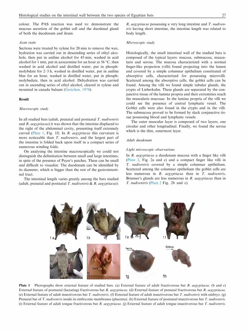
In all studied bats (adult, prenatal and postnatal T. nudiventrisand R. aegyptiacus) it was shown that the intestine displaced to
the right of the abdominal cavity, presenting itself extremelycurved (Plate 1, Fig. 1f). In R. aegyptiacus this curvature ismore noticeable than T. nudiventris, and the largest part ofthe intestine is folded back upon itself in a compact series of
numerous winding folds.On analysing the intestine macroscopically we could not
distinguish the delimitation between small and large intestines,
in spite of the presence of Peyer’s patches. These can be smalland difficult to visualize. The duodenum can be identified byits diameter, which is bigger than the rest of the gastrointesti-
nal tract.The intestinal length varies greatly among the bats studied
(adult, prenatal and postnatal T. nudiventris & R. aegyptiacus):
Plate 1 Photographs show external feature of studied bats. (a) Ext
External feature of postnatal (lactating) fructivorous bat R. aegyptiacu
(e) External feature of adult insectivorous bat T. nudiventris. (f) Extern
Prenatal bat of T. nudiventris inside its embryonic membranes (placenta
(i) External feature of adult tongue fructivorous bat R. aegyptiacus. (j)
R. aegyptiacus possessing a very long intestine and T. nudiven-tris having short intestine, the intestine length was related tobody length.
Microscopic study
Histologically, the small intestinal wall of the studied bats is
composed of the typical layers: mucosa, submucosa, muscu-laris and serosa. The mucosa characterized with a normalfinger-like projection (villi) found projecting into the lumen
and covered by a simple columnar epithelium constituted ofabsorptive cells, characterized for possessing microvilli.Scattered among the absorptive cells, the goblet cells can be
found. Among the villi we found simple tubular glands, thecrypts of Lieberkuhn. These glands are separated by the con-junctive tissue of the lamina propria and their extremities reachthe muscularis mucosae. In the lamina propria of the villi we
could see the presence of central lymphatic vessel. TheGoblet cells were also found in the crypts and in the villi.The submucosa proved to be formed by slack conjunctive tis-
sue possessing blood and lymphatic vessels.The outer muscular layer is composed of two layers, one
circular and other longitudinal. Finally, we found the serosa
which is the thin, outermost layer.
Adult duodenum
Light microscopic observations
In R. aegyptiacus a duodenum mucosa with a finger like villi(Plate 2, Fig. 2a and c) and a compact finger like villi in
T. nudiventris covered by a simple columnar epithelium.Scattered among the columnar epithelium the goblet cells areless numerous in R. aegyptiacus than in T. nudiventris,
Brunner’s glands are less numerous in R. aegyptiacus than inT. nudiventris (Plate 2 Fig. 2b and c).
ernal feature of adult fructivorous bat R. aegyptiacus. (b and c)
s. (d) External feature of prenatal fructivorous bat R. aegyptiacus.
al feature of adult insectivorous bat T. nudiventris with embryo. (g)
). (h) External feature of postnatal insectivorous bat T. nudiventris.
External feature of adult tongue insectivorous bat T. nudiventris.
Plate 2 Photomicrographs of adult duodenum sections of R. aegyptiacus and T. nudiventris. (a and b) Transverse sections of R.
aegyptiacus duodenum showing the muscularis layer (ME), submucosa (SU.M), Brunner’s gland (BG), a finger like villi (V) and Goblet
cells (G.C) H&E. (c and d) Transverse sections of T. nudiventris duodenum showing the muscularis layer (ME), submucosa (SU.M),
Brunner’s gland (BG), a compact finger like villi (V) and Goblet cells (G.C) H&E. (e) Transverse sections of R. aegyptiacus duodenum
showing the goblet cells and the lamina propria of the villi giving strong reaction with PAS reaction. (f) Transverse sections of T.
nudiventris duodenum showing the Goblet cells (G.C) only give strong reaction with PAS reaction.
28 A. Selim, E. El Nahas
Histochemical investigations
By using PAS stain we observe that the brush border of thecolumnar cells and the goblet cells gives moderate reaction in
R. aegyptiacus (Plate 2, Fig. 2e) but in T. nudiventris onlythe goblet cells give strong PAS reaction (Plate 2, Fig. 2f).
Prenatal duodenum
Light microscopic observations
In prenatal R. aegyptiacus a duodenum mucosa with villi withvery sharp apices and a compact villi in prenatal T. nudiventriscovered by a simple columnar epithelium. Scattered among the
Plate 3 Photomicrographs of prenatal duodenum sections of R. ae
aegyptiacus duodenum showing the muscularis layer (ME), submucosa
Goblet cells (G.C) H&E. (c and d) Transverse sections of T. nudive
(SU.M), Brunner’s gland (BG), a compact villi (V) and Goblet cells
showing the mucosa of the villi giving moderate reaction with PAS sta
Brunner’s glands (BG) giving strong reaction with PAS stain.
columnar epithelium the goblet cells are less numerous in R.aegyptiacus than in T. nudiventris, Brunner’s glands are lessnumerous also in R. aegyptiacus than in T. nudiventris, andthe cytoplasmic contents of Brunner’s glands in T. nudiventris
are different from those in R. aegyptiacus (Plate 3, Fig. 3a–d).
Histochemical investigations
By using PAS stain we observe that the brush border of thecolumnar cells and the goblet cells gives moderate reactionbut Brunner’s glands give weak reaction in R. aegyptiacus(Plate 3, Fig. 3e), but in T. nudiventris the goblet cells and
the Brunner’s glands give strong reaction (Plate 3, Fig. 3f).
gyptiacus and T. nudiventris. (a and b) Transverse sections of R.
(SU.M), Brunner’s gland (BG), villi with very sharp apices (V) and
ntris duodenum showing the muscularis layer (ME), submucosa
(G.C) H&E. (e) Transverse section of R. aegyptiacus duodenum
in. (f) Transverse section of T. nudiventris duodenum showing the

Histological studies on the intestinal wall between the two species of Egyptian bats 29
The post natal duodenum
Light microscopic observations
In postnatal R. aegyptiacus a duodenum mucosa with a pyra-
midal villi in R. aegyptiacus (Plate 4, Fig. 4a and c) and a fin-ger like villi in T. nudiventris. Scattered among the columnarepithelium the goblet cells are less numerous in R. aegyptiacusthan in T. nudiventris. Brunner’s glands are less numerous also
in R. aegyptiacus than in T. nudiventris and the cytoplasmiccontents of Brunner’s glands in T. nudiventris are different
Plate 4 Photomicrographs of postnatal duodenum sections of R. ae
aegyptiacus duodenum showing the muscularis layer (ME), submucosa
cells (G.C) H&E. (c and d) Transverse sections of T. nudiventris d
muscularis layer (ME), submucosa (SU.M), Brunner’s gland (BG), a fin
sections of R. aegyptiacus duodenum showing the Goblet cells and the
Transverse sections of T. nudiventris duodenum showing the Brunner’
Plate 5 Photomicrographs of adult ileum sections of R. aegyptiacus
ileum showing the muscularis layer (ME), submucosa (SU.M), a norm
H&E. (c and d) Transverse sections of T. nudiventris ileum showing la
submucosa (SU.M), Brunner’s gland (BG), a normal finger-like villi (V
aegyptiacus ileum showing the Goblet cells and the villi giving weak
nudiventris ileum showing the intestinal glands giving moderate reactio
from those in R. aegyptiacus. We observe in the post natal ofR. aegyptiacus are enormous in number than those of prenatalof T. nudiventris (Plate 4, Fig. 4b and c).
Histochemical investigations
By using PAS stain we observe that the brush border of thecolumnar cells and the goblet cells give moderate reaction and
Brunner’s glands give strong reaction in R. aegyptiacus (Plate4, Fig. 4e and f) while in T. nudiventris the goblet cells andBrunner’s glands give strong reaction (Plate 4, Fig. 4g and h).
gyptiacus and T. nudiventris. (a and b) Transverse sections of R.
(SU.M), Brunner’s gland (BG), a pyramidal villi (V) and Goblet
uodenum showing large numbers of Brunner’s gland (BG), the
ger like villi (V) and Goblet cells (G.C) H&E. (e and f) Transverse
Brunner’s glands giving strong reaction with PAS stain. (g and h)
s glands (BG) giving strong reaction with PAS stain.
and T. nudiventris. (a and b) Transverse sections of R. aegyptiacus
al finger-like villi (V), intestinal glands (IG) and Goblet cells (G.C)
rge numbers of intestinal glands (IG), the muscularis layer (ME),
) and Goblet cells (G.C) H&E. (e and f) Transverse sections of R.
reaction with (PAS) stain. (g and h) Transverse sections of T.
n with PAS stain.
30 A. Selim, E. El Nahas
Adult ileum
Light microscopic observations
In adult R. aegyptiacus and T. nudiventris ileum the mucosa
characterized with normal finger-like projection (villi) foundprojecting into the lumen and covered by a simple columnarepithelium constituted of absorptive cells, characterized forpossessing microvilli. Scattered among the absorptive cells,
the goblet cells can be found (Plate 5, Fig. 5c). The intestinalglands are less numerous in R. aegyptiacus than in T. nudiven-tris (Plate 5, Fig. 5a and d).
Histochemical investigations
By using PAS stain we observe that the brush border of thecolumnar cells (absorptive cells) and the goblet cells give mod-
erate reaction in R. aegyptiacus (Plate 5, Fig. 5e and f), but inT. nudiventris the goblet cells and the intestinal glands givestrong reaction (Plate 5, Fig. 5g and h).
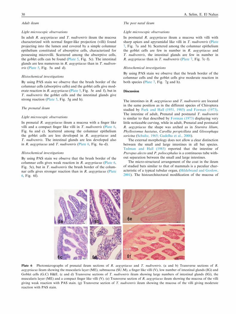
The prenatal ileum
Light microscopic observations
In prenatal R. aegyptiacus ileum a mucosa with a finger likevilli and a compact finger like villi in T. nudiventris (Plate 6,Fig. 6a and c). Scattered among the columnar epithelium
the goblet cells are less developed in R. aegyptiacus andT. nudiventris. The intestinal glands are less developed alsoin R. aegyptiacus and T. nudiventris (Plate 6, Fig. 6a–d).
Histochemical investigations
By using PAS stain we observe that the brush border of thecolumnar cells gives weak reaction in R. aegyptiacus (Plate 6,
Fig. 5e), but in T. nudiventris the brush border of the colum-nar cells gives stronger reaction than in R. aegyptiacus (Plate6, Fig. 6f).
Plate 6 Photomicrographs of prenatal ileum sections of R. aegyp
aegyptiacus ileum showing the muscularis layer (ME), submucosa (SU.
Goblet cells (G.C) H&E. (c and d) Transverse sections of T. nudive
muscularis layer (ME) and a compact finger like villi (V). (e) Transver
giving weak reaction with PAS stain. (g) Transverse section of T. nu
reaction with PAS stain.
The post natal ileum
Light microscopic observations
In postnatal R. aegyptiacus ileum a mucosa with villi with
sharp apices and apyramidal like villi in T. nudiventris (Plate7, Fig. 7a and b). Scattered among the columnar epitheliumthe goblet cells are few in number in R. aegyptiacus andT. nudiventris, the intestinal glands are few in number in
R. aegyptiacus than in T. nudiventris (Plate 7, Fig. 7c–f).
Histochemical investigations
By using PAS stain we observe that the brush border of thecolumnar cells and the goblet cells give moderate reaction inboth species (Plate 7, Fig. 7g and h).
Discussion
The intestines in R. aegyptiacus and T. nudiventris are locatedin the same position as in the different species of Chiroptera
studied by Park and Hall (1951, 1985) and Forman (1973).The intestine of adult, Prenatal and postnatal T. nudiventrisis similar to that described by Forman (1973) displaying very
little noticeable curving, while in adult, Prenatal and postnatalR. aegyptiacus the shape was arched as in Sturnira lilium,Phyllostomus hastatus, Carollia perspicillata and Glossophaga
soricina (Schultz, 1965; Gadelha et al., 2008).The external morphology does not allow a clear distinction
between the small and large intestines in all bat species.Tedman and Hall (1985) reported that the intestine of
Pteropus alecto and P. poliocephalus is a continuous tube with-out separation between the small and large intestines.
The micro-structural arrangement of the coat in the ileum
of studied bats similar to that of mammals is a peculiar char-acteristic of a typical tubular organ, (Hildebrand and Goslow,2001). The histoarchitectural modification of the mucosa of
tiacus and T. nudiventris. (a and b) Transverse sections of R.
M), a finger like villi (V), low number of intestinal glands (IG) and
ntris ileum showing large numbers of intestinal glands (IG), the
se section of R. aegyptiacus ileum showing the mucosa of the villi
diventris ileum showing the mucosa of the villi giving moderate

Plate 7 Photomicrographs of postnatal ileum sections of R. aegyptiacus and T. nudiventris. (a and b) Transverse sections of R.
aegyptiacus ileum showing the muscularis layer (ME), submucosa (SU.M), A mucosa with villi with sharp apices (V), low number of
intestinal glands (IG) and Goblet cells (G.C) H&E. (c and g) Transverse sections of T. nudiventris ileum showing large numbers of
intestinal glands (IG), the muscularis layer (ME) and a pyramidal like villi (v) H&E. (h) Transverse section of R. aegyptiacus ileum
showing the mucosa of the villi giving strong reaction with PAS stain. (i) Transverse section of T. nudiventris ileum showing the mucosa of
the villi giving weak reaction with PAS stain.
Histological studies on the intestinal wall between the two species of Egyptian bats 31
the pangolin’s ileum into plicae circulares (Ofusori et al., 2008)in contrast to studied bats is an adaptation needed to enhancethe surface area and attenuate the forward flow of intra lumi-
nal contents, thus, increasing the time of contact with absorp-tive surfaces. This adaptation may probably be a strategy tomake up for the short intestine and absent colon specially in
adult, prenatal and postnatal T. nudiventris. These findingsagreed with the works of Rubin, 1979.
Gadelha et al. (2008) stated that in S. lilium and D. rotundus
a greater number of aggregates of lymphoid nodular tissue, arewell distributed along the tract. In P. hastatus and C. perspicil-lata Peyer’s patches were observed in the distal portion next tothe large intestine, and the two portions of the intestine (small
and large) could be distinguished. In G. soricina Peyer’spatches could not be visualized externally, and so it was notpossible to delimit the small/large intestine junction, as also
observed by Ishikawa et al., 1985 in the insectivorous batMyotis frater.
The absence of the caecum and of the appendix is consid-
ered characteristic of the gastrointestinal tract of chiropterans(Tedman and Hall, 1985), another feature being the absence ofthe ascending and transverse colons for several species of bats(Park and Hall, 1951; Ishikawa et al., 1985; Tedman and Hall,
1985; Makanya and Maina, 1994; Makanya et al., 2001), andthis also applies to adult, Prenatal and postnatal R. aegyptia-cus and T. nudiventris. Most bats have a smaller intestine than
do terrestrial mammals of comparable size (Klite, 1965).Several comparisons of bat intestinal length measurements(Robin, 1881; Park and Hall, 1951; Forman et al., 1979;
Tedman and Hall, 1985; Makanya, 1997) reveal that frugivo-rous species have a longer intestine, relative to body length,than species with other feeding habits.
Keegan (1975) indicated that long intestines would nothamper the flight of frugivorous bats, since food transit andabsorption time is quite rapid, the gland of Brunner is easilyfound in the duodenum of studied bats. The villi of the small
intestine mucosa presented differences, similar to the ones
described by Schultz (1965) and Forman et al. (1979). Themucosa epithelium, similar to that found in other mammals,of the simple columnar type, was also observed in M. frater
by Ishikawa et al. (1985) in Pteropus alecto and P. polio-cephalus by Tedman and Hall (1985) and in Epomophoruswahlbergi by Makanya et al. (2001). Further, the similarities
in the histo architectural configuration of the villi in studiedbats are believed to be indicative of similar way of coping withtheir high fibre diets. The increase in length of the villi in stud-
ied bat underlines its functional implication. It appears to alsoplay a ciliary role, which helps to complement the peristalticmovement of chyle against gravity when the animals are intheir normal (up-side-down) posture.
The cell types found in the small intestine of the adult,Prenatal and postnatal R. aegyptiacus and T. nudiventris arebasically the same found in mammals in general, i.e. absorptive
cells which are microvilli and goblet cells which become morenumerous along the tube. This was observed in other species ofbats by Ishikawa et al. (1985), Tedman and Hall (1985),
Makanya and Maina (1994), Makanya et al. (1995, 2001) pre-sented less goblet cells than the S. lilium, P. hastatus, C. perspi-cillata and G. soricinia (Gadelha et al., 2008), a fact alsoobserved in Pteropus alecto by Tedman and Hall, 1985 This
can be associated to the type of food consumed, which is liquidand easily absorbed. The Paneth cells were observed by Schaaf(1970) in the insectivorous species group: Artibeus jamaicensis,
Brachyphylla nana, Phyllonycteris polyi and Monophyllus red-mani. These cells were also observed in adult prenatal andpostnatal T. nudiventris at the base of crypts of Lieberkuhn.
The muscularis mucosae and submucosa are not highly devel-oped in the Prenatal and postnatal R. aegyptiacus and T. nudi-ventris, a fact also observed inM. frater (Ishikawa et al., 1985).
Many characteristics generally found in mammals were alsoseen in bats, such as the presence of central lacteals in the lam-ina propria of the villi, and an outer muscular layer composedof a circular and a longitudinal layer. The serosa, was formed
by slack conjunctive tissue covered by a mesothelium.

32 A. Selim, E. El Nahas
References
Allen, A., 1981. Structure and function of gastrointestinal mucus. In:
Johnson, L.R. (Ed.), Physiology of the Gastrointestinal Tract.
Raven Press, New York, pp. 617–639.
Bancroft, J.D., Stevens, A., 1990. Theory and Practice of Histological
Techniques, third ed., pp. 43–80.
Coico R.F., Sunshine G., Benjamini E., 2003. Immunology: Short
Course, 5th ed.
Choi, B.Y., Sohn, Y.S., Choi, C., Chae, C., 2003. Lectin histochem-
istry for glycoconjugates in the small intestines of piglets naturally
infected with Isospora Suis. Vet. Med. Sci. J. 65 (3), 389–392.
Filipe, M.I., 1979. Mucins in the human gastrointestinal epithelium: a
review. Invest. Cell Pathol. J. 2 (3), 195–216.
Forman, G.L., Phillips A.J., Rouk C.S. 1979. Alimentary Tract pp.
205–227.
Forman, G.L., 1972. Comparative morphological and histochemical
studies of stomachs of selected North American bats. Univ. Kans.
Sci. Bull. 26, 591–729.
Forman, G.L., 1971. Histochemical differences in gastric mucus of
bats. Mammal. J. 52, 191–193.
Forman, G.L., 1973. Studies of gastric morphology in North
American Chiroptera (Emballonuridae, Noctilionidae and
Phyllostomatidae). Mammal. J. 54, 909–923.
Forman, G.L., 1974. Gastric morphology in selected mormoopid and
glossophagine bats as related to systematic problems. Trans.
Illinois State Acad. Sci. 64, 273–282.
Gadelha,A.R.,Rozensztranch,A.M.S.,Rocha, B.O., 2008.Comparative
intestinal histomorphology of five species of Phyllostomid Bats
(Phyllostomidae, Microchiroptera): ecomorphological relations with
alimentary habits. Int. Morphol. J. 26 (3), 591–602.
Gretchen L.H., 1974. Animal Tissue Technique, fourth ed.
Hildebrand, M., Goslow, G.E., 2001. Analysis of Vertebrate Structure,
5th ed. John Wily and Sons Inc, New York, pp. 201–204.
Ishikawa, K., Matoba, M., Tanaka, H., Ono, K., 1985. Anatomical
study of the intestine of the insect feeder bat, Myotis frater kaguae.
Anat. J. 12 (1), 41–50.
Keegan, D.J., 1975. Aspects of absorption of fructose in Rousettus
aegypticus. S. Afr. Med. Sci. J. 40, 49–55.
Klite, P.D., 1965. Intestinal bacterial flora and transit time in three
neotropical bat species. Bacteriol. J. 90 (37), 5–9.
Liquori, G.E., Scillitani, G., Mastrodonato, M., Ferri, D., 2002.
Histochemical investigations on the secretory cells in the oesoph-
agogastric tract of the Eurasian green toad, Bufo viridis.
Histochem. J. 34, 517–524.
Madkour, G.A., 1977. Comparative study of certain features of the
alimentary canal and disposition of the viscera in Egyptian bats.
Ann. Zool. (Agra) 13, 63–81.
Madkour, G.A., Hammouda, E.M., Ibrahim, I.G., 1982. Histology of
the alimentary tract of two common Egyptian bats. Ann. Zool. 19,
53–74.
Makanya, N.A., Maina, J.N., 1994. The morphology of the intestine
of the insectivorous horseshoe bat (Rhinolophus hildebrandti,
Peters): a scanning electron and light microscopic study. Afr. J.
Ecol. 32 (2), 158–168.
Makanya, A.N., 1997. The morphology of the intestine of the
entomophagous longfingered bat, Miniopterus inflatus: mucosal
topography and possible landmarks. Acta Biol. J. Hung. 48, 15–27.
Makanya, A.N., Mayhew, T.M., Maina, J.N., 1995. Stereological
methods for estimating the functional surfaces in the chiropteran
small intestine. Anat. J. 187, 361–368.
Park, H., Hall, E.R., 1951. The gross anatomy of the tongues and
stomachs of eight NewWorld Bats. Trans. Kans. Acad. Sci. 54, 64–
72.
Okon, E.E., 1977. Functional anatomy of the alimentary canal in the fruit
bat, Eidolon helvum and the insect bat, Tadarida nigeriae. Acta Zool.
(Stockh) 58, 83–93.
Ofusori, O.A., Enaibe, B.U., Falana, B.A., Adeeyo, O.A., Yusuf,
U.A., Ajayi, S.A., 2008. A comparative morphometric analysis of
stomach in rat (Rattus norvegicus), bat (Eidolon helvum) and
pangolin (Manis tricuspis). Cell Anim. Biol. J. 2 (3), 079–083.
Pedini, V., Scocco, P., Gargiulo, A.M., Ceccarelli, P., 2001.
Carbohydrate histochemistry of lamb duodenum. Acta
Histochem. 103 (3), 315–323.
Rubin, W., 1979. The epithelial ‘‘membrane’’ of small intestine. Am.
Clin. Nutr. J. 24 (1), 45–64.
Robin, H.A., 1881. Recherches anatomiques sur les Mammiferesde
l’ordre des Chiropteres. Ann. Sci. Nat. Zool. 12, 1–180.
Rouck, C.S., Glass, B.P., 1970. Comparative gastric histology of five
North and Central American bats. Mammal. J. 51, 455–476.
Sato, A., Spicer, S.S., 1980. Ultrastructural cytochemistry of complex
carbohydrates of gastric epithelium in the guinea pig. Am. Anat. J.
159 (3), 307–329.
Schumacher, U., Duku, M., Katoh, M., Jorns, J., Krause, W.J., 2004.
Histochemical similarities of mucins produced by Brunner’s glands
and pyloric glands: a comparative study. Anat. Rec. A Discov.
Mol. Cell. Evol. Biol. 278 (2), 540–550.
Suganuma, T., Katsuyama, T., Tsukahara, M., Tatematsu, M.,
Sakakura, Y., Murata, F., 1981. Comparative histochemical study
of alimentary tracts with special reference to the mucous neck cells
of the stomach. Am. Anat. J. 61 (2), 219–238.
Schultz, W., 1965. Studien uber den Magen-Darm-Kanal der
Chiropteren. Z. Wiss. Zool. 171, 240–391.
Schaaf, V.P., 1970. Untersuchungen uber das histochemischeVerhalten
der Panethschen Kornerzellen bei mittelamerikanischen
Fledermausarten mitunterschiedlichen Ernkhrungsweisen. Anat.
Anz. Bd. 126, 275–277.
Sheahan, D.G., Jervis, H.R., 1976. Comparative histochemistry of
gastrointestinal mucosubstances. Am. Anat. J. 146 (2), 103–131.
Tedman, R.A., Hall, L.S., 1985. The morphology of the gastrointesti-
nal tract and food transit time in the fruit bats Pteropus alecto and
P. poliocephalus (Megachiroptera). Aust. J. Zool. 33, 625–640.
Further reading
Makanya, A.N., Mayhew, T.M., Maina, J.N., 1846. Morphometry in
the epauletted Wahlberg’s fruit bat (Epomophorus Wahlbergi,
Sundervall. Acta Biol. Hung. 52 (1), 75–89.
Scillitani, G., Zizza, S., Liquori, G.E., Ferri, D., 2005. Histochemical
and immunohisto-chemical evidence for a gradient in gastric juice
production in the greater horseshoe bat, Rhinolophus ferrume-
quinum (Schreber, 1774). Acta Chiropterol. 7 (2), 301–308.
Stevens, A., Lowe, J.S., 2005. Human Histology, third ed. Elsevier
Limited, USA, pp. 190–194.