Intra- and interhemispheric connectivity between face-selective
Journal de Neurochirurgie Juillet 2017 N° 25
THE INTERHEMISPHERIC TRANSCALLOSAL APPROACH IN THE TREATMENT OF CAVERNOUS
MALFORMATION OF THE THIRD VENTRICLECASE REPORT AND LITERATURE REVIEW
BENMAMAR.T, SHABAY.Z, BOUAITA.KNeurosurgery, EHS Ali Ait Idir,
Algiers, Algeria
46
ABSTRACT : Intracerebral cavernomas represent 5-10% of all vascular malformations ofthe central nervous system. Their location in the third ventricle is very rare. Only about a hundredcases have been reported in the literature so far. We present for the first time a case of thirdventricular cavernoma encountered in a 39-year-old patient admitted in our department, whiledetailing the various difficulties encountered in the management of this rare entity. Key words : Cavernoma, Third ventricle, Transcallosal approach
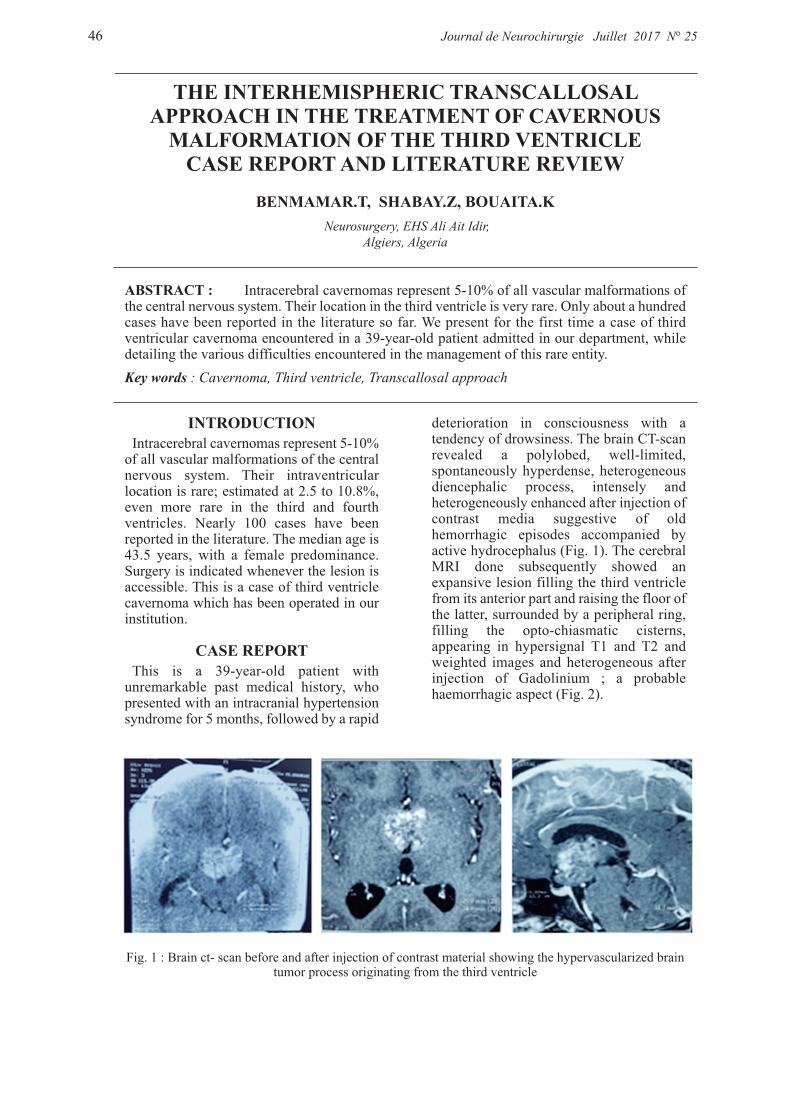
Fig. 1 : Brain ct- scan before and after injection of contrast material showing the hypervascularized braintumor process originating from the third ventricle
INTRODUCTIONIntracerebral cavernomas represent 5-10%
of all vascular malformations of the centralnervous system. Their intraventricularlocation is rare; estimated at 2.5 to 10.8%,even more rare in the third and fourthventricles. Nearly 100 cases have beenreported in the literature. The median age is43.5 years, with a female predominance.Surgery is indicated whenever the lesion isaccessible. This is a case of third ventriclecavernoma which has been operated in ourinstitution.
CASE REPORTThis is a 39-year-old patient with
unremarkable past medical history, whopresented with an intracranial hypertensionsyndrome for 5 months, followed by a rapid
deterioration in consciousness with atendency of drowsiness. The brain CT-scanrevealed a polylobed, well-limited,spontaneously hyperdense, heterogeneousdiencephalic process, intensely andheterogeneously enhanced after injection ofcontrast media suggestive of oldhemorrhagic episodes accompanied byactive hydrocephalus (Fig. 1). The cerebralMRI done subsequently showed anexpansive lesion filling the third ventriclefrom its anterior part and raising the floor ofthe latter, surrounded by a peripheral ring,filling the opto-chiasmatic cisterns,appearing in hypersignal T1 and T2 andweighted images and heterogeneous afterinjection of Gadolinium ; a probablehaemorrhagic aspect (Fig. 2).

THE INTERHEMISPHERIC TRANSCALLOSAL - APPROACH IN THE TREATMENT OF CAVERNOUS MALFORMATION OF THE THIRD VENTRICLE - CASE REPORT AND LITERATURE REVIEW 47
The patient initially benefited from anemergency ventriculo-peritoneal shunt. Thepatient was then operated for her lesion in asecond sitting by anterior transcallosal
interhemispheric approach. Subtotal reddishblack tumor excision was done in view of itsintimate adhesion to the floor of the thirdventricle (Fig. 4).
Fig.2 : Brain MRI in T1 and T2 weighted sequence and in angiographic sequence showing the same tumor process that fills the third ventricle, encapsulated and heterogeneous signal
due to intratumoral haemorrhages
Fig. 3 : Brain ct- scan after placement of the ventriculo-peritoneal shunt and after tumor excision.
Fig. 4 : Visualization of the caverns at the end of tumor excision ventricle
RESULTSThe histopathological study revealed a
cavernoma. The post-surgical outcome wasmarked by the persistance of a state ofdrowsiness. Postoperative brain CT-scanshowed the presence of a tumor residue
adjacent to the floor of the third ventricle (Fig. 3). The hormonal imbalancedetected an imbalance of the thyroid and corticotropic axes which weresubsequently supplemented.

THE INTERHEMISPHERIC TRANSCALLOSAL - APPROACH IN THE TREATMENT OF CAVERNOUS MALFORMATION OF THE THIRD VENTRICLE - CASE REPORT AND LITERATURE REVIEW48
DISCUSSION Intraventricular cavernomas represent
about 2.5% of all endocranial cavernomas,Houtteville in 1985, found 6 cases in thethird ventricle. The median age is 43.5years, with a female predominance of 2/1.Due to their location, they may manifest byintracranial hypertension, sub arachnoid orintraventricular haemorrhage. However, intraparenchymal haemorrhage
even from intraventricular location is notexcluded. In the literature, only 14% ofpublished cases of intraventricularcavernomas presented with intraventricularhemorrhage. Generally, resection ofintraventricular cavernomas does notsignificantly pose a risk for permanentneurological deficit. Of the 89 published cases of intra
ventricular cavernomas, only one patientdied during surgery for cavernoma resectiondue to rebleeding in the temporal horn of thelateral ventricle with massive intra-ventricular hemorrhage and intra cerebralhematoma. Brain MRI often leads to itsdiagnosis. On the T1 and T2 weighted sequences, the
center appears as a heterogeneous signal,while the periphery appears hyposignalwhich suggests a circle of hemosiderinproving repeated bleeding of the lesion. TheT1-weighted sequence with gadoliniuminjection makes it possible to obtain acontrast of this process, which can vary fromhigh enhancement to the absence of contrastenhancement. The gradient echo-weighted MRI sequence
is the master sequence in the diagnosis ofcavernomas. One can also appreciate thepresence of mass effect, of an overlyinghydrocephalus, and sometimes of littleperilesional edema. Surgery is alwaysindicated whenever the lesion is accessibleand the CSF circulation must be restored.Transcallosal interhemispheric approachavoids trauma of the brain parenchyma,although the prognosis of these lesions isdifficult to predict because some of them arestrongly attached to the floor of the thirdventricle, particularly the hypothalamus, butalso to their tendency for rapid growth due to intra-lesional hemorrhage, thisphenomenon being probably explained bythe absence of great resistance at theperiphery of the lesion. For this reason, it is essential, as far as
possible; to perform the most completeresection, even if it is necessary to re-operate in case of partial excision. This isshown in the literature which shows a high
number of deaths by bleeding amongpatients who have undergone partialresection
CONCLUSIONDespite their rarity, cavernomas of the third
ventricle are lesions that should besuspected when a tumor is detected near thethird ventricle, their total excision isessential for cure.
REFERENCES1] MOHANA RAO PATIBANDLA,
AMIT KUMAR THOTAKURA & MANAS KUMAR PANIGRAHI, Third ventricular cavernous malformation, an unusual lesion, British Journal of Neurosurgery, Aug 2013
2] FAROPOULOS K, PANAGIO TOPOULOS V, PARTHENI M, TZORTZIDIS F, KONSTANTINOU D Therapeutic management of intra ventricular cavernoma: case series and review of the literature, Neurol Surg, 2015 May
3] Z. MILENKOVIC Z. MILENKOVIC, Neurosurgical Clinic, Medical Faculty of Nis, Postural intermittent headaches as the initial symptom of a cavernoma in the third ventricle, Acta Neurochirurgica, 30 Sept. 2004
4] A. ALVES de Sousa Hôpital Santa Casa, hôpital Lifecenter, Belo Horizonte, Brésil, Cavernomes profonds (corps calleux, intra ventriculaires, ganglions de la base, insulaires) et du tronc cérébral, expérience d'une série brésilienne, j.neuchi 2007.03.008
5] HIROAKI NAGASHIMA KAZUHIROTANAKA, TAKASHI SASAYAMA, YUSUKE OKAMURA, MASAAKI TANIGUCHI, KYOKO OTANI, TAKASHI YAMASAKI, TOMOO ITOH, EIJI KOHMURA, A large cavernous malformation of the third ventricle floor : A case report Department of Neurosurgery, Kobe University Graduate School of Medicine, Kusunoki cho, Chuoku, Kobe, Japan, 2015.08.004
6] NIKOLAY L. MARTIROSYAN, MD, M. YASHAR S. KALANI, MD, PHD, PETER NAKAJI, MD, AND ROBERT F. The anterior inter hemispheric approach to a V3 cavernous malformation department of Neuro

THE INTERHEMISPHERIC TRANSCALLOSAL - APPROACH IN THE TREATMENT OF CAVERNOUS MALFORMATION OF THE THIRD VENTRICLE - CASE REPORT AND LITERATURE REVIEW 49
surgery, Barrow Neurological Institute St. Joseph’s Hospital and Medical Center, Phoenix, Arizona, Oct. 14, 2015
7] O.VERNET B.RILLIET, Cavernomes du troisième ventricule chez un enfant de 18 mois, service de neurochirurgie, centre hospitalier universitaire Vaudois (CHUV), Lausanne Suisse 2007
8] KIVELEV J, NIEMELÄ M, KIVISAARI R, ET AL. Intra ventricular cerebral cavernomas: a series of 12 patients and review of the literature. J Neurosurg. 2010 ; 112 (1) :140-9
9] ITOH J, USUI K. Cavernous angiomain the fourth ventricular floor--case report. Neurol Med Chir (Tokyo). 1991 ; 31 (2) : 100- 3
10] KENDALL B, REIDER-GROSSWASSER I, VALENTINE A. Diagnosis of masses presenting within the ventricles on computed tomography. Neuroradiology. 1983 25 (1) : 11- 22